Embed Size (px)
Citation preview

Review Special Issue: Resolution of Acute Inflammation and the Role of Lipid Mediators TheScientificWorldJOURNAL (2010) 10, 832–856 ISSN 1537-744X; DOI 10.1100/tsw.2010.77
*Corresponding author. ©2010 with author. Published by TheScientificWorld; www.thescientificworld.com
832
Resolution of Adipose Tissue Inflammation
Ana González-Périz and Joan Clària*
Department of Biochemistry and Molecular Genetics, Hospital Clínic, IDIBAPS, CIBEK, and CIBERehd, University of Barcelona
E-mail: [email protected]; [email protected]
Received December 22, 2009; Revised March 30, 2010; Accepted April 6, 2010; Published May 4, 2010
The presence of the so-called “low-grade” inflammatory state is recognized as a critical event in adipose tissue dysfunction in obesity. This chronic “low-grade” inflammation in white adipose tissue is powerfully augmented through the infiltration of macrophages, which, together with adipocytes, perpetuate a vicious cycle of macrophage recruitment and secretion of free fatty acids and deleterious adipokines that predispose the development of obesity-related comorbidities, such as insulin resistance and nonalcoholic fatty liver disease. In the last decade, many factors have been identified that contribute to mounting uncontrolled inflammation in obese adipose tissue. Among them, bioactive lipid mediators derived from the cyclooxygenase and 5-lipoxygenase pathways, which convert the ω-6-polyunsaturated fatty acid (PUFA) arachidonic acid into potent proinflammatory eicosanoids (i.e., prostaglandins [PGs] and leukotrienes), have emerged. Interestingly, the same lipid mediators that initially trigger the inflammatory response also signal the termination of inflammation by stimulating the biosynthesis of anti-inflammatory and proresolving lipid autacoids. This review discusses the current status, characteristics, and progress in this class of “stop signals”, including the lipoxins, which were the first identified ω-6 PUFA–derived lipid mediators with potent anti-inflammatory properties; the recently described ω-3 PUFA–derived lipid mediators resolvins and protectins; and the cyclopentenone PGs of the D series. Special emphasis is given to the participation of these bioactive lipid autacoids in the resolution of adipose tissue inflammation and in preventing the development of obesity-related complications.
KEYWORDS: adipose tissue, adipocytes, macrophages, inflammation, insulin signaling, adiponectin, AMPK, omega-3 PUFA, omega-3 PUFA–derived lipid autacoids, resolvins, protectins
ADIPOSE TISSUE IS AN ENDOCRINE ORGAN
Two types of adipose tissue exist in mammals: white and brown. Compared to white, brown adipose
tissue contains smaller adipocytes and is rich in mitochrondia, and is used for production of heat by
nonshivering thermogenesis, especially in hibernating mammals. Brown adipose tissue is difficult to find
in adult humans, since brown fat pads existing within the posterior neck in neonatal humans to provide
cold-adaptive thermogenesis for newborns are lost soon after birth[1]. Therefore, white adipose tissue is
that which is relevant in terms of human physiology and disease. Until recently, storage of energy in the

González-Périz and Clària: Resolution of Adipose Tissue Inflammation TheScientificWorldJOURNAL (2010) 10, 832–856
833
form of fat and a mechanical role in thermal insulation were the most widely recognized functions of
white adipose tissue. White adipose tissue was also recognized for its role as a major secretory organ
responsible for the release of fatty acids into the circulation. In fact, adipose tissue is the major source of
fatty acids that are used as energy substrates for generation of adenosine triphosphate (ATP) high-energy
bonds for metabolic functions from oxidative phosphorylation[2]. For decades, the focal point of adipose
tissue biology has been the study of the mechanisms that regulate lipolysis and lipogenesis, and the fine
equilibrium between more energy expenditure (i.e., increased circulating levels of free fatty acids from
adipose tissue lipolysis) or, conversely, less energy expenditure (i.e., enhanced fat storage)[2].
However, in addition to serving as the main fat depot and energy storage area of our body, white
adipose tissue is a very active endocrine organ that secretes a number of hormones and signaling peptides
that carry diverse biological functions with direct actions on our body’s homeostasis. The most relevant
actions of these hormones secreted by adipose tissue include the regulation of appetite and satiety, glucose
and lipid metabolism, blood pressure homeostasis, inflammation, and immune functions[2,3]. The initial
finding that changed the perspective on the physiological role of adipose tissue was the cloning, in 1994, of
the ―ob‖ gene that codes for leptin, a hormone that signals to the hypothalamus for the regulation of appetite
and energy balance (see below)[4]. The identification of this hormone, together with other observations
showing that the proinflammatory cytokine tumor necrosis factor (TNF)-α and a secreted protein, later
identified as the complement-related factor adipsin, were released by adipocytes[5], led to the recognition
that adipose tissue is an endocrine organ with adipocytes as the major endocrine cells. Since then, an ever-
increasing number of factors, now numbering more than 50 different molecular entities secreted from
adipose tissue and generically known as adipocytokines or adipokines (preferred designation), have been
described. An updated list of adipokines released by adipose tissue is given in Table 1. This list includes
cytokines and related proteins (leptin, TNF-α, interleukin [IL]-6, IL-10, and monocyte chemotactic protein-1
[MCP-1]), proteins of the fibrinolytic cascade (plasminogen activator inhibitor-1 [PAI-1]), complement and
complement-related proteins (visfatin and adiponectin), and other biologically active peptides, such as
resistin and chemerin. In the current review, we will limit our discussion to adipokines, which have been
shown to participate in the cross-talk among adipose tissue cells, and are implicated in adipose tissue
inflammation and in the development of the metabolic syndrome.
Leptin
Leptin is the product of the obese ―ob‖ gene and is predominantly expressed in adipocytes[6]. Leptin
controls appetite/satiety and regulates energy balance by sending antiobesity signals to the central nervous
system[6]. The lack of leptin in ob/ob mice as a consequence of a recessive autosomic mutation in the
―ob‖ gene results in hyperphagia and severe obesity[7]. However, patients with obesity have elevated
levels of circulating leptin, which suggests the presence of a generalized leptin resistance, explaining why
leptin therapy in this condition has been unsuccessful. Leptin has been shown to modulate both innate and
adaptive immune responses by stimulating Th1 proinflammatory adaptive immune response and the
secretion of proinflammatory cytokines, such as interferon (IFN)- and TNF-α, leading to macrophage
activation and cell-mediated inflammatory response[8]. As a cytokine, leptin is significantly increased in
response to inflammatory stimuli and, in turn, induces the secretion of IL-1 and TNF-α[9]. Consistent
with its proinflammatory role, leptin-deficient (ob/ob) mice display limited cell-mediated imflammatory
and immune responses[10]. Similar findings have been found in leptin receptor–deficient (db/db) mice,
which have increased infection susceptibility and reduced allogenic graft rejection[11].
TNF-α, IL-6, and MCP-1
White adipose tissue has been identified as a major site of production of TNF-α. Similar to other
adipokines, TNF-α acts in both paracrine and endocrine fashions by binding to structurally related receptor

González-Périz and Clària: Resolution of Adipose Tissue Inflammation TheScientificWorldJOURNAL (2010) 10, 832–856
834
TABLE 1 List of Selected Adipokines Secreted by Adipose Tissue
Adipokine Cellular Origin
Reported Actions Adipocytes Stromal Cells
Leptin ++ — Signals satiety in the hypothalamus; proinflammatory effects in peripheral tissues
TNF-α + ++ Proinflammatory effects; insulin resistance
IL-6 + ++ Proinflammatory effects; insulin resistance
IL-10 (?) + Anti-inflammatory effects
MCP-1 + ++ Monocyte/macrophage recruitment; insulin resistance (?)
PAI-1 + + Thrombosis; insulin resistance (?)
MIP-1α (?) + Monocyte/macrophage recruitment
Angiotensinogen + (?) Cardiovascular function; mediator of chronic inflammation
Resistin ++ — Proinflammatory effects (mouse); insulin resistance (mouse)
Visfatin + — Proinflammatory effects (?); natural insulin mimetic (?)
Adiponectin ++ — Anti-inflammatory effects; insulin-sensitizing effects
Chemerin + (?) Regulates adipogenesis; anti-inflammatory actions (?)
Stromal cells include endothelial cells, macrophages, and preadipocytes. ++, High expression; +, clear evidence; (?),
data not confirmed yet. MIP-1: macrophage inflammatory protein 1
proteins known as the TNF receptor superfamily that embraces at least 12 different receptors. Most of the
effects of TNF-α appear to be mediated by interaction with two membrane receptors: TNF-R1 and TNF-
R2[12,13]. In most circumstances, TNF-α is able to activate other cytokine networks, including the
release of IL-1 and IL-6, thereby amplifying inflammatory response and tissue injury[13]. TNF-α is also a
critical mediator of insulin resistance, activating proinflammatory pathways, such as nuclear factor kappa
B (NF-B) and c-jun N-terminal kinase (JNK)[14,15]. White adipose tissue also produces IL-6, which is
the most relevant member of a family of cytokines that comprise IL-11, oncostatin M, ciliary neurotropic
factor, and cardiotrophin-1[16]. IL-6 is a proinflammatory and insulin-resistant cytokine expressed in
both adipocytes and stromal cells of the adipose tissue. IL-6 signals through gp130, activation of the
Jak/Stat pathway similarly to leptin, and by induction of SOCS3 expression[15]. IL-6 plays divergent
roles, and this adipokine appears to confer protection against acute and chronic liver injury[17]. Finally,
adipose tissue also produces MCP-1. MCP-1, also known as chemokine (C-C motif) ligand 2 (CCL-2), is
a potent chemoattractant protein that plays a role in the recruitment of monocytes/macrophages and in the
maintenance of the inflammatory infiltrate at the site of inflammation[18]. Although MCP-1 is also
secreted from adipocytes, it is more abundant in adipose tissue stromal cells[19]. MCP-1 has also been
postulated as an adipokine that contributes to insulin resistance[20].
Resistin
Resistin is a 10-kDa polypeptide member of a family of cysteine-rich proteins that is secreted from rodent
adipocytes[21]. Resistin is an inflammatory adipokine that stimulates secretion of proinflammatory
cytokines (i.e., TNF-α and IL-6) by monocytes, worsens inflammation, and parallels the expression of
proinflammatory chemokines and NF-B activation in activated myofibroblasts[14,15]. The effects of
resistin in humans are not clearly defined and its role in human disease may differ from that observed in
mouse models. Remarkably, human resistin protein is only 55% identical to its mouse counterpart, which
indicates that it might not be evolutionarily well conserved across species.

González-Périz and Clària: Resolution of Adipose Tissue Inflammation TheScientificWorldJOURNAL (2010) 10, 832–856
835
Visfatin
Visfatin is secreted by adipocytes in visceral fat, although its expression can be extended to many other
cells and tissues. Visfatin was originally identified as a protein involved in immune B-cell maturation
(pre–B colony enhancing factor)[22]. Visfatin binds and activates the insulin receptor, and reduces insulin
resistance, but does not compete with insulin[14]. Despite its beneficial insulin-sensitizing actions,
visfatin may also work as a proinflammatory adipokine, indicating that future studies on this natural
insulin mimetic are needed in order to define its role in insulin resistance and inflammation.
Adiponectin
Originally identified as complement-related protein 30 (ACRP30), monomeric adiponectin is a 30-kDa
protein, structurally related to the complement 1q family that comprises a collagen-like tail linked by a
disulfide bond at Cys-39 to a globular head domain[23]. In humans and rodents, full-length adiponectin
circulates in a wide range of oligomeric forms from trimers (low-molecular-weight adiponectin) and
hexamers consisting of two trimers (middle-molecular-weight adiponectin) to high-molecular weight
multimers (12–18 mer)[23]. Adiponectin expression is almost exclusive to adipocytes[24]. Adiponectin
binds to two distinct receptors, termed AdipoR1 and AdipoR2, which are integral membrane proteins
with the N-terminus internal and the C-terminus external[23]. AdipoR1 is ubiquitously expressed and
abundantly present in skeletal muscle, whereas AdipoR2 appears to be mostly expressed in the liver[23].
AdipoR1 and R2 serve as receptors for both globular and full-length adiponectin, and mediate increased
AMPK and PPARα activities[23]. Contrary to the other adipokines, adiponectin exerts pleiotropic anti-
inflammatory and insulin-sensitizing effects in the liver, adipose tissue, and skeletal muscle[23].
Adiponectin stimulates secretion of anti-inflammatory cytokines (i.e., IL-10), blocks NF-B activation,
and inhibits release of TNF-α, IL-6, and inflammatory chemokines[14]. On the other hand, adiponectin
directly improves glucose metabolism and insulin sensitivity by reducing hepatic glucose production, and
increasing glucose uptake and fatty acid oxidation in the skeletal muscle[25].
IL-10
IL-10 is an anti-inflammatory cytokine mainly expressed in adipose tissue stromal cells, specifically
macrophages. Type 2 diabetes and metabolic syndrome have been associated with decreased IL-10
production and, indeed, circulating IL-10 levels are positively correlated with insulin sensitivity[26,27].
Moreover, IL-10 counteracts IL-6–induced insulin resistance, reduces MCP-1 secretion, and reverses the
detrimental effects of TNF-αon GLUT-4 and insulin receptor substrate 1 (IRS-1) tyrosine
phosphorylation. Since adipocytes express IL-10 receptor, IL-10 is likely to act as an anti-inflammatory
and insulin-sensitizing adipokine in adipose tissue.
Chemerin
Chemerin, also known as RARRES2 or TIG2, is a recently discovered chemoattractant protein that serves
as a ligand for the G protein-coupled receptor CMKLR1 (ChemR23 or DEZ)[28]. Chemerin is secreted as
an 18-kDa inactive proprotein and undergoes cleavage of the C-terminal portion to generate the 16-kDa
active protein. Chemerin undergoes further proteolytic cleavage to generate a number of peptides with
anti-inflammatory properties[29]. Interestingly, the chemerin receptor (ChemR23) binds resolvin E1, an
endogenous anti-inflammatory lipid mediator generated from the ω-3-polyunsaturated fatty acid (PUFA)
eicosapentaenoic acid (EPA) (see below). High expression of chemerin as well as its receptor ChemR23
has been identified in the white adipose tissue of mice, suggesting this tissue as a source and target for

González-Périz and Clària: Resolution of Adipose Tissue Inflammation TheScientificWorldJOURNAL (2010) 10, 832–856
836
chemerin signaling. In adipose tissue, chemerin regulates adipogenesis as well as adipocyte metabolism,
and is currently considered to play a role in obesity and the metabolic syndrome[28,30].
ADIPOSE TISSUE INFLAMMATION IN OBESITY
Adipose tissue is vital for life and its functional integrity is required for balanced body metabolism of a
healthy organism. However, excessive fat mass is deleterious and increases the incidence of
comorbidities. In this regard, obese individuals exhibit a propensity to develop glucose intolerance and
insulin resistance leading to type 2 diabetes, hypertension, dyslipidemia, and nonalcoholic fatty liver
disease (NALFD)[31]. The mechanism by which obesity is detrimental to our health is thought to be
directly related to the release of higher amounts of fatty acids and proinflammatory adipokines from fat,
which cause inflammation and insulin resistance not only in adipose tissue, but also in remote tissues.
However, in recent years, a wealth of evidence indicates that adipose tissue dysfunction and associated
pathologies are aggravated by the development of a state of chronic mild inflammation in this
tissue[20,32,33]. In addition, this state of chronic ―low-grade‖ inflammation is powerfully augmented
through the infiltration of macrophages into white adipose tissue[34]. Infiltrated macrophages, together
with adipocytes present in the obese adipose tissue, perpetuate a vicious cycle of macrophage recruitment
and production of proinflammatory adipokines. In obese adipose tissue, macrophages form ―crown-like‖
structures that surround necrotic adipocytes that scavenge adipocyte debris[35] (Fig. 1). In addition to
augmented infiltration of macrophages, obesity also induces a phenotypic switch in these cells.
Specifically, diet-induced obesity leads to a shift in the phenotype of macrophages from an M2-polarized
state (―alternative activated‖ or anti-inflammatory phenotype) in lean animals to an M1 proinflammatory
state (―classically activated‖) in obese mice[36]. On the other hand, recent papers have provided evidence
that other inflammatory cell types, such as T lymphocytes, also infiltrate the obese adipose tissue and
contribute to adipose tissue inflammation[37,38,39].
FIGURE 1. Representative photomicrographs of adipose tissue sections from lean and obese mice immunostained with the
macrophage specific marker F4/80. Adipose tissue from obese mice shows a remarkable infiltration of macrophages that form ―crown-like‖ structures that surround necrotic adipocytes and scavenging adipocyte debris.
One of the most important consequences of adipose tissue inflammation is the development of insulin
resistance. Indeed, proinflammatory stimuli simultaneously activate both the JNK and NF-B pathways
through classical receptor-mediated mechanisms[32]. JNK activation induces insulin resistance by

González-Périz and Clària: Resolution of Adipose Tissue Inflammation TheScientificWorldJOURNAL (2010) 10, 832–856
837
disrupting insulin signaling through serine phosphorylation of IRS-1. Unlike JNK, NF-B does not
phosphorylate IRS-1, but induces the transcriptional activation of numerous target genes whose products
induce insulin resistance. Moreover, increased production of proinflammatory and insulin-resistant
adipokines (i.e., TNF-α, IL-6, and MCP-1) and decreased adiponectin secretion have been postulated to play
a pivotal role in insulin resistance[40]. Accordingly, obese and insulin-resistant ob/ob mice that were made
deficient for TNF-α or TNF-α receptors experienced a significant improvement in insulin sensitivity. In
obese subjects, epidemiological studies have demonstrated a direct correlation between the levels of
proinflammatory factors, such as TNF-αand IL-6, and glucose intolerance and insulin resistance[5].
Obesity-induced adipose tissue inflammation associated with the development of insulin resistance
also affects liver function. In fact, one of the major metabolic consequences of obesity-driven
inflammation is NAFLD, a condition ranging from simple overaccumulation of triglycerides in the
cytoplasm of hepatocytes (steatosis or fatty liver) to steatosis combined with inflammation (steatohepatitis
or NASH)[41,42,43]. Although generally asymptomatic, hepatic steatosis is no longer regarded as a
neutral bystander, but rather as a premorbid condition that increases the vulnerability of this organ to
progress to steatohepatitis and to more severe forms of liver damage[41,42]. In this regard, steatotic livers
are more susceptible to the tissue-damaging effects of oxidative stress and inflammatory mediators, and
its transition to steatohepatitis represents a critical step in the progression to hepatic fibrosis and
cirrhosis[41,42]. Not surprisingly, the prevalence of NAFLD is directly correlated with body mass index,
as the prevalence of NAFLD and metabolic syndrome in the general population is coincidental (22 and
20%, respectively), supporting the notion that NAFLD is the hepatic manifestation of the metabolic
syndrome[41,44]. Insulin resistance plays a crucial role in the pathogenesis of NAFLD. Indeed, in the
absence of obesity, even in patients with total lipodistrophy, insulin resistance leads to hepatic
steatosis[41,45]. Although the mechanisms underlying the association of insulin resistance to hepatic
steatosis remain unclear, several events related to insulin resistance may ultimately lead to NAFLD,
including (1) increased free fatty acid efflux to the liver owing to increased lipolysis from visceral fat or
increased intake of dietary fat; (2) increased levels of proinflammatory and insulin-resistant adipokines by
adipose tissue, such as increased secretion of TNF-α and IL-6; (3) decreased secretion of the anti-
inflammatory and insulin-sensitizing adipokine, adiponectin; (4) decreased hepatic oxidation of free fatty
acids; (5) increased de novo hepatic lipogenesis secondary to altered insulin sensitivity; and (6) decreased
hepatic lipid export via VLDL (very low-density lipoprotein) assembly[46].
MEDIATORS OF ADIPOSE TISSUE INFLAMMATION
UAdipokines
In addition to serving as endocrine mediators, proinflammatory adipokines overproduced with increasing
adiposity also exert autocrine actions in adipose tissue cells, thus contributing to the initiation and
exacerbation of the ―low-grade‖ inflammatory state present in obese fat tissue (see previous section).
UBioactive Lipid Mediators with Proinflammatory Activity
0BIn addition to inflammatory adipokines, recent data have implicated bioactive inflammatory lipid
mediators in the development of obesity-induced adipose tissue inflammation. These lipid mediators
originate from the cleavage of structural lipid components of cellular membranes and represent one of the
most potent classes of endogenous inflammatory mediators. Eicosanoids, which are a large family of
compounds generated from arachidonic acid, represent a paradigmatic example of this class of lipid
mediator. Arachidonic acid is an essential ω-6 PUFA primarily found esterified in the 2-acyl position of
phospholipids in all mammalian outer and intracellular membranes. Upon activation of phospholipase A2,
arachidonic acid is released from membrane phospholipids and its free acid becomes available as a
González-Périz and Clària: Resolution of Adipose Tissue Inflammation TheScientificWorldJOURNAL (2010) 10, 832–856
838
substrate for the intracellular biosynthesis of eicosanoids through two major enzymatic routes: the
cyclooxygenase (COX) and lipoxygenase (LO) pathways[47,48,49]. The COX pathway results in the
formation of prostaglandins (PGs) and thromboxane (TX) A2, which are known for their powerful
physiological properties and their critical role in the inflammatory response[48,49]. On the other hand, the
LO pathway comprises three major LOs, designated 5-, 12-, and 15-LO; 5-LO converts arachidonic acid
into 5(S)-hydroxyeicosatetraenoic acid (5-HETE) and leukotrienes (LTs), a consolidated pharmacological
target in inflammation[48,49].
COX
There are two distinct isozymes of COX, designated COX-1 and COX-2. Although the products
generated by these two isozymes are the same, COX-1 is a constitutive enzyme virtually expressed in all
cells, whereas COX-2 expression is induced in most tissues by inflammatory stimuli and is the isoform
involved in inflammatory response[50,51]. COX-2 was originally identified as a unique, inducible gene
product in studies addressing cell growth signaling pathways, as well as in investigations on COX activity
in response to cytokines and other inflammatory mediators[51,52]. In fact, COX-2 is consistently induced
by IL-1/, TNF-α, IFN-lipopolysaccharide (LPS), epidermal growth factor, platelet-derived growth
factor, fibroblast growth factor, and oncogenes (v-src and v-ras), and its expression is critical in
inflammation[51,52]. Both COX isozymes sequentially transform arachidonic acid into PGG2 and,
subsequently, into PGH2, which is finally converted by specific synthases into PGs of the D2, E2, F2, and
I2 series, as well as into TXA2. Biosynthesis of COX products is cell specific and any given cell type tends
to specialize in the formation of one of these PGs as its major product. For example, endothelial cells
mainly produce PGI2 (prostacyclin) from PGH2 by means of PGI synthase, and platelets release TXA2
from PGH2 through the action of TX synthase. Both PGI2 and TXA2 have a very short half-life and are
rapidly hydrolyzed to the inactive compounds 6-keto-PGF1 and TXB2, respectively[53]. PGH2 can be
alternatively converted into PGF2 by PGF synthase, which is mainly expressed in the uterus. PGH2 is
also converted into PGD2 by the action of PGD synthase, of which two distinct types have been
identified: lipocalin-type PGD synthase and hematopoietic-type PGD synthase[49]. PGD2 is readily
dehydrated to the cyclopentenone PGs of the J2 series, PGJ2 and 15-deoxy-12,14
-PGJ2 (15d-PGJ2) (see
Cyclopentenone PGs section, below). PGE2 is formed by the enzyme PGE synthase (PGES) present in
virtually every cell type. There are three different PGES isoforms (mPGES-1, cPGES-1, and mPGES-2),
of which mPGES-1 was the first to be identified and characterized[54].
PGs have been detected in almost every tissue and body fluid. With the exception of seminal fluid,
PGs are not stored in tissues or cells. Instead, once synthesized, COX products are released and/or
exported to the extracellular space. Owing to instability, PGs and TX exert their functions mainly in
proximity to their sites of synthesis. Thus, they typically act as autocrine or paracrine hormones,
maintaining homeostasis within their cells of origin or in neighboring cells in the tissue. PGs produce a
broad spectrum of biological effects, including inflammation, regulation of smooth muscle tone,
gastrointestinal and renal cytoprotection, and progression of cancer. With regard to adipose tissue, it was
first described in the late 1960s that rat adipose tissue had the ability to release COX-derived prostanoids
such as PGE2 and PGI2[55] (Table 2). Although it was first proposed that stromal cells were the only cell
type responsible for PG biosynthesis in adipose tissue[56], it was later reported that PGs were also
produced by adipocytes[57,58,59,60] (Table 2). With regard to function, PGs, especially PGE2, which is
the most abundant COX product in adipose tissue, are established modulators of adipogenesis[60,61,62,
63,64]. In addition, PGE2 has been shown to play an antilipolytic role in adipose tissue contributing to fat
mass expansion[65,66]. More recently, it was reported that PGs other than PGE2, such as PGI2 or PGF2α,
were not detected at high enough concentrations in adipose tissue to bind their receptors effectively[66].
In contrast, a recent study measuring the PGF2α-derived metabolite, 15-keto-dihydro-PGF2α in 274
male and female adolescents aged between 13 and 17 years as an indicator of COX-mediated inflammatory

González-Périz and Clària: Resolution of Adipose Tissue Inflammation TheScientificWorldJOURNAL (2010) 10, 832–856
839
TABLE 2 List of Selected ω-6 PUFA– and ω-3 PUFA–Derived Lipid Mediators Produced by Adipose Tissue
Lipid Mediator
Cellular Origin Reported Actions
Adipocytes Stromal Cells
PGE2 + ++ Adipogenic effects; antilipolytic effects
PGI2 + ++ Unknown
PGF2 (?) (?) Unknown
LTB4 + + Proinflammatory effects
LTD4 + + Proinflammatory effects
12-HETE (?) (?) Adipocyte differentiation (?); proinflammatory effects (?)
15-HETE (?) (?) Adipocyte differentiation (?); proinflammatory effects (?)
15d-PGJ2 (?) (?) Anti-inflammatory effects
Resolvin E1 (?) (?) Antisteatotic effects; anti-inflammatory; insulin-sensitizing effects
Resolvin D1 (?) (?) Unknown
Protectin D1 (?) (?) Increases adiponectin expression
Stromal cells include endothelial cells, macrophages, and preadipocytes. ++, High production; +, clear evidence; (?), data not confirmed yet.
response, revealed that levels of this PGF2α metabolite were significantly correlated with body mass
index, waist circumference, and insulin levels[67]. Nevertheless, the role of COX products in
adipogenesis appears to be complex since both induction and repression of adipocyte differentiation have
been reported following selective COX-2 inhibition[68,69].
COX-2 is a key executor of inflammation in many cell types, including fibroblasts; monocytes and
macrophages; epithelial, endothelial, smooth muscle, mesangial, and mast cells; synoviocytes;
osteoblasts; neurons; and adipocytes[51,52]. Given that obesity is characterized by the presence of ―low-
grade‖ chronic inflammation in adipose tissue, it is not surprising that COX-2 expression as well as PGE2
production are altered in obesity. In fact, COX-2 expression and PGE2 production in adipose tissue are
markedly increased in experimental obesity[60,66,70]. Accordingly, selective COX-2 inhibition impairs
obesity development in mice with high-fat-diet– and leptin-deficient–induced obesity[71]. Furthermore,
COX-2-mediated inflammation has been shown to be crucial in the development of insulin resistance and
fatty liver in a rat model of high-fat-diet–induced obesity[70]. Consistent with this, mice lacking both
COX-2 alleles (COX-2–/–
mice) are protected against obesity induced by a high-fat/high-sugar diet[72].
Surprisingly, in this study, heterozygous mutant mice (COX-2+/–
mice) showed increased PGE2 and 6-
keto PGF1 levels in adipose tissue and became more obese in response to the high-fat/high-sugar
diet[72].
3B5-LO
5-LO, the key enzyme in the biosynthesis of 5(S)-HETE and LTs, is a 674-amino-acid protein with an
apparent molecular weight of between 72 and 80 kDa[73]. The 5-LO gene is highly conserved across
species[74]. It consists of 14 exons and 13 introns, and contains a promoter region that encompasses
consensus regions for transcription regulators of the Egr, Sp, NF-B, GATA, myb, and AP families[75].
Upon cellular activation, cytosolic or nuclear 5-LO translocates to the nuclear envelope where it interacts
with phospholipase A2, which makes free arachidonic acid available to 5-LO[76]. In the nuclear envelope,
5-LO transforms arachidonic acid into 5(S)-HpETE with the concerted interaction of five lipoxygenase-
González-Périz and Clària: Resolution of Adipose Tissue Inflammation TheScientificWorldJOURNAL (2010) 10, 832–856
840
activating protein (FLAP), a 18-kDa resident integral protein that functions as a transfer protein
facilitating the binding of arachidonic acid to 5-LO[77]. Subsequently, 5(S)-HpETE is either reduced to
5(S)-HETE or converted to the highly unstable allylic epoxide LTA4. Once formed, LTA4 is rapidly
transformed to either LTB4 via stereoselective hydration by LTA4 hydrolase or to LTC4 through
glutathione conjugation catalyzed by LTC4 synthase[75,78]. Sequential metabolic reactions catalyzed by
-glutamyl transferase and a specific membrane-bound dipeptidase convert LTC4 into LTD4 and LTE4,
respectively. Together, LTC4, D4, and E4 are termed cysteinyl-leukotrienes (cys-LTs) and were referred to
in the past as the slow-reacting substances of anaphylaxis.
A recent study by our laboratory demonstrated the constitutive expression of all enzymes of the 5-LO
pathway as well as the specific receptors (B-LT1, B-LT2, cys-LTs, and cys-LT2) necessary for the
formation of 5-LO products and their signaling in adipose tissue[79] (Table 2). In this study, the enzymes
and receptors of the 5-LO pathway were detected in both adipocytes and stromal cells. Moreover, adipose
FLAP expression and LTB4 levels were significantly increased in adipose tissue isolated from obese mice
as compared to that from lean mice, findings that are in agreement with previous studies in humans and
animals with experimental obesity[80,81]. Importantly, Horrillo et al. demonstrated that 5-LO products
enhance the nuclear translocation of the key proinflammatory transcription factor NF-B and induce
secretion of several proinflammatory adipokines, including MCP-1, MIP-1and IL-6 from adipose
tissue[79]. Consistent with these findings, pharmacological inhibition of the 5-LO pathway with a
selective FLAP inhibitor resulted in amelioration of the inflammatory status in the adipose tissue and
consequently in a reduction in high-fat-diet–induced hepatic steatosis[79].
12/15-LO
Mouse 12/15-LO is a 663-amino-acid enzyme, homologous to the human 662-amino-acid 15-LO protein
that catalyzes the insertion of molecular oxygen in arachidonic acid at the 12th and/or 15
th carbon,
resulting in the formation of 12- or 15-HETEs[82]. 12/15-LO was originally found to be expressed in
differentiated macrophages, dendritic cells, and endothelial and smooth muscle cells. Recently, 12/15-LO
expression has also been described in murine adipocytes[83]. In fact, products derived from 12/15-LO
were among the most abundant eicosanoids produced in the adipose tissue of obese ob/ob mice[84]. A
role for 12/15-LO in adipocyte differentiation has been suggested since 12/15-LO inhibition prevents
3T3-L1 preadipocyte differentiation into mature adipocytes[85] (Table 2). Moreover, 12/15-LO has been
postulated to play a role in the development of tissue inflammation and insulin resistance, since 12/15-LO
knockout mice are more resistant to high-fat-diet–induced adipose tissue inflammation, macrophage
infiltration, and cytokine production than wild-type mice[86,87].
RESOLUTION OF ADIPOSE TISSUE INFLAMMATION
UThe Resolution Process
Inflammation is part of the innate immunity response and is characterized by the rapid influx of
specialized leukocytes (polymorphonuclear neutrophils [PMN] and eosinophils) into injured tissues to
neutralize and eliminate injurious stimuli, such as a microbial infection or surgical trauma. The innate
immunity response not only acts as the first line of defense against a noxious agent, but it also provides
the necessary signals to instruct the adaptive immune system to provide an effective response to deal with
the injurious stimulus. Although inflammation per se is a beneficial response because it is a limited
wound-healing process that walls off tissue injury or infection, prolonged inflammation results in tissue
damage and loss of function, and represents the underlying basis for many disease conditions. In addition,
chronic inflammation is associated with continual activation of the adaptive immune system that
significantly contributes to the exacerbation of the inflammatory response.
González-Périz and Clària: Resolution of Adipose Tissue Inflammation TheScientificWorldJOURNAL (2010) 10, 832–856
841
Since prolonged inflammation is detrimental to the host, higher organisms have developed protective
mechanisms to ensure resolution of the inflammatory response in a limited and specific time and space
manner[88,89,90]. In cellular terms, resolution of acute inflammation is defined as the interval from
maximum infiltration of PMN to the point when they are lost from the tissue as a consequence of limited
PMN infiltration and apoptosis of recruited PMN[89,90]. In this interval, mononuclear leukocytes
(monocytes, macrophages) are introduced to phagocytose apoptotic PMN and cell debris in a
nonphlogistic fashion. They also release hydrolytic and proteolytic enzymes, and generate reactive
oxygen species that eliminate and digest invading organisms. Finally, the injurious stimulus is cleared and
normal tissue structure and function are restored, thus completing the tissue repair process.
The mediators and mechanisms implicated in the resolution of inflammation have remained largely
ignored. However, at present, resolution of inflammation is envisioned not as a mere passive process of
dilution of inflammation, but as a highly orchestrated and complex process in which many endogenous
anti-inflammatory and proresolving mediators counteract the effects of proinflammatory
mediators[89,91]. Indeed, a temporal order of events that follow a molecular program is precisely
conserved for the effective function of inflammatory response. Initially, tissue injury, microbes, and
surgical trauma all activate the local formation of vasoactive amines, lipid mediators, cytokines, and
chemokines that coordinately regulate the initial events of acute inflammation. Of special interest in
this process is the biosynthesis of bioactive lipid mediators derived from arachidonic acid, such as PGs
and LTB4, through pathways involving COX-1, COX-2, and 5-LO[75]. PGs and LTB4 modify the
vascular permeability, blood flow, and vascular dilation needed for the recruitment of inflammatory
cells (i.e., leukocytes) from the peripheral circulation to the inflammatory site via adhesion to the
endothelial cells and diapedesis[48]. These changes are permissive for the initial increase in protein
exudation and PMN accumulation in the inflamed tissue, which efficiently destroy the injurious insult.
However, the same factors that initially trigger the inflammatory response also signal the termination of
inflammation by stimulating the biosynthesis of proresolving lipid mediators[92,93]. For instance, both
PGE2 and PGD2 transcriptionally activate the expression of 15-LO in human PMN, switching the
mediator profile of these cells from the proinflammatory LTB4 to the anti-inflammatory lipoxin (LX)
A4[92]. This class switch in eicosanoid production and phenotypic change in mediator profiles
generated from arachidonic acid in resolving tissues provide a temporal and spatial dissociation of
eicosanoid biosynthesis that is emerging as a critical factor in the resolution of inflammation.
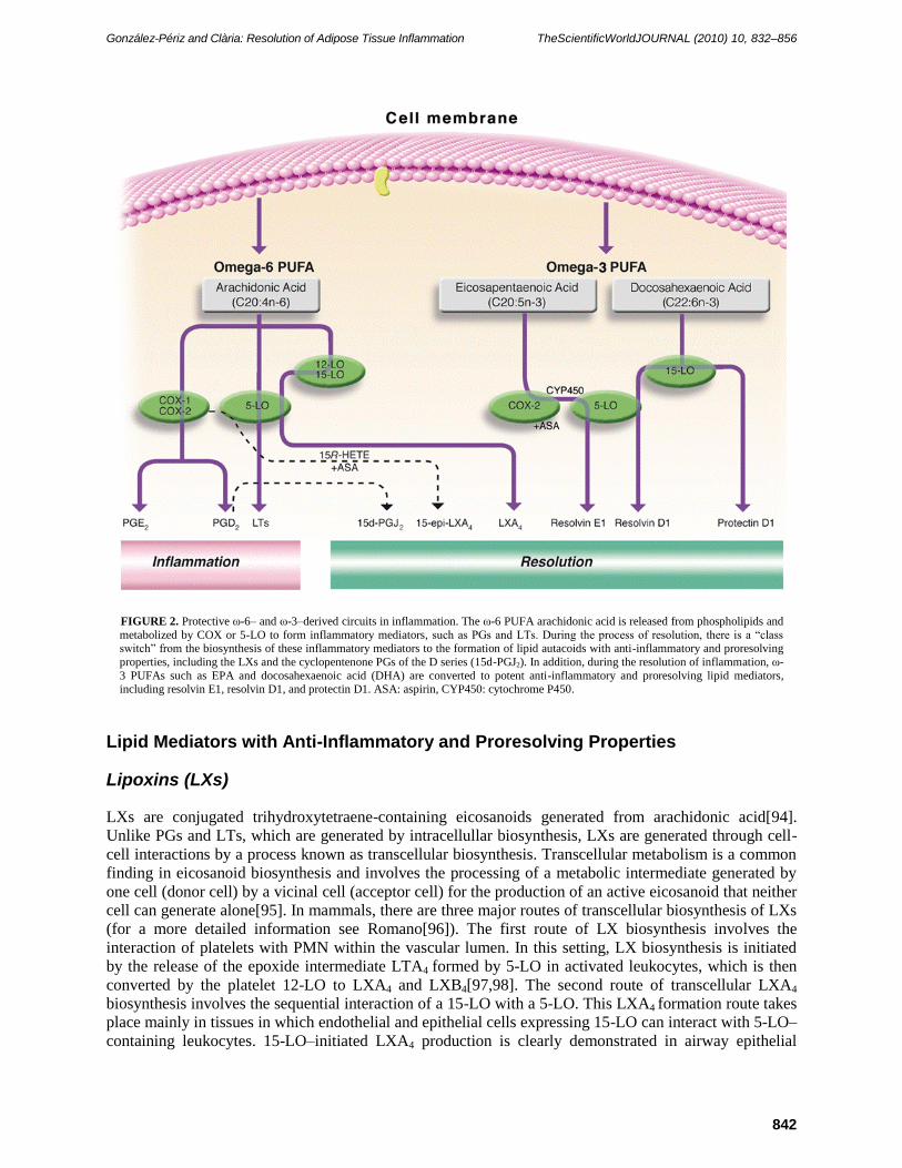
Nowadays, the most recognized ―stop signals‖ are (1) the lipoxins (LXs), which were the first
identified ω-6 PUFA–derived lipid mediators with potent immunomodulatory and anti-inflammatory
properties;(2) the recently described ω-3 PUFA–derived mediators resolvins and protectins; and (3) the
cyclopentenone PGs of the D series. A schematic diagram of the pathways involved in the biosynthesis
of these mediators is shown in Fig. 2. Interestingly, these families of endogenous proresolution
molecules are not immunosuppressive, but instead stimulate and accelerate resolution of inflammation
by activating specific mechanisms to restore tissue homeostasis. These anti-inflammatory and
proresolving mediators exert a strict control of the resolution process and not only stop PMN and
eosinophil functions, but also pave the way for monocyte migration and their differentiation to
phagocytosing macrophages, which remove dead cells and then terminate the inflammatory
response[89,92,93]. A concept that is currently of interest is that loss or deterioration of tissue function
during chronic inflammation is the result of an inappropriate inflammatory response that remains
uncontrolled because of the lack of the intrinsic capacity of the tissue for complete resolution.
Therefore, the modulation of these ―stop signals‖ that promote the timely resolution of inflammation is
emerging as a strategy to maintain inflammation self-limiting, and to prevent tissue injury and disease.
González-Périz and Clària: Resolution of Adipose Tissue Inflammation TheScientificWorldJOURNAL (2010) 10, 832–856
842
UFIGURE 2. Protective ω-6– and ω-3–derived circuits in inflammation. The ω-6 PUFA arachidonic acid is released from phospholipids and
metabolized by COX or 5-LO to form inflammatory mediators, such as PGs and LTs. During the process of resolution, there is a ―class switch‖ from the biosynthesis of these inflammatory mediators to the formation of lipid autacoids with anti-inflammatory and proresolving
properties, including the LXs and the cyclopentenone PGs of the D series (15d-PGJ2). In addition, during the resolution of inflammation, ω-
3 PUFAs such as EPA and docosahexaenoic acid (DHA) are converted to potent anti-inflammatory and proresolving lipid mediators, including resolvin E1, resolvin D1, and protectin D1. ASA: aspirin, CYP450: cytochrome P450.
Lipid Mediators with Anti-Inflammatory and Proresolving Properties
Lipoxins (LXs)
LXs are conjugated trihydroxytetraene-containing eicosanoids generated from arachidonic acid[94].
Unlike PGs and LTs, which are generated by intracellullar biosynthesis, LXs are generated through cell-
cell interactions by a process known as transcellular biosynthesis. Transcellular metabolism is a common
finding in eicosanoid biosynthesis and involves the processing of a metabolic intermediate generated by
one cell (donor cell) by a vicinal cell (acceptor cell) for the production of an active eicosanoid that neither
cell can generate alone[95]. In mammals, there are three major routes of transcellular biosynthesis of LXs
(for a more detailed information see Romano[96]). The first route of LX biosynthesis involves the
interaction of platelets with PMN within the vascular lumen. In this setting, LX biosynthesis is initiated
by the release of the epoxide intermediate LTA4 formed by 5-LO in activated leukocytes, which is then
converted by the platelet 12-LO to LXA4 and LXB4[97,98]. The second route of transcellular LXA4
biosynthesis involves the sequential interaction of a 15-LO with a 5-LO. This LXA4 formation route takes
place mainly in tissues in which endothelial and epithelial cells expressing 15-LO can interact with 5-LO–
containing leukocytes. 15-LO–initiated LXA4 production is clearly demonstrated in airway epithelial

González-Périz and Clària: Resolution of Adipose Tissue Inflammation TheScientificWorldJOURNAL (2010) 10, 832–856
843
cells, monocytes, and eosinophils following the activation of 15-LO[94]. Once activated, these cells
generate and release 15(S)-HETE, which is rapidly taken up and converted by PMN to LXA4 via the
action of 5-LO[99]. Concomitant with the biosynthesis of LXA4 by the 15-LO–initiated route, LT
biosynthesis is blocked at the 5-LO level, resulting in an inverse relationship between LT and LXA4
formation[94,100]. Interestingly, the formation of these anti-inflammatory compounds appears to be
temporarily and spatially distinct from the formation of PGs[92,101]. Formation of LXA4 from
endogenous arachidonic acid through the sequential actions of 15-LO and 5-LO can also occur in a single
cell type, particularly in granulocytes and macrophages primed with cytokines. Formation of LXA4 from
a single cell type has been demonstrated in leukocytes isolated from asthmatic patients and patients with
chronic liver disease[102,103]. A third major route of LX biosynthesis that does not involve LO-LO
interactions has also been uncovered[104]. This LX biosynthetic route is initiated by aspirin, which
acetylates COX-2 and switches its catalytic activity from PG synthase to 15-LO. Following this
conformational change, PG biosynthesis is inhibited and COX-2 transforms arachidonic acid to 15(R)-
HETE[104]. 15(R)-HETE is subsequently transformed by activated leukocytes possessing 5-LO to a new
series of carbon-15 epimers of LXA4 that carry their 15 alcohol in the R configuration (15-epi-
LXA4)[104]. The formation of these lipid mediators is specific for aspirin treatment and the term aspirin-
triggered LXA4 has been coined for these compounds[104].
LXA4 and aspirin-triggered LXA4 display a unique spectrum of biological activities. Unlike LTs,
which are proinflammatory compounds that facilitate PMN adhesion to the vascular wall and recruitment
at the site of inflammation, and leukocyte respiratory burst and degranulation[48,75,78], LXA4 and
aspirin-triggered LXA4 display potent anti-inflammatory actions. These eicosanoids appear to work as
―stop signals‖ for inflammation and, for instance, LXA4 inhibits PMN and eosinophil chemotaxis, blocks
selectin- and integrin-mediated PMN adhesion to and transmigration across endothelial monolayers, and
blocks PMN migration across postcapillary venules and PMN entry into inflamed tissues[105,106,
107,108,109,110]. LXA4 and aspirin-triggered LXA4 have also been shown to inhibit TNF-α–stimulated
superoxide generation, and degranulation and cytokine release by activated PMN[105,106,
107,108,109,110]. Interestingly, intravenous delivery of LXA4 and aspirin-triggered LXA4 inhibits acute
dermal inflammation and neutrophil infiltration of skin microabcesses and lungs in LTB4 receptor
transgenic mice[111]. In contrast to their inhibitory effects on PMN, LXA4 and aspirin-triggered LXA4
promote monocyte activity by stimulating monocyte adherence to vascular endothelium and
chemotaxis[112], and promote the phagocytic clearance of apoptotic cells by macrophages[113]. This is
important because the resolution of inflammation depends on the phagocytosis of degranulated PMN by
activated monocyte-derived macrophages, which eventually exit the inflamed site in the draining
lymphatics. Therefore, resolution of the inflammatory lesion depends, in part, on the activation state of
the monocytes.
Owing to their very short half-life, a range of stable, biologically active analogs have been designed
and tested in animal models for their anti-inflammatory and proresolving activities. These LXA4 stable
analogs significantly inhibit LTB4-induced leukocyte rolling and adherence, and neutrophil margination
and extravasation[114]. LXA4 analogs inhibit TNF-α–stimulated leukocyte trafficking and chemokine
secretion in murine air pouches, and when applied topically to mouse ears, dramatically inhibit leukocyte
infiltration and vascular permeability[112,115]. Aspirin-triggered LXA4 analogs protect mice from renal
ischemia-reperfusion injury and glomerulonephritis, and attenuate gingivitis and leukocyte
recruitment[116,117]. In a murine model of asthma, stable LXA4 and aspirin-triggered LXA4 analogs
attenuate airway hyper-reactivity and inflammation, and accelerate resolution of pulmonary edema[118].
Administration of a metabolically stable LXA4 analog in a mouse model of chronic airway inflammation
and infection associated with cystic fibrosis suppresses neutrophilic inflammation, decreases pulmonary
bacterial burden, and attenuates disease severity[119]. Finally, ZK-192, a β-oxidation–resistant LXA4
analog with enhanced chemical stability and oral pharmacokinetics potently attenuates hapten-induced
colitis in rats[120]. Based on their biosynthetic pathways and their biological activities, it is quite likely that LXs play a
role in the resolution of adipose tissue inflammation. Although this possibility has not been directly

González-Périz and Clària: Resolution of Adipose Tissue Inflammation TheScientificWorldJOURNAL (2010) 10, 832–856
844
addressed, expression of key enzymes involved in LXA4 biosynthesis (i.e., 5-LO and 12/15-LO) as well
as expression for the LXA4 receptor have been detected in adipose tissue in mice[121,122]. Whether the
signal for the LXA4 receptor in adipose tissue is due to its expression in adipocytes or stromal cells is, at
present, unknown, but recent findings from our laboratory point to the direction that both 5-LO and
12/15-LO are constitutively expressed in both cell fractions[79].
1BCyclopentenone PGs (CyPGs)
CyPGs are products of the nonenzymatic dehydration of PGs. CyPGs are structurally defined by the
presence of a highly reactive α,β-unsaturated carbonyl moiety in the cyclopentenone ring[123]. From a
biological point of view, the most relevant CyPGs are those derived from the dehydration of PGD2,
including the PGs of the J2 series: PGJ2, 12
-PGJ2 and 15d-PGJ2. Interestingly, it has been demonstrated
that the dominant source of 15d-PGJ2 formation in vivo is COX-2[124]. Gilroy and colleagues
demonstrated that there is a switch in PG synthesis from proinflammatory PGs (i.e., PGE2) at the onset of
inflammation to anti-inflammatory PGs (i.e., 15d-PGJ2) at the resolution stage of inflammation[125].
Unlike other PGs, to date, no specific transmembrane receptor has been identified for these PGs. Instead,
15d-PGJ2 appears to exert its effects through binding and activation of PPARγ, a member of the nuclear
receptor superfamily of ligand-activated transcription factors[62]. Other actions independent of PPARγ
have also been reported for CyPGs, including down-regulation of NF-B transcriptional activity[126],
inhibition of cytokine production by monocytes[127], and direct inhibition of key enzymes of the
eicosanoid cascade, namely cytosolic phospholipase A2, COX-2, and mPGES-1[128,129].
CyPGs have a broad spectrum of biological effects including powerful immunomodulatory and anti-
inflammatory properties[130,131,132]. Moreover, in vitro studies have shown the ability of 15d-PGJ2 to
promote apoptosis in leukocytes and myofibroblasts[130,131,132,133,134,135,136,137,138]. The
apoptotic pathways induced by 15d-PGJ2 depend on the cell type. In granulocytes, PGJ2 and 15d-PGJ2
induce caspase-dependent apoptosis via inhibition of IB degradation[134], whereas in basophilic
leukemia cells and myofibroblasts, PGJ2-induced apoptosis is primarily mediated by activation of
caspase-3 and -9[135,136]. In macrophages, 15d-PGJ2 may also exert its apoptotic effects by mechanisms
involving activation of protein kinase C δ-induced imbalance between MAPKs and NF-B[138]. Detailed
information on CyPGs can be found in Díez-Dacal and Pérez-Sala[139].
Little is known concerning the role of CyPGs in the resolution of adipose tissue inflammation.
Although 15d-PGJ2 has not yet been described in adipose tissue, the addition of exogenous 15d-PGJ2 to
human adipocytes stimulates the production of a protective adipokine identified as macrophage inhibitory
cytokine-1[140]. Moreover, a significant down-regulation in the expression and secretion of the
proinflammatory adipokine leptin has been reported in adipocytes exposed to 15d-PGJ2[141]. Since
activation of PPARγ by 15d-PGJ2 results in inhibitory effects on the NF-B, STAT, and AP-1 families in
many cell types and tissues, testing the effects of 15d-PGJ2 on inflamed adipose tissue appears to be
worth trying.
Resolvins and Protectins
Resolvins and protectins are potent bioactive lipid mediators derived from long-chain ω-3 PUFAs. Long-
chain PUFAs contain a carboxyl head group and an even-numbered carbon chain higher than 18 carbons,
with two or more methylene-interrupted double unsaturated bonds. Long-chain PUFAs are classified into
two families, ω-3 and ω-6, according to the number of carbons, double carbons, and proximity to the
methyl (ω) terminal of the fatty acid acyl chain. Fatty acids of the ω-3 family contain a double bond at the
third carbon, whereas those of the ω-6 family contain a double bond at the sixth carbon. The most
representative members are docosahexaenoic acid (DHA, C22:6n-3) and EPA (C20:5n-3) for the ω-3
PUFA family, and arachidonic acid (C20:4n-6) for the ω-6 PUFA family. These compounds are essential

González-Périz and Clària: Resolution of Adipose Tissue Inflammation TheScientificWorldJOURNAL (2010) 10, 832–856
845
fatty acids because animals and humans cannot synthesize PUFAs of the ω-3 and ω-6 series from
endogenous sources, and these compounds need to be incorporated from the diet. In mammals, precursors
for the synthesis of PUFAs of the ω-6 and ω-3 series are linoleic acid (C18:2n-6) and linolenic acid
(C18:3n-3), respectively. The most widely available source of ω-3 PUFAs is coldwater oily fish, such as
salmon, herring, mackerel, anchovies, and sardines, and the main dietary source of ω-6 PUFAs is
vegetable oils and minor quantities of meat and other products of animal origin.
Omega-3 PUFAs have recognized beneficial effects on human health. Omega-3 PUFAs have been
known as essential for normal growth and health since the 1930s, although awareness of their health
benefits has dramatically increased in the past few years. In fact, ω-3 PUFAs have been shown to exert
anti-inflammatory and protective actions in a number of disease conditions including, among others,
cystic fibrosis, ulcerative colitis, asthma, atherosclerosis, and metabolic and neuronal diseases[142]. The
results of the Gruppo Italiano per lo Studio della Sopravvivenza nell’Infarto Miocardico (GISSI) trial
performed in more than 11,000 patients with cardiovascular disease provided solid evidence that dietary
supplementation with approximately 1 g of ω-3 PUFAs per day significantly reduced the incidence of a
second cardiovascular event[143]. Omega-3 PUFAs have also been shown to reduce blood triglyceride
and cholesterol levels, and to lower blood pressure[144,145]. In addition, significant benefits have been
reported in inflammatory conditions such as rheumatoid arthritis[146]. The mechanisms underlying these
beneficial and protective effects of ω-3 PUFAs were not completely elucidated until recently. Initially, a
rather widely accepted mechanism of action for these compounds was that EPA competed with
arachidonic acid as a substrate to the COX and 5-LO pathways, thus preventing the formation of classical
inflammatory mediators derived from arachidonic acid (e.g., PGs and LTs)[147]. However, this
hypothesis was not subsequently confirmed since DHA is a poor substrate for COX activity; arachidonic
acid is a better substrate than EPA for COX activity, whereas the opposite is true for LO activity[148]. A
second possibility initially postulated was that both EPA and DHA directly inhibit the activity of the
COX enzyme, thus decreasing the formation of PGs and ameliorating inflammatory status[142,149]. A
third alternative explanation initially postulated was that EPA was converted to PGs and LTs of the 3 and
5 series, respectively, which carry significantly lower potency as inflammatory mediators than PGs of the
2 series and LTs of the 2 series generated from the ω-6 PUFA arachidonic acid[150].
Recently, Serhan and collaborators shed new light on the mechanisms underlying the recognized
therapeutic values of ω-3 PUFAs. These authors discovered an array of endogenous anti-inflammatory
and proresolving mediators generated from ω-3 PUFAs[151]. By means of a lipidomics-based approach
that combines liquid chromatography and tandem mass spectrometry, these authors identified a library of
ω-3 PUFA–derived lipid mediators present within exudates obtained from mice dorsal skin pouches
during the ―spontaneous resolution‖ phase of acute inflammation[151,152,153,154]. Resolving exudates
in these mice contained several related bioactive lipid mediators termed resolvins (derived from
resolution phase interaction products) and protectins (derived from EPA and DHA), the most
representative members of which are resolvin E1, resolvin D1, and protectin D1[151,153] (see Fig. 2 for a
schematic diagram of the biosynthetic pathways involved in the formation of these mediators). Resolvins
are classified as either resolvin E1 if the biosynthesis is initiated from EPA and resolvin D1 if they are
generated from DHA[93]. Protectin D1 (formerly known as neuroprotectin D1) is a product generated
from DHA[155]. On the other hand, resolvin E1 biosynthesis is initiated when EPA is converted to 18R-
hydroperoxy-EPE by endothelial cells expressing COX-2 and treated with aspirin[151,156].
Alternatively, 18R-hydroperoxy-EPE can be produced through cytochrome P450 activity[157]. Similar to
15R-HETE in 15-LXA4 formation, 18R-hydroperoxy-EPE generated by endothelial cells can be
transformed by 5-LO of neighboring leukocytes into resolvin E1 (5S,12R,18R-trihydroxy-EPA) via a
5(6)epoxide intermediate[151,153]. Resolvin D1 was also originally discovered in resolving exudates of
mice. In this pathway of transcellular biosynthesis, endothelial cells expressing COX-2 treated with
aspirin transform DHA into 17R-hydroxy-DHA, which is further transformed by leukocyte 5-LO into
resolvin D1[151,153]. More importantly from a physiological point of view, resolvin D1 can also be
formed from endogenous sources of DHA without the requirement of aspirin. In this case, endogenous
DHA is converted via 15-LO/5-LO interactions that give rise to a 17S alcohol–containing series of
González-Périz and Clària: Resolution of Adipose Tissue Inflammation TheScientificWorldJOURNAL (2010) 10, 832–856
846
resolvins, including resolvin D1 and resolvin D2[152,158]. Finally, DHA is also transformed into a
dihydroxy-containing DHA derivative, 17S-hydroxy-DHA, via an intermediate epoxide that opens via
hydrolysis and subsequent rearrangements to form protectin D1 (10R,17S-dihydroxy-docosa-
DHA)[151,152,153,154]. Among all the mediators generated from ω-3 PUFAs, resolvin E1 is the most
interesting since it is the most effective drug candidate and the compound with the most developed
biology[93,156,159,160]. Recently, a resolvin E1 cognate receptor was identified as the G protein-
coupled receptor ChemR23, which binds the peptide chemerin[156]. Chemerin also transduces anti-
inflammatory signals and is expressed in monocytes, dendritic cells, and adipocytes (see the section
Adipose Tissue as an Endocrine Organ). Resolvin E1 can also interact with the LTB1 receptor, leading to
partial agonist/antagonist effects to dampen LTB4 actions on leukocytes[161]. A more comprehensive
description of the pathways involved in the biosynthesis of resolvins and protectins is provided in
Bannenberg[162] and Seki et al.[163].
Resolvins and protectins display potent anti-inflammatory and proresolution properties[93,154,155,
156,158,159,160,161,164,165). Resolvin E1, in particular, decreases PMN infiltration and T-cell
migration, reduces TNF-α and IFN-γ secretion, inhibits chemokine formation, and blocks IL-1–induced
NF-B activation[155,158,164,166]. Resolvin E1 also stimulates macrophage phagocytosis of apoptotic
PMN and is a potent modulator of proinflammatory leukocyte expression adhesion molecules (i.e., L-
selectin)[167,168]. In vivo resolvin E1 exerts potent anti-inflammatory actions in experimental models of
periodontitis, colitis, and peritonitis, and protects mice against brain ischemia-reperfusion and corneal
injury[155,156,158,160,164]. Similar protective actions have been reported for protectin D1, although
this DHA-derived mediator is a more potent ―stop signal‖ of leukocyte-mediated tissue damage in stroke
brain injury and retinal pigmented cellular damage degeneration[158,169]. Recently, Levy et al.
demonstrated that the administration of protectin D1 before aeroallergen challenge resulted in reduced
eosinophilic and T-cell–mediated inflammation and accelerated resolution of airway inflammation in a
murine model of asthma[170]. These authors also identified a resolvin E1–initiated resolution program
for allergic airway response[171]. Finally, a recent study identified resolvin D2 as a potent endogenous
regulator of excessive inflammatory responses in mice with microbial sepsis[172]. Since the production
of resolvins and protectins seems to be regulated through the availability of a different substrate than LXs
(ω-3 vs. ω-6 PUFAs), synergism between these anti-inflammatory pathways that share function similarity
may help to accelerate the resolution of inflammation.
Studies concerning the effects of ω-3 PUFAs on adipose tissue have documented unequivocally
beneficial actions of dietary DHA and EPA on the inflammatory status of this tissue. In human studies, ω-
3 PUFA treatment showed additive benefits in insulin sensitivity, lipid profile, and inflammation during
the management of weight loss in overweight hyperinsulinemic women[173]. Interestingly, adipose tissue
represents the main storage site of ω-3 PUFAs in obese individuals[174]. Additionally, animal studies
have demonstrated that ω-3 PUFAs protect against weight gain, adipose tissue inflammation, and obesity-
related complications, including insulin resistance, dyslipidemia, cardiovascular disease, and NAFLD
induced by a high-fat diet[84,175,176,177,178]. Consistent with these findings, dietary deprivation of ω-3
PUFAs in rats induces changes in tissue fatty acid composition leading to severe metabolic alterations,
such as augmented adipose tissue mass and plasma glucose, decreased insulin sensitivity, and hepatic
steatosis[179,180]. In contrast, mice with transgenic expression of the ω-3 fatty acid desaturase (fat-1),
which converts ω-6 PUFAs into ω-3 PUFAs, thus enriching the ratio between ω-3 and ω-6 in various
tissues, display improved glucose tolerance and reduced body weight[181].
Our laboratory recently provided some mechanistic insights into the beneficial effects of ω-3 PUFAs
on adipose tissue biology in ob/ob mice, an experimental model of obesity-induced insulin resistance and
fatty liver disease[84]. In these mice, dietary intake of an ω-3 PUFA–enriched diet for 5 weeks
significantly alleviated hepatic steatosis[84]. This antisteatotic effect was associated with improved
insulin tolerance and changes in the expression of specific adipocyte-derived factors (i.e., adipokines) that
orchestrate the interaction between adipose tissue and the liver. Among these soluble factors, we
identified adiponectin, an adipokine with antidiabetic and anti-inflammatory properties, which was
significantly increased in adipose tissue isolated from obese mice receiving the ω-3 PUFA–enriched
González-Périz and Clària: Resolution of Adipose Tissue Inflammation TheScientificWorldJOURNAL (2010) 10, 832–856
847
diet[84]. Adiponectin is produced mainly by adipocytes and interacts with at least two different
membrane receptors, the activation of which results in a reduction of hyperglycemia and insulin
resistance, as well as in the regulation of many biological processes related to the inflammatory and
immune responses. Moreover, in obese mice receiving ω-3 PUFAs, there was an up-regulation of PPARγ,
which is a member of the nuclear hormone receptor superfamily that binds to specific DNA response
elements as heterodimers with the retinoid X receptor[182]. PPARγ activation results in insulin
sensitization, and this nuclear factor is the cognate receptor and the established target for the
thiazolidinenione class of antidiabetic agents, of which rosiglitazone is a representative member[182,183].
Interestingly, the ω-3 PUFA DHA and its derivative 17-HDHA are potent PPARγ agonists[164], suggesting
that induction of PPARγ expression and activation of this nuclear receptor by ω-3–derived products
contribute to the insulin-sensitizing actions exerted by dietary ω-3 PUFAs. More to the point, the finding
that putative metabolites of DHA are strong PPARγ activators has stirred much interest in developing ω-3
PUFA derivatives as potent antidiabetic agents targeting PPARγ184]. In parallel with increased
adiponectin and PPARγ, the ω-3 PUFA DHA induced the phosphorylation of AMPK, a fuel-sensing
enzyme that acts as a gatekeeper of the systemic energy balance by regulating glucose and lipid
homeostasis in adipose tissue[185]. AMPK responds to changes in the cellular energy state; thus, when
the AMP/ATP ratio is increased, this enzyme is phosphorylated and becomes active to restore the energy
levels by inhibiting ATP-consuming pathways and activating ATP-producing pathways[185]. The
insulin-sensitizing effects of adiponectin are likely mediated by a mechanism involving AMPK-
dependent PPARγactivation[186]. Moreover, in our study, ω-3 PUFAs up-regulated genes coding for
insulin signaling (i.e., IRS-1, the substrate protein for the insulin receptor) as well as glucose transport (i.e.,
GLUT-4, the glucose transporter) in adipose tissue[84]. A summary of the effects and the proposed
mechanisms mediating the anti-inflammatory and insulin-sensitizing actions of ω-3 PUFAs on adipose
tissue is illustrated in Fig. 3.
One of the most interesting findings of the study by González-Périz et al.[84] was the identification of
endogenous levels of 17-hydroxy-DHA, protectin D1, and resolvin D1 by liquid chromatography-tandem
mass spectrometry (LC/MS/MS) in adipose tissue from obese mice[84]. In these samples, significant
levels of eicosanoids derived from the ω-6 PUFA, arachidonic acid, such as those produced through the
COX (i.e., PGE2, PGF2α and TXB2) and LO (5-HETE, 12-HETE, and 15-HETE) pathways were also
detected[84]. Interestingly, products derived from the 12/15-LO were among the most abundant
eicosanoids produced in adipose tissue, suggesting that formation of anti-inflammatory and proresolving
compounds (i.e., LXA4) may be primed. The administration of an ω-3 PUFA–enriched diet to these obese
mice amplified the formation of 17-hydroxy-DHA, protectin D1, and resolvin D1 in the adipose tissue, an
effect that was accompanied by an inhibition of the formation of ω-6 PUFA–derived inflammatory
mediators[84]. In our study, ω-3 PUFAs specifically reduced the formation of eicosanoids derived from
5-LO, a major pathway of arachidonic acid metabolism recently established as a potent steatogenic factor
in obese ob/ob mice[187]. Importantly, representative members of ω-3 PUFA–derived mediators
mimicked the beneficial actions observed during the dietary administration of ω-3 PUFA to obese
mice[84]. In this regard, intraperitoneal injection of resolvin E1 at nanomolar levels elicited significant
insulin-sensitizing effects by inducing adiponectin, GLUT-4, IRS-1, and PPARγexpression in the adipose
tissue, and conferred significant protection against hepatic steatosis[84]. An interesting point was that the
effect of resolvin E1 was more potent than its ω-3 PUFA precursor. Another interesting point that should
be considered is that resolvin E1 binds a specific G protein-coupled receptor, namely, the chemerin
receptor ChemR23, which is an adipokine with potent anti-inflammatory properties highly expressed in
mouse and human adipocytes[156,161,188].
CONCLUDING REMARKS
Advances in adipose tissue biology over the last years have led to a better understanding of the mechanisms
linking obesity with the metabolic syndrome and associated complications. Obesity is characterized by a

González-Périz and Clària: Resolution of Adipose Tissue Inflammation TheScientificWorldJOURNAL (2010) 10, 832–856
848
FIGURE 3. Anti-inflammatory and insulin-sensitizing effects of
ω-3 PUFA–derived mediators in obese adipose tissue. The dietary intake of an ω-3 PUFA–enriched diet amplifies the
formation of resolvins and protectins, which induce the secretion
of adiponectin and up-regulate PPARγ expression and AMPK phosphorylation in the adipose tissue. Adiponectin is a potent
antidiabetic and anti-inflammatory adipokine, whereas
PPARγand AMPK act as gatekeepers of energy balance by regulating glucose and lipid homeostasis in adipose tissue.
Interestingly, the effects of adiponectin appear to be mediated by
a mechanism involving AMPK-dependent PPARγactivation. Moreover, resolvins and protectins up-regulate genes coding for
insulin receptor signaling (i.e., IRS-1, the substrate protein for the
insulin receptor) as well as glucose transport (i.e., GLUT-4, the glucose transporter) in adipose tissue.
chronic ―low-grade‖ state of mild inflammation in adipose tissue leading to altered lipid profile and
adipokine secretion, which is essential for the development of insulin resistance and other obesity-
associated complications, such as type 2 diabetes and NAFLD. Therefore, disruption of the inflammatory
pathways in adipose tissue may suggest novel treatments and prevention strategies aimed at reducing
obesity-associated morbidities and mortality. An emerging strategy to combat inflammation is to enhance
the natural host defenses and favor the formation of anti-inflammatory and proresolution mediators. Such
therapeutic approaches might be based on the use of dietary supplements enriched in ω-3 PUFAs that
boost the formation of endogenous anti-inflammatory signals, such as resolvins and protectins, or by the
exogenous administration of these bioactive lipid autacoids together with the use of stable LX analogs
that may expedite resolution of inflammation in the obese adipose tissue.

González-Périz and Clària: Resolution of Adipose Tissue Inflammation TheScientificWorldJOURNAL (2010) 10, 832–856
849
ACKNOWLEDGMENTS
Our research laboratory is supported by a grant from the Ministerio de Ciencia e Innovación (SAF
09/08767). A. González-Périz has a contract with CIBERehd funded by the Instituto de Salud Carlos III.
We would like to thank Ms. Claire Redhead for her assistance in the compilation of this Special Issue. We
apologize to our many colleagues whose work was not cited due to space limitations.
REFERENCES
1. Cannon, B. and Nedergaard, J. (2004) Brown adipose tissue: function and physiological significance. Physiol. Rev.
84, 277–359. 2. Redinger, R.N. (2009) Fat storage and the biology of energy expenditure. Transl. Res. 154, 52–60. 3. Guilherme, A., Virbasius, J.V., Puri, V., and Czech, M.P. (2008) Adipocyte dysfunctions linking obesity to insulin
resistance and type 2 diabetes. Nat. Rev. Mol. Cell. Biol. 9, 367–377. 4. Zhang, Y., Proenca, R., Maffei, M., Barone, M., Leopold, L., and Friedman, J.M. (1994) Positional cloning of the
mouse obese gene and its human homologue. Nature 372, 425–432. 5. Hotamisligil, G.S., Shargill, N.S., and Spiegelman, B.M. (1993) Adipose expression of tumor necrosis factor-alpha:
direct role in obesity-linked insulin resistance. Science 259, 87–91. 6. Myers, M.G., Cowley, M.A., and Münzberg, H. (2008) Mechanisms of leptin action and leptin resistance. Annu. Rev.
Physiol. 70, 537–556.
7. Ingalls, A.M., Dickie, M.M., and Snell, G.D. (1950) Obese, a new mutation in the house mouse. J. Hered. 41, 317–
318. 8. La Cava, A., Alviggi, C., and Matarese, G. (2004) Unraveling the multiple roles of leptin in inflammation and
autoimmunity. J. Mol. Med. 82, 4–11.
9. Loffreda, S., Yang, S.Q., Lin, H.Z., Karp, C.L., Brengman, M.L., Wang, D.J., Klein, A.S., Bulkley, G.B., Bao, C.,
Noble, P.W., Lane, M.D., and Diehl, A.M. (1998) Leptin regulates proinflammatory immune responses. FASEB J. 12,
57–65. 10. Chandra, R.K. (1980) Cell-mediated immunity in genetically obese C57BL/6J (ob/ob) mice. Am. J. Clin. Nutr. 33,
13–16. 11. Mandel, M.A. and Mahmoud, A.A. (1978) Impairment of cell-mediated immunity in mutation diabetic mice (db/db).
J. Immunol. 120, 1375–1377.
12. Locksley, R.M., Killeen, N., and Lenardo, M.J. (2001) The TNF and TNF receptor superfamilies: integrating
mammalian biology. Cell 104, 487–501.
13. Tacke, F., Luedde, T., and Trautwein, C. (2009) Inflammatory pathways in liver homeostasis and liver injury. Clin.
Rev. Allergy Immunol. 36, 4–12.
14. Tilg, H. and Moschen, A.R. (2006) Adipocytokines: mediators linking adipose tissue, inflammation and immunity.
Nat. Rev. Immunol. 6, 772–783.
15. Marra, F. and Bertolani, C. (2009) Adipokines in liver diseases. Hepatology 50, 957–969.
16. Streetz, K.L., Luedde, T., Manns, M.P., and Trautwein, C. (2000) Interleukin 6 and liver regeneration. Gut 47, 309–
312.
17. Ramadori, G., Van Damme, J., Rieder, H., and Meyer zum Büschenfelde, K.H. (1988) Interleukin 6, the third
mediator of acute-phase reaction, modulates hepatic protein synthesis in human and mouse. Comparison with
interleukin 1 beta and tumor necrosis factor-alpha. Eur. J. Immunol. 18, 1259–1264.
18. Kanda, H., Tateya, S., Tamori, Y., Kotani, K., Hiasa, K., Kitazawa, R., Kitazawa, S., Miyachi, H., Maeda, S.,
Egashira, K., and Kasuga, M. (2006) MCP-1 contributes to macrophage infiltration into adipose tissue, insulin
resistance, and hepatic steatosis in obesity. J. Clin. Invest. 116, 1494–1505.
19. Bruun, J.M., Lihn, A.S., Pedersen, S.B., and Richelsen, B. (2005) Monocyte chemoattractant protein-1 release is
higher in visceral than subcutaneous human adipose tissue (AT): implication of macrophages resident in the AT. J.
Clin. Endocrinol. Metab. 90, 2282–2289.
20. Maury, E. and Brichard, S.M. (2010) Adipokine dysregulation, adipose tissue inflammation and metabolic syndrome.
Mol. Cell. Endocrinol. 314, 1–16.
21. Steppan, C.M., Bailey, S.T., Bhat, S., Brown, E.J., Banerjee, R.R., Wright, C.M., Patel, H.R., Ahima, R.S., and Lazar,
M.A. (2001) The hormone resistin links obesity to diabetes. Nature 409, 307–312.
22. Samal, B., Sun, Y., Stearns, G., Xie, C., Suggs, S., and McNiece, I. (1994) Cloning and characterization of the cDNA
encoding a novel human pre-B-cell colony-enhancing factor. Mol. Cell. Biol. 14, 1431–1437.
23. Kadowaki, T. and Yamauchi, T. (2005) Adiponectin and adiponectin receptors. Endocr. Rev. 26, 439–451.
24. Hu, E., Liang, P., and Spiegelman, B.M. (1996) AdipoQ is a novel adipose-specific gene dysregulated in obesity. J.
Biol. Chem. 271, 10697–10703.

González-Périz and Clària: Resolution of Adipose Tissue Inflammation TheScientificWorldJOURNAL (2010) 10, 832–856
850
25. Yamauchi, T., Kamon, J., Minokoshi, Y., Ito, Y., Waki, H., Uchida, S., Yamashita, S., Noda, M., Kita, S., Ueki, K.,
Eto, K., Akanuma, Y., Froguel, P., Foufelle, F., Ferre, P., Carling, D., Kimura, S., Nagai, R., Kahn, B.B., and
Kadowaki, T. (2002) Adiponectin stimulates glucose utilization and fatty-acid oxidation by activating AMP-activated
protein kinase. Nat. Med. 8, 1288–1295.
26. van Exel, E., Gussekloo, J., de Craen, A.J., Frölich, M., Bootsma-Van Der Wiel, A., and Westendorp, R.G. (2002)
Low production capacity of interleukin-10 associates with the metabolic syndrome and type 2 diabetes : the Leiden
85-Plus Study. Diabetes 51,1088–1092.
27. Straczkowski, M., Kowalska, I., Nikolajuk, A., Krukowska, A., and Gorska, M. (2005) Plasma interleukin-10
concentration is positively related to insulin sensitivity in young healthy individuals. Diabetes Care 28, 2036–2037.
28. Goralski, K.B., McCarthy, T.C., Hanniman, E.A., Zabel, B.A., Butcher, E.C., Parlee, S.D., Muruganandan, S., and
Sinal, C.J. (2007) Chemerin, a novel adipokine that regulates adipogenesis and adipocyte metabolism. J. Biol. Chem.
282, 28175–28188.
29. Cash, J.L., Hart, R., Russ, A., Dixon, J.P., Colledge, W.H., Doran, J., Hendrick, A.G., Carlton, M.B., and Greaves,
D.R. (2008) Synthetic chemerin-derived peptides suppress inflammation through ChemR23. J. Exp. Med. 205, 767–
775.
30. Bozaoglu, K., Bolton, K., McMillan, J., Zimmet, P., Jowett, J., Collier, G., Walder, K., and Segal, D. (2007)
Chemerin is a novel adipokine associated with obesity and metabolic syndrome. Endocrinology 148, 4687–4694.
31. Trayhurn, P. (2005) Endocrine and signalling role of adipose tissue: new perspectives on fat. Acta Physiol. Scand.
184, 285–293.
32. Shoelson, S.E., Lee, J., and Goldfine, A.B. (2006) Inflammation and insulin resistance. J. Clin. Invest. 116, 1793–
1801.
33. Trayhurn, P. and Wood, I.S. (2005) Signalling role of adipose tissue: adipokines and inflammation in obesity.
Biochem. Soc. Trans. 33, 1078–1081.
34. Ferrante, A.W., Jr. (2007) Obesity-induced inflammation: a metabolic dialogue in the language of inflammation. J.
Intern. Med. 262, 408–414.
35. Cancello, R., Henegar, C., Viguerie, N., Taleb, S., Poitou, C., Rouault, C., Coupaye, M., Pelloux, V., Hugol, D.,
Bouillot, J.L., Bouloumié, A., Barbatelli, G., Cinti, S., Svensson, P.A., Barsh, G.S., Zucker, J.D., Basdevant, A.,
Langin, D., and Clément, K. (2005) Reduction of macrophage infiltration and chemoattractant gene expression
changes in white adipose tissue of morbidly obese subjects after surgery-induced weight loss. Diabetes 54, 2277–
2286.
36. Lumeng, C.N., Bodzin, J.L., and Saltiel, A.R. (2007) Obesity induces a phenotypic switch in adipose tissue
macrophage polarization. J. Clin. Invest. 117, 175–184.
37. Nishimura, S., Manabe, I., Nagasaki, M., Eto, K., Yamashita, H., Ohsugi, M., Otsu, M., Hara, K., Ueki, K., Sugiura,
S., Yoshimura, K., Kadowaki, T., and Nagai, R. (2009) CD8+ effector T cells contribute to macrophage recruitment
and adipose tissue inflammation in obesity. Nat. Med. 15, 914–920.
38. Winer, S., Chan, Y., Paltser, G., Truong, D., Tsui, H., Bahrami, J., Dorfman, R., Wang, Y., Zielenski, J., Mastronardi,
F., Maezawa, Y., Drucker, D.J., Engleman, E., Winer, D., and Dosch, H.M. (2009) Normalization of obesity-
associated insulin resistance through immunotherapy. Nat. Med. 15, 921–929.
39. Feuerer, M., Herrero, L., Cipolletta, D., Naaz, A., Wong, J., Nayer, A., Lee, J., Goldfine, A.B., Benoist, C., Shoelson,
S., and Mathis, D. (2009) Lean, but not obese, fat is enriched for a unique population of regulatory T cells that affect
metabolic parameters. Nat. Med. 15, 930–939.
40. Furukawa, S., Fujita, T., Shimabukuro, M., Iwaki, M., Yamada, Y., Nakajima, Y., Nakayama, O., Makishima, M.,
Matsuda, M., and Shimomura, I. (2004) Increased oxidative stress in obesity and its impact on metabolic syndrome. J.
Clin. Invest. 114, 1752–1761.
41. Angulo, P. (2002) Nonalcoholic fatty liver disease. N. Engl. J. Med. 346, 1221–1231.
42. Sanyal, A.J. (2005) Mechanisms of disease: pathogenesis of nonalcoholic fatty liver disease. Nat. Clin. Pract.
Gastroenterol. Hepatol. 2, 46–53.
43. Gastaldelli, A., Kozakova, M., Højlund, K., Flyvbjerg, A., Favuzzi, A., Mitrakou, A., and Balkau, B.; The RISC
Investigators (2009) Fatty liver is associated with insulin resistance, risk of coronary heart disease, and early
atherosclerosis in a large European population. Hepatology 49, 1537–1544.
44. Sanyal, A.J. (2002) AGA technical review on non-alcoholic fatty liver disease. Gastroenterology 123, 1705–1725.
45. Ginsberg, H.N. (2006) Is the slippery slope from steatosis to steatohepatitis paved with triglyceride or cholesterol?
Cell Metab. 4, 179–181.
46. Tilg, H. and Moschen, A.R. (2008) Insulin resistance, inflammation, and non-alcoholic fatty liver disease. Trends
Endocrinol. Metab. 19, 371–379.
47. Pérez-Chacón, G., Astudillo, A.M., Balgoma, D., Balboa, M.A., and Balsinde, J. (2009) Control of free arachidonic
acid levels by phospholipases A(2) and lysophospholipid acyltransferases. Biochim. Biophys. Acta 1791, 1103–1113.
48. Romano, M. and Clària, J. (2003) Cyclooxygenase-2 and 5-lipoxygenase converging functions on cell proliferation
and tumor angiogenesis: implications for cancer therapy. FASEB J. 17, 1986–1995.
49. Dudzinski, D.M. and Serhan, C.N. (2004) Pharmacology of eicosanoids. In Principles of Pharmacology: The
Pathophysiologic Basis of Drug Therapy. Golan, D.E., Tashjian, A.H., Armstrong, E.J., Galanter, J.M., Armstrong, A.W.,
Arnaout, R.A., and Rose, H.S., Eds. Lippincott Williams & Wilkins, Philadelphia. pp. 627–646.
González-Périz and Clària: Resolution of Adipose Tissue Inflammation TheScientificWorldJOURNAL (2010) 10, 832–856
851
50. Clària, J. (2003) Cyclooxygenase-2 biology. Curr. Pharm. Des. 9, 2177–2190.
51. Morita, I. (2002) Distinct functions of COX-1 and COX-2. Prostaglandins Other Lipid Mediat. 68-69, 165–175.
52. Herschman, H.R. (1996) Prostaglandin synthase 2. Biochim. Biophys. Acta 1299, 125–140.
53. Smith, W.L. and Song, I. (2002) The enzymology of prostaglandin endoperoxide H synthases-1 and -2.
Prostaglandins Other Lipid Mediat. 68–69, 115–128.
54. Jakobsson, P.J., Thoren, S., Morgenstern, R., and Samuelsson, B. (1999) Identification of human prostaglandin E
synthase: a microsomal, glutathione-dependent, inducible enzyme, constituting a potential novel drug target. Proc.
Natl. Acad. Sci. U. S. A. 96, 7220–7225.
55. Shaw, J.E. and Ramwell, P.W. (1968) Release of prostaglandin from rat epididymal fat pad on nervous and hormonal
stimulation. J. Biol. Chem. 243, 1498–1503.
56. Chatzipanteli, K., Rudolph, S., and Axelrod, L. (1992) Coordinate control of lipolysis by prostaglandin E2 and
prostacyclin in rat adipose tissue. Diabetes 41, 927–935.
57. Richelsen, B. (1987) Factors regulating the production of prostaglandin E2 and prostacyclin (prostaglandin I2) in rat
and human adipocytes. Biochem. J. 247, 389–394.
58. Richelsen, B., Børglum, J.D., and Sørensen, S.S. (1992) Biosynthetic capacity and regulatory aspects of prostaglandin
E2 formation in adipocytes. Mol. Cell. Endocrinol. 85, 73–81.
59. Richelsen, B. (1992) Release and effects of prostaglandins in adipose tissue. Prostaglandins Leukot. Essent. Fatty
Acids 47, 171–182.
60. Fain, J.N., Kanu, A., Bahouth, S.W., Cowan, G.S., Jr., Hiler, M.L., and Leffler C.W. (2002) Comparison of PGE2,
prostacyclin and leptin release by human adipocytes versus explants of adipose tissue in primary culture.
Prostaglandins Leukot. Essent. Fatty Acids 67, 467–473.
61. Aubert, J., Saint-Marc, P., Belmonte, N., Dani, C., Négrel, R., and Ailhaud, G. (2000) Prostacyclin IP receptor up-
regulates the early expression of C/EBPbeta and C/EBPdelta in preadipose cells. Mol. Cell. Endocrinol. 160, 149–
156.
62. Forman, B.M., Tontonoz, P., Chen, J., Brun, R.P., Spiegelman, B.M., and Evans, R.M. (1995) 15-Deoxy-delta 12, 14-
prostaglandin J2 is a ligand for the adipocyte determination factor PPAR gamma. Cell 83, 803–812.
63. Reginato, M.J., Krakow, S.L., Bailey, S.T., and Lazar, M.A. (1998) Prostaglandins promote and block adipogenesis
through opposing effects on peroxisome proliferator-activated receptor gamma. J. Biol. Chem. 273, 1855–1858.
64. Vassaux, G., Gaillard, D., Ailhaud, G., and Négrel, R. (1992) Prostacyclin is a specific effector of adipose cell
differentiation. Its dual role as a cAMP- and Ca(2+)-elevating agent. J. Biol. Chem. 267, 11092–11097.
65. Fain, J.N., Leffler, C.W., Cowan, G.S., Jr., Buffington, C., Pouncey, L., and Bahouth, S.W. (2001) Stimulation of
leptin release by arachidonic acid and prostaglandin E(2) in adipose tissue from obese humans. Metabolism 50, 921–
928.
66. Jaworski, K., Ahmadian, M., Duncan, R.E., Sarkadi-Nagy, E., Varady, K.A., Hellerstein, M.K., Lee, H.Y., Samuel,
V.T., Shulman, G.I., Kim, K.H., de Val, S., Kang, C., and Sul, H.S. (2009) AdPLA ablation increases lipolysis and
prevents obesity induced by high-fat feeding or leptin deficiency. Nat. Med. 15, 159–168.
67. Basu, S. (2007) Novel cyclooxygenase-catalyzed bioactive prostaglandin F2alpha from physiology to new principles
in inflammation. Med. Res. Rev. 27, 435–468.
68. Fajas, L., Miard, S., Briggs, M.R., and Auwerx, J. (2003) Selective cyclo-oxygenase-2 inhibitors impair adipocyte
differentiation through inhibition of the clonal expansion phase. J. Lipid Res. 44, 1652–1659.
69. Yan, H., Kermouni, A., Abdel-Hafez, M., and Lau, D.C. (2003) Role of cyclooxygenases COX-1 and COX-2 in
modulating adipogenesis in 3T3-L1 cells. J. Lipid Res. 44, 424–429.
70. Hsieh, P.S., Jin, J.S., Chiang, C.F., Chan, P.C., Chen, C.H., and Shih, K.C. (2009) COX-2-mediated inflammation in
fat is crucial for obesity-linked insulin resistance and fatty liver. Obesity 17, 1150–1157.
71. Lijnen, H.R., Van Hoef, B., Lu, H.R., and Gallacher, D.J. (2008) Rofecoxib impairs adipose tissue development in a
murine model of nutritionally induced obesity. Thromb. Haemost. 100, 338–342.
72. Fain, J.N., Ballou, L.R., and Bahouth, S.W. (2001) Obesity is induced in mice heterozygous for cyclooxygenase-2.
Prostaglandins Other Lipid Mediat. 65, 199–209.
73. Rådmark, O. (2002) Arachidonate 5-lipoxygenase. Prostaglandins Other Lipid Mediat. 68-69, 211–234.
74. Funk, C.D., Hoshiko, S., Matsumoto, T., Rådmark, O., and Samuelsson, B. (1989) Characterization of the human 5-
lipoxygenase gene. Proc. Natl. Acad. Sci. U. S. A. 86, 2587–2591.
75. Samuelsson, B., Dahlen, S.E., Lindgren, J.A., Rouzer, C.A., and Serhan, C.N. (1987) Leukotrienes and lipoxins:
structures, biosynthesis, and biological effects. Science 237, 1171–1176.
76. Borgeat, P., Hamberg, M., and Samuelsson, B. (1976) Transformation of arachidonic acid and homo-gamma-linolenic
acid by rabbit polymorphonuclear leukocytes. Monohydroxy acids from novel lipoxygenases. J. Biol. Chem. 251,
7816–7820.
77. Dixon, R.A., Diehl, R.E., Opas, E., Rands, E., Vickers, P.J., Evans, J.F., Gillard, J.W., and Miller, D.K. (1990)
Requirement of a 5-lipoxygenase-activating protein for leukotriene synthesis. Nature 343, 282–284. 78. Funk, C.D. (2001) Prostaglandins and leukotrienes: advances in eicosanoid biology. Science 294, 1871–1875.
79. Horrillo, R., González-Périz, A., Martínez-Clemente, M., López-Parra, M., Ferré, N., Titos, E., Morán-Salvador, E.,
Deulofeu, R., Arroyo, V., and Clària, J. (2010) 5-Lipoxygenase activating protein signals adipose tissue inflammation
and lipid dysfunction in experimental obesity. J. Immunol. 184, 3978–3987.

González-Périz and Clària: Resolution of Adipose Tissue Inflammation TheScientificWorldJOURNAL (2010) 10, 832–856
852
80. Bäck, M., Sultan, A., Ovchinnikova, O., and Hansson, G.K. (2007) 5-Lipoxygenase-activating protein: a potential
link between innate and adaptive immunity in atherosclerosis and adipose tissue inflammation. Circ. Res. 100, 946–
949.
81. Kaaman, M., Rydén, M., Axelsson, T., Nordström, E., Sicard, A., Bouloumié, A., Langin, D., Arner, P., and
Dahlman, I. (2006) ALOX5AP expression, but not gene haplotypes, is associated with obesity and insulin resistance.
Int. J. Obes. (Lond.) 30, 447–452.
82. Funk, C.D. (1996) The molecular biology of mammalian lipoxygenases and the quest for eicosanoid functions using
lipoxygenase-deficient mice. Biochim. Biophys. Acta 1304, 65–84.
83. Chakrabarti, S.K., Cole, B.K., Wen, Y., Keller, S.R., and Nadler, J.L. (2009) 12/15-Lipoxygenase products induce
inflammation and impair insulin signaling in 3T3-L1 adipocytes. Obesity (Silver Spring) 17, 1657–1663.
84. González-Périz, A., Horrillo, R., Ferré, N., Gronert, K., Dong, B., Morán-Salvador, E., Titos, E., Martínez-Clemente,
M., López-Parra, M., Arroyo, V., and Clària, J. (2009) Obesity-induced insulin resistance and hepatic steatosis are
alleviated by omega-3 fatty acids: a role for resolvins and protectins. FASEB J. 23, 1946–1957.
85. Madsen, L., Petersen, R.K., Sørensen, M.B., Jørgensen, C., Hallenborg, P., Pridal, L., Fleckner, J., Amri, E.Z., Krieg,
P., Furstenberger, G., Berge, R.K., and Kristiansen, K. (2003) Adipocyte differentiation of 3T3-L1 preadipocytes is
dependent on lipoxygenase activity during the initial stages of the differentiation process. Biochem. J. 375, 539–549.
86. Sears, D.D., Miles, P.D., Chapman, J., Ofrecio, J.M., Almazan, F., Thapar, D., and Miller, Y.I. (2009) 12/15-
Lipoxygenase is required for the early onset of high fat diet-induced adipose tissue inflammation and insulin
resistance in mice. PloS One 4, e7250.
87. Nunemaker, C.S., Chen, M., Pei, H., Kimble, S.D., Séller, S.R., Carter, J.D., Yang, Z., Smith, K.M., Wu, R., Bevard,
M.H., Garmey, J.C., and Nadler, J.L. (2008) 12-Lipoxygenase-knockout mice are resistant to inflammatory effects of
obesity induced by Western diet. Am. J. Physiol. Endocrinol. Metab. 295, E1065–1075.
88. Perretti, M. and D'Acquisto, F. (2009) Annexin A1 and glucocorticoids as effectors of the resolution of inflammation.
Nat. Rev. Immunol. 9, 62–70.
89. Serhan, C.N., Brain, S.D., Buckley, C.D., Gilroy, D.W., Haslett, C., O'Neill, L.A., Perretti, M., Rossi, A.G., and
Wallace, J.L. (2007) Resolution of inflammation: state of the art, definitions and terms. FASEB J. 21, 325–332. 90. Lawrence, T., Willoughby, D.A., and Gilroy, D.W. (2002) Anti-inflammatory lipid mediators and insights into the
resolution of inflammation. Nat. Rev. Immunol. 2, 787–795.
91. Kohli, P. and Levy, B.D. (2009) Resolvins and protectins: mediating solutions to inflammation. Br. J. Pharmacol.
158, 960–971.
92. Levy, B.D., Clish, C.B., Schmidt, B., Gronert, K., and Serhan, C.N. (2001) Lipid mediator class switching during
acute inflammation: signals in resolution. Nat. Immunol. 2, 612–619.
93. Serhan, C.N., Chiang, N., and Van Dyke, T.E. (2008) Resolving inflammation: dual anti-inflammatory and pro-
resolution lipid mediators. Nat. Rev. Immunol. 8, 349–361.
94. Chiang, N., Arita, M., and Serhan, C.N. (2005) Anti-inflammatory circuitry: lipoxin, aspirin-triggered lipoxins and
their receptor ALX. Prostaglandins Leukot. Essent. Fatty Acids 73, 163–177.
95. Marcus, A.J. (1986) Transcellular metabolism of eicosanoids. Prog. Hemost. Thromb. 8, 127–142.
96. Romano, M. (2010) Lipoxins and aspirin-triggered lipoxins. TheScientificWorldJOURNAL 10, 1048-1064.
97. Serhan, C.N. and Sheppard, K.A. (1990) Lipoxin formation during human neutrophil-platelet interactions. Evidence
for the transformation of leukotriene A4 by platelet 12-lipoxygenase in vitro. J. Clin. Invest. 85, 772–780.
98. Serhan, C.N. and Romano, M. (1995) Lipoxin biosynthesis and actions: role of the human platelet LX-synthase. J.
Lipid Mediat. Cell Signal. 12, 293–306.
99. Serhan, C.N., Hamberg, M., and Samuelsson, B. (1984) Lipoxins: novel series of biologically active compounds
formed from arachidonic acid in human leukocytes. Proc. Natl. Acad. Sci. U. S. A. 81, 5335–5339.
100. Clària, J., Lee, M.H., and Serhan, C.N. (1996) Aspirin-triggered lipoxins (15-epi-LX) are generated by the human
lung adenocarcinoma cell line (A549)-neutrophil interactions and are potent inhibitors of cell proliferation. Mol. Med.
2, 583–596.
101. Bannenberg, G.L., Chiang, N., Ariel, A., Arita, M., Tjonahen, E., Gotlinger, K.H., Hong, S., and Serhan, C.N. (2005)
Molecular circuits of resolution: formation and actions of resolvins and protectins. J. Immunol. 174, 4345–4355.
102. Chavis, C., Vachier, I., Chanez, P., Bousquet, J., and Godard, P. (1996) 5(S),15(S)-Dihydroxyeicosatetraenoic acid
and lipoxin generation in human polymorphonuclear cells: dual specificity of 5-lipoxygenase towards endogenous
and exogenous precursors. J. Exp. Med. 183, 1633–1643.
103. Clària, J., Titos, E., Jiménez, W., Ros, J., Ginès, P., Arroyo, V., Rivera, F., and Rodés, J. (1998) Altered biosynthesis of
leukotrienes and lipoxins and host defense disorders in patients with liver cirrhosis. Gastroenterology 115, 147–156.
104. Clària, J. and Serhan, C.N. (1995) Aspirin triggers previously undescribed bioactive eicosanoids by human
endothelial cell-leukocyte interactions. Proc. Natl. Acad. Sci. U. S. A. 92, 9475–9479.
105. Lee, T.H., Horton, C.E., Kyan-Aung, U., Haskard, D., Crea, A.E., and Spur, B.W. (1989) Lipoxin A4 and lipoxin B4
inhibit chemotactic responses of human neutrophils stimulated by leukotriene B4 and N-formyl-L-methionyl-L-
leucyl-L-phenylalanine. Clin. Sci. 77, 195–203.
106. Colgan, S.P., Serhan, C.N., Parkos, C.A., Delp-Archer, C., and Madara, J.L. (1993) Lipoxin A4 modulates
transmigration of human neutrophils across intestinal epithelial monolayers. J. Clin. Invest. 92, 75–82.

González-Périz and Clària: Resolution of Adipose Tissue Inflammation TheScientificWorldJOURNAL (2010) 10, 832–856
853
107. Papayianni, A., Serhan, C.N., and Brady, H.R. (1996) Lipoxin A4 and B4 inhibit leukotriene-stimulated interactions of
human neutrophils and endothelial cells. J. Immunol. 56, 2264–2272.
108. Gronert, K., Gewirtz, A., Madara, J.L., and Serhan, C.N. (1998) Identification of a human enterocyte lipoxin A4
receptor that is regulated by interleukin (IL)-13 and interferon gamma and inhibits tumor necrosis factor alpha-
induced IL-8 release. J. Exp. Med. 187, 1285–1294.
109. Hachicha, M., Pouliot, M., Petasis, N.A., and Serhan, C.N. (1999) Lipoxin (LX)A4 and aspirin-triggered 15-epi-
LXA4 inhibit tumor necrosis factor 1alpha-initiated neutrophil responses and trafficking: regulators of a cytokine-
chemokine axis. J. Exp. Med. 189, 1923–1930.
110. Planagumà, A., Titos, E., López-Parra, M., Gaya, J., Pueyo, G., Arroyo, V., and Clària, J. (2002) Aspirin (ASA) regulates
5-lipoxygenase activity and peroxisome proliferator-activated receptor α-mediated CINC-1 release in rat liver cells: novel
actions of lipoxin A4 (LXA4) and ASA-triggered 15-epi-LXA4. FASEB J. 16, 1937–1939.
111. Chiang, N., Gronert, K., Clish, C.B., O'Brien, J.A., Freeman, M.W., and Serhan, C.N. (1999) Leukotriene B4 receptor
transgenic mice reveal novel protective roles for lipoxins and aspirin-triggered lipoxins in reperfusion. J. Clin. Invest.
104, 309–316.
112. Takano, T., Fiore, S., Maddox, J.F., Brady, H.R., Petasis, N.A., and Serhan, C.N. (1997) Aspirin-triggered 15-epi-lipoxin
A4 (LXA4) and LXA4 stable analogues are potent inhibitors of acute inflammation: evidence for anti-inflammatory
receptors. J. Exp. Med. 185, 693–704.
113. Godson, C., Mitchell, S., Harvey, K., Petasis, N.A., Hogg, N., and Brady, H.R. (2000) Cutting edge: lipoxins rapidly
stimulate nonphlogistic phagocytosis of apoptotic neutrophils by monocyte-derived macrophages. J. Immunol. 164,
1663–1667.
114. Scalia, R., Gefen, J., Petasis, N.A., Serhan, C.N., and Lefer, A.M. (1997) Lipoxin A4 stable analogs inhibit leukocyte
rolling and adherence in the rat mesenteric microvasculature: role of P-selectin. Proc. Natl. Acad. Sci. U. S. A. 94,
9967–9972.
115. Takano, T., Clish, C.B., Gronert, K., Petasis, N.A., and Serhan, C.N. (1998) Neutrophil-mediated changes in vascular
permeability are inhibited by topical application of aspirin-triggered 15-epi-lipoxin A4 and novel lipoxin B4 stable
analogues. J. Clin. Invest. 101, 819–826.
116. Pouliot, M., Clish, C.B., Petasis, N.A., Van Dyke, T.E., and Serhan, C.N. (2000) Lipoxin A(4) analogues inhibit
leukocyte recruitment to Porphyromonas gingivalis: a role for cyclooxygenase-2 and lipoxins in periodontal disease.
Biochemistry 39, 4761–4768.
117. Van Dyke, T.E. and Serhan, C.N. (2003) Resolution of inflammation: a new paradigm for the pathogenesis of
periodontal diseases. J. Dent. Res. 82, 82–90.
118. Levy, B.D., De Sanctis, G.T., Devchand, P.R., Kim, E., Ackerman, K., Schmidt, B.A., Szczeklik, W., Drazen, J.M.,
and Serhan, C.N. (2002) Multi-pronged inhibition of airway hyper-responsiveness and inflammation by lipoxin A(4).
Nat. Med. 8, 1018–1023.
119. Karp, C.L., Flick, L.M., Park, K.W., Softic, S., Greer, T.M., Keledjian, R., Yang, R., Uddin, J., Guggino, W.B.,
Atabani, S.F., Belkaid, Y., Xu, Y., Whitsett, J.A., Accurso, F.J., Wills-Karp, M., and Petasis, N.A. (2004) Defective
lipoxin-mediated anti-inflammatory activity in the cystic fibrosis airway. Nat. Immunol. 5, 388–392.
120. Fiorucci, S., Wallace, J.L., Mencarelli, A., Distrutti, E., Rizzo, G., Farneti, S., Morelli, A., Tseng, J.L., Suramanyam,
B., Guilford, W.J., and Parkinson, J.F. (2004) A beta-oxidation-resistant lipoxin A4 analog treats hapten-induced
colitis by attenuating inflammation and immune dysfunction. Proc. Natl. Acad. Sci. U. S. A. 101, 15736–15741.
121. Bäck, M., Sultan, A., Ovchinnikova, O., and Hansson, G.K. (2007) 5-Lipoxygenase-activating protein: a potential
link between innate and adaptive immunity in atherosclerosis and adipose tissue inflammation. Circ. Res. 100, 946–
949.
122. Vaughn, M.W., Proske, R.J., and Haviland, D.L. (2002) Identification, cloning, and functional characterization of a
murine lipoxin A4 receptor homologue gene. J. Immunol. 169, 3363–3369.
123. Straus, D.S. and Glass, C.K. (2001) Cyclopentenone prostaglandins: new insights on biological activities and cellular
targets. Med. Res. Rev. 21, 185–210.
124. Bell-Parikh, L.C., Ide, T., Lawson, J.A., McNamara, P., Reilly, M., and FitzGerald, G.A. (2003) Biosynthesis of 15-
deoxy-delta12,14-PGJ2 and the ligation of PPARgamma. J. Clin. Invest. 112, 945–955.
125. Gilroy, D.W., Colville-Nash, P.R., Willis, D., Chivers, J., Paul-Clark, M.J., and Willoughby, D.A. (1999) Inducible
cyclooxygenase may have anti-inflammatory properties. Nat. Med. 5, 698–701.
126. Cernuda-Morollon, E., Pineda-Molina, E., Canada, F.J., and Perez-Sala, D. (2001) 15-Deoxy-delta 12,14-
prostaglandin J2 inhibition of NF-kappaB-DNA binding through covalent modification of the p50 subunit. J. Biol.
Chem. 276, 35530–35536.
127. Thieringer, R., Fenyk-Melody, J.E., Le Grand, C.B., Shelton, B.A., Detmers, P.A., Somers, E.P., Carbin, L., Moller,
D.E., Wright, S.D., and Berger, J. (2000) Activation of peroxisome proliferator-activated receptor gamma does not
inhibit IL-6 or TNF-alpha responses of macrophages to lipopolysaccharide in vitro or in vivo. J. Immunol. 164, 1046–
1054.
128. Tsubouchi, Y., Kawahito, Y., Kohno, M., Inoue, K., Hla, T., and Sano, H. (2001) Feedback control of the
arachidonate cascade in rheumatoid synoviocytes by 15-deoxy-delta(12,14)-prostaglandin J2. Biochem. Biophys. Res.
Commun. 283, 750–755.

González-Périz and Clària: Resolution of Adipose Tissue Inflammation TheScientificWorldJOURNAL (2010) 10, 832–856
854
129. Quraishi, O., Mancini, J.A., and Riendeau, D. (2002) Inhibition of inducible prostaglandin E(2) synthase by 15-
deoxy-delta(12,14)-prostaglandin J(2) and polyunsaturated fatty acids. Biochem. Pharmacol. 63, 1183–1189.
130. Planagumà, A., Clària, J., Miquel, R., López-Parra, M., Titos, E., Masferrer, J.L., Arroyo, V., and Rodés, J. (2005)
The selective cyclooxygenase-2 inhibitor SC-236 reduces liver fibrosis by mechanisms involving non-parenchymal
cell apoptosis and PPARgamma activation. FASEB J. 19, 1120–1122.
131. Bishop-Bailey, D. and Hla, T. (1999) Endothelial cell apoptosis induced by the peroxisome proliferator-activated
receptor (PPAR) ligand 15-deoxy-delta12, 14-prostaglandin J2. J. Biol. Chem. 274, 17042–17048.
132. Kawahito, Y., Kondo, M., Tsubouchi, Y., Hashiramoto, A., Bishop-Bailey, D., Inoue, K., Kohno, M., Yamada, R.,
Hla, T., and Sano, H. (2000) 15-Deoxy-12,14-PGJ2 induces synoviocyte apoptosis and suppresses adjuvant-induced
arthritis in rats. J. Clin. Invest. 106, 189–197.
133. Gilroy, D.W., Colville-Nash, P.R., McMaster, S., Sawatzky, D.A., Willoughby, D.A., and Lawrence, T. (2003)
Inducible cyclooxygenase-derived 15- deoxy(delta)12-14PGJ2 brings about acute inflammatory resolution in rat
pleurisy by inducing neutrophil and macrophage apoptosis. FASEB J. 17, 2269–2271.
134. Ward, C., Dransfield, I., Murray, J., Farrow, S.N., Haslett, C., and Rossi, A.G. (2002) Prostaglandin D2 and its
metabolites induce caspase-dependent granulocyte apoptosis that is mediated via inhibition of I kappa B alpha
degradation using a peroxisome proliferator-activated receptor-gamma-independent mechanism. J. Immunol. 168,
6232–6243.
135. Li, L., Tao, J., Davaille, J., Feral, C., Mallat, A., Rieusset, J., Vidal, H., and Lotersztajn, S. (2001) 15-Deoxy-delta
12,14-prostaglandin J2 induces apoptosis of human hepatic myofibroblasts. A pathway involving oxidative stress
independently of peroxisome-proliferator-activated receptors. J. Biol. Chem. 276, 38152–38158.
136. Emi, M. and Maeyama, K. (2004) The biphasic effects of cyclopentenone prostaglandins, prostaglandin J(2) and 15-
deoxy-delta(12,14)-prostaglandin J(2) on proliferation and apoptosis in rat basophilic leukemia (RBL-2H3) cells.
Biochem. Pharmacol. 67, 1259–1267.
137. Kondo, M., Shibata, T., Kumagai, T., Osawa, T., Shibata, N., Kobayashi, M., Sasaki, S., Iwata, M., Noguchi, N., and
Uchida, K. (2002) 15-Deoxy-delta(12,14)-prostaglandin J(2): the endogenous electrophile that induces neuronal
apoptosis. Proc. Natl. Acad. Sci. U. S. A. 99, 7367–7372.
138. Castrillo, A., Traves, P.G., Martin-Sanz, P., Parkinson, S., Parker, P.J., and Bosca, L. (2003) Potentiation of protein
kinase C zeta activity by 15-deoxy-delta(12,14)-prostaglandin J(2) induces an imbalance between mitogen-activated
protein kinases and NF-kappa B that promotes apoptosis in macrophages. Mol. Cell. Biol. 23, 1196–1208.
139. Díez-Dacal, B. and Pérez-Sala, D. (2010) Anti-inflammatory prostanoids: focus on the interactions between
electrophile signaling and resolution of inflammation. TheScientificWorldJOURNAL 10, 655–675.
140. Ding, Q., Mracek, T., Gonzalez-Muniesa, P., Kos, K., Wilding, J., Trayhurn, P., and Bing, C. (2009) Identification of
macrophage inhibitory cytokine-1 in adipose tissue and its secretion as an adipokine by human adipocytes.
Endocrinology 150, 1688–1696. 141. Sinha, D., Addya, S., Murer, E., and Boden, G. (1999) 15-Deoxy-delta(12,14) prostaglandin J2: a putative
endogenous promoter of adipogenesis suppresses the ob gene. Metabolism 48, 786–791.
142. Calder, P.C. (2006) n-3 Polyunsaturated fatty acids, inflammation, and inflammatory diseases. Am. J. Clin. Nutr. 83,
1505S–1519S.
143. GISSI Researchers (1999) Dietary supplementation with n-3 polyunsaturated fatty acids and vitamin E after
myocardial infarction: results of the GISSI-Prevenzione trial. Gruppo Italiano per lo Studio della Sopravvivenza
nell'Infarto miocardico. Lancet 354, 447–455. 144. Mori, T.A., Bao, D.Q., Burke, V., Puddey, I.B., and Beilin, L.J. (1999) Docosahexaenoic acid but not
eicosapentaenoic acid lowers ambulatory blood pressure and heart rate in humans. Hypertension 34, 253–260.
145. Harris, W.S. (1997) n-3 Fatty acids and serum lipoproteins: human studies. Am. J. Clin. Nutr. 65, 1645S–1654.
146. Kremer, J.M., Bigauoette, J., Michalek, A.V., Timchalk, M.A., Lininger, L., Rynes, R.I., Huyck, C., Zieminski, J.,
and Bartholomew, L.E. (1985) Effects of manipulation of dietary fatty acids on clinical manifestations of rheumatoid
arthritis. Lancet 1, 184–187.
147. Bagga, D., Wang, L., Farias-Eisner, R., Glaspy, J.A., and Reddy, S.T. (2003) Differential effects of prostaglandin
derived from omega-6 and omega-3 polyunsaturated fatty acids on COX-2 expression and IL-6 secretion. Proc. Natl.
Acad. Sci. U. S. A. 100, 1751–1756.
148. Smith, W.L. (2005) Cyclooxygenases, peroxide tone and the allure of fish oil. Curr. Opin. Cell Biol. 17, 174–182.
149. Vecchini, A., Ceccarelli, V., Susta, F., Caligiana, P., Orvietani, P., Binaglia, L., Nocentini, G., Riccardi, C., Calviello,
G., Palozza, P., Maggiano, N., and Di Nardo, P. (2004) Dietary alpha-linolenic acid reduces COX-2 expression and
induces apoptosis of hepatoma cells. J. Lipid Res. 45, 308–316.
150. James, M.J., Gibson, R.A., and Cleland, L.G. (2000) Dietary polyunsaturated fatty acids and inflammatory mediator
production. Am. J. Clin. Nutr. 71, 343S–348S. 151. Serhan, C.N., Clish, C.B., Brannon, J., Colgan, S.P., Chiang, N., and Gronert, K. (2000) Novel functional sets of
lipid-derived mediators with antiinflammatory actions generated from omega-3 fatty acids via cyclooxygenase 2-
nonsteroidal antiinflammatory drugs and transcellular processing. J. Exp. Med. 192, 1197–1204. 152. Hong, S., Gronert, K., Devchand, P.R., Moussignac, R.L., and Serhan, C.N. (2003) Novel docosatrienes and 17S-
resolvins generated from docosahexaenoic acid in murine brain, human blood, and glial cells. Autacoids in anti-
inflammation. J. Biol. Chem. 278, 14677–14687.

González-Périz and Clària: Resolution of Adipose Tissue Inflammation TheScientificWorldJOURNAL (2010) 10, 832–856
855
153. Serhan, C.N., Hong, S., Gronert, K., Colgan, S.P., Devchand, P.R., Mirick, G., and Moussignac, R.L. (2002) Resolvins: a
family of bioactive products of omega-3 fatty acid transformation circuits initiated by aspirin treatment that counter
proinflammation signals. J. Exp. Med. 196, 1025–1037.
154. Serhan, C.N., Gotlinger, K., Hong, S., and Arita, M. (2004) Resolvins, docosatrienes, and neuroprotectins, novel
omega-3-derived mediators, and their aspirin-triggered endogenous epimers: an overview of their protective roles in
catabasis. Prostaglandins Other Lipid Mediat. 73, 155–172. 155. Serhan, C.N., Gotlinger, K., Hong, S., Lu, Y., Siegelman, J., Baer, T., Yang, R., Colgan, S.P., and Petasis, N.A.
(2006) Anti-inflammatory actions of neuroprotectin D1/protectin D1 and its natural stereoisomers: assignments of
dihydroxy-containing docosatrienes. J. Immunol. 176, 1848–1859. 156. Arita, M., Bianchini, F., Aliberti, J., Sher, A., Chiang, N., Hong, S., Yang, R., Petasis, N.A., and Serhan, C.N. (2005)
Stereochemical assignment, antiinflammatory properties, and receptor for the omega-3 lipid mediator resolvin E1. J.
Exp. Med. 201, 713–722. 157. Haas-Stapleton, E.J., Lu, Y., Hong, S., Arita, M., Favoreto, S., Nigam, S., Serhan, C.N., and Agabian, N. (2007)
Candida albicans modulates host defense by biosynthesizing the pro-resolving mediator resolvin E1. PLoS One 2,
e1316. 158. Marcheselli, V.L., Hong, S., Lukiw, W.J., Tian, X.H., Gronert, K., Musto, A., Hardy, M., Gimenez, J.M., Chiang, N.,
Serhan, C.N., and Bazan, N.G. (2003) Novel docosanoids inhibit brain ischemia-reperfusion-mediated leukocyte
infiltration and pro-inflammatory gene expression. J. Biol. Chem. 278, 43807–43817.
159. Serhan, C.N. (2007) Resolution phase of inflammation: novel endogenous anti-inflammatory and proresolving lipid
mediators and pathways. Annu. Rev. Immunol. 25, 101–137.
160. Arita, M., Yoshida, M., Hong, S., Tjonahen, E., Glickman, J.N., Petasis, N.A., Blumberg, R.S., and Serhan, C.N.
(2005) Resolvin E1, an endogenous lipid mediator derived from omega-3 eicosapentaenoic acid, protects against
2,4,6-trinitrobenzene sulfonic acid-induced colitis. Proc. Natl. Acad. Sci. U. S. A. 102, 7671–7676. 161. Arita, M., Ohira, T., Sun, Y.P., Elangovan, S., Chiang, N., and Serhan, C.N. (2007) Resolvin E1 selectively interacts
with leukotriene B4 receptor BLT1 and ChemR23 to regulate inflammation. J. Immunol. 178, 3912–3917.
162. Bannenberg, G. (2010) Therapeutic applicability of anti-inflammatory and proresolving polyunsaturated fatty acid–
derived lipid mediators. TheScientificWorldJOURNAL 10, 676–712. 163. Seki, H., Sasaki, T., Ueda, T., and Arita, M. (2010) Resolvins as regulators of the immune system.
TheScientificWorldJOURNAL 10, 818–831. 164. Gronert, K., Maheshwari, N., Khan, N., Hassan, I.R., Dunn, M., and Laniado Schwartzman, M. (2005) A role for the
mouse 12/15-lipoxygenase pathway in promoting epithelial wound healing and host defense. J. Biol. Chem. 280,
15267–15278. 165. González-Périz, A., Planagumà, A., Gronert, K., Miquel, R., López-Parra, M., Titos, E., Horrillo, R., Ferré, N.,
Deulofeu, R., Arroyo, V., Rodés, J., and Clària, J. (2006) Docosahexaenoic acid (DHA) blunts liver injury by
conversion to protective lipid mediators: protectin D1 and 17S-hydroxy-DHA. FASEB J. 20, 2537–2539. 166. Ariel, A., Li, P.L., Wang, W., Tang, W.X., Fredman, G., Hong, S., Gotlinger, K.H., and Serhan, C.N. (2005) The
docosatriene protectin D1 is produced by TH2 skewing and promotes human T cell apoptosis via lipid raft clustering.
J. Biol. Chem. 280, 43079–43086.
167. Schwab, J.M., Chiang, N., Arita, M., and Serhan, C.N. (2007) Resolvin E1 and protectin D1 activate inflammation-
resolution programmes. Nature 447, 869–874. 168. Dona, M., Fredman, G., Schwab, J.M., Chiang, N., Arita, M., Goodarzi, A., Cheng, G., von Andrian, U.H., and
Serhan, C.N. (2008) Resolvin E1, an EPA-derived mediator in whole blood, selectively counterregulates leukocytes
and platelets. Blood 112, 848–855.
169. Mukherjee, P.K., Marcheselli, V.L., Serhan, C.N., and Bazan, N.G. (2004) Neuroprotectin D1: a docosahexaenoic
acid-derived docosatriene protects human retinal pigment epithelial cells from oxidative stress. Proc. Natl. Acad. Sci.
U. S. A. 101, 8491–8496.
170. Levy, B.D., Kohli, P., Gotlinger, K., Haworth, O., Hong, S., Kazani, S., Israel, E., Haley, K.J., and Serhan, C.N.
(2007) Protectin D1 is generated in asthma and dampens airway inflammation and hyperresponsiveness. J. Immunol.
178, 496–502. 171. Haworth, O., Cernadas, M., Yang, R., Serhan, C.N., and Levy B.D. (2008) Resolvin E1 regulates interleukin 23,
interferon-gamma and lipoxin A4 to promote the resolution of allergic airway inflammation. Nat. Immunol. 9, 873–
879. 172. Spite, M., Norling, L.V., Summers, L., Yang, R., Cooper, D., Petasis, N.A., Flower, R.J., Perretti, M., and Serhan,
C.N. (2009) Resolvin D2 is a potent regulator of leukocytes and controls microbial sepsis. Nature 461, 1287–1291.
173. Krebs, J.D., Browning, L.M., McLean, N.K., Rothwell, J.L., Mishra, G.D., Moore, C.S., and Jebb, S.A. (2006)
Additive benefits of long-chain n-3 polyunsaturated fatty acids and weight-loss in the management of cardiovascular
disease risk in overweight hyperinsulinaemic women. Int. J. Obes. (Lond.) 30, 1535–1544.
174. Lundbom, J., Heikkinen, S., Fielding, B., Hakkarainen, A., Taskinen, M.R., and Lundbom, N. (2009) PRESS echo
time behavior of triglyceride resonances at 1.5T: detecting omega-3 fatty acids in adipose tissue in vivo. J. Magn.
Reson. 201, 39–47.

González-Périz and Clària: Resolution of Adipose Tissue Inflammation TheScientificWorldJOURNAL (2010) 10, 832–856
856
175. Pérez-Echarri, N., Pérez-Matute, P., Marcos-Gómez, B., Baena, M.J., Marti, A., Martínez, J.A., and Moreno-Aliaga,
M.J. (2008) Differential inflammatory status in rats susceptible or resistant to diet-induced obesity: effects of EPA
ethyl ester treatment. Eur. J. Nutr. 47, 380–386.
176. Kuda, O., Jelenik, T., Jilkova, Z., Flachs, P., Rossmeisl, M., Hensler, M., Kazdova, L., Ogston, N., Baranowski, M.,
Gorski, J., Janovska, P., Kus, V., Polak, J., Mohamed-Ali, V., Burcelin, R., Cinti, S., Bryhn, M., and Kopecky, J.
(2009) n-3 Fatty acids and rosiglitazone improve insulin sensitivity through additive stimulatory effects on muscle
glycogen synthesis in mice fed a high-fat diet. Diabetologia 52, 941–951.
177. Zeyda, M., Farmer, D., Todoric, J., Aszmann, O., Speiser, M., Györi, G., Zlabinger, G.J., and Stulnig, T.M. (2007)
Human adipose tissue macrophages are of an anti-inflammatory phenotype but capable of excessive pro-inflammatory
mediator production. Int. J. Obes. (Lond.) 31, 1420–1428.
178. Todoric, J., Löffler, M., Huber, J., Bilban, M., Reimers, M., Kadl, A., Zeyda, M., Waldhäusl, W., and Stulnig, T.M.
(2006) Adipose tissue inflammation induced by high-fat diet in obese diabetic mice is prevented by n-3
polyunsaturated fatty acids. Diabetologia 49, 2109–2119. 179. Pachikian, B.D., Neyrinck, A.M., Cani, P.D., Portois, L., Deldicque, L., De Backer, F.C., Bindels, L.B., Sohet, F.M.,
Malaisse, W.J., Francaux, M., Carpentier, Y.A., and Delzenne, N.M. (2008) Hepatic steatosis in n-3 fatty acid
depleted mice: focus on metabolic alterations related to tissue fatty acid composition. BMC Physiol. 8, 21.
180. Sener, A., Zhang, Y., Bulur, N., Louchami, K., Malaisse, W.J., and Carpentier, Y.A. (2009) The metabolic syndrome
of omega3-depleted rats. II. Body weight, adipose tissue mass and glycemic homeostasis. Int. J. Mol. Med. 24, 125–
129. 181. Ji, S., Hardy, R.W., and Wood, P.A. (2009) Transgenic expression of n-3 fatty acid desaturase (fat-1) in C57/BL6
mice: effects on glucose homeostasis and body weight. J. Cell. Biochem. 107, 809–817.
182. Glass, C.K. (2006) Going nuclear in metabolic and cardiovascular disease. J. Clin. Invest. 116, 556–522.
183. Semple, R.K., Chartejee, V.K., and O’Rahilly, S. (2006) PPAR gamma and human metabolic disease. J. Clin. Invest.
116, 581–589.
184. Yamamoto, K., Itoh, T., Abe, D., Shimizu, M., Kanda, T., Koyama, T., Nishikawa, M., Tamai, T., Ooizumi, H., and
Yamada, S. (2005) Identification of putative metabolites of docosahexaenoic acid as potent PPARgamma agonists
and antidiabetic agents. Bioorg. Med. Chem. Lett. 15, 517–522.
185. Long Y.C. and Zierath J.R. (2006) AMP-activated protein kinase signaling in metabolic regulation. J. Clin. Invest.
116, 1776–1783. 186. Nawrocki, A.R., Rajala, M.W., Tomas, E., Pajvani, U.B., Saha, A.K., Trumbauer, M.E., Pang, Z., Chen, A.S.,
Ruderman, N.B., Chen, H., Rossetti, L., and Scherer, P.E. (2006) Mice lacking adiponectin show decreased hepatic
insulin sensitivity and reduced responsiveness to peroxisome proliferator-activated receptor gamma agonists. J. Biol.
Chem. 281, 2654–2660. 187. López-Parra, M., Titos, E., Horrillo, R., Ferré, N., González-Périz, A., Martínez-Clemente, M., Planagumà, A.,
Masferrer, J., Arroyo, V., and Clària, J. (2008) Regulatory effects of arachidonate 5-lipoxygenase on hepatic
microsomal TG transfer protein activity and VLDL-triglyceride and apoB secretion in obese mice. J. Lipid Res. 49,
2513–2523. 188. Campbell, E.L., Louis, N.A., Tomassetti, S.E., Canny, G.O., Arita, M., Serhan, C.N., and Colgan, S.P. (2007)
Resolvin E1 promotes mucosal surface clearance of neutrophils: a new paradigm for inflammatory resolution. FASEB
J. 21, 3162–3170.
This article should be cited as follows:
González-Périz, A. and Clària, J. (2010) Resolution of adipose tissue inflammation. TheScientificWorldJOURNAL 10, 832–856.
DOI 10.1100/tsw.2010.77.

Submit your manuscripts athttp://www.hindawi.com
Hindawi Publishing Corporationhttp://www.hindawi.com Volume 2014
Anatomy Research International
PeptidesInternational Journal of
Hindawi Publishing Corporationhttp://www.hindawi.com Volume 2014
Hindawi Publishing Corporation http://www.hindawi.com
International Journal of
Volume 2014
Zoology
Hindawi Publishing Corporationhttp://www.hindawi.com Volume 2014
Molecular Biology International
GenomicsInternational Journal of
Hindawi Publishing Corporationhttp://www.hindawi.com Volume 2014
The Scientific World JournalHindawi Publishing Corporation http://www.hindawi.com Volume 2014
Hindawi Publishing Corporationhttp://www.hindawi.com Volume 2014
BioinformaticsAdvances in
Marine BiologyJournal of
Hindawi Publishing Corporationhttp://www.hindawi.com Volume 2014
Hindawi Publishing Corporationhttp://www.hindawi.com Volume 2014
Signal TransductionJournal of
Hindawi Publishing Corporationhttp://www.hindawi.com Volume 2014
BioMed Research International
Evolutionary BiologyInternational Journal of
Hindawi Publishing Corporationhttp://www.hindawi.com Volume 2014
Hindawi Publishing Corporationhttp://www.hindawi.com Volume 2014
Biochemistry Research International
ArchaeaHindawi Publishing Corporationhttp://www.hindawi.com Volume 2014
Hindawi Publishing Corporationhttp://www.hindawi.com Volume 2014
Genetics Research International
Hindawi Publishing Corporationhttp://www.hindawi.com Volume 2014
Advances in
Virolog y
Hindawi Publishing Corporationhttp://www.hindawi.com
Nucleic AcidsJournal of
Volume 2014
Stem CellsInternational
Hindawi Publishing Corporationhttp://www.hindawi.com Volume 2014
Hindawi Publishing Corporationhttp://www.hindawi.com Volume 2014
Enzyme Research
Hindawi Publishing Corporationhttp://www.hindawi.com Volume 2014
International Journal of
Microbiology