Embed Size (px)
Citation preview
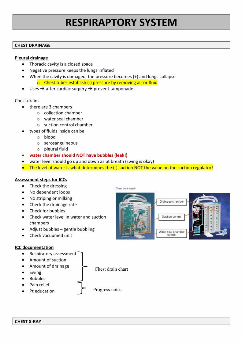
RESPIRAPTORY SYSTEM CHEST DRAINAGE Pleural drainage
• Thoracic cavity is a closed space • Negative pressure keeps the lungs inflated • When the cavity is damaged, the pressure becomes (+) and lungs collapse
o Chest tubes establish (-) pressure by removing air or fluid • Uses à after cardiac surgery à prevent tamponade
Chest drains
• there are 3 chambers o collection chamber o water seal chamber o suction control chamber
• types of fluids inside can be o blood o serosanguineous o pleural fluid
• water chamber should NOT have bubbles (leak!) • water level should go up and down as pt breath (swing is okay) • The level of water is what determines the (-) suction NOT the value on the suction regulator!
Assessment steps for ICCs
• Check the dressing • No dependent loops • No striping or milking • Check the drainage rate • Check for bubbles • Check water level in water and suction
chambers • Adjust bubbles – gentle bubbling • Check vacuumed unit
ICC documentation
• Respiratory assessment • Amount of suction • Amount of drainage • Swing • Bubbles • Pain relief • Pt education
CHEST X-RAY
Chest drain chart
Progress notes
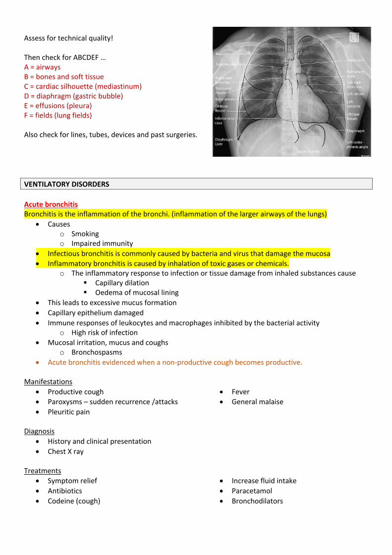
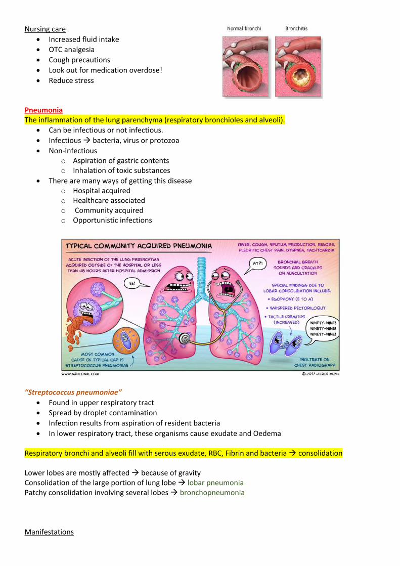
Assess for technical quality! Then check for ABCDEF … A = airways B = bones and soft tissue C = cardiac silhouette (mediastinum) D = diaphragm (gastric bubble) E = effusions (pleura) F = fields (lung fields) Also check for lines, tubes, devices and past surgeries. VENTILATORY DISORDERS Acute bronchitis Bronchitis is the inflammation of the bronchi. (inflammation of the larger airways of the lungs)
• Causes o Smoking o Impaired immunity
• Infectious bronchitis is commonly caused by bacteria and virus that damage the mucosa • Inflammatory bronchitis is caused by inhalation of toxic gases or chemicals.
o The inflammatory response to infection or tissue damage from inhaled substances cause § Capillary dilation § Oedema of mucosal lining
• This leads to excessive mucus formation • Capillary epithelium damaged • Immune responses of leukocytes and macrophages inhibited by the bacterial activity
o High risk of infection • Mucosal irritation, mucus and coughs
o Bronchospasms • Acute bronchitis evidenced when a non-productive cough becomes productive.
Manifestations
• Productive cough • Paroxysms – sudden recurrence /attacks • Pleuritic pain
• Fever • General malaise
Diagnosis
• History and clinical presentation • Chest X ray
Treatments
• Symptom relief • Antibiotics • Codeine (cough)
• Increase fluid intake • Paracetamol • Bronchodilators
Nursing care • Increased fluid intake • OTC analgesia • Cough precautions • Look out for medication overdose! • Reduce stress
Pneumonia The inflammation of the lung parenchyma (respiratory bronchioles and alveoli).
• Can be infectious or not infectious. • Infectious à bacteria, virus or protozoa • Non-infectious
o Aspiration of gastric contents o Inhalation of toxic substances
• There are many ways of getting this disease o Hospital acquired o Healthcare associated o Community acquired o Opportunistic infections
“Streptococcus pneumoniae”
• Found in upper respiratory tract • Spread by droplet contamination • Infection results from aspiration of resident bacteria • In lower respiratory tract, these organisms cause exudate and Oedema
Respiratory bronchi and alveoli fill with serous exudate, RBC, Fibrin and bacteria à consolidation Lower lobes are mostly affected à because of gravity Consolidation of the large portion of lung lobe à lobar pneumonia Patchy consolidation involving several lobes à bronchopneumonia Manifestations
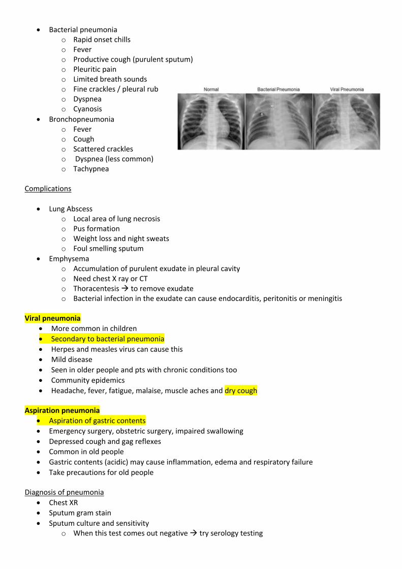
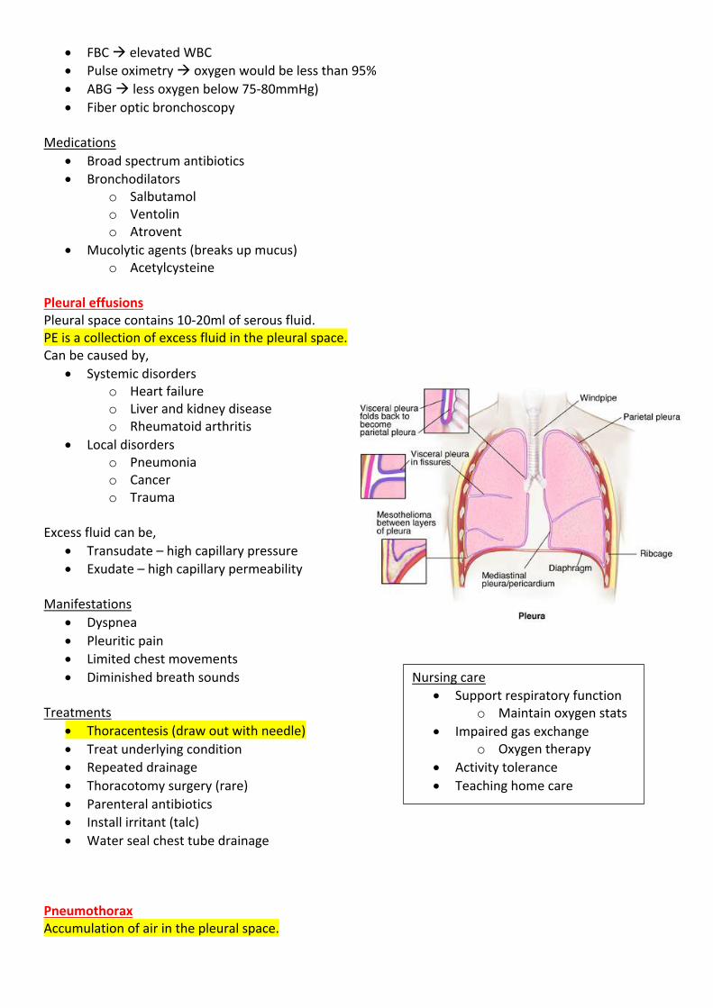
• Bacterial pneumonia o Rapid onset chills o Fever o Productive cough (purulent sputum) o Pleuritic pain o Limited breath sounds o Fine crackles / pleural rub o Dyspnea o Cyanosis
• Bronchopneumonia o Fever o Cough o Scattered crackles o Dyspnea (less common) o Tachypnea
Complications
• Lung Abscess o Local area of lung necrosis o Pus formation o Weight loss and night sweats o Foul smelling sputum
• Emphysema o Accumulation of purulent exudate in pleural cavity o Need chest X ray or CT o Thoracentesis à to remove exudate o Bacterial infection in the exudate can cause endocarditis, peritonitis or meningitis
Viral pneumonia
• More common in children • Secondary to bacterial pneumonia • Herpes and measles virus can cause this • Mild disease • Seen in older people and pts with chronic conditions too • Community epidemics • Headache, fever, fatigue, malaise, muscle aches and dry cough
Aspiration pneumonia
• Aspiration of gastric contents • Emergency surgery, obstetric surgery, impaired swallowing • Depressed cough and gag reflexes • Common in old people • Gastric contents (acidic) may cause inflammation, edema and respiratory failure • Take precautions for old people
Diagnosis of pneumonia
• Chest XR • Sputum gram stain • Sputum culture and sensitivity
o When this test comes out negative à try serology testing
• FBC à elevated WBC • Pulse oximetry à oxygen would be less than 95% • ABG à less oxygen below 75-80mmHg) • Fiber optic bronchoscopy
Medications
• Broad spectrum antibiotics • Bronchodilators
o Salbutamol o Ventolin o Atrovent
• Mucolytic agents (breaks up mucus) o Acetylcysteine
Pleural effusions Pleural space contains 10-20ml of serous fluid. PE is a collection of excess fluid in the pleural space. Can be caused by,
• Systemic disorders o Heart failure o Liver and kidney disease o Rheumatoid arthritis
• Local disorders o Pneumonia o Cancer o Trauma
Excess fluid can be,
• Transudate – high capillary pressure • Exudate – high capillary permeability
Manifestations
• Dyspnea • Pleuritic pain • Limited chest movements • Diminished breath sounds
Treatments
• Thoracentesis (draw out with needle) • Treat underlying condition • Repeated drainage • Thoracotomy surgery (rare) • Parenteral antibiotics • Install irritant (talc) • Water seal chest tube drainage
Pneumothorax Accumulation of air in the pleural space.
Nursing care • Support respiratory function
o Maintain oxygen stats • Impaired gas exchange
o Oxygen therapy • Activity tolerance • Teaching home care
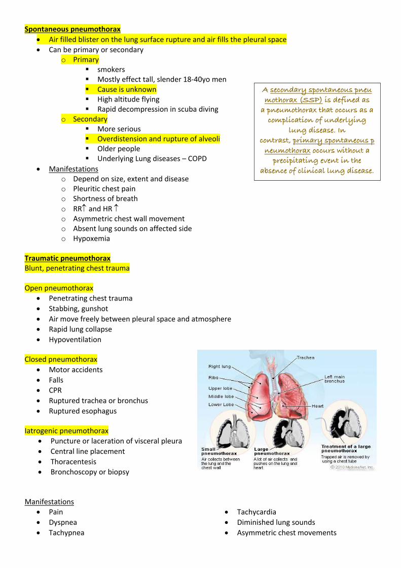
Spontaneous pneumothorax • Air filled blister on the lung surface rupture and air fills the pleural space • Can be primary or secondary
o Primary § smokers § Mostly effect tall, slender 18-40yo men § Cause is unknown § High altitude flying § Rapid decompression in scuba diving
o Secondary § More serious § Overdistension and rupture of alveoli § Older people § Underlying Lung diseases – COPD
• Manifestations o Depend on size, extent and disease o Pleuritic chest pain o Shortness of breath o RR and HR o Asymmetric chest wall movement o Absent lung sounds on affected side o Hypoxemia
Traumatic pneumothorax Blunt, penetrating chest trauma Open pneumothorax
• Penetrating chest trauma • Stabbing, gunshot • Air move freely between pleural space and atmosphere • Rapid lung collapse • Hypoventilation
Closed pneumothorax
• Motor accidents • Falls • CPR • Ruptured trachea or bronchus • Ruptured esophagus
Iatrogenic pneumothorax
• Puncture or laceration of visceral pleura • Central line placement • Thoracentesis • Bronchoscopy or biopsy
Manifestations
• Pain • Dyspnea • Tachypnea
• Tachycardia • Diminished lung sounds • Asymmetric chest movements
A secondary spontaneous pneumothorax (SSP) is defined as
a pneumothorax that occurs as a complication of underlying
lung disease. In contrast, primary spontaneous p
neumothorax occurs without a precipitating event in the
absence of clinical lung disease.
• Hemothorax
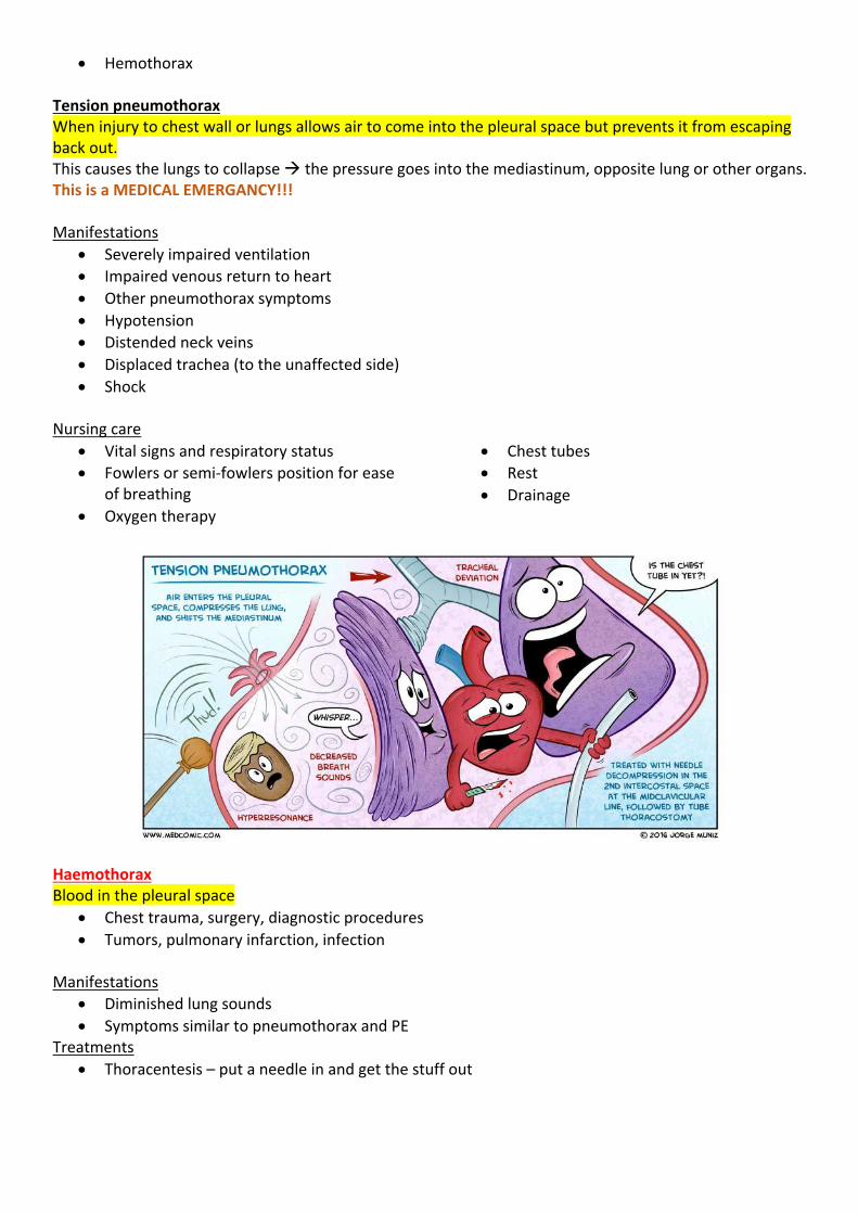
Tension pneumothorax When injury to chest wall or lungs allows air to come into the pleural space but prevents it from escaping back out. This causes the lungs to collapse à the pressure goes into the mediastinum, opposite lung or other organs. This is a MEDICAL EMERGANCY!!! Manifestations
• Severely impaired ventilation • Impaired venous return to heart • Other pneumothorax symptoms • Hypotension • Distended neck veins • Displaced trachea (to the unaffected side) • Shock
Nursing care
• Vital signs and respiratory status • Fowlers or semi-fowlers position for ease
of breathing • Oxygen therapy
• Chest tubes • Rest • Drainage
Haemothorax Blood in the pleural space
• Chest trauma, surgery, diagnostic procedures • Tumors, pulmonary infarction, infection
Manifestations
• Diminished lung sounds • Symptoms similar to pneumothorax and PE
Treatments • Thoracentesis – put a needle in and get the stuff out
• Thoracotomy - surgical opening à put a drain tube • Chest tube drainage • Blood transfusion
Nursing care
• Maintain respiratory function • Maintain cardiac output • Impaired gas exchange • Fluid deficits • No smoking
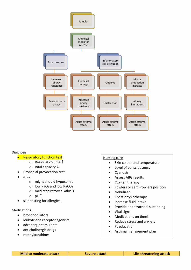
GAS EXCHANGE DISORDERS Asthma Chronic inflammatory disorder. A condition in which a person's airways become inflamed, narrow and swell and produce extra mucus, which makes it difficult to breathe. Wheezing, breathlessness, chest tightness and coughing often seen.
• Risk factors o Allergies o Genetics o Exercise o Pollution
o Workplace exposure o Infections o Stress o Cold air
• Manifestations o Dyspnea o Tachypnea o Tachycardia o Chest tightness
o Wheezing o Cough o Anxiety o Lung crackles
Status asmaticus is the prolonged, severe asthma that does not respond to routine treatment.
• Endotracheal intubation • Mechanical ventilation • Aggressive drugs
Diagnosis
• Respiratory function test o Residual volume o Vital capacity ¯
• Bronchial provocation test • ABG
o might should hypoxemia o low PaO2 and low PaCO2 o mild respiratory alkalosis o pH
• skin testing for allergies Medications
• bronchodilators • leukotriene receptor agonists • adrenergic stimulants • anticholinergic drugs • methylxanthines
Mild to moderate attack Severe attack Life-threatening attack
Stimulus
Chemical mediator release
Bronchospasm
Increased airway
resistance
Acute asthma attack
Inflammatory cell activation
Epithelial damage
Increased airway
resistance
Acute asthma attack
Oedema
Obstruction
Acute asthma attack
Mucus production
increase
Airway limitations
Acute asthma attack
Nursing care • Skin colour and temperature • Level of consciousness • Cyanosis • Assess ABG results • Oxygen therapy • Fowlers or semi-fowlers position • Nebulizer • Chest physiotherapy • Increase fluid intake • Provide endotracheal suctioning • Vital signs • Medications on time! • Reduce stress and anxiety • Pt education • Asthma management plan
Give salbutamol 4-12 puffs via pMDI and spacer
Repeat every 20-30 min for the first hour if needed. (sooner if
needed to relieve breathlessness)
Give salbutamol 12 puffs via pMDI and spacer
If pt is unable to breathe through the spacer, give 5mg nebule via
nebuliser.
Start oxygen therapy if oxygen saturation is below 95%.
Adults: 92%-95%
Children: 95% or higher
Repeat salbutamol as needed. Give at least every 20 min for the
first hour.
Give salbutamol 2x5mg nebules via continuous nebulisation
driven by oxygen.
Maintain oxygen saturations
Adult: 92% or higher Children: 95% or higher
Arrange immediate transfer to
higher level care.
When dyspnoea improves, consider changing to salbutamol
via pMDI plus spacer or intermittent nebuliser (doses as
for severe acute asthma)
CHRONIC OBSTRUCTIVE PULMONARY DISEASE (COPD)
Chronic Obstructive Pulmonary Disease (COPD) is an umbrella term for a group of progressive lung conditions including: Emphysema, Chronic bronchitis and Chronic asthma which causes chronic airflow
obstructions. Risk factors – middle aged and smokers Chronic bronchitis Disorder of excessive bronchial mucus secretion. Chronic bronchitis specifically refers to chronic cough and daily mucus production for at least three months of two or more consecutive years. Manifestations
• Vasodilation • Congestion • Oedema • Impaired ciliary action • Goblet cells increase in
size and number • Thick tenacious mucus
• Recurrent infections • Hypoxemia • Hypercapnia • Hypertension • Productive cough • Cyanosis
• Right sided heart failure
• Distended neck veins • Enlarged liver and
heart • Wheezing lung sounds
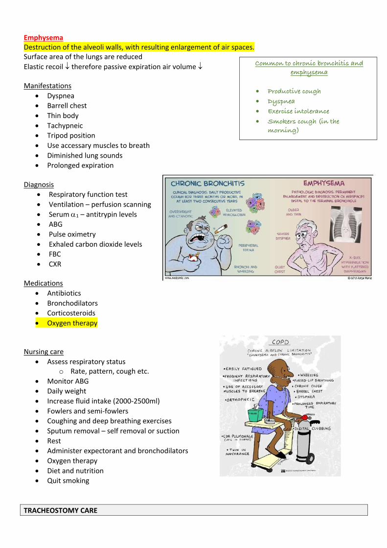
Emphysema Destruction of the alveoli walls, with resulting enlargement of air spaces. Surface area of the lungs are reduced Elastic recoil ¯ therefore passive expiration air volume ¯ Manifestations
• Dyspnea • Barrell chest • Thin body • Tachypneic • Tripod position • Use accessary muscles to breath • Diminished lung sounds • Prolonged expiration
Diagnosis
• Respiratory function test • Ventilation – perfusion scanning • Serum a1 – antitrypin levels • ABG • Pulse oximetry • Exhaled carbon dioxide levels • FBC • CXR
Medications
• Antibiotics • Bronchodilators • Corticosteroids • Oxygen therapy
Nursing care
• Assess respiratory status o Rate, pattern, cough etc.
• Monitor ABG • Daily weight • Increase fluid intake (2000-2500ml) • Fowlers and semi-fowlers • Coughing and deep breathing exercises • Sputum removal – self removal or suction • Rest • Administer expectorant and bronchodilators • Oxygen therapy • Diet and nutrition • Quit smoking
TRACHEOSTOMY CARE
Common to chronic bronchitis and emphysema
• Productive cough
• Dyspnea
• Exercise intolerance
• Smokers cough (in the morning)
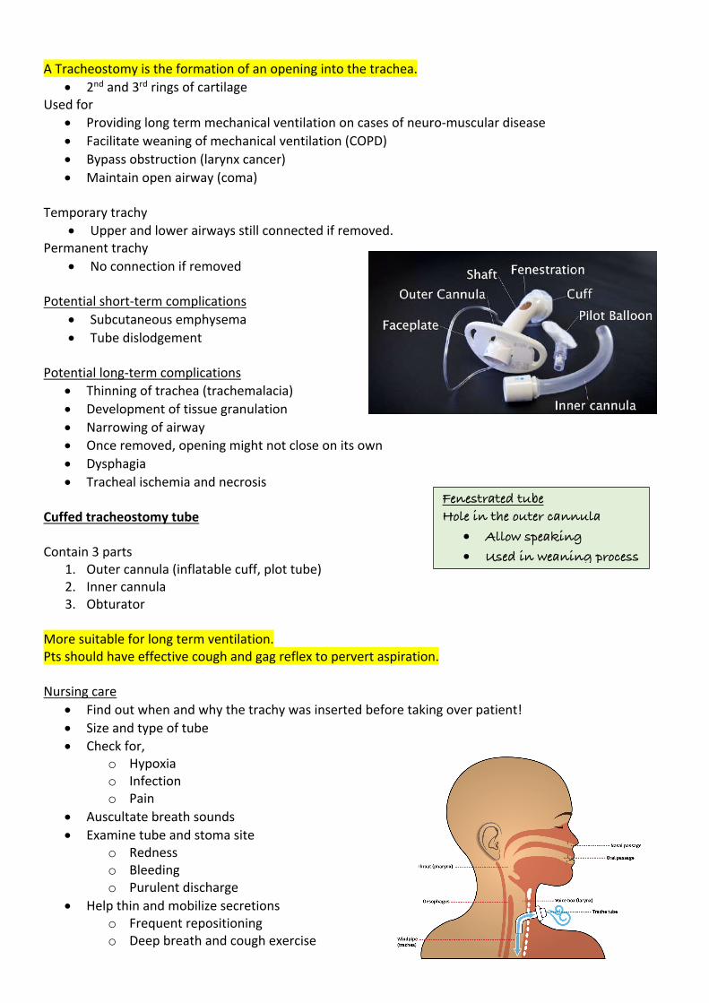
A Tracheostomy is the formation of an opening into the trachea.
• 2nd and 3rd rings of cartilage Used for
• Providing long term mechanical ventilation on cases of neuro-muscular disease • Facilitate weaning of mechanical ventilation (COPD) • Bypass obstruction (larynx cancer) • Maintain open airway (coma)
Temporary trachy
• Upper and lower airways still connected if removed. Permanent trachy
• No connection if removed Potential short-term complications
• Subcutaneous emphysema • Tube dislodgement
Potential long-term complications
• Thinning of trachea (trachemalacia) • Development of tissue granulation • Narrowing of airway • Once removed, opening might not close on its own • Dysphagia • Tracheal ischemia and necrosis
Cuffed tracheostomy tube Contain 3 parts
1. Outer cannula (inflatable cuff, plot tube) 2. Inner cannula 3. Obturator
More suitable for long term ventilation. Pts should have effective cough and gag reflex to pervert aspiration. Nursing care
• Find out when and why the trachy was inserted before taking over patient! • Size and type of tube • Check for,
o Hypoxia o Infection o Pain
• Auscultate breath sounds • Examine tube and stoma site
o Redness o Bleeding o Purulent discharge
• Help thin and mobilize secretions o Frequent repositioning o Deep breath and cough exercise
Fenestrated tube Hole in the outer cannula
• Allow speaking
• Used in weaning process
o Chest physiotherapy o Oral and parenteral hydration o Supplemental humidification
DRUGS ACTING ON THE LOWER RESPIRATORY TRACT Preventative and treatment measure for COPD
• Reduce environmental exposure to irritants • Stop smoking • Filter allergens from the air • Avoid exposure to known irritants and allergens • Open the conducting airways through muscular bronchodilation
Drug type Actions Indications &
Contraindications
Pharmacokinetics Adverse effects Drug-drug interactions
Drugs that affect the Lower respiratory tract Xanthines
(Aminophylline)
Direct effect on the smooth
muscles of the respiratory
system, both in the bronchi and blood vessels.
Indications Symptomatic
relief or prevention of
bronchial asthma and for reversal of
bronchospasm associated with
COPD Contraindications
GI problems Coronary disease
Respiratory dysfunction
Renal and hepatic disease
Alcoholism Hyperthyroidism
Narrow therapeutic
margin. Quickly absorbed
by the GI tract. Metabolized in
the liver. Excreted by urine.
Related to theophylline levels in the
blood.
GI upsets Nausea
Irritability Tachycardia
Seizures Brain damage
Even death
Many drugs interact
xanthines.
Nicotine increase
metabolism.
Sympathomimetics
(Adrenaline)
Beta2 selective adrenergic agonist
Indications Acute asthma
attacks. Bronchospasm in acute or chronic
asthma. Prevention of
exercise induced asthma.
Contraindications Depends of the
severity of underlying condition
Rapidly distributed after
injection. Transformed in
the liver to metabolites that are excreted in
urine.
Sympathomimetic stimulation.
CNS stimulation
GI upsets
Arrhythmias Hypertension
Bronchospasm Sweating
Pallor Flushing
General anaesthetics

Anticholinergic bronchodilators
(Ipratropium)
Blocks vagally medicated reflexes by
antagonising the action of
acetylcholine
Indications Maintenance of bronchospasm associated with
COPD
Contraindications Any condition that
would be aggravated by
cholinergic drugs.
Onset of action is 15min when
inhaled.
Peaks in 1-2 hours.
Duration of action is 3-4
hours.
Related to anticholinergic
effect of the drug.
Dizziness Headache
Fatigue Nervousness Dry mouth Sore throat Palpitations
Urinary retention
-
Inhaled steroids
(Budesonide)
Decrease the inflammatory
response of the airway.
Indications Prevention and
treatment of asthma.
Treating chronic steroid dependent bronchial asthma. Contraindications
Not used for emergency during an acute attack or status asthmatics.
Pregnancy or breastfeeding.
Well absorbed from respiratory
tract.
Metabolised in natural systems, mostly within the liver and excreted
in urine.
Sore throat Hoarseness Coughing
Dry mouth
Pharyngeal or laryngeal fungal
infections.
-
Leukotriene receptor agonists
(Zafirlukast)
Selectively and competitively
block or antagonise
receptors for the production of leukotriene.
Indications Prophylaxis and
chronic treatment of bronchial
asthma in adults and in individuals younger than 12
years of age. Contraindications Hepatic and renal
impairment. Pregnancy or breastfeeding.
Rapidly absorbed from the GI tract,
extensively metabolised in
the live and primarily
excreted in faeces.
Headache Dizziness Myalgia Nausea
Diarrhoea Abdominal pain
Vomiting
Elevated liver enzyme
concentration
Generalized pain
Propranolol Theophylline
Warfarin
Calcium channel blockers
Cyclosporine
Aspirin
Mast cell stabilisers
(Sodium cromoglicate)
Works at the cellular level to
inhibit the release of histamines and
inhibits the release of SRSA.
Indications Treatment of
chronic bronchial asthma.
Exercise induced asthma.
Allergic rhinitis.
Absorption is largely from the respiratory tract. Normally inhaled
– more puffs more absorption.
Cough
Runny nose Throat irritation Unpleasant taste
Headache
isoprenaline
Drugs that affect the upper respiratory tract Antitussives
(Dextromethorphan)
Block the cough reflex by acting on
the medullary cough centre
Indications Control non-
productive cough
Contraindications
Rapidly absorbed
Metabolised in
the liver Excreted in urine.
Drying effect in the mucus
membrane.
CNS adverse effects. GI upset
-
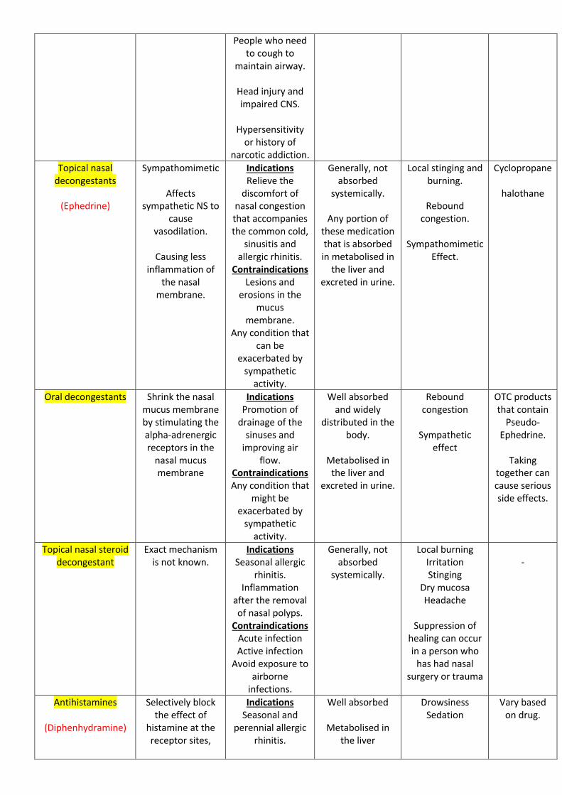
People who need to cough to
maintain airway.
Head injury and impaired CNS.
Hypersensitivity
or history of narcotic addiction.
Topical nasal decongestants
(Ephedrine)
Sympathomimetic
Affects sympathetic NS to
cause vasodilation.
Causing less
inflammation of the nasal
membrane.
Indications Relieve the
discomfort of nasal congestion that accompanies the common cold,
sinusitis and allergic rhinitis.
Contraindications Lesions and
erosions in the mucus
membrane. Any condition that
can be exacerbated by
sympathetic activity.
Generally, not absorbed
systemically.
Any portion of these medication that is absorbed in metabolised in
the liver and excreted in urine.
Local stinging and burning.
Rebound
congestion.
Sympathomimetic Effect.
Cyclopropane
halothane
Oral decongestants Shrink the nasal mucus membrane by stimulating the alpha-adrenergic receptors in the
nasal mucus membrane
Indications Promotion of
drainage of the sinuses and
improving air flow.
Contraindications Any condition that
might be exacerbated by
sympathetic activity.
Well absorbed and widely
distributed in the body.
Metabolised in
the liver and excreted in urine.
Rebound congestion
Sympathetic
effect
OTC products that contain
Pseudo- Ephedrine.
Taking
together can cause serious side effects.
Topical nasal steroid decongestant
Exact mechanism is not known.
Indications Seasonal allergic
rhinitis. Inflammation
after the removal of nasal polyps.
Contraindications Acute infection Active infection
Avoid exposure to airborne
infections.
Generally, not absorbed
systemically.
Local burning Irritation Stinging
Dry mucosa Headache
Suppression of
healing can occur in a person who
has had nasal surgery or trauma
-
Antihistamines
(Diphenhydramine)
Selectively block the effect of
histamine at the receptor sites,
Indications Seasonal and
perennial allergic rhinitis.
Well absorbed
Metabolised in the liver
Drowsiness Sedation
Vary based on drug.
decreasing the allergic reaction.
Allergic congestivitis.
Uncomplicated urticaria
Angio-oedema
Contraindications Pregnancy or
breastfeeding. Renal and hepatic
impairment. History of
arrhythmias.
Excreted in urine and faeces.
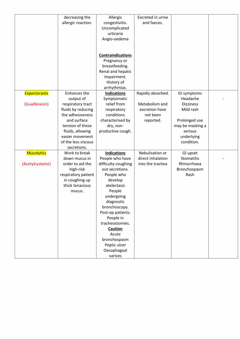
Expectorants
(Guaifenesin)
Enhances the output of
respiratory tract fluids by reducing the adhesiveness
and surface tension of these fluids, allowing
easier movement of the less viscous
secretions.
Indications Symptomatic
relief from respiratory conditions
characterised by dry, non-
productive cough.
Rapidly absorbed.
Metabolism and excretion have
not been reported.
GI symptoms Headache Dizziness Mild rash
Prolonged use
may be masking a serious
underlying condition.
-
Mucolytics
(Acetylcysteine)
Work to break down mucus in order to aid the
high-risk respiratory patient
in coughing up thick tenacious
mucus.
Indications People who have
difficulty coughing out secretions.
People who develop
atelectasis People
undergoing diagnostic
bronchoscopy. Post-op patients.
People in tracheostomies.
Caution Acute
bronchospasm Peptic ulcer
Oesophageal varices
Nebulisation or direct inhalation into the trachea
GI upset Stomatitis
Rhinorrhoea Bronchospasm
Rash
-


![Index [s3.studentvip.com.au]](https://img.pdfslide.net/doc/110x75/61eb3d4de79bdf67c17284a8/index-s3-.jpg)