Embed Size (px)
Citation preview

Respiratory Care
Training Package for Nurses Version 1.1. March 2020.
#RisingToTheChallenge

Contents
1. Summary of training package
2. Competency document
3. Supportive materials: training cards
4. Oxygen titration tool
5. Staff information leaflet

Summary
As the COVID-19 situation develops we anticipate a large increase in patients requiring
respiratory care in our ward areas. Traditionally many of these patients would be cared for in a
respiratory ward area, however during the COVID-19 epidemic it is unlikely the traditional units
will be able to meet the demand.
This training package has been designed to re-familiarise and refresh our nursing teams
knowledge around caring for patient with respiratory conditions outside of the usual specialist
respiratory ward areas.
The ‘Respiratory Care Training Package for Nurses’ is intended to provide an agile training
platform suitable for bedside teaching, small groups and act as a resource that can be referred
to. The programme has been a collaborative effort involving the Acute Care Team, Education
Teams from Manchester Royal Infirmary and Wythenshawe Hospital, Corporate Nursing
Teams and Physiotherapy.

2. Competency document

1. Anatomy and Physiology
I can demonstrate through discussion:
Structure of respiratory system
Mechanism of breathing
Respiratory Failure
Type I respiratory failure
Type II respiratory failure
Signs and symptoms of respiratory failure
Effects of poor ventilation and oxygenation on other systems:
Cardiac
Renal
Gastrointestinal
Cerebral
Skin
2. Assessment and investigations
I can demonstrate through discussion:
Normal parameters for respiratory observations – need to observe for one full
minute. Look at rate, rhythm, depth, symmetry, accessory muscle use
Trends in observation charts
Respiratory Assessment
Look
Listen
Feel
Skin colour:
Peripheral cyanosis
Central cyanosis
Respiratory Competencies for Ward Based Staff

Cough Strength
Effective and ineffective
Ability to expectorate vs retention
Sputum assessment
Prescribed target oxygen ranges
Indications and methods for Oxygen therapy and titration
3. Management
I can demonstrate through discussion:
Oxygen therapy:
Non-rebreathe mask
Venturi
Humidified
Nasal cannula
Treatment
Nebulisation technique
Inhaler technique
Patient positioning:
Maximising ventilation
Postural draining positioning
Deep breathing exercises/ testing procedures:
Incentive spirometry
Secretion clearance/ oral suctioning
Provision of emotional reassurance and support for patient
Escalation for higher level of respiratory support
Recognition of level of care requirements
Escalation procedures

Self-assessment
Staff name/grade/ward area
Assessor sign and date
Please return to Yvon Poland to populate training database,
email. [email protected]

3. Supportive material: training
cards


4. Oxygen titration tool

Oxygen titration
Venturi 24% 2-4l/min
Nasal cannula 4 l/min Venturi 35% 8-10 l/min
Venturi 28% 4-6 l/min Nasal cannula 2-3
l/min
Nasal cannula 1 l/min
Venturi 40% 10-12 l/min or simple face mask 5-6 l/min
Venturi 60% 12-15 l/min or simple face mask 7-10 l/min
Reservoir mask at 15 l/min oxygen flow
If Reservoir mask is required or ≥60%, seek senior medical input immediately
Patients in peri-arrest situation and critically ill patients should receive maximum oxygen via a reservoir mask or bag valve mask while help is being
summoned.
If patient requires
increasing oxygen
delivery or shows signs of
respiratory deterioration, seek medical
advice

5. Staff information sheet

Look
Count the respiration rate for one full minute
What is the rhythm of the breathing – is it irregular?
What is the depth of the breathing – is it shallow?
Is the chest moving equally/symmetrically?
Is the patient mouth breathing, pursing the lips on
expiration, using the abdominal muscles or flaring
the nostrils?
Are there any signs of bluish colouration in the skin,
fingertips or lips (cyanosis)?
Is there new confusion or agitation?
What are the oxygen saturations – are they within
the prescribed oxygen saturation requirements?
Check the position of the trachea: deviation to one
side indicates mediastinal shift (e.g. pneumothorax,
lung fibrosis or pleural fluid) – this an emergency
situation if new deviation
Listen Listen to the patient’s breath sounds a short
distance from his face: rattling airway noises
indicate the presence of airway secretions, usually
caused by the inability of the patient to cough
sufficiently or to take a deep breath. Stridor or
wheeze suggests partial, but significant, airway
obstruction
Feel Auscultate the chest if trained to: bronchial
breathing indicates lung consolidation with patent
airways; absent or reduced sounds suggest a
pneumothorax or pleural fluid or lung consolidation
caused by complete obstruction
Feel the chest for symmetry of movement
Information for Respiratory Assessment and Management of Respiratory Failure
Respiratory Assessment
A respiratory assessment is an external assessment of ventilation that includes observations of the rate, depth and pattern of respirations.
Steps of a respiratory assessment

Type 1 respiratory failure
(Hypoxemic respiratory failure)
Type 2 respiratory failure
(Hypercapnic respiratory failure)
Hypoxemic respiratory failure means
that you don’t have enough oxygen in
your blood, but your levels of carbon
dioxide are close to normal
A common cause of hypoxemic
respiratory failure is an abnormality of
the lung tissue, such as acute
respiratory distress syndrome, severe
pneumonia, excess fluid in the lungs
(for example, caused by heart failure or
kidney failure). Such abnormalities
disrupt the usual ability of the lung
tissues to take in oxygen from the air
Hypercapnic respiratory failure means that
there’s too much carbon dioxide in your
blood, and near normal or not enough
oxygen in your blood
With hypercapnic respiratory failure, the
level of carbon dioxide is usually too high
because something prevents the person
from breathing normally. Blockage or
narrowing of the airways, weakness of
muscles that normally inflate the lungs,
and an overdose of opioids or alcohol all
decrease the unconscious reflex that drives
people to breathe. Blockage or narrowing
of the airways can result from disorders
(such as asthma and chronic obstructive
pulmonary disease) as well as inhaled
foreign objects
Respiratory failure
Respiratory failure is a condition in which your blood doesn't have enough oxygen or has too much carbon dioxide.
In COVID-19 we are tending to see patients with acute Type 1 respiratory failure
Signs of respiratory failure
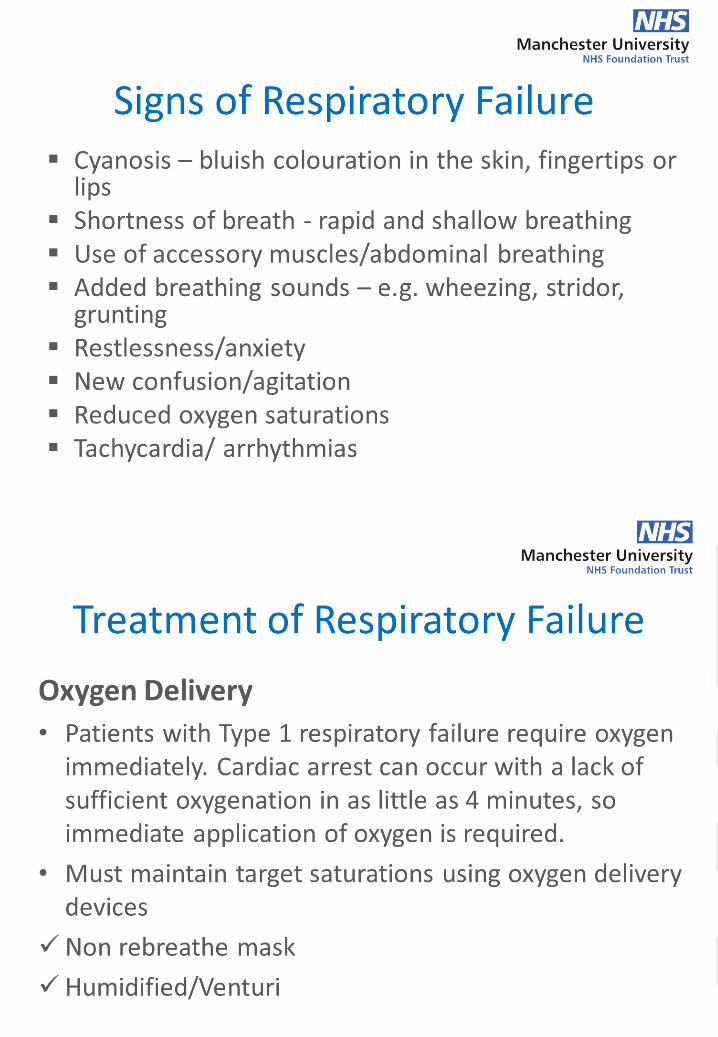
• Cyanosis – bluish colouration in the skin, fingertips or lips • Shortness of breath - rapid and shallow breathing • Use of accessory muscles/abdominal breathing • Added breathing sounds – e.g. wheezing, stridor, grunting • Restlessness/anxiety • New confusion/agitation • Reduced oxygen saturations • Tachycardia/ arrhythmias
If your patient is showing signs of respiratory failure, apply 15 litres of oxygen
via a non-rebreathe mask and escalate to the medical team immediately. If your
patient is losing consciousness or goes in to a peri-arrest state ring 2222
immediately and commence BLS as required

Treatment of respiratory failure
To manage respiratory failure we need to look at three steps
Oxygen delivery Treat cause Mechanical ventilation
Oxygen delivery Patients with Type 1 respiratory failure require oxygen immediately. Cardiac arrest can occur with a lack of sufficient oxygenation in as little as 4 minutes, so immediate application of oxygen is required. In the acute phase of respiratory failure you should apply 15 litres of oxygen via a non-rebreathe mask until the patient has received a medical review.
Types of oxygen delivery systems
Non-rebreathe mask Advantages: With a good fit, the mask can deliver between 60% and 80% FiO2 (fraction of inspired oxygen). The flow meter should be set to deliver O2 at 10 to 15 L/min. Flow rate must be high enough to ensure that the reservoir bag remains partially inflated during inspiration. Disadvantages: These masks have a risk of suffocation if the gas flow is interrupted. The bag should never totally deflate. The patient should never be left alone and should be monitored continuously. The mask also requires a tight seal and may be hot and confining for the patient.
Humidified oxygen Advantages: Able to provide humidification prevent the oxygen therapy from drying out the mucous membranes and reduces secretions becoming tenacious. Can provide 28-60% O2 at 4-10 L/min. Disadvantages: Can feel cold against the face and can be noisy
Venturi masks Advantages: The system can provide 24% to 60% O2 at 4 to 12 L/min. delivers a more precise level of oxygen by controlling the specific amounts of oxygen delivered. The port on the corrugated tubing (base of the mask) sets the oxygen concentration. Disadvantages: The mask may be hot and confining for some patients, and it interferes with talking and eating. Need a properly fitting mask.
Nasal cannula - It is used for short- or long-term therapy and is best used with stable patients who require low amounts of oxygen. Advantages: Can provide 24% to 36% O2 (1 to 4 litres per minute).It is convenient as patient can talk and eat while receiving oxygen. Limitations: Easily dislodged, not as effective is a patient is a mouth breather or has blocked nostrils.

If your patient requires oxygen to maintain saturations – the patient is not stable
and should have their observations taken by a qualified staff member. Oxygen is a
drug and should be prescribed and signed for on medication rounds.
If you commence oxygen therapy or are having to increase the flow of oxygen to
maintain saturations, you must escalate to the medical team immediately.
Treat cause
The treatment of the cause of the respiratory failure will be set out by the medical
team. This may include the COVID-19 treatment, or other drugs such as antibiotics
or steroids depending on the diagnosis
Patients will require other tests such as chest x-ray, sputum samples and peak flow.
Any treatments that are aerosol producing such as suction, physio and
nebulisation, you must wear PPE equipment as per trust policy.
Mechanical Ventilation
A number of patients will have respiratory failure that is so severe that they will require help with their breathing. Invasive mechanical ventilation may be required and the patient will be taken to an intensive care setting for this. Some patients may not need invasive ventilation bit may require non-invasive ventilation, such as CPAP (continuous positive airway pressure), which may be given in some ward areas. Further training will be given to ward staff if required.
Escalation
Continue to escalate your patients according to the Early Warning Score Policy
Make sure you familiarise yourself with specific hospital escalation for COVID-
19 positive patients