Embed Size (px)
Citation preview
Respiratory Crisis Patient’s with Case Study
Objectives
• Review Respiratory System • Review ETCO2
• Review Asthma and COPD • Review Assembly of CPAP • Review Patient Presentations
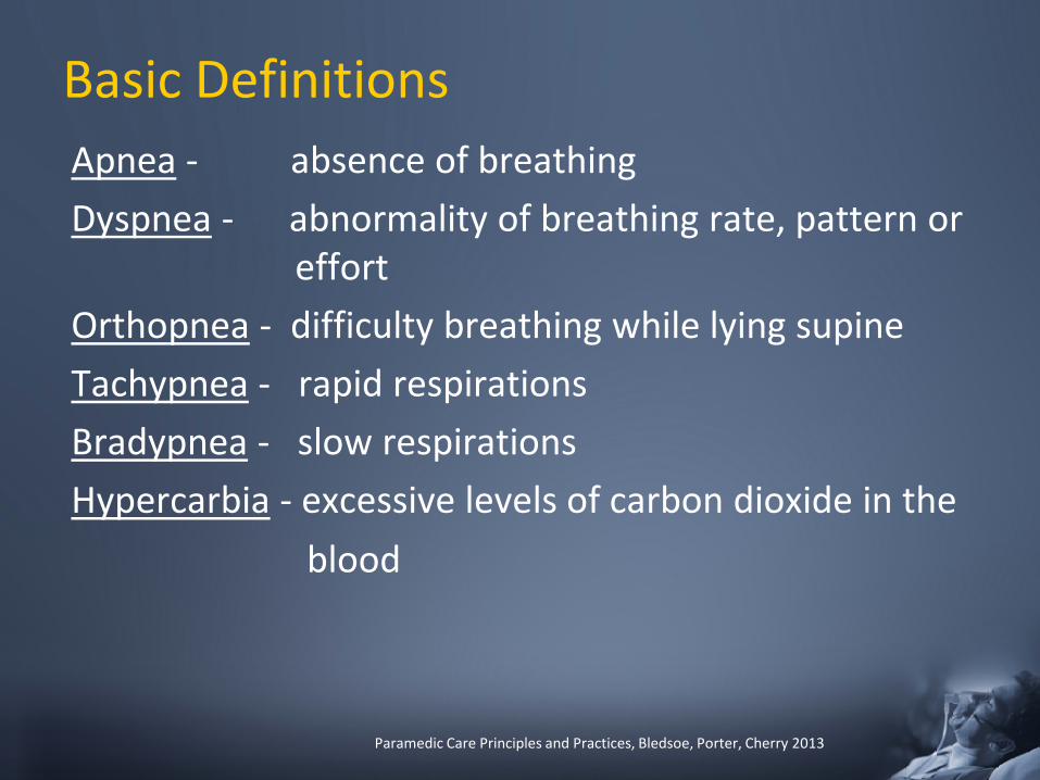
Basic Definitions Apnea - absence of breathing Dyspnea - abnormality of breathing rate, pattern or
effort Orthopnea - difficulty breathing while lying supine Tachypnea - rapid respirations Bradypnea - slow respirations Hypercarbia - excessive levels of carbon dioxide in the
blood
Paramedic Care Principles and Practices, Bledsoe, Porter, Cherry 2013
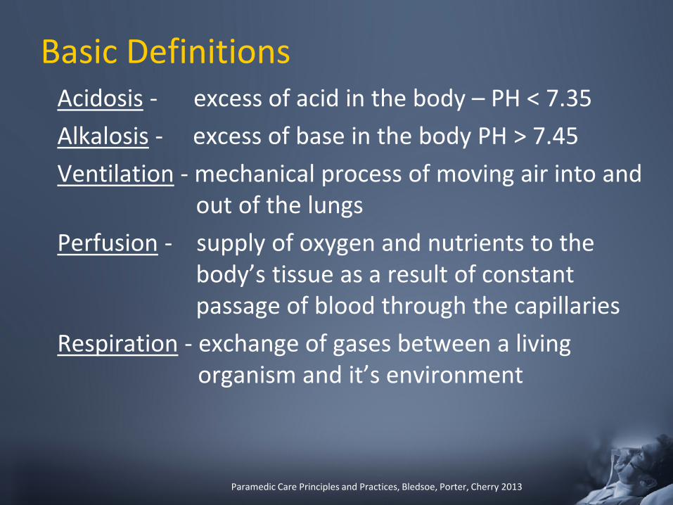
Basic Definitions Acidosis - excess of acid in the body – PH < 7.35 Alkalosis - excess of base in the body PH > 7.45 Ventilation - mechanical process of moving air into and
out of the lungs Perfusion - supply of oxygen and nutrients to the
body’s tissue as a result of constant passage of blood through the capillaries
Respiration - exchange of gases between a living organism and it’s environment
Paramedic Care Principles and Practices, Bledsoe, Porter, Cherry 2013
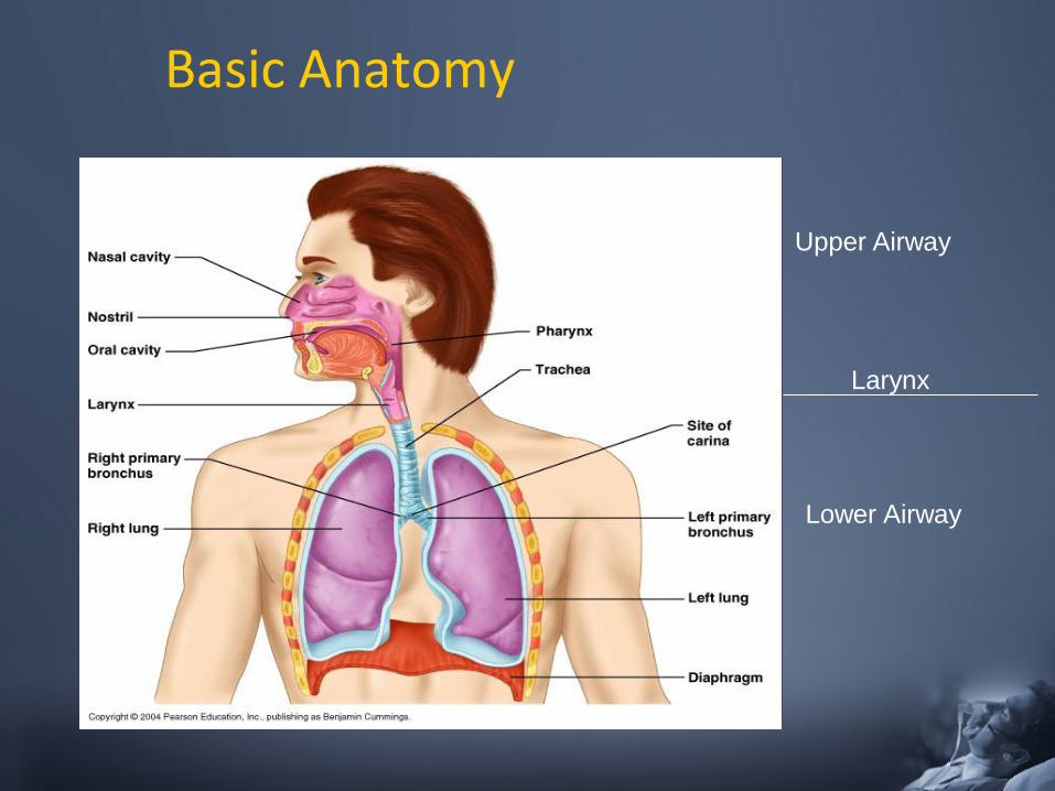
Basic Anatomy
Upper Airway
Lower Airway
Larynx
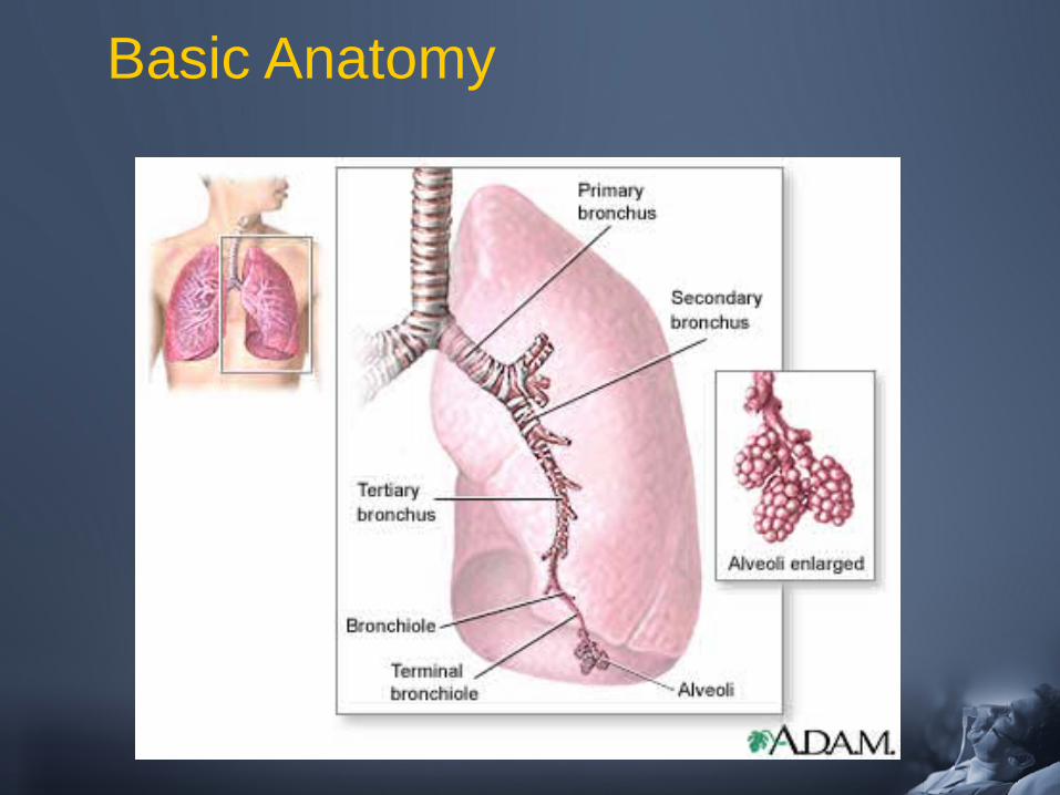
Basic Anatomy
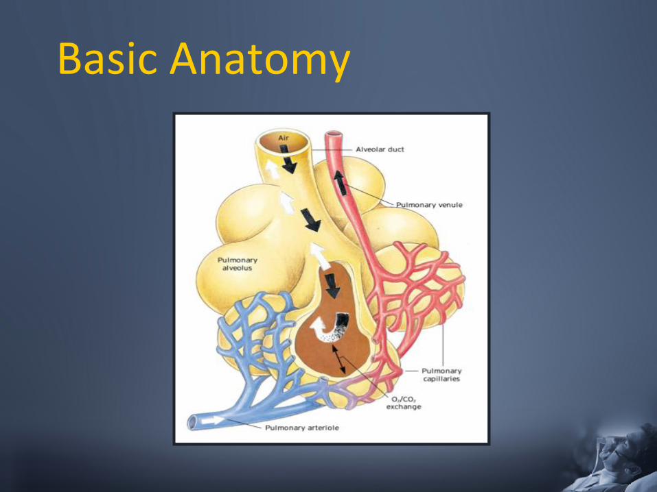
Basic Anatomy
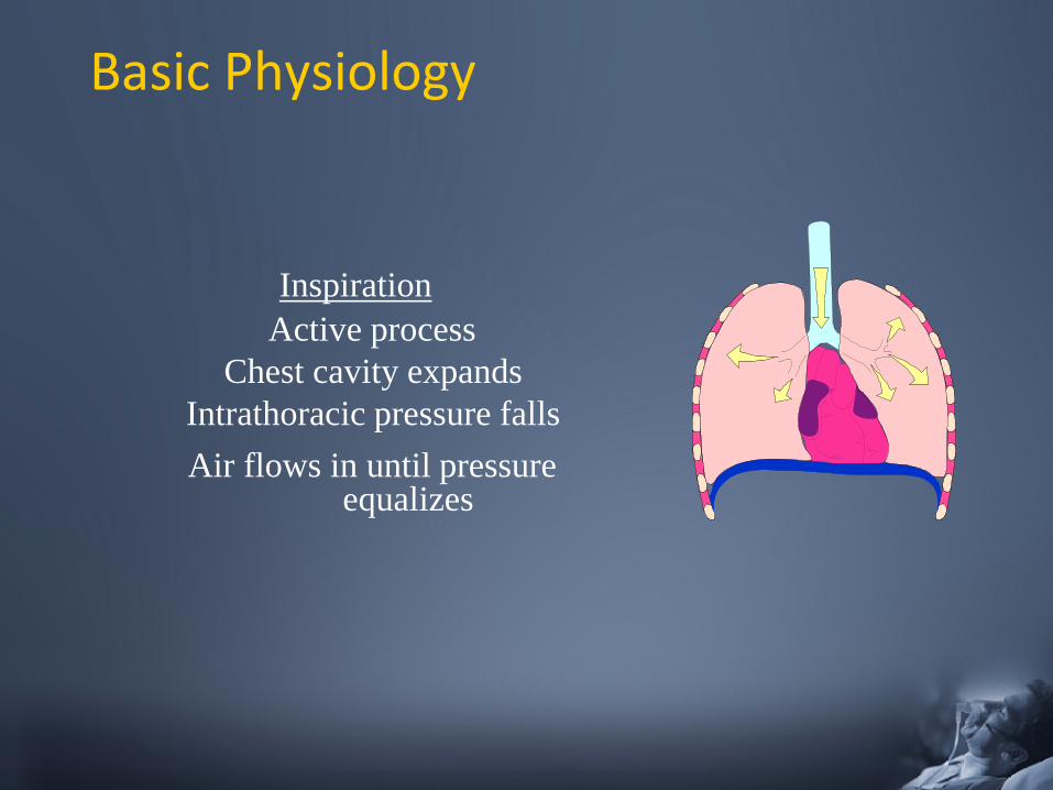
Basic Physiology
Inspiration Active process
Chest cavity expands Intrathoracic pressure falls Air flows in until pressure
equalizes
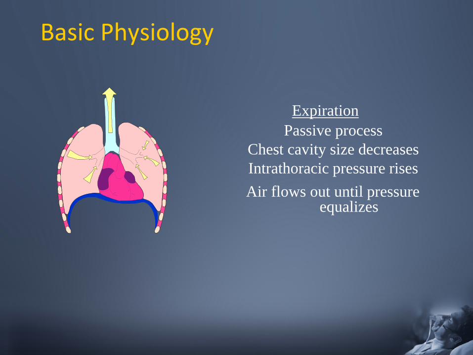
Basic Physiology
Expiration Passive process
Chest cavity size decreases Intrathoracic pressure rises Air flows out until pressure
equalizes
Autonomic Function (Why do we breath)
Primary drive: increase in arterial CO2
(Hypercarbia) – need to get rid of excess CO2
Secondary drive: decrease in arterial O2
(Hypoxia) – need to get more O2
Hering – Breuer reflex: a reflex triggered to prevent over-inflation of
the lungs - Pulmonary stretch receptors present in the smooth muscle of the airways respond to excessive stretching of the lung during large inspirations
Respiratory Cycle
• Respiration - process of oxygen taken into body and distributed to the cells for energy – Carbon dioxide is returned to the lungs by the circulatory
system and exhaled.
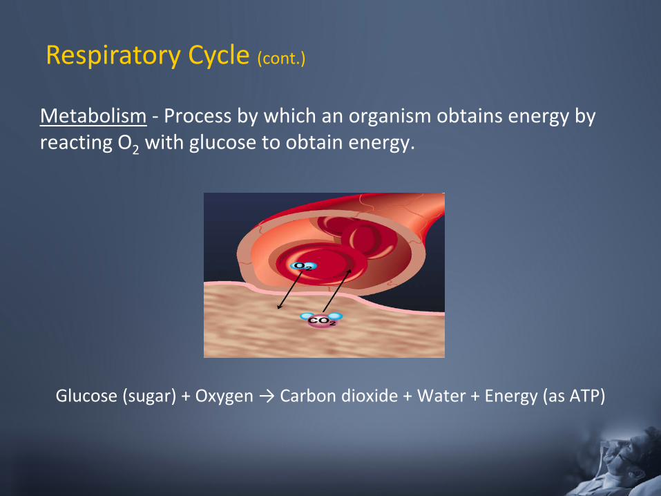
Metabolism - Process by which an organism obtains energy by reacting O2 with glucose to obtain energy.
Glucose (sugar) + Oxygen → Carbon dioxide + Water + Energy (as ATP)
Respiratory Cycle (cont.)
Respiratory Cycle (cont.)
• Ventilation - Rate that gases enter and leave the lungs
– Minute Ventilation - Total volume of gas entering lungs per minute
– Alveolar Ventilation - Volume of gas that reaches the alveoli – Dead Space Ventilation - Volume of gas that does not reach
the respiratory portions ( 150 ml)

CAPNOGRAPHY………..……
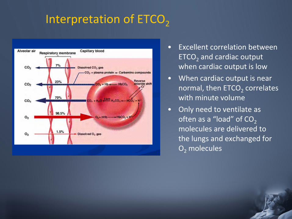
Interpretation of ETCO2
• Excellent correlation between ETCO2 and cardiac output when cardiac output is low
• When cardiac output is near normal, then ETCO2 correlates with minute volume
• Only need to ventilate as often as a “load” of CO2 molecules are delivered to the lungs and exchanged for O2 molecules
How is ETCO2 Measured?
• Capnometry - Analysis only of the presence of gases – no waveforms
• Capnography - Continuous analysis and recording of Carbon Dioxide concentrations in respiratory gases – waveforms and numeric values
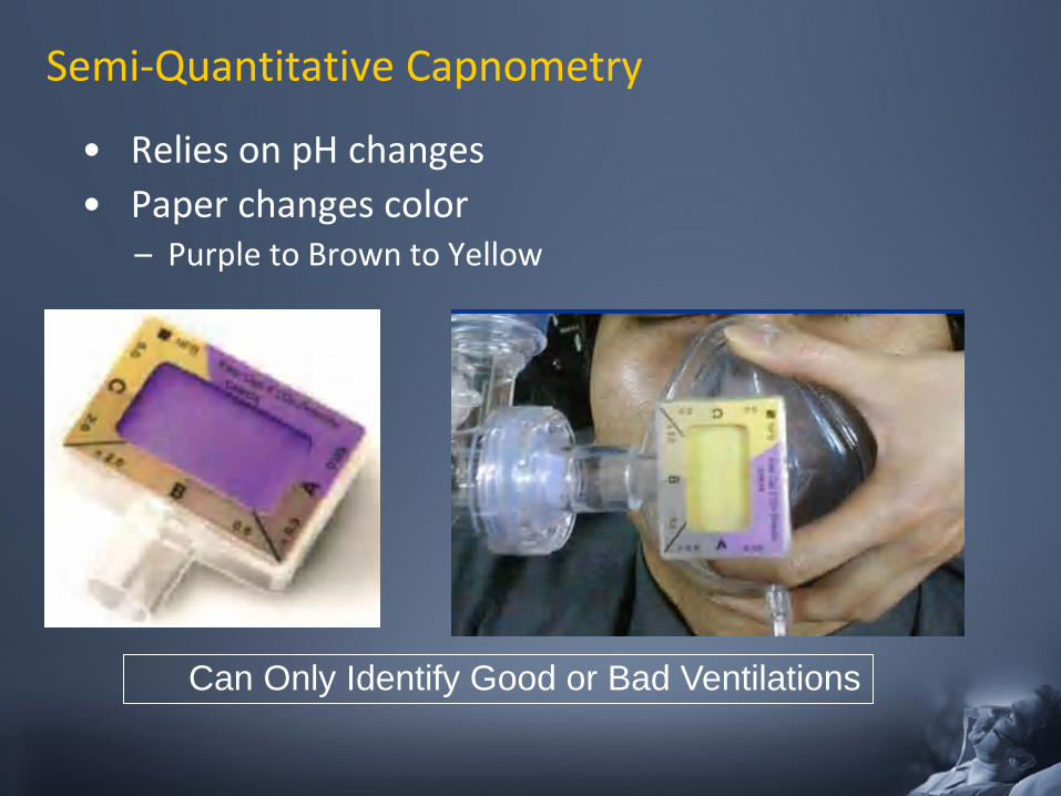
Semi-Quantitative Capnometry
• Relies on pH changes • Paper changes color
– Purple to Brown to Yellow
Can Only Identify Good or Bad Ventilations
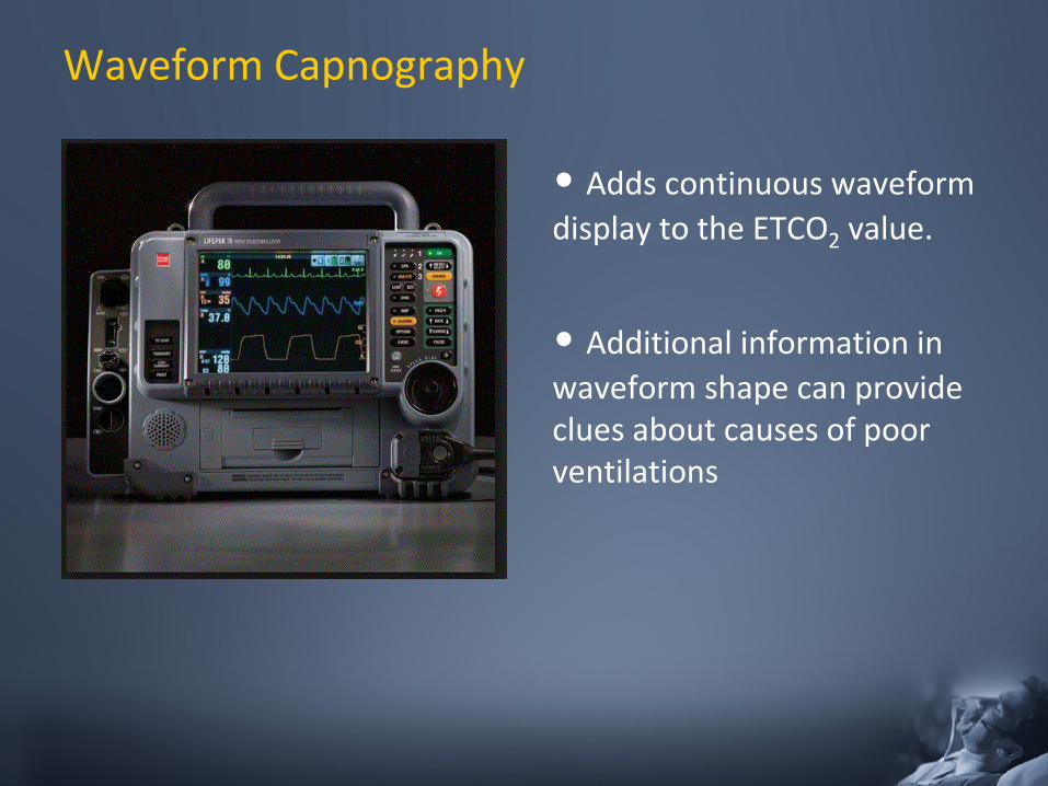
Waveform Capnography
• Adds continuous waveform display to the ETCO2 value.
• Additional information in waveform shape can provide clues about causes of poor ventilations
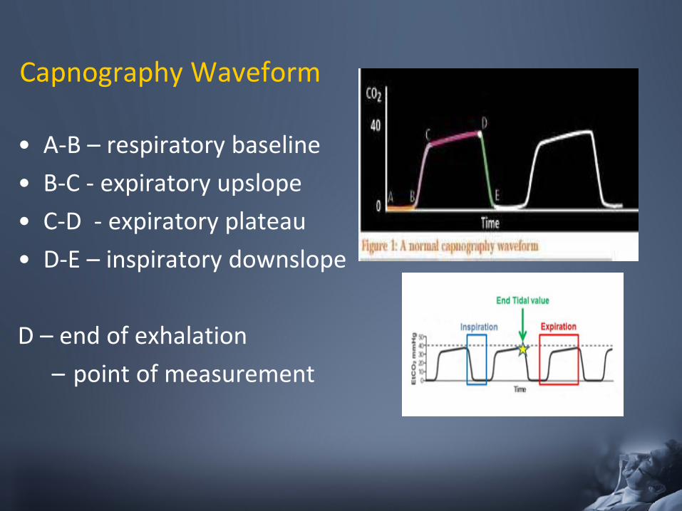
Capnography Waveform
• A-B – respiratory baseline • B-C - expiratory upslope • C-D - expiratory plateau • D-E – inspiratory downslope
D – end of exhalation
– point of measurement
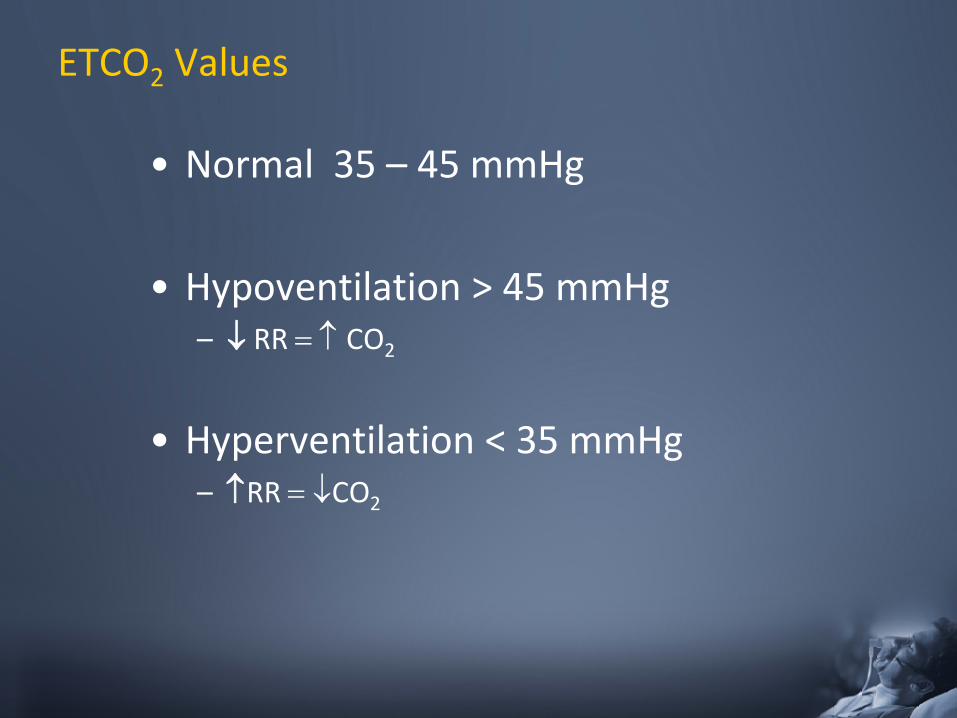
ETCO2 Values
• Normal 35 – 45 mmHg
• Hypoventilation > 45 mmHg – ↓ RR = ↑ CO2
• Hyperventilation < 35 mmHg – ↑RR = ↓CO2
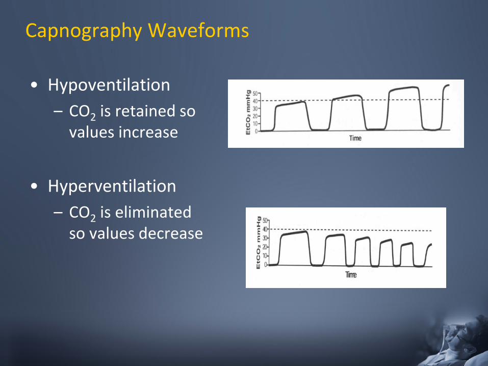
Capnography Waveforms
• Hypoventilation – CO2 is retained so
values increase • Hyperventilation
– CO2 is eliminated so values decrease
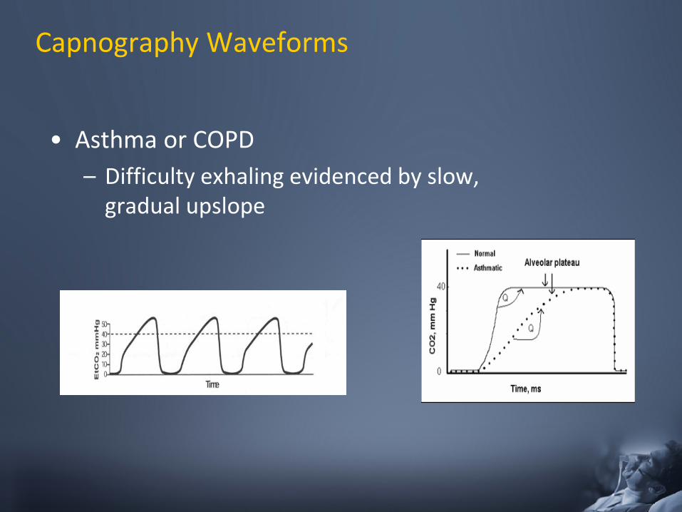
Capnography Waveforms
• Asthma or COPD – Difficulty exhaling evidenced by slow,
gradual upslope
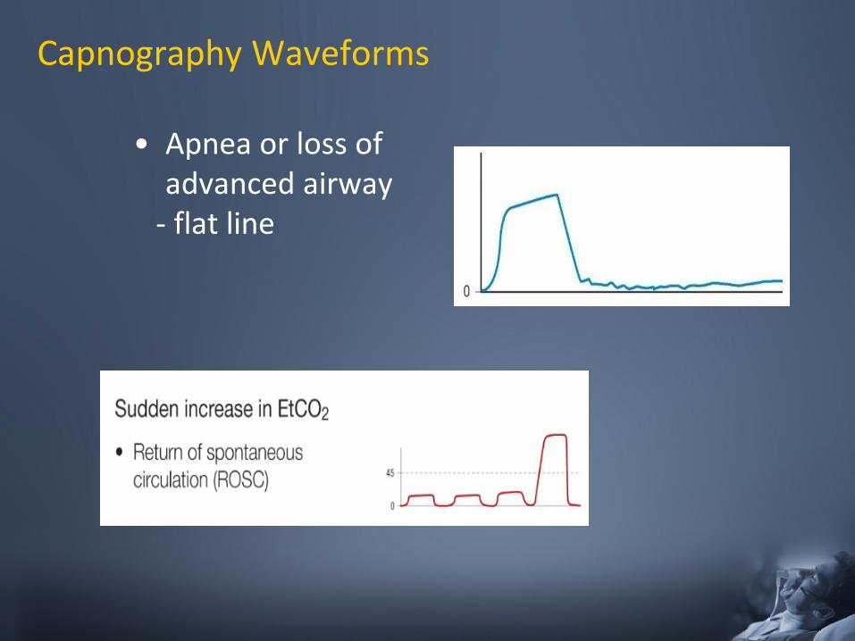
Capnography Waveforms
• Apnea or loss of advanced airway
- flat line
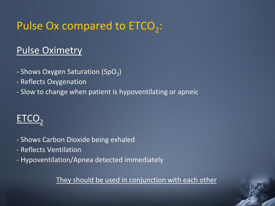
Pulse Ox compared to ETCO2:
Pulse Oximetry
- Shows Oxygen Saturation (SpO2) - Reflects Oxygenation - Slow to change when patient is hypoventilating or apneic
ETCO2
- Shows Carbon Dioxide being exhaled - Reflects Ventilation - Hypoventilation/Apnea detected immediately
They should be used in conjunction with each other
Now what does all this mean to EMS?
• ETCO2 is a great tool to help monitor the patients breath to breath status.
• Can help recognize airway obstructions before the patient has signs of problem
• Helps you control the ETCO2 of head injuries
• Can help to identify ROSC in cardiac arrest
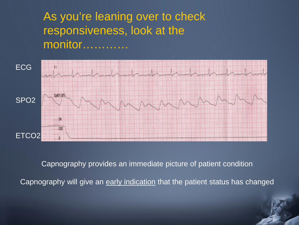
Capnography provides an immediate picture of patient condition
Capnography will give an early indication that the patient status has changed
As you’re leaning over to check responsiveness, look at the monitor…………
ECG
SPO2
ETCO2
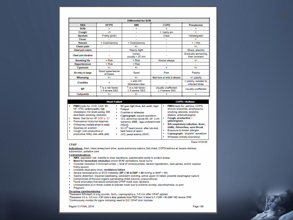
All that wheezes is not asthma…..
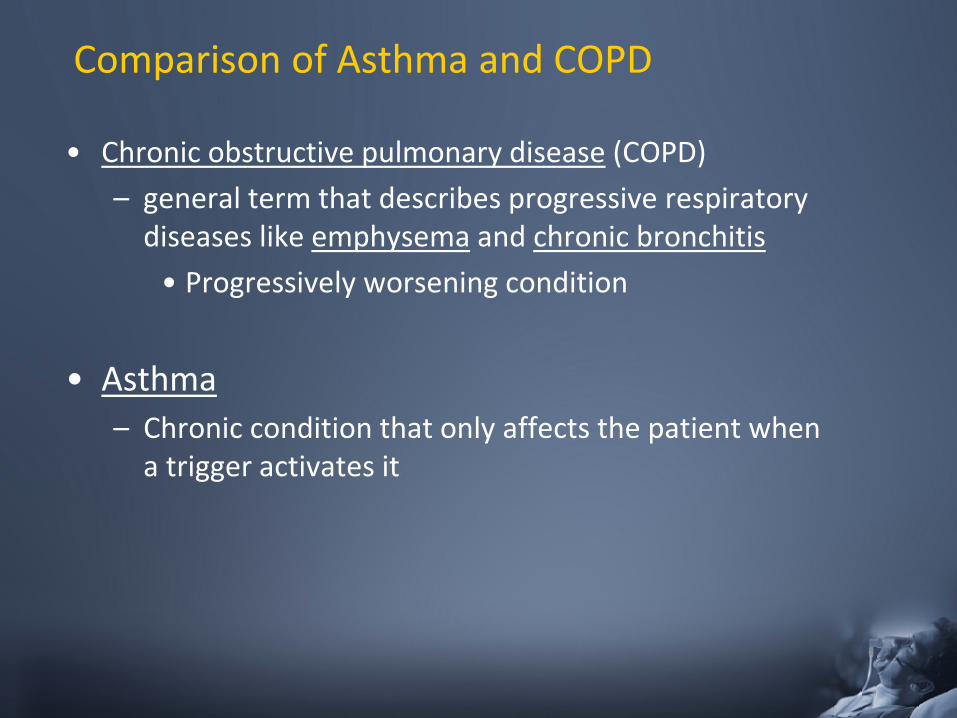
Comparison of Asthma and COPD
• Chronic obstructive pulmonary disease (COPD) – general term that describes progressive respiratory
diseases like emphysema and chronic bronchitis • Progressively worsening condition
• Asthma – Chronic condition that only affects the patient when
a trigger activates it
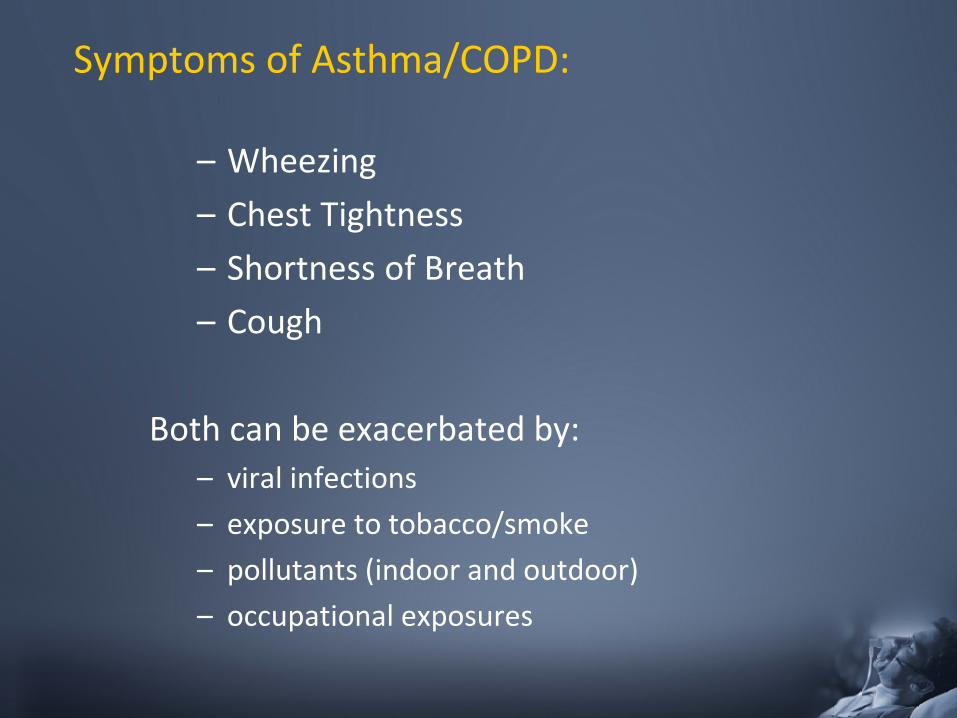
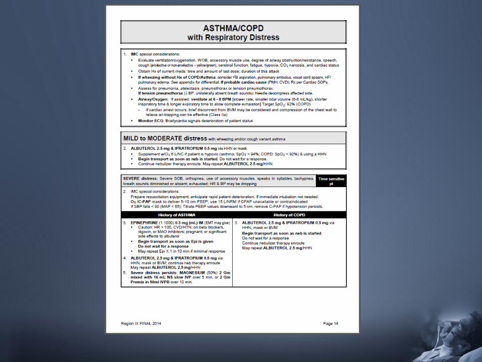
Symptoms of Asthma/COPD:
– Wheezing – Chest Tightness – Shortness of Breath – Cough
Both can be exacerbated by:
– viral infections – exposure to tobacco/smoke – pollutants (indoor and outdoor) – occupational exposures
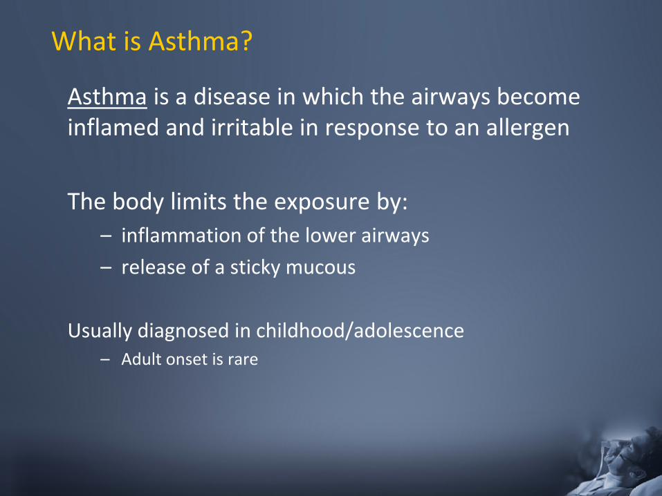
What is Asthma?
Asthma is a disease in which the airways become inflamed and irritable in response to an allergen
The body limits the exposure by:
– inflammation of the lower airways – release of a sticky mucous
Usually diagnosed in childhood/adolescence
– Adult onset is rare
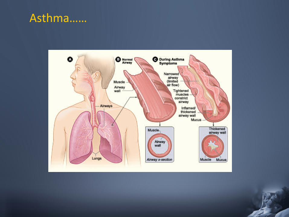
Asthma……
What is Chronic Obstructive Pulmonary Disease (COPD)?
• In COPD, the lungs become damaged following exposure to certain irritants – most common irritant - chronic smoking – chronic exposure causes damage
• leads to airway obstruction & hyperinflation – commonly due to bacteria growth
• Usually diagnosed in Adulthood
– Symptoms get progressively worse
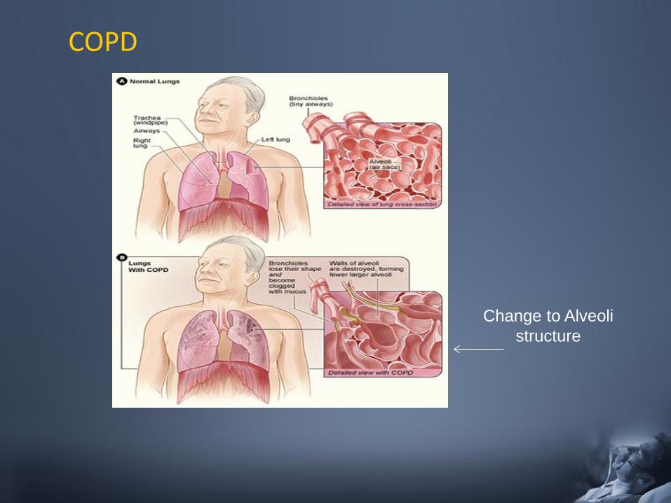
COPD
Change to Alveoli structure
Treatment Philosophy:
• Asthma – attempt to lower or suppress inflammation
• COPD – reduce symptoms

MECHANICAL INTERVENTIONS……….

CPAP: Just a reminder on how it works
• Maintains constant pressure within the airway and through-out the respiratory cycle – Keeps alveoli open and expanded
• Also decreases venous return to the heart thereby lessening its work load
• In CHF, forces excess fluid out of alveoli & interstitial space back into the vasculature
• Can avert the need for advanced airway and mechanical ventilation if applied early enough

THIS IS HOW CPAP FEELS TO THE PATIENT!!
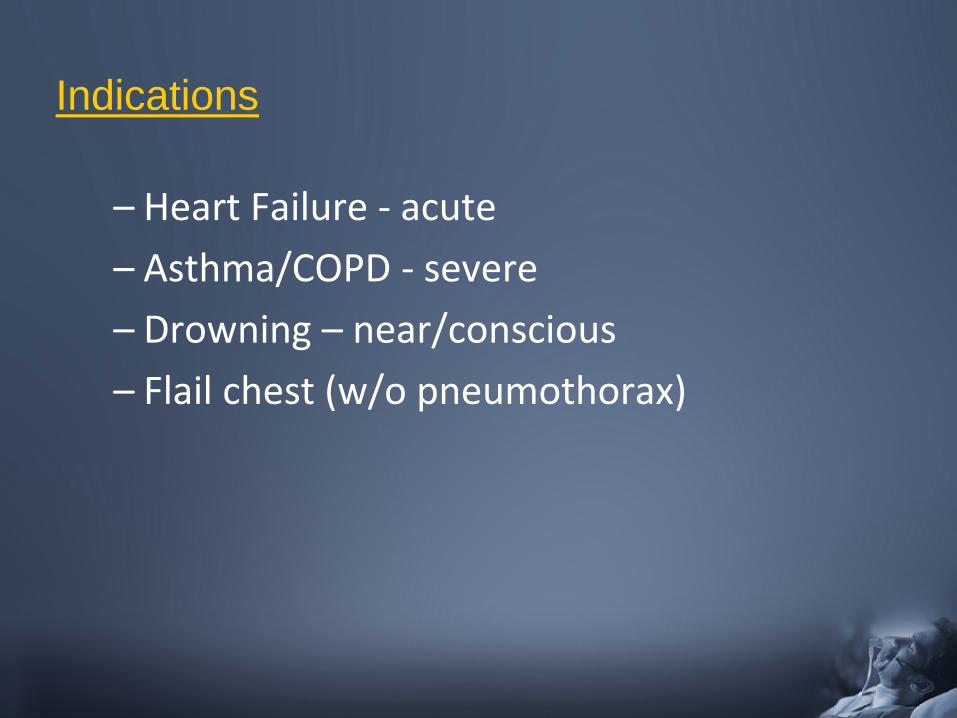
– Heart Failure - acute – Asthma/COPD - severe – Drowning – near/conscious – Flail chest (w/o pneumothorax)
Indications
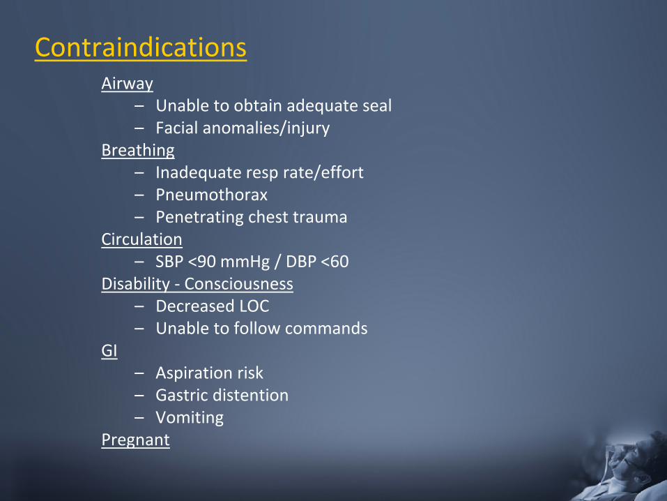
Contraindications Airway
– Unable to obtain adequate seal – Facial anomalies/injury
Breathing – Inadequate resp rate/effort – Pneumothorax – Penetrating chest trauma
Circulation – SBP <90 mmHg / DBP <60
Disability - Consciousness – Decreased LOC – Unable to follow commands
GI – Aspiration risk – Gastric distention – Vomiting
Pregnant
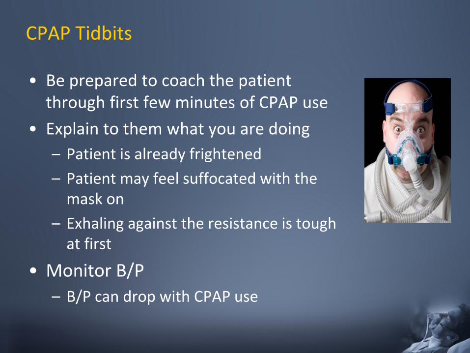
CPAP Tidbits
• Be prepared to coach the patient through first few minutes of CPAP use
• Explain to them what you are doing – Patient is already frightened – Patient may feel suffocated with the
mask on – Exhaling against the resistance is tough
at first
• Monitor B/P – B/P can drop with CPAP use
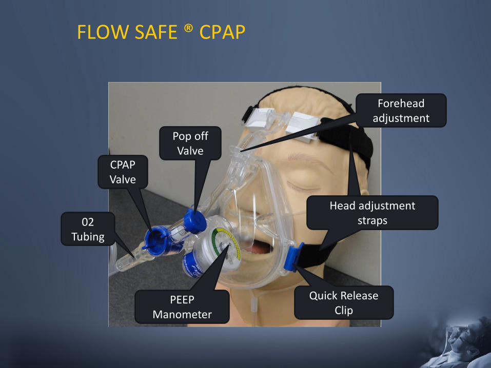
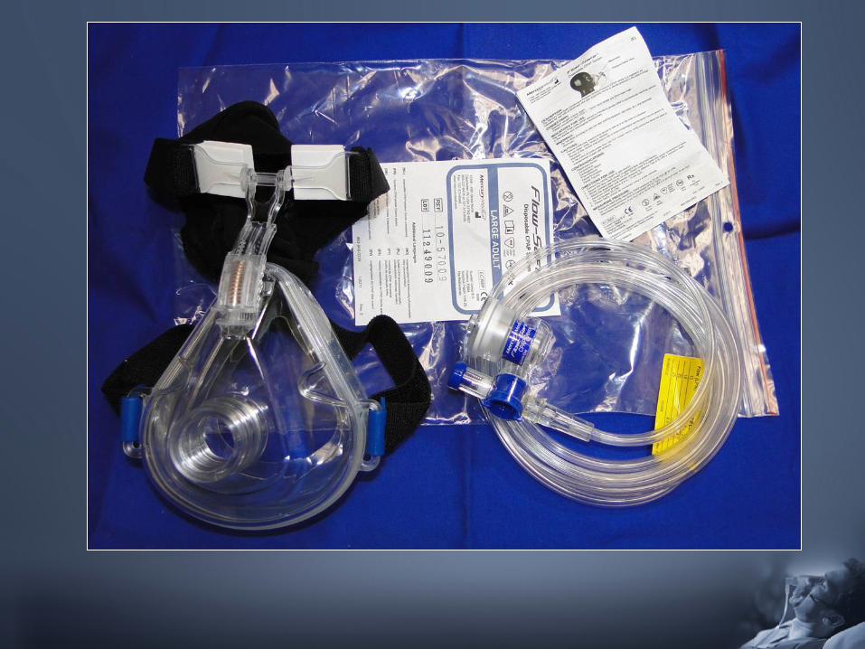
FLOW SAFE ® CPAP
PEEP Manometer
Quick Release Clip
Head adjustment straps
Forehead adjustment
Pop off Valve
CPAP Valve
02 Tubing
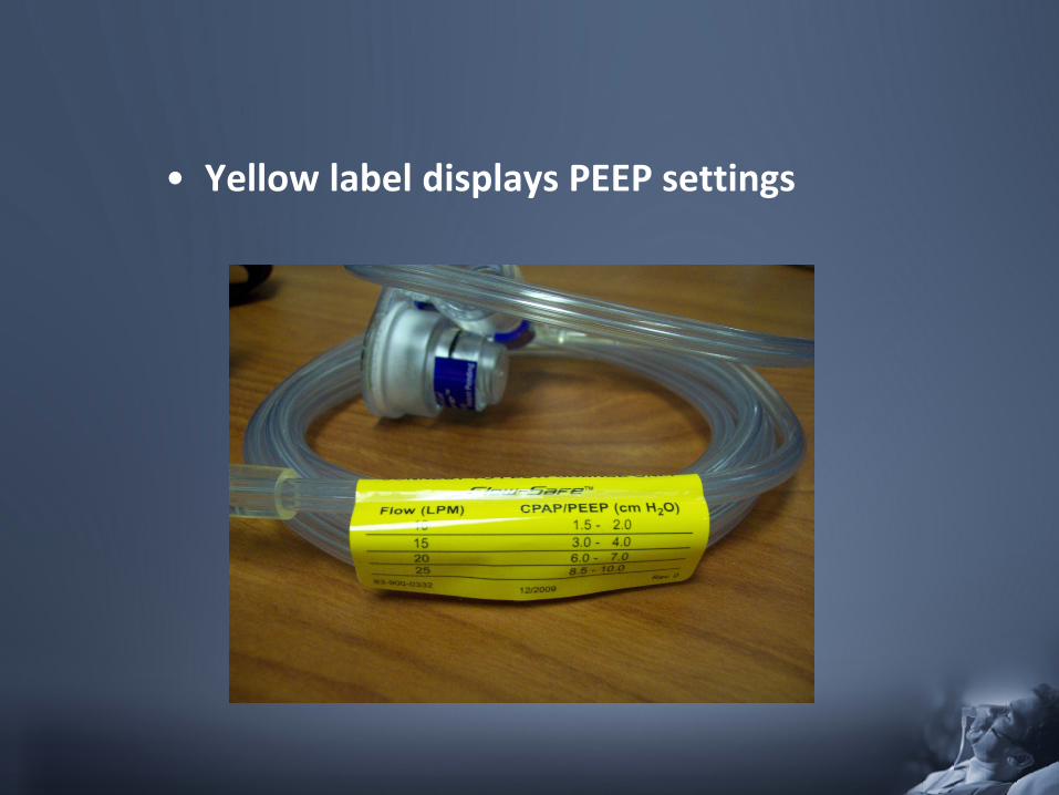
• Yellow label displays PEEP settings
Flow-Safe® CPAP Device
PEEP depends on - O2 flow rate - Face mask seal (leak) Oxygen concentration depends on - O2 flow rate - Respiratory rate - Tidal volume
TITRATE PEEP TO PATIENT’S WORK OF BREATHING
– Start at 15 L which equals 3-4 cm H2O PEEP
– Titrate up to patient response (decreased work of breathing)
– Maximum PEEP 8.5 - 10 cm at 25 L O2
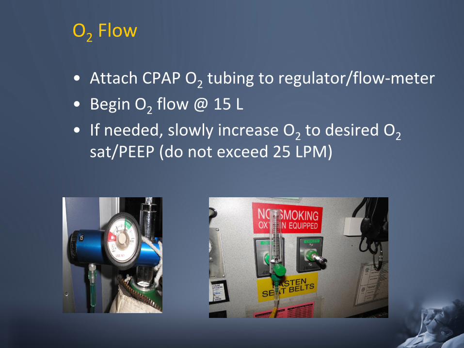
O2 Flow
• Attach CPAP O2 tubing to regulator/flow-meter • Begin O2 flow @ 15 L • If needed, slowly increase O2 to desired O2
sat/PEEP (do not exceed 25 LPM)
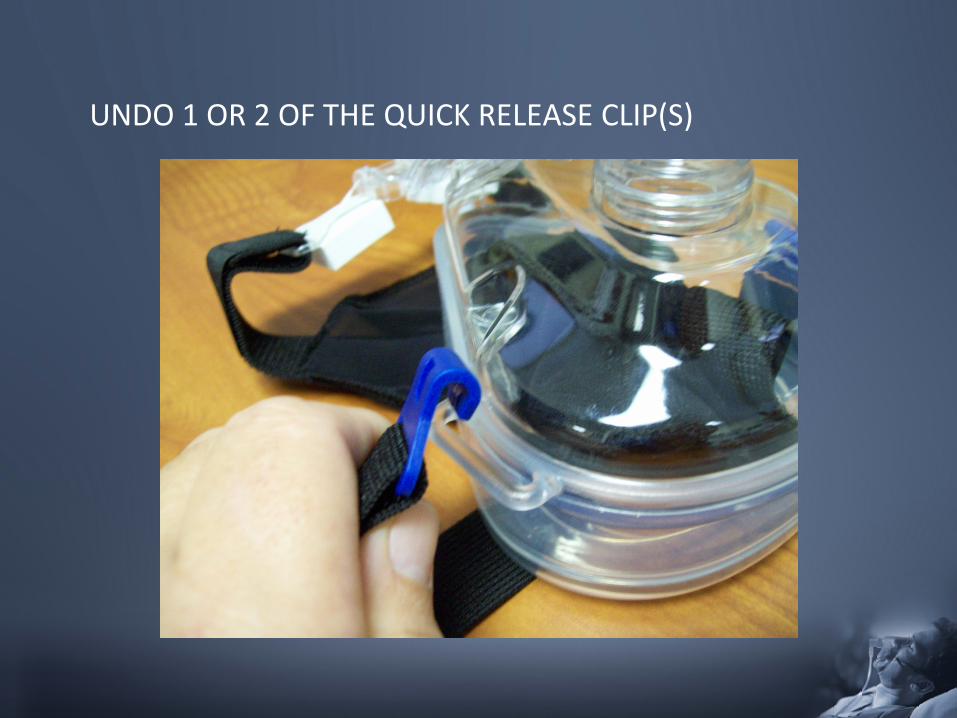
UNDO 1 OR 2 OF THE QUICK RELEASE CLIP(S)
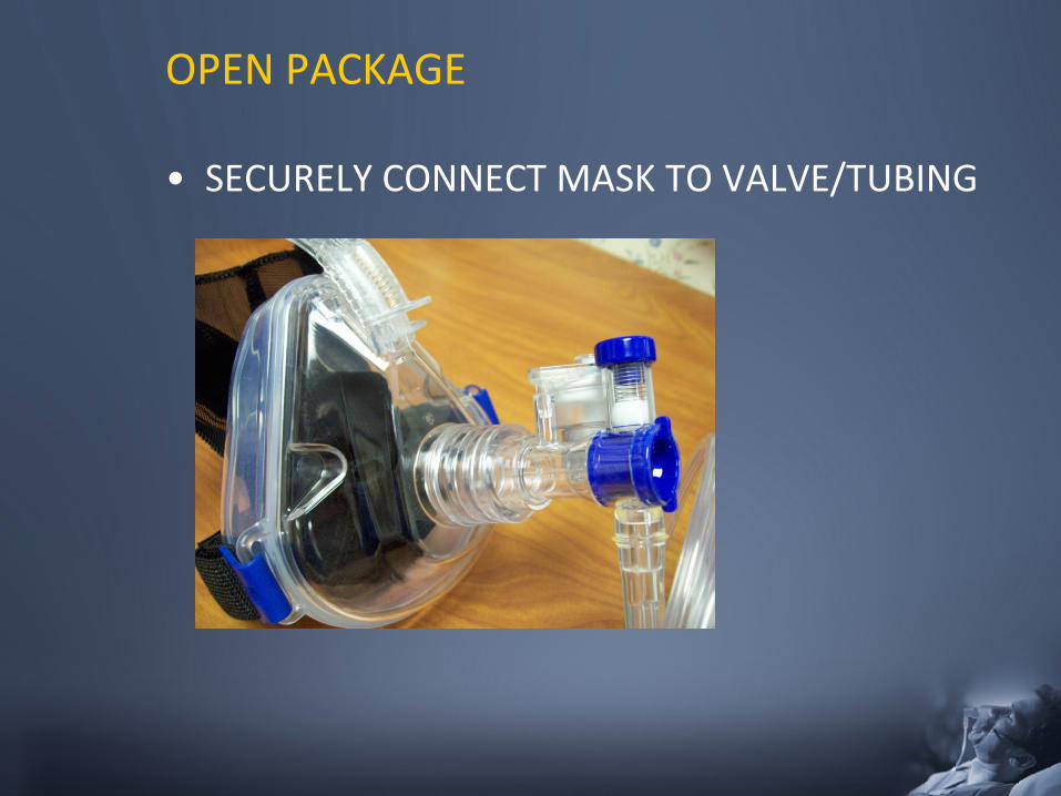
OPEN PACKAGE
• SECURELY CONNECT MASK TO VALVE/TUBING
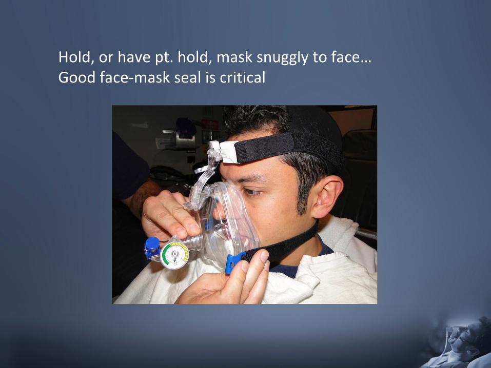
Hold, or have pt. hold, mask snuggly to face… Good face-mask seal is critical
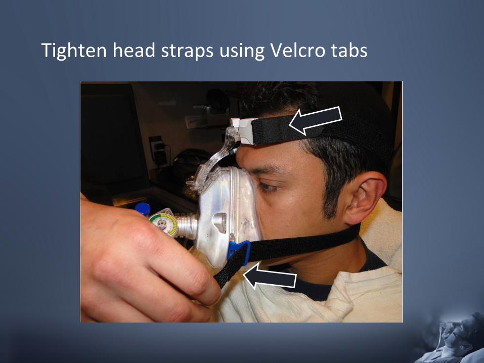
Tighten head straps using Velcro tabs
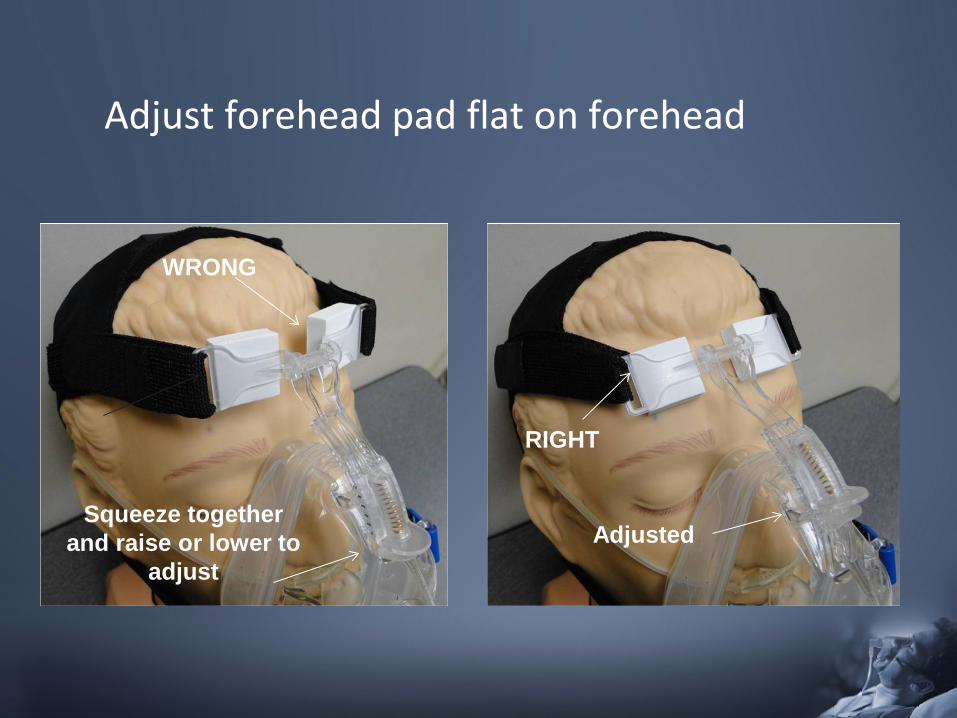
Adjust forehead pad flat on forehead
Squeeze together and raise or lower to
adjust
WRONG
RIGHT
Adjusted
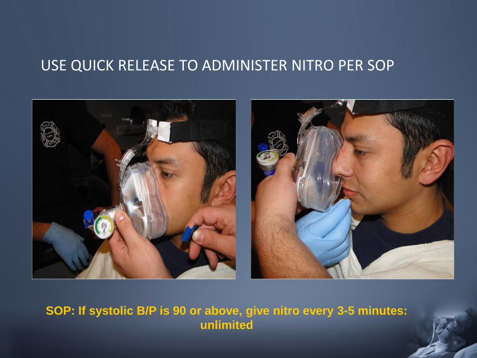
USE QUICK RELEASE TO ADMINISTER NITRO PER SOP
SOP: If systolic B/P is 90 or above, give nitro every 3-5 minutes: unlimited
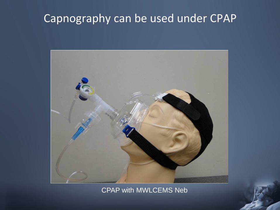
Capnography can be used under CPAP
CPAP with MWLCEMS Neb
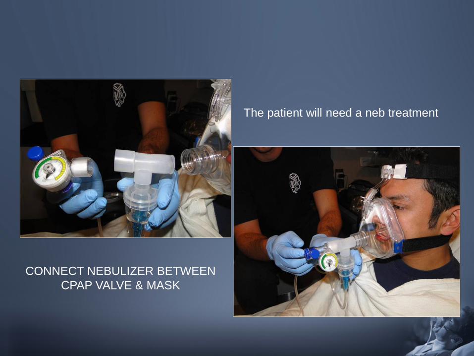
The patient will need a neb treatment
CONNECT NEBULIZER BETWEEN CPAP VALVE & MASK
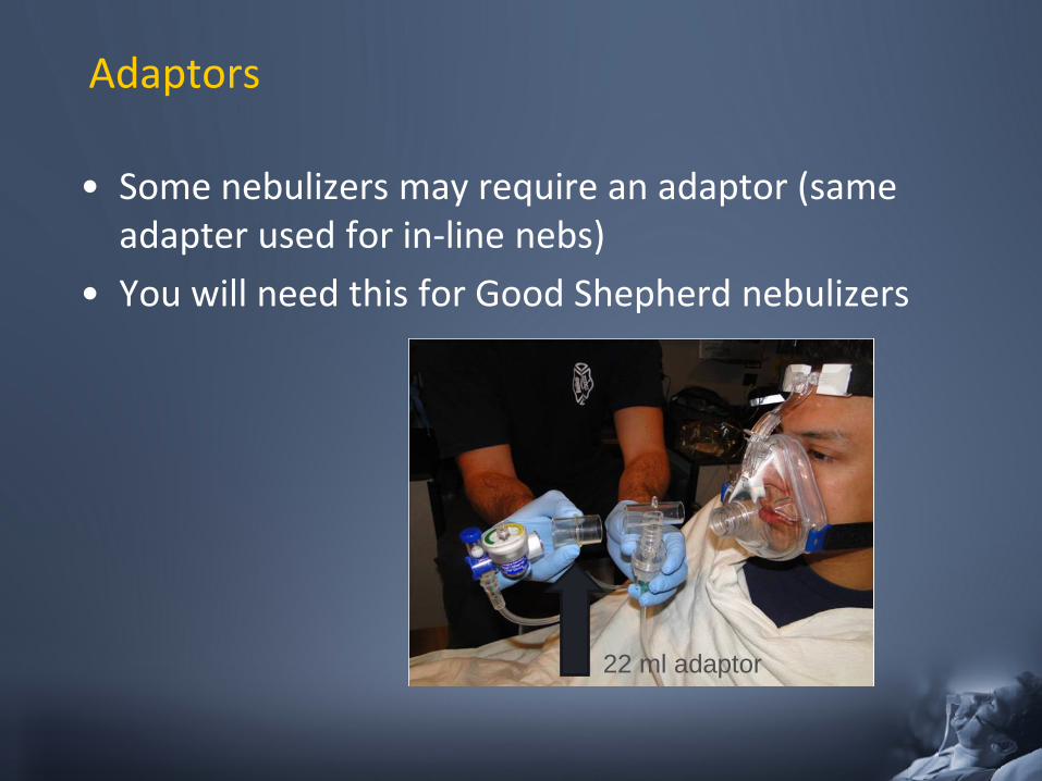
Adaptors
• Some nebulizers may require an adaptor (same adapter used for in-line nebs)
• You will need this for Good Shepherd nebulizers
22 ml adaptor
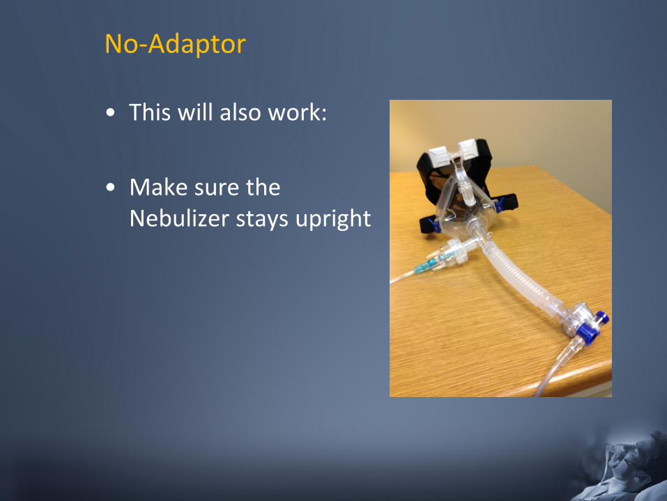
No-Adaptor
• This will also work:
• Make sure the Nebulizer stays upright
Documentation
• Document the oxygen flow rate
• Document the PEEP reading on the manometer
• This can be done in your narrative
• Remember to use the manometer reading when patient exhales
58
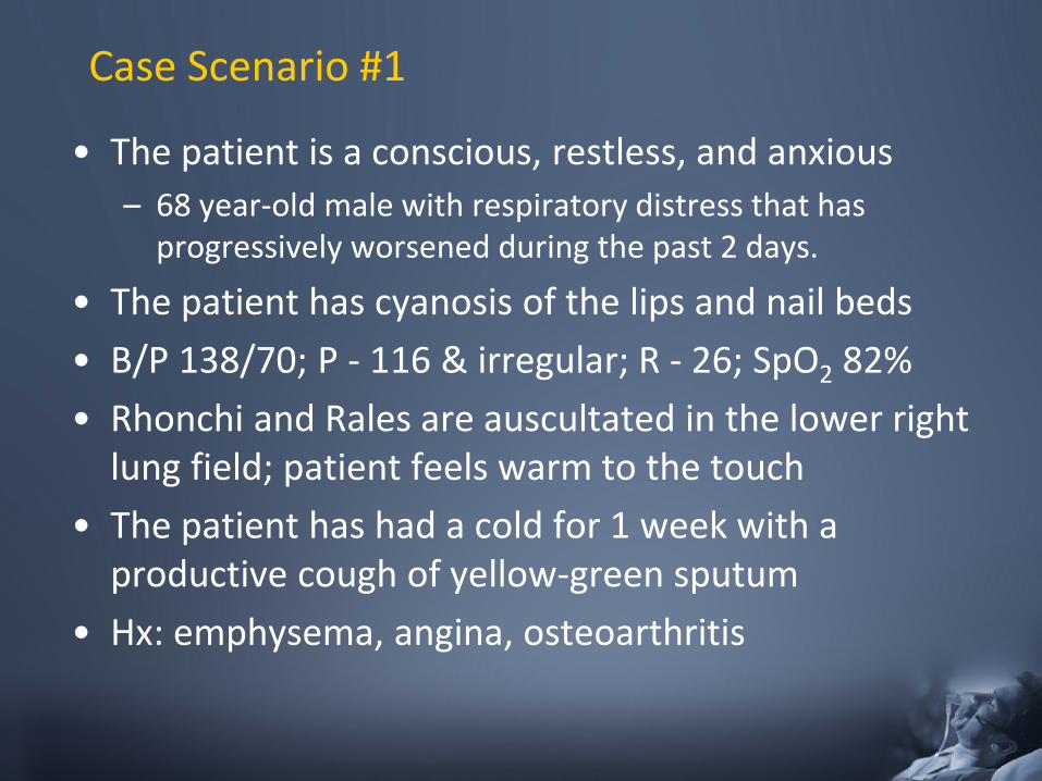
Case Scenario #1
• The patient is a conscious, restless, and anxious – 68 year-old male with respiratory distress that has
progressively worsened during the past 2 days.
• The patient has cyanosis of the lips and nail beds • B/P 138/70; P - 116 & irregular; R - 26; SpO2 82% • Rhonchi and Rales are auscultated in the lower right
lung field; patient feels warm to the touch • The patient has had a cold for 1 week with a
productive cough of yellow-green sputum • Hx: emphysema, angina, osteoarthritis
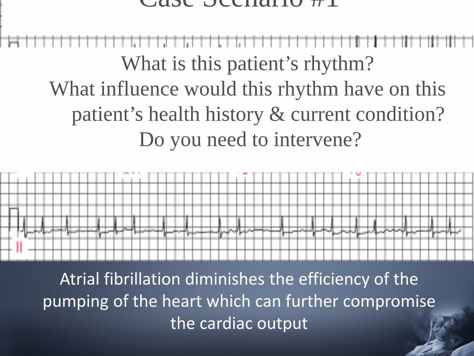
Case Scenario
What is this patient’s rhythm? What influence would this rhythm have on this patient’s health history & current condition?
Do you need to intervene?
Case Scenario #1
Atrial fibrillation diminishes the efficiency of the pumping of the heart which can further compromise
the cardiac output
Case Scenario #1
• Impression & intervention? • The patient has COPD most likely complicated by
pneumonia – a “cold” over the last week – productive cough of yellow-green sputum – warm to the touch (temperature 100.60F) – rhonchi & rales in the right lung field base
• Routine medical care – supplemental oxygen – ↑ heart rate most likely due to pneumonia and does
not need specific treatment
Case Scenario #2 • A 68 year-old female calls 911 due to severe
respiratory distress which suddenly woke her up from sleep. She is unable to speak in complete sentences and is using accessory muscles to breathe. Lips and nail beds are cyanotic; ankles are swollen.
• B/P 186/100; P - 124; R - 34; SpO2 - 88% • Crackles are auscultated in the lower half of the
lung fields.
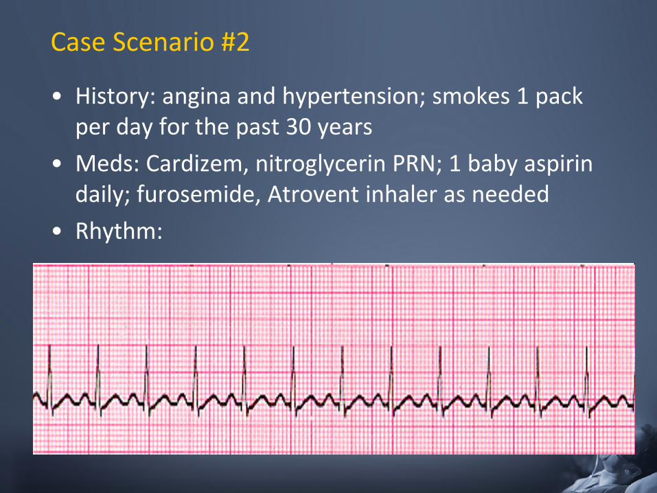
Case Scenario #2
• History: angina and hypertension; smokes 1 pack per day for the past 30 years
• Meds: Cardizem, nitroglycerin PRN; 1 baby aspirin daily; furosemide, Atrovent inhaler as needed
• Rhythm:
Case Scenario #2
• What is your impression? • What intervention(s) are appropriate following
Region IX SOP’s? • What is the rationale for these interventions? • What is this patient’s rhythm and do you need
to administer any medications for the rhythm?
Case Scenario #2
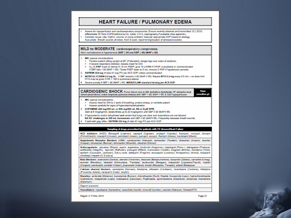
• Impression: congestive heart failure with pulmonary edema – paroxysmal nocturnal dyspnea (sudden shortness of
breath at night) – bilateral crackles in the lungs – peripheral edema – cardiac history - hypertension and angina
• Rhythm - sinus tachycardia – determine and treat the underlying cause
Case Scenario #2 • Interventions
– Sit the patient upright, have their feet dangle off the sides of the cart
• This encourages the patient to sit in a better, upright position
– CPAP – Prepare to assist breathing via BVM
• have BVM out on the counter and ready for use – IV-O2-monitor – Meds: ASA, NTG, Midazolam for anxiety
Case Scenario #3
• 7 year-old with history of asthma has sudden onset of difficulty breathing and wheezing while playing outside
• Patient has an increased respiratory rate and is using accessory muscles
• B/P - 108/70; P - 90; R - 24; SpO2 - 97% • Upon auscultation, left lung is clear and wheezing
is present on the right side • Impression and intervention?
Case Scenario #3
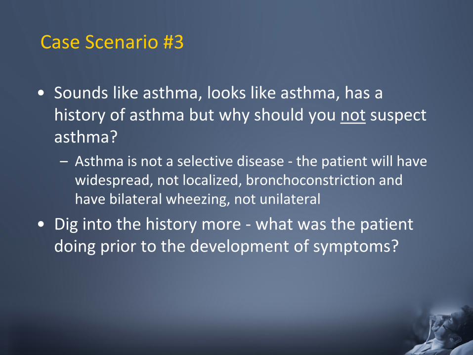
• Sounds like asthma, looks like asthma, has a history of asthma but why should you not suspect asthma? – Asthma is not a selective disease - the patient will have
widespread, not localized, bronchoconstriction and have bilateral wheezing, not unilateral
• Dig into the history more - what was the patient doing prior to the development of symptoms?
Case Scenario #3
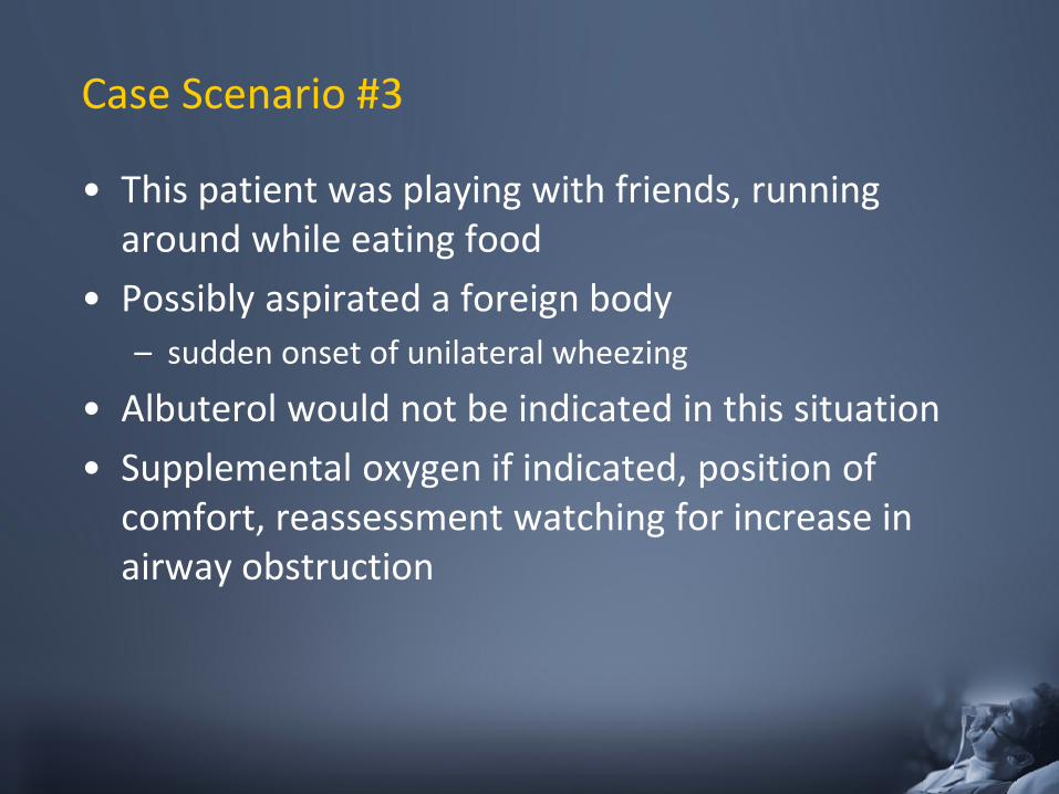
• This patient was playing with friends, running around while eating food
• Possibly aspirated a foreign body – sudden onset of unilateral wheezing
• Albuterol would not be indicated in this situation • Supplemental oxygen if indicated, position of
comfort, reassessment watching for increase in airway obstruction

WRAP UP………..
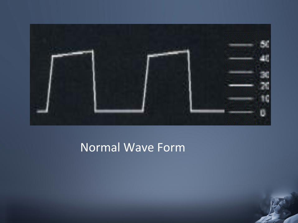
Normal Wave Form
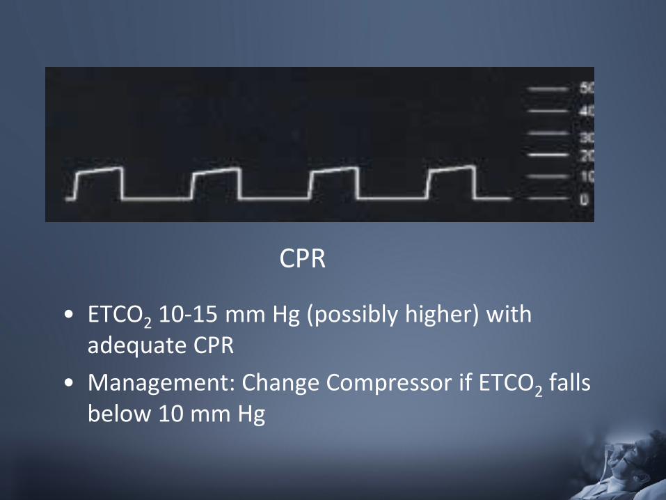
CPR
• ETCO2 10-15 mm Hg (possibly higher) with adequate CPR
• Management: Change Compressor if ETCO2 falls below 10 mm Hg
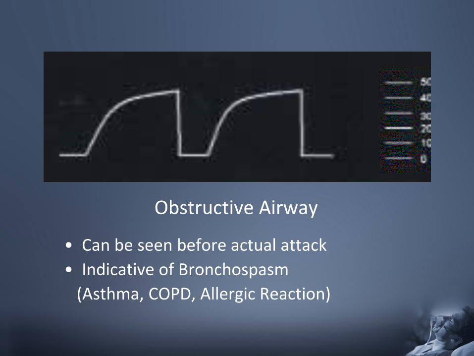
Obstructive Airway
• Can be seen before actual attack • Indicative of Bronchospasm (Asthma, COPD, Allergic Reaction)

ROSC (Return of Spontaneous Circulation)
• During CPR sudden increase of ETCO2 above 10-15 mm Hg
• Management: Check for pulse
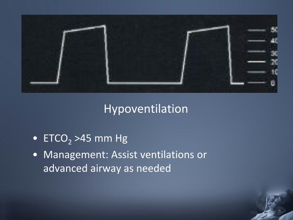
Hypoventilation
• ETCO2 >45 mm Hg • Management: Assist ventilations or
advanced airway as needed
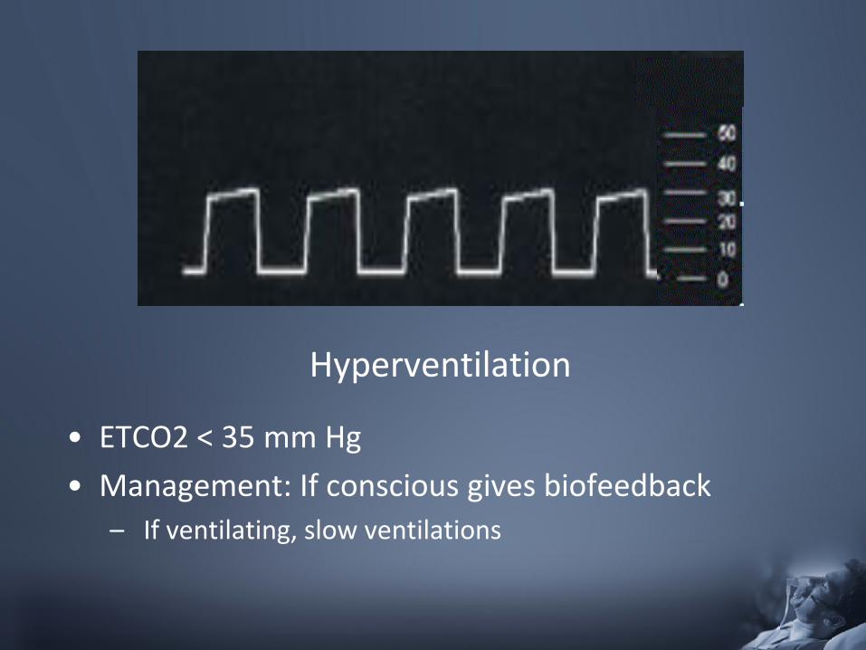
Hyperventilation
• ETCO2 < 35 mm Hg • Management: If conscious gives biofeedback
– If ventilating, slow ventilations
Quick Field Guidelines…..
• If you are thinking about giving O2, then give it!
• If you can’t tell whether a patient is breathing adequately, then they aren’t!
• If you’re thinking about assisting a patient’s breathing, you probably should be!
• When a patient quits fighting it does not mean that they are getting better!

Questions?
Be Safe!