Embed Size (px)
Citation preview
75
Review
www.expert-reviews.com © 2012 Expert Reviews Ltd ISSN 1747-634810.1586/ERS.11.81
Any impairment in striated muscle function can interfere with the performance of daily activities, particularly for a patient already liv-ing with a respiratory disorder. However, the interaction between the respiratory and mus-culoskeletal systems is not always considered in the clinical management of these patients. Striated muscles are contractile elements that provide organisms with physiological func-tions, such as movement and generation of both air- and blood-flow. The latter two func-tions are essential for respiratory gas exchange. The generation of airflow requires the action of inspiratory muscles. When this muscle group contracts, the changes in intrathoracic pres-sure allow air to enter the lungs. When these muscles relax, the air exits from the respira-tory system. If additional effort is required to exhale, the expiratory muscle group contracts, increasing the alveolar–atmosphere pressure gradient. Although the diaphragm is the main inspiratory muscle, specifically in young and healthy subjects when they are at rest, other muscles progressively participate in the effort as ventilatory demands increase. These include external intercostals, parasternal and, to a lesser degree, the scalenes, sternocleidomas-toid, latissimus dorsi, serratus and pectoralis muscles. The main expiratory muscles are the
internal intercostals and those constituting the abdominal wall. Skeletal muscles located in the limbs (and also called peripheral muscles), are involved in the movements of the body. Any impairment in their function can interfere with the performance of daily activities.
Muscle function becomes impaired in many different respiratory disorders, such as chronic obstructive pulmonary disease (COPD), cystic fibrosis, bronchial asthma, obstructive sleep apnea syndrome (OSAS), kyphoscoliosis and lung cancer. Although changes in respiratory mechanics in these diseases primarily target respiratory muscles, limb muscles can also be affected. In addition, respiratory and limb muscle dysfunction can occur in patients with myopathies, neurological and neuromuscular junction disorders, chronic heart failure, sepsis and other critical illness. In the intensive care unit (ICU), the condition called ICU muscle weakness is not only a limb problem but can also hamper weaning from mechanical ventilation.
This review aims to brief ly present basic concepts of skeletal muscle physiology and to describe in-depth the muscle function impair-ment occurring in some of the most prevalent respiratory conditions, defining the factors and mechanisms involved in the etiopathogenesis of muscle dysfunction in this setting.
Joaquim Gea*, Carme Casadevall, Sergi Pascual, Mauricio Orozco-Levi and Esther BarreiroServei de Pneumologia, Hospital del Mar – IMIM, Departament de Ciències Experimentals i de la Salut (CEXS), Universitat Pompeu Fabra, CIBER de Enfermedades Respiratorias (CIBERES) ISC III, Barcelona, Catalunya, Spain *Author for correspondence: Tel.: +34 93 248 3138 Fax: +34 93 248 3425 [email protected]
Many respiratory diseases lead to impaired function of skeletal muscles, influencing quality of life and patient survival. Dysfunction of both respiratory and limb muscles in chronic obstructive pulmonary disease has been studied in depth, and seems to be caused by the complex interaction of general (inflammation, impaired gas exchange, malnutrition, comorbidity, drugs) and local factors (changes in respiratory mechanics and muscle activity, and molecular events). Some of these factors are also present in cystic fibrosis and asthma. In obstructive sleep apnea syndrome, repeated exposure to hypoxia and the absence of reparative rest are believed to be the main causes of muscle dysfunction. Deconditioning appears to be crucial for the functional impairment observed in scoliosis. Finally, cachexia seems to be the main mechanism of muscle dysfunction in advanced lung cancer. A multidimensional therapeutic approach is recommended, including pulmonary rehabilitation, an adequate level of physical activity, ventilatory support and nutritional interventions.
Keywords: drugs • exercise limitation – muscle remodeling • hypoventilation • inflammation • limb muscles • muscle dysfunction • respiratory diseases • respiratory muscles
Respiratory diseases and muscle dysfunctionExpert Rev. Respir. Med. 6(1), 75–90 (2012)
For reprint orders, please contact [email protected]
76
Review
Expert Rev. Respir. Med. 6(1), (2012)
Muscle physiologyThe two main functional properties of both respiratory and limb muscles are: strength and endurance. The former can be defined as the ability to develop a brief maximal effort, whereas the lat-ter could be described as the ability to maintain a submaximal contraction over time. Strength mainly depends on muscle mass, although other factors also contribute, such as muscle length, innervation, fiber size and the proportion of predominantly anaerobic fibers. Endurance is related to the aerobic properties of the muscle, which in turn, are conditioned by capillary density, proportion of type I fibers and enzyme activity in the oxida-tive pathways, among other factors. When the strength and/or endurance of skeletal muscles is reduced, this is called muscle dysfunction and can be characterized in two ways: weakness and fatigue. Muscle weakness is related to the loss of muscle strength. Therefore, it can be identified easily in clinical conditions through the assessment of muscle force (determination of pressures gener-ated by respiratory muscles and standard dynamometry to assess limb muscles). Weakness is a constitutive and relatively stable situation, and the muscle requires long-term therapeutic meas-ures (training and nutritional interventions). By contrast, muscle fatigue is a temporary dysfunction related to endurance, and is primarily resolved by rest. It can be identified by neurophysi-ological (changes in the high/low frequencies ratio or in centroid frequency) or mechanical (transient inability to perform a target task) indicators. Both conditions, weakness and fatigue, can be present simultaneously in the same patient, as a weak muscle will become fatigued much more easily.
Respiratory musclesInspiratory muscles ensure an appropriate level of ventilation to facilitate pulmonary gas exchange. Therefore, their dysfunc-tion will result in hypoxemia and hypercapnia, and in venti-lated patients can lead to difficulties in the weaning process. Malfunction of expiratory muscles will, in turn, give rise to dif-ficulties upon exertion, coughing and attempts to expectorate secretions from the airways. Functional assessment of respiratory muscles is slightly more complicated than that of limb muscles, but can be achieved through determination of respiratory pres-sures. A large number of studies have demonstrated that respi-ratory muscle function can also be impaired in widely diverse disorders. These include COPD, cystic fibrosis, chronic asthma, scoliosis, neuromuscular diseases and also ICU muscle weakness and sepsis.
Molecular and cellular events occurring in respiratory muscles with an impaired function are identified through biopsy analysis. However, the sampling of these muscles is always very invasive, and therefore their structural and molecular properties are studied less frequently than in limb muscles. Most studies have been per-formed in the diaphragm, with samples obtained during thoracic or abdominal surgery owing to associated diseases [1–4]. However, data are also available regarding other inspiratory muscles, such as external intercostals, parasternals or even accessory muscles, such as latissimus dorsi [5]. Regarding expiratory muscles, data are scarce, but there are some reports on the major oblique muscle [6].
Disuse of respiratory muscles only occurs under very specific con-ditions, such as mechanical ventilation. Conversely, overloading occurs more frequently because many different situations result in increased breathing. On the one hand, airway resistances increase in obstructive diseases. On the other, pulmonary hyperinflation or changes in thorax geometry can deny respiratory muscles their optimal length for contraction. Although some studies support that these factors are essential for respiratory muscle dysfunction, additional elements cannot be excluded.
Peripheral musclesThe striated muscles of the upper and lower limbs constitute the peripheral muscles; the concept can also include the muscles of the shoulder and pelvic girdles. The muscles of the upper limbs are essential for manipulating objects, and for many of the tasks involved in personal care. In addition, some of them can be recruited to serve as ventilatory muscles when these are overburdened by respiratory loads [7,8]. In turn, the muscles of the lower limbs are essential for locomotion and exercise, and are crucial for many daily activities. The clinical implications of peripheral muscle dysfunction are important, as patients may be unable to work or take care of themselves, become extremely dependent on those around them and experience a reduction in their quality of life. Since tests for the functional proper-ties of peripheral muscles are relatively simple [9], we know that many disorders can induce (or are associated with) limb muscle dysfunction, including COPD, scoliosis, chronic heart failure and cancer cachexia, among others. Peripheral muscles are very sensitive to disuse (deconditioning) and nutritional abnormali-ties, two factors that many authors believe are pivotal for the occurrence of muscle dysfunction. However, other factors can also be implicated.
Peripheral muscles are readily accessible for tissue sampling, facilitating analysis of the cellular and molecular changes that are associated with muscle dysfunction. In the following sections, these changes and their possible mechanisms will be reviewed. However, it is important to clarify that many of these structural and molecular studies have been performed in samples from the quadriceps muscle, particularly its external part (vastus latera-lis). It is possible to speculate that not all the findings should be directly extrapolated to other peripheral muscles, whose functions are essentially different from those of the anterior thigh. Some studies performed in the upper limb muscles appear to confirm this hypothesis [10,11].
Muscle dysfunction can occur not only as a consequence of a disease or a treatment, but also in some more physiological circumstances, such as aging and extreme sedentary lifestyle. As mentioned above, it has been well described in COPD, bron-chial asthma and lung cancer cachexia, among others. It is also characteristic of other disorders targeting the respiratory system, such as OSAS, neuromuscular and rib cage abnormalities [12–15]. The following sections review the causes and mechanisms of muscle dysfunction in the most common disorders that target the respiratory system and also respiratory and/or peripheral muscles (Box 1).
Gea, Casadevall, Pascual, Orozco-Levi & Barreiro
www.expert-reviews.com 77
Review
Respiratory disordersChronic obstructive pulmonary diseaseCOPD is a highly prevalent condition characterized by nonreversible airf low obstruction [301]. The main cause of COPD is tobacco smoking, which results in inflammatory phenomena leading to destruction of lung parenchyma and air-way remodeling. In addition to airway obstruction, COPD results in pulmonary hyperinflation, increased airway resistance, changes in pulmonary compliance and gas exchange abnormalities, characterized by hypoxemia and, in some cases, hypercap-nia. As a result, patients experience ventila-tory limitations that impair their exercise tolerance. However, COPD is a heteroge-neous disease and patients may also show extrapulmonary involvement, including malnutrition, and abnormalities in skel-etal muscles, blood cells, renal and nerv-ous systems and even bone metabolism [16–19]. Muscle dysfunction is probably the best-studied extrapulmonary manifestation occurring in COPD. It includes abnormali-ties in the strength and endurance of both respiratory and limb muscles [3,20], and is believed to be of mul-tifactorial origin, with local and general factors interacting to modify the phenotype and function of any particular muscle. Muscle dysfunction is relevant because it reduces exercise capac-ity, which ultimately affects quality of life and influences patient survival [21].
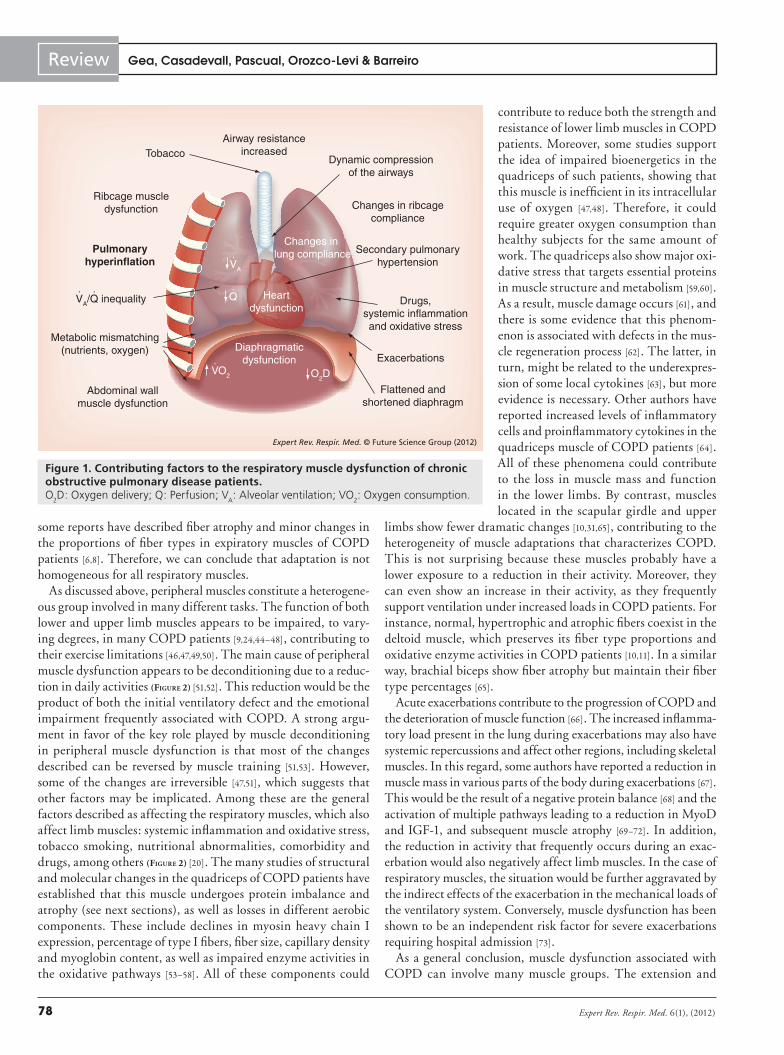
Respiratory muscle dysfunction is frequently observed in many COPD patients [22–24], mostly in those with more advanced stages of the disease, and can involve both inspiratory and expiratory muscle groups. Regarding inspiratory muscle dysfunction, it is believed to mainly be the consequence of changes in lung function [23,25]. On the one hand, pulmonary hyperinflation, present in many patients, has a dramatic impact in the length–tension rela-tionships of both the diaphragm and intercostal muscles (Figure 1)
[7]. These muscles become shorter and larger, respectively, than their optimal length for generating force [25]. In addition, the cos-tal and crural parts of the diaphragm lose their summatory action and become physiologically independent [26]. On the other hand, increased airway resistance and impaired gas exchange lead to an imbalance between demand and supply in the muscle [7,8]. One of the reasons to believe that hyperinflation is crucial for the develop-ment of respiratory muscle dysfunction in COPD is that although these patients develop lower respiratory pressures than healthy subjects, they can generate even greater force than the controls when both groups perform a test maneuver at similar high lung volumes [23]. This finding also suggests some muscle adaptation to pulmonary hyperinflation. However, respiratory muscles are subjected to the same deleterious general factors as other muscles,
including inflammation, oxidative stress, nutritional abnormali-ties and the effects of tobacco and some drugs (Figure 1) [27–30]. The combination of these systemic factors and the adaptive changes that occur in the face of increased mechanical loads leads to important changes in the phenotype of respiratory muscles. On the one hand, the percentages of myosin heavy chain I, type I fibers (with a predominantly aerobic metabolism), capillary and mitochondria increase in the diaphragm, leading to a more oxida-tive phenotype [1–3,31,32]. On the other hand, sarcomerae appear to shorten in this muscle, partially restoring their optimal length for contraction [2]. Interestingly, these positive phenomena coex-ist with signs of myopathy (paracristalline inclusions), as well as oxidative stress, changes in the expression of local cytokines and protein imbalance [33–35], all of which can contribute to the loss of muscle function. Data from external intercostal, parasternal and accessory muscles are much more scarce, but also suggest a com-bination of adaptive and negative phenomena [5,36–39]. Regarding expiratory muscles, the information is even more scant. Although their function deteriorates in many COPD patients [24,40], some of the factors should be different from those acting in the inspira-tory or peripheral muscle groups. Changes in lung volumes will only negatively affect the length–tension relationships of inter-nal intercostals, not those of abdominal muscles [41]. Moreover, deconditioning is unlikely since expiratory muscles are chronically activated for both the breathing effort and coughing in COPD patients [42,43]. However, they are also subject to all of the systemic factors present in other muscles. All of these circumstances result in changes to their phenotype and metabolism [7,8]. In this regard,
Box 1. Respiratory system and muscle dysfunction.
Respiratory disorders, frequent comorbidities and drugs known to alter muscle structure and/or function are listed.
Respiratory disorders:• Chronic obstructive pulmonary disease
• Bronchial asthma
• Sleep apnea–hypopnea and related syndromes
• Cystic fibrosis
• Scoliosis and other thoracic deformities
• Idiopathic pulmonary hypertension
Other conditions:• ICU muscle weakness – deleterious effects of mechanical ventilation
• Lung cancer (cachexia)
Frequent comorbidities:• Chronic heart failure
• Sepsis
• Diabetes mellitus
• Aging – sarcopenia
Drugs:• Corticosteroids
• Antagonists of b-adrenergic receptors
• Statins
• Diuretic drugs
• Phosphodiesterase 5 inhibitors
ICU: Intensive care unit.
Respiratory diseases & muscle dysfunction
78
Review
Expert Rev. Respir. Med. 6(1), (2012)
some reports have described fiber atrophy and minor changes in the proportions of fiber types in expiratory muscles of COPD patients [6,8]. Therefore, we can conclude that adaptation is not homogeneous for all respiratory muscles.
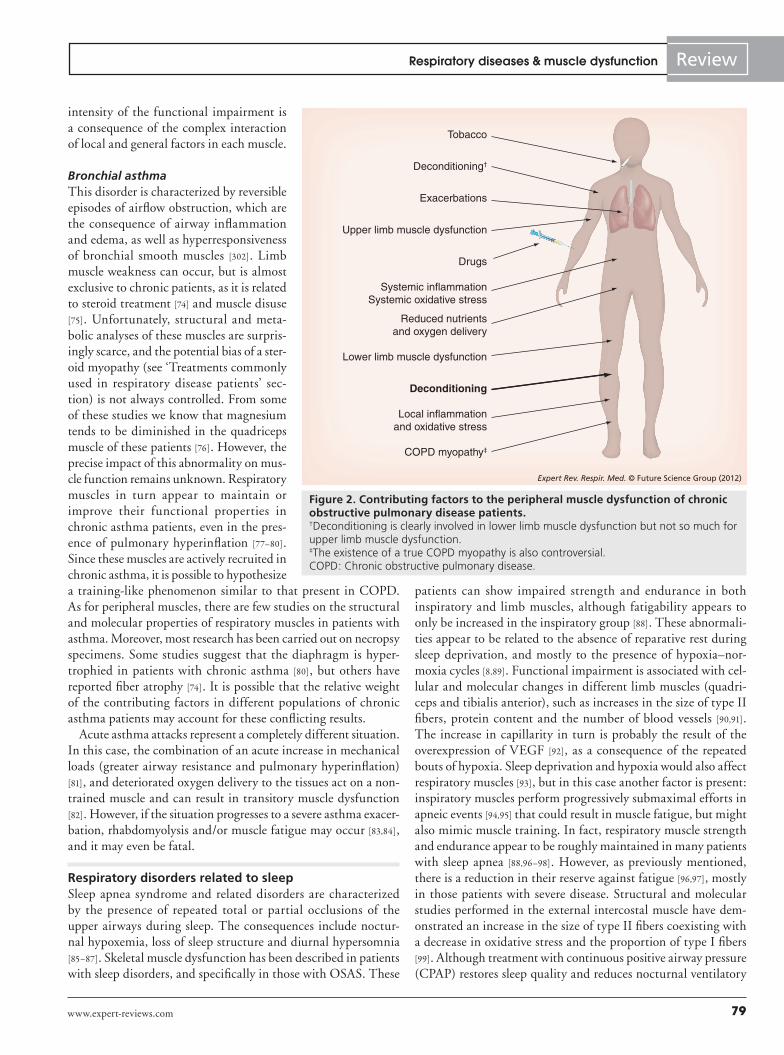
As discussed above, peripheral muscles constitute a heterogene-ous group involved in many different tasks. The function of both lower and upper limb muscles appears to be impaired, to vary-ing degrees, in many COPD patients [9,24,44–48], contributing to their exercise limitations [46,47,49,50]. The main cause of peripheral muscle dysfunction appears to be deconditioning due to a reduc-tion in daily activities (Figure 2) [51,52]. This reduction would be the product of both the initial ventilatory defect and the emotional impairment frequently associated with COPD. A strong argu-ment in favor of the key role played by muscle deconditioning in peripheral muscle dysfunction is that most of the changes described can be reversed by muscle training [51,53]. However, some of the changes are irreversible [47,51], which suggests that other factors may be implicated. Among these are the general factors described as affecting the respiratory muscles, which also affect limb muscles: systemic inflammation and oxidative stress, tobacco smoking, nutritional abnormalities, comorbidity and drugs, among others (Figure 2) [20]. The many studies of structural and molecular changes in the quadriceps of COPD patients have established that this muscle undergoes protein imbalance and atrophy (see next sections), as well as losses in different aerobic components. These include declines in myosin heavy chain I expression, percentage of type I fibers, fiber size, capillary density and myoglobin content, as well as impaired enzyme activities in the oxidative pathways [53–58]. All of these components could
contribute to reduce both the strength and resistance of lower limb muscles in COPD patients. Moreover, some studies support the idea of impaired bioenergetics in the quadriceps of such patients, showing that this muscle is inefficient in its intra cellular use of oxygen [47,48]. Therefore, it could require greater oxygen consumption than healthy subjects for the same amount of work. The quadriceps also show major oxi-dative stress that targets essential proteins in muscle structure and metabolism [59,60]. As a result, muscle damage occurs [61], and there is some evidence that this phenom-enon is associated with defects in the mus-cle regeneration process [62]. The latter, in turn, might be related to the underexpres-sion of some local cytokines [63], but more evidence is necessary. Other authors have reported increased levels of inflammatory cells and proinflammatory cytokines in the quadriceps muscle of COPD patients [64]. All of these phenomena could contribute to the loss in muscle mass and function in the lower limbs. By contrast, muscles located in the scapular girdle and upper
limbs show fewer dramatic changes [10,31,65], contributing to the heterogeneity of muscle adaptations that characterizes COPD. This is not surprising because these muscles probably have a lower exposure to a reduction in their activity. Moreover, they can even show an increase in their activity, as they frequently support ventilation under increased loads in COPD patients. For instance, normal, hypertrophic and atrophic fibers coexist in the deltoid muscle, which preserves its fiber type proportions and oxidative enzyme activities in COPD patients [10,11]. In a similar way, brachial biceps show fiber atrophy but maintain their fiber type percentages [65].
Acute exacerbations contribute to the progression of COPD and the deterioration of muscle function [66]. The increased inflamma-tory load present in the lung during exacerbations may also have systemic repercussions and affect other regions, including skeletal muscles. In this regard, some authors have reported a reduction in muscle mass in various parts of the body during exacerbations [67]. This would be the result of a negative protein balance [68] and the activation of multiple pathways leading to a reduction in MyoD and IGF-1, and subsequent muscle atrophy [69–72]. In addition, the reduction in activity that frequently occurs during an exac-erbation would also negatively affect limb muscles. In the case of respiratory muscles, the situation would be further aggravated by the indirect effects of the exacerbation in the mechanical loads of the ventilatory system. Conversely, muscle dysfunction has been shown to be an independent risk factor for severe exacerbations requiring hospital admission [73].
As a general conclusion, muscle dysfunction associated with COPD can involve many muscle groups. The extension and
Expert Rev. Respir. Med. © Future Science Group (2012)
TobaccoAirway resistance
increasedDynamic compression
of the airways
Changes in ribcagecompliance
Ribcage muscledysfunction
Metabolic mismatching(nutrients, oxygen)
Secondary pulmonaryhypertension
Abdominal wallmuscle dysfunction
Flattened andshortened diaphragm
Exacerbations
Drugs,systemic inflammationand oxidative stress
Diaphragmaticdysfunction
Changes in lung compliancePulmonary
hyperinflation
Heartdysfunction
VA/Q inequality
VA
VO2
Q
O2D
Figure 1. Contributing factors to the respiratory muscle dysfunction of chronic obstructive pulmonary disease patients.O2D: Oxygen delivery; Q: Perfusion; VA: Alveolar ventilation; VO2: Oxygen consumption.
Gea, Casadevall, Pascual, Orozco-Levi & Barreiro
www.expert-reviews.com 79
Review
intensity of the functional impairment is a consequence of the complex interaction of local and general factors in each muscle.
Bronchial asthmaThis disorder is characterized by reversible episodes of airflow obstruction, which are the consequence of airway inflammation and edema, as well as hyperresponsiveness of bronchial smooth muscles [302]. Limb muscle weakness can occur, but is almost exclusive to chronic patients, as it is related to steroid treatment [74] and muscle disuse [75]. Unfortunately, structural and meta-bolic analyses of these muscles are surpris-ingly scarce, and the potential bias of a ster-oid myopathy (see ‘Treatments commonly used in respiratory disease patients’ sec-tion) is not always controlled. From some of these studies we know that magnesium tends to be diminished in the quadriceps muscle of these patients [76]. However, the precise impact of this abnormality on mus-cle function remains unknown. Respiratory muscles in turn appear to maintain or improve their functional properties in chronic asthma patients, even in the pres-ence of pulmonary hyperinflation [77–80]. Since these muscles are actively recruited in chronic asthma, it is possible to hypothesize a training-like phenomenon similar to that present in COPD. As for peripheral muscles, there are few studies on the structural and molecular properties of respiratory muscles in patients with asthma. Moreover, most research has been carried out on necropsy specimens. Some studies suggest that the diaphragm is hyper-trophied in patients with chronic asthma [80], but others have reported fiber atrophy [74]. It is possible that the relative weight of the contributing factors in different populations of chronic asthma patients may account for these conflicting results.
Acute asthma attacks represent a completely different situation. In this case, the combination of an acute increase in mechanical loads (greater airway resistance and pulmonary hyperinflation) [81], and deteriorated oxygen delivery to the tissues act on a non-trained muscle and can result in transitory muscle dysfunction [82]. However, if the situation progresses to a severe asthma exacer-bation, rhabdomyolysis and/or muscle fatigue may occur [83,84], and it may even be fatal.
Respiratory disorders related to sleepSleep apnea syndrome and related disorders are characterized by the presence of repeated total or partial occlusions of the upper airways during sleep. The consequences include noctur-nal hypoxemia, loss of sleep structure and diurnal hypersomnia [85–87]. Skeletal muscle dysfunction has been described in patients with sleep disorders, and specifically in those with OSAS. These
patients can show impaired strength and endurance in both inspiratory and limb muscles, although fatigability appears to only be increased in the inspiratory group [88]. These abnormali-ties appear to be related to the absence of reparative rest during sleep deprivation, and mostly to the presence of hypoxia–nor-moxia cycles [8,89]. Functional impairment is associated with cel-lular and molecular changes in different limb muscles (quadri-ceps and tibialis anterior), such as increases in the size of type II fibers, protein content and the number of blood vessels [90,91]. The increase in capillarity in turn is probably the result of the overexpression of VEGF [92], as a consequence of the repeated bouts of hypoxia. Sleep deprivation and hypoxia would also affect respiratory muscles [93], but in this case another factor is present: inspiratory muscles perform progressively submaximal efforts in apneic events [94,95] that could result in muscle fatigue, but might also mimic muscle training. In fact, respiratory muscle strength and endurance appear to be roughly maintained in many patients with sleep apnea [88,96–98]. However, as previously mentioned, there is a reduction in their reserve against fatigue [96,97], mostly in those patients with severe disease. Structural and molecular studies performed in the external intercostal muscle have dem-onstrated an increase in the size of type II fibers coexisting with a decrease in oxidative stress and the proportion of type I fibers [99]. Although treatment with continuous positive airway pressure (CPAP) restores sleep quality and reduces nocturnal ventilatory
Tobacco
Systemic inflammationSystemic oxidative stress
Upper limb muscle dysfunction
Drugs
Lower limb muscle dysfunction
Deconditioning†
Deconditioning
COPD myopathy‡
Exacerbations
Local inflammationand oxidative stress
Reduced nutrientsand oxygen delivery
Expert Rev. Respir. Med. © Future Science Group (2012)
Figure 2. Contributing factors to the peripheral muscle dysfunction of chronic obstructive pulmonary disease patients. †Deconditioning is clearly involved in lower limb muscle dysfunction but not so much for upper limb muscle dysfunction. ‡The existence of a true COPD myopathy is also controversial.COPD: Chronic obstructive pulmonary disease.
Respiratory diseases & muscle dysfunction
80
Review
Expert Rev. Respir. Med. 6(1), (2012)
effort, it only partially improves respiratory muscle function [97,99]. The lack of complete restoration might be explained by the per-sistence of oxidative stress in the muscle [99] and the probable presence of pulmonary hyperinflation due to the CPAP treatment. Finally, upper airway muscles, which have an important role in the pathophysiology of OSAS, have shown mechanical, structural and metabolic changes in patients suffering from this condition. Musculus uvulae, for instance, shows an increased strength along with larger fibers, a higher protein content and better anaerobic enzyme capacity in OSAS patients than in nonapneic snorers [100,101]. Although genioglossus dysfunction has been reported in patients with OSAS, cellular findings are less impressive [102] and only a mild increase in the proportion of anaerobic fibers has been reported [103]. Interestingly, CPAP reverse these functional and structural changes, suggesting that these are the consequence, and not the cause, of the obstructive problem.
Cystic fibrosisMuscle dysfunction is also frequent in cystic fibrosis. Cachexia, systemic inflammation and gas exchange abnormalities are fre-quently associated with advanced stages of the disease, potentially targeting all skeletal muscles [104,105]. These factors, along with deconditioning, will determine the weakness reported for limb muscles. However, respiratory muscles will face a chronic increase in ventilatory workloads, which would have effects similar to those reported in COPD patients. Therefore, although structural studies are lacking, this muscle group would exhibit phenotypes and function resulting from the complex interaction of multiple deleterious factors with a training effect [105].
Scoliosis & other thoracic deformitiesScoliosis is defined by a lateral curvature of the spine associated with vertebral rotation. This also results in chest deformity, back pain, ventilatory restriction, respiratory and limb muscle weakness and exercise limitation [15,106]. Although respiratory muscle dys-function has classically been attributed to chest deformity, limb muscle dysfunction appears to be the consequence of decondition-ing, probably thorough the development of local oxidative stress [106]. Therefore, especially in adolescents with scoliosis, muscle training would improve their physical performance. By contrast, chest surgery options do not appear to improve muscle function in these patients [Gea J et al., Unpublished Data]. In individuals with advanced stages of thoracic deformity and chronic respiratory fail-ure, noninvasive mechanical ventilation can be useful to improve ventilation and gas exchange [107,108].
Idiopathic pulmonary hypertensionThis condition is characterized by an increase in the blood pres-sure in pulmonary vessels. One of the most frequent symptoms in idiopathic pulmonary hypertension is exercise intolerance. Although this is believed to be caused mainly by vascular fac-tors, some authors have suggested a role for muscle dysfunction [109,110]. This would affect both respiratory and peripheral mus-cles, suggesting the involvement of systemic factors. Moreover, the impairment in functional outcomes has been shown to be
associated with structural and molecular changes, including mus-cle atrophy and a reduction in the proportion of aerobic fibers in the quadriceps muscle [111].
Other circumstances related to respiratory system targeting of skeletal muscles ICU muscle weakness Many different factors, such as systemic inflammation, sepsis, multiorganic failure, malnutrition, malposition, drugs, dyselec-trolytemia and mechanical ventilation [112–115], can contribute to muscle weakness in critically ill patients. These and other still unknown factors can result in axonal and demyelinating neuropathies, defects in the neuromuscular junction and acute myopathies. The latter have specific characteristics, including fiber atrophy, the loss of myosin and the presence of mitochon-dria with paracrystalline inclusions [116,117]. The consequences of muscle dysfunction in ICU patients are very relevant because the weaning process will be more difficult as a result [118,119], and intense rehabilitation is required to ensure reintegration into everyday activities.
The different modalities of mechanical ventilation (MV) assist or substitute for respiratory muscles in their function of providing ventilation to maintain pulmonary gas exchange. Classical MV with anesthesia-paralysis results in early respiratory and peripheral muscle dysfunction. This appears to mainly be the consequence of inactivity, a very harmful factor that can induce diaphragm atrophy at only 48 h of MV [113]. By contrast, noninvasive MV and those forms of classical MV involving the periodic use of respiratory muscles do not appear to induce severe dysfunction because contractile activity is preserved. On the contrary, in most cases they provide rest to fatigued muscles and reduce the work of weakened muscles.
Lung cancer cachexia Cachexia is a complex metabolic syndrome characterized by the loss of muscle mass, which is common in advanced malignant diseases, including lung cancer. Anorexia is frequently associ-ated with cachexia [120,121], which targets muscle by inducing protein imbalance and atrophy [122], both resulting in muscle weakness. On the one hand, gluconeogenesis degrades structural and functional muscle proteins as a source of energy. On the other, protein synthesis also becomes affected [123–125]. Moreover, some cytokines, such as TNF-a, IL-1b, IL-6 and IFN-g [126,127], oxidative stress derived from metabolic changes and the use of antineoplastic drugs appear to be directly involved in this pro-tein imbalance [128,129]. Peripheral muscle weakness occurring in cachectic patients causes their quality of life to deteriorate through progressive limitation of their daily activities. In addition, ventila-tory failure may occur when respiratory muscles become affected. In fact, a third of deaths in cancer patients have been attributed to muscle dysfunction.
Skeletal muscle wasting is a prominent feature in patients with lung cancer [130], even in those with normal bodyweight [131]. In addition, this has been shown to be not only a risk factor for prognosis, but a predictor of cancer treatment toxicity [131]. In the
Gea, Casadevall, Pascual, Orozco-Levi & Barreiro
www.expert-reviews.com 81
Review
same regard, proteolysis has been shown to be negatively related to survival in non-small-cell lung cancer [132].
General factors involved in muscle dysfunction in respiratory disordersAlthough some general factors, such as tobacco smoking, are more specific to COPD, others (systemic inflammation, sedentarism, comorbidity, aging, drug effects and so on) are common to vari-ous respiratory diseases. Below, we review the most relevant gen-eral factors believed to influence muscle function in respiratory patients:
Tobacco smoking Different reports appear to indicate that tobacco smoking per se can induce muscle dysfunction by different mechanisms, includ-ing oxidative stress and a decrease in protein synthesis [30,133–135]. Therefore, a point that still remains unclear is whether the initial stimulus that affects the muscle is just the direct result of the aggressive action of smoking itself or is secondary to the inflam-mation caused by smoking in lung parenchyma, pulmonary blood vessels and the airways [136]. In either case, and indeed they prob-ably coexist, inflammation will affect systemic circulation reach-ing various organs (including muscles) and contributing to their dysfunction [137,138]. An intriguing question is what causes the inflammatory response to persist after the initial noxious stimulus has disappeared. Current thinking on the answer to this ques-tion is based on the hypothesis that these mechanisms may be immunological [139].
Inflammation There is overwhelming evidence to support the hypothesis that a certain level of systemic inflammation is present in COPD [52]. It has been shown that serum levels of certain inflammatory biomarkers (C-reactive protein, fibrinogen and several cytokines) are elevated in these patients [27,137], and higher white blood cell counts have also been found [137,140]. Moreover, it has recently been suggested that the systemic manifestations of COPD may be an expression of an attenuated form of the systemic inflammatory response syndrome [141]. This syndrome has traditionally been conceptualized in the context of the multiorgan failure associ-ated with sepsis [112], but could also be present in a minor form in other chronic conditions, such as COPD [141,142], cystic fibrosis [105] or asthma. Systemic inflammation can have important effects on muscles: various cytokines can induce an increase in local protein degradation through oxidative stress or the activation of proteolytic pathways [143,144], both found in COPD muscles [29,59,145,146]. Furthermore, certain proinflammatory cytokines can inhibit muscle contraction [147], although they appear to be necessary for muscle repair [148].
Oxidative stressThis is closely related to inflammatory mediators, which in conjunction with other factors also present in chronic diseases, such as COPD, lead to oxidative and nitrosative stress [8,149,150]. Inversely, the stress can act as a signal for increased expression
of proinflammatory cytokines [150]. Oxidative stress is the result of an imbalance between reactive oxygen species, a product of aerobic metabolism, and antioxidant mechanisms present in cells and tissues. When the action of oxidants overcomes that of anti-oxidants, certain molecules become modified and their function is impaired as a result. Oxidative stress has been found in various organs of COPD patients, including the lungs, blood and mus-cles [4,34,59,151]. Although both respiratory and peripheral muscles exhibit oxidative stress, it appears to be greater in the limbs of both these patients and animal models of COPD [4,60,152].
Nutritional abnormalities Although relatively frequent in COPD, cystic fibrosis, scoliosis and lung cancer [104,105,130,153,154], nutritional impairment is rela-tively rare in other conditions, such as bronchial asthma. In the particular case of COPD there is wide geographical diversity in the prevalence of nutritional abnormalities. The range is from approximately 5% in Mediterranean countries to 25% or more in Northern Europe and North America [155]. Nutritional abnor-malities have been attributed to factors such as lifestyle, a reduc-tion in food intake [156], an increase in metabolic costs [53] and, more recently, the presence of systemic inflammation [27,157] and changes in the metabolism of certain substances, such as leptin [156]. Nutritional status is a good predictor of mortality in COPD and lung cancer patients [131,158] and can influence their mus-cles. Specifically, malnutrition results in decreased muscle mass, changes in fiber type percentages and muscle dysfunction [159,160].
Comorbidity & agingThe increased life expectancy in developed societies has resulted in a high percentage of elderly patients [161]. In addition, as a con-sequence of the etiopathogenic factors shared by respiratory con-ditions and other disorders, comorbidity is frequently observed. In these circumstances, muscle dysfunction has been attributed to sarcopenia, characteristic of elderly subjects, and to abnor-malities in muscle function also present in highly prevalent dis-eases, such as chronic heart failure, diabetes and rheumatological diseases [46,162,163].
Extreme sedentarismThis is very common in developed countries and leads to cardio-vascular and muscle deconditioning. In fact, the level of physical activity, which is the determinant for an appropriate muscle phe-notype, is a prognostic factor for exacerbations and even for life expectancy in patients with COPD [164–166]. However, this factor seems to be especially important in peripheral muscles because the activity of respiratory muscles would even be even increased in sedentary individuals with respiratory disorders.
Gas-exchange abnormalitiesHypoxemia and hypercapnia are frequently observed in many respiratory diseases and can result in a decrease in muscle strength or endurance [167–169]. Hypoxemia can reduce oxygen delivery to the muscle. This reduction will be even higher in the presence of anemia, a circumstance also very prevalent in chronic diseases,
Respiratory diseases & muscle dysfunction
82
Review
Expert Rev. Respir. Med. 6(1), (2012)
such as COPD. If tissue hypoxia develops it can result in a reduc-tion in stored energy and protein synthesis, impairing muscle con-traction [170–172]. In a similar way the presence of hypercapnia will affect muscle contractility, both directly and indirectly, through the development of muscle acidosis [167,173].
Treatments commonly used in respiratory disease patientsCorticosteroids can induce both acute and chronic myopathies, mostly when used systemically [174]. Although the use of sys-temic corticosteroids has declined considerably, these agents are still necessary in the management of certain seriously ill patients. Corticosteroids also appear to be used more liberally in certain European countries and in North America, perhaps owing to the particular characteristics of the health systems in those countries. Acute myopathy can develop following administration of high doses of corticosteroids [175] and is characterized by marked weakness that can affect various muscle groups [176]. The structural bases of this functional impairment are the loss of myosin filaments and rhab-domyolysis [177,178]. Chronic myopathy in turn is the consequence of long-term administration of steroids, even at relatively low doses [179]. It is mainly characterized by muscle weakness in proximal muscle groups (girdles and trunk) [179]. Cellular and biochemical abnormalities underlying chronic myopathy include type II fiber atrophy, an imbalance in protein synthesis and degradation, and abnormalities in carbohydrate metabolism [179,180].
Anticholinergics are used in respiratory patients as bronchodi-lators to block muscarinic receptors of acetylcholine, and thus relax airway smooth muscles. Although their effects on skeletal muscles are irrelevant at standard doses, higher levels can impair diaphragm contraction [181] and reduce muscle reaction time [182].b-blockers are competitive antagonists of b-adrenergic receptors
and are widely used in cardiovascular disorders, such as hyper-tension and ischemic heart disease. Since many patients with respiratory diseases also present these comorbidities, they fre-quently receive systemic b-blockers. This occurs even in subjects also receiving inhaled b-agonists. b-blockers decrease myocardial contractility [183,184] and have also been shown to facilitate skeletal muscle fatigue [185], although they do not appear to reduce skeletal muscle strength [186].
Calcium channel blockers inhibit channels that mediate the entry of extracellular Ca2+ into muscle cells. Calcium channel blockers are extensively used in respiratory patients with cardio-vascular comorbidities (systemic hypertension, angina pectoris and so on) and have negative inotropic effects on myocardium. However, this effect has not been described for skeletal muscles. Nevertheless, there is some evidence about the action of calcium channel blockers on attenuation of contraction-induced muscle damage [187] and the differentiation of muscle myoblasts/satel-lite cells [188]. This could have some negative influence in the remodeling process undergone by respiratory muscles in obstruc-tive diseases, but might also attenuate the progression of some myopathies.
Statins are widely used for dyslipidemia because they inhibit an enzyme that catalyzes an early step in cholesterol biosynthesis.
Their major adverse effect is a myopathy, mainly characterized by rhabdomyolysis and intense muscle pain, that initiates in the arms and thighs [183,184]. Note that drugs, such as macrolides, frequently used in exacerbations of COPD, interact with statins and can augment their effects [183].
Diuretic drugs can lead to electrolytic imbalance, which in turn can deteriorate muscle function. In this regard, abnormalities in the plasma levels of Na+, K+, Cl- and other ions can result in impaired contraction and easier fatigability.
Phosphodisterase 5 inhibitors augment cyclic GMPc by inhibit-ing a key enzyme, which results in smooth muscle relaxation and vasodilation. Therefore, they are being used for pulmonary hyper-tension and erectile dysfunction [183,184]. Some of these drugs have also been shown to inhibit the muscle effects of insulin (capillary recruitment and glucose uptake) [189], which might result in early muscle fatigue. However, there is also some evidence in animal models that phosphodisterase 5 inhibitors might ameliorate muscle damage in muscle dystrophy [190].
Expert commentaryRespiratory disorders (e.g., COPD, bronchial asthma and thoracic deformities) and their treatments (drugs and mechanical ventila-tion) are frequently associated with respiratory and/or limb mus-cle dysfunction. Whereas respiratory muscle dysfunction results in ventilatory problems, limb muscle dysfunction leads to a reduc-tion in exercise tolerance and limitation of many everyday tasks. Muscle dysfunction is attributed to the complex interaction of general and local factors, including inflammation, oxidative stress, comorbidities, drugs, increases and decreases in muscle activity and changes in thorax geometry. Greater knowledge about the causes and consequences of the muscle dysfunction that occurs in respiratory disorders would open new therapeutic strategies, including a more rational use of current drugs and muscle train-ing, and perhaps, to the adoption of new antioxidants, NSAIDs, anabolic agents and calcium sensitizers as they become available.
Five-year viewThere are good reasons to believe that the next few years will bring not only increased knowledge about the mechanisms of muscle dysfunction in respiratory diseases but also new therapeu-tic approaches to the management and treatment of muscle dys-function. In this respect, recent conceptual advances have opened the way to optimizing classical instruments. These include the expanded use of rehabilitation programs. Although rehabilitation is indicated in many COPD guidelines [191,301], the actual use of this integral therapy is still relatively limited. However, particularly when it involves muscle and general exercise training, rehabilitation has a considerable effect, not only on muscle function, but also on reduction of exacerbations and improved exercise tolerance, quality of life and even patient survival [51,192–194]. This can be applied, not only to COPD, but also to many other respiratory conditions, such as cystic fibrosis and scoliosis. It is important to note, however, that not all COPD patients will respond to muscle training. In the more advanced stages of the disease, when nutritional abnormalities become very relevant and/or exacerbations are extremely frequent,
Gea, Casadevall, Pascual, Orozco-Levi & Barreiro
www.expert-reviews.com 83
Review
these subjects would need additional measures in order to restore even minimal performance. Noninvasive MV, a technique that is already well accepted in the management of COPD exacerba-tions and restrictive disorders, could also prove to be useful in selected stable patients with obstructive diseases. However, this will depend on better identification of the most appropriate candidates [141,195]: those in whom ventilatory support allows the muscles the rest they need. In addition, it should be taken into account that mechanical ventilation can also have deleterious effects on muscle function in specific groups of patients. The administration of drugs with anabolic, anti-inflammatory or antioxidant properties can be expected to increase dramatically in the coming years [152,196]. Specifically, nutritional supplements, testosterone and other ana-bolic agents appear to have a beneficial effect on muscle mass, muscle strength, quality of life and survival in particular groups of patients [154,197,198]. For example, the use of nutritional support has been shown to be beneficial in subjects who have lost weight. One novel prospect is the potential use of ghrelin (a growth hor-mone secretagogue) and growth factors similar to those produced by healthy muscle during training (mechano growth factor) [199] or substances inhibiting myostatin [200]. By contrast, drugs with anti-inflammatory properties should be used with more caution, since some of the proinflammatory cytokines have dual effects on the muscles. On the one hand, they can cause damage and impair contraction, but on the other they appear to be necessary for muscle growth and regeneration [201,202]. Since one of the factors impli-cated in muscle dysfunction is oxidative stress, it is not surprising that there is increased evidence on the potentially beneficial effects on muscles of antioxidants, such as N-acetylcysteine, vitamin E and a-tocopherol [152,203]. Another active research field studies the use of nonsteroidal anti-inflammatory agents to modulate muscle
structure and function [204]. Other drugs widely used in patients with cardiovascular disorders, such as angiotensin-converting enzyme inhibitors, have been shown to prevent cachexia and improve muscle strength [205,206]. More recently, calcium sensi-tizers, widely used in chronic heart failure, have demonstrated their ability to improve contractility of diaphragmatic fibers from COPD patients, as well as respiratory muscle function of healthy individuals [207]. This finding opens new perspectives for drug management of muscle dysfunction in the near future. In addition, surgical and endoscopic procedures can result in reductions of lung volume that reshape the diaphragm in COPD, thus restoring its mechanical properties [208,209]. Although these procedures are still only used in very specific patients, it is to be expected that new techniques will extend their use.
Finally, we should not forget that tobacco smoking is known to negatively influence muscle function in respiratory diseases. Health policies that have been implemented in many countries to eradicate smoking, along with measures devoted to increasing the level of physical activity, are expected to definitively decrease muscle dysfunction as a complication for our respiratory patients.s
AcknowledgementThe authors would like to acknowledge Elaine Lilly, PhD, for editing help.
Financial & competing interests disclosureFunded, in part, by CIBERES ISC III, Plan Nacional SAF2007-62719 and FUCAP. The authors have no other relevant affiliations or financial involvement with any organization or entity with a financial interest in or financial conflict with the subject matter or materials discussed in the manuscript apart from those disclosed.
No writing assistance was utilized in the production of this manuscript.
Key issues
• Muscle dysfunction is frequent in many disorders targeting the respiratory system. These include chronic obstructive pulmonary disease, chronic asthma, obstructive sleep apnea syndrome, cystic fibrosis, lung cancer, thoracic deformities and neuromuscular disorders.
• Respiratory muscle dysfunction results in hypoventilation and can lead to death. It can also hamper the weaning process in mechanically ventilated patients. Peripheral muscle dysfunction results in exercise limitation and restrictions in many daily activities.
• Muscle dysfunction associated with chronic obstructive pulmonary disease is probably the result of the complex interaction between local and systemic factors. While pulmonary hyperinflation appears to be the main factor contributing to respiratory muscle dysfunction, deconditioning is believed to play a determinant role in peripheral muscle dysfunction.
• Drugs typically used in respiratory disorders, including corticosteroids, b-blockers and diuretics, can impair muscle function.
• A wider and better use of both rehabilitation programs and noninvasive mechanical ventilation, as well as appropriate nutritional support and pharmacological strategies (antioxidants, nonsteroidal anti-inflammatory agents, growth factors and calcium sensitizers), will open new possibilities in the treatment of muscle dysfunction.
ReferencesPapers of special note have been highlighted as:• of interest•• of considerable interest
1 Levine S, Kaiser L, Leferovich J, Tikunov B. Cellular adaptations in the diaphragm in chronic obstructive pulmonary disease. N. Engl. J. Med. 337, 1799–1806 (1997).
2 Orozco-Levi M, Gea J, Lloreta J et al. Subcellular adaptation of the human diaphragm in chronic obstructive pulmonary disease. Eur. Respir. J. 13, 371–378 (1999).
3 Orozco-Levi M, Lloreta J, Minguella J, Serrano S, Broquetas J, Gea J. Injury of the human diaphragm associated with exertion and COPD. Am. J. Respir. Crit. Care Med. 164, 1734–1739 (2001).
4 Barreiro E, de la Puente B, Minguella J et al. Oxidative stress and respiratory muscle dysfunction in severe COPD. Am. J. Respir. Crit. Care Med. 171, 1116–1124 (2005).
5 Orozco-Levi M, Gea J, Sauleda J et al. Structure of the latissimus dorsi muscle and respiratory function. J. Appl. Physiol. 78, 1132–1139 (1995).
Respiratory diseases & muscle dysfunction
84
Review
Expert Rev. Respir. Med. 6(1), (2012)
6 Barreiro E, Ferrer A, Hernández-Frutos N, Palacio J, Broquetas J, Gea J. Expiratory function and cellular properties of the external oblique muscle in patients with extremely severe COPD. Am. J. Crit. Care Med. 159(Suppl.), A588 (1999) (Abstract).
7 Gea J, Orozco-Levi M, Barreiro E, Ferrer A, Broquetas J. Structural and functional changes in the skeletal muscles of COPD patients: The ‘Compartments’ Theory. Mon. Arch. Chest Dis. 56, 214–224 (2001).
8 Gea J, Barreiro E, Orozco-Levi M. Skeletal muscle adaptation to disease states. In: Skeletal Muscle Plasticity in Health and Disease: From Genes to Whole Muscle. Bottinelli R, Reggiani C (Eds). Springer, Doordrecht, The Netherlands, 315–360 (2006).
•• Achapterdevotedtomuscledysfunctioninrespiratoryandnonrespiratorydisorders.Causesandconsequencesarediscussedindepth.
9 Coronell C, Orozco-Levi M, Mendez R, Ramirez A, Galdiz JB, Gea J. Relevance of assessing quadriceps endurance in patients with COPD. Eur. Respir. J. 24, 129–136 (2004).
10 Hernandez N, Orozco-Levi M, Belalcazar V et al. Dual morphometrical changes of the deltoid muscle in patients with COPD. Respir. Physiol. Neurobiol. 134, 219–229 (2003).
11 Gea J, Pasto M, Carmona M, Orozco-Levi M, Palomeque J, Broquetas J. Metabolic characteristics of the deltoid muscle in patients with chronic obstructive pulmonary disease. Eur. Respir. J. 17, 939–945 (2001).
12 Epstein SK. An overview on respiratory muscle function. Clin. Chest Med. 15, 619–639 (1994).
13 Martyn J, Powell E, Shore S, Emrich J, Engel LA. The role of respiratory muscles in the hyperinflation of bronchial asthma. Am. Rev. Respir. Dis. 121, 441–447 (1980).
14 Appendini L, Purro A, Patessio A et al. Partitioning of inspiratory muscle workload and pressure assistance in ventilator-dependent COPD patients. Am. J. Respir. Crit. Care Med. 154, 1301–1309 (1996).
15 Martínez-Llorens J, Ramírez M, Colomina MJ et al. Muscle dysfunction and exercise limitation in adolescent idiopathic scoliosis. Eur. Respir. J. 36, 393–400 (2010).
• Recentpaperdescribingtheextensionandintensityofmuscledysfunctioninscoliosis,suggestingakeyrolefordeconditioningand/orprimarymuscleinvolvementinthepathogenesisofthedisease.
16 Casaburi R. Skeletal muscle function in COPD. Chest 117, S267–S271 (2000).
17 Noguera A, Sala E, Pons AR, Iglesias J, MacNee W, Agusti AG. Expression of adhesion molecules during apoptosis of circulating neutrophils in COPD. Chest 125, 1837–1842 (2004).
18 Bolton C, Ionescu A, Shiels K et al. Associated loss of fat-free mass and bone mineral density in chronic obstructive pulmonary disease. Am. J. Respir. Crit. Care Med. 170, 1286–1293 (2004).
19 Agustí AG, Noguera A, Sauleda J, Sala E, Pons J, Busquets X. Systemic effects of chronic obstructive pulmonary disease. Eur. Respir. J. 21, 347–360 (2003).
20 American Thoracic Society. Skeletal muscle dysfunction in chronic obstructive pulmonary disease: a statement of the American Thoracic Society and European Respiratory Society. Am. J. Respir. Crit. Care Med. 159, S1–S40 (1999).
•• Althoughpublished12yearsago,thisisanexcellentreviewofthecausesandconsequencesofmuscledysfunctionassociatedwithchronicobstructivepulmonarydisease(COPD).
21 Swallow EB, Reyes D, Hopkinson NS et al. Quadriceps strength predicts mortality in patients with moderate to severe chronic obstructive pulmonary disease. Thorax 62, 115–120 (2007).
• ClinicalstudyshowingthatlimbmuscledysfunctioninCOPDisanindependentfactorforprognosis.
22 Rochester DF, Braun NMT, Arora NS. Respiratory muscle strength in chronic obstructive pulmonary disease. Am. Rev. Respir. Dis. 119, 151–154 (1979).
23 Similowsky T, Yan S, Gaithier AP, Macklem PT. Contractile properties of the human diaphragm during chronic hyperinflation. N. Engl. J. Med. 325, 917–923 (1991).
24 Gosselink R, Troosters T, Decramer M. Peripheral muscle weakness contributes to exercise limitation in COPD. Am. J. Respir. Crit. Care Med. 153, 976–980 (1996).
25 Goldman MD, Grassino A, Mead J, Sears A. Mechanics of the human diaphragm during voluntary contraction: dynamics. J. Appl. Physiol. 44, 840–848 (1978).
26 Macklem PT, Gross D, Grassino GA, Roussos C. Partitioning of inspiratory pressure swings between diaphragm and intercostal/accessory muscles. J. Appl. Physiol. 44, 200–208 (1978).
27 Di Francia M, Barbier D, Mege JL, Orehek J. Tumor necrosis factor-alpha levels and
weight loss in chronic obstructive pulmonary disease. Am. J. Respir. Crit. Care Med. 150, 1453–1455 (1994).
28 Gosker HR, Wouters EF, van der Vusse GJ, Schols AM. Skeletal muscle dysfunction in chronic obstructive pulmonary disease and chronic heart failure: underlying mechanisms and therapy perspectives. Am. J. Clin. Nutr. 71, 1033–1047 (2000).
29 Barreiro E, Gea J, Di Falco M, Kriazhev L, James S, Hussain SN. Protein carbonyl formation in the diaphragm. Am. J. Respir. Mol. Cell Biol. 32, 9–17 (2005).
30 Barreiro E, Peinado VI, Galdiz JB et al. ENIGMA in COPD Project. Cigarette smoke-induced oxidative stress: a role in chronic obstructive pulmonary disease skeletal muscle dysfunction. Am. J. Respir. Crit. Care Med. 182, 477–488 (2010).
•• KeypaperintheunderstandingoftheroleplayedbytobaccosmokinginthedevelopmentofmuscledysfunctioninCOPD.
31 Gea J, Pastó M, Ennion S, Goldspink G, Broquetas JM. Expression of the genes corresponding to myosin heavy chain isoforms (MyHC I, IIa and IIx) in the diaphragm of patients suffering from COPD. Eur. Respir. J. 12(Suppl. 28), S267 (1998) (Abstract).
32 Hards JM, Reid WD, Pardy RL, Paré PD. Respiratory muscle morphometry: correlation with pulmonary function and nutrition. Chest 97, 1037–1044 (1990).
33 Lloreta J, Orozco-Levi M, Gea J, Broquetas J. Selective diaphragmatic mitochondrial abnormalities in a patient with marked airflow obstruction. Ultrastruct. Pathol. 20, 67–71 (1996).
34 Marin-Corral J, Minguella J, Ramírez-Sarmiento AL, Hussain SN, Gea J, Barreiro E. Oxidised proteins and superoxide anion production in the diaphragm of severe COPD patients. Eur. Respir. J. 33, 1309–1319 (2009).
35 Clanton TL, Levine S. Respiratory muscle fiber remodeling in chronic hyperinflation: dysfunction or adaptation? J. Appl. Physiol. 107, 324–335 (2009).
• NicereviewonthenatureofchangesoccurringinrespiratorymusclesofCOPDpatients,showingthatadaptationsinthehumandiaphragmcanrepresenteitheraformofdysfunction,secondarytothesystemicdisease,oranadaptiveprocessoccurringinaworkingenvironment.
36 Campbell JA, Hughes RL, Shagal V, Frederiksen J, Shields TW. Alterations in intercostal muscle morphology and
Gea, Casadevall, Pascual, Orozco-Levi & Barreiro
www.expert-reviews.com 85
Review
biochemistry in patients with chronic obstructive lung disease. Am. Rev. Respir. Dis. 122, 679–686 (1980).
37 Gea J. Myosin gene expression in the respiratory muscles. Eur. Respir. J. 10, 2404–2410 (1997).
38 Jimenez M, Gea J, Aguar MC et al. Capillary density and respiratory function in the external intercostal muscle. Arch. Bronconeumol. 35, 471–476 (1999).
39 Levine S, Nguyen T, Friscia M et al. Parasternal intercostal muscle remodeling in severe chronic obstructive pulmonary disease. J. Appl. Physiol. 101, 1297–1302 (2006).
40 Ramírez-Sarmiento A, Orozco-Levi M, Barreiro E et al. Expiratory muscle endurance in chronic obstructive pulmonary disease. Thorax 57, 132–136 (2002).
41 Arnold JS, Thomas AJ, Kelsen SG. Length-tension relations of abdominal expiratory muscles: effect of emphysema. J. Appl. Physiol. 62, 739–745 (1987).
42 Dodd DS, Brancatisano T, Engel LA. Chest wall mechanics during exercise in patients with severe chronic airflow obstruction. Am. Rev. Respir. Dis. 129, 33–38 (1984).
43 Ninane V, Rypens F, Yernault J, De Troyer A. Abdominal muscle use during breathing in patients with chronic airflow obstruction. Am. Rev. Respir. Dis. 146, 16–21 (1992).
44 Seymour JM, Spruit MA, Hopkinson NS et al. The prevalence of quadriceps weakness in COPD and the relationship with disease severity. Eur. Respir. J. 36, 81–88 (2010).
•• BroadstudyontheprevalenceofquadricepsweaknessinCOPD.ThestudydemonstratesthatapproximatelyathirdofCOPDpatients,eventhosewithmild-to-moderatedisease,havelimbmuscledysfunction.
45 Bernard S, LeBlanc P, Whittom F et al. Peripheral muscle weakness in patients with chronic obstructive pulmonary disease. Am. J. Respir. Crit. Care Med. 158, 629–634 (1998).
46 Hamilton AL, Killian KJ, Summers E, Jones NL. Muscle strength, symptom intensity, and exercise capacity in patients with cardiorespiratory disorders. Am. J. Respir. Crit. Care Med. 52, 2021–2031 (1995).
47 Sala E, Roca J, Marrades RM et al. Effect of endurance training on skeletal muscle bioenergetic in chronic obstructive
pulmonary disease. Am. J. Respir. Crit. Care Med. 159, 1726–1734 (1999).
48 Engelen MP, Schols AM, Does JD, Gosker HR, Deutz NE, Wouters EF. Exercise-induced lactate increase in relation to muscle substrates in patients with chronic obstructive pulmonary disease. Am. J. Respir. Crit. Care Med. 162, 1697–1704 (2000).
49 Killian KJ, Leblanc P, Martin DH, Summers E, Jones NL, Campbell EJM. Exercise capacity and ventilatory, circulatory, and symptom limitation in patients with chronic airflow obstruction. Am. Rev. Respir. Dis. 146, 935–940 (1992).
50 Eisner MD, Iribarren C, Blanc PD et al. Development of disability in chronic obstructive pulmonary disease: beyond lung function. Thorax 66, 108–114 (2011).
51 Maltais F, Leblanc P, Simard C et al. Skeletal muscle adaptation to endurance training in patients with chronic obstructive pulmonary disease. Am. J. Respir. Crit. Care Med. 154, 442–447 (1996).
52 Doucet M, Dubé A, Joanisse DR et al. Atrophy and hypertrophy signalling of the quadriceps and diaphragm in COPD. Thorax 65, 963–970 (2010).
53 O’Shea SD, Taylor NF, Paratz JD. Progressive resistance exercise improves muscle strength and may improve elements of performance of daily activities for people with COPD: a systematic review. Chest 136, 1269–1283 (2009).
54 Jakobsson P, Jorfeldt L, Brundin A. Skeletal muscle metabolites and fiber types in patients with advanced chronic obstructive pulmonary disease (COPD), with and without chronic respiratory failure. Eur. Respir. J. 3, 192–196 (1990).
55 Jakobsson P, Jordfelt L, Henriksson J. Metabolic enzyme activity in the quadriceps femoris muscle in patients with severe chronic obstructive pulmonary disease. Am. J. Respir. Crit. Care Med. 151, 374–377 (1995).
56 Simard C, Maltais F, Leblanc P, Simard PM, Jobin J. Mitochondrial and capillarity changes in vastus lateralis muscle of COPD patients: electron microscopy study. Med. Sci. Sports Exerc. 28, S95 (1996).
57 Satta A, Migliori GB, Spanevello A et al. Fibre types in skeletal muscles of chronic obstructive pulmonary disease patients related to respiratory function and exercise tolerance. Eur. Respir. J. 10, 2853–2860 (1997).
58 Whittom F, Jobin J, Simard PM et al. Histochemical and morphological characteristics of the vastus lateralis muscle in COPD patients. Med. Sci. Sports Exerc. 30, 1467–1474 (1998).
• ClassicpaperonthemorphologicalcharacteristicsoflimbmusclesinCOPDpatients.Itdemonstratedthatthevastuslateralismuscleischaracterizedbyfiberatrophyandamarkeddecreaseinthepercentageofaerobicfibers.
59 Barreiro E, Gea J, Corominas JM, Hussain SN. Nitric oxide synthases and protein oxidation in the quadriceps femoris of patients with chronic obstructive pulmonary disease. Am. J. Respir. Cell Mol. Biol. 29, 771–778 (2003).
60 Barreiro E, Gea J, Matar G, Hussain SNA. Expression and carbonylation of creatine kinase in the quadriceps femoris muscles of COPD patients. Am. J. Respir. Cell Mol. Biol. 33, 636–642 (2005).
61 Orozco-Levi M, Ferrer MD, Coronell C et al. Evidence of sarcomere disruption and myofibrillar degeneration in the quadriceps muscle of stable COPD patients. Eur. Respir. J. 22(Suppl. 45), S552 (2003) (Abstract).
62 Gea J, Suelves M, Casadevall C, Orozco-Levi M, Barreiro E, Muñoz-Canoves P. Both urokinase-type plasminogen activator (uPA) and uPA receptor are upregulated in respiratory muscles but not in peripheral muscles of severe COPD patients. Proc. Am. Thorac. Soc. 2, A530 (2005) (Abstract).
63 Barreiro E, Schols A, Polkey M et al. On behalf of the ENIGMA in COPD project. Cytokine profile in quadriceps muscles of patients with severe chronic obstructive pulmonary disease. Thorax 63, 100–107 (2008).
64 Montes de Oca M, Torres SH, De Sanctis J, Mata A, Hernández N, Tálamo C. Skeletal muscle inflammation and nitric oxide in patients with COPD. Eur. Respir. J. 26, 390–397 (2005).
65 Sato Y, Asoh T, Honda Y, Fujimatso Y, Higuchi I, Oizumi K. Morphological and histochemical evaluation of biceps muscle in patients with chronic pulmonary emphysema manifesting generalized emaciation. Eur. Neurol. 37, 116–121 (1997).
66 Spruit MA, Gosselink R, Troosters T et al. Muscle force during an acute exacerbation in hospitalised patients with COPD and its relationship with CXCL8 and IGF-I. Thorax 58, 752–756 (2003).
Respiratory diseases & muscle dysfunction
86
Review
Expert Rev. Respir. Med. 6(1), (2012)
67 Güerri R, Gayete A, Balcells E et al. Mass of intercostal muscles associates with risk of multiple exacerbations in COPD. Respir. Med. 104, 378–388 (2010).
68 Saudny-Unterberger H, Martin JG, Gray-Donald K. Impact of nutritional support on functional status during an acute exacerbation of chronic obstructive pulmonary disease. Am. J. Respir. Crit. Care Med. 156, 794–799 (1997).
69 Crul T, Testelmans D, Spruit MA et al. Gene expression profiling in vastus lateralis muscle during an acute exacerbation of COPD. Cell Physiol. Biochem. 25, 491–500 (2010).
70 Creutzberg EC, Wouters EF, Vanderhoven-Augustin IM, Dentener MA, Schols AM. Disturbances in leptin metabolism are related to energy imbalance during acute exacerbations of chronic obstructive pulmonary disease. Am. J. Respir. Crit. Care Med. 162, 1239–1245 (2000).
71 Crul T, Spruit MA, Gayan-Ramirez G et al. Markers of inflammation and disuse in vastus lateralis of chronic obstructive pulmonary disease patients. Eur. J. Clin. Invest. 37, 897–904 (2007).
72 Decramer M, Rennard S, Troosters T et al. COPD as a lung disease with systemic consequences – clinical impact, mechanisms, and potential for early intervention. COPD 5, 235–256 (2008).
73 Vilaró J, Ramirez-Sarmiento A, Martínez-Llorens JM et al. Global muscle dysfunction as a risk factor of readmission to hospital due to COPD exacerbations. Respir. Med. 104, 1896–1902 (2010).
74 Picado C, Fiz JA, Montserrat JM et al. Respiratory and skeletal muscle function in steroid-dependent bronchial asthma. Am. Rev. Respir. Dis. 141, 14–20 (1990).
75 Mancuso CA, Sayles W, Robbins L et al. Barriers and facilitators to healthy physical activity in asthma patients. J. Asthma 43, 137–143 (2006).
76 Gustafson T, Boman K, Rosenhall L, Sandström T, Wester PO. Skeletal muscle magnesium and potassium in asthmatics treated with oral beta 2-agonists. Eur. Respir. J. 9, 237–240 (1996).
77 Rochester DF, Arora NS. The respiratory muscles in asthma. In: Diagnostic Aspects and Management of Asthma. Lavietes MH, Reichman LB (Eds). Purdue-Frederick, Norwalk, CO, USA (1981).
78 Mckenzie DK, Gandevia SC. Strength and endurance of inspiratory and limb muscles in asthma. Am. Rev. Respir. Dis. 134, 999–1004 (1986).
79 Peress L, Sybrecht G, Macklem PT. The mechanism of increase in total lung capacity during acute asthma. Am. J. Med. 61, 165–169 (1976).
80 de Bruin PF, Ueki J, Watson A, Pride NB. Size and strength of the respiratory and quadriceps muscles in patients with chronic asthma. Eur. Respir. J. 10, 59–64 (1997).
81 De Troyer A, Wilson TA. Effect of acute inflation on the mechanics of the inspiratory muscles. J. Appl. Physiol. 107, 315–323 (2009).
82 Laviettes MH, Grocela JA, Maniatis T, Potulski F, Ritter AB, Sunderam G. Inspiratory muscle strenght in asthma. Chest 93, 1043–1048 (1988).
83 Lovis C, Mach F, Unger PF, Bouillie M, Chevrolet JC. Elevation of creatine kinase in acute severe asthma is not of cardiac origin. Intensive Care Med. 27, 528–533 (2001).
84 Tsushima K, Koyama S, Ueno M et al. Rhabdomyolysis triggered by an asthmatic attack in a patient with McArdle disease. Intern. Med. 40, 131–134 (2001).
85 Hoffmann M, Bybee K, Accurso V, Somers VK. Sleep apnea and hypertension. Minerva Med. 95, 281–290 (2004).
86 Parish JM, Somers VK. Obstructive sleep apnea and cardiovascular disease. Mayo. Clin. Proc. 79, 1036–1046 (2004).
87 Yaggi H, Mohsenin V. Obstructive sleep apnoea and stroke. Lancet Neurol. 3, 333–342 (2004).
88 Chien MY, Wu YT, Lee PL, Chang YJ, Yang PC. Inspiratory muscle dysfunction in patients with severe obstructive sleep apnoea. Eur. Respir. J. 35, 373–380 (2010).
• Recentpaperonrespiratoryandlimbmuscledysfunctionassociatedwiththesleepapneasyndrome.Causesmightincluderepetitiveinspiratoryeffortsagainstanoccludedairway,intermittenthypoxiaandimpairedsleepquality.
89 Chen H, Tang Y. Sleep loss impairs inspiratory muscle endurance. Am. Rev. Respir. Dis. 140, 907–909 (1989).
90 Wåhlin-Larsson B, Kadi F, Ulfberg J, Piehl Aulin K. Skeletal muscle morphology and aerobic capacity in patients with obstructive sleep apnoea syndrome. Respiration 76, 21–27 (2008).
91 Sauleda J, García-Palmer FJ, Tarraga S, Maimó A, Palou A, Agustí AG. Skeletal muscle changes in patients with obstructive sleep apnoea syndrome. Respir. Med. 97, 804–810 (2003).
92 Wåhlin-Larsson B, Ulfberg J, Aulin KP, Kadi F. The expression of vascular endothelial growth factor in skeletal muscle of patients with sleep disorders. Muscle Nerve 40, 556–561 (2009).
93 Garpestad E, Hatayama H, Parker J et al. Stroke volume and cardiac output decrease at termination of obstructive apneas. J. Appl. Physiol. 73, 1743–1748 (1992).
94 Wilcox PG, Paré PD, Road JD, Fleetham JA. Respiratory muscle function during obstructive sleep apnea. Am. Rev. Respir. Dis. 142, 533–539 (1990).
95 Kimoff J, Cheong T, Olha A et al. Mechanisms of apnea termination in obstructive sleep apnea. Am. J. Respir. Crit. Care Med. 149, 707–714 (1994).
96 Griggs G, Findley L, Suratt P, Esau S, Wilhoit S, Rochester D. Prolonged relaxation rate of inspiratory muscles in patients with sleep apnea. Am. Rev. Respir. Dis. 140, 706–710 (1989).
97 Aran X, Félez MA, Gea J, Orozco-Levi M, Sauleda J, Broquetas JM. Respiratory muscle force and resistance in patients with SAHS. The effect of using nighttime CPAP. Arch. Bronconeumol. 35, 440–445 (1999).
98 Shepherd KL, Jensen CM, Maddison KJ, Hillman DR, Eastwood PR. Relationship between upper airway and inspiratory pump muscle force in obstructive sleep apnea. Chest 130, 1757–1764 (2006).
99 Barreiro E, Nowinski A, Gea J, Sliwinski P. Oxidative stress in the external intercostal muscles of patients with obstructive sleep apnoea. Thorax 62, 1095–1101 (2007).
100 Sériès F, Côté C, Simoneau JA et al. Physiologic, metabolic, and muscle fiber type characteristics of musculus uvulae in sleep apnea hypopnea syndrome and in snorers. J. Clin. Invest. 95, 20–25 (1995).
101 Sériès F, Côté C, St Pierre S. Dysfunctional mechanical coupling of upper airway tissues in sleep apnea syndrome. Am. J. Respir. Crit. Care Med. 159, 1551–1555 (1999).
102 Sériès FJ, Simoneau SA, St Pierre S, Marc I. Characteristics of the genioglossus and musculus uvulae in sleep apnea hypopnea syndrome and in snorers. Am. J. Respir. Crit. Care Med. 153, 1870–1874 (1996).
103 Carrera M, Barbé F, Sauleda J, Tomás M, Gómez C, Agustí AG. Patients with obstructive sleep apnea exhibit genioglossus dysfunction that is normalized after treatment with continuous positive airway pressure. Am. J. Respir. Crit. Care Med. 159, 1960–1966 (1999).
Gea, Casadevall, Pascual, Orozco-Levi & Barreiro
www.expert-reviews.com 87
Review
104 Von Haehling S, Anker SD. Cachexia as a major underestimated and unmet medical need: facts and numbers. J. Cachexia Sarcopenia Muscle 1, 1–5 (2010).
105 Dufresne V, Knoop C, Van Muylem A et al. Effect of systemic inflammation on inspiratory and limb muscle strength and bulk in cystic fibrosis. Am. J. Respir. Crit. Care Med. 180, 153–158 (2009).
106 Swallow EB, Barreiro E, Gosker H et al. Quadriceps muscle strength in scoliosis. Eur. Respir. J. 34, 1429–1435 (2009).
107 Gustafson T, Franklin KA, Midgren B, Pehrsson K, Ranstam J, Ström K. Survival of patients with kyphoscoliosis receiving mechanical ventilation or oxygen at home. Chest 130, 1828–1833 (2006)
108 Duiverman ML, Bladder G, Meinesz AF, Wijkstra PJ. Home mechanical ventilatory support in patients with restrictive ventilatory disorders: a 48-year experience. Respir. Med. 100, 56–65 (2006).
109 Meyer FJ, Lossnitzer D, Kristen AV et al. Respiratory muscle dysfunction in idiopathic pulmonary arterial hypertension. Eur. Respir. J. 25, 125–130 (2005).
110 Bauer R, Dehnert C, Schoene P et al. Skeletal muscle dysfunction in patients with idiopathic pulmonary arterial hypertension. Respir. Med. 101, 2366–2369 (2007).
111 Mainguy V, Maltais F, Saey D et al. Peripheral muscle dysfunction in idiopathic pulmonary arterial hypertension. Thorax 65, 113–117 (2010).
112 Barreiro E, Comtois AS, Mohammed S, Lands L, Hussain SNA. Role of heme oxygenases in sepsis-induced diaphragmatic contractile dysfunction and oxidative stress. Am. J. Physiol. Lung Cell Mol. Physiol. 283, L476–L484 (2002).
113 Le Bourdelles G, Viires N, Boczkowski J et al. Effects of mechanical ventilation on diaphragmatic contractile properties in rats. Am. J. Respir. Crit. Care Med. 149, 1539–1544 (1994).
114 Deconinck N, Van Parijs V, Bleeckers-Bleukx G et al. Critical illness myopathy unrelated to corticosteroids or neuromuscular blocking agents. Neuromuscul. Disord. 8, 186–192 (1998).
115 Polkey MI, Moxham J. Clinical aspects of respiratory muscle dysfunction in the critically ill. Chest 119, 926–939 (2001).
116 Matsubara S, Okada T, Yoshida M. Mitochondrial changes in acute myopathy after treatment of respiratory failure with mechanical ventilation (acute relaxant-steroid myopathy). Acta Neuropathol. 88, 475–478 (1994).
117 Lacomis D, Giuliani M, Van Cott A et al. Acute myopathy of intensive care: clinical, electromyographic and pathological aspects. Ann. Neurol. 40, 645–654 (1996).
118 Spizer AR, Giancarlo T, Mahler L. Nueromuscular causes of prolonged ventilator dependency. Muscle Nerve 15, 682–686 (1992).
119 Mehta S, Nelson DL, Klinger JR, Buczko GB, Levy MM. Prediction of post-extubation work of breathing. Crit. Care Med. 28, 1341–1346 (2000).
120 Blum D, Omlin A, Baracos VE et al.; European Palliative Care Research Collaborative. Cancer cachexia: a systematic literature review of items and domains associated with involuntary weight loss in cancer. Crit. Rev. Oncol. Hematol. 80(1), 114–144 (2011).
121 Evans WJ, Morley JE, Argilés J et al. Cachexia: a new definition. Clin. Nutr. 27, 793–799 (2008).
122 DeWys W. Management of cancer cachexia. Semin. Oncol. 12, 452–460 (1985).
123 Costelli P, Carbó N, Tessitore L et al. Tumour necrosis factor-alpha mediates changes in muscle protein turnover in a cachectic rat tumour model. J. Clin. Invest. 92, 2783–2789 (1993).
124 García-Martínez C, López-Soriano FJ, Argilés JM. Amino acid uptake in skeletal muscle of rats bearing the Yoshida AH-130 ascites hepatoma. Mol. Cell Biochem. 148, 17–23 (1995).
125 Kirchberger M. Excitation and contraction of skeletal muscle. In: Physiological Basis of Medical Practice. West JB (Ed.). Williams & Wilkins, Baltimore, MD, USA, 62–102 (1991).
126 Llovera M, Carbó N, López-Soriano FJ et al. Different cytokines modulate ubiquitin-gene expression in rat skeletal muscle. Cancer Lett. 133, 83–87 (1998).
127 Fortunati N, Manti R, Birocco N et al. Pro-inflammatory cytokines and oxidative stress/ antioxidant parameters characterize the bio-humoral profile of early cachexia in lung cancer patients. Oncol. Rep. 18, 1521–1527 (2007).
128 Gomes-Marcondes MC, Tisdale MJ. Induction of protein catabolism and the ubiquitin-proteasome pathway by mild oxidative stress. Cancer Lett. 180, 69–74 (2002).
129 Chevari S, Andial T, Benke K, Shtrenger IA, Free radical reactions and cancer, Vopr. Med. Khim. 38, 4–5 (1992).
130 Muscaritoli M, Bossola M, Aversa Z, Bellantone R, Rossi-Fanelli F. Prevention and treatment of cancer cachexia: new insights into an old problem. Eur. J. Cancer 42, 31–41 (2006).
131 Baracos VE, Reiman T, Mourtzakis M, Gioulbasanis I, Antoun S. Body composition in patients with non-small cell lung cancer: a contemporary view of cancer cachexia with the use of computed tomography image analysis. Am. J. Clin. Nutr. 91, S1133–S1137 (2010).
• Population-basedstudyofcachexiainpatientswithlungcancer,demonstratingthatmusclewastingisaprominentfeaturedespitenormalorheavybodyweightinsuchpatients.
132 Wang Q, Lu JB, Wu B, Hao LY. Expression and clinicopathologic significance of proteolysis-inducing factor in non-small-cell lung cancer: an immunohistochemical analysis. Clin. Lung Cancer 11, 346–351 (2010).
133 Zhang J, Liu Y, Shi J, Larson DF, Watson RR. Side-stream cigarette smoke induces dose-response in systemic inflammatory cytokine production and oxidative stress. Exp. Biol. Med. 227, 823–829 (2002).
134 Montes de Oca M, Loeb E, Torres SH, De Sanctis J, Hernández N, Tálamo C. Peripheral muscle alterations in non-COPD smokers. Chest 133, 13–18 (2008).
135 Petersen AM, Magkos F, Atherton P et al. Smoking impairs muscle protein synthesis and increases the expression of myostatin and MAFbx in muscle. Am. J. Physiol. Endocrinol. Metab. 293, E843–E848 (2007).
136 Sinden NJ, Stockley RA. Systemic inflammation and comorbidity in COPD: a result of ‘overspill’ of inflammatory mediators from the lungs? Review of the evidence. Thorax 65, 930–936 (2010).
137 Gan WQ, Man WQ, Senthilselvan A, Sin DD. Association between chronic obstructive pulmonary disease and systemic inflammation: a systematic review and a meta-analysis. Thorax 59, 574–580 (2004).
138 Nordskog BK, Fields WR, Hellmann GM. Kinetic analysis of cytokine response to cigarette smoke condensate by human endothelial and monocytic cells. Toxicology 212, 87–97 (2005).
139 Borchers MT, Wesselkamper SC, Curull V et al. Sustained CTL activation by murine pulmonary epithelial cells promotes the development of COPD-like disease. J. Clin. Invest. 119, 636–649 (2009).
Respiratory diseases & muscle dysfunction
88
Review
Expert Rev. Respir. Med. 6(1), (2012)
140 Sauleda J, García-Palmer FJ, González G, Palou A, Agustí AG. The activity of cytochrome oxidase is increased in circulating lymphocytes of patients with chronic obstructive pulmonary disease, asthma, and chronic arthritis. Am. J. Respir. Crit. Care Med. 161, 32–35 (2000).
141 Gea J, Orozco-Levi M, Barreiro E. Skeletal and respiratory muscle dysfunction in the systemic inflammatory response syndrome associated with COPD. In: COPD as a Systemic Disease. Cazzola M, Stockley RA, Wouters EFM (Eds). Clinical Publishing, Boca Raton, FL, USA (2008).
142 Creutzberg EC, Wouters EF, Vanderhoven-Augustin IM, Dentener MA, Schols AM. Disturbances in leptin metabolism are related to energy imbalance during acute exacerbations of chronic obstructive pulmonary disease. Am. J. Respir. Crit. Care Med. 162, 1239–1245 (2000).
143 Flores EA, Bristain BR, Pomposelli JJ, Dinarello CA, Blackburn GL, Istfan NW. Infusion of tumor necrosis factor /cachectin promotes muscle metabolism in the rat. J. Clin. Invest. 83, 1614–1622 (1989).
144 Hall-Angeras M, Angeras U, Zamir O, Hasselgren PO, Fischer JE. Interaction between corticosterone and tumor necrosis factor simulated protein breakdown in rat skeletal muscle similar to sepsis. Surgery 108, 460–466 (1990).
145 Plant PJ, Brooks D, Faughnan M et al. Cellular markers of muscle atrophy in chronic obstructive pulmonary disease. Am. J. Respir. Cell Mol. Biol. 42, 461–471 (2010).
146 Fermoselle C, Rabinovich R, Ausin P et al. Severe COPD patients exhibit increased muscle proteolysis in their vastus lateralis. Eur. Respir. J. 36(Suppl.), 338s (2010) (Abstract).
147 Reid MB, Lannergren J, Westerblad H. Respiratory and limb muscle weakness induced by tumor necrosis factor-alpha: involvement of muscle myofilaments. Am. J. Respir. Crit. Care Med. 166, 479–484 (2002).
148 Pascual S, Casadevall C, Martínez-Llorens J et al. Functional blockade of TNF-alpha and myogenic differentiation in cells from peripheral muscles of COPD patients. Am. J. Respir. Crit. Care Med. (2011) (In press) (Abstract).
149 Supinski G. Free radical-induced respiratory muscle dysfunction. Mol. Cell Biochem. 179, 99–110 (1998).
150 Gea J, Barreiro E, Orozco-Levi M. Free radicals, cytokines and respiratory muscles in COPD patients. Clin. Pulm. Med. 14, 117–126 (2007).
• RelativelyrecentreviewonthepotentialroleplayedbycytokinesandoxidantsinmuscledysfunctionassociatedwithCOPD.
151 Gosker HR, Bast A, Haenen GR et al. Altered antioxidant status in peripheral skeletal muscle of patients with COPD. Respir. Med. 99, 118–125 (2005).
152 Barreiro E, Gáldiz JB, Mariñán M, Álvarez FJ, Hussain SN, Gea J. Respiratory loading intensity and diaphragm oxidative stress: N-acetylcysteine effects. J. Appl. Physiol. 100, 555–563 (2006).
• Interestingpapershowingthebenefitsofanantioxidanttherapyinrespiratorymuscledysfunctionoccurringinananimalmodelofinspiratoryloading.
153 Schols AM. Nutrition in chronic obstructive pulmonary disease. Curr. Opin. Pulm. Med. 6, 110–115 (2000).
154 Schols AM, Wouters EF. Nutritional abnormalities and supplementation in chronic obstructive pulmonary disease. Clin. Chest Med. 21, 753–762 (2000).
155 Coronell C, Orozco-Levi M, Gea J. COPD and body weight in a Mediterranean population. Clin. Nutr. 21, 437 (2002).
156 Schols AM, Creutzberg E, Buurman W, Campfield L, Saris W, Wouters E. Plasma leptin is related to proinflammatory status and dietary intake in patients with chronic obstructive pulmonary disease. Am. J. Respir. Crit. Care Med. 160, 1220–1226 (1999).
157 Schols AM, Buurman W, van den Brekel S et al. Evidence for a relation between metabolic derangements and increased levels of inflammatory mediators in a subgroup of patients with chronic obstructive pulmonary disease. Thorax 51, 819–824 (1996).
158 Schols AM, Slangen J, Volovics L, Wouters EF. Weight loss is a reversible factor in the prognosis of chronic obstructive pulmonary disease. Am. J. Respir. Crit. Care Med. 157, 1791–1797 (1998).
159 Kelsen SG, Ference M, Kapoor S. Effects of prolonged undernutrition on structure and function of the diaphragm. J. Appl. Physiol. 58, 1354–1359 (1985).
160 Lopes J, Russell DM, Whitwell J, Jeejeebhoy KN. Skeletal muscle function in malnutrition. Am. J. Clin. Nutr. 36, 602–610 (1982).
161 Valente S, Pasciuto G, Bernabei R, Corbo GM. Do we need different treatments for very elderly COPD patients? Respiration 80, 357–368 (2010).
162 Barreiro E, Coronell C, Laviña B, Ramírez-Sarmiento A, Orozco-Levi M, Gea J; PENAM Project. Aging, sex differences, and oxidative stress in human respiratory and limb muscles. Free Radic. Biol. Med. 41, 797–809 (2006).
163 Chua TP, Anker SD, Harrington D, Coats AJ. Inspiratory muscle strength is a determinant of maximum oxygen consumption in chronic heart failure. Br. Heart J. 74, 381–385 (1995).
164 García-Aymerich J, Farrero E, Félez MA, Izquierdo J, Marrades RM, Antó JM; the EFRAM investigators. Risk factors of readmission to hospital for a COPD exacerbation: a prospective study. Thorax 58, 100–105 (2003).
165 Pitta F, Troosters T, Probst VS, Lucas S, Decramer M, Gosselink R. Potential consequences for stable chronic obstructive pulmonary disease patients who do not get the recommended minimum daily amount of physical activity. J. Bras. Pneumol. 32, 301–308 (2006).
166 Pitta F, Troosters T, Spruit MA, Decramer M, Gosselink R. Activity monitoring for assessment of physical activities in daily life in patients with chronic obstructive pulmonary disease. Arch. Phys. Med. Rehabil. 86(10), 1979–1985 (2005).
167 Aguar MC, Gea J, Aran X et al. Modifications in diaphragmatic mechanics induced by inhaled CO
2 in COPD
patients. Arch. Bronconeumol. 29, 226–228 (1993).
168 Gea J, Orozco-Levi M, Aran X, Sauleda J, Aguar MC, Broquetas JM. Acute hypoxemia and respiratory muscles function in COPD patients. Am. Rev. Respir. Dis. 149(Suppl.), A140 (1994) (Abstract).
169 Kent BD, Mitchell PD, McNicholas WT. Hypoxemia in patients with COPD: cause, effects, and disease progression. Int. J. Chron. Obstruct. Pulmon. Dis. 6, 199–208 (2011).
170 Pastoris O, Dossena M, Foppa P et al. Modifications by chronic intermittent hypoxia and drug treatment on skeletal muscle metabolism. Neurochem. Res. 20, 143–150 (1995).
171 Romer LM, Haverkamp HC, Amann M, Lovering AT, Pegelow DF, Dempsey JA. Effect of acute severe hypoxia on peripheral fatigue and endurance capacity in healthy humans. Am. J. Physiol. Regul. Integr. Comp. Physiol. 292, R598–R606 (2007).
172 Caquelard F, Burnet H, Tagliarini F, Cauchy E, Richalet JP, Jammes Y. Effects of prolonged hypobaric hypoxia on human
Gea, Casadevall, Pascual, Orozco-Levi & Barreiro
www.expert-reviews.com 89
Review
skeletal muscle function and electromyographic events. Clin. Sci. (Lond.) 98, 329–337 (2000).
173 Rafferty GF, Lou Harris M, Polkey MI, Greenough A, Moxham J. Effect of hypercapnia on maximal voluntary ventilation and diaphragm fatigue in normal humans. Am. J. Respir. Crit. Care Med. 160, 1567–1571 (1999).
174 Goldberg A, Goodman H. Relationship between cortisone and muscle work in determining muscle size. J. Physiol. 200, 667–675 (1969).
175 Kaminski HJ, Ruff RL. Endocrine myopathies (hyper- and hypofunction of adrenal, thyroid, pituitary, and parathyroid glands, and iatrogenic corticosteroid myopathy. In: Myology. Engel AG, Franzini-Armstrong C (Eds). McGraw-Hill Medical, New York, NY, USA, 1726–1733 (1994).
176 Nava S, Bruschi C, Franchia C, Fezicetti G, Callezsri G, Ambrosino N. Acute treatment with corsticosteroids (CS) alters respiratory (RM) and skeletal muscles (SM) function in humans. Eur. Respir. J. 9(Suppl.), S298 (1996) (Abstract).
177 Waclawik AJ, Sufit RL, Beinlich BR, Schutta HS. Acute myopathy with selective degeneration of myosin filaments following status asthmaticus treated with methylprednisolone and vecuronium. Neuromuscul. Disord. 2, 19–26 (1992).
178 Ramsay DA, Zochodne DW, Robertson DM, Nag S, Ludwin SK. A syndrome of acute severe muscle necrosis in intensive care unit patients. J. Neuropathol. Exp. Neurol. 52, 387–398 (1993).
179 Decramer M, deBock V, Dom R. Functional and histologic picture of steroid-induced myopathy in chronic obstructive pulmonary disease. Am. J. Respir. Crit. Care Med. 153, 1958–1964 (1996).
• Paperexaminingthefunctionalandhistologicalpictureofsteroid-inducedmyopathy.Thiswasassociatedwithventilatoryandlimbmuscleweakness,fiberatrophyandnecrosis,aswellasabnormalmuscleenzymelevelsinblood.
180 Lieu F, Powers SK, Herb RA et al. Exercise and glucocorticoid-induced diaphragmatic myopathy. J. Appl. Physiol. 75, 763–771 (1993).
181 Qiu Z, Zhao D, Shi Y, Liu Y. [The effects of high dose atropine on function of isolated diaphragmatic preparation of rats with omethoate poisoning]. Zhonghua Nei Ke Za Zhi 40, 187–189 (2001).
182 Karataş GK, Günendi Z. Do anticholinergics affect reaction time? A possible impact on the course of rehabilitation. NeuroRehabilitation 27, 141–145 (2010).
183 The Pharmacological Basis of Therapeutics. XI Edition. Brunton LL, Lazo JS, Parker KL (Eds). Goldman & Gilman’s, McGraw-Hill, New York, NY, USA (2006).
184 Applied Therapeutics. IX Edition. Koda-Kimble MA, Young LY, Alldredge BK et al. (Eds). Lippincott Williams & Wilkins. PA, USA (2009).
185 Kaiser P, Hylander B, Eliasson K, Kaijer L. Effect of beta1-selective and nonselective beta blockade on blood pressure relative to physical performance in men with systemic hypertension. Am. J. Cardiol. 55, D79–D84 (1985).
186 Cupido CM, Hicks AL, McKelvie RS, Sale DG, McComas AJ. Effect of selective and nonselective beta-blockade on skeletal muscle excitability and fatigability. J. Appl. Physiol. 76, 2461–2466 (1994).
187 Nemirovskaia TL, Kitina IuN, Zgelesniakova AV, Vikhliandtsev IM. [Cytoskeletal proteins and heat shock proteins 27 under eccentric exercise of rats: effects of calcium L-type channel blockade]. Ross. Fiziol . Zh. Im. IM Sechenova 94, 293–300 (2008).
188 Porter GA Jr, Makuck RF, Rivkees SA. Reduction in intracellular calcium levels inhibits myoblast differentiation. J. Biol. Chem. 277, 28942–28947 (2002).
189 Mahajan H, Richards SM, Rattigan S, Clark MG. T-1032, a cyclic GMP phosphodiesterase-5 inhibitor, acutely blocks physiologic insulin-mediated muscle haemodynamic effects and glucose uptake in vivo. Br. J. Pharmacol. 140, 1283–1291 (2003).
190 Asai A, Sahani N, Kaneki M, Ouchi Y, Martyn JA, Yasuhara SE. Primary role of functional ischemia, quantitative evidence for the two-hit mechanism, and phosphodiesterase-5 inhibitor therapy in mouse muscular dystrophy. PLoS One 2, e806 (2007).
191 Celli BR, MacNee W, and committee members. ATS/ERS Task Force. Standards for the diagnosis and treatment of patients with COPD: a summary of the ATS/ERS position paper. Eur. Respir. J. 23, 932–946 (2004).
192 American Association for Cardiovascular and Pulmonary Rehabilitation. Guidelines for Cardiac Rehabilitation and Secondary Prevention Programs. 4th Edition. Human
Kinetics Publishers, Champaign, IL, USA (2004).
193 Salman GF, Mosier MC, Beasley BW, Clakins DR. Rehabilitation for patients with chronic obstructive pulmonary disease: meta-analysis of randomized controlled trials. J. Gen. Intern. Med. 18, 213–221 (2003).
194 Ramírez-Sarmiento A, Orozco-Levi M, Güell R et al. Inspiratory muscle training in patients with chronic obstructive pulmonary disease: structural adaptation and physiologic outcomes. Am. J. Respir. Crit. Care Med. 166, 1491–1497 (2002).
195 Wijkstra PJ. Non-invasive positive pressure ventilation (NIPPV) in stable patients with chronic obstructive pulmonary disease (COPD). Respir. Med. 97, 1086–1093 (2003).
196 Svartberg J. Androgens and chronic obstructive pulmonary disease. Curr. Opin. Endocrinol. Diabetes Obes. 17, 257–261 (2010).
197 Schols AM. Nutritional modulation as part of the integrated management of chronic obstructive pulmonary disease. Proc. Nutr. Soc. 62, 783–791 (2003).
198 Casaburi R, Bhasin S, Cosentino L et al. Effects of testosterone and resistance training in men with chronic obstructive pulmonary disease. Am. J. Respir. Crit. Care Med. 170, 870–878 (2004).
199 Goldspink G. Research on mechano growth factor: its potential for optimising physical training as well as misuse in doping. Br. J. Sports Med. 39, 787–788 (2005).
200 Tsuchida K. Targeting myostatin for therapies against muscle-wasting disorders. Curr. Opin. Drug Discov. Devel. 11, 487–494 (2008).
201 Kuru S, Inukai A, Kato T, Liang Y, Kimura S, Sobue G. Expression of tumor necrosis factor-alpha in regenerating muscle fibers in inflammatory and non-inflammatory myopathies. Acta Neuropathol. (Berl.) 105, 217–224 (2003).
202 Warren GL, Hulderman T, Jensen N et al. Physiological role of tumor necrosis factor alpha in traumatic muscle injury. FASEB J. 16, 1630–1632 (2002).
203 Supinski GS, Stofan D, Ciufo R, DiMarco A. N-acetylcysteine administration alters the response to inspiratory loading in oxygen-supplemented rats. J. Appl. Physiol. 82, 1119–1125 (1997).
204 Barnes PJ. Cytokine modulators as novel therapies for airway disease. Eur. Respir. J. 34(Suppl.), S67–S77 (2001).
Respiratory diseases & muscle dysfunction
90
Review
Expert Rev. Respir. Med. 6(1), (2012)
205 Maggio M, Ceda GP, Lauretani F et al. Relation of angiotensin converting enzyme inhibitor treatment to insulin-like growth factor-1 serum levels in subjects of 65 years of age (the InCHIANTI study). Am. J. Cardiol. 97, 1525–1529 (2006).
206 Coirault C, Hagege A, Chemla D, Fratacci MD, Guerot C, Lecarpentier Y. Angiotensin-converting enzyme inhibitor therapy improves respiratory muscle strength in patients with heart failure. Chest 119, 1755– 1760 (2001).
207 Doorduin J, Sinderby CA, Beck J et al. The calcium sensitizer levosimendan improves human diaphragm function. Am. J. Respir. Crit. Care Med. doi:201107-1268OCv1 (2011) (Epub ahead of print).
208 Estenne M. Effect of lung transplant and volume reduction surgery on respiratory muscle function. J. Appl. Physiol. 107, 977–986 (2009).
209 Emery MJ, Eveland RL, Eveland K, Couetil LL, Hildebrandt J, Swenson ER. Lung volume reduction by bronchoscopic administration of steam. Am. J. Respir. Crit. Care Med. 182, 1282–1291 (2010).
Websites
301 Global Initiative for Chronic Obstructive Lung Disease. Global Strategy for the Diagnosis, Management and Prevention of Chronic Obstructive Pulmonary Disease. NHLBI/WHO workshop report. Bethesda, updated 2010. National Heart, Lung and Blood Institute. Update of the Management Sections, GOLD. http://goldcopd.com
302 Global Initiative for Asthma. Global Strategy for Asthma Management and Prevention. NHLBI. Bethesda, updated 2009. http://ginasthma.com
Gea, Casadevall, Pascual, Orozco-Levi & Barreiro

Reproduced with permission of the copyright owner. Further reproduction prohibited without permission.