Embed Size (px)
DESCRIPTION
RESPIRATORY FAILURE AND MECHANICAL VENTILATION. ALOK SINHA Department of Medicine Manipal College of Medical Sciences Pokhara , Nepal. - PowerPoint PPT Presentation
Citation preview
ALOK SINHADepartment of Medicine
Manipal College of Medical SciencesPokhara, Nepal
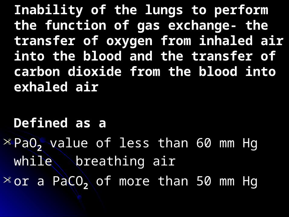
Inability of the lungs to perform the function of gas exchange- the transfer of oxygen from inhaled air into the blood and the transfer of carbon dioxide from the blood into exhaled air
Defined as a • PaO2 value of less than 60 mm Hg while
breathing air • or a PaCO2 of more than 50 mm Hg
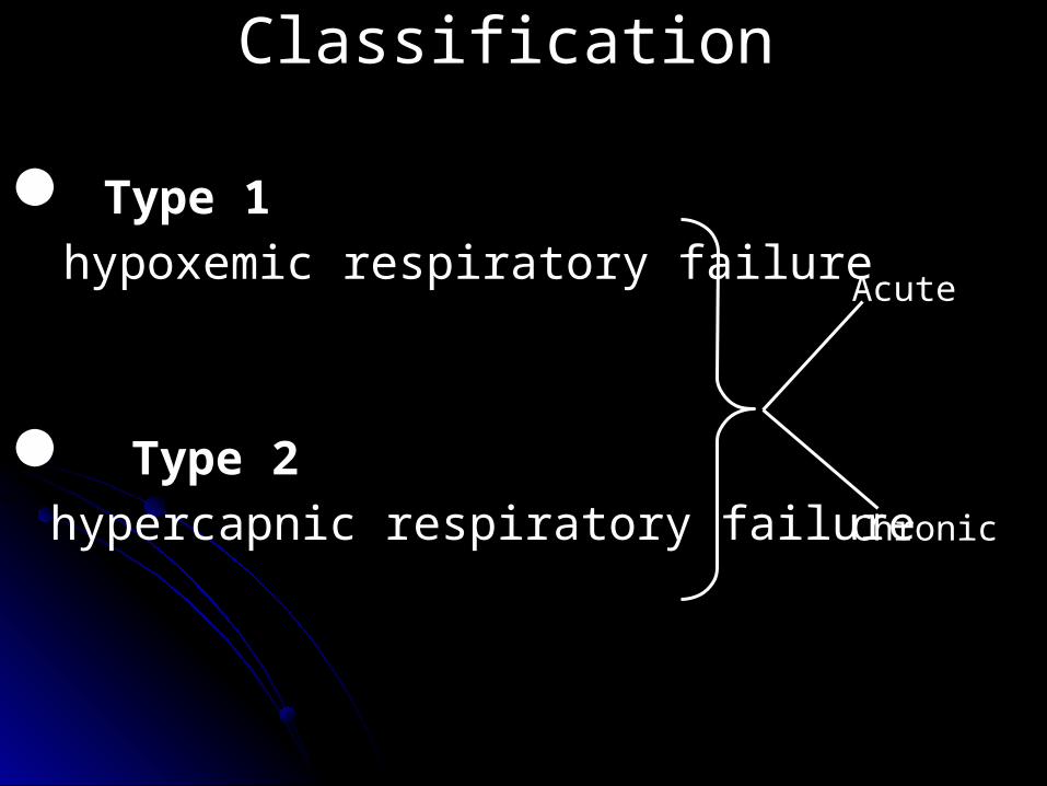
Classification
Type 1hypoxemic respiratory failure
Type 2hypercapnic respiratory failure
Acute
Chronic
Respiratory failure is caused by:
1. failure to oxygenate characterized by decreased PaO2
2. failure to ventilatecharacterized by increased PCO2
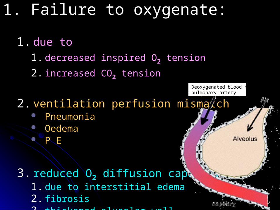
1. Failure to oxygenate:
1. due to 1. decreased inspired O2 tension
2. increased CO2 tension
2. ventilation perfusion mismatch Pneumonia Oedema P E
3. reduced O2 diffusion capacity 1. due to interstitial edema 2. fibrosis3. thickened alveolar wall
Deoxygenated blood from pulmonary artery
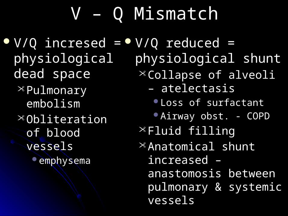
V – Q MismatchV/Q incresed =
physiological dead space• Pulmonary
embolism• Obliteration of
blood vesselsemphysema
V/Q reduced = physiological shunt• Collapse of alveoli –
atelectasisLoss of surfactantAirway obst. - COPD
• Fluid filling• Anatomical shunt
increased – anastomosis between pulmonary & systemic vessels
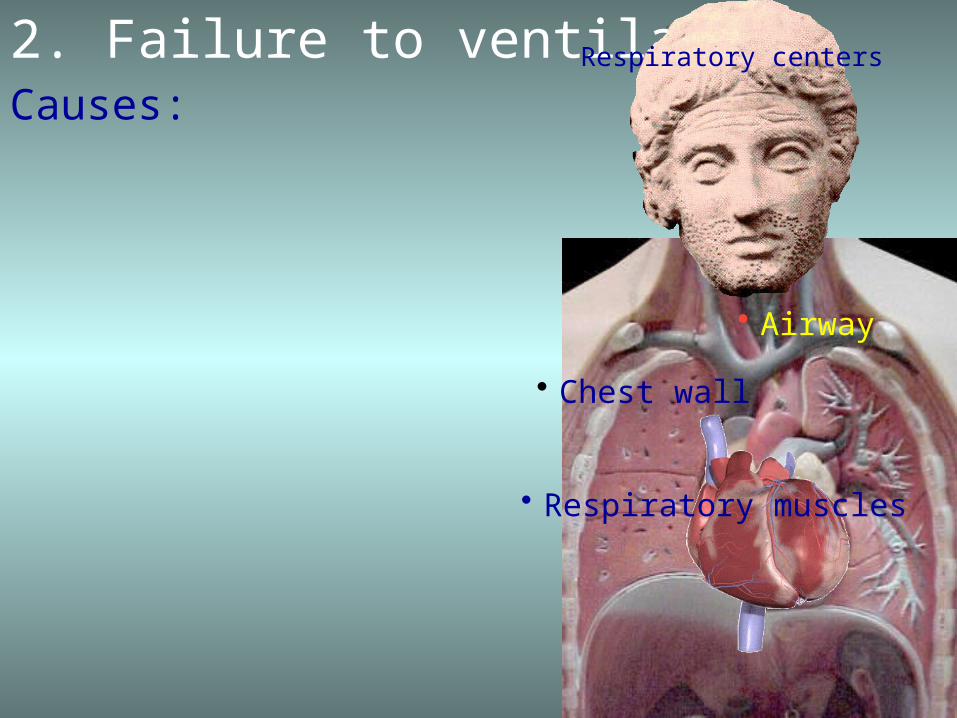
2. Failure to ventilateCauses:
• Airway
• Respiratory muscles
• Chest wall
Respiratory centers
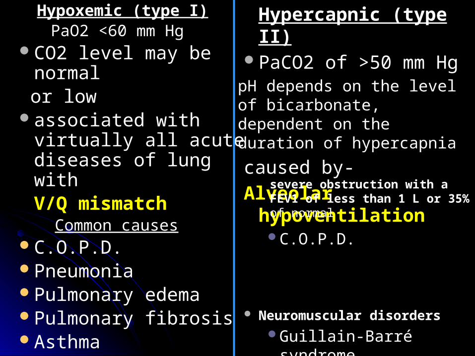
Hypoxemic (type I) PaO2 <60 mm Hg
CO2 level may be normal or lowassociated with virtually
all acute diseases of lung withV/Q mismatch
Common causesC.O.P.D.Pneumonia Pulmonary edema Pulmonary fibrosis Asthma
Hypercapnic (type II)PaCO2 of >50 mm Hg
pH depends on the level of bicarbonate, dependent on the duration of hypercapnia caused by- Alveolar hypoventilation
C.O.P.D.
Neuromuscular disorders Guillain-Barré syndrome Diaphragm paralysis Amyotrophic lateral
sclerosis Muscular dystrophy Myasthenia gravis
severe obstruction with a FEV1 of less than 1 L or 35% of normal
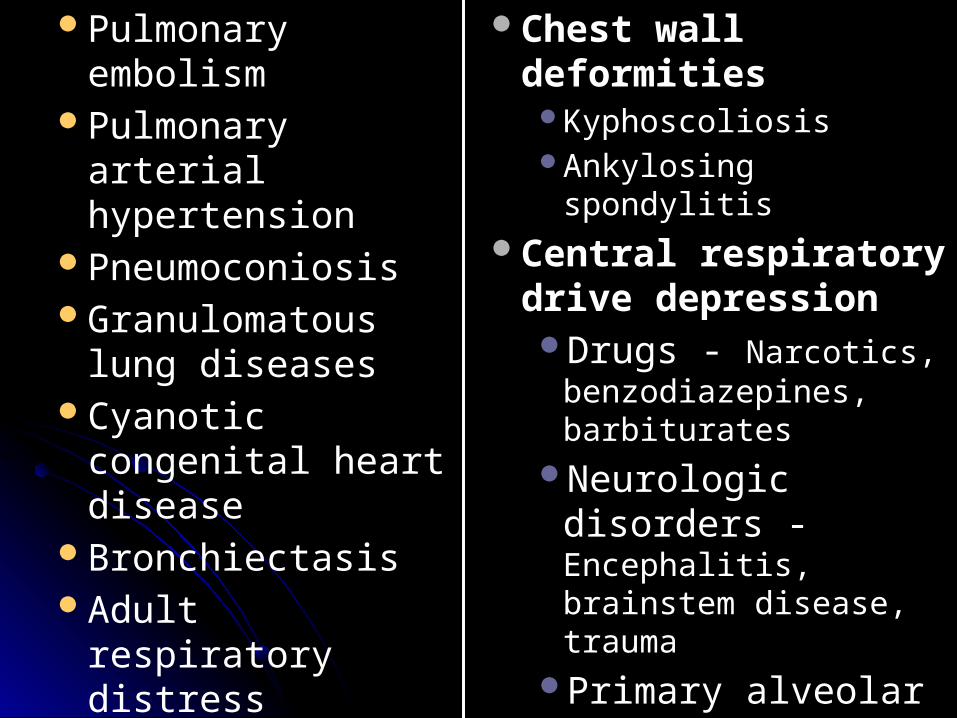
Pulmonary embolism Pulmonary arterial
hypertension Pneumoconiosis Granulomatous lung
diseases Cyanotic congenital
heart disease Bronchiectasis Adult respiratory
distress syndrome Fat embolism
syndrome
Chest wall deformities KyphoscoliosisAnkylosing spondylitis
Central respiratory drive depression Drugs - Narcotics,
benzodiazepines, barbiturates
Neurologic disorders - Encephalitis, brainstem disease, trauma
Primary alveolar hypoventilation
Obesity hypoventilation syndrome (Pickwickian Syn)
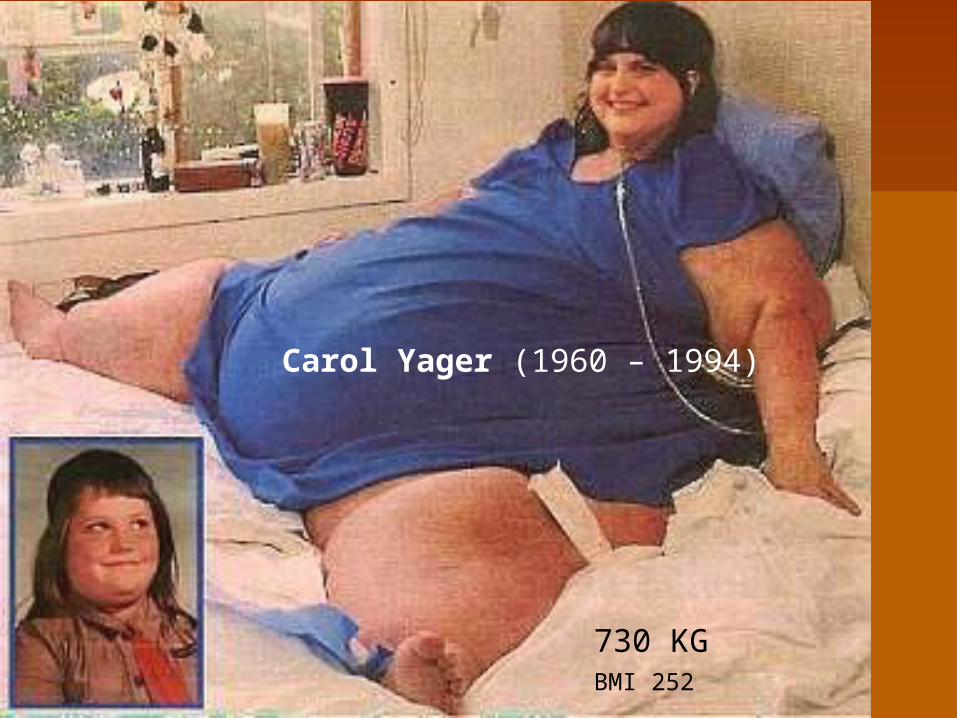
730 KGBMI 252
Carol Yager (1960 – 1994)
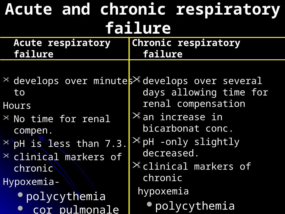
Acute and chronic respiratory failure Acute respiratory failure
• develops over minutes toHours• No time for renal compen.• pH is less than 7.3.• clinical markers of chronicHypoxemia-
polycythemia cor pulmonale
Are absent
Chronic respiratory failure
• develops over several days allowing time for renal compensation
• an increase in bicarbonat conc.• pH -only slightly decreased.• clinical markers of chronic hypoxemia
polycythemia cor pulmonaleAre present
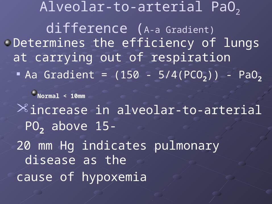
Alveolar-to-arterial PaO2 difference (A-a Gradient)
Determines the efficiency of lungs at carrying out of respiration Aa Gradient = (150 - 5/4(PCO2)) - PaO2
Normal < 10mm
• increase in alveolar-to-arterial PO2 above 15-
20 mm Hg indicates pulmonary disease as thecause of hypoxemia
• Normal in Hypoventilation

Underlying disease process (pneumonia, pulmonary edema, asthma, COPD)
associated hypoxemia
hypercapnia
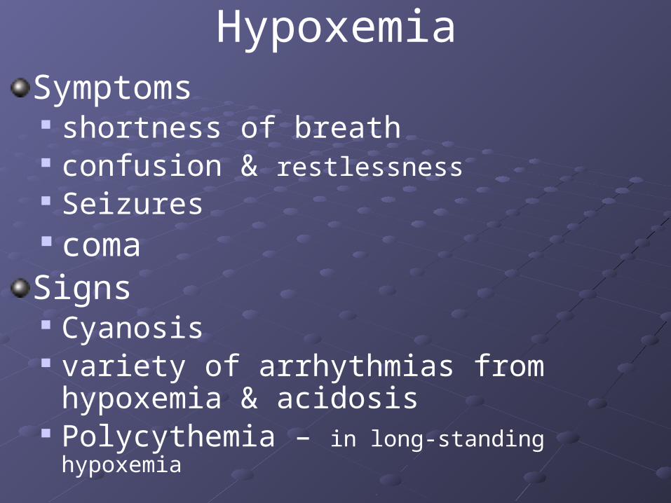
HypoxemiaSymptoms shortness of breath confusion & restlessness Seizures coma Signs Cyanosis variety of arrhythmias from hypoxemia &
acidosis Polycythemia – in long-standing hypoxemia
Hypercapnia
Vasodilation leading to Morning headache flushed skin & warm moist palms full & bounding pulse Extrasystoles & other arrythmias muscle twitches flapping tremors - asterixisdrowsiness
Asterix

Now answer this question-If you are forced to choose one of these, which one youwill like to have
Hypoxia Hypercapnia
?

.
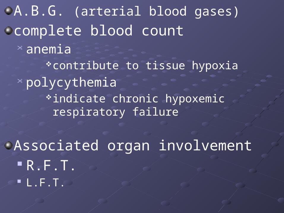
A.B.G. (arterial blood gases) complete blood count • anemia
contribute to tissue hypoxia• polycythemia
indicate chronic hypoxemic respiratory failure
Associated organ involvement R.F.T. L.F.T.
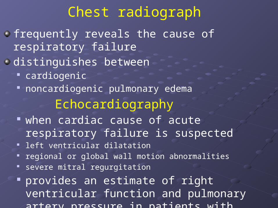
Chest radiograph frequently reveals the cause of respiratory failuredistinguishes between
cardiogenic noncardiogenic pulmonary edema
Echocardiography when cardiac cause of acute respiratory failure is
suspected left ventricular dilatation regional or global wall motion abnormalities severe mitral regurgitation provides an estimate of right ventricular function
and pulmonary artery pressure in patients with chronic hypercapnic respiratory failure
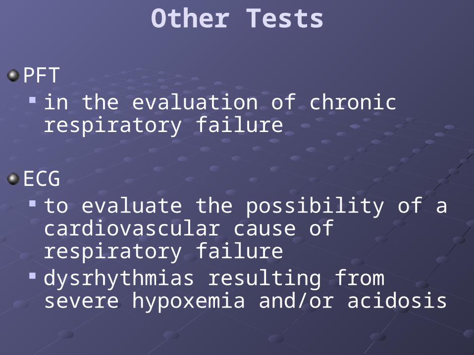
Other Tests
PFT in the evaluation of chronic respiratory failure
ECG to evaluate the possibility of a cardiovascular
cause of respiratory failure dysrhythmias resulting from severe
hypoxemia and/or acidosis

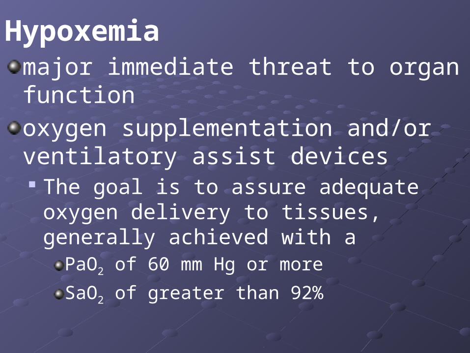
Hypoxemiamajor immediate threat to organ functionoxygen supplementation and/or ventilatory assist devices The goal is to assure adequate oxygen
delivery to tissues, generally achieved with a PaO2 of 60 mm Hg or moreSaO2 of greater than 92%
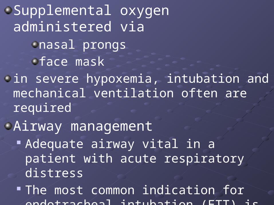
Supplemental oxygen administered via nasal prongs face mask
in severe hypoxemia, intubation and mechanical ventilation often are requiredAirway management Adequate airway vital in a patient with acute
respiratory distress The most common indication for
endotracheal intubation (ETT) is respiratory failure
What is the role of tracheostomy??
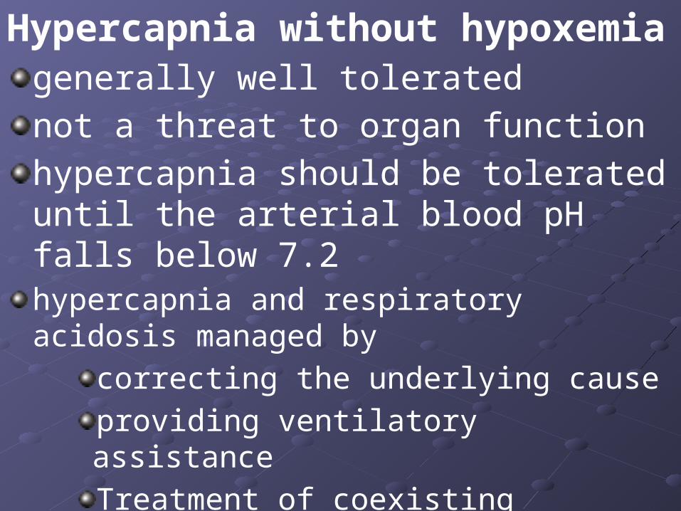
Hypercapnia without hypoxemia generally well tolerated not a threat to organ function hypercapnia should be tolerated until the arterial blood pH falls below 7.2hypercapnia and respiratory acidosis managed by
correcting the underlying cause providing ventilatory assistanceTreatment of coexisting condition with approptiate drugs
Mechanical ventilation is a method to
mechanically assist or
replace spontaneous breathing
Mechanical Ventilatior What is it?
Machine that generates a controlled flow of gas into a patient’s airways
Oxygen and air are received from cylinders or wall outlets blended according to the prescribed inspired oxygen tension (FiO2)
Delivered to the patient using one of many available modes of ventilation.The magnitude of rate and duration of flow are determined by the operator
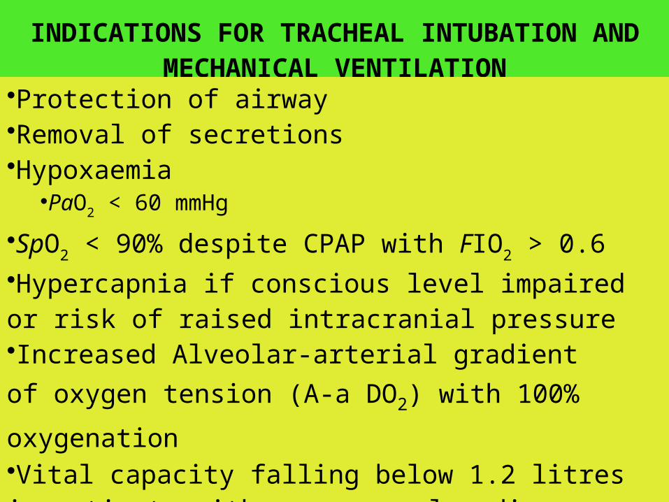
INDICATIONS FOR TRACHEAL INTUBATION AND MECHANICAL VENTILATION
Body_ID: B008019
•Protection of airway •Removal of secretions •Hypoxaemia
•PaO2 < 60 mmHg•SpO2 < 90% despite CPAP with FIO2 > 0.6 •Hypercapnia if conscious level impaired or risk of raised intracranial pressure•Increased Alveolar-arterial gradient of oxygen tension (A-a DO2) with 100% oxygenation •Vital capacity falling below 1.2 litres in patients with neuromuscular disease •Removing the work of breathing in exhausted patients
Ventilatory workload is increased by loss of lung compliance inspiration/ventilation is usually supported to
reduce O2 requirements and increase patient comfort
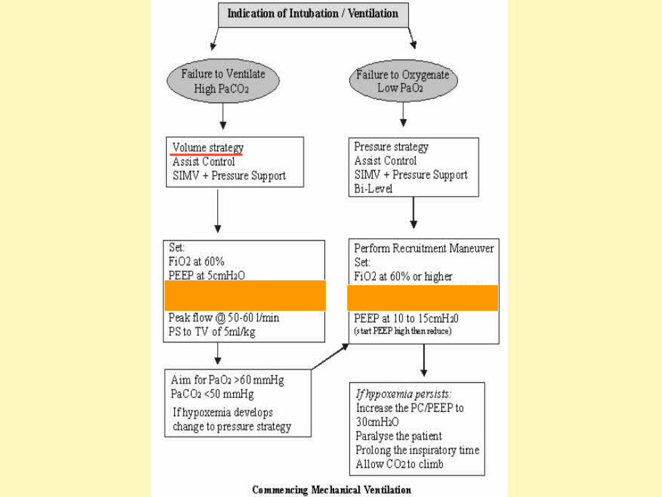
Respiratory failure is caused by 1. Failure to ventilate
characterized by increased PCO2
2. Failure to oxygenate characterized by decreased PaO2
Failure to ventilate Increase the patient’s alveolar ventilation
rate depth of breathing
by using mechanical ventilation
Failure to oxygenate Restoration and maintenance of lung volumes
by using recruitment maneuvers
Recruitment maneuvers are used to reinflate collapsed alveoli: due to pressure generated by ventilator during inspiration alveoli are inflated
PEEP is used to prevent derecruitment

What do we mean by PEEP ?
Girl’s changing room
PEEPamount of pressure above atmospheric
pressure present in the airway at the end of the expiratory cycle
PEEP improves gas exchange by preventing alveolar collapse recruiting more lung unitsincreasing functional residual capacity redistributing fluid in the alveoli
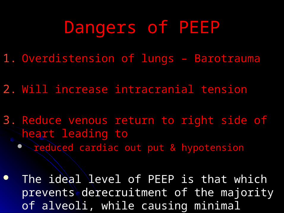
Dangers of PEEP
1. Overdistension of lungs – Barotrauma
2. Will increase intracranial tension
3. Reduce venous return to right side of heart leading to
reduced cardiac out put & hypotension
The ideal level of PEEP is that which prevents derecruitment of the majority of alveoli, while causing minimal overdistension
Modes of ventilation: Air flow continues until
a predetermined volume has been delivered – volume controlled
airway pressure generated – pressure controlled
Flow reverses, when the machine cycles into the expiratory phase, the message to do this is either at a preset timepreset tidal volume preset percentage of peak flow
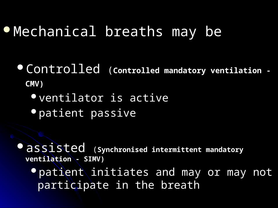
Mechanical breaths may be
Controlled (Controlled mandatory ventilation -CMV) ventilator is active patient passive
assisted (Synchronised intermittent mandatory ventilation - SIMV)
patient initiates and may or may not participate in the breath
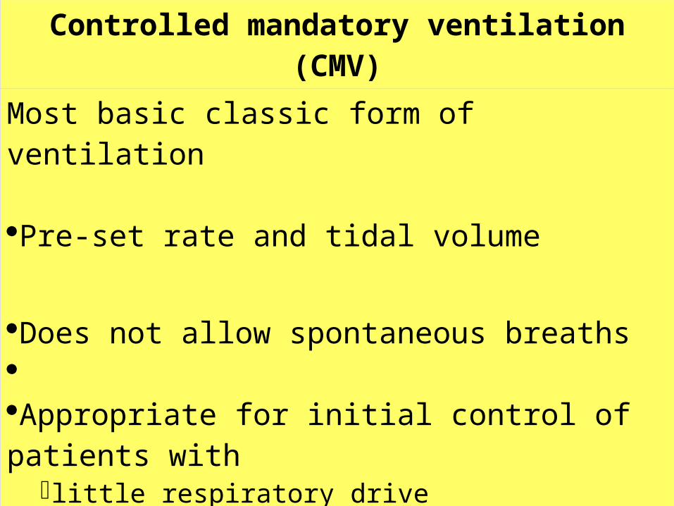
Controlled mandatory ventilation (CMV)
Most basic classic form of ventilation Pre-set rate and tidal volume
Does not allow spontaneous breaths Appropriate for initial control of patients with
little respiratory drivesevere lung injury circulatory instability
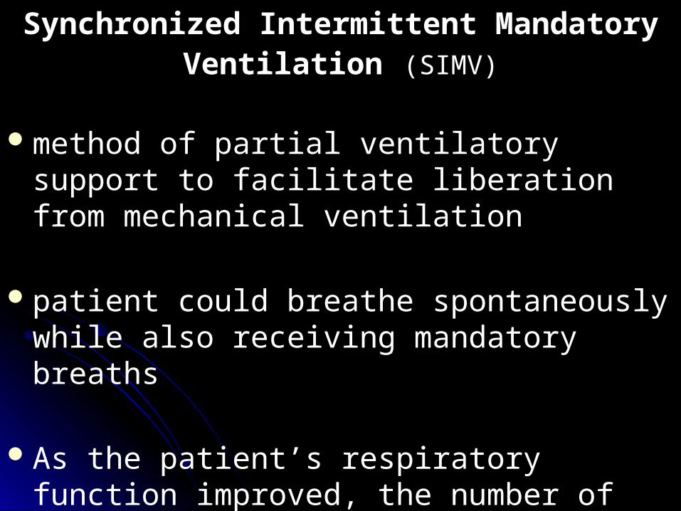
Synchronized Intermittent Mandatory Ventilation (SIMV)
method of partial ventilatory support to facilitate liberation from mechanical ventilation
patient could breathe spontaneously while also receiving mandatory breaths
As the patient’s respiratory function improved, the number of assisted is decreased, until the patient breaths unassisted
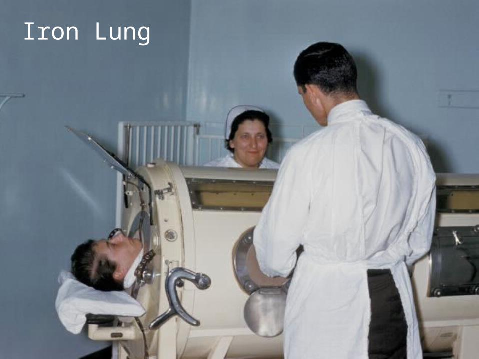
Iron Lung
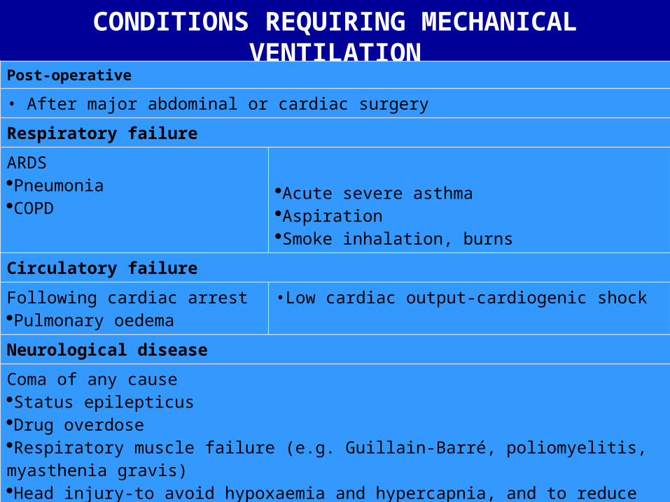
CONDITIONS REQUIRING MECHANICAL VENTILATION
Post-operative
• After major abdominal or cardiac surgery
Respiratory failureARDS Pneumonia COPD
Acute severe asthma Aspiration Smoke inhalation, burns
Circulatory failureFollowing cardiac arrest Pulmonary oedema
•Low cardiac output-cardiogenic shock
Neurological diseaseComa of any cause Status epilepticus Drug overdose Respiratory muscle failure (e.g. Guillain-Barré, poliomyelitis, myasthenia gravis) Head injury-to avoid hypoxaemia and hypercapnia, and to reduce intracranial pressure Bulbar abnormalities causing risk of aspiration (CVA, myasthenia gravis)
Multiple trauma
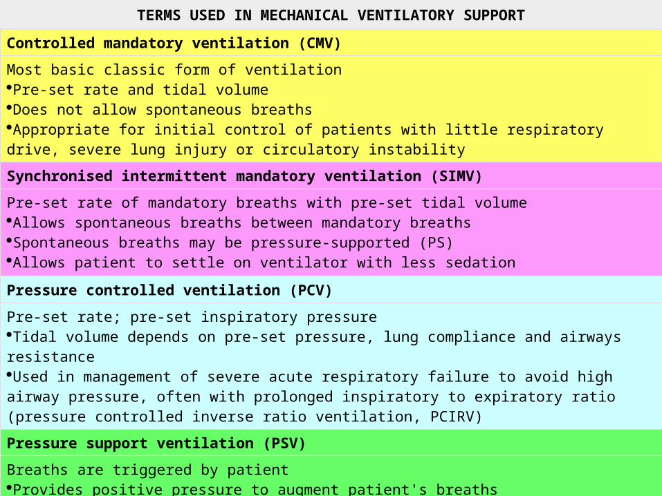
TERMS USED IN MECHANICAL VENTILATORY SUPPORT
Controlled mandatory ventilation (CMV)Most basic classic form of ventilation Pre-set rate and tidal volume Does not allow spontaneous breaths Appropriate for initial control of patients with little respiratory drive, severe lung injury or circulatory instability
Synchronised intermittent mandatory ventilation (SIMV)Pre-set rate of mandatory breaths with pre-set tidal volume Allows spontaneous breaths between mandatory breaths Spontaneous breaths may be pressure-supported (PS) Allows patient to settle on ventilator with less sedation
Pressure controlled ventilation (PCV)Pre-set rate; pre-set inspiratory pressure Tidal volume depends on pre-set pressure, lung compliance and airways resistance Used in management of severe acute respiratory failure to avoid high airway pressure, often with prolonged inspiratory to expiratory ratio (pressure controlled inverse ratio ventilation, PCIRV)
Pressure support ventilation (PSV)Breaths are triggered by patient Provides positive pressure to augment patient's breaths Useful for weaning Usually combined with CPAP; may be combined with SIMV Pressure support is titrated against tidal volume and respiratory rate
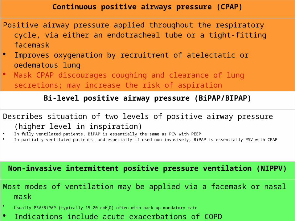
Continuous positive airways pressure (CPAP)
Positive airway pressure applied throughout the respiratory cycle, via either an endotracheal tube or a tight-fitting facemask
Improves oxygenation by recruitment of atelectatic or oedematous lung Mask CPAP discourages coughing and clearance of lung secretions; may increase
the risk of aspiration
Bi-level positive airway pressure (BiPAP/BIPAP)
Describes situation of two levels of positive airway pressure (higher level in inspiration) In fully ventilated patients, BiPAP is essentially the same as PCV with PEEP In partially ventilated patients, and especially if used non-invasively, BiPAP is essentially PSV with CPAP
Non-invasive intermittent positive pressure ventilation (NIPPV)
Most modes of ventilation may be applied via a facemask or nasal mask Usually PSV/BiPAP (typically 15-20 cmH2O) often with back-up mandatory rate Indications include acute exacerbations of COPD
Of mechanical ventilation
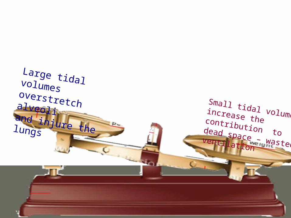
Large tidal volumes overstretch alveoli and injure the lungs Small tidal volumes increase the contribution to dead space – wasted ventilation
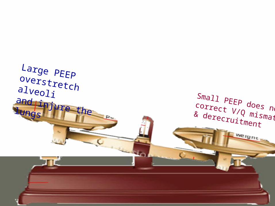
Large PEEP overstretch alveoli and injure the lungsSmall PEEP does not correct V/Q mismatch & derecruitment
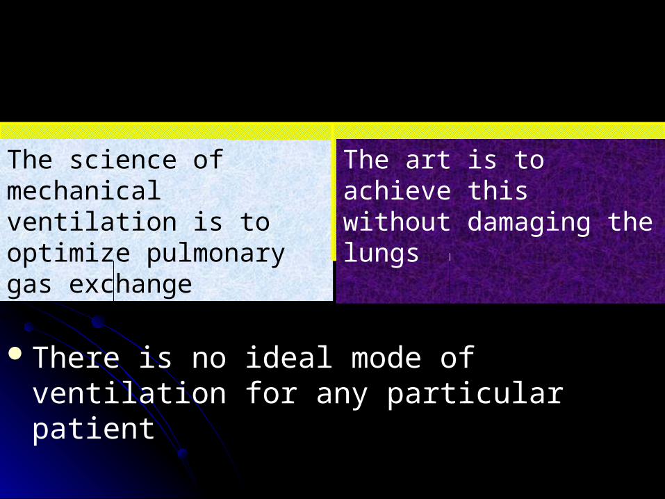
There is no ideal mode of ventilation for any particular patient
The science of mechanical ventilation is to optimize pulmonary gas exchange
The art is to achieve this without damaging the lungs.

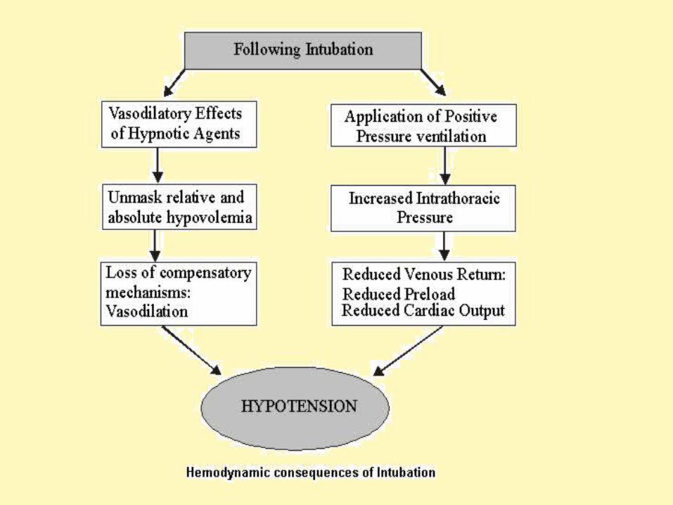
Major immediate complication1. Hypotension
due to vasodilatory effects of hypnotic drugsTreated with vasoconstrictors
have an ampule of phenylephrine (a selective alpha adrenoceptor agonist) at hand to reverse vasodilatory hypotension
Increase in intrathoracic pressure Treated with fluid boluses
Always have an intravenous fluid drip running and be prepared to run in a liter or more of fluid quickly
2. Barotrauma Pneumothoraxsubcut emphysema
3. VALI (Ventilator Associated Lung Injury)
4. O2 toxicity 5. From prolonged immobility and inability to
eat normallyvenous thromboembolic diseaseskin breakdown atelectasis
Late complications
6. From endotracheal intubationventilator-associated pneumonia (VAP) tracheal stenosis vocal cord injury tracheal-esophageal or tracheal-vascular
fistula
Measures to reduce complicationsElevating the head of the bed to > 30°
decreases risk of ventilator-associated pneumonia
routine turning of patient every 2 h decreases the risk of skin breakdown
Keep the PEEP & TV in optimal rangeAll patients receiving mechanical ventilation
should receive deep venous thrombosis prophylaxis
Some special techniques
1. Inhaled nitric oxidevery short-acting pulmonary vasodilator
Delivered to the airway in concentrations of between 1 and 20 parts per million
Improves blood flow to ventilated alveoli, thus improving V/Q mismatch, Oxygenation can be improved markedly
benefit only lasts for 48 hours and outcome is not improved
2. techniques to reduce the high inflation pressures resulting from the stiff lungs (low compliance)
1. Low tidal volumes to reduce inflation pressures
(6 ml/kg ideal body weight compared to 12 ml/kg) reduces mortality Minute ventilation reducedPaCO2 rises – permissive hypercapnia

3. Inverse ratio ventilation : • may improve oxygenation• PCO2 may rise further
4. Prone positioning: • improves oxygenation in ~70% of patients
with ARDS