Embed Size (px)
DESCRIPTION
FINAL for Practicum Health Science II
Citation preview
A PROFESSIONAL PROJECT SUBMISSION FOR PARTIAL COMPLETION OF PRACTICUM IN HEALTH SCIENCE II
RESPIRATORY PROJECT FINAL
KELECHI ONWUMERE AND ANTHONY TUCARL WUNSCHE SENIOR HIGH SCHOOL
MEDICAL TOWERD’HANIA MILLER
1st/2nd and 4th/5th
SPRING SEMESTER2012-2013
Vocabulary PageEpiglottis- a piece of tissue covering the top of the larynx that prevents food and liquid from entering the respiratory tract.Trachea- a tube extending from the larynx to the bronchi (windpipe)Bronchi- the two main branches which lead from the trachea to the lungs, providing a passageway for air.Bronchioles- smaller branches or subdivisions of the bronchiAlveoli- small grape-like sacs in the lungs found at the end of the bronchioles where the air meets the capillaries.Pulmono- one of the two cone shaped, spongy organs of the respiratory system contained within the pleural cavity of the thorax. Contains bronchi, bronchioles, alveoli, and capillaries.Thoracic cavity- the part of the body between the base of the neck and the diaphragm.Pleura- a membrane that enfold the lungs and is moistened with a secretion that reduces friction during respiration movements.Larynx- (voicebox) triangular chamber below the pharynx; within the larynx are vocal cords (glottis)Sputum- an abnormal, thick fluid expelled from deep within the lungs by coughing. May contain cellular debris, blood, pus, and microoragnisms.
A&P Page: Respiratory System
Respiration: The process where exchange gases, especially oxygen and carbon dioxide, with the environment. In air-breathing vertebrates, respiration takes place in the lungs. Respiration is also the diffusion of oxygen from alveoli to blood and of carbon dioxide from blood to alveoli; and transport of oxygen to and carbon dioxide from body cells.The respiratory system consists of the nose/nasal cavity, mouth (oral cavity), pharynx (throat), larynx (voice box), trachea (windpipe), bronchi & bronchioles, lungs, alveoli, and diaphragm.The nose/nostrils/nasal cavity brings air into the nose, where the air is warmed and humidified. The air must be humidified because it can be damaging to the respiratory tract otherwise. Tiny little hairs called Cilia filters out dust and other particles in the air and protect the nasal passage and other regions of the respiratory tract. It is also the preferred entrance for the outside air into the respiratory system. Mouth (oral cavity) is also a point of entrance of air into the respiratory system, and is used alternatively to the nasal cavity to intake air by people that have a mouth-breathing habit or whose nasal passage is obstructed. The pharynx (throat) collects the incoming air from the nasal/oral cavity and passes it downward to the trachea (windpipe) to further the process of respiration. The pharynx consists of 3 subsections: nasopharynx, oropharynx, and the laryngopharynx. The larynx (voice box) is the part of the respiratory system that contains the vocal cords. It is the place where air being breathed in and out creates voice sounds because of the air passing through. This part of the respiratory system is what makes speech and vocal communication possible. The trachea (windpipe) is the passage leading from the pharynx to the lungs. The trachea is what leads the air into each lung. From the trachea, it diverges into two main bronchi (tubes), one for each lung. The bronchi then subdivide into bronchioles. It lets the air have direction to the lungs.The lungs are what house the air once inside the body, and hold the bronchioles & alveoli that actually allow the exchange between oxygen and carbon dioxide to occur. The lungs hold the air during the exchanged called respiration. The alveoli are the actual site of where the gas exchange of carbon dioxide and oxygen are exchanged during respiration. The diaphragm is a muscular structure between the thoracic and abdominal cavity. Contraction of the diaphragm causes the chest or thorax cavity to expand, which occur during inhalation. During exhalation, the release of the diaphragm causes the chest or thorax cavity to contract. The diaphragm aides in breathing, moving the intercostals, and allowing the lungs to expand and contract during respiration.
Disease PagesAsthma
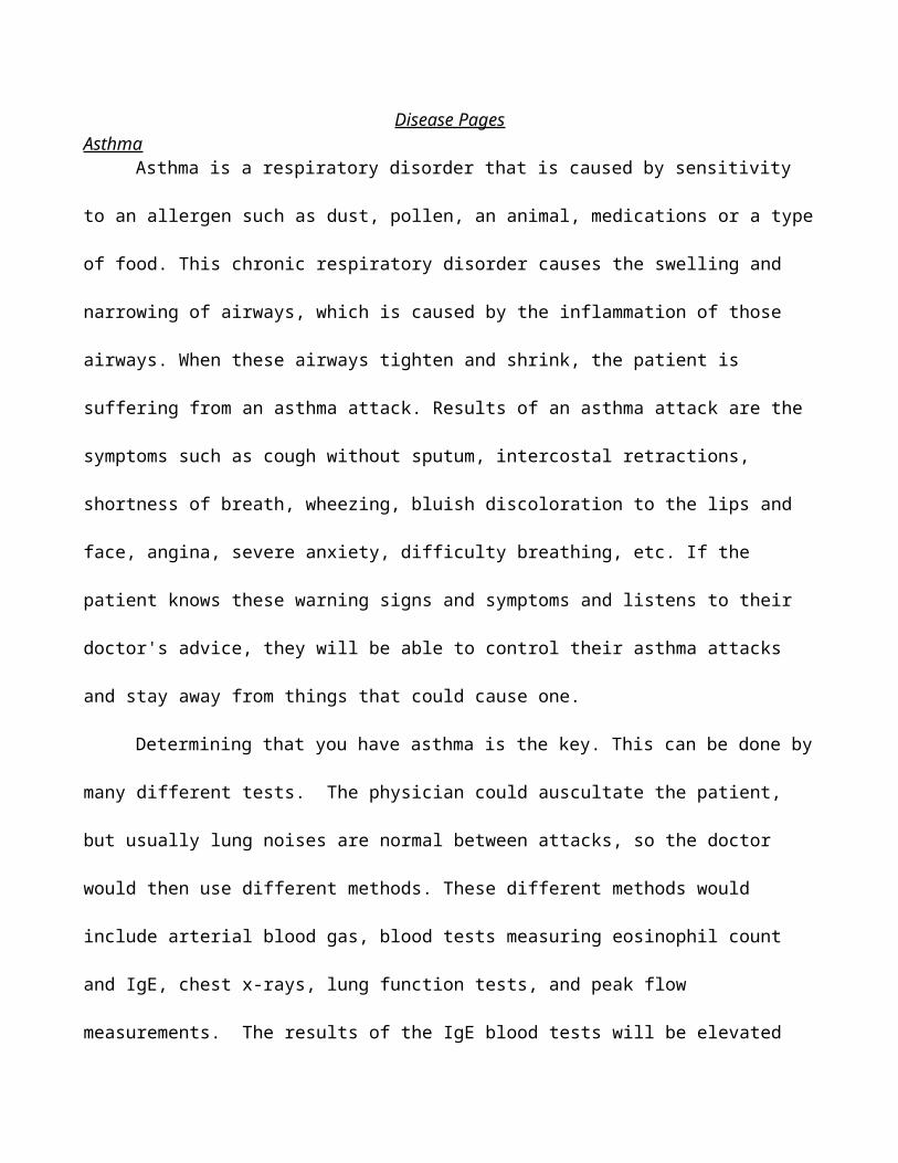
Asthma is a respiratory disorder that is caused by sensitivity to an allergen such as dust, pollen,
an animal, medications or a type of food. This chronic respiratory disorder causes the swelling and
narrowing of airways, which is caused by the inflammation of those airways. When these airways
tighten and shrink, the patient is suffering from an asthma attack. Results of an asthma attack are the
symptoms such as cough without sputum, intercostal retractions, shortness of breath, wheezing, bluish
discoloration to the lips and face, angina, severe anxiety, difficulty breathing, etc. If the patient knows
these warning signs and symptoms and listens to their doctor's advice, they will be able to control their
asthma attacks and stay away from things that could cause one.
Determining that you have asthma is the key. This can be done by many different tests. The
physician could auscultate the patient, but usually lung noises are normal between attacks, so the doctor
would then use different methods. These different methods would include arterial blood gas, blood tests
measuring eosinophil count and IgE, chest x-rays, lung function tests, and peak flow measurements.
The results of the IgE blood tests will be elevated from a normal patient, without asthma. According to
the Oxford Journals, “Eosinophils were shown to produce large amounts of the sulphidopeptide
leukotrienes (LTC4/D4 and E4) and platelet activating factor (PAF) which were thought to be involved
in causing bronchospasm in asthma” (pg. 986). The peak flow meter measures how quickly air can be
moved in and out of the lungs. MedlinePlus states that, “peak flow values of 50% - 80% of a specific
person's best results are a sign of a moderate asthma attack, while values below 50% are a sign of a
severe attack” (Web). A continual check up of the peak flow meter will allow for a general
understanding of the normal asthmatic levels and when they have changed and become lesser, which
will be how the patient can evaluate when they are reaching an attack.
The treatment of asthma is to control the asthma, it’s swelling and it’s triggers. Allergy testing is
what doctors use to test what allergen causes your reaction. There are different types of treatment for
asthma, the long-term control that is meant to be preventative, and the quick relief treatment that
controls asthma attacks and symptoms. The long-term controls are inhaled steroids to prevent swelling
of the airways, long-acting beta-agonist inhalers that prevent symptoms, omalizumab, leukotriene
inhibitors, and cromolyn sodium or nedocromil sodium. The quick relief controllers are short-acting
bronchodilators (inhalers) and oral corticosteroids prescribed by your physician. The best way to treat
asthma is to prevent it and know how to deal with it. When at home, knowing the symptoms to look for
and what triggers your attacks will help keep the asthma at bay.
Even though there is no cure for asthma, it is a disorder that can be handled and not hinder a
generally normal lifestyle. As long as the patient takes proper precautions to reduce risks of asthma
attacks and maintain the medications necessary, the patient will lead a normal life.
Chronic Obstructive Pulmonary Disease
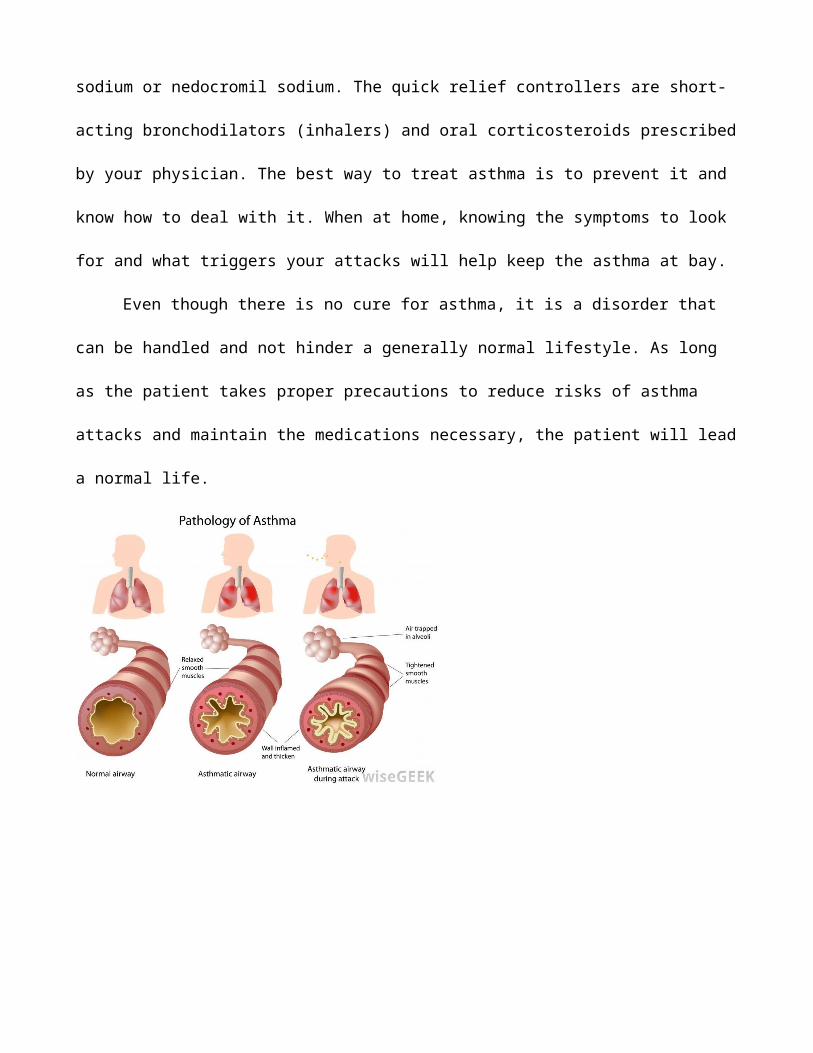
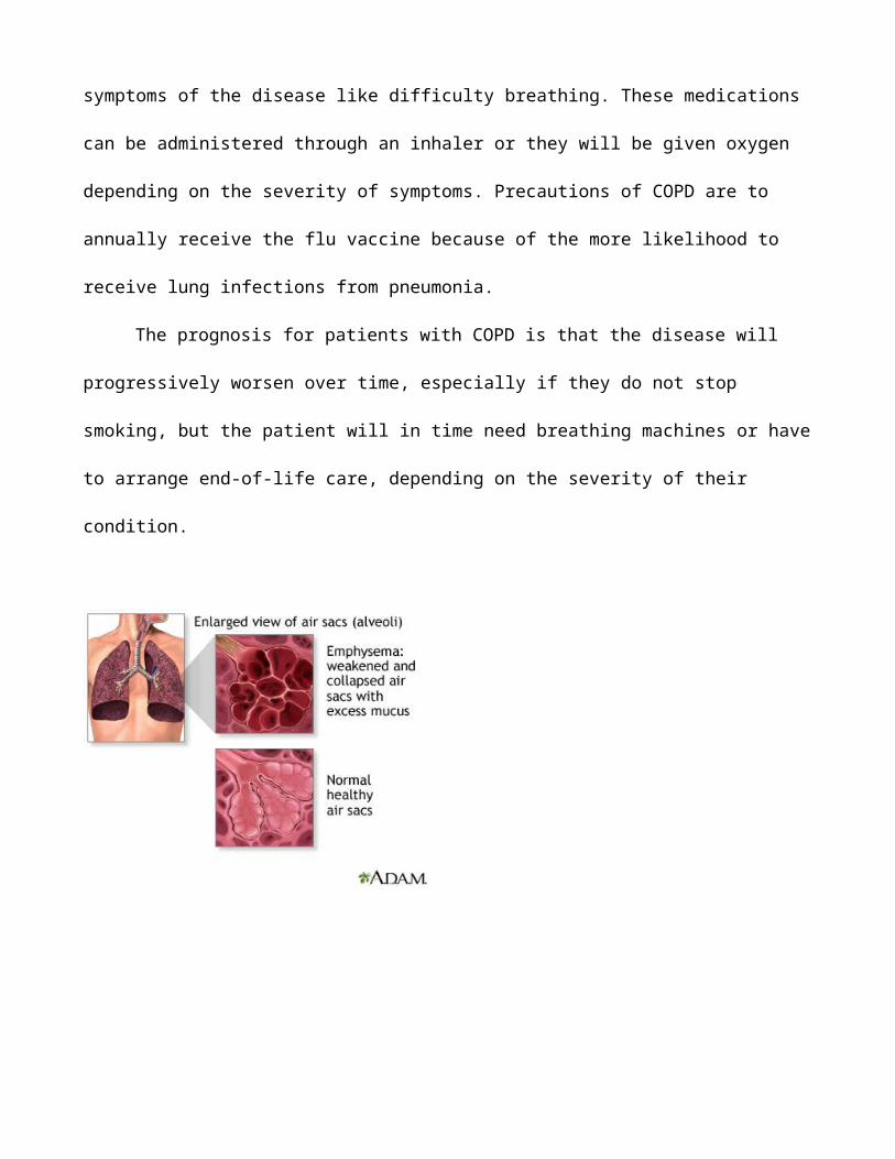
Chronic obstructive pulmonary disease is a group of diseases that is a result of an obstruction to
the airways and breathing related problems. The causes of COPD are tobacco smoke, exposure to air
pollutants, chemical fumes, or dust, genetic factors, and respiratory infections. On the onset of the
disease there will be mild or no symptoms, but as
it gets progressively worse, there will be more severe symptoms. These symptoms include a mucous
productive cough (usually called “smoker's cough”), shortness of breath, wheezing, and tightness of the
chest.
COPD is seen most in people 65-74 years old, non-Hispanic whites, women, those with a
history of asthma, current or former smokers, people with lower incomes, individuals who were
divorced, widowed, or separated, individuals with less than a high school education, and those who
were unemployed, retired, or unable to work.
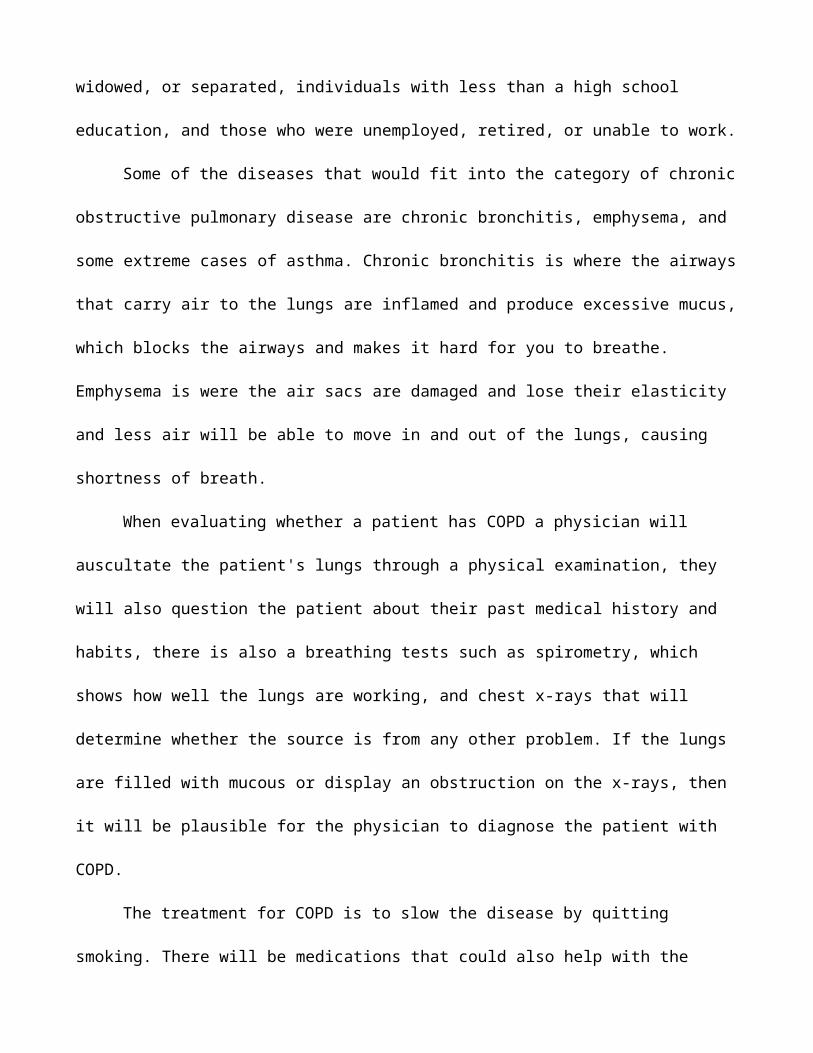
Some of the diseases that would fit into the category of chronic obstructive pulmonary disease
are chronic bronchitis, emphysema, and some extreme cases of asthma. Chronic bronchitis is where the
airways that carry air to the lungs are inflamed and produce excessive mucus, which blocks the airways
and makes it hard for you to breathe. Emphysema is were the air sacs are damaged and lose their
elasticity and less air will be able to move in and out of the lungs, causing shortness of breath.
When evaluating whether a patient has COPD a physician will auscultate the patient's lungs
through a physical examination, they will also question the patient about their past medical history and
habits, there is also a breathing tests such as spirometry, which shows how well the lungs are working,
and chest x-rays that will determine whether the source is from any other problem. If the lungs are
filled with mucous or display an obstruction on the x-rays, then it will be plausible for the physician to
diagnose the patient with COPD.
The treatment for COPD is to slow the disease by quitting smoking. There will be medications
that could also help with the symptoms of the disease like difficulty breathing. These medications can
be administered through an inhaler or they will be given oxygen depending on the severity of
symptoms. Precautions of COPD are to annually receive the flu vaccine because of the more likelihood
to receive lung infections from pneumonia.
The prognosis for patients with COPD is that the disease will progressively worsen over time,
especially if they do not stop smoking, but the patient will in time need breathing machines or have to
arrange end-of-life care, depending on the severity of their condition.
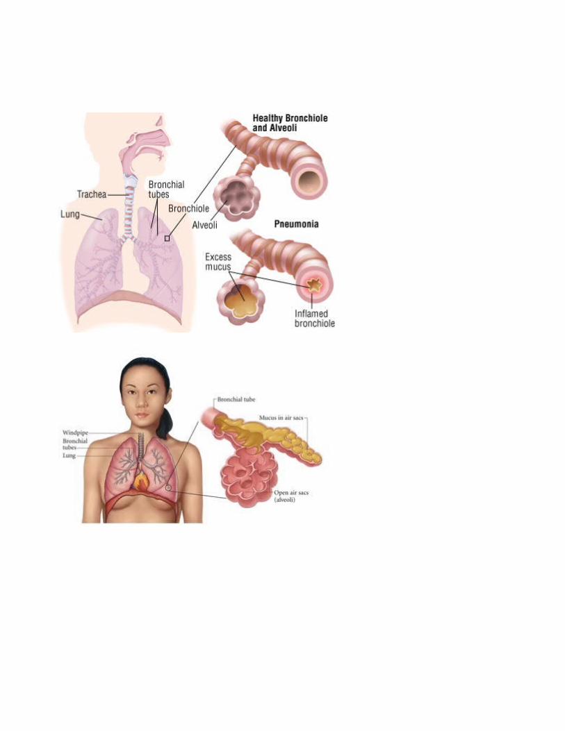
Pneumonia
Pneumonia is an infection or inflammation of the lungs by a buildup of fluid in the alveoli. This
infection is a common illness that affects many people in our country year to year. Pneumonia is caused
by bacteria, viruses, and fungi. The weaker your immune system the easier it is to get pneumonia. Long
term diseases such as asthma, cancer, heart disease, or diabetes make it more likely to get pneumonia.
The symptoms of pneumonia are a cough, fever, rapid breathing, shaking, angina, weakness,
nausea and vomiting, and diarrhea. The mild symptoms of pneumonia are called “walking pneumonia.”
Other symptoms of pneumonia like confusion, clammy skin, headaches, loss of appetite, fatigue, and
stabbing chest pains. The tests required to see if the patient has pneumonia are arterial blood gases,
white blood cell count, CT scan of the chest, culture of the sputum, pleural fluid culture. When you are
looking at the CBC white blood cell count will be increased from the normal levels. The CT chest scan
will display the mucous and fluid in the chest cavity, which should not be present. The culture of the
sputum will show the type of bacteria or virus causing the symptoms. The arterial blood gases display
the amounts of oxygen getting into your blood from the lungs, if the pneumonia is present this will be
low because the patient will not be getting enough air because of their shortness of breath.
When treating pneumonia, the physician must determine whether the cause is from a bacterial,
viral or fungal infection. After determining this the patient will be given antibiotics, antiviral
medications, and fever reducers and cough medicine which will treat the symptoms and whatever that
has weakened the immune system.
The prognosis of this disorder is that the patient will improve significantly within the next two
weeks. This may not be the case with the elderly, patients with autoimmune diseases, and young
children, in those cases it may take longer to heal.
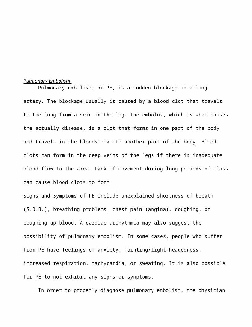
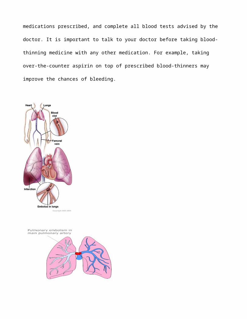
Pulmonary Embolism Pulmonary embolism, or PE, is a sudden blockage in a lung artery. The blockage usually is
caused by a blood clot that travels to the lung from a vein in the leg. The embolus, which is what
causes the actually disease, is a clot that forms in one part of the body and travels in the bloodstream to
another part of the body. Blood clots can form in the deep veins of the legs if there is inadequate blood
flow to the area. Lack of movement during long periods of class can cause blood clots to form.
Signs and Symptoms of PE include unexplained shortness of breath (S.O.B.), breathing problems, chest
pain (angina), coughing, or coughing up blood. A cardiac arrhythmia may also suggest the possibility of
pulmonary embolism. In some cases, people who suffer from PE have feelings of anxiety,
fainting/light-headedness, increased respiration, tachycardia, or sweating. It is also possible for PE to
not exhibit any signs or symptoms.
In order to properly diagnose pulmonary embolism, the physician will ask about past medical
history to figure out DVT and PE risk factors, the likelihood, and eliminate other causes for the
exhibited symptoms. The doctor will conduct a physical exam as well to assess legs for DVT, check
blood pressure, as well as the heart and lungs. Possible diagnostic test that can be ordered to help
diagnose PE include ultrasound, computed tomography scans, lung ventilation/perfusion scan,
pulmonary angiography, blood tests, echocardiography, electrocardiogram (EKG), chest x-ray, or chest
MRI. Normal findings would be a normal blood pressure, heart sounds, and no blockage shown in
scans/x-rays. What would be found with a person that is affected by PE is an increase in blood
pressure, tachycardia, abnormal heart sounds, and maybe even visual imaging of the embolus from
CT/MRI scan.
Treatment must be administered as soon as possible to prevent serious problems or even death.
Medications that can be taken to treat pulmonary embolism are anticoagulants and thrombolytic (clot
dissolvers). In some severe cases, physicians may suggest a clot removal, vein filter, or surgery in
general to remove the embolus.
The prognosis or outlook on a patient suffering from PE will usually be treated in a hospital, but
upon discharge, will need to take medicine at home for 6 months or longer. It is vital to take all
medications prescribed, and complete all blood tests advised by the doctor. It is important to talk to
your doctor before taking blood-thinning medicine with any other medication. For example, taking
over-the-counter aspirin on top of prescribed blood-thinners may improve the chances of bleeding.
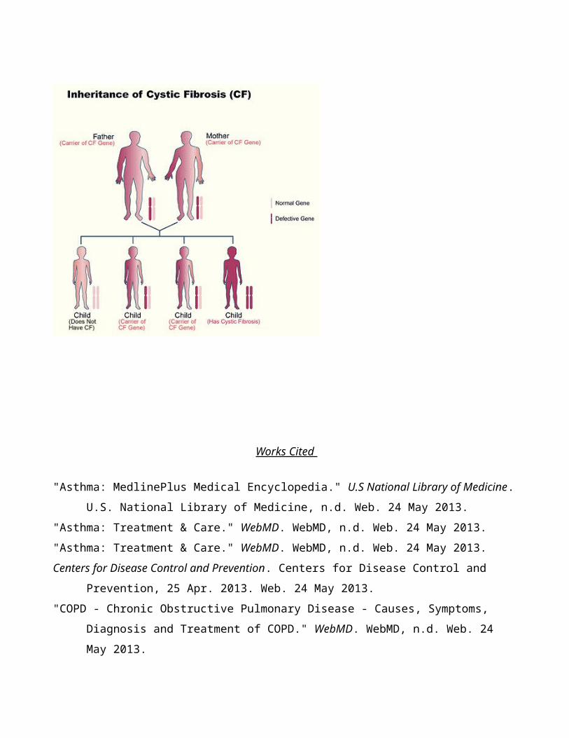
Cystic FibrosisCystic fibrosis (CF) is an inherited disease of the mucus and sweat glands. “Inherited” means
that the disease is passed from parents to children through genes. CF mainly affects the lungs, pancreas,
liver, intestines, sinuses, and sex organs (pretty much glands that secrete). CF causes your mucus to
become thick and sticky. There is mucus build up in your lungs and block out your airways. This can
lead to bacterial infection. This leads to serious lung infections, and over time will cause severe damage
to your lungs.
Signs and symptoms of cystic fibrosis vary from individuals over time. At times, you will have
few symptoms, but other times, the ailments and symptoms will become more severe. One of the first
signs that parents might notice is that their baby’s skin tastes salty when kissed, or the infant does not
have bowel movements when first born. Most of the other signs and symptoms of CF happen later.
They’re related to how CF affects the respiratory, digestive, and/or reproductive system of the body.
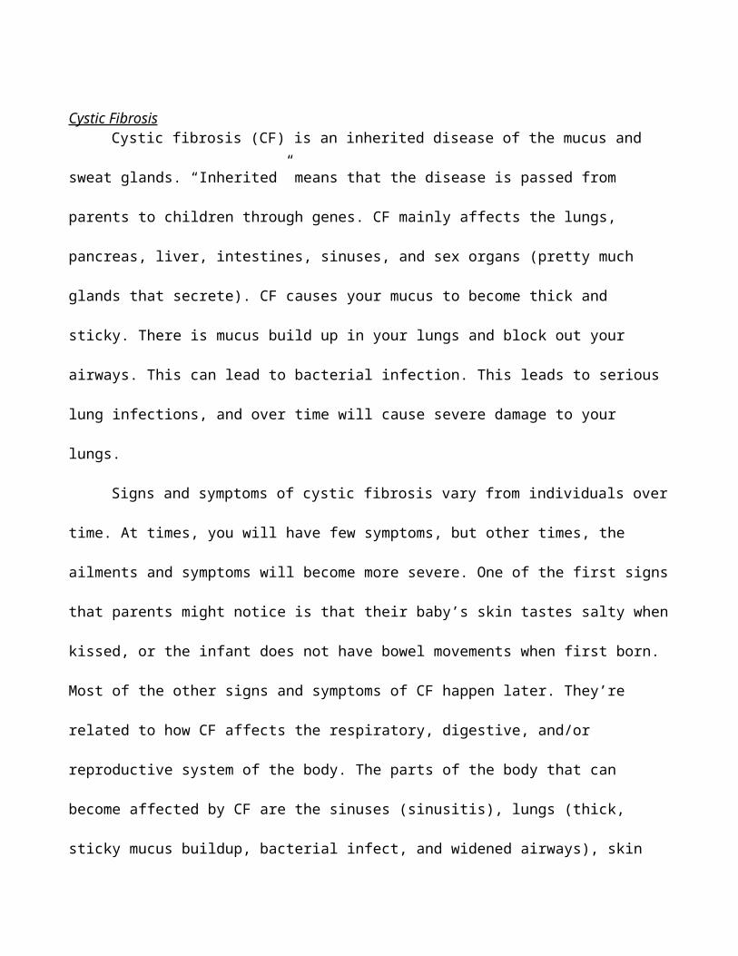
The parts of the body that can become affected by CF are the sinuses (sinusitis), lungs (thick, sticky
mucus buildup, bacterial infect, and widened airways), skin (sweat glands produce salty sweat), liver
(blocked biliary ducts), pancreas (blocked pancreatic ducts), intestines (inability to fully absorb
nutrients), and reproductive (male & female complications).
Diagnostic tests that can be used to diagnose cystic fibrosis are newborn screening, sweat test,
prenatal screening, cystic fibrosis carrier testing, genetics test, chest x-ray, sinus x-ray, lung function
tests, and sputum culture. In a newborn screening, either a genetic test or a blood test will be used to
find CF. the genetic test shows whether a newborn has faulty CFTR genes. If the genetic test comes up
negative, the newborn doesn’t have CF. The blood test shows whether a newborn’s pancreas is
working properly. If the pancreas is not working, the chances of CF are higher. If a genetic test or blood
test suggests cystic fibrosis, the physician will attempt to confirm the diagnosis using a sweat test,
where it measures the amount of salt concentration in sweat. If it comes up with a normal salt level,
then it isn’t likely that it is cystic fibrosis. Prenatal screening can show if your fetus has cystic fibrosis.
These tests include amniocentesis and chorionic villus sampling. Amniocentesis withdraws a small
amount of fluid from the sac around the baby to test to see whether both of the baby’s CTFR genes are
normal, or suggest the possibility of cystic fibrosis if abnormal. CVS takes a tissue sample from the
placenta, and runs tests to see whether the baby has CF or not. The cystic fibrosis carrier test takes a
blood or saliva sample to see if an individual has a faulty CF gene. If the test is negative, the individual
is not a carrier of CF. A chest x-ray creates pictures of the structures in your chest, and can show if your
lungs are inflamed or scarred, or if they trap air. If there aren’t any observations of such things, CF is
not likely as a cause of other symptoms. A sinus x-ray will show signs of sinusitis, a complication of
CF. If there is not a diagnosis of sinusitis, and CF isn’t as likely. Lung function test measures how
much air you can breathe in and out, how fast you can breathe air out, and how well your lungs deliver
oxygen to your blood. Normal lung functions will go against the initial suspect of cystic fibrosis. A
sputum culture will take a sample of your spit to see what kind of bacteria are growing in it. If you have
mucoid Pseudomonas, you may have more advanced CF that needs aggressive treatment.
Possible treatment options include treatment from a cystic fibrosis specialist, treatment for lung
problems, chest physical therapy, and exercise, prescribed medication, treatment for advanced lung
disease, pulmonary rehabilitation, treatment for digestive problems, and treatment for cystic fibrosis
complications. Cystic fibrosis has no cure, but all treatments have an aim to improve the quality of life
for the patient.
Prognosis for a patient suffering from cystic fibrosis is to have ongoing care from health care
professionals to keep them updated with conditions of the patient. The patient must have a good
transition to attain proper age-treatment against CF. Between medical checkups, practicing good self-
care and following a healthy lifestyle are vital to improving the quality of life. CF requires daily care;
most people who have the disease can go on to lead normal lives. Adults with CF may have difficulty
concerning fertility.
Works Cited
"Asthma: MedlinePlus Medical Encyclopedia." U.S National Library of Medicine. U.S. National Li-
brary of Medicine, n.d. Web. 24 May 2013.
"Asthma: Treatment & Care." WebMD. WebMD, n.d. Web. 24 May 2013.
"Asthma: Treatment & Care." WebMD. WebMD, n.d. Web. 24 May 2013.
Centers for Disease Control and Prevention. Centers for Disease Control and Prevention, 25 Apr.
2013. Web. 24 May 2013.
"COPD - Chronic Obstructive Pulmonary Disease - Causes, Symptoms, Diagnosis and Treatment of
COPD." WebMD. WebMD, n.d. Web. 24 May 2013.
"COPD (Chronic Obstructive Pulmonary Disease): MedlinePlus." U.S National Library of Medicine.
U.S. National Library of Medicine, n.d. Web. 24 May 2013.
"Cystic Fibrosis: MedlinePlus." U.S National Library of Medicine. U.S. National Library of Medicine,
n.d. Web. 20 May 2013. <http://www.nlm.nih.gov/medlineplus/cysticfibrosis.html>.
"Pneumonia - Adults (community Acquired): MedlinePlus Medical Encyclopedia." U.S National Li-
brary of Medicine. U.S. National Library of Medicine, n.d. Web. 24 May 2013.
"Pneumonia Symptoms, Causes, Treatments & More." WebMD. WebMD, n.d. Web. 24 May 2013.
"Pulmonary Embolism: MedlinePlus." U.S National Library of Medicine. U.S. National Library of
Medicine, n.d. Web. 20 May 2013. <http://www.nlm.nih.gov/medlineplus/pulmonaryembolis-
m.html>.
"The Respiratory System." Lung.ca. N.p., n.d. Web. 20 May 2013. <https://www.lung.ca/children/
grades7_12/respiratory/respiratory_system.html>.
"The Results List of Search Bronchial Asthma Disease." Bronchial Asthma Disease. N.p., n.d. Web. 24
May 2013.
Staff, Mayo Clinic. "Cystic Fibrosis." Mayo Clinic. Mayo Foundation for Medical Education and Re-
search, 13 June 2012. Web. 20 May 2013. <http://www.mayoclinic.com/health/cystic-
fibrosis/DS00287>.
Staff, Mayo Clinic. "Definition." Mayo Clinic. Mayo Foundation for Medical Education and Research,
21 May 2013. Web. 24 May 2013.
Staff, Mayo Clinic. "Pulmonary Embolism." Mayo Clinic. Mayo Foundation for Medical Education
and Research, 19 Jan. 2013. Web. 20 May 2013.
Advertisement
Mind Map
Organs & Structures
Nose- The nose is the primary upper respiratory organ in which air enters into and exits from the body.
Cilia and mucus line the nasal cavity and traps bacteria and foreign particles that enter in through the
nose. In addition, air that passes through the nasal cavity is humidified and moistened. The nasal
septum divides the nose into two narrow nasal cavities: one area is responsible for smell and the other
area is responsible for respiration
Pharynx- Besides the nose, air can enter into the lungs through the mouth. The pharynx is a tubular
structure, positioned behind the oral and nasal cavities, that allows air to pass from the mouth to the
lungs. The pharynx contains three parts: The nasopharynx, which connects the upper part of the throat
with the nasal cavity; the oropharynx, positioned between the top of the epiglottis and the soft palate;
and the laryngopharynx, located below the epiglottis.
Larynx- From the pharynx, air enters into the larynx, commonly called the voice box. The larynx is part
of the upper respiratory tract that has two main functions: a passageway for air to enter into the lungs,
and a source of vocalization. The larynx is made up of the hyoid bone and cartilage, which helps
regulate the flow of air. The epiglottis is a flap-like cartilage structure contained in the larynx that
protects the trachea against food aspiration.
Bronchi- The bronchi allow the passage of air to the lungs. The trachea is made of c-shaped ringed
cartilage that divides into the right and left bronchus. The right main bronchus is shorter and wider than
the left main bronchus. The right bronchus is subdivided into three lobar bronchi, while the left one is
divided into two lobar bronchi.
Lungs- The lungs are spongy, air-filled organs located on both sides of the chest cavity. The left lung is
divided into a superior and inferior lobe, and the right lung is subdivided into a superior, middle, and
inferior lobe. Pleura, a thin layer of tissue, line the lungs to allow the lungs to expand and contract with
ease. Respiration is the primary function of the lungs, which includes the transfer of oxygen found in

the atmosphere into the blood stream and the release of carbon dioxide into the air.
Alveoli- The average adult has about 600 million alveoli, which are tiny grape-like sacs at the end of
the respiratory tree. The exchange of oxygen and carbon dioxide gases occurs at the alveolar level.
Although effort is required to inflate the alveoli (similar to blowing up a balloon), minimal effort is
needed to deflate the alveoli (similar to the deflating of a balloon).
Diaphragm- The diaphragm is a muscular structure located between the thoracic and abdominal cavity.
Contraction of the diaphragm causes the chest or thorax cavity to expand, which occurs during
inhalation. During exhalation, the release of the diaphragm causes the chest or thorax cavity to contract.
Symptoms
Typical symptoms seen in the respiratory system is shortness of breath, coughing, and wheezing.
Treatment
Some treatments that can be used to treat respiratory ailments are bronchodilators, inhalers, anti-
histamines, and decongestants.
Relationship to Other Body Systems
The respiratory system is one of the most key body systems because if there is not sufficient oxygen
flow to the rest of the other systems, then there won’t be adequate function and response from the other
parts of the body. The respiratory system works hand in hand with the cardiovascular system, as the
cardiovascular system helps to transport the oxygen collected from the environment through the
respiratory system to the rest of the body through RBC’s.
WebQuest
1. What causes asthma?2. Which diagnostic test is necessary to diagnose asthma?3. Name 3 symptoms of asthma.4. Is asthma a fatal disease? Why or why not?5. What daily changes in their life must an asthmatic take?
Answers
1. Allergens are usually the cause of asthma and varies from person to person2. Chest x-ray and allergy tests3. Coughing, wheezing, and shortness of breath4. Asthma is something that people can continue on with a normal life, as long as it is
controlled. Though it is a chronic disease, it is not usually fatal unless there is a pre-existing condition present.
5. The patient must try not to exacerbate their condition and know their signs and symptoms and take proper precautions.
http://www.webmd.com/asthma/guide/asthma-overview-facts
1. What does COPD stand for?2. What is COPD?3. What most common conditions make up COPD?4. Name 3 symptoms of COPD?5. What is a major cause of COPD?
Answers
1. Chronic obstructive pulmonary disease2. A group of diseases that block airflow and make breathing difficult.3. Emphysema and chronic bronchitis4. Shortness of breath, wheezing, chest tightness5. Smoking
http://www.mayoclinic.com/health/copd/DS00916
1. What is the definition of pneumonia?2. What types of infection are pneumonia?3. What is the prognosis of pneumonia?4. What types of people are at highest risk of pneumonia?5. Name 3 treatment options for pneumonia.
Answers
1. It is an infection that inflames the air sacs in one or both lungs.2. Bacterial, viral, and fungal.3. Pneumonia ranges in seriousness from mild to life-threatening. It depends on the severity.4. Infants and children younger than 2 yo and people older than 65 yo5. Antibiotics, fever reducers, cough medicine
http://www.mayoclinic.com/health/pneumonia/DS00135
1. What is a pulmonary embolism?2. Can a pulmonary embolism hit anyone?
3. What puts you at risk of a pulmonary embolism?4. What causes a pulmonary embolism?5. How can you prevent a pulmonary embolism?
Answers
1. A blockage in one or more arteries in your lungs.2. Yes. A pulmonary embolism can occur in otherwise healthy people.3. Prolonged immobility, Age, Family History, Surgery, Etc.4. A pulmonary embolism occurs when a clump of material, most of a blood clot, gets wedged
into an artery in your lungs.5. You can stay moving and exercise. In the hospital you will be given anticoagulant therapy,
graduated compression stockings, and use pneumatic compression.http://www.mayoclinic.com/health/pulmonary-embolism/DS00429
1. What is the definition of cystic fibrosis?2. What is the cause of cystic fibrosis?3. Is cystic fibrosis a congenital disease?4. In what ethnicities is cystic fibrosis most prevalent in?5. Name 2 complications when it comes to cystic fibrosis.
Answers
1. Cystic fibrosis is a life-threatening disorder that causes severe damage to the lungs and digestive system.
2. Cystic fibrosis is a defect in a gene that changes a protein that regulates the movement of salt in and out of cells. This creates a thick, sticky mucus in the respiratory, digestive, and reproductive systems and an increase of salt in sweat.
3. Yes. It is inherited by a parent/ family member.4. Cystic fibrosis is most common in white people of Northern European ancestry, but also in
Hispanics, African-Americans and some Native Americans. It is rare in Asians and Middle Easterners.
5. Bronchiectasis, Chronic infections, Nasal polyps, etc.http://www.mayoclinic.com/health/cystic-fibrosis/DS00287
Case Study
1. Respiratory
A. A young man has chest pain, dough, difficulty breathing, and a fever. Obviously these could be caused by a variety
of diseases. What diagnostic procedures would be helpful in diagnosing the man’s disease? Two diagnostic procedures that
could aide in diagnosing this man are to conduct a CBC (complete blood count) test to see if he has a possible infection or
not, with an increased white blood cell count, and chest x-ray to visually have an image to see if there is any inflammation
or obstruction in his chest region.
B. At age 62, Harry decided to retire from the grain mill where he had worked for 40 years. He was walking much
slower and felt weaker even though he was walking less. He had enjoyed his smoke breaks and lunches with the guys, but
now he had difficulty getting his breath, while his breathing rate had increased and his chest seemed inflated. What disease
seems apparent here, and how may it be addressed? Harry sounds like he has emphysema due to continual smoking and
exposure to the grain mills and its fumes. Emphysema is a group of Chronic Obstruction Pulmonary Disease (COPD). To
address his problem, that is progressively getting worse, Harry must seek medical attention immediately. At the hospital he
will be diagnosed based on pulmonary function tests, chest x-rays, CT scans, and arterial blood gas analysis. The treatment
Harry will need is a smoking cessation, medications such as bronchodilators, inhaled steroids, and antibiotics. This
treatment will not reverse the effects of the emphysema, but it will slow down the progression.
C. Sara love to run. She watched the rack meets at school even as a first grader. An asthmatic, she occasionally had to
use an inhaler, but seemed determined to be an athlete. Should Sara be discouraged from pursing her goal? Why or why
not? Sara should not be discouraged because as she grew up just occasionally using an inhaler isn't that bad. Her condition
is not that bad to prevent her from leading a normal life, and going for whatever she wants to do. She loves to run, so
whatever obstacles come in her life should never divert her eyes from her goal to be an athlete. Whether it is being asthmatic
or having a broken leg, it takes hard work and dedication to attain your dreams. Being slightly asthmatic shouldn't hold Sara
back. She should just always take the proper precautions while running, as she does not want to put herself at any
unnecessary risks.