Embed Size (px)
Citation preview

Respiratory Physiologyin Pregnancy
Matthew J. Hegewald, MDa,b,*, Robert O. Crapo, MDcKEYWORDS
� Respiratory physiology � Pregnancy� Pulmonary function testing
This review discusses respiratory physiologicchanges during normal pregnancy. Cardiovascularphysiology is also reviewed, given the importantinteractions between the respiratory and cardio-vascular systems in pregnancy. The combinationof hormonal changes, mechanical effects of theenlarging uterus, and marked circulatory changesresult in significant changes in pulmonary andcardiovascular physiology. These adaptations arenecessary to meet the increased metabolicdemands of the mother and fetus. It is importantfor the clinician to be familiar with the normal phys-iologic changes in pregnancy. Understandingthese changes is critical in distinguishing thecommon dyspnea that occurs during normal preg-nancy from pathophysiologic states associatedwith cardiopulmonary diseases seen inpregnancy,1 and in anticipating disease worseningin pregnancy and the peripartum period in thosewomen with cardiopulmonary disorders.
UPPER AIRWAY CHANGES IN PREGNANCY
There are significant changes to the mucosa of thenasopharynx and oropharynx during pregnancy.The mucosal changes in the upper airway includehyperemia, edema, leakage of plasma into thestroma, glandular hypersecretion, increasedphagocytic activity, and increased mucopolysac-charide content.2,3 All of these result in nasalcongestion often called rhinitis of pregnancy.The clinical definition of rhinitis of pregnancy is
a Division of Pulmonary and Critical Care Medicine, InteSouth Cottonwood Street, UT 84157, USAb Pulmonary Function Laboratory, Intermountain Medica84157, USAc Division of Pulmonary and Critical Care Medicine, LDS HSalt Lake City, UT 84103, USA* Corresponding author. Pulmonary Department, Interm84157.E-mail address: [email protected]
Clin Chest Med 32 (2011) 1–13doi:10.1016/j.ccm.2010.11.0010272-5231/11/$ – see front matter � 2011 Elsevier Inc. All
“nasal congestion present during the last 6 ormore weeks of pregnancy without other signs ofrespiratory tract infection and with no knownallergic cause, disappearing completely within 2weeks after delivery.”2 The incidence of rhinitis ofpregnancy has been reported to be between18% and 42%.4–6 Nasal congestion is often notedearly in the first trimester, peaks late in pregnancy,and disappears within 48 hours of delivery.2,4 Theetiology of rhinitis of pregnancy is not clear,2
though increased blood volume and hormonalfactors likely play a role. Blood volume changesare discussed later. Hormonal factors includeestrogen and placental growth hormone. Nasalmucosal biopsies obtained during pregnancy andfrom women taking oral contraceptive medica-tions have implicated estrogen as a cause.3
However, other factors are likely involved, asestrogen levels are not higher in women whohave rhinitis of pregnancy than in women who donot.7 Placental growth hormone may contributeto rhinitis of pregnancy, as levels are significantlyhigher in patients with the syndrome.2 Nonhor-monal factors implicated in nasal congestion inpregnancy include smoking, nasal allergy, infec-tions, and chronic use of topical vasoconstrictormedication.
Rhinitis of pregnancy has the potential tocontribute to maternal-fetal complications. Nasalobstruction contributes to snoring and sleep-disordered breathing, both of which are associatedwith hypertension and preeclampsia and may
rmountain Medical Center, University of Utah, 5121
l Center, 5121 South Cottonwood Street, Murray, UT
ospital, University of Utah, 8th Avenue and C Street,
ountain Medical Center, PO Box 577000, Murray, UT
rights reserved. chestm
ed.th
eclinics.com

Hegewald & Crapo2
contribute to intrauterine growth retardation,although the relationship between sleep-disorderedbreathing and intrauterine growth retardation iscontroversial.2,8 Nasal congestion and resultantmouth breathing reduces concentrations of inhalednitric oxide, primarily produced in the maxillarysinuses.9 Nitric oxide is a potent mediator of pulmo-nary vascular tone, and reduced nitric oxide maycontribute to the complications associated withsnoring. The upper airway congestion and obstruc-tion common in pregnancy may adversely affect theability of air to pass through nasal and oral tubes.Mallampati score, a common predictor of airway
patency, has been shown to increase during thecourse of pregnancy.10 Neck circumference hasalso been found to be increasedwith pregnancy,11,12
and decreases in the postpartum period.11
Using acoustic reflectance measurements,oropharyngeal junction size is smaller in the seatedposition, and mean pharyngeal cross-sectionalarea is smaller in the supine, lateral, and seatedposition in pregnant women compared withnonpregnant controls.11 In addition, there wasa much larger drop in the size of the upper airwayon laying down in pregnant women in their thirdtrimester compared with nonpregnant controls inone study13 but not in another.11 Factors poten-tially affecting airway collapsibility in thesepatients include reduced lung volumes—leadingto less caudal traction on the upper airway14—and fat infiltration of the upper airway. Functionalresidual capacity is reduced in pregnancy in theupright position with an additional reductionoccurring in the supine position. Pregnant womenmay gain an average of 25 to 35 pounds (11–16 kg)during the course of pregnancy. However, in thestudy by Iczi and colleagues,13 the drop in airwaysize between the seated and the supine positionin the pregnant group did not appear to be relatedto body mass index, suggesting that other factorssuch as changes in functional residual capacity orchanges in the upper airway related to interstitialfluid may play a role. Mean pharyngeal cross-sectional area increases significantly postpartumcompared with intrapartum,11 but it is not clearwhen or whether these changes return to precon-ception size. The measurements discussed heremay be even more pronounced during sleep,with the loss of upper airway muscle dilation, butthis theory needs to be tested further.
CHEST WALL CHANGES IN PREGNANCY
The thoraxundergoessignificant structural changesin pregnancy: The subcostal angle of the rib cageand the circumference of the lower chest wallincrease and the diaphragm moves up.15–18 These
changes are necessary to accommodate theenlarging uterus and increasing maternal weight,but the changes occur early in pregnancy beforethe uterus is significantly enlarged.15,18 Hormonalchanges rather than the mechanical effects of theenlarginguteruscause relaxationof the ligamentousattachments of the lower ribs. Relaxin, the hormoneresponsible for relaxation of the pelvic ligaments,likely also causes relaxation of the lower rib-cageligaments.19 The subcostal angle progressivelywidens from 68.5� to 103.5� during pregnancy.18
The anterior-posterior and transverse diameters ofthe chest wall each increase by 2 cm, resulting inan increase of 5 to 7 cm in the circumference ofthe lower rib cage. The anatomic changes of thechest wall peak at week 37. The chest wall configu-ration normalizes by 24 weeks postpartum but thesubcostal angle remains about 20% wider thanthe baseline value.15 The enlarging uterus causesthe diaphragm to be displaced cephalad 4 cm inlate pregnancy, but the increase in chest wall sizemitigates any changes in lung volumes caused bythe upward displacement of the diaphragm. Theanatomic changes of the thorax with pregnancyare illustrated in Fig. 1.
RESPIRATORY MUSCLE FUNCTION
There is no significant change in respiratorymusclestrength during pregnancy despite the cephaladdisplacement of the diaphragm and changes inthe chest wall configuration. Maximal inspiratoryand expiratory mouth pressures and maximumtransdiaphragmatic pressure, measured as gastricpressure minus esophageal pressure, in late preg-nancy and after delivery show no significantchanges.15,20 Despite the upward displacementof the diaphragm by the gravid uterus, diaphragmexcursion actually increases by 2 cm comparedwith the nonpregnant state.18,20 Increased dia-phragmatic excursion and preserved respiratorymuscle strength are important adaptations, giventhe increase in tidal volume and minute ventilationthat accompanies pregnancy. Improved dia-phragm mechanics in pregnancy are explained byan increased area of apposition of the diaphragmto the rib cage.20
LUNG FUNCTION IN PREGNANCYStatic Lung Function
Static lung function stays the same in pregnancyexcept for decreases in functional residualcapacity (FRC) and its components: expiratoryreserve volume (ERV) and residual volume (RV).FRC depends on 2 opposing forces: the elasticrecoil of the lungs and the outward and downward

2cm
4cm
103.5°68.5°
5-7cm
Uterus(37 weeks)
Fig. 1. Chest wall changes that occur during pregnancy. The subcostal angle increases, as does the anterior-posterior and transverse diameters of the chest wall and the chest wall circumference. These changes compensatefor the 4-cm elevation of the diaphragm so that total lung capacity is not significantly reduced.
Respiratory Physiology in Pregnancy 3
pull of the chest wall and abdominal contents.A reduction in FRC in pregnancy is expectedgiven the 4-cm elevation of the diaphragm,decreased downward pull of the abdomen, andchanges in chest wall configuration that decreaseoutward recoil.17 As anticipated, chest wallcompliance is decreased antepartum comparedwith postpartum.21 Lung compliance is unaffectedby pregnancy.21,22
Several studies have measured serial static lungvolumes during pregnancy and after delivery.23–27
The changes in lung function in pregnancy areillustrated in Fig. 2. FRC decreases by approxi-mately 20% to 30% or 400 to 700 mL during preg-nancy. FRC is composed of ERV, which decreases15% to 20% or 200 to 300 mL, and RV, whichdecreases 20% to 25% or 200 to 400 mL. Signifi-cant reductions in FRC are noted at 6 months’gestation with a progressive decline as pregnancycontinues.23,28 At term, there is a further 25%
decrease in FRC in the supine position comparedwith sitting.26
Inspiratory capacity (IC), the maximum volumethat can be inhaled from FRC, increases by 5%to 10% or 200 to 350 mL during pregnancy. Totallung capacity (TLC), the combination of FRC andIC, is unchanged or decreases minimally (lessthan 5%) at term.
Lung volume measurements can be made usinginert gas techniques and by body plethysmo-graphy.29 In patients without lung disease the 2techniques produce similar results.30 Garcia-Rioand colleagues27 measured lung volumes byplethysmography and inert gas (helium dilution)techniques during pregnancy and postpartum. At36 weeks of pregnancy there were significantdifferences in lung volumes between the 2 tech-niques. FRC measured by body plethysmographywas decreased by 27% compared with post-partum whereas FRC measured by helium dilution

Fig. 2. Changes in lung volumes with pregnancy. The most significant changes are reductions in FRC and itssubcomponents ERV and RV, and increases in IC and VT.
Hegewald & Crapo4
was decreased 38% compared with postpartum.FRCwas larger by 18% or 350mLwhenmeasuredby plethysmography. The underestimation of lungvolumes by inert gas technique has been attrib-uted to airway closure during tidal breathing inlate pregnancy.27
Both pregnancy and obesity are associated withan increase in abdominal mass, resulting in a reduc-tion in FRC. However, significant differences areseen inother lungvolumesbetweenthe2processes.Specifically, RV is decreased in pregnancy andincreased in obesity.31 The increase in RV in obesityis attributed to significant air trapping.31
Spirometry
Airflow mechanics during pregnancy have beenextensively studied and are well characterized.Beginning with the classic study by Cugell in1953, several investigators have measured lungfunction serially during pregnancy and afterdelivery.23,25,27,32–36
Routine spirometric measurements (forced expi-ratory volume in 1 second [FEV1] and FEV1/forcedvital capacity [FVC] ratio) are not significantlydifferent compared with nonpregnant values. FVChas been reported to be eitherminimally increased,decreased, or unchanged during pregnancycompared with the nonpregnant state; on average,there is no significant change.23,25,27,32–36 Theshape of the flow-volume curve and instantaneousflows that reflect larger airway caliber (peak expira-tory flow) and smaller airway caliber (forced expira-tory flow at 50% and 25%of vital capacity) are alsounchanged.35,37 Maximum breathing capacity,
also referred to as maximum voluntary ventilation,a measure of respiratory muscle strength andairway mechanics, is not significantly changedwith pregnancy.23,33
The stability of spirometry during pregnancysuggests that there is no significant change in expi-ratory airflow resistance with pregnancy. Spirom-etry is also not significantly different in womenwith twin pregnancy as compared with singletonpregnancy.38 Clinicians caring for pregnantpatients should be alert to these findings: abnormalspirometry in a pregnant patient is likely not relatedto pregnancy and suggests respiratory disease.
Airway Resistance/Conductance
Several studies have addressed airway resistanceand its reciprocal, airway conductance, duringpregnancy.22,32,34,39 Measurements of airwayresistance and conductance quantify the easewith which air flows through the tracheobronchialtree for a given driving pressure. These parametersare primarily determined by the caliber of the largeand medium-sized bronchi and because of this,lung volume is a key factor.40 Investigators havefound either a decrease in total pulmonaryresistance22,32 or no change34,39 during preg-nancy. The reduced or stable airway resistanceindicates that there is no change in the caliber ofthe large and medium-sized airways in pregnancydespite factors that would be expected to increaseairway resistance, including a reduction in FRC,reduced nasopharyngeal caliber due to upperairway congestion, and bronchoconstriction asso-ciated with the significant reduction in alveolar
Respiratory Physiology in Pregnancy 5
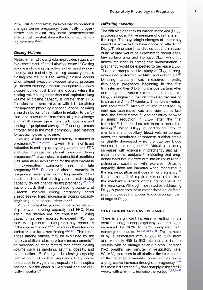
PCO2. This outcomemay be explained by hormonalchanges during pregnancy. Specifically, proges-terone and relaxin may have bronchodilatoryeffects that counterbalance the bronchoconstrict-ing elements.22,34
Closing Volume
Measurementofclosingvolumeprovidesaquantita-tive assessment of small airway closure.41 Closingvolume and closing capacity are often used synony-mously, but technically, closing capacity equalsclosing volume plus RV. Airway closure occurswhen pleural pressure exceeds airway pressure(ie, transpulmonary pressure is negative). Airwayclosure during tidal breathing occurs when theclosing volume is greater than end-expiratory lungvolume or closing capacity is greater than FRC.The closure of small airways with tidal breathinghas important physiologic consequences, includinga maldistribution of ventilation in relation to perfu-sion, and a resultant impairment of gas exchangeand small airway injury from cyclic opening andclosing of peripheral airways.41 The single-breathnitrogen test is the most commonly used methodfor assessing closing volume.41
Closing volume has been extensively studied inpregnancy.24,27,35,42–45 Given the significantreduction in end-expiratory lung volume and FRCand the increase in pleural pressure duringpregnancy,15 airway closure during tidal breathingwas seen as an explanation for the mild decreasein oxygenation commonly seen in latepregnancy.18,46 Studies of closing capacity inpregnancy have given conflicting results. Moststudies indicate that closing volume and closingcapacity do not change during pregnancy,35,43,45
but one study that measured closing capacity at2-month intervals during pregnancy noteda progressive, linear increase in closing capacitybeginning in the second trimester.24
More important for gas exchange is the relation-ship between closing capacity and FRC. Hereagain, the studies are not consistent. Closingcapacity has been reported to exceed FRC in upto 60% of patients in late pregnancy, especiallyin the supine position,42,45 whereas others have re-ported this to be a rare finding.35,43,44 The differ-ences among studies may be explained by thelarge variability in closing volume measurements47
or presence of other factors that affect closingvolume such as smoking, asthma, obesity, andkyphoscoliosis.48 Changes in closing capacityrelative to FRC in late pregnancy likely causea decrease in oxygenation, especially in the supineposition, but the effect is likely small and not clin-ically important.44
Diffusing Capacity
The diffusing capacity for carbon monoxide (DLCO)provides a quantitative measure of gas transfer inthe lungs. The physiologic changes of pregnancywould be expected to have opposing effects onDLCO. The increase in cardiac output and intravas-cular volume would be expected to recruit capil-lary surface area and increase DLCO while theknown reduction in hemoglobin concentration inpregnancy would be expected to decrease DLCO.The most comprehensive study of DLCO in preg-nancy was performed by Milne and colleagues.49
Diffusing capacity was measured monthlythroughout pregnancy beginning in the firsttrimester and then 3 to 5 months postpartum. Aftercorrecting for alveolar volume and hemoglobin,DLCO was highest in the first trimester, decreasingto a nadir at 24 to 27 weeks with no further reduc-tion thereafter.49 Alveolar volume measured byinert gas techniques was also significantly lessafter the first trimester.49 Another study showeda similar reduction in DLCO after the firsttrimester,37 but this has not been a consistentfinding.50 When DLCO is partitioned into itsmembrane and capillary blood volume compo-nents, the membrane component is either stableor slightly decreased while the capillary bloodvolume is unchanged.37,50 Diffusing capacityincreases with exercise in pregnancy just as itdoes in normal subjects,51 indicating that preg-nancy does not interfere with the ability to recruitpulmonary capillaries with exercise. Diffusingcapacity does not increase when measured inthe supine position as it does in nonpregnancy,26
likely as a result of impaired venous return fromthe mechanical effects of the gravid uterus onthe vena cava. Although most studies addressingDLCO in pregnancy have methodological defects,pregnancy does not appear to cause a significantchange in DLCO.
VENTILATION AND GAS EXCHANGE
There is a significant increase in resting minuteventilation (VE) during pregnancy. At term, VE isincreased by 20% to 50% compared withnonpregnant values.15,23,25,28,52–55 The increasein VE is associated with a 30% to 50% (fromapproximately 450 to 650 mL) increase in tidalvolume with no change or only a small increase(1–2 breaths per minute) in respiratory rate.While VE increases in all studies, the time courseof the increase is variable. Some studies reveala progressive increase throughout pregnancy23,28
but most indicate that VE rises sharply in the first 12weeks with a minimal increase thereafter.15,25,53,55

Hegewald & Crapo6
The increase in tidal volume occurs without anincrease in inspiratory time or the duration of therespiratory cycle, indicating that inspiratory flowis increased.15,17,55
The dead space to tidal volume ratio (VD/VT) inpregnancy is unchanged at approximately30%.17,56,57 Given the significant increase in tidalvolume, this indicates that dead space ventilationis also increased. Studies of dead space ventila-tion in pregnancy have produced conflictingresults.56–58 Dead space ventilation would be ex-pected to decrease in pregnancy because of theincreases in cardiac output and perfusion to thelung apices. However, most studies indicate thatthere is an increase in dead space ventilation.56,57
Anatomic dead space is unlikely to be altered bypregnancy, so an increase in alveolar dead spaceis the likely cause. The mechanism for the increasein alveolar dead space is not clear.Hyperventilation in pregnancy is primarily
caused by a progesterone effect augmented byan increased metabolic rate and increased CO2
production. There is convincing evidence thatprogesterone is a respiratory stimulant. Increasedventilation occurs during the luteal phase of themenstrual cycle corresponding to increasedplasma progesterone levels.59 Exogenous proges-terone administered to males causes increasedminute ventilation and CO2 chemosensitivity.60,61
The mechanism by which progesterone causesan increase in ventilation is not completely under-stood, although progesterone decreases thethreshold and increases the sensitivity of thecentral ventilatory chemoreflex response toCO2.
59,62,63 Independent of its effect on CO2
sensitivity, there is also evidence that proges-terone, either alone or in combination with estra-diol, stimulates central neural sites in the medullaoblongata, thalamus, and hypothalamus, involvedin controlling ventilation.59,62 Progesteronealso has a direct effect on the carotid body so asto increase the peripheral ventilatory response tohypoxia. This effect is potentiated by estrogen.64
In summary, progesterone and estradiol act syner-gistically to increase minute ventilation and reducePaCO2 by multiple mechanisms.The discordance between increasing proges-
terone after the first trimester and relative stabilityof minute ventilation has not been explained.Progesterone increases progressively duringpregnancy. Most studies reveal a sharp increasein minute ventilation early in pregnancy and thenonly a minimal increase during the remainder ofpregnancy.15,25,53,55 There is a direct relationshipbetween respiratory drive, quantified by mouthocclusion pressure (P0.1), the pressure measuredat the mouth 100 milliseconds following airway
occlusion at FRC, and progesterone levelsthroughout pregnancy.15 The lack of an associa-tion between P0.1 and minute ventilation may beexplained by increased respiratory impedancesecondary to mechanical changes in the chestwall and abdomen.15
Dyspnea is a common complaint in healthypregnant women. “Physiologic dyspnea” occursin 60% to 70% of normal pregnant women by 30weeks of gestation.53,65,66 Women with physio-logic dyspnea when compared with asymptomaticpregnant women have a higher P0.1 and anincreased ventilatory response to both CO2 andhypoxia.55 The increased minute ventilation andchemosensitivity are not explained by higherprogesterone levels in patients with physiologicdyspnea compared with those without thissymptom.55 Physiologic dyspnea is likely relatedto an increased awareness of this augmenteddrive to breathe.13
The increased metabolic demands of the fetus,uterus, and maternal organs result in increasedoxygen consumption (VO2), carbon dioxideproduction (VCO2), and basal metabolic rate. VO2
and VCO2 at term are approximately 20% and35% greater, respectively, than nonpregnantvalues.28,52,54,56,67–69 The respiratory exchangeratio (VCO2/VO2) is unchanged or minimallyincreased with pregnancy.52,54,56,69 The increasein VE exceeds the increase in VCO2 and VO2. Thedisproportionate increase in VE leads to anincrease in alveolar and arterial partial pressuresof oxygen (PAO2 and PaO2) and a decrease in alve-olar and arterial partial pressures of CO2 (PACO2
and PaCO2).Normal arterial blood values in pregnancy at sea
level are listed in Table 1. Templeton andKelman57 measured serial arterial blood gases ina cohort of healthy women throughout pregnancyand postpartum at sea level. PaO2 was 106 mmHg during the first trimester and decreased to102 mm Hg near term. PaO2 values during preg-nancy were greater than those measured post-partum and in a control group (PaO2 93–95 mmHg). There was no change in the PAO2-PaO2 differ-ence throughout pregnancy compared with thepostpartum period or the control group. Otherstudies have also documented an increase inPaO2 during pregnancy.63,70,71
The increased oxygen tension during pregnancyis an important adaptation that facilitatesoxygen transfer across the placenta. Despite anincreased oxygen tension, the combination ofincreased oxygen consumption and a lower reser-voir of oxygen stores due to reduced functionalresidual capacity decreases maternal oxygenreserves. Pregnant women are more susceptible
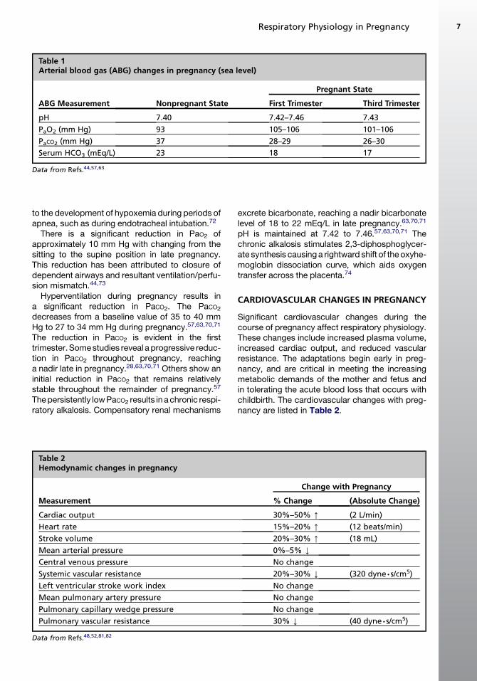
Table 1Arterial blood gas (ABG) changes in pregnancy (sea level)
ABG Measurement Nonpregnant State
Pregnant State
First Trimester Third Trimester
pH 7.40 7.42–7.46 7.43
PaO2 (mm Hg) 93 105–106 101–106
PaCO2 (mm Hg) 37 28–29 26–30
Serum HCO3 (mEq/L) 23 18 17
Data from Refs.44,57,63
Respiratory Physiology in Pregnancy 7
to the development of hypoxemia during periods ofapnea, such as during endotracheal intubation.72
There is a significant reduction in PaO2 ofapproximately 10 mm Hg with changing from thesitting to the supine position in late pregnancy.This reduction has been attributed to closure ofdependent airways and resultant ventilation/perfu-sion mismatch.44,73
Hyperventilation during pregnancy results ina significant reduction in PaCO2. The PaCO2decreases from a baseline value of 35 to 40 mmHg to 27 to 34 mm Hg during pregnancy.57,63,70,71
The reduction in PaCO2 is evident in the firsttrimester.Somestudies reveal aprogressive reduc-tion in PaCO2 throughout pregnancy, reachinga nadir late in pregnancy.28,63,70,71 Others show aninitial reduction in PaCO2 that remains relativelystable throughout the remainder of pregnancy.57
Thepersistently lowPaCO2 results in achronic respi-ratory alkalosis. Compensatory renal mechanisms
Table 2Hemodynamic changes in pregnancy
Measurement
Cardiac output
Heart rate
Stroke volume
Mean arterial pressure
Central venous pressure
Systemic vascular resistance
Left ventricular stroke work index
Mean pulmonary artery pressure
Pulmonary capillary wedge pressure
Pulmonary vascular resistance
Data from Refs.48,52,81,82
excrete bicarbonate, reaching a nadir bicarbonatelevel of 18 to 22 mEq/L in late pregnancy.63,70,71
pH is maintained at 7.42 to 7.46.57,63,70,71 Thechronic alkalosis stimulates 2,3-diphosphoglycer-ate synthesis causing a rightward shift of the oxyhe-moglobin dissociation curve, which aids oxygentransfer across the placenta.74
CARDIOVASCULAR CHANGES IN PREGNANCY
Significant cardiovascular changes during thecourse of pregnancy affect respiratory physiology.These changes include increased plasma volume,increased cardiac output, and reduced vascularresistance. The adaptations begin early in preg-nancy, and are critical in meeting the increasingmetabolic demands of the mother and fetus andin tolerating the acute blood loss that occurs withchildbirth. The cardiovascular changes with preg-nancy are listed in Table 2.
Change with Pregnancy
% Change (Absolute Change)
30%–50% [ (2 L/min)
15%–20% [ (12 beats/min)
20%–30% [ (18 mL)
0%–5% Y
No change
20%–30% Y (320 dyne∙s/cm5)
No change
No change
No change
30% Y (40 dyne∙s/cm5)

Hegewald & Crapo8
Blood Volume Changes
Total body volume expansion occurs secondary tosodium and water retention during pregnancy.Total body volume expansion accounts forapproximately 6 kg of the weight gained duringpregnancy, and is distributed among the maternalextracellular and intracellular spaces, amnioticfluid, and the fetus.17,75 Maternal blood volumeincreases progressively during pregnancy, peak-ing at a value approximately 40% greater thanbaseline by the third trimester. The increase inmaternal plasma blood volume is physiologicallyimportant. Smaller increases in maternal plasmablood volume are associated with intrauterinegrowth retardation and poor fetal outcomes.76
The increase in plasma blood volume is best ex-plained by the underfill hypothesis. Vasodilation ofthe maternal circulation and shunt-like effectscreated by the low-resistance uteroplacentalcirculation activate the renin-angiotensin system,and stimulate increased renal sodium reabsorp-tion and water retention. Animal models suggestthat the nitric oxide system is the main mediatorof the primary peripheral vasodilation inpregnancy.77 Hormonal factors also contribute tovolume expansion and activation of the renin-angiotensin system75,78,79 However, elevatedlevels of atrial natriuretic peptide early in preg-nancy are not consistent with the underfillhypothesis.80 The exact mechanism for volumehomeostasis in normal pregnancy remains unex-plained. The 40% increase (1.2 L on average) inmaternal plasma blood volume exceeds the 20%to 30% increase in maternal red blood cell mass,resulting in hemodilution and the relative anemiaof pregnancy.76
Cardiac Output
Cardiac output begins to increase as early as thefifth week of pregnancy and peaks at 30% to50% (approximately 2 L per minute) abovenonpregnant values at 25 to 32 weeks.81–85
The increase in cardiac output is a result ofa 20% increase in heart rate and a 20% to 30%increase in stroke volume. The increase in strokevolume is a consequence of an increase in pre-load, due to increased plasma volume anda 20% to 30% decrease in systemic vascularresistance with no significant change in ventricularcontractility.82–84,86 Studies that have usedrepeated echocardiograms during pregnancyhave revealed no significant change in left ventric-ular ejection fraction, with increases in left ventric-ular mass and wall thickness as well as increasesin all cardiac chamber dimensions.83,84 Cardiacoutput may decrease slightly in the third trimester,
but is highly dependent on body position. In latepregnancy, cardiac output in the supine positiondecreases by up to 30% compared with the leftlateral decubitus position as a result of compres-sion of the inferior vena cava by the gravid uterusand reduced venous return.87
Central Hemodynamic and PulmonaryVascular Changes
In addition to the significant increase in cardiacoutput and reduction in systemic vascular resis-tance discussed earlier, right heart catheterizationstudies in normal pregnant women revealed nochanges in central venous pressure and pulmo-nary capillary wedge pressure compared withnonpregnant values.81,82 Mean pulmonary arterypressure is also unchanged during pregnancy.81,82
Pulmonary artery pressures increase minimallywith exercise but remain within the normalrange.81 Pulmonary vascular resistance signifi-cantly decreases during pregnancy, as would beexpected given the stable mean pulmonary arterypressure and pulmonary wedge pressure as wellas the increased cardiac output.
EXERCISE PHYSIOLOGY IN PREGNANCY
Moderate aerobic exercise during pregnancyappears to be safe for the mother and fetus, andmay improve some pregnancy outcomes such asgestational diabetes and preeclampsia.88–91 Moststudies addressing the physiologic response toexercise in pregnancy used submaximal, constantload exercise protocols to avoid potential risk tothe fetus.52,56,67,69,89,92,93
At moderate intensity of exercise, pregnantwomen respond differently to nonpregnantwomen. The increase in VO2 for a given workloadis greater.68,69,92 However, VO2 varies with thetype of exercise. In pregnant subjects weight-bearing exercise, such as treadmill walking, isassociated with a significantly higher VO2 than iscycle ergometry compared with the differencebetween these 2 modes in nonpregnantcontrols.52 When non–weight-bearing exercise isperformed and VO2 is adjusted for body weight(VO2 expressed in mL O2/kg/min), the VO2
increases are similar for a given workload in preg-nant and nonpregnant subjects.89,92
During pregnancy, minute ventilation (VE) andalveolar ventilation (VA) are higher at rest and therate of increase with exercise is more rapidcompared with the nongravid state. Both VE andVA increase 20% to 25% compared with nonpreg-nant values at submaximal work rates.56,69,89,92
The increase in VE exceeds the increase in VO2,

Respiratory Physiology in Pregnancy 9
resulting in an increase in the ventilatory equivalentfor oxygen (VE/VO2).
Cardiac output is higher for a given exerciselevel in pregnant subjects compared withnonpregnant subjects. This difference is primarilydue to an increase in stroke volume.67,92 The pro-portionally greater increase in cardiac output withexercise results in a reduction in the differencebetween the arterial and mixed venous oxygencontent differences (CaO2–CvO2) compared withthe nonpregnant state, leading to increasedoxygen delivery to the fetus during maternalexercise.67
Few studies have performed symptom-limitedmaximal cardiopulmonary exercise tests in latepregnancy.69,89 Maximal O2 consumption hasbeen noted to be reduced in sedentary pregnantwomen, but is associated with lower peak heartrates and may be partly due to effort. MaximalO2 consumption is preserved in physically fit preg-nant women but anaerobic working capacity maybe reduced.
The fetus usually responds to submaximalmaternal exercise with an increase in fetal heartrate. The fetal heart rate gradually returns tonormal after the mother stops exercise.89 Tran-sient fetal bradycardia may develop with maximalexercise. The significance of fetal bradycardiawith intense exercise is unclear. Prolongedsubmaximal exercise (greater than 30 minutes) inlate pregnancy often results in a moderate reduc-tion in maternal blood glucose concentration andmay transiently reduce fetal glucose availability.Maternal core body temperature increases withmoderate exercise, but generally less than1.5�C.89 The clinical significance of these changesis not clear, but moderate exercise does notappear to be associated with adverse effects tothe mother or fetus.
RESPIRATORY PHYSIOLOGY IN LABOR,DELIVERY, AND POSTPARTUM
Hyperventilation, beyond the usual pregnancy-mediated increase in minute ventilation, iscommon during labor and delivery. Several factorsinteract to influence minute ventilation during laborand delivery. Pain, anxiety, and coached breathingtechniques increase minute ventilation whereasnarcotic analgesics have the opposite effect. Theresult is a wide variation in minute ventilation andbreathing patterns, as illustrated by a study of 25patients during labor that found tidal volumesranging from 330 to 2250 mL and minute ventila-tion varying from 7 to 90 L per minute.94 Paincontrol with narcotic analgesics minimizes labor-induced hyperventilation, suggesting that the
main contributing factor for the hyperventilationis painful uterine contractions.11 Hyperventilationduring labor and delivery results in a reduction inalveolar CO2 levels from 32 mm Hg during earlylabor to 26 mm Hg during the second stage.95
The significant hyperventilation during labor anddelivery and resultant hypocarbia can causeuterine vessel vasoconstriction and decreasedplacental perfusion, and may have deleteriouseffects in patients with marginal placentalreserves.43 Within 72 hours of delivery, the minuteventilation decreases halfway back toward thenonpregnant value and returns to baseline withina few weeks.49,51
The static lung volume changes that occurduring pregnancy rapidly normalize after deliverywith decompression of the diaphragm and lungs.FRC and RV return to baseline values within 48hours.20 The chest wall changes that occur duringpregnancy normalize by 24 weeks postpartum, butthe subcostal angle does not return to its prepreg-nancy value, remaining about 20% wider thanbaseline.15
CARDIOVASCULAR PHYSIOLOGY IN LABOR,DELIVERY, AND POSTPARTUM
Labor, delivery and the postpartum period areassociated with significant cardiovascularchanges. Cardiac output increases 10% to 15%above late pregnancy levels during early laborand by 50% during the second stage of labor.87,96
The increase in cardiac output is caused by anincrease in both heart rate and stroke volume.Factors contributing to the hemodynamic changesduring labor include pain and anxiety with resultantincreases in circulating catecholamines, anduterine contractions with resultant cyclic“autotransfusions” and increases in centralblood volume and preload. Narcotic analgesicsand regional anesthesia minimize the pain-mediated changes in cardiac output. Immediatelypostpartum there is a 60% to 80% increase incardiac output and stroke volume relative to prel-abor values, due to increased preload associatedwith “autotransfusion” of approximately 500 mLof blood that is no longer diverted to the uteropla-cental vascular bed, and the relief of aortocavalcompression with delivery.82,91 Systolic and dia-stolic blood pressure and systemic vascular resis-tance increase during active labor, although themagnitude depends on the severity of maternalpain, anxiety, and the intensity of uterine contrac-tions. It is important for the clinician to recognizethat patients with underlying cardiac or pulmonaryvascular disease may not be able to augment their

Hegewald & Crapo10
cardiac output in the peripartum period, resultingin decompensated heart failure.There is conflicting information regarding the
time course for the resolution of the cardiovascularphysiologic changes after delivery. Most studiesreveal a significant reduction in cardiac outputand stroke volume by 2 weeks after delivery, withfurther reductions to near baseline values over 12to 24 weeks.77,79,81,97 However, another studyindicated that cardiac output and stroke volumeremained significantly elevated above prepreg-nancy values at 12 weeks postpartum.98 Some ofthe cardiovascular changes resolve more slowly.Left ventricular wall thickness and mass remainsignificantly greater than nonpregnant controls at24 weeks after delivery, suggesting an elementof residual hypertrophy.92
SUMMARY
Significant anatomic and physiologic adaptationsinvolving the respiratory and cardiac systemsoccur during pregnancy and are necessary tomeet the increased metabolic demands of boththe mother and the fetus. The prominentrespiratory changes include: mechanical alter-ations to the chest wall and diaphragm to accom-modate the enlarging uterus; a reduction in FRCand its components ERV and RV, with little or nochange in TLC; and an increase in minute ventila-tion, resulting in reduced PaCO2 and chronic respi-ratory alkalosis. There is no significant change inspirometry, DLCO, or oxygenation. The majorcardiovascular changes include increased plasmablood volume, increased cardiac output, anda reduction in systemic vascular resistance. Abasic knowledge of these expected changes willhelp health providers distinguish the commonphysiologic dyspnea from breathlessness causedby the various cardiopulmonary diseases thatcoexist with pregnancy.
REFERENCES
1. Lapinsky SE. Cardiopulmonary complications of
pregnancy. Crit Care Med 2005;33(7):1616–22.
2. Ellegard EK. Pregnancy rhinitis. Immunol Allergy
Clin North Am 2006;26(1):119–35, vii.
3. Toppozada H, Michaels L, Toppozada M, et al. The
human respiratory nasal mucosa in pregnancy. An
electron microscopic and histochemical study.
J Laryngol Otol 1982;96(7):613–26.
4. Bende M, Gredmark T. Nasal stuffiness during preg-
nancy. Laryngoscope 1999;109(7 Pt 1):1108–10.
5. Ellegard E. [Nasal congestion among women. At
least every fifth pregnant woman suffers–most
common among smokers]. Lakartidningen 2000;
97(34):3619–20, 3623 [in Swedish].
6. Mabry RL. Rhinitis of pregnancy. South Med J 1986;
79(8):965–71.
7. Ellegard EK. Clinical and pathogenetic characteris-
tics of pregnancy rhinitis. Clin Rev Allergy Immunol
2004;26(3):149–59.
8. Pien GW, Schwab RJ. Sleep disorders during preg-
nancy. Sleep 2004;27(7):1405–17.
9. Lundberg JO, Weitzberg E. Nasal nitric oxide in
man. Thorax 1999;54(10):947–52.
10. Pilkington S, Carli F, Dakin MJ, et al. Increase in Mal-
lampati score during pregnancy. Br J Anaesth 1995;
74(6):638–42.
11. Izci B, Vennelle M, Liston WA, et al. Sleep-disor-
dered breathing and upper airway size in pregnancy
and post-partum. Eur Respir J 2006;27(2):321–7.
12. Pien GW, Fife D, Pack AI, et al. Changes in symp-
toms of sleep-disordered breathing during preg-
nancy. Sleep 2005;28(10):1299–305.
13. Izci B, Riha RL, Martin SE, et al. The upper airway in
pregnancy and pre-eclampsia. Am J Respir Crit
Care Med 2003;167(2):137–40.
14. White DP. Pathogenesis of obstructive and central
sleep apnea. Am J Respir Crit Care Med 2005;
172(11):1363–70.
15. Contreras G, Gutierrez M, Beroiza T, et al. Ventilatory
drive and respiratory muscle function in pregnancy.
Am Rev Respir Dis 1991;144(4):837–41.
16. Elkus R, Popovich J Jr. Respiratory physiology in
pregnancy. Clin Chest Med 1992;13(4):555–65.
17. Crapo RO. Normal cardiopulmonary physiology
during pregnancy. Clin Obstet Gynecol 1996;39(1):
3–16.
18. Weinberger SE, Weiss ST, Cohen WR, et al. Preg-
nancy and the lung. Am Rev Respir Dis 1980;
121(3):559–81.
19. Goldsmith LT, Weiss G, Steinetz BG. Relaxin and its
role in pregnancy. Endocrinol Metab Clin North Am
1995;24(1):171–86.
20. Gilroy RJ, Mangura BT, Lavietes MH. Rib cage and
abdominal volume displacements during breathing in
pregnancy. Am Rev Respir Dis 1988;137(3):668–72.
21. Marx GF, Murthy PK, Orkin LR. Static compliance
before and after vaginal delivery. Br J Anaesth
1970;42(12):1100–4.
22. Gee JB, Packer BS, Millen JE, et al. Pulmonary
mechanics during pregnancy. J Clin Invest 1967;
46(6):945–52.
23. Cugell DW, Frank NR, Gaensler EA, et al. Pulmonary
function in pregnancy. I. Serial observations in
normal women. Am Rev Tuberc 1953;67(5):568–97.
24. Garrard GS, Littler WA, Redman CW. Closing
volume during normal pregnancy. Thorax 1978;
33(4):488–92.
25. Alaily AB, Carrol KB. Pulmonary ventilation in preg-
nancy. Br J Obstet Gynaecol 1978;85(7):518–24.

Respiratory Physiology in Pregnancy 11
26. Norregaard O, Schultz P, Ostergaard A, et al. Lung
function and postural changes during pregnancy.
Respir Med 1989;83(6):467–70.
27. Garcia-Rio F, Pino-Garcia JM, Serrano S, et al.
Comparison of helium dilution and plethysmo-
graphic lung volumes in pregnant women. Eur Re-
spir J 1997;10(10):2371–5.
28. Prowse CM, Gaensler EA. Respiratory and acid-
base changes during pregnancy. Anesthesiology
1965;26:381–92.
29. Wanger J, Clausen JL, Coates A, et al. Standardisa-
tion of the measurement of lung volumes. Eur Respir
J 2005;26(3):511–22.
30. Ferris BG. Epidemiology standardization project
(American Thoracic Society). Am Rev Respir Dis
1978;118(6 Pt 2):1–120.
31. Unterborn J. Pulmonary function testing in obesity,
pregnancy, and extremes of body habitus. Clin
Chest Med 2001;22(4):759–67.
32. Goucher D, Rubin A, Russo N. The effect of preg-
nancy upon pulmonary function in normal women.
Am J Obstet Gynecol 1956;72(5):963–9.
33. Ihrman K. A clinical and physiological study of preg-
nancy in a material from northern Sweden. II. Vital
capacity and maximal breathing capacity during and
after pregnancy. Acta Soc Med Ups 1960;65:147–54.
34. Milne JA, Mills RJ, Howie AD, et al. Large airways
function during normal pregnancy. Br J Obstet Gy-
naecol 1977;84(6):448–51.
35. Baldwin GR, Moorthi DS, Whelton JA, et al. New lung
functions and pregnancy. Am J Obstet Gynecol
1977;127(3):235–9.
36. Puranik BM, Kaore SB, Kurhade GA, et al.
A longitudinal study of pulmonary function tests
during pregnancy. Indian J Physiol Pharmacol
1994;38(2):129–32.
37. Gazioglu K, Kaltreider NL, Rosen M, et al. Pulmo-
nary function during pregnancy in normal women
and in patients with cardiopulmonary disease.
Thorax 1970;25(4):445–50.
38. McAuliffe F, Kametas N, Costello J, et al. Respiratory
function in singleton and twin pregnancy. BJOG
2002;109(7):765–9.
39. Kerr JH. Bronchopulmonary resistance in preg-
nancy. Can Anaesth Soc J 1961;8:347–55.
40. Levitzky MG. Pulmonary physiology. 7th edition.
New York (NY): McGraw-Hill; 2007.
41. Milic-Emili J, Torchio R, D’Angelo E. Closing volume:
a reappraisal (1967–2007). Eur J Appl Physiol 2007;
99(6):567–83.
42. Bevan DR, Holdcroft A, Loh L, et al. Closing volume
and pregnancy. Br Med J 1974;1(5896):13–5.
43. Craig DB, Toole MA. Airway closure in pregnancy.
Can Anaesth Soc J 1975;22(6):665–72.
44. Awe RJ, Nicotra MB, Newsom TD, et al. Arterial
oxygenation and alveolar-arterial gradients in term
pregnancy. Obstet Gynecol 1979;53(2):182–6.
45. Russell IF, Chambers WA. Closing volume in normal
pregnancy. Br J Anaesth 1981;53(10):1043–7.
46. Fishburne JI. Physiology and disease of the respira-
tory system in pregnancy. A review. J Reprod Med
1979;22(4):177–89.
47. Burki NK, Barker DB, Nicholson DP. Variability of the
closing volume measurement in normal subjects.
Am Rev Respir Dis 1975;112(2):209–12.
48. Camann WR, Ostheimer GW. Physiological adapta-
tions during pregnancy. Int Anesthesiol Clin 1990;
28(1):2–10.
49. Milne JA, Mills RJ, Coutts JR, et al. The effect of
human pregnancy on the pulmonary transfer factor
for carbon monoxide as measured by the single-
breath method. Clin Sci Mol Med 1977;53(3):271–6.
50. Krumholz RA, Echt CR, Ross JC. Pulmonary
diffusing capacity, capillary blood volume, lung
volumes, and mechanics of ventilation in early and
late pregnancy. J Lab Clin Med 1964;63:648–55.
51. Bedell GN, Adams RW. Pulmonary diffusing
capacity during rest and exercise. a study of normal
persons and persons with atrial septal defect, preg-
nancy, and pulmonary disease. J Clin Invest 1962;
41(10):1908–14.
52. Knuttgen HG, Emerson K Jr. Physiological response
to pregnancy at rest and during exercise. J Appl
Physiol 1974;36(5):549–53.
53. Milne JA. The respiratory response to pregnancy.
Postgrad Med J 1979;55(643):318–24.
54. Rees GB, Broughton Pipkin F, Symonds EM, et al.
A longitudinal study of respiratory changes in
normal human pregnancy with cross-sectional data
on subjects with pregnancy-induced hypertension.
Am J Obstet Gynecol 1990;162(3):826–30.
55. Garcia-Rio F, Pino JM, Gomez L, et al. Regulation of
breathing and perception of dyspnea in healthy
pregnant women. Chest 1996;110(2):446–53.
56. Pernoll ML, Metcalfe J, Kovach PA, et al. Ventilation
during rest and exercise in pregnancy and post-
partum. Respir Physiol 1975;25(3):295–310.
57. Templeton A, Kelman GR. Maternal blood-gases,
(PAo2-Pao2), physiological shunt andVD/VT in normal
pregnancy. Br J Anaesth 1976;48(10):1001–4.
58. Shankar KB, Moseley H, Vemula V, et al. Physiolog-
ical dead space during general anaesthesia for
Caesarean section. Can J Anaesth 1987;34(4):
373–6.
59. Bayliss DA, Millhorn DE. Central neural mechanisms
of progesterone action: application to the respiratory
system. J Appl Physiol 1992;73(2):393–404.
60. Zwillich CW, Natalino MR, Sutton FD, et al. Effects of
progesterone on chemosensitivity in normal men.
J Lab Clin Med 1978;92(2):262–9.
61. Schoene RB, Pierson DJ, Lakshminarayan S, et al.
Effect of medroxyprogesterone acetate on respira-
tory drives and occlusion pressure. Bull Eur Physio-
pathol Respir 1980;16(5):645–53.

Hegewald & Crapo12
62. Jensen D, Wolfe LA, Slatkovska L, et al. Effects of
human pregnancy on the ventilatory chemoreflex
response to carbon dioxide. Am J Physiol Regul In-
tegr Comp Physiol 2005;288(5):R1369–75.
63. Liberatore SM, Pistelli R, Patalano F, et al. Respira-
tory function during pregnancy. Respiration 1984;
46(2):145–50.
64. Hannhart B, Pickett CK, Moore LG. Effects of
estrogen and progesterone on carotid body neural
output responsiveness to hypoxia. J Appl Physiol
1990;68(5):1909–16.
65. Gilbert R, Auchincloss JH Jr. Dyspnea of pregnancy.
Clinical and physiological observations. Am J Med
Sci 1966;252(3):270–6.
66. Tenholder MF, South-Paul JE. Dyspnea in preg-
nancy. Chest 1989;96(2):381–8.
67. Ueland K, Novy MJ, Metcalfe J. Cardiorespiratory
responses to pregnancy and exercise in normal
women and patients with heart disease. Am J Obstet
Gynecol 1973;115(1):4–10.
68. Pernoll ML, Metcalfe J, Schlenker TL, et al. Oxygen
consumption at rest and during exercise in preg-
nancy. Respir Physiol 1975;25(3):285–93.
69. Artal R, Wiswell R, Romem Y, et al. Pulmonary
responses to exercise in pregnancy. Am J Obstet
Gynecol 1986;154(2):378–83.
70. Lucius H, Gahlenbeck H, Kleine HO, et al. Respira-
tory functions, buffer system, and electrolyte
concentrations of blood during human pregnancy.
Respir Physiol 1970;9(3):311–7.
71. Dayal P, Murata Y, Takamura H. Antepartum and
postpartum acid-base changes in maternal blood
in normal and complicated pregnancies. J Obstet
Gynaecol Br Commonw 1972;79:612–24.
72. Archer GW Jr, Marx GF. Arterial oxygen tension
during apnoea in parturient women. Br J Anaesth
1974;46(5):358–60.
73. Ang CK, Tan TH, Walters WA, et al. Postural influ-
ence on maternal capillary oxygen and carbon
dioxide tension. Br Med J 1969;4(5677):201–3.
74. Tsai CH, de Leeuw NK. Changes in 2,3-diphospho-
glycerate during pregnancy and puerperium in
normal women and in beta-thalassemia heterozy-
gous women. Am J Obstet Gynecol 1982;142(5):
520–3.
75. Brown MA, Gallery ED. Volume homeostasis in
normal pregnancy and pre-eclampsia: physiology
and clinical implications. Baillieres Clin Obstet Gy-
naecol 1994;8(2):287–310.
76. Hytten F. Blood volume changes in normal preg-
nancy. Clin Haematol 1985;14(3):601–12.
77. Cadnapaphornchai MA, Ohara M, Morris KG Jr,
et al. Chronic NOS inhibition reverses systemic
vasodilation and glomerular hyperfiltration in preg-
nancy. Am J Physiol Renal Physiol 2001;280(4):
F592–8.
78. Longo LD. Maternal blood volume and cardiac
output during pregnancy: a hypothesis of endocri-
nologic control. Am J Physiol 1983;245(5 Pt 1):
R720–9.
79. Wilson M, Morganti AA, Zervoudakis I, et al. Blood
pressure, the renin-aldosterone system and sex
steroids throughout normal pregnancy. Am J Med
1980;68(1):97–104.
80. Sala C, Campise M, Ambroso G, et al. Atrial natri-
uretic peptide and hemodynamic changes during
normal human pregnancy. Hypertension 1995;
25(4 Pt 1):631–6.
81. Bader RA, Bader ME, Rose DF, et al. Hemody-
namics at rest and during exercise in normal preg-
nancy as studies by cardiac catheterization. J Clin
Invest 1955;34(10):1524–36.
82. Clark SL, Cotton DB, Lee W, et al. Central hemody-
namic assessment of normal term pregnancy. Am
J Obstet Gynecol 1989;161(6 Pt 1):1439–42.
83. Robson SC, Hunter S, Boys RJ, et al. Serial study of
factors influencing changes in cardiac output during
human pregnancy. Am J Physiol 1989;256(4 Pt 2):
H1060–5.
84. Mabie WC, DiSessa TG, Crocker LG, et al.
A longitudinal study of cardiac output in normal
human pregnancy. Am J Obstet Gynecol 1994;
170(3):849–56.
85. van Oppen AC, Stigter RH, Bruinse HW. Cardiac
output in normal pregnancy: a critical review. Obstet
Gynecol 1996;87(2):310–8.
86. Hunter S, Robson SC. Adaptation of the maternal
heart in pregnancy. Br Heart J 1992;68(6):540–3.
87. Ueland K, Metcalfe J. Circulatory changes in preg-
nancy. Clin Obstet Gynecol 1975;18(3):41–50.
88. Kramer MS, McDonald SW. Aerobic exercise for
women during pregnancy. Cochrane Database
Syst Rev 2006;3:CD000180.
89. Wolfe LA, Weissgerber TL. Clinical physiology of
exercise in pregnancy: a literature review. J Obstet
Gynaecol Can 2003;25(6):473–83.
90. Bung P, Bung C, Artal R, et al. Therapeutic exercise
for insulin-requiring gestational diabetics: effects on
the fetus–results of a randomized prospective longi-
tudinal study. J Perinat Med 1993;21(2):125–37.
91. Dempsey JC, Sorensen TK, Williams MA, et al.
Prospective study of gestational diabetes mellitus
risk in relation to maternal recreational physical
activity before and during pregnancy. Am J Epide-
miol 2004;159(7):663–70.
92. Gorski J. Exercise during pregnancy: maternal and
fetal responses. A brief review. Med Sci Sports Ex-
erc 1985;17(4):407–16.
93. Wolfe LA, Walker RM, Bonen A, et al. Effects of preg-
nancy and chronic exercise on respiratory
responses to graded exercise. J Appl Physiol
1994;76(5):1928–36.

Respiratory Physiology in Pregnancy 13
94. Cole PV, Nainby-Luxmoore RC. Respiratory volumes
in labour. Br Med J 1962;1(5285):1118.
95. Reid DH. Respiratory changes in labour. Lancet
1966;1(7441):784–5.
96. Fujitani S, Baldisseri MR. Hemodynamic assessment
in a pregnant and peripartum patient. Crit Care Med
2005;33(Suppl 10):S354–61.
97. Robson SC, Hunter S, Moore M, et al. Haemody-
namic changes during the puerperium: a Doppler
and M-mode echocardiographic study. Br J Obstet
Gynaecol 1987;94(11):1028–39.
98. Capeless EL, Clapp JF. When do cardiovascular
parameters return to their preconception values?
Am J Obstet Gynecol 1991;165(4 Pt 1):883–6.