Embed Size (px)
Citation preview

Respiratory Physiology & Neurobiology 164 (2008) 429–440
Contents lists available at ScienceDirect
Respiratory Physiology & Neurobiology
journa l homepage: www.e lsev ier .com/ locate / resphys io l
Respiratory rhythm of brainstem–spinal cord preparations: Effects ofmaturation, age, mass and oxygenation
Angelina Y. Fonga,∗, Andrea E. Corcorana, M. Beth Zimmerb, Denis V. Andradec, William K. Milsoma
a Department of Zoology, University of British Columbia, Vancouver, BC, V6T 1Z4, Canadab Department of Biological Sciences, Ferris State University, Big Rapids, MI, USAc Departamento de Zoologia, Universidade Estadual Paulista, Rio Claro, SP, Brazil
a r t i c l e i n f o
Article history:Accepted 12 September 2008
Keywords:Respiratory rhythmogenesisPonsPostnatal development
a b s t r a c t
We examined the effect of age, mass and the presence of the pons on the longevity (length of time sponta-neous respiratory-related activity is produced) of brainstem–spinal cord preparations of neonatal rodents(rats and hamsters) and the level of oxygenation in the medulla respiratory network in these preparations.We found the longevity of the preparations from both species decreased with increasing postnatal age.Physical removal of the pons increased respiratory frequency and the longevity of the preparation. How-ever, tissue oxygenation at the level of the medullary respiratory network was not affected by removal ofthe pons or increasing postnatal age (up to postnatal day 4). Taken together, these data suggest that the
RatHamster effect of removing the pons on respiratory frequency and the longevity of brainstem–spinal cord prepara-
atalls of
1
tdmsuenrfTa(vaittg
C1
paGtaoHrVtrsmtAt
1d
tions with increasing postnto mass or changes in leve
. Introduction
The respiratory network, including the centers required forhe neurogenesis of respiratory rhythm, must be sufficiently welleveloped at the time of birth to cope with the immediate require-ents of respiration. Current evidence, however, indicates that the
ystem continues to undergo substantial postnatal growth and mat-ration (Liu and Wong-Riley, 2001; Dutschmann et al., 2004; Kront al., 2007). We have been particularly interested in the post-atal changes that occur in cold tolerance and the ability of theespiratory system to restart (autoresuscitate) in neonatal rodentsollowing hypothermic respiratory arrest (Mellen et al., 2002;attersall and Milsom, 2003; Zimmer and Milsom, 2004). We havettempted to address these questions using brainstem–spinal corden bloc) preparations from neonatal rodents (rats and hamsters) ofarious ages with the pons intact (Pontomedullary preparations)nd with the pons removed (Medullary preparations). In attempt-
ng to interpret our data we have been confronted by the needo dissociate the effects of increasing tissue mass from postna-al growth that may result in hypoxia at the respiratory rhythmenerators from the effects of postnatal maturation.∗ Corresponding author at: Department of Zoology, University of Britisholumbia, Biological Sciences Building, 6270 University Blvd, Vancouver, BC, V6TZ4, Canada. Tel.: +1 604 822 5799; fax: +1 604 822 2416.
E-mail address: [email protected] (A.Y. Fong).
g(etppacoa
569-9048/$ – see front matter © 2008 Elsevier B.V. All rights reserved.oi:10.1016/j.resp.2008.09.008
age are primarily due to postnatal development and appear to be unrelatedtissue oxygenation.
© 2008 Elsevier B.V. All rights reserved.
The en bloc preparation has been widely used in the study of res-iratory rhythmogenesis (Suzue, 1984; Hilaire et al., 1989; Smith etl., 1990; Errchidi et al., 1991; Ito et al., 2002; Viemari et al., 2003;uimaraes et al., 2007). Although it is commonly acknowledged
hat older preparations have reduced viability, this is generallyssumed to be due to the effects of increasing mass which reducesxygen diffusion from the superfusate to the respiratory neurons.owever, the same trend in reduced viability occurs in neonatal
odents of very different sizes (rats vs. mice) (Errchidi et al., 1991;iemari et al., 2003), suggesting that oxygen diffusion may not be
he primary cause for the reduced longevity of the en bloc prepa-ation. Although, one may argue that given the increase in masspecific metabolic rate seen with decreasing size in vivo in mam-als, the smaller mouse tissue may have a higher metabolic rate
han rat tissue resulting in similar PO2 profiles within the medulla.lso while numerous studies have shown that preparations con-
aining the pons have lower respiratory frequencies and this isenerally attributed to descending inhibition arising from the ponsHilaire et al., 1989; Errchidi et al., 1991; Ito et al., 2002; Viemarit al., 2003; Guimaraes et al., 2007), part of this effect may be dueo increased mass and reduced oxygenation at the medullary res-iratory rhythm generators. As a result of these observations, the
resent study was designed to attempt to dissociate the effects ofge, mass, postnatal development and levels of oxygenation, on thehanges that occur in respiratory frequency and temporal longevityf brainstem–spinal cord preparations from neonatal rodents withnd without the pons.
4 ogy & Neurobiology 164 (2008) 429–440
2
Ug
2
SaocScKbwoptbTavdopmicacfiWO
2o
ptag(Cwupfmmc
2r
rmsPrp
Fig. 1. (A) Schematic illustration of the double perfusion chamber used for sep-arate superfusion of the pons and medulla/spinal cord. The vaseline (grease gap)was placed at the level of the caudal cerebellar artery and fictive respiratory burstswere recorded from C4 nerve rootlets. (B) Preliminary experiments superfusingtetrodotoxin (TTX, 500 (M) over the medulla and spinal cord resulted in cessation ofburst activity within 8 min. (C) Superfusion of artificial cerebral spinal fluid (aCSF)containing 0.2 mM Ca2+ and 5 mM Mg2+ (low Ca2+/high Mg2+) over the medulla andsnf
30 A.Y. Fong et al. / Respiratory Physiol
. Methods
All experiments were performed with prior approval from theniversity of British Columbia Animal Care Committee, under theuidelines of the Canadian Council for Animal Care (CCAC).
.1. Brainstem–spinal cord preparation
Neonatal Sprague–Dawley rat pups (0–8 days postnatal) oryrian (Golden) hamster pups (0–8 days postnatal) were deeplynaesthetized with 2–4% halothane or isoflurane until the absencef a withdrawal reflex from toe pinch. The brainstem and spinalord were isolated en bloc (preparation previously described byuzue, 1984) at room temperature while submerged in artificialerebral spinal fluid (aCSF, 1.5 mM CaCl2, 113.0 mM NaCl, 3.0 mMCl, 1.2 mM NaH2PO4, 30.0 mM NaHCO3, 30.0 mM d-glucose) bub-led with 95% O2–5% CO2, to obtain a pH of 7.4. The brainstemas trimmed rostrally at one of two locations: (1) just rostralf the pons at the level of the superior cerebellar peduncles forons–medulla–spinal cord (Pontomedullary) preparations or (2)he rostral end of the pyramids and just rostral of the caudal cere-ellar artery for medulla–spinal cord (Medullary) preparations.he spinal cord was transected caudal to the seventh cervical rootnd the entire en bloc preparation was transferred and pinnedentral side up on a stainless steel grid in a plexiglass recordingish. The stainless steel grid allows for simultaneous superfusionf the preparation over both the ventral and dorsal surfaces. Thereparation was continuously superfused with oxygenated aCSFaintained at 27 ◦C by a Lauda water bath (Model RC6) and mon-
tored using a thermocouple placed in the bathing fluid in thehamber. The flow rate of the aCSF was maintained between 5nd 9 ml/min. Whole nerve activity was recorded from C1 or C4ervical rootlets via a glass suction electrode, amplified (20–50 K),ltered (100 Hz to 3 kHz) and recorded (2000 samples/s) using aindaq data acquisition system (DI200; DataQ Instruments, Akron,
H, USA).
.2. Experiment 1: longevity of respiratory-related motor outputf en bloc preparations at different ages
To examine the effect of postnatal age, brain mass and theresence of the pons on the longevity of the en bloc prepara-ions, Pontomedullary and Medullary preparations from both ratsnd hamsters at specific postnatal days were divided into five ageroups: P0 (day of birth), P2 (2 Postnatal days), P4, P6 and P8n = 5–9, in each group). Fictive breathing was recorded from C1 or4 nerve rootlets via a glass suction electrode and the preparationsere maintained in oxygenated aCSF at 27 ◦C at a constant flow ratentil nerve activity ceased. Wet weights (mass) of the transectedons and the medulla with spinal cord (up to C7) were collectedrom brains of rats and hamsters after experimentation. The total
ass was obtained by adding the pons and medulla/spinal cordasses together. The tissue was removed from the experimental
hamber and blotted dry with kimwipes and weighed immediately.
.3. Experiment 2: chemical inhibition of the pons vs. pontineemoval or transection
We examined the effect of pontine influence on respiratoryhythm at the ages of P0, P2 and P4 (n = 7–11 per age per treat-
ent). Only preparations from rat pups up to P4 were used in thiseries of experiments as preparations from rat pups older than4 did not last long enough for the protocol, as determined fromesults from Experiment 1. Pontomedullary preparations were pre-ared as described above and placed in an acrylic recording dish
cttp
pinal cord resulted in a gradual decrease in burst amplitude until C4 bursts couldot be detected after 30 min. Arrows indicate when the superfusate was switched
rom regular aCSF to the treatment indicated.
onsisting of two chambers joined by a narrow gap. The prepara-ion was placed in the chamber with the caudal cerebellar artery athe narrow gap (Fig. 1A). The gap between the two chambers wasacked with Vaseline (grease gap) to completely separate the two

ogy &
ccpmu
b2bF≤itcMpmstorofBwfwdirapititbbbmi3l
2
twrwfFtamb
aPaOast
a>fnfmtc
2m
tpaiThiOhtOaesabts1mlPOowrm
1s
2
bct0cFZ
2
avo
A.Y. Fong et al. / Respiratory Physiol
hambers, with one chamber containing the pons and the otherontaining the medulla and spinal cord. Each chamber had its ownerfusion circuit allowing for separate superfusion of the pons andedulla (Fig. 1A). Fictive breathing was recorded from C4 rootlets
sing a glass suction electrode.The preparation was allowed to stabilize for 20–30 min, with
oth chambers superfused with oxygenated aCSF maintained at7 ◦C, as described above. The integrity of the grease gap was testedy addition of a small amount of dye (∼50–100 (l of 1% Fast GreenCF w/v, into 200 ml aCSF, resulting in equivalent concentration of0.0005%) into each chamber separately to check for leaks dur-
ng the stabilization period. At the end of the stabilization period,he superfusate to the pontine chamber was replaced with oneontaining either (1) 0.2 mM Ca2+ and 5 mM Mg2+ (low Ca2+/highg2+) for 45–60 min (pH 7.4), or (2) tetrodotoxin (TTX, 500 (M,
H 7.4) for 15 min. In preliminary experiments, superfusion of theedulla and spinal cord with TTX (500 (M) resulted in a rapid and
udden cessation of C4 activity approximately 8 min after the addi-ion of TTX (Fig. 1B). Based on this data, the time course of 15 minf TTX exposure was determined to be sufficient to inhibit neu-onal activity. Similarly, in preliminary experiments, superfusionf the medulla and spinal cord with low Ca2+/high Mg2+ super-usate abolished C4 discharge progressively over 30 min (Fig. 1C).ased on this data, a time course of 45 min superfusion of the ponsas chosen as sufficient to inhibit neuronal activity. After super-
usion with TTX or low Ca2+/high Mg2+, the pontine superfusateas switched back to regular aCSF for 20 min to ensure that allrug was washed out of the pontine chamber and tubing. Follow-
ng washout of TTX or low Ca2+/high Mg2+, the grease gap wasemoved and the pons was manually transected using a razor bladet the same level as the grease gap. In experiments with TTX, theontine tissue was removed from the perfusion chamber, while
n experiments using low Ca2+/high Mg2+ superfusate, the pon-ine tissue mass was not removed following transection but leftn situ in the chamber. This was done to examine the effect of pon-ine removal vs. pontine transection (descending inputs severedut without altering total tissue mass in the perfusion cham-er). The preparation was again allowed to stabilize and fictivereathing was recorded for 30 min. At the end of the experi-ent, the preparation was removed from the chamber and fixed
n 10% neutral buffered formalin followed by cryoprotection in0% sucrose for later histological verification of the transection
evel.
.4. Experiment 3: oxygenation of the medulla
En bloc preparations were prepared as described above withhe pons intact and transferred to an acrylic chamber, superfusedith oxygenated aCSF maintained at 27 ◦C and at a constant flow
ate. The preparation was always orientated in the same directionith respect to the flow of the superfusate, such that the super-
usate flowed over the pons prior to the medulla and spinal cord.ictive breathing was recorded from C4 nerve rootlets throughouthe experiment to ensure that the preparation was rhythmicallyctive. The preparation was allowed to stabilize for 30 min beforeeasurement of PO2 began. All preparations maintained fictive
reathing throughout the experimental protocol.Measurement of PO2 was performed using a commercially avail-
ble fiber-optic microsensor (Optode, OxyMicro System, Worldrecision Instruments) with a tapered tip diameter of ∼30 (m that
llowed for high spatial resolution. Prior to each experiment, theptodes were calibrated for two points: water-vapor saturatedir and oxygen-free solution. The measurement for water-vaporaturated air was obtained by placing wet cotton wool in a con-ainer with two access holes for the introduction of the Optodegpn
d
Neurobiology 164 (2008) 429–440 431
nd temperature sensor. The system was allowed to equilibrate for10 min, to ensure water-vapor saturation. Oxygen-free solutionor calibration was obtained by bubbling 10 ml of aCSF with 100%itrogen for >10 min, in a small container with two access holes
or the introduction of the Optode and temperature sensor. PO2easurements were taken every 5 s and were acquired directly by
he data acquisition software. All measurements were temperatureorrected.
.4.1. Effect of the presence of the pons on oxygenation of theedulla
PO2 measurements were taken in the medulla in both Pon-omedullary (n = 6) and Medullary (n = 6) preparations from P4 ratups at three mediolateral tracts. PO2 was also measured at thepproximate level of the PreBötC (midline + 1.0 mm, depth 400 (m)n P2 Pontomedullary (n = 7) and P2 Medullary (n = 8) preparations.he Optode was mounted in a stereotaxic frame in an electrodeolder attached to a hydraulic microdrive to enable fine control
n the vertical plane. Using a dissecting microscope, the tip of theptode was placed rostrocaudally at the level of the rostral mostypoglossal nerve rootlet, corresponding to the level containinghe preBötC (Smith et al., 1990; Ruangkittisakul et al., 2007). Theptode was then placed on the ventral surface of the brainstemt one of three mediolateral co-ordinates: 0.5, 1.0 and 1.5 mm lat-ral from midline, chosen at random to minimize the effect ofequence on the PO2 measurements. The Optode was placed 1.0 mmbove the ventral surface of the brainstem and PO2 measurementsegan. The surface of the brainstem was confirmed by observinghe tip of the Optode as it was advanced into the tissue. PO2 mea-urements were taken every 200 (m in the superfusate and every00 (m in the brainstem until a PO2 of 0 Torr was recorded. PO2easurements were taken at 5 s intervals and allowed to equi-
ibrate for at least 1 min at each depth, a time sufficient for theO2 measurements to stabilize. At the completion of each tract, theptode was retracted and moved to a different mediolateral co-rdinate on the same side of the brainstem until measurementsere obtained from all three mediolateral tracts. The pons was then
emoved and the measurements repeated on the other side of theedulla.At the conclusion of the experiment, the tissue was fixed in
0% neutral buffered formalin and cryoprotected in 30% sucrose forubsequent histological processing to verify the level of transection.
.5. Histology
For histological analysis, brainstems were fixed in 10% neutraluffered formalin prior to cryoprotection in 30% sucrose. Coronalryosections (50 (m) were cut for analysis of the level of brainstemransection. The slide-mounted sections were counterstained in.5% neutral red, dehydrated in increasing concentration of alcohol,leared in xylene and coversliped with mounting media (Permount,isher Scientific). Counterstained sections were examined using aeiss Axioskop under standard brightfield conditions.
.6. Data and statistical analysis
All data were recorded online using commercially available datacquisition hardware and software (DI200 analog-to-digital con-erter, Windaq software, DataQ, Akron, OH, USA) and analyzedffline. Raw nerve recordings were full wave rectified and inte-
rated for measurements of burst frequency, determined as burstser minute and burst amplitude measured in arbitrary units andormalized to baseline.In Experiment 1, the time from the start of recording untilischarge from cervical rootlets ceased (time to last burst) was

4 ogy &
rrmmetuadifoicfcw
o3otcA
tOawelcdoA
fs
3
3o
3p
fbtiphb(rpon
oo(prtpioP
3
FaoPd
32 A.Y. Fong et al. / Respiratory Physiol
ecorded for preparations from P0 to P8 pups. For simplicity, weefer to the time to last burst as ‘longevity’ for the remainder of theanuscript. Fictive breathing frequency and amplitude were alsoeasured in preparations from P0 to P4 pups for a 2-min period
very 30 min, beginning at the start of recording (Time 0) and con-inuing until discharge stopped. A two-way ANOVA on ranks wassed to examine the effect of age and preparation type (Factors:ge and presence of pons) on longevity (not normally distributedata). A two-way repeated measures ANOVA was used to exam-
ne the effect of age and time (Factors: age and time, Repeatedactor: time) on both the consistency of frequency and amplitudever time within each type of preparation of ages P0–P4. The start-ng frequency at different ages within each preparation type wasompared using a one-way ANOVA and the difference in startingrequency between preparation types within the same age wasompared using a t-test. The masses of brainstems at different agesithin each species were compared using a one-way ANOVA.
In Experiment 2, burst frequency was measured over a periodf 5 min at three different time points: (1) at the end of the0 min stabilization period (baseline), (2) the last 5 min of TTXr low Ca2+/high Mg2+ and (3) 30 min after pontine transec-ion or removal. Data from each age group was combined andompared between conditions using one-way repeated measuresNOVA.
In Experiment 3, PO2 measurements at each depth were taken ashe average of all values recorded over the entire duration that theptode remained at that depth. PO2 measurements from 800 (mbove the surface of the medulla to 800 (m below the surface, orhen PO2 reached 0 Torr, were used for statistical analysis. The
ffect of depth and preparation type on PO2 at each mediolateralevel was examined using two-way repeated measures ANOVA toompare the presence of the pons and depth (Factors: pons andepth, Repeated Factor: depth). The effect of age and the presence
n the pons on PO2 in the medulla was tested using a two-wayNOVA (Factors: age and presence of the pons).A Student Newman–Kuel’s post hoc analysis was performedollowing the ANOVA where appropriate. P < 0.05 was consideredignificant in all cases.
ttwt
ig. 2. Effect of postnatal age on longevity of rat (A) and hamster (B) Pontomedullary andnd Medullary preparations decreased with postnatal age beyond P2. Pontomedullary pref the same age. (B) Time to last burst (longevity) for hamster Pontomedullary and Medul4 hamster pups also had significantly reduced longevity. *P < 0.05, Pontomedullary vs. Meifference between the ages within the same preparation type in each species. Two-way
Neurobiology 164 (2008) 429–440
. Results
.1. Experiment 1: longevity and respiratory-related motorutput of en bloc preparations
.1.1. Effect of age and pons on the longevity of en blocreparations
To examine the viability of the en bloc preparations at dif-erent postnatal ages we recorded the longevity (time to lasturst) of rat and hamster preparations with the pons intact (Pon-omedullary) and the pons removed (Medullary) (Fig. 2). In rats,ncreasing age significantly decreased longevity of the en blocreparations (P < 0.001). P0 and P2 Pontomedullary preparationsad similar longevity (354.9 ± 37 and 333.8 ± 65 min, respectively)ut longevity significantly decreased with increasing postnatal ageFig. 2A). In rat Medullary preparation, the longevity of P2 prepa-ations was significantly greater than all other preparations. Theresence of the pons also had a significant effect on the longevityf the preparation (P < 0.001), with pontine removal leading to sig-ificantly prolonged longevity in P2 and P4 preparations (Fig. 2A).
In hamster en bloc preparations, age also had a significant effectn the longevity of the preparations (P < 0.001). The longevityf P0 and P2 hamster Pontomedullary preparations was similar600.0 ± 118 min vs. 756.0 ± 112 min) and significantly longer thanreparations from older pups (Fig. 2B). In hamster Medullary prepa-ations, the longevity of P2 preparations was significantly longerhan all other ages. In hamster preparations, the presence of theons did not affect the longevity of the en bloc preparations, except
n P4 hamster preparations where the pons reduced the longevityf the preparation by half (P4 Medullary: 530.5 ± 74.1 min vs. P4ontomedullary: 208.9 ± 67.1 min, Fig. 2B).
.1.2. Effect of brain mass on longevity of en bloc preparations
In Figs. 3 and 4, the data shown in Fig. 2 are plotted as a func-ion of mass rather than age to examine the relationship betweenhese two variables. Since mass of both pons and medulla increasedith postnatal age in both hamsters and rats (Table 1), the rela-
ionship between longevity and mass (Fig. 3) was similar to the
Medullary preparations. (A) Time to last burst (longevity) for rat Pontomedullaryparations from P2 and P4 pups had shorter longevity than Medullary preparations
lary preparations decreased with postnatal age. Pontomedullary preparations fromdullary within the same age in the same species. Same letter indicates no statisticalANOVA, followed by Student Newman–Kuel’s post hoc test.

A.Y. Fong et al. / Respiratory Physiology & Neurobiology 164 (2008) 429–440 433
F rat PoM ration
rsin
Fslip
ig. 3. Increasing mass correlated with a decrease in time to last burst (longevity) inedullary (B2) preparations. There was also a correlation between the mass, prepa
elationship between longevity and age (Fig. 2). In rat and ham-ter preparations, both with and without the pons, longevity wasnversely correlated to mass (Fig. 3). However, the lowest mass didot have the greatest longevity, as P2 preparations had the greatest
lP
e
ig. 4. (A1) In rats, Pontomedullary preparations (closed symbols) had greater mass andymbols). (B1) In hamsters, the relationship between preparation type and longevity wasongevity of a hypothetical 50 mg preparation taken from the regression line between Pont appears that rat preparations from postnatal day 2 had the greatest longevity indepenreparations (B2). Vertical dashed line in (C1) and (D1) indicates 50 mg.
ntomedullary (A1), rat Medullary (A2), hamster Pontomedullary (B1) and hamstertype and time to last burst in both rats and hamsters.
ongevity, although the mass of P2 preparations was greater than0 preparations (Fig. 3).
Removing the pons reduced the mass of the preparation but theffect on longevity was variable. In general, the longevity of rat en
shorter time to last burst compared to age matched Medullary preparations (openmore variable depending on the age of the preparation. By plotting the estimatedtomedullary and Medullary preparations for each age group in rats and hamsters,
dent of mass (A2), while hamster P0 and P2 preparations lasted longer than older
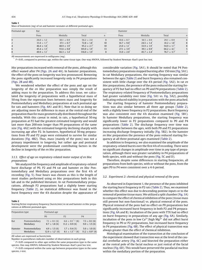
434 A.Y. Fong et al. / Respiratory Physiology & Neurobiology 164 (2008) 429–440
Table 1Mass of brainstem (mg) of rat and hamster neonates at different postnatal ages.
Postnatal age Rat Hamster
Pons Medulla Total n Pons Medulla Total n
0 26.0 ± 1.2 30.1 ± 0.9 56.2 ± 2.0 8 10.6 ± 0.7 18.5 ± 0.9 29.1 ± 0.9 82 35.8 ± 1.7* 37.1 ± 0.7* 72.8 ± 1.8* 10 16.7 ± 9.0* 24.5 ± 1.4* 41.2 ± 1.8* 84 46.4 ± 1.6* 48.9 ± 1.9* 95.2 ± 2.7* 10 21.0 ± 1.1* 33.9 ± 1.9* 54.9 ± 1.7* 86 45.4 ± 1.5* 55.6 ± 0.8* 101.0 ± 1.9* 13 27.1 ± 3.9* 39.1 ± 0.9* 66.2 ± 4.1* 58
AA, fol
bett(
lal5Pfapmplsitdpdd
3p
btrmlrfb
TSr
P
R
H
AN
s
s
ctIbsbqThi
twwIspmieq
rnrb
pa
3
49.9 ± 1.1* 66.3 ± 1.4* 116.0 ± 1.9*
ll measurements are expressed in milligrams (mg).* P < 0.05, compared to previous age, within the same tissue type. One-way ANOV
loc preparations increased with removal of the pons, although thisffect waned after P4 (Figs. 2A and 4A). In hamster preparations,he effect of the pons on longevity was less pronounced. Removinghe pons significantly increased longevity only in P4 preparationsFigs. 2B and 4B).
We wondered whether the effect of the pons and age on theongevity of the en bloc preparation was simply the result ofdding mass to the preparation. To address this issue, we calcu-ated the longevity of preparations with a hypothetical mass of0 mg from the regression lines between mass and longevity forontomedullary and Medullary preparations at each postnatal ageor rats and hamsters (Fig. 4A1 and B1). Note that in so doing were adjusting more for difference in mass at the rostral end of thereparation than for mass geometrically distributed throughout theedulla. With this caveat in mind, in rats, a hypothetical 50 mg
reparation at P2 had the greatest estimated longevity and wouldast more than 200 min longer than P0 preparations of the sameize (Fig. 4A2) with the estimated longevity declining sharply withncreasing age after P2. In hamsters, hypothetical 50 mg prepara-ions from P0 and P2 pups were estimated to survive for similarurations (Fig. 4B2). Thus, mass (mainly of the pons) was not arimary impediment to longevity, but rather age and postnatalevelopment were the predominant contributing factors in theecline in longevity of the en bloc preparation.
.1.3. Effect of age on respiratory-related motor output of en blocreparations
We analyzed the frequency and amplitude of respiratory-relatedurst discharge of P0, P2 and P4 rat and hamster en bloc Pon-omedullary and Medullary preparations over the first 4 h ofecording (Fig. 5). Four hours was chosen as this is the length of
ost studies performed using en bloc preparations both in thisab and in the published literature. In rat Pontomedullary prepa-ations, although P2 preparations had a slightly lower startingrequency (Table 2), no statistical difference was found in theurst frequency over the 4 h duration despite the appearance of
able 2tarting fictive respiratory frequency (bursts/min) in rat and hamster en bloc prepa-ations at different postnatal ages.
reparation type Postnatal age
P0 P2 P4
at Pontomedullary 7.1 ± 0.1 (6) 6.6 ± 0.1*,† (6) 7.3 ± 0.1 (6)Medullary 8.7 ± 1.8 (6) 11.2 ± 0.9 (6) 9.0 ± 1.5 (6)
amster Pontomedullary 4.9 ± 1.5 (6) 1.7 ± 0.4 (5) 5.0 ± 1.0 (6)Medullary 12.9 ± 1.2† (6) 8.1 ± 1.3*,† (6) 11.2 ± 0.9† (6)
ll values are expressed as bursts/min.umber in parentheses indicates number of observations in each group.* P < 0.05 compared to other ages within the same preparation type in the same
pecies. One-way ANOVA, followed by Student Newman–Kuel’s post hoc test.† P < 0.05 compared within the same age between preparation types in the same
pecies, t-test.
twrtsPstoifia
tdanw
8 31.0 ± 2.0* 45.7 ± 2.4* 76.7 ± 3.7* 7
lowed by Student Newman–Kuel’s post hoc test.
onsiderable variation (Fig. 5A1). It should be noted that P4 Pon-omedullary preparations stopped bursting after 150 min (Fig. 5A1).n rat Medullary preparations, the starting frequency was similaretween the ages (Table 2) and burst frequency also remained con-istent with little change over the 4 h period (Fig. 5A2). In rat enloc preparations, the presence of the pons reduced the starting fre-uency of P2 but had no effect on P0 and P4 preparations (Table 2).he respiratory related frequency of Pontomedullary preparationsad greater variability over time (Fig. 5A1 vs. Fig. 5A2), possibly
ndicating reduced stability in preparations with the pons attached.The starting frequency of hamster Pontomedullary prepara-
ions was also similar between all three age groups (Table 2),ith a slightly lower frequency in P2 preparations. Burst frequencyas also consistent over the 4 h duration examined (Fig. 5B1).
n hamster Medullary preparations, the starting frequency wasignificantly lower in P2 preparations compared to P0 and P4reparations (Table 2). The discharge frequency over time wasore variable between the ages, with both P0 and P2 preparations
ncreasing discharge frequency initially (Fig. 5B2). In the hamstern bloc preparation the presence of the pons reduced starting fre-uency at all three postnatal ages examined (Table 2).
In addition to frequency, we also examined the amplitude of theespiratory-related bursts over the first 4 h of recording. There wereo significant changes in amplitude over time in any type of prepa-ation, although there was greater variability in P4 preparations ofoth species, with and without the pons (Fig. 5C and D).
Therefore, despite some differences in starting frequencies, allreparations from both species, with or without the pons, from theges P0 to P4 were consistent over a 4-h period.
.2. Experiment 2: chemical and physical removal of the pons
As observed in Experiment 1, the presence of the pons inhibitedhe starting burst frequency in P2 rats (Table 2). Thus, we examinedhether this effect was due to descending pontine inputs or to the
emoval of pontine tissue mass. We utilized a split chamber (Fig. 1A)o compare the effect of chemical inhibition of the pons (tissue masstill present but non-functional) vs. physical removal of the pons.hysical removal of the pons had no effect on P0 preparations butignificantly increased burst frequency in both P2 and P4 prepara-ions (Fig. 6A and B). Incubation of the pons with TTX had no effectn burst frequency in preparations of any age (Fig. 6A). Similarly,ncubation of the pons in low Ca2+/high Mg2+ did not affect burstrequency in P0 or P2 preparations, but increased burst frequencyn P4 preparations (Fig. 6B). The effect of physical transection waslways greater than the effect of chemical inhibition.
Histological examination of the transection at the conclusion ofhe experiment showed that transection was at the level of the cau-al cerebellar artery (Fig. 6C) and bisected the preparation eithert the rostral pole of the facial nucleus or just rostral of the facialucleus (Fig. 6D). This would have preserved the parafacial nucleusithin the medullary portion of the preparation.

A.Y. Fong et al. / Respiratory Physiology & Neurobiology 164 (2008) 429–440 435
Fig. 5. Burst frequency of rat Pontomedullary (A1) and rat Medullary (A2) preparations was consistent over the first 4 h of recording at different postnatal ages. (B1) HamsterPontomedullary preparations had consistent burst frequency over the 4 h period at all three ages. (B2) Hamster Medullary preparations had differing patterns of burstfrequency over the 4 h period depending on the age of the preparation. Burst amplitude remained consistent in the rat Pontomedullary (C1), rat Medullary (C2), hamsterPontomedullary (D1) and hamster Medullary (D2) preparations for all three ages examined. Gray symbols denote significant difference from time 0 (P < 0.05, one way repeatedmeasures ANOVA, followed by Student Newman–Kuel’s post hoc test).

436 A.Y. Fong et al. / Respiratory Physiology & Neurobiology 164 (2008) 429–440
Fig. 6. (A) Quantitative changes in burst frequency following chemical inhibition ofthe pons by superfusion with TTX and pontine transection of P0, P2 and P4 rat prepa-rations. (B) Quantitative changes in burst frequency following pontine superfusionwith low Ca2+/high Mg2+ and pontine transection of P0, P2 and P4 rat preparations.(C) Photograph of an en bloc preparation showing the level of the pontine transec-tion, and (D) a representative photomicrograph of a hemisection at the cut surfaceon the medullary side, showing the rostral pole of the facial nucleus (VII) and thelaNi
3
wdtptm
3
ir
Table 3Effect of age on PO2
(Torr) within the medulla at the level of the PreBötzinger Com-plex of rat en bloc preparations from pups of different postnatal ages.
Age Preparation type
Pontomedullary Medullary
P2 54.2 ± 11.1 (7) 37.8 ± 14.0 (8)P
Ap
ab5tbsaoamsMeo6P
mmrTsrsmltstw
3
4b(p3
4
pqpamor
ocation of the fourth ventricle (4 V). Arrows indicate direction for dorsal surface (D)nd midline (M). *P < 0.05, one-way repeated measures ANOVA, followed by Studentewman–Kuel’s post hoc test. Numbers in the bars indicate number of preparations
n each age group.
.3. Experiment 3: oxygenation of medulla
The results of Experiment 2 did not allow us to clearly determinehether the inhibition of burst frequency by the pons was due toescending pontine inputs or due to the presence of the pontineissue mass. To better examine whether the effect of age, mass orresence of the pons on burst frequency was due to alteration inissue oxygenation in the medulla, we directly measured PO2 in the
edulla in various preparations.
.3.1. Effect of pons on PO2 in the medullaRemoval of pontine inputs by chemical and physical transection
nfluenced the frequency of respiratory-like activity in P4 prepa-ations. We measured the PO2 medulla of P4 preparations with
sspdm
4 42.1 ± 17.3 (6) 40.1 ± 3.8 (6)
ll measurements are shown in Torr. Number in parentheses indicates number ofreparations in each group.
nd without the pons attached. The PO2 in the superfusate (bub-led with 95% O2) at the start of each set of measurements was54.8 ± 5.8 Torr (n = 12) and began to decrease 300–400 (m abovehe surface of the brainstem, becoming significantly different to theulk superfusate PO2 200–300 (m above the surface of the brain-tem (Fig. 7A). This boundary layer effect resulted in an average PO2t the surface of the brainstem of 273.9 ± 12.4 Torr. The presencef the pons had no effect on the PO2 measured at the surface atny of the mediolateral co-ordinates (Fig. 7A and B). The PO2 in theedulla decreased significantly with greater depth in the brain-
tem at all mediolateral co-ordinates in both Pontomedullary andedullary preparations (P < 0.001). The presence of the pons had no
ffect on the PO2 levels at any depth within the medulla, regardlessf the mediolateral co-ordinates (Fig. 7B). PO2 reached zero between00 and 700 (m below the ventral surface of the brainstem in bothontomedullary and Medullary preparations.
To better examine the relationship between locations within theedulla and PO2 , the average of measurements taken at the threeediolateral tracts within Pontomedullary and Medullary prepa-
ations (shown in Fig. 7) were used to calculate a regression plot.his regression plot was then overlaid on a drawing of a repre-entative neonatal rat brainstem section illustrating the location ofespiratory nuclei of interest, as shown in Fig. 8. Regression analy-is demonstrated a layer of hyperoxygenated tissue throughout theediolateral aspect of the medulla, with PO2 decreasing to anoxic
evels with increasing distance from the surface, reaching 0 Torr athe level of Nucleus Ambiguus. The pattern of oxygenation was theame in both Pontomedullary and Medullary preparations. PO2 athe PreBötC in both Pontomedullary and Medullary preparationsere similar and ranged between 50 and 140 Torr (Fig. 8).
.3.2. Effect of age on PO2 at the PreBötCMeasurement of PO2 at a single location (1.0 mm from midline,
00 (m depth, the approximate level of the PreBötC) was similaretween P2 and P4 Pontomedullary and Medullary preparationsTable 3). PO2 at the PreBötC in both Pontomedullary and Medullaryreparations, regardless of age, were similar and ranged between8 and 54 Torr (Table 3).
. Discussion
While it is widely recognized that the longevity of the en blocreparation decreases with postnatal age, this study is the first touantify the relationship between longevity, age and mass of thereparations, both with the pons-intact and pons-removed in ratsnd hamsters. We found that postnatal development rather thanass appeared to be the major factor determining the longevity
f the en bloc preparations. This was re-enforced by the similarelationships between longevity and age in both rats and ham-
ters despite significant differences in brain mass between thepecies. We also found that pontine inhibition of medullary res-iratory rhythmogenesis increased with postnatal age and was notue to changes in oxygenation at the rhythm generating sites in theedulla. In en bloc rat preparations, regardless of mass, or pres-
A.Y. Fong et al. / Respiratory Physiology & Neurobiology 164 (2008) 429–440 437
Fig. 7. Measurement of PO2(Torr) in the medulla of rat Pontomedullary and Medullary preparations shows a boundary layer of 300 (m over the ventral surface of the brainstem
where P decreased from superfusate levels at all three mediolateral tracts (A). Increasing depth in the medulla resulted in decreasing P until 0 Torr was reached between6 tweenf n eitht depth
eltm
4
crw(otd
ioitltainr
O200 and 700 (m below the ventral surface. No difference in PO2
levels was found beound between the PO2
at matching depths in different mediolateral co-ordinates ihe medulla to a depth of 700 (m. The shaded area in panel B indicates the reported
nce of the pons, or age (from P2 to P4), the PO2 at the approximateevel of the PreBötzinger complex at 27 ◦C was similar and withinhe physiological range, indicating sufficient oxygenation of the
edullary respiratory network.
.1. The effect of age and mass on the en bloc preparation
We were interested in evaluating the effect of age-relatedhanges in mass vs. developmental effects on the en bloc prepa-ation. Our study provides a systematic examination of the
ell-established and widely observed phenomenon that longevitytime to last burst) of the en bloc preparation decreases with devel-pmental age. This reduced longevity has generally been attributedo increasing tissue mass with age and a corresponding increase iniffusion distance for oxygen and thus, reduced oxygen availabil-
dtmPp
O2the Pontomedullary and Medullary preparations at any depth. No difference was
er preparation types. Panel B shows expanded profiles for PO2from the surface of
of respiratory neurons in the medulla of the en bloc preparation.
ty to the medullary respiratory network. A number of noteworthybservations from our study suggest that the reduced longevitys likely due to developmental and maturational changes, ratherhan just increasing size. First, during the first two postnatal days,ongevity increased despite increasing total mass. Secondly, whilehere was an inverse correlation between tissue mass and longevityfter P2, the PO2 at the level of the ventral respiratory column (VRG)n the medulla, including the preBötzinger Complex (PreBötC), didot change with increasing age. Lastly, both hamster and rat prepa-ations exhibited a similar relationship between age and longevity
espite the larger mass of rat brainstems (Table 1). Equally, prepara-ions from rats and hamsters at different postnatal ages with similarass did not have the same longevity. For example, the masses of8 hamster Medullary preparations (45.7 mg) and P4 rat Medullaryreparations (46.4 mg) are similar, however, the rat Medullary

438 A.Y. Fong et al. / Respiratory Physiology & Neurobiology 164 (2008) 429–440
F Medulm re is nl lex; N
pa1eDiPvdbtl
4
aif(Tlre2apcrimttpia
tpoitts
miccocortmAvtttHpns
4
omoiaOwlwtbw
ti(
ig. 8. Psuedocolored plot of PO2level within the medulla in Pontomedullary and
edulla. The location of the PreBötzinger Complex (PBC) is indicated in white. Theevels of PO2
within the PBC (50–140 Torr). Abbreviations: IOC, inferior olivary comp
reparations produced spontaneous fictive respiratory activity forlmost 5 h, while P8 hamster Medullary preparations lasted only6 min. Rats and hamsters are developmentally equivalent by P8,ven though hamsters are born more altricial to rats (Finlay andarlington, 1995; Clancy et al., 2001, 2007). Thus, despite the sim-
lar mass between the P8 hamster Medullary preparations and the4 rat Medullary preparation, the younger preparation has greateriability. Taken together, these observations suggest that postnatalevelopmental is the primary determinant of longevity of the enloc preparation and the decreased longevity of en bloc prepara-ions with age is not due to limitation of oxygen diffusion due toarger size.
.2. The effect of the pons on the en bloc preparation
In the present study, we report that the pons reduced longevitynd inhibited burst frequency on the en bloc preparation. Thiss in agreement with numerous reports of increasing respiratoryrequency following pontine removal in the en bloc preparationHilaire et al., 1989; Smith et al., 1990; Errchidi et al., 1991;anabe et al., 2005). In our study, this change in frequency fol-owing transection was not due to removal of the parafacialespiratory group, a proposed second respiratory rhythm gen-rator (Onimaru and Homma, 2003; Janczewski and Feldman,006). Our level of transection consistently left the facial nucleusnd therefore, the adjacent pFRG intact within the medullaryreparation (Ruangkittisakul et al., 2007). In addition, chemi-al inhibition of the pons in P4 preparations produced similaresults to physical transection (Fig. 6B), suggesting that the increasen burst frequency was not due to removal of pontine tissue
ass, but rather the removal of pontine inputs. Furthermore,he removal of the pons did not alter the level of oxygen inhe medulla (see next section). Together, these observations sup-ort the conclusion that the pons provides direct descending
nhibitory inputs to the medulla (Hilaire et al., 1989; Errchidi etl., 1991).
While pontine inhibition produced similar results to physicalransection, chemical inhibition was always less effective thanhysical transaction. The reason for this is not clear. As the level
f the grease gap isolating the chemical inhibition to the pons wasdentical to the level of transection, the tissue that was bathed byhe pontine superfusate was identical to that removed by transec-ion. It is possible that the chemicals did not penetrate to the deepertructures in the pons. Or the concentrations of TTX or Ca2+/Mg2+wtdBs
lary preparations overlaid on a coronal schematic representation of a neonatal rato difference in the oxygenation profile between the two preparations with similarA, nucleus ambiguous; PBC, pre-Bötzinger complex; ROb, raphé obscurus.
ay have been insufficient to completely inhibit neuronal activ-ty throughout the pons. Preliminary experiments showed that theoncentrations of TTX and low Ca2+/high Mg2+ superfusate wereapable of silencing the medulla when applied to the medulla sidef the split bath, therefore, the same concentrations of chemi-al was presumed to be sufficient to silence the neuronal activityf the pons. However, as the medullary structures involved inespiratory rhythmogenesis are generally more superficial thanhe pontine sites known to modulate respiratory activity, these
edullary regions may have had greater accessibility to the drugs.nother possibility is that the grease covered some of the pontineentral surface limiting the access of the superfusate (and drug) tohis small portion of the pons, especially regions located close tohe ventral surface, such as the A5 region which is known to par-icipate in modulation of respiratory rhythm (Errchidi et al., 1991;ilaire et al., 2004). The potential pontine mechanisms involved inroviding the inhibition to medullary respiratory networks wereot examined in the current study but will be the focus of a futuretudy.
.3. Tissue oxygenation in the medulla of the en bloc preparation
Another objective in the present study was to examine the effectf age, mass and the presence of the pons on oxygenation of theedulla in the en bloc preparation. It was possible that the effects
f pontine removal on longevity and burst frequency were due toncreased oxygenation to the medullary respiratory rhythm gener-tors as pontine inhibition was not as effective as pontine removal.ur data, however, show that PO2 measurements in the medullaere unaffected by age or the presence of the pons. In particu-
ar, the PO2 in the preBötzinger Complex (PreBötC) did not changeith increasing age. The distance between the ventral surface and
he ventral edge of the Nucleus Ambiguus was roughly constantetween P0 and P4 (540–580 (m), placing the PreBötC within aell-oxygenated region of the medulla.
A number of postnatal developmental events may contribute tohe reduced longevity of the en bloc preparations. Current evidencendicates the appearance of myelin in the brainstem from birthin spinal trigeminal tract) to appearance after the first postnatal
eek (in pyramidal tract) (Hamano et al., 1998). Myelin continueso develop over early postnatal period reaching mature levels atifferent rates in different regions (Hamano et al., 1998; Butt anderry, 2000). This postnatal increase in myelin may alter the diffu-ion of oxygen through the tissue, increasing the distance between

ogy &
tsdrds
wis3(a(Pti
tamfbrhVrtand
oedmdadTocofBs
4
forpuartdodtrwd
A
tB
R
B
B
C
C
D
E
F
G
H
H
H
H
H
I
J
K
L
L
M
N
O
P
R
A.Y. Fong et al. / Respiratory Physiol
he ventral surface and the PreBötC or alter metabolism of the brain-tem. Since the oxygenation of the medullary respiratory networkid not change over this period (P0–P4), our data suggest that theeduced longevity of the brainstem–spinal cord preparation is notue to events that alter O2 tissue diffusion by developmental eventsuch as postnatal myelination.
PO2 measurements in the medulla in our study are in agreementith earlier study (Brockhaus et al., 1993) and the PO2 at the PreBötC
s comparable to data obtained in vivo. Several studies have demon-trated oxygen tension in different brain regions ranging from 2 to8 Torr under normoxic conditions in in vivo, anaesthetized ratsSick et al., 1982; Nwaigwe et al., 2000; Hou et al., 2003; Hare etl., 2006). Compared to these in vivo values, the superficial regionsup to 400 (m) of the en bloc preparations are hyperoxic, while theO2 in regions between 400 and 600 (m from the surface, includinghe respiratory neurons of the VRG, are similar to those observedn vivo.
Although the VRG is well oxygenated, regions located deeper inhe medulla, such as the midline raphé, will be situated within annoxic region. The role of the midline raphé in respiratory rhythmodulation is well established (Li et al., 2006) and excitatory drive
rom this region is essential for respiratory rhythmogenesis in therainstem slice preparation (Pace et al., 2007). The en bloc prepa-ation continuously produces rhythmic motor output for manyours with spontaneously active respiratory neurons within theRG (Suzue, 1984; Smith et al., 1990) indicating that the respi-
atory network is functional in these preparations. This suggestshat deep regions such as midline raphé is not required for gener-tion of respiratory rhythm in en bloc preparations. However, it isot clear whether the roles of these sites increase during postnatalevelopment.
One criticism of oxygen probes is that the physical introductionf the sensor into the brainstem may provide a conduit around thelectrode for the highly oxygenated superfusate to reach the deeperepths of the tissue, leading to an overestimation of the PO2 level. Toinimize this possibility, we used optical electrodes with a small
iameter (30 (m) to minimize the size of the opening in the tissuend reduce the likelihood of damage and any inadvertent intro-uction of the highly oxygenated aCSF to the depths of the tissue.his effect of introducing the electrode would be similar regardlessf age and the presence of the pons and thus, does not alter ouronclusion that PO2 within the medulla is unaffected by presencef the pontine tissue mass or age. Furthermore, PO2 measurementsrom our study are in agreement with those previously reported byrockhaus et al. (1993) supporting the PO2 levels measured in ourtudy.
.4. Conclusion
This study demonstrates that postnatal development has a pro-ound effect on the longevity of the en bloc preparation. In addition,ur data support earlier reports of direct pontine inhibition of respi-atory rhythm and adds that this pontine inhibition increases withostnatal development. Finally, we show that, under the conditionssed in the current study, the VRG is well oxygenated and the PO2t the PreBötzinger Complex at 27 ◦C is within the physiologicalange of PO2 in vivo and is unaffected by age or the presence ofhe pons in en bloc preparations from rats. Taken together, theseata suggest that the effects of postnatal age or pontine removaln respiratory rhythmogenesis in the en bloc preparation are not
ue to reduced availability of oxygen at the medullary respira-ory network. The exact manner by which postnatal developmenteduces the longevity of the en bloc preparation and the extent tohich this also applies to even older preparations, remains to beetermined.S
S
Neurobiology 164 (2008) 429–440 439
cknowledgements
This work was funded by a grant from the NSERC of Canadao WKM. DVA was supported by grants from FAPESP and CNPq ofrazil.
eferences
rockhaus, J., Ballanyi, K., Smith, J.C., Richter, D.W., 1993. Microenvironment of res-piratory neurons in the in vitro brainstem–spinal cord of neonatal rats. J. Physiol.462, 421–445.
utt, A.M., Berry, M., 2000. Oligodendrocytes and the control of myelination invivo: new insights from the rat anterior medullary velum. J. Neurosci. Res. 59,477–488.
lancy, B., Darlington, R.B., Finlay, B.L., 2001. Translating developmental time acrossmammalian species. Neuroscience 105, 7–17.
lancy, B., Finlay, B.L., Darlington, R.B., Anand, K.J., 2007. Extrapolating brain devel-opment from experimental species to humans. Neurotoxicology 28, 931–937.
utschmann, M., Morschel, M., Kron, M., Herbert, H., 2004. Development of adaptivebehaviour of the respiratory network: implications for the pontine Kolliker-Fusenucleus. Resp. Physiol. Neurobiol. 143, 155–165.
rrchidi, S., Monteau, R., Hilaire, G., 1991. Noradrenergic modulation of the medullaryrespiratory rhythm generator in the newborn rat: an in vitro study. J. Physiol. 443,477–498.
inlay, B.L., Darlington, R.B., 1995. Linked regularities in the development and evo-lution of mammalian brains. Science 268, 1578–1584.
uimaraes, L., Dominguez-del-Toro, E., Chatonnet, F., Wrobel, L., Pujades, C., Mon-teiro, L.S., Champagnat, J., 2007. Exposure to retinoic acid at the onset ofhindbrain segmentation induces episodic breathing in mice. Eur. J. Neurosci.25, 3526–3536.
amano, K., Takeya, T., Iwasaki, N., Nakayama, J., Ohto, T., Okada, Y., 1998. A quan-titative study of the progress of myelination in the rat central nervous system,using the immunohistochemical method for proteolipid protein. Brain Res. Dev.Brain Res. 108, 287–293.
are, G.M., Harrington, A., Liu, E., Wang, J.L., Baker, A.J., Mazer, C.D., 2006. Effect ofoxygen affinity and molecular weight of HBOCs on cerebral oxygenation andblood pressure in rats. Can. J. Anaesth. 53, 1030–1038.
ilaire, G., Monteau, R., Errchidi, S., 1989. Possible modulation of the medullaryrespiratory rhythm generator by the noradrenergic A5 area: an in vitro study inthe newborn rat. Brain Res. 485, 325–332.
ilaire, G., Viemari, J.C., Coulon, P., Simonneau, M., Bevengut, M., 2004. Modulationof the respiratory rhythm generator by the pontine noradrenergic A5 and A6groups in rodents. Resp. Physiol. Neurobiol. 143, 187–197.
ou, H., Grinberg, O.Y., Taie, S., Leichtweis, S., Miyake, M., Grinberg, S., Xie,H., Csete, M., Swartz, H.M., 2003. Electron paramagnetic resonance assess-ment of brain tissue oxygen tension in anesthetized rats. Anesth. Analg. 96,1467–1472, table.
to, Y., Oyamada, Y., Hakuno, H., Yamaguchi, K., 2002. Morphological analysis ofdevelopmental changes in pontine noradrenergic neuronal groups in the neona-tal rat. Brain Res. 925, 107–109.
anczewski, W.A., Feldman, J.L., 2006. Distinct rhythm generators for inspiration andexpiration in the juvenile rat. J. Physiol. 570, 407–420.
ron, M., Morschel, M., Reuter, J., Zhang, W., Dutschmann, M., 2007. Developmentalchanges in brain-derived neurotrophic factor-mediated modulations of synap-tic activities in the pontine Kolliker-Fuse nucleus of the rat. J. Physiol. 583,315–327.
i, A., Zhou, S., Nattie, E., 2006. Simultaneous inhibition of caudal medullary rapheand retrotrapezoid nucleus decreases breathing and the CO2 response in con-scious rats. J. Physiol. 577, 307–318.
iu, Y.Y., Wong-Riley, M.T., 2001. Developmental study of cytochrome oxidase activ-ity in the brain stem respiratory nuclei of postnatal rats. J. Appl. Physiol. 90,685–694.
ellen, N.M., Milsom, W.K., Feldman, J.L., 2002. Hypothermia and recovery fromrespiratory arrest in a neonatal rat in vitro brain stem preparation. Am. J. Physiol.282, R484–R491.
waigwe, C.I., Roche, M.A., Grinberg, O., Dunn, J.F., 2000. Effect of hyperventila-tion on brain tissue oxygenation and cerebrovenous PO2 in rats. Brain Res. 868,150–156.
nimaru, H., Homma, I., 2003. A novel functional neuron group for respiratoryrhythm generation in the ventral medulla. J. Neurosci. 23, 1478–1486.
ace, R.W., Mackay, D.D., Feldman, J.L., Del Negro, C.A., 2007. Role of persistentsodium current in mouse preBotzinger Complex neurons and respiratory rhythmgeneration. J. Physiol. 580, 485–496.
uangkittisakul, A., Secchia, L., Bornes, T.D., Palathinkal, D.M., Ballanyi, K., 2007.Dependence on extracellular Ca2+/K+ antagonism of inspiratory centre rhythmsin slices and en bloc preparations of newborn rat brainstem. J. Physiol. 584,489–508.
ick, T.J., Lutz, P.L., LaManna, J.C., Rosenthal, M., 1982. Comparative brain oxygena-tion and mitochondrial redox activity in turtles and rats. J. Appl. Physiol. 53,1354–1359.
mith, J.C., Greer, J.J., Liu, G.S., Feldman, J.L., 1990. Neural mechanisms generating res-piratory pattern in mammalian brain stem-spinal cord in vitro. I. Spatiotemporalpatterns of motor and medullary neuron activity. J. Neurophysiol. 64, 1149–1169.

4 ogy &
S
T
T
Viemari, J.C., Burnet, H., Bevengut, M., Hilaire, G., 2003. Perinatal maturation of
40 A.Y. Fong et al. / Respiratory Physiol
uzue, T., 1984. Respiratory rhythm generation in the in vitro brain stem-spinal cord
preparation of the neonatal rat. J. Physiol. 354, 173–183.anabe, A., Fujii, T., Onimaru, H., 2005. Facilitation of respiratory rhythm by a mu-opioid agonist in newborn rat pons–medulla–spinal cord preparations. Neurosci.Lett. 375, 19–22.
attersall, G.J., Milsom, W.K., 2003. Hypothermia-induced respiratory arrest andrecovery in neonatal rats. Resp. Physiol. Neurobiol. 137, 29–40.
Z
Neurobiology 164 (2008) 429–440
the mouse respiratory rhythm-generator: in vivo and in vitro studies. Eur. J.Neurosci. 17, 1233–1244.
immer, M.B., Milsom, W.K., 2004. Effect of hypothermia on respiratory rhythm gen-eration in hamster brainstem–spinal cord preparations. Resp. Physiol. Neurobiol.142, 237–249.