Embed Size (px)
Citation preview
Respiratory System
Christopher RobinsonEmanuel Williams
Maya VarnerJovanni Rivers
Tiffany CorneliusAsya Pace
Maycee Labares
Bill Nye Video Linkhttps://www.youtube.com/watch?v=CNJYKgLRPJ0

Respiratory System
● Consists of the respiratory and conducting zones
● Respiratory zone
o Site of gas exchange
o Consists of bronchioles, alveolar ducts, and alveoli
● Conducting zone
o Provides rigid conduits for air to reach the sites of gas exchange
o Includes all other respiratory structures (e.g., nose, nasal cavity, pharynx, trachea)
Respiratory muscles – diaphragm and other muscles that promote ventilation
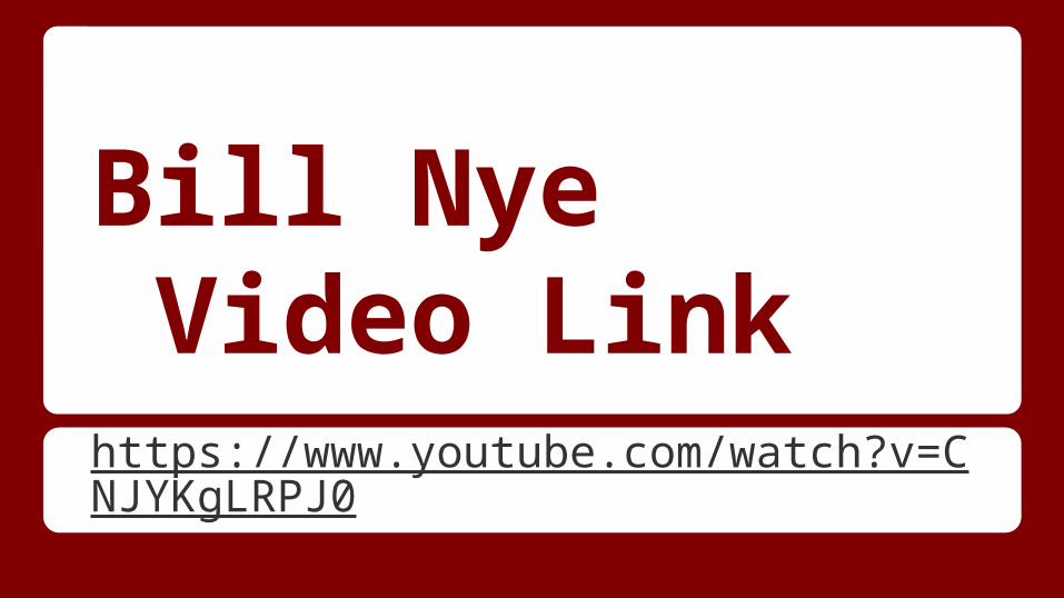
The Respiratory System
Major Functions
● To supply the body with oxygen and dispose of carbon dioxide
● Respiration – four distinct processes must happen
o Pulmonary ventilation – moving air into and out of the lungs
o External respiration – gas exchange between the lungs and the blood
● Transport – transport of oxygen and carbon dioxide between the lungs and tissues
● Internal respiration – gas exchange between systemic blood vessels and tissues
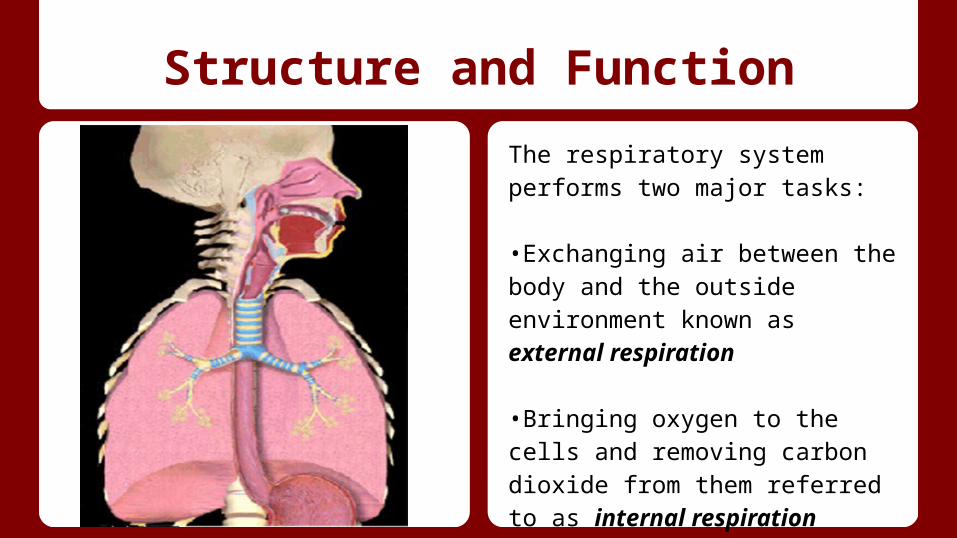
Structure and Function
The respiratory system performs two major tasks:
•Exchanging air between the body and the outside environment known as external respiration
•Bringing oxygen to the cells and removing carbon dioxide from them referred to as internal respiration
Structure and Function
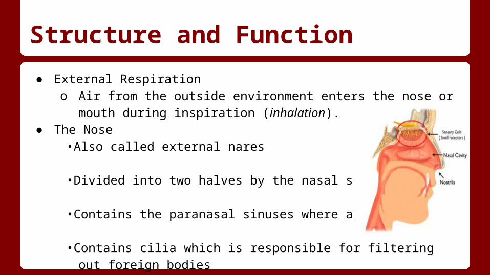
● External Respirationo Air from the outside environment enters the nose or mouth during
inspiration (inhalation). ● The Nose
•Also called external nares
•Divided into two halves by the nasal septum
•Contains the paranasal sinuses where air is warmed
•Contains cilia which is responsible for filtering out foreign bodies
Structure and Function
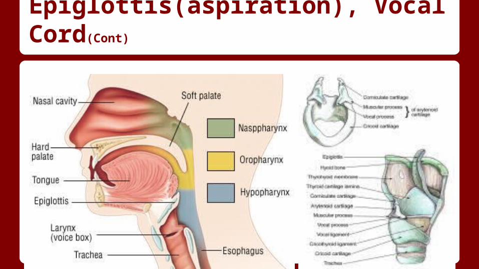
● Pharynxo Also known as the throat is a passageway for both air and food
Three Sections of the Pharynx● Nasopharynx- contains the pharyngeal tonsils (adenoids) which aid in the
body’s immune defense● Oropharynx- back portion of the mouth that contains the palatine tonsils
which aid in the body’s immune defense
● Laryngopharynx- -bottom section of the pharynx where the respiratory tract divides into the esophagus and the larynx
Structure and Function
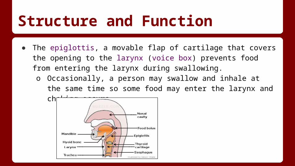
● The epiglottis, a movable flap of cartilage that covers the opening to the larynx (voice box) prevents food from entering the larynx during swallowing.o Occasionally, a person may swallow and inhale at the same time so
some food may enter the larynx and choking occurs.
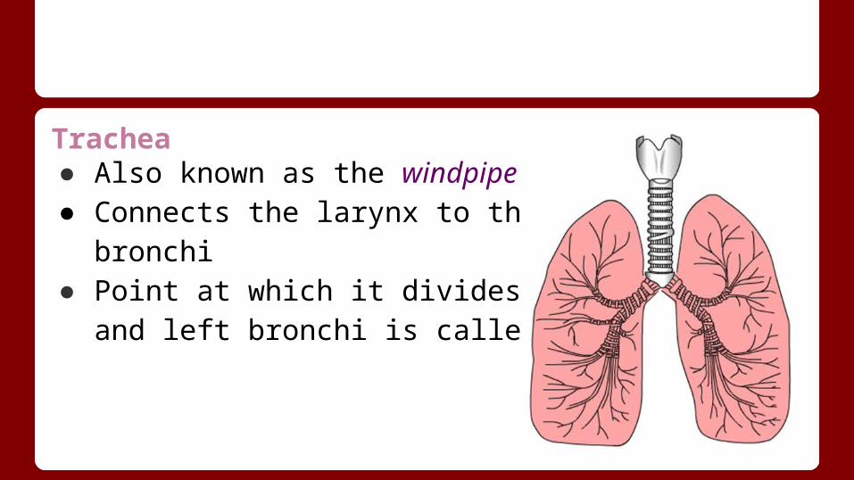
Trachea● Also known as the windpipe● Connects the larynx to the right and left bronchi● Point at which it divides into the right and left bronchi is
called the mediastinum
Pharynx, Larynx, Trachea, Epiglottis(aspiration), Vocal Cord
● Epiglottis- leaf-shaped cartilage;forms a lid over the glottis that protects the larynx from aspiration of foods or liquids being swallowed.
● Pharynx- Cone-shaped passageway leading from the oral and nasal cavities in the head to the esophagus and larynx;Consists of Circular and Longitudinal muscles. Circular helps push food down and prevents air from being swallowed; Longitudinal helps lift the walls of the pharynx
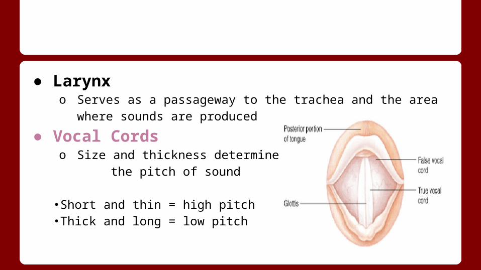
● Larynxo Serves as a passageway to the trachea and the area where sounds
are produced
● Vocal Cords o Size and thickness determine
the pitch of sound
•Short and thin = high pitch•Thick and long = low pitch
Pharynx, Larynx, Trachea, Epiglottis(aspiration), Vocal Cord(Cont)
Larynx- Its primary function is to protect the lower airway by closing abruptly upon mechanical stimulation, thereby halting respiration and preventing the entry of foreign matter into the airway. Trachea-Commonly known as the windpipe, the trachea widens and lengthens slightly with each breath in, returning to its resting size with each breath out.
Pharynx, Larynx, Trachea, Epiglottis(aspiration), Vocal Cord(Cont)

Internal Respiration● Air from the bronchi travels to the bronchioles then to
the tiny air sacs (alveoli) which connect to lung capillaries.
● Oxygen and carbon dioxide are exchanged and oxygen is delivered to the body cells. alveolus
capillaries
The Lungs
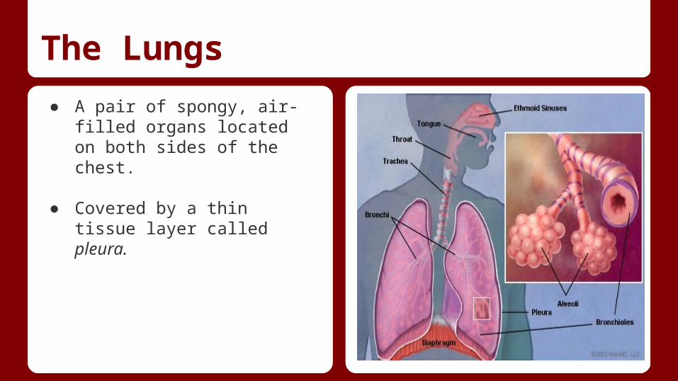
● A pair of spongy, air-filled organs located on both sides of the chest.
● Covered by a thin tissue layer called pleura.
Structure and Function (Lungs)
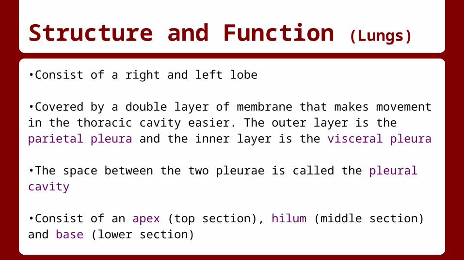
•Consist of a right and left lobe
•Covered by a double layer of membrane that makes movement in the thoracic cavity easier. The outer layer is the parietal pleura and the inner layer is the visceral pleura
•The space between the two pleurae is called the pleural cavity
•Consist of an apex (top section), hilum (middle section) and base (lower section)
Structure and Function
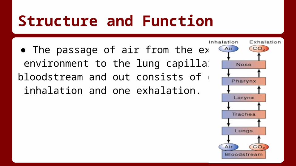
● The passage of air from the external environment to the lung capillaries, bloodstream and out consists of one inhalation and one exhalation.
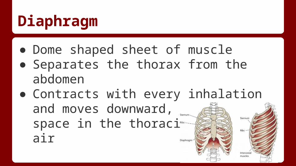
Diaphragm
● Dome shaped sheet of muscle● Separates the thorax from the abdomen● Contracts with every inhalation and moves
downward, causing more space in the thoracic cavity for air
Diaphragm Cont.
● Separated into two parts1. Peripheral Muscle - Consists of many radiating muscle fibers1. Central Tendon - Made up of dense collagen fibers
Respiratory Mucosa
● Mucous membrane lining the respiratory tracto Nasal Cavityo Larynxo Tracheao Bronchi
● Consist of various types of epithelial cells: o ciliated columnaro simple squamous
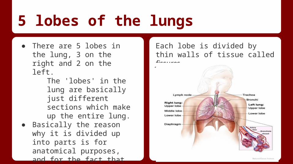
5 lobes of the lungs
● There are 5 lobes in the lung, 3 on the right and 2 on the left.
The 'lobes' in the lung are basically just different sections which make up the entire lung.
● Basically the reason why it is divided up into parts is for anatomical purposes, and for the fact that each section functions as a separate but interconnected unit.
Each lobe is divided by thin walls of tissue called fissures.
Lobes of the lung ..● In the right lung, there is the superior, middle and inferior lobes. The left lung however only has a superior
and inferior lobe. The superior is at the top, middle is in the middle (in the right lung), and inferior at the bottom of the lung.
Superior Lobe: The lobe of the right lung that lies above the oblique and horizontal fissures and includes the apical, posterior and anterior bronchopulmonary segments; in the left lung, the lobe lies above the oblique fissure and contains the apicoposterior, anterior, superior lingular and inferior lingular segments.
Middle Lobe: Located anteriorly between the horizontal and oblique fissures and includes lateral and medial bronchopulmonary segments.
Inferior Lobe: Located below and behind the oblique fissure and contains five bronchopulmonary segments: superior, medial basal, anterior basal, lateral basal, and posterior basal.
Overall, each section or lobe have similar functions, they each contain branches of alveoli and their job is to work together to filter air and provide oxygen for the bloodstream.
Alveoli, Alveoli sac,and alveolar duct
● Oxygen exchange in the lungs takes place in small balloon like structures called Alveoli. They are attach to the bronchial branches in our lungs.
● the alveolar sac is the space in which the alveolar duct open distally and makes the Alveoli rise in the lung.
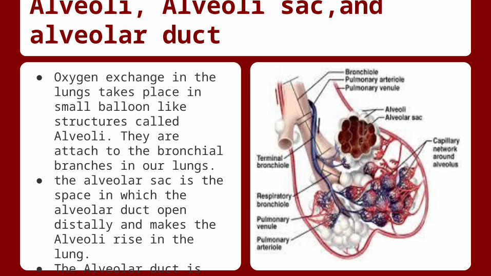
Alveoli, Alveoli sac,and alveolar duct
● Oxygen exchange in the lungs takes place in small balloon like structures called Alveoli. They are attach to the bronchial branches in our lungs.
● the alveolar sac is the space in which the alveolar duct open distally and makes the Alveoli rise in the lung.
● The Alveolar duct is the stem that passes through the Alveoli
Air-Blood Barrier
This material intervening between alveolar air and capillary blood it consists of a nonstructural film or surfactant.
Major functions of the Respiratory System
●To supply the body with oxygen and dispose of carbon dioxide●Respiration – four distinct processes must happen●Pulmonary ventilation – moving air into and out of the lungs●External respiration – gas exchange between the lungs and the blood
Continued
●Transport – transport of oxygen and carbon dioxide between the lungs and tissues
●Internal respiration – gas exchange between systemic blood vessels and tissues
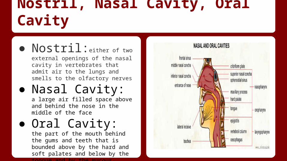
Nostril, Nasal Cavity, Oral Cavity
● Nostril:either of two external
openings of the nasal cavity in vertebrates that admit air to the lungs and smells to the olfactory nerves
● Nasal Cavity: a large air
filled space above and behind the nose in the middle of the face
● Oral Cavity: the part of the
mouth behind the gums and teeth that is bounded above by the hard and soft palates and below by the tongue and by the mucous membrane connecting it with the inner part of the mandible
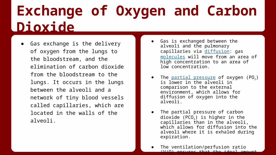
Exchange of Oxygen and Carbon Dioxide
● Gas exchange is the delivery of oxygen
from the lungs to the bloodstream, and
the elimination of carbon dioxide from the
bloodstream to the lungs. It occurs in the
lungs between the alveoli and a network
of tiny blood vessels called capillaries,
which are located in the walls of the
alveoli.
● Gas is exchanged between the alveoli and the pulmonary capillaries via diffusion: gas molecules will move from an area of high concentration to an area of low concentration.
● The partial pressure of oxygen (PO2) is lower in the alveoli in comparison to the external environment, which allows for diffusion of oxygen into the alveoli.
● The partial pressure of carbon dioxide (PCO2) is higher in the capillaries than in the alveoli, which allows for diffusion into the alveoli where it is exhaled during expiration.
● The ventilation/perfusion ratio (V/Q) ensures that the ideal amount of blood and gas is received by the alveoli for efficient gas exchange.
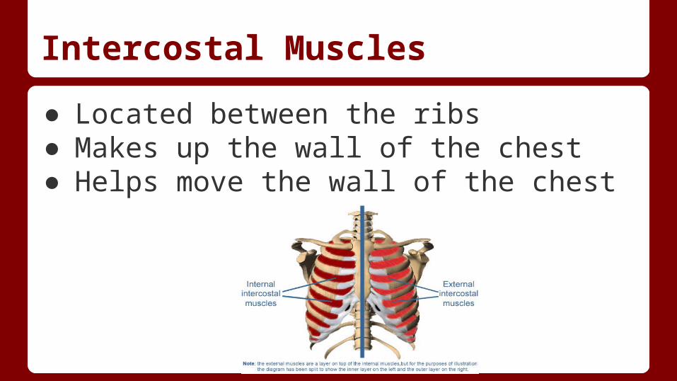
Intercostal Muscles
● Located between the ribs● Makes up the wall of the chest● Helps move the wall of the chest
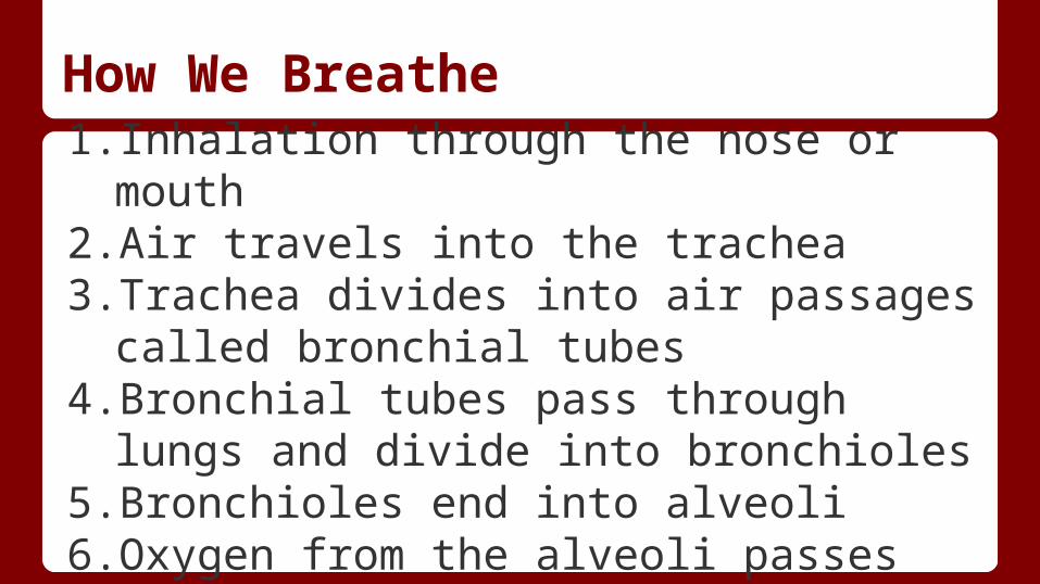
How We Breathe1. Inhalation through the nose or mouth2. Air travels into the trachea3. Trachea divides into air passages called
bronchial tubes4. Bronchial tubes pass through lungs and
divide into bronchioles 5. Bronchioles end into alveoli 6. Oxygen from the alveoli passes into
capillaries
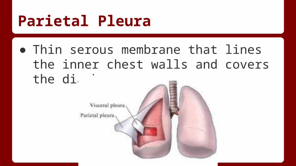
Parietal Pleura
● Thin serous membrane that lines the inner chest walls and covers the diaphragm
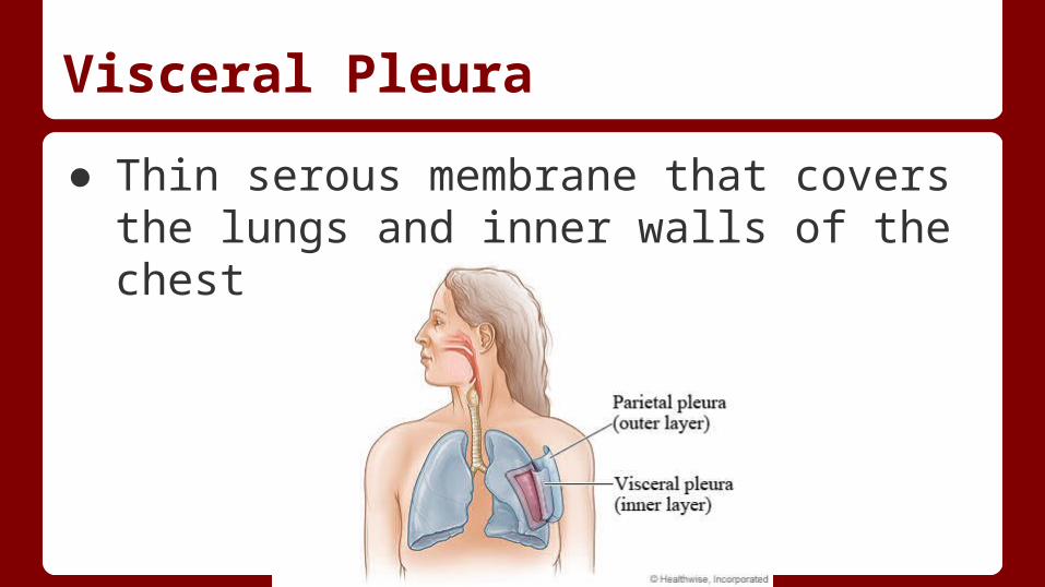
Visceral Pleura
● Thin serous membrane that covers the lungs and inner walls of the chest
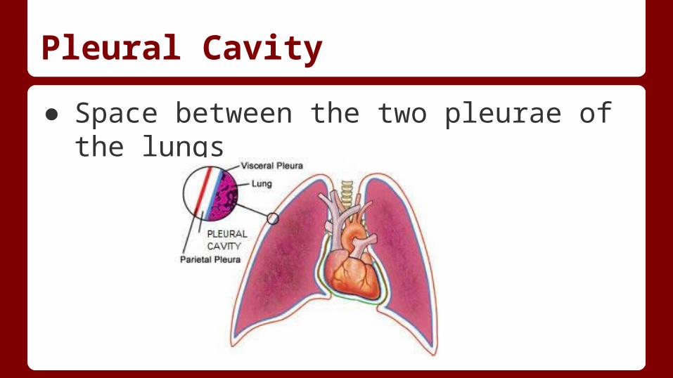
Pleural Cavity
● Space between the two pleurae of the lungs
Primary Bronchi, Secondary Bronchi
● Bronchi- Bronchi are the main passageway into the lungs.
● Primary bronchi- located in the upper portion of the lungs.o The right primary bronchus enters the right lung nearly opposite the fifth thoracic vertebra.
The left primary bronchus divides into bronchi for the superior and anterior lobes of the lung.
● Secondary bronchi- near the center of the lungs. o No gas exchanges occur in any of the bronchi. o The right primary bronchus enters the right lung nearly opposite the fifth thoracic vertebra.
The left primary bronchus divides into bronchi for the superior and anterior lobes of the lung.
Pulmonary Arteries and Veins
● Your pulmonary vein are a group of blood vessels that drain oxygenated blood from your lungs and return it to your heart.
Yawning
- Helps cool the brain- Lack of sleep is not the only cause of yawning.- contagious- Tends to occur in the summer because it cools the brain- stress and anxiety causes the brain to get hotter and
yawning can help it cool off.- the receptors that turn yawning on and off works with
dopamine.- we yawn when waking up in the morning because
dopamine levels are usually high during that time.

Diseases
- Chronic Obstructive Pulmonary Disease: intersection of three related disease.
- bronchitis, chronic asthma and emphysema- makes it difficult to breath
- Asthma: inflammation of lung airways causing coughing, wheezing, shortness of breath- Lung cancer: may be common with smoker, but can actually happen to anyone.
Cited Sources
http://www.innerbody.com/image/musc06.htmlhttp://www.innerbody.com/image_chest1/chest01.html#full-descriptionhttp://www.webmd.com/lung/how-we-breathehttp://www.thefreedictionary.com/parietal+pleura