Embed Size (px)
Citation preview

Respiratory System
Health CareersCanadian Valley Technology Center

Respiratory system
• Complex organs and structures that perform 1. pulmonary ventilation (external
respiration) - process of inhaling and exhaling air 2. cellular respiration (internal respiration) - process of oxygen carried by the blood passing into the cells and being used
by the cells

Functions of the Respiratory System
• exchange of carbon dioxide and oxygen
• regulate the pH of blood

Characteristics of pulmonary ventilation
• Mechanical portion is breathing• Two stages - inhalation/exhalation• Contolled by - medulla oblongata - pons• Sends impulses to phrenic nerve - out to diaphragm
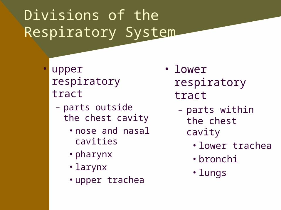
Divisions of the Respiratory System
• upper respiratory tract– parts outside the
chest cavity• nose and nasal
cavities• pharynx• larynx• upper trachea
• lower respiratory tract– parts within the
chest cavity• lower trachea• bronchi• lungs

Nose and Nasal Cavities
• bone and cartilage• two nasal cavities
– divided by the nasal septum
• palate forms the floor of the nasal cavity– hard palate
• anterior portion– soft palate
• posterior portion

• soft palate ends in a projection called the uvula– directs food into the oropharynx
• nasal mucosa consists of ciliated epithelium– filters the air– produces mucus that traps foreign material– cilia propels mucus with trapped particles
toward the pharynx where it is swallowed• gastric juices in the stomach will then destroy
most of the microorganisms

Nasal Conchae
• three bony ridges, called nasal conchae are found on the lateral wall of each nasal cavity– superior, middle,
inferior– increase the surface
area of the cavity– warm and moisten the
air and to direct air flow

Paranasal Sinuses
• air-filled cavities open into the nasal cavity
• frontal, maxillae, ethmoid, and sphenoid bones
• functions of paranasal sinues are to:– reduce the weight of the skull– produce mucus – influence voice quality

• olfactory receptors– upper nasal
cavities– detect vaporized
chemicals – olfactory nerves
pass through the ethmoid bone to the brain

Passage of air
air entersexternal nares
openings intothe pharynx are the internal nares

Functions of the Nose
• passageway for air going to the lungs• warms, moistens, and filters air of
impurities• organ of smell• aids in phonation

Pharnyx
• “throat”• muscular tube-like
structure, 5 inches – extends from base of the
skull to 6th cervical vertebrae
• lined with mucous membrane
• passageway for air• three regions
– nasopharynx– oropharynx– laryngopharynx

Nasopharynx
• uppermost portion– behind the nose and
above the soft palate
• auditory tubes (Eustachian) open into the nasopharynx– equalize air pressure
• lymphoid tissue on the posterior wall of the nasopharynx– pharyngeal tonsils
(adenoids)

Oropharynx
• posterior to the oral cavity– between the palate
and hyoid bone– soft palate and uvula
elevate during swallowing to prevent material from going into the nasopharynx
• serves as a passageway for air and food

• contains masses of lymphoid tissues called tonsils– palantine tonsils– lingual tonsils palantine
lingual

Laryngopharynx
• most inferior portion– extends from
hyoid bone to the lower margin of the larynx

Larynx
• “voice box”• passageway for air
between the pharynx and trachea– extends from the 4th
to 6th cervical vertebrae
• mucosa is ciliated epithelium except for vocal cords

• formed by nine cartilages– thyroid
• Adam’s apple– cricoid– epiglottis
• uppermost cartilage• during swallowing,
closes to prevent entry of food into larynx

Vocal Cords
• two pair of ligaments• upper pair
– vestibular folds (false vocal cords)
– prevent particles from entering larynx
• lower pair– true vocal cords– sound production
• opening between the true vocal cords is the glottis– leads to trachea

• muscles control length and tension of the true vocal cords– relaxed during normal breathing– when speaking, muscles pull the vocal cords
across the glottis• exhaled air vibrates the vocal cords to
produce sounds for speech– length of vocal cord determines pitch– force of air regulates loudness

Trachea
• “windpipe”• extends from larynx
to mediastinum– divided into right and
left bronchi
• mucosa is ciliated epithelium – sweeps foreign
material upward toward pharnyx

• supported by 15 to 20 C-shaped pieces of hyaline cartilage– holds trachea open
• posterior part consists of smooth muscle and connective tissue– allows for expansion of the esophagus
• provides passageway for air from larynx to bronchi

Bronchi
• trachea branches into right and left primary bronchi as it enters the lungs– 5th thoracic vertebrae– hilum
• blood vessels, nerves, and lymphatics
• branches into secondary bronchi

• secondary bronchi branch to form tertiary bronchi and then into bronchioles
• terminal bronchioles branch into smaller bronchioles when then lead into microscopic alveolar ducts
• alveolar ducts terminate in clusters of tiny air sacs called alveoli

Lungs
• located on either side of the heart
• separated by the mediastinum
• protected by the rib cage
• base rests on diaphragm• apex at level of clavicle• soft, spongy tissue
– air spaces surrounded by alveolar cells and connective tissue

Right Lung
• shorter, broader • greater volume than left lung• divided into three lobes by two fissures
– superior (upper)– middle (middle)– inferior (lower)
• each lobe is supplied by one of the secondary bronchi

Left Lung
• longer and narrower• indentation called the cardiac notch
– apex of the heart
• divided into two lobes be a fissure– upper– lower

Functions of the Lungs
• essential organs for respiration• site where gaseous exchanges occur• excretory organ

Pleural Membranes
• each lung is enclosed by a double layered serous membrane, called the pleura– visceral pleura
• attached to surface of lung
– parietal pleura• lines the wall of the
thorax• space between visceral
and parietal pleura is the pleural cavity– reduces friction

Alveoli
• functional unit of the lung• millions of alveoli in each
lung• surrounded by a network
of pulmonary capillaries• lined with a thin layer of
tissue fluid for transportation of gases– alveoli secrete a lipoprotein
called surfactant decreases surface tension

Mechanics of Ventilation
• pulmonary ventilation is commonly called “breathing”
• air flowing into the lungs during inspiration and out of the lungs during expiration
• 14 to 20 breaths/minute• air flows because of pressure
differences between the atmosphere and the gases in the lungs

Pressures in Pulmonary Ventilation
• atmospheric pressure– pressure of the air around us
• intrapleural pressure– pressure within the pleural space– slightly lower than atmospheric pressure
• intrapulmonic pressure– pressure within the bronchial tree and
alveoli– fluctuates below and above atmospheric

Respiratory Muscles
• diaphragm– dome shaped
muscle located below the lungs
• external and internal intercostal muscles– between the ribs

Inspiration
• motor impulses from medulla along the phrenic nerves and intercostal nerves– diaphragm contracts, moves downward and
expands chest cavity from top to bottom– external intercostal muscles pulls ribs up
and out to expand chest cavity from side to side and front to back
– parietal pleura expands and intrapleural pressure becomes even more negative
– visceral pleura expands, followed by expansion of the lungs

– intrapulmonic pressure falls below atmospheric pressure
– air enters nose and travels to alveoli– continues until intrapulmonic pressure is
equal to atmospheric pressure

Expiration
• impulse stops• diaphragm and external intercostal muscles
relax• chest cavity becomes smaller, lungs are
compressed, alveoli compresses• intrapulmonic pressure rises above
atmospheric pressure, air is forced out until two pressures are equal

Exchange of Gases
• two sites – lungs
• External Respiration• exchange of oxygen and carbon dioxide
between the air in the lungs and the blood surrounding the capillaries
– tissues of the body• Internal Respiration• exchange of gases between the tissue cells
and the blood in the tissue capillaries

Partial Pressures
• concentration of each gas in a particular site is expressed in a value called partial pressures– air in the alveoli has a high PO2
and a low PCO2– blood in the capillaries has low
PO2 and high PCO2• gas will diffuse from an area of
higher concentration to an area of lesser concentration
• O2 diffuses from the air in the alveoli to the blood and CO2 diffuses from the blood to the air in the alveoli

• blood returning to the heart has high PO2 and low PCO2 and is pumped to the body
• tissue fluid has low PO2 and high PCO2
• O2 diffuses from the blood to the tissue fluid and CO2 diffuses from tissue fluid to the blood
• returns to right atrium with low PO2 and high PCO2
• pumped to lungs

Transport of Gases
• Oxygen transportation– carried in the blood bonded to the hemoglobin in red
blood cells (oxygen-hemoglobin) which is formed in the lungs
– passes through tissue bond is broken and oxygen is released into the tissues
• Carbon Dioxide transportation– some is dissolved in the plasma– some is carried by hemoglobin
(carbaminohemoglobin)– most is carried in the plasma in the form of HCO3
ions

• CO2 enters the blood• presence of the enzyme, carbonic anhydrase
– catalyzes the reaction of CO2 and H2O to form carbonic acid
– CO2 + H2O H2CO3 (carbonic acid)• carbonic acid then dissociates
– H2CO3 H + HCO3 (bicarbonate)• bicarbonate diffuse out of red blood cells into the
plasma, leaving H ions in the red blood cells– neutralized by hemoglobin

• HCO3 reaches the lungs– combine with H ions to form carbonic acid– HCO3 + H H2CO3 (carbonic acid)
• carbonic acid dissociates into H2O and CO2– H2CO3 H2O + CO2– CO2 diffuses into the alveoli and is exhaled

Regulation of Respiration
• two types of mechanisms– nervous mechanisms
• respiratory centers located in the medulla and pons
– inspiratory center– expiratory center
»controls rate and depth of breathing– chemical mechanisms
• chemoreceptors in the carotid and aortic bodies• sensitive to changes in CO2 and hydrogen ion
concentration

Nervous Regulation
• inspiration center– generates impulses in
rhythmic spurts– inhalation– baroreceptors detect
stretching– send message to medulla
to depression inspiration center
– lungs relax• expiration center
– need for more forceful expirations

• two respiratory centers in the pons work with inspiration center to produce normal rhythm– apneustic center
• prolongs inhalation– pneumotaxic center
• interrupts stimulus from apneustic center

Chemical Regulation
• decrease in oxygen is detected• chemoreceptors send sensory impulses along
the glassopharyngeal and vagus nerves to the medulla
• responds by increasing respiratory rate or depth
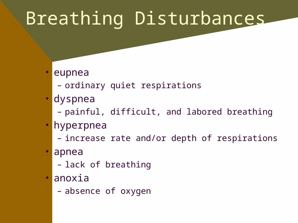
Breathing Disturbances
• eupnea– ordinary quiet respirations
• dyspnea– painful, difficult, and labored breathing
• hyperpnea– increase rate and/or depth of respirations
• apnea– lack of breathing
• anoxia– absence of oxygen

• hypoxia– decreased amount of oxygen reaching the body cells
• suffocation– stoppage of respirations caused by strangulation,
aspiration, foreign object, or drowning
• Cheyne-Stokes– alternating cycles of hyperpnea and apnea
• cyanosis– bluish gray discoloration – indicates insufficient amount of oxygen