Embed Size (px)
Citation preview

Part 2
Respiratory System

Respiration
Exchange of gases between air and body cells
Three steps
1. Ventilation
2. External Respiration
3. Internal Respiration

Ventilation
Pulmonary ventilation consists of two phases
1. Inspiration: gases flow into the lungs
2. Expiration: gases exit the lungs
Relies on pressure gradients
Atmosphere and alveoli
Negative pressure breathing
Thoracic pressures
Atmospheric pressure
Boyle’s Law
The relationship between the pressure and volume of a fixed
quantity of gas in a closed container
Pressure (P) varies inversely with volume (V):
P1V1 = P2V2
While one increases the other decreases

Ventilation
Intrapleural pressure is subatmospheric
Inward elastic recoil of lung tissue
Outward elastic recoil of chest wall
Resting lung volume

Ventilation
Atmospheric pressure (Patm)
Pressure exerted by the air surrounding the body
760 mm Hg at sea level
Thoracic pressures are described relative to Patm
Negative respiratory pressure is less than Patm
Positive respiratory pressure is greater than Patm

Figure 22.12
Atmospheric pressure
Intrapleural
pressure
756 mm Hg
(–4 mm Hg)
Transpulmonary
pressure
760 mm Hg
–756 mm Hg
= 4 mm Hg
Thoracic wall
Diaphragm
Lung Intrapulmonary
pressure 760 mm Hg
(0 mm Hg)
Parietal pleura
Pleural cavity
Visceral pleura
756
760

Ventilation
Intrapulmonary (intra-alveolar) pressure (Ppul)
Pressure in the alveoli
Fluctuates with breathing
Always eventually equalizes with Patm

Ventilation
Intrapleural pressure (Pip):
Pressure in the pleural cavity
Fluctuates with breathing
Always a negative pressure (<Patm and <Ppul)

Ventilation
Negative Pip is caused by opposing forces
Two inward forces promote lung collapse
Elastic recoil of lungs decreases lung size
Surface tension of alveolar fluid reduces alveolar size
One outward force tends to enlarge the lungs
Elasticity of the chest wall pulls the thorax outward

Figure 22.12
Atmospheric pressure
Intrapleural
pressure
756 mm Hg
(–4 mm Hg)
Transpulmonary
pressure
760 mm Hg
–756 mm Hg
= 4 mm Hg
Thoracic wall
Diaphragm
Lung Intrapulmonary
pressure 760 mm Hg
(0 mm Hg)
Parietal pleura
Pleural cavity
Visceral pleura
756
760
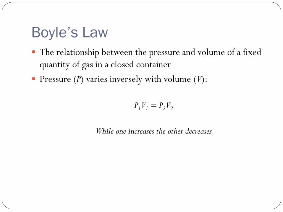
Pulmonary Ventilation
Inspiration and expiration
Mechanical processes that depend on volume changes in the
thoracic cavity
Volume changes pressure changes
Pressure changes pressure gradient gases flow to equalize
pressure


Inspiration
An active process
Inspiratory muscles contract
Thoracic volume increases
Lungs are stretched and intrapulmonary volume increases
Intrapulmonary pressure drops
Air flows into the lungs, down its pressure gradient
o Ppul = Patm

Figure 22.13 (1 of 2)
Sequence of events
Changes in anterior-
posterior and superior- inferior dimensions
Changes in lateral
dimensions (superior view)
Ribs are elevated and sternum flares
as external intercostals
contract.
Diaphragm moves inferiorly
during contraction.
External intercostals contract.
Inspiratory muscles contract (diaphragm descends; rib cage rises).
2
1
Thoracic cavity volume increases.
3 Lungs are stretched; intrapulmonary volume increases.
4 Intrapulmonary pressure drops (to –1 mm Hg).
5 Air (gases) flows into lungs down its pressure gradient until intrapulmonary pressure is 0 (equal to atmospheric pressure).

Expiration Quiet expiration is normally a passive process
Inspiratory muscles relax Thoracic cavity volume decreases
Elastic lungs recoil and intrapulmonary volume decreases
Ppul rises
Air flows out of the lungs down its pressure gradient until Ppul = 0

Figure 22.13 (2 of 2)
Sequence
of events
Changes in anterior-
posterior and superior- inferior dimensions
Changes in
lateral dimensions
(superior view)
Ribs and sternum are depressed
as external intercostals
relax.
External intercostals relax.
Diaphragm moves superiorly as it relaxes.
1 Inspiratory muscles relax (diaphragm rises; rib cage descends due to recoil of costal cartilages).
2 Thoracic cavity volume decreases.
3 Elastic lungs recoil passively; intrapulmonary volume decreases.
4 Intrapulmonary pres- sure rises (to +1 mm Hg).
5 Air (gases) flows out of lungs down its pressure gradient until intra- pulmonary pressure is 0.

Figure 22.14
5 seconds elapsed
Volume of breath
Intrapulmonary pressure
Expiration
Intrapleural pressure
Trans- pulmonary pressure
Inspiration Intrapulmonary
pressure. Pressure inside lung decreases as lung volume increases during inspiration; pressure increases during expiration.
Intrapleural pressure.
Pleural cavity pressure becomes more negative as chest wall expands during inspiration. Returns to initial value as chest wall recoils.
Volume of breath.
During each breath, the pressure gradients move 0.5 liter of air into and out of the lungs.

Air Flow Physical factors influencing efficiency of air flow
Inspiratory muscles overcome three factors that hinder air
passage and pulmonary ventilation
1. Airway resistance
2. Alveolar surface tension
3. Lung compliance
6b. Friction is inversely proportional to airway diameter

Air Flow
Broncoconstriction
Smooth muscle contracts
Reduced air flow

Lung Compliance
Diminished by
Nonelastic scar tissue (fibrosis)
Reduced production of surfactant
Decreased flexibility of the thoracic cage

Gas Law’s
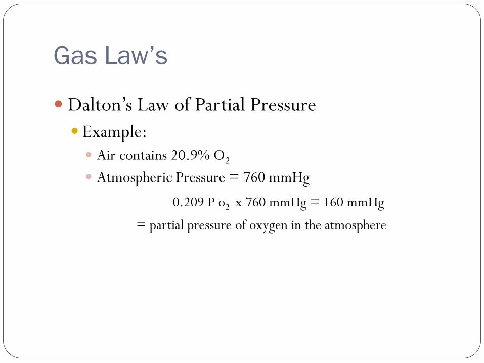
Dalton’s Law of Partial Pressure
Describes how a gas behaves when it is part of a mixture
Partial pressure = pressure exerted by a single gas in the mixture =
proportional to the percentage of gas in a mixture
Total pressure exerted by a mixture of gases is the sum of the pressures
exerted by each gas
Gas Law’s
Dalton’s Law of Partial Pressure
Example:
Air contains 20.9% O2
Atmospheric Pressure = 760 mmHg
0.209 P o2 x 760 mmHg = 160 mmHg
= partial pressure of oxygen in the atmosphere

Gas Law’s
Fick’s Law of Diffusion
Gives the rate of diffusion for a given gas across a membrane
CO2 has a high diffusion coefficient and diffuses 20 times more
rapidly across a membrane than O2
Vg = (A) (P1-P2) (D)
(T)

Gas Law’s
Diseases related to gas diffusion
Pulmonary edema
COPD

Gas Exchange
External respiration
Pulmonary gas exchange
Internal respiration
Gas exchange between blood capillaries and tissues

Figure 22.17
Inspired air:
P 160 mm Hg
P 0.3 mm Hg
Blood leaving
lungs and
entering tissue
capillaries:
P 100 mm Hg
P 40 mm Hg
Alveoli of lungs:
P 104 mm Hg
P 40 mm Hg O2
Heart
Blood leaving
tissues and
entering lungs:
P 40 mm Hg
P 45 mm Hg
Systemic
veins Systemic
arteries
Tissues:
P less than 40 mm Hg
P greater than 45 mm Hg
Internal
respiration
External
respiration
Pulmonary
veins (P
100 mm Hg)
Pulmonary
arteries
CO2
O2
CO2
O2
CO2 O2
CO2
O2
CO2
O2

External Respiration
Exchange of O2 and CO2 across the respiratory membrane
From lung to blood
Influenced by
Partial pressure gradients
Gas solubilities
Structural characteristics of the respiratory membrane

External Respiration
Respiratory membrane
Membrane of lung epithelium
Pulmonary capillary membrane
Surfactant

Figure 22.9c
Capillary
Type II (surfactant-
secreting) cell
Type I cell
of alveolar wall
Endothelial cell nucleus
Macrophage
Alveoli (gas-filled
air spaces)
Red blood cell
in capillary
Alveolar pores
Capillary
endothelium
Fused basement
membranes of the
alveolar epithelium
and the capillary
endothelium
Alveolar
epithelium
Respiratory
membrane
Red blood
cell
O2
Alveolus
CO2
Capillary
Alveolus
Nucleus of type I
(squamous
epithelial) cell
(c) Detailed anatomy of the respiratory membrane

External Respiration
Partial pressure gradient for O2 in the lungs is steep
Venous blood Po2 = 40 mm Hg
Alveolar Po2 = 104 mm Hg
Oxygen readily diffuses from alveoli to lung capillaries

External Respiration
Partial pressure gradient for CO2 in the lungs
Venous blood Pco2 = 45 mm Hg
Alveolar Pco2 = 40 mm Hg
But…
CO2 is 20 times more soluble in plasma than oxygen
CO2 diffuses in equal amounts with oxygen!

Figure 22.17
Inspired air:
P 160 mm Hg
P 0.3 mm Hg
Blood leaving
lungs and
entering tissue
capillaries:
P 100 mm Hg
P 40 mm Hg
Alveoli of lungs:
P 104 mm Hg
P 40 mm Hg O2
Heart
Blood leaving
tissues and
entering lungs:
P 40 mm Hg
P 45 mm Hg
Systemic
veins Systemic
arteries
Tissues:
P less than 40 mm Hg
P greater than 45 mm Hg
Internal
respiration
External
respiration
Pulmonary
veins (P
100 mm Hg)
Pulmonary
arteries
CO2
O2
CO2
O2
CO2 O2
CO2
O2
CO2
O2

Factors Affecting External Respiration
Anatomical adaptations
Moist surfaces
Thickness and surface area
Narrow capillaries = RBC’s single file
Physiological and physical factors
Pulmonary disease
Affect of drugs on minute volume
Partial pressure changes with altitude

Internal Respiration
Capillary gas exchange in body tissues
Partial pressures and diffusion gradients are reversed
compared to external respiration
Po2 in tissue is always lower than in systemic arterial blood
PCO2 in tissue is higher than in systemic arterial blood

Figure 22.17
Inspired air:
P 160 mm Hg
P 0.3 mm Hg
Blood leaving lungs and
entering tissue capillaries:
P 100 mm Hg
P 40 mm Hg
Alveoli of lungs:
P 104 mm Hg
P 40 mm Hg O2
Heart
Blood leaving
tissues and
entering lungs:
P 40 mm Hg
P 45 mm Hg
Systemic
veins Systemic
arteries
Tissues:
P less than 40 mm Hg
P greater than 45 mm Hg
Internal
respiration
External
respiration
Pulmonary
veins (P
100 mm Hg)
Pulmonary
arteries
CO2
O2
CO2
O2
CO2 O2
CO2
O2
CO2
O2


![Presentation1 - Linn–Benton Community Collegecf.linnbenton.edu/mathsci/bio/waitea/upload/Lecture_01_Neurons.pdfMicrosoft PowerPoint - Presentation1 [Compatibility Mode] Author: U0076978](https://img.pdfslide.net/doc/110x75/5f0eb6cd7e708231d44093df/presentation1-linnabenton-community-microsoft-powerpoint-presentation1-compatibility.jpg)