Embed Size (px)
Citation preview

Respiratory Tract Mucus
Ciba Foundation Symposium 54 (new series)
1978
Elsevier - Excerpta Medica - North-Holland
Amsterdam. Oxford - New York


Respiratory Tract Mucus
The Ciba Foundation for the promotion of international cooperation in medical and chemical research is a scientific and educational charity established by ClBA Limited - now CIBA-GEIG Y Limited - of Basle. The Foundation operates independentry in London under English trust law.
Ciba Foundation Symposia are published in collaboration with Elsevier Scientific Publishing Company / Excerpta Medica 1 North-Holland Publishing Company in Amsterdam
Elsevier / Excerpta Media / North-Holland, P.O. Box 21 1, Amsterdam

Respiratory Tract Mucus
Ciba Foundation Symposium 54 (new series)
1978
Elsevier - Excerpta Medica - North-Holland
Amsterdam. Oxford - New York

0 Copyright 1978 Ciba Foundation
All rights reserved. No part of this publication may be reproduced or transmitted in any form or by any means, electronic or mechanical, including photocopying and recording, or by any information storage and retrieval system, without permission in writing from the publishers.
First printing January 1978 Second printing February 1979
ISBN 0-444-90016-0
Published in January 1978 by Elsevier/Excerpta Medica/North-Holland, P.O. Box 21 1, Amsterdam and Elsevier/North-Holland Inc., 52 Vanderbilt Avenue, New York, N.Y. 10017.
Suggested series entry for library catalogues : Ciba Foundation Symposia. Suggested publisher’s entry for library catalogues : Elsevier/Excerpta Medica/North-Holland
Ciba Foundation Symposium 54 (new series)
342 pages, 79 figures, 22 tables
Library of Congress Cataloging in Publication Data
Symposium on Respiratory Tract Mucus, London, 1977. Respiratory tract mucus.
(Ciba Foundation symposium; new ser., 54) ‘Symposium . . . held at the Ciba Foundation, London, 22-24 March 1977.” Includes bibliographies and index. 1. Mucus-Congresses. 2. Respiratory organs-Congresses. I. Title. 11. Series :
Ciba Foundation. Symposium; new ser., 54. DNLM : 1. Mucus-Congresses. 2. Respiratory system-Physiology-Congresses. 3. Respiratory tract diseases-Physiopathology-Congresses. W3 C161F v. 54 / WF102 R441 QP215S95 1975 559’.01’2 77-16019 ISBN 0-444-90016-0
Printed in The Netherlands by Casparie, Alkmaar
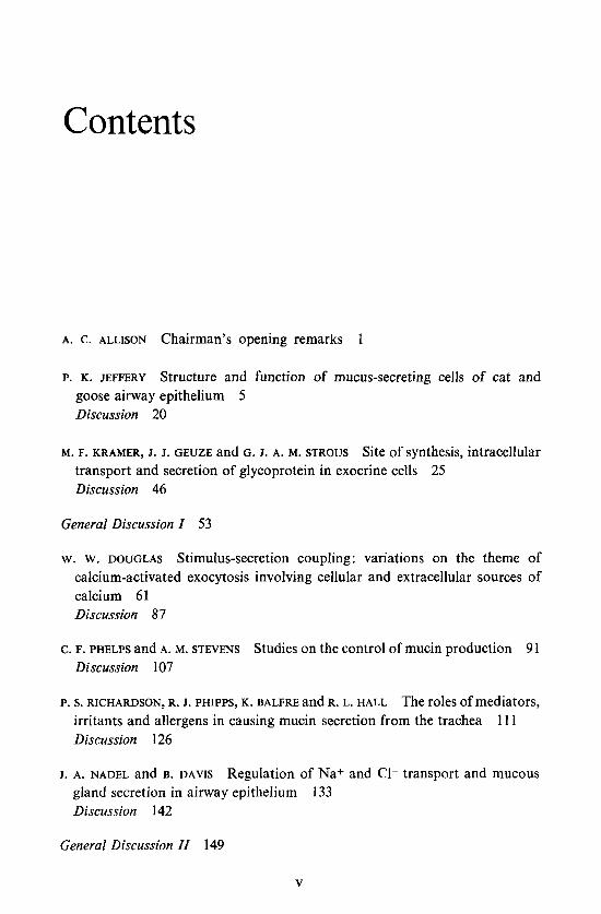
Contents
A. c . ALLISON Chairman’s opening remarks 1
P. K. JEFFERY Structure and function of mucus-secreting cells of cat and goose airway epithelium 5 Discussion 20
M. F. KRAMER, J. J. GEUZE and G. J. A. M. STROUS Site of synthesis, intracellular transport and secretion of glycoprotein in exocrine cells 25 Discussion 46
General Discussion I 53
w. w. DOUGLAS Stimulus-secretion coupling: variations on the theme of calcium-activated exocytosis involving cellular and extracellular sources of calcium 61 Discussion 87
C. F. PHELPS and A. M. STEVENS Studies on the control of mucin production 91 Discussion 107
P. s. RICHARDSON, R. J. PHIPPS, K. BALFRE and R. L. HALL The roles of mediators, irritants and allergens in causing mucin secretion from the trachea 1 1 1 Discussion 126
J. A. NADEL and B. DAVIS Regulation of Na+ and C1- transport and mucous gland secretion in airway epithelium Discussion 142
133
General Discussion II 149
V

CONTENTS VI
P. w. KENT Chemical aspects of tracheal glycoproteins I55 Discussion 17 1
R. JONES The glycoproteins of secretory cells in airway epithelium I75 Discussion 188
General Discussion III 195
F. A. MEYER and A. SILBERBERG Structure and function of mucus 203 Discussion 2 I 1
D. F. PROCTOR, G. K. ADAMS, I. ANDERSEN and S. F. P. MAN Nasal mucociliary clearance in man 219 Discussion 230
J. IRAVANI, G. N. MELVILLE and G. HORSTMANN Tracheobronchial clearance in health and disease: with special reference to interciliary fluid 235 Discussion 249
J. SCHRAGER and G. CUMMING The isolation and partial characterization of the major bronchial glycoproteins 253 Discussion 263
General Discussion IV 265
P. SADOUL, E. PUCHELLE and F. GIRARD Criteria for evaluating mucus functions and their disorders in chronic bronchitis 277 Discussion 29 1
L. REID Animal models in clinical disease 297 Discussion 307
Final discussion: Clinical aspects 3 1 1
A. c. ALLISON Closing remarks 321
Index of contributors 327
Subject index 329

Participants Symposium on Respiratory Tract Mucus held at the Ciba Foundation, London, 22-24 March 1977
A. c . ALLISON (Chairman) Division of Cell Pathology, MRC Clinical Research Centre, Watford Road, Harrow, Middlesex HA1 3UJ, UK
I . CARLSTEDT Department of Physiological Chemistry, Faculty of Medicine, University of Lund, PO Box 750, S-220 07 Lund 7, Sweden
G. GUMMING Midhurst Medical Research Institute, Midhurst, Sussex GU29 OBL, UK
B. DAVIS School of Medicine Cardiovascular Research Institute, University of California, San Francisco, California 94122, USA
K. w. DONALD Nant-Y-Celyn, Cloddiau, Welshpool SY21 9JE, Powys, Wales, UK
w. w. DOUGLAS Department of Pharmacology, Sterling Hall of Medicine, Yale University School of Medicine, 333 Cedar Street, New Haven, Conn. 06510, USA
R. c. GODFREY Western Hospital, Oakley Road, Mill Brook, Southampton SO9 4WQ, UK
A. GUZ Department of Medicine, Charing Cross Hospital, Fulham Palace Road, London W6 9HH, UK
J. IRAVANI Institut fur Anatomie Lehrstuhl fur Anatornie 11, Postfach 102148/MA6/148, Ruhr-Universitat Bochum, Universitatsstrasse 150, 4630 Bochum 1, West Germany
P. K. JEFFERY Basic Medical Sciences Group, Department of Physiology, Chelsea College, University of London, Manress Road, London SW3 6LX, UK
VII

VIII PARTICIPANTS
J. G. JONES Division of Anaesthesia, MRC Clinical Research Centre, Watford Road, Harrow, Middlesex HA1 3UJ, UK
R. JONES Department of Pathology, Children’s Hospital Medical Center, 300 Longwood Avenue, Boston, Massachusetts 021 15, USA
P. w. KENT Glycoprotein Research Unit, Science Laboratories, University of Durham, South Road, Durham DHl 3LE, UK
M. F. KRAMER Department of Histology and Cell Biology, Rijksuniversiteit Utrecht, Nic. Beetsstraat 22, Utrecht 2501, The Netherlands
J. A. NADEL School of Medicine Cardiovascular Research Institute, University of California, San Francisco, California 94122, USA
c. F. PHELPS Department of Biological Sciences, University of Lancaster, Lancaster LA1 4YQ, UK
D. F. PROCTOR Department of Otolaryngology, The Johns Hopkins Hospital, Baltimore, Maryland 21205, USA
L. REID Department of Pathology, Children’s Hospital Medical Center, 300 Longwood Avenue, Boston, Massachusetts 021 15, USA
P. s. RICHARDSON Department of Physiology, St George’s Hospital Medical School, Tooting, London SW17 OQT, UK
P. SADOUL INSERM U 14, Case officielle no. 10,54500 Vandoeuvre-les-Nancy, France
A. SILBERBERG Polymer Department, Weizmann Institute of Science, Rehovot, Israel
w. M. THURLBECK Department of Pathology, Health Sciences Centre, The University of Manitoba, 700 William Avenue, Winnipeg, Manitoba, Canada R3E 0 2 3
J. G. WIDDICOMBE Department of Physiology, St George’s Hospital Medical School, Tooting, London SW17 OQT, UK
Editors: Ruth Porter (Organizer), John Rivers and Maeve O’Connor

Chairman’s opening remarks
A. C. ALLISON
Division of Cell Pathology, MRC Clinical Research Centre, Harrow, Middlesex
Many of you will be wondering why I am here, and I wonder more than you do; but it is a long-established British tradition to have non-experts participating in everything from local dispensation of justice to hospital administration.
I suggest that we approach this whole subject from the point of view of cell biologists and look at respiratory tract mucus in the way in which I was originally taught by Sir Howard (later Lord) Florey. He was one of the first to point out that mucus is not just gubbins but an interesting substance which performs important and distinct functions in different places. When Florey was active, very little was known about the chemistry of mucus. Now we are able to use established techniques to discover the structure both of mucus from different sites and of related substances such as synovial fluid, and to compare structure with mechanical and other properties. Synovial fluid has interesting rheological properties, for example non-Newtonian viscosity, a property associated with joint lubrication which would be disastrous if it existed in respiratory tract mucus. One of the features of mucus is that it acts as the mechanical link between the energy provided by ciliary beats and translocation of particles. Professor Silberberg, who has been a pioneer in this field, has done a lot to associate the rheological properties and chemistry of mucus. This link is important not only in the respiratory tract but also, for example, in the female genital tract. Mucus is associated with translocation of the ovum down the Fallopian tube, the probability of fertilization and, in the uterus itself, with the site of implantation and the allowing of implantation. There, hormonal influences are strong and one of the main ways in which the contraceptive pill works is by changing the properties of mucus in the genital tract.
We should like to know the structure of mucus and how it is related to particular functions. The old concept of a uniform blanket of mucus now

2 A. C. ALLISON
seems to be disappearing in favour of the concept of isolated patches. We shall also hear a good deal about the chemical structure and I regret that Dr Pierre Masson cannot be with us, because one of the points which he has made is that instead of rather long protein chains-with carbohydrate side chains, which is true of proteoglycan in synovial fluid-there may be uniform units of protein which are quite short, with a molecular weight of about 30 000 daltons on the basis of published N-terminal determinations and more recent C-terminal determinations (Masson 1973). Because of the larger sizes of the units observed and their properties there is the interesting possibility that the subunits may be cross-linked by carbohydrate chains. So I hope that the chemists here will address themselves to the questions: what is the basic structure of respiratory tract mucus? is Masson's model correct? and what would be the properties of material of this kind in contrast to other proteoglycans with which we are all familiar?
In synovial fluid there are specific interactions between proteoglycan and hyaluronic acid, mediated by specific bonds, and these interactions determine the properties of the material. Although hyaluronic acid is absent, there may be other constituents of respiratory t'ract mucus which interact in a specific way with proteoglycan to generate particular behavioural properties. For example, calcium and magnesium ions may interact with the carbohydrate side chains and with the cell coat. It may be possible to generate, from combinations of different secretions, material which behaves differently from the original secretions themselves. Professor Silberberg makes the point that the behaviour of this material is quite evanescent and that if fresh mucus is isolated its mechanical properties are different from those of material that has been frozen and then thawed. Such material is convenient for biochemical analysis but its mechanical properties may in fact be greatly changed. So we want to know what the constituents are, how they change, whether they are changed by enzymic action, by cross-linking, or by degradation of either the carbohydrate units or the protein units of which they are composed, and whether any changes result from interaction of different components.
The anatomical basis of mucus secretion is itself quite complex. We were taught that there are in respiratory tract epithelium three cell types-an undifferentiated cell, a ciliated cell and a goblet cell. Now we are told that there are many cell types-11 according to Dr Jeffery, who will be describing their properties-and during the meeting we shall want to look at the changes which occur in these cell types. Can a serous cell be transformed into a mucus- secreting cell and if so in what circumstances? What induces the differentiation of these relatively undifferentiated cells into cells of particular types, and is the process reversible? We know, for example, that in the development of

CHAIRMAN’S OPENING REMARKS 3
the blood system there is a series of stem cells; the early ones are multipotential, but at each stage of development the potentialities are further restricted. One starts with cells which can become lymphocytes, erythrocytes or granulocytes. At later stages they are committed, for example, to the erythrocyte series of differentiation or to the granulocyte-monocyte series, and cells in the latter series can become either monocytes, neutrophils or eosinophils. Particular local factors, some of which are now defined, determine which pathway of differentiation these cells will follow. We know very little about analogous processes in the respiratory tract but these are questions to which we should address ourselves, not looking at structure just from a static viewpoint but trying to bear in mind how cells can follow one pathway of differentiation or anothcr.
Again, very little is known about the control of mucus secretion. We know a great deal about the mechanisms underlying secretion in other cells, particularly the role of calcium, the relationship to microtubules, micro- filaments, membrane fusion and so on. Professor Douglas, who is one of the experts in that field, will remind us of the general background and I think it would be appropriate in the near future to apply to respiratory tract mucus the general principles and information which have emerged from other systems. We know very little about whether the control is determined by local mediators: interesting possibilities are emerging about the role of prostaglandins with the use of indomethacin and other drugs to inhibit the formation of prosta- glandins or divert their metabolism along particular pathways. From studies of inflammation it is already clear that this is a very complicated process. For example, it is not usually the final stages of prostaglandins, the E and F series, which are so easily obtained and analysed, that are particularly important in inflammation, but rather the transient intermediates-prosta- glandin A and the thromboxanes. We don’t know the relative importance of local humoral factors and nervous control of respiratory tract mucus secretion. There is an intimate structural relationship between nerves and the cells of the respiratory tract epithelium and we suspect that reflex stimulation may be important; interesting examples have been studied in frogs and other experimental animals. We know a great deal more about the new neuro- transmitters and how to block transmission, and there is now good opportunity for applying accumulated physiological or cytopbhrmacological knowledge to the respiratory tract.
I hope, then, that we shall try to apply the principles of cell biology and developmental biology to our subject so as to provide the scientific background for the discussions of the clinical aspects which will follow. It will be more appropriate to discuss general points, such as the nature of the linkages between

4 A. C. ALLISON
polypeptide and carbohydrate chains, rather than go into enormous detail on the actual amino-acid and sugar sequences which are involved, unless they have some bearing on general structural problems. In the same way I am sure we don’t want elaborate descriptions of the different cell types in several species of animals.
Finally, we can already see from the contributions to this meeting how much there is to be learnt from other fields. For example, Dr Nadel will be reminding us of the application to the respiratory tract of the basic work on the toad bladder, which yielded a great deal of information about ion movements across membranes, and of the micropuncture techniques which were used so successfully in kidney physiology. Dr Kramer will be describing the beautiful work which has been done in cell biology on finding out the sites of synthesis of proteins and carbohydrates in different cell types-work which was pioneered by Leblond, Jamieson and Palade and is now applied to mucus secretion. Many special disciplines impose in an interesting way on the subject of this meeting and I am sure that we shall have fruitful discussions.
Reference
MASSON, P. L. (1973) Carbohydrate component of cervical mucus, in Cervical Mucus in Human Reproduction (Elstein, M. et al., eds.), p. 82, Scriptor, Copenhagen

Structure and function of mucus-secreting cells of cat and goose airway epithelium
PETER K. JEFFERY
Chelsea College and St George’s Hospital Medical School, London
Abstract In the cat and goose, studies have been undertaken to determine the ultrastructure of airway epithelia, the concentration and distribution of the secretory cells which produce respiratory tract mucus, and the histochemistry of mucins located within cells and on their luminal surfaces. By electron microscopy all the 11 cell types so far described can be found in the airways of the cat but not the goose. Both goblet cells and submucosal glands are abundant in the cat whereas the trachea of the goose lacks the latter, having instead abundant goblet cells many of which form ‘intraepithelial glands’. Histochemically, the goblet cells of the cat and goose are similar in that they contain mucins with a predominance of sulphate esters. A surface mucosubstance can be demonstrated which, histochemically, is similar to that described in dog and man. Interestingly, this surface layer may be sloughed in response to an inhaled irritant such as ammonia and thereby contribute to the respiratory tract mucus recovered experimentally. Incorporation into macromolecules of radioactively labelled mucin precursors is assessed by autoradiography of tissue sections, and preliminary results of experiments designed to test the respolise of mucus-secreting cells to airway irritation and the parasympathomimetic drug, pilocarpine, are also presented.
The airways which supply the respiratory portion of the lung are responsible for the clearance of inhaled particulate and gaseous pollutants deposited on their mucosal lining. To this end, the lining mucosa produces a mucous secretion in which pollutants become dissolved or entangled, and which is then moved by cilia to the pharynx, where it is normally swallowed. The mucous secretion so produced (normally between 10 and 100 ml/day) is a complex mixture of about 95% water and 5 % carbohydrate, protein, lipid and inorganic material-usually in the form of a number of different glyco- proteins, each differing in the ratio of its protein to carbohydrate component and its degree of acidification. The physical (i.e. viscoelastic) properties of
5

6 P. K. JEFFERY
the secretion are largely due to these glycoproteins which are produced by a variety of cell types comprising the airway wall.
In man, respiratory mucus may come from two anatomical sources-the secretory cells of the surface epithelium and those of the submucosal glands (Reid 1954; McCarthy & Reid 1964). This is also true of the respiratory tract of the rat, dog and pig, three animals which have been used as experimental animal ‘models’ for mucous studies (Reid 1963; Chakrin & Saunders 1974; Jones et al. 1975). More recently, both the cat and the goose have been used in studies of the control of mucin secreted in a tracheal segment isolated in situ in the anaesthetized animal (Gallagher et al. 1975; Phipps et al. 1977). Whereas the fine structural anatomy of the airway mucosal linings of the human, rat, dog and pig have been well described (Rhodin 1966; Jeffery & Reid 1975; Frasca et al. 1968; Baskerville 1970), those of the cat and goose have not. In particular, the goose may be a useful animal to study in that its airways, like those of other species of birds, have an abundance of epithelial goblet cells but no submucosal glands, thus allowing mucin from a purely epithelial source to be investigated (Cover 1953; Purcell 1971; Walsh & McLelland 1974).
The work presented here is the result of new studies we have undertaken to determine, in the cat and goose, the ultrastructure of airway epithelia, the concentration and distribution of the secretory cells which produce the respiratory tract mucus found in these two species, and the histochemistry of the mucin, whether it be located within cells or on their surfaces. Preliminary results on the histology of tracheal ‘segments’ taken from cats or geese which have been stimulated (by neural or pharmacological means) to increase their output of tracheal mucin will also be given.
ULTRASTRUCTURE OF THE EPITHELIUM
As reviews of the ultrastructure and diversity of cell types which make up the airway epithelium of different species have recently been published (see Breeze et al. 1976; Jeffery & Reid 1977), this section will give only a general survey of the cell types which have now been found in the airways of the cat and goose.
Up to 1 1 distinct cell types (nine epithelial and two migratory) have been described in airway epithelia, depending upon the species studied and the anatomical location of the respiratory tract examined (Fig. 1). Except for the cat, each species is deficient in one or more of the 11 cell types (Table 1). The cat airway epithelium has the full complement of cell types as well as the occasional mast cell and neutrophil (polymorphonuclear leucocyte) (Jeffery

AIRWAY STRUCTURE AND FUNCTION 7
Epithelial
, n; SecrTrh
"Special type" Intermediate
Leucocyte
FIG. 1 . Epithelial cell types described by electron microscopy.
TABLE 1
Epithelial cell types identified by electron microscopy in various species
~~ ____
Ciliated + + + + + + + + + + + + + + - + + + + + - + - - + - - - +
Goblet (lucent granule) + + Serous (dense granule) + -
+ + - - _ - - - - 'Special type' + - Clara + + - + + - + + ? + - Intermediate + - - + + - + - - - + Brush ? - - + + - + - - ? - Basal + + + + + + + + + + + Kulchitsky + - - + - - + + - + + Globule leucocyte - 'migratory' + + - - + - - - - Lymphocyte + 'migratory' - + - - + - - - - Intraepithelial nerve + - - + - - + + - + + + identified; - not yet identified; ? unconfirmed.

8 P. K. JEFFERY
FIG. 2. An electron micrograph of cat tracheal epithelium showing goblet (gc), ciliated (cc) and basal cells (bc). Goblet cells contain a mixture of electron-lucent and electron-dense secretory granules. Airway lumen (I) and basement membrane (bm). Glutaraldehyde and osmium tetroxide: uranyl acetate and lead citrate. x 3000
& Das 1977). In particular, the cat has all four secretory cell types, i.e. goblet cells, ‘epithelial serous cells’ (Jeffery & Reid 1975), Clara cells (most frequent in bronchioli), and ‘special type cells’, which have also been described in the airways of dog and man (Frasca et al. 1968).
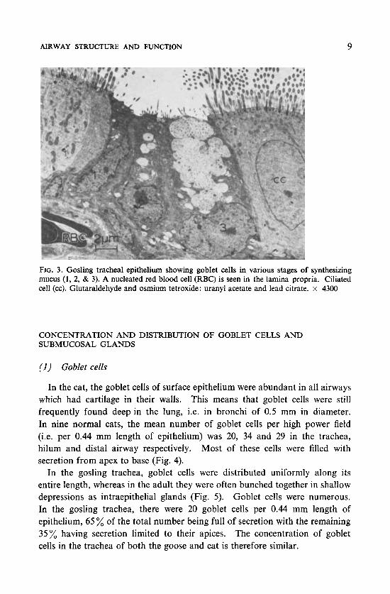
Fig. 2 shows cat tracheal epithelium. The most frequent type of secretory cell is the goblet cell, which in the cat constitutes between 7 and 15% of all epithelial cells, depending on the location of the airway examined. Unlike the rat, many goblet cells of the cat contain a mixture of electron-lucent and electron-dense granules within the same cell, which suggests that there is a mixture of distinct glycoproteins within the same cell (see below). The ‘epithelial serous cell’ and ‘special type cell’ are infrequently found. Fig. 3 shows gosling tracheal epithelium. Goblet and ‘epithelial serous’ cells have been identified, the latter being found infrequently (Phipps et al. 1977). The goblet cells are unique in containing electron-lucent secretory granules each of which is ‘speckled’ with electron-dense areas. This morphological hetero- geneity suggests that within each granule there may be two or more distinct types of glycoprotein. Goblet cells of the trachea are present as 12% of the epithelial cells. The intrapulmonary airways of the goose have not yet been examined histologically.
AIRWAY STRUCTURE AND FUNCTION 9
FIG. 3. Gosling tracheal epithelium showing goblet cells in various stages of synthesizing mucus (1, 2, & 3). A nucleated red blood cell (RBC) is seen in the lamina propria. Ciliated cell (cc). Glutaraldehyde and osmium tetroxide: uranyl acetate and lead citrate. x 4300
CONCENTRATION AND DISTRIBUTION OF GOBLET CELLS AND SUBMUCOSAL GLANDS
(1) Goblet cells
In the cat, the goblet cells of surface epithelium were abundant in all airways which had cartilage in their walls. This means that goblet cells were still frequently found deep in the lung, i.e. in bronchi of 0.5 mm in diameter. In nine normal cats, the mean number of goblet cells per high power field (i.e. per 0.44 mm length of epithelium) was 20, 34 and 29 in the trachea, hilum and distal airway respectively. Most of these cells were filled with secretion from apex to base (Fig. 4).
In the gosling trachea, goblet cells were distributed uniformly along its entire length, whereas in the adult they were often bunched together in shallow depressions as intraepithelial glands (Fig. 5). Goblet cells were numerous. In the gosling trachea, there were 20 goblet cells per 0.44 mm length of epithelium, 65 % of the total number being full of secretion with the remaining 35% having secretion limited to their apices. The concentration of goblet cells in the trachea of both the goose and cat is therefore similar.

10 P. K. JEFFERY
FIG. 4. Light microscopic appearance of tracheal epithelium stained with periodic acid- Schiff (PAS) for the carbohydrate component of mucus. Goblet cells are abundant and filled with intracellular secretion. Gland duct (arrow) and cartilage (c). x 130
(2 ) Submucosal glands
In the cat, submucosal glands occupied a significant proportion of the tracheal and hilar airway walls and submucosal glands were still found in the distal airway, albeit to a lesser extent. The Reid index, i.e. mean gland to wall ratio, in the tracheas of five ‘conventional’ cats was 0.75, and in four clean or ‘specific pathogen-free’ cats it was 0.65. Both values are considerably higher than 0.33, the value for normal adult man (Reid 1960). By point counting, Lamb (see Gallagher et al. 1975) has shown that the submucosal glands of the cat trachea form between 61 and 94% of the total secretory cell volume (i.e. goblet cells and glands) of which the greater part (up to 80 %) is composed of serous cells.
HISTOCHEMISTRY
(I) Goblet cells
The histochemical characteristics of the goblet cells of the cat airway have

AIRWAY STRUCTURE AND FUNCTION 11
FIG. 5. Photomicrograph of goose epithelium to show shallow epithelial depressions in which goblet cells are grouped to form ‘intraepithelial’ glands (arrow). Alcian blue/PAS. X 650
been described qualitatively by Lamb (see Gallagher et al. 1975). The goblet cells contain mainly sulphated mucin but a proportion have sialomucin which is resistant to digestion by the enzyme sialidase (neuraminidase, EC 3.2.1.18). Some cells contain mixtures of sialomucin and sulphomucin.
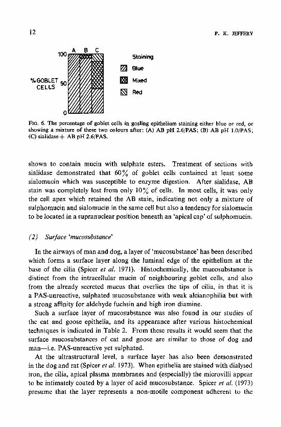
In the goose trachea, a histochemical analysis of some 6000 goblet cells showed that, after they were stained with combined Alcian blue, pH 2.61 periodic acid Schiff (AB 2.6/PAS), most goblet cells stained blue-i.e. although both acid and neutral glycoproteins were present, the acid predominated and was present in more than 95% of goblet cells (Fig. 6). When the cells were stained with combined AB l.O/PAS, more than 90% of goblet cells were
12 P. K. JEFFERY
A B C 100 Staining
E4 Blue
69 Red
OIoGOBLET Mixed CELLS
0-
FIG. 6. The percentage of goblet cells in gosling epithelium staining either blue or red, or showing a mixture of these two colours after: (A) AB pH 2.6PAS; (B) AB pH l.O/PAS; (C) sialidase + AB pH 2.6/PAS.
shown to contain mucin with sulphate esters. Treatment of sections with sialidase demonstrated that 60 % of goblet cells contained at least some sialomucin which was susceptible to enzyme digestion. After sialidase, AB stain was completely lost from only 10% of cells. In most cells, it was only the cell apex which retained the AB stain, indicating not only a mixture of sulphomucin and sialomucin in the same cell but also a tendency for sialomucin to be located in a supranuclear position beneath an ‘apical cap’ of sulphomucin.
(2) Surface ‘mucosubstance’
In the airways of man and dog, a layer of ‘mucosubstance’ has been described which forms a surface layer along the luminal edge of the epithelium at the base of the cilia (Spicer et al. 1971). Histochemically, the mucosubstance is distinct from the intracellular mucin of neighbouring goblet cells, and also from the already secreted mucus that overlies the tips of cilia, in that it is a PAS-unreactive, sulphated mucosubstance with weak alcianophilia but with a strong affinity for aldehyde fuchsin and high iron diamine.
Such a surface layer of mucosubstance was also found in our studies of the cat and goose epithelia, and its appearance after various histochemical techniques is indicated in Table 2. From these results it would seem that the surface mucosubstances of cat and goose are similar to those of dog and man-i.e. PAS-unreactive yet sulphated.
At the ultrastructural level, a surface layer has also been demonstrated in the dog and rat (Spicer et al. 1973). When epithelia are stained with dialysed iron, the cilia, apical plasma membranes and (especially) the microvilli appear to be intimately coated by a layer of acid mucosubstance. Spicer et al. (1973) presume that the layer represents a non-motile component adherent to the

AIRWAY STRUCTURE AND FUNCTION 13
TABLE 2
Histochemistry of epithelial surface mucosubstance in the trachea
Staina Species
Cat Goose
PAS alone AB2.6/PAS ABl.Q/PAS AH/AB2.6/PAS AH/ABl .Q/PAS Sialidase/AB2.6/PAS AFlAB2.6
-b ? +" + (slight) + + ? + + + (patchy) ? + (AF)
- -
5 PAS = periodic acid-Schiff; AF = aldehyde fuchsin b - unreactive; + reactive; ? untested c not into ciliated duct of glands
AB = Alcian blue at given pH; AH = acid hydrolysis;
cell, corresponding to the histochemically distinctive mucosubstance visible under the light microscope.
(3) Submucosal glands
In the cat submucosal glands, the mucous cells contain mainly much with sulphate esters. The histochemical results for serous cells are not clear but there are indications that they may also produce a sulphated glycoprotein (Gallagher et al. 1975).
AUTORADIOGRAPHY
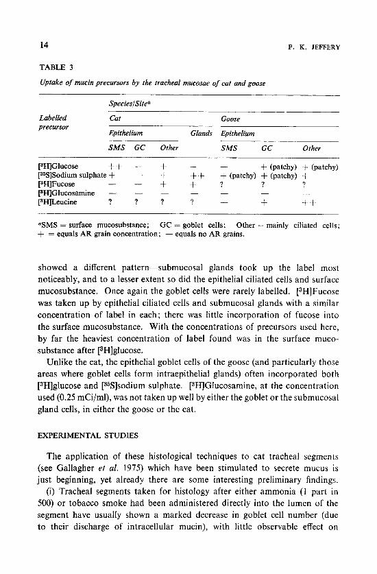
We have recently studied, by autoradiography of tracheal sections, the uptake and incorporation into macromolecules of several radioactively labelled precursors of mucin (Table 3). The sections were taken from animals which earlier had had the radioactive precursor given directly into the tracheal lumen by a method described by Gallagher et al. (1975). Satisfactory labelling and localization was found after [3H]glucose (0.1 mCi/ml), [35S]sodium sulphate (0.25 mCi/ml) and [3H]fucose (0.25 mCi/ml).
In the cat, [3H]glucose was selectively taken up and incorporated into the surface mucosubstance, epithelial ciliated cells, submucosal glands and, rarely, the epithelial goblet cells, in that order of priority. [35S]Sodium sulphate
P. K. JEFFERY 14
TABLE 3
Uptake of mucin precursors by the tracheal mucosae of cat and goose
SpecieslSite'
Labelled Cat Goose precursor
Epithelium Glands Epithelium
SMS GC Other SMS GC Other -.
[3H]Glucose ++ - + + - + (patchy) 4- (patchy) [35S]Sodium sulphate + - + ++ + (patchy) + (patchy) -t + + ? ? ? [3HlFucose - - [SH]Glucosamine - - - - -
[SHILeucine ? ? ? ? + ++ - _ _
-
'SMS = surface mucosubstance; GC = goblet cells; Other = mainly ciliated cells; + = equals AR grain concentration; - equals no AR grains.
showed a different pattern-submucosal glands took up the label most noticeably, and to a lesser extent so did the epithelial ciliated cells and surface mucosubstance. Once again the goblet cells were rarely labelled. [3H]Fucose was taken up by epithelial ciliated cells and submucosal glands with a similar concentration of label in each; there was little incorporation of fucose into the surface mucosubstance. With the concentrations of precursors used here, by far the heaviest concentration of label found was in the surface muco- substance after [3H]glucose.
Unlike the cat, the epithelial goblet cells of the goose (and particularly those areas where goblet cells form intraepithelial glands) often incorporated both [3H]glucose and [35S]sodium sulphate. [3H]Glucosamine, at the concentration used (0.25 mCi/ml), was not taken up well by either the goblet or the submucosal gland cells, in either the goose or the cat.
EXPERIMENTAL STUDIES
The application of these histological techniques to cat tracheal segments (see Gallagher et af. 1975) which have been stimulated to secrete mucus is just beginning, yet already there are some interesting preliminary findings.
(i) Tracheal segments taken for histology after either ammonia (1 part in 500) or tobacco smoke had been administered directly into the lumen of the segment have usually shown a marked decrease in goblet cell number (due to their discharge of intracellular mucin), with little observable effect on

AIRWAY STRUCTURE AND FUNCTION 15
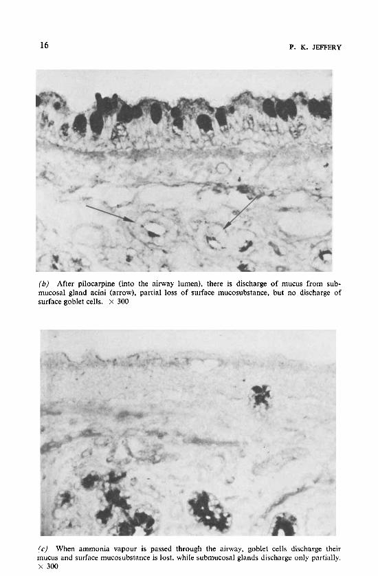
submucosal gland. For example, in a comparison made between part of the trachea (i.e. below the segment) not isolated for treatment and the ammonia- treated ‘segment’, the goblet cell number fell from a mean of 23.5 f 2.2 per microscopic field to 1.6 & 0.4 respectively. In another experiment, pilocarpine nitrate (a parasympathomimetic) was administered directly into the ‘segment’ at a concentration of 7 ,ug/ml. Histology showed that this had little effect on goblet cell number but submucosal glands were emptied of their intracellular secretion (Fig. 7).
(ii) In another experiment, two tracheal segments were isolated and each was labelled with [3H]glucose. Ammonia was then administered into one of the segments, causing a marked increase in 3H-labelled mucus recovered in the tracheal washings of that segment. Histology and autoradiography of sections from both the ammonia-treated and untreated segments showed that not only were goblet cells decreased in number in the ammonia-treated
FIG. 7. Three photomicrographs of cat tracheal epithelia. In each case [3H]glucose was incorporated into the tissue. After the experimental stimulus the tissue was then sectioned, prepared for autoradiocraphy and stained with PAS: ( a ) No stimulus (i.e. control) showing intracellular secretion within goblet and gland cells. The black line along the epithelial surface represents autoradiographic (AR) grains and hence localization of [3H]glucose. x 300. Figs. 7b and 7c , see p. 16.
16 P. K. JEFFERY
(b ) After pilocarpine (into the airway lumen), there is discharge of mucus from sub- mucosal gland acini (arrow), partial loss of surface mucosubstance, but no discharge of surface goblet cells. X 300
(c) When ammonia vapour is passed through the airway, goblet cells discharge their mucus and surface mucosubstance is lost, while submucosal glands discharge only partially. X 300

AIRWAY STRUCTURE AND FUNCTION 17
segment, but there was also a marked loss of labelled surface mucosubstance. This suggests that surface mucosubstance contributes to the increased 3H-
labelled mucus recovered from the segment treated with ammonia. In a similar experiment where the stimulant for secretion was pilocarpine
nitrate, the labelling of surface mucosubstance remained intact in spite of exhaustion of the submucosal glands by this parasympathomimetic drug.
SUMMARY AND CONCLUSIONS
(I) Species variation
It is clear from these and other histological studies that there is considerable inter-species variation in the mucus-producing cells of the respiratory tract.
In any airway, the number of goblet cells and the amount of gland varies. Notably the specific pathogen-free rat has a deficiency of goblet cells (less than 1 % of cells are goblet) when compared to the cat, dog, goose and man (see Jeffery & Reid 1975). With regard to the amounts of submucosal glands cats have an abundance (by human criteria they would be considered bronchitic!), while the lack of submucosal glands in the goose makes it a useful animal for studies of pure epithelial secretion.
The variation in the electron microscopic appearance of the secretory granules of goblet cells suggests that there may be heterogeneity of glycoprotein composition both within a single secretory granule (e.g. goose) or between granules present in the same cell (e.g. cat). The latter suggestion is confirmed by the histochemical results.
Histochemically, the goblet cells of the cat, goose and dog are similar in that they contain a mucin with a predominance of sulphate esters. The normal rat differs in having goblet cells which in the main secrete a neutral glycoprotein (Jones et al. 1973). A surface mucosubstance has been demonstrated in the cat and goose which, histochemically, is similar to that described in the dog and human (Spicer et al. 1971). Far from being an integral and non-mobile part of the surface membrane of ciliated epithelial cells, it appears that the mucosubstance may be easily sloughed and may contribute to respiratory mucus.
(2 ) Structure and function
Histology may help to explain the results of some of the physiological experiments now in progress (Richardson et al., this symposium, pp. 11 1-125), on the isolated, in situ, cat-tracheal preparation.
For example, in experiments where two radioactive precursors of mucin,
18 P. K. JEFFERY
[35S]sodium sulphate and PHIglucose, were given simultaneously into one tracheal segment, stimulation by pilocarpine nitrate resulted in an outflow of mucin which was heavily labelled with 35s (with little 3H), whereas when ammonia was given (after pilocarpine) a significantly higher proportion of the mucin was labelled with 3H than with 35S. Our histological and auto- radiographic results suggest that, in the main, the 3%-labelled mucin came from the epithelium and most probably from the layer of surface mucosubstance. The chemical analysis of each of these distinct fractions is now in progress.
Finally, after parasympathetic stimulation, secretion of mucus ensues. Histology shows that, although gland cells have discharged, surface goblet cells have not. With the electron microscope, the concentration and type of intraepithelial nerve fibre have been recently determined in airways of the cat (Das et al. 1977a). Although intraepithelial nerve fibres are often found, their lack of neuro- secretory vesicles suggests that most are sensory in function. Examination of the cat submucosal gland, however, shows nerve fibres, each packed with neurosecretory vesicles (and therefore thought to be motor), lying adjacent to secretory (serous) cells. Furthermore, after unilateral infra-nodose vagotomy, most intraepithelial nerve fibres of the cat degenerate on the ipsilateral and vagotomized side (Das et al. 19776). This procedure is designed to destroy vagal afferents selectively, leaving postganglionic parasympathetic motor fibres intact.
In the goose, where only surface goblet cells are present, stimulation of the peripheral cut ends of the descending oesophageal nerves causes a marked secretion of tracheal mucus ; most of this response is cholinergically mediated since three-quarters of it is blocked by atropine (Phipps et al. 1977). Preliminary electron microscopic studies of gosling tracheal epithelium have shown that, unlike the cat, many intraepithelial nerve fibres of the goose contain an abundance of neurosecretory vesicles. In this regard, they resemble many of the nerve fibres found in the airway epithelium of the rat (Jeffery & Reid 1973). Clearly there is species variation in the nature and concentration of this epithelial innervation also.
Ultrastructural studies indicate why this may be so.
ACKNOWLEDGEMENTS
I wish to acknowledge the technical assistance of Dr L. Al-Ugaily and Miss S. Barker
The work presented here was funded in part by the Medical Research Council and by the and to thank Professor J. G. Widdicombe for the facilities made available to me.
Cystic Fibrosis Research Trust.

AIRWAY STRUCTURE AND FUNCTION 19
References
BASKERVILLE, A. (1970) Ultrastructure of the bronchial epithelium of the pig. Zentralbl. Veterinarmed. A. 17, 796-802
BREEZE, R. G., WHEELDON, E. B. & PIRIE, H. M. (1976) Cell structure and function in the mammalian lung: the trachea, bronchi and bronchioles. Vet. Bull. 46, 319-337
CHAKRIN, L. W. & SAUNDERS, L. Z. (1974) Experimental chronic bronchitis. Pathology in the dog. Lab. Invest. 30, 145-154
COVER, M. S. (1953) Gross and microscopic anatomy of the respiratory system of the turkey. 11. The larynx, trachea, syrinx, bronchi and lungs. Am. J . Res. 14, 230-238
DAS, R. M., JEFFERY, P. K . & WIDDICOMBE, J. G. (1977a) The epithelial innervation of the lower respiratory tract of the cat. J. Anat., in press
DAS, R. M., JEFFERY, P. K. & WIDDICOMBE, J. A. (1977b) Experimental degeneration of intra-epithelial nerve fibres in cat airways. J. Anat. in press.
FRASCA, J. M., AUERBACK, O., PARKS, V. R. & JAMIESON, J. D. (1968) Electron microscopic observations of the bronchial epithelium of dogs. 1. Control dogs. Exp. Mol. Pathol. 9,
GALLAGHER, J. T., KENT, P. W., PASSATORE, M., PHIPPS, R. J. & RICHARDSON, P. S. (1975) The composition of tracheal mucus and the nervous control of its secretion in the cat. Proc. R. SOC. Lond. B Biol. Sci. 192,49-76
363-379
JEFFERY, P. K. & DAS, R. M. (1977) Paper in preparation. JEFFERY, P. K. & REID, L. (1973) Intra-epithelial nerves in normal rat airways: a quantitative
electron microscopic study. J . Anat. 114, 35-45 JEFFERY, P. K. & REID, L. (1975) New observations of rat airway epithelium: a quantitative
and electron microscopic study. J . Anat. 120, 295-320 JEFFERY, P . K. & REID, L. (1977) The respiratory mucous membrane, in Respiratory Defense
Mechanisms (Brain, J. et al., eds.) pp. 193-245, Dekker, New York JONES, R., BOLDUC, P. & REID, L. (1973) Goblet cell glycoprotein and tracheal gland hyper-
trophy in rat airways: the effect of tobacco smoke with or without the anti-inflammatory agent phenylmethyloxadiazole. Br. J . Exp. Pathol. 54, 229-239
JONES, R., BASKERVILLE, A. & REID, L. (1975) Histochemical identification of glycoproteins in pig bronchial epithelium. (a) Normal (b) Hypertrophied from enzootic pneumonia. J. Pathol. 116, 1-11
MCCARTHY, C. & REID, L. (1964) Intracellular mucopolysaccharides in the normal human bronchial tree. Q. J . Exp. Physiol. Cogn. Med. Sci. 49, 85-94
PHIPPS, R. J., RICHARDSON, P. S., CORFIELD, A., GALLAGHER, J. T., JEFFERY, P. K., KENT, P. W. & PASSATORE, M. (1977) A physiological, biochemical and histological study of goose tracheal mucin and its secretion. Proc. R. SOC. Lond. B Biol. Sci. 279, 513-543
PURCELL, D. A. (1971) The ultrastructure of tracheal epithelium in the fowl. Res. Vet. Sci.
REID, L. (1954) Pathology of chronic bronchitis. Lancet I, 275-279 REID, L. (1960) Measurement of the bronchial mucous gland layer: a diagnostic yardstick
in chronic bronchitis. Thorax 15, 132-141 REID, L. (1963) An experimental study of hypersecretion of mucus in the bronchial tree.
Br. J. Exp. Pathol. 44, 437-445 RHODIN, J. (1966) Ultrastructure and function of the human tracheal mucosa. Am. Rev.
Respir. Dis. 93, 1-15 RICHARDSON, P. S., ~HIPPS, R. J., BALFRE, K. & HALL, R. L. (1978) The roles of mediators,
irritants and allergens in causing much secretion from the trachea, in this volume,
SPICER, S. S., CHAKRIN, L. W., WARDELL, J. R . Jr. & KENDRICK, W. (1971) Histochemistry
12, 327-329
pp. 111-126
of mucosubstances in the canine and human respiratory tract. Lab. Invest. 25, 483

20 DISCUSSION
SPICER, S. S., CHAKRIN, L. W. & WARDELL, J. R. Jr. (1973) Respiratory mucous secretion, in Sputum. Fundamentals and Clinical Pathology (Dulfano, M. J., ed.), pp. 22-68, Thomas, Springfield, Ill.
WALSH, C. & MCLELLAND, J. (1974) The ultrastructure of the avian extrapulmonary respiratory epithelium. Acta Anat. 89, 412-422
Discussion
Guz: What is the chemical basis for staining with substances like Alcian blue? Jeffery: There is a lot of controversy about this dye but most will agree
that Alcian blue stains acid glycoprotein and, in particular at pH 2.6, it stains weakly acidic groups. This is said to be by salt linkage to the acid groupings. It has been shown by a number of workers, e.g. Jones & Reid (1973a), that if one reduces the pH of the Alcian blue the staining becomes more specific for sulphated groups.
Guz: Are we talking about chemicals-in a test tube-or biological material? R. Jones: We have looked at Alcian blue staining of epithelial glycoproteins
in tissue. It is a very specific dye for these groups provided that the pH level of the staining solution is controlled (Jones & Reid 19730, b). Quite a lot is known about the mechanism of its staining from the work of Dr John Scott and his colleagues (Scott et al. 1964, 1968; Quintarelli et al. 1964a, b). Their studies included in vitro work but they have also looked at the interaction of the dye and tissue groups. It is essentially a positive/negative charge attraction between polyanions in the tissue and cations in the dye.
Jeffery: One can compare the affinity of tissue for Alcian blue with two other techniques as well; for example, with the staining of cells with aldehyde fuchsin and especially with the uptake of radioactive sulphate by these same cells. There has been quite good correlation, but not 100% correlation, with the uptake of sulphate and the affinity for sulphate-specific stains of these cells.
R. Jones: But in fact very little is known about the way other dyes for sulphate groups, such as aldehyde fuchsin and high iron diamine, stain tissue -unlike Alcian blue, about which relatively speaking a good deal is known.
Silberberg: Could you indicate the nature of the electron-dense stain that was used in the electron micrographs? What does it indicate in terms of the chemistry?
Jeffery: It is glutaraldehyde and osmium tetroxide double fixation. It is the glutaraldehyde, coupled with uranyl acetate and lead citrate (which are the heavy metal stains used in these preparations), that imparts the electron- density to the serous cell granule. As the granules are not electron-dense after osmium alone, these results indicate a high protein content in the