Embed Size (px)
Citation preview

50Publicación en línea, enero 2019
Vallejo-Gutiérrez AJ, Mejía-Carranza J, García Velasco R and Ramírez-Gerardo MG. 2018. Response of Cap-sicum pubescens genotypes to damage caused by the fungal wilt complex. Revista Mexicana de Fitopatología 37(1): 50-70.DOI: 10.18781/R.MEX.FIT.1809-3
Primera publicación DOI: 15 de Diciembre, 2018.First DOI publication: December 15, 2018.
Resumen. En el sur del Estado de México el chi-le manzano (Capsicum pubescens R. y P.) es un cultivo económicamente importante, sin embargo, es afectado por la “marchitez”, enfermedad de raíz que provoca la muerte de la planta. El objetivo fue identificar los organismos asociados y evaluar la variación en respuesta al daño de la marchitez en 16 genotipos (M1-M16) de chile manzano. Se sem-braron segmentos de plantas infectadas en medio de cultivo PDA y 3P y se identificaron morfológica
Response of Capsicum pubescens genotypes to damage caused by the fungal wilt complex
Respuesta de genotipos de Capsicum pubescens al daño ocasionado por el complejo fúngico de la marchitez
Alma Janeth Vallejo-Gutiérrez, Jaime Mejía-Carranza*, Rómulo García-Velasco, Centro Universitario Tenancingo, Universidad Autónoma del Estado de México, Km 1.5. Carretera Tenancingo-Villa Guerrero, Te-nancingo Estado de México, C.P. 52400, México; Marithza Guadalupe Ramírez-Gerardo, División de In-geniería en Innovación Agrícola Sustentable, Tecnológico de Estudios Superiores de Villa Guerrero, Carretera Federal México-Ixtapan de la Sal km 64.5. La Finca, Villa Guerrero, Estado de México. C.P. 51760, México. *Autor de correspondencia: [email protected]
Recibido: 27 de Septiembre, 2018. Aceptado: 19 de Noviembre, 2018.
Abstract. In the south of the State of Mexico the manzano pepper (Capsicum pubescens R. and P.) is an economically important crop, but is affected by the “wilt disease”, a root disease that causes the death of the plant. The objective was to identify the associated organisms and evaluate the variation in response to the damage of wilt using 16 genotypes (M1-M16) of manzano pepper. Segments of infected plants were sown in PDA and 3P culture medium. The associated organisms were morphologically and molecularly identified. To evaluate the pathogen-genotype interaction, associated organisms were inoculated alone and their combinations in C. pubescens. Fusarium oxysporum, Phytophthora capsici and Rhizoctonia solani, were identified as the main cause of wilt, in which there were differences in severity and incidence between colonies of F. oxysporum (P≤0.01), and P. capsici (P≤0.05). There were significant differences (P≤0.01) in pathogenicity between P. capsici, F. oxysporum and R. solani, and

Publicación en línea, enero 2019 51
Fully BilingualRevista Mexicana de FITOPATOLOGÍA
Mexican Journal of Phytopathology
y molecularmente a los organismos asociados. Para la interacción patógeno-genotipo, se inocularon los organismos asociados solos y sus combinaciones en C. pubescens. Se identificaron a Fusarium oxys-porum, Phytophthora capsici y Rhizoctonia sola-ni como responsables de la marchitez, en los que hubo diferencias en severidad e incidencia entre colonias de F. oxysporum (P≤0.01), y de P. capsici (P≤0.05). Hubo diferencias significativas (P≤0.01) en patogenicidad entre P. capsici, F. oxysporum y R. solani, y combinaciones entre estos. Se observó variación en la resistencia a la marchitez, don-de M8 fue el genotipo que presentó resistencia a F. oxysporum y R. solani, y M9 tolerancia a F. oxysporum. Éstos pueden ser aprovechados en el mejoramiento genético para desarrollo de genoti-pos resistentes.
Palabras clave: chile manzano, Phytophthora capsici, Rhizoctonia solani, Fusarium oxysporum.
El género Capsicum, que incluye a los chiles dulces y picantes, son verduras y especias cultiva-das y consumidas en todo el mundo (Carrizo et al., 2016). De acuerdo a USDA-ARS (2011), el géne-ro Capsicum cuenta con 38 especies, de las cuales Capsicum annuum, Capsicum frutescens, Capsi-cum chinense, Capsicum pubescens y Capsicum baccatum son cultivadas. De las cinco especies C. annuum y C. frutescens fueron domesticados en Mesoamérica y C. chinense, C. baccatum y C. pubescens en América del Sur (Pickersgill, 2007). El chile en México, por su tradición e historia es un cultivo importante, en 2016 alcanzó 2.3 mill/t, con un valor que rebasa los 22 mil 500 millones de pesos (SAGARPA, 2017). En la diversidad de chiles que se cultivan en el territorio nacional, el chile manzano (C. pubescens) ha cobrado mayor importancia en la última década y de acuerdo con
combinations among them. Variation in resistance to wilt was observed, where M8 was the genotype that showed resistance to F. oxysporum and R. solani, and M9 tolerance to F. oxysporum. These can be used in breeding for the development of resistant genotypes.
Key words: Manzano pepper, Phytophthora capsici, Rhizoctonia solani, Fusarium oxysporum.
The Capsicum genus, which includes sweet and hot chilies, are vegetables and spices cultivated and consumed worldwide (Carrizo et al., 2016). According to USDA-ARS (2011), the genus Capsicum has 38 species, from which the most grown are Capsicum annuum, Capsicum frutescens, Capsicum chinense, Capsicum pubescens and Capsicum baccatum. From the five species, C. annuum and C. frutescens were domesticated in Mesoamerica, and C. chinense, C. baccatum and C. pubescens in South America (Pickersgill, 2007). Traditionally and historically, chili is an important crop in Mexico, whose production in 2016 was of 2.3 million tons valued at more than 22,500 million Mexican pesos (SAGARPA, 2017). Among the diversity of chili species cultivated in Mexico, the importance of manzano pepper (C. pubescens) increased in the last decade, and according to SAGARPA (2018), 4,221.83 tons are intended only for export to the United States, 86.3% of which are produced in the southern zone of the State of Mexico. Manzano pepper cultivation is intensive in that region. However, manzano pepper has agronomic production limitations, such as wilt susceptibility, a disease that causes root damage and death in manzano pepper plants. This disease, which is considered the most destructive worldwide (Zhang et al., 2013), was first detected in C. annuum by Leonian (1922), who identified

Publicación en línea, enero 2019 52
Fully BilingualRevista Mexicana de FITOPATOLOGÍAMexican Journal of Phytopathology
SAGARPA (2018), 4,221.83 t están destinadas solo para exportación a los Estados Unidos, de las cua-les el 86.3 % es producido en el sur del Estado de México. En esta región el cultivo de chile manzano es intensivo. Sin embargo, tiene limitaciones agro-nómicas en la producción, como la susceptibilidad a la “marchitez”, enfermedad que daña a la raíz y causa la muerte de la planta. Esta enfermedad con-siderada como una de las más destructivas en todo el mundo (Zhang et al., 2013), fue reportada por primera vez en C. annuum por Leonian (1922), quien identificó a Phytophthora capsici como su agente causal. Los organismos asociados pueden afectar al cultivo en cualquier etapa de desarro-llo, causan la pérdida de turgencia en la planta y su muerte posterior (Kousik et al., 2012). Para C. annuum en México, se han reportado como agen-tes causales de la marchitez, además de P. capsici, a Rhizoctonia solani y Fusarium oxysporum (Uc-Arguelles et al., 2017).
De acuerdo con González-Pérez et al. (2014), C. pubescens cuenta con una baja diversidad gé-nica como consecuencia de un efecto fundador durante su domesticación que lo hace una especie menos polimórfica. Aun así, en la zona sur del Es-tado de México, con un variado número de nichos ecológicos, se cultivan diferentes genotipos de C. pubecens, que representan considerable variación dentro de la especie y por lo tanto, un valioso re-servorio de germoplasma que pudiera ser de utili-dad en el mejoramiento de esta especie (Arias et al., 2017), no solo para aspectos morfológicos sino también en la resistencia a enfermedades. Con la presencia de variación natural, algunos individuos de la población pueden expresar ciertas caracterís-ticas con mayor o menor grado que otros, las cuales pueden dar al individuo ciertas ventajas ligadas a su ciclo de vida. Estos individuos pueden variar en la producción de frutos, tanto en número como en tamaño, color, textura, sabor, maduración, apariencia
Phytophthora capsici as the causal agent. The associated organisms affect the crop at any development stage, cause loss of turgidity and the subsequent death of plants (Kousik et al., 2012). Besides P. capsici, in Mexico, Rhizoctonia solani and Fusarium oxysporum have been reported as causal agents of wilt in C. annuum (Uc-Arguelles et al., 2017).
According to González-Pérez et al. (2014), C. pubescens has low genetic diversity as a result of a founder effect during its domestication that makes it a less polymorphic species. However, in the southern zone of the State of Mexico, with a varied number of ecologic niches, different genotypes of C. pubecens are cultivated, which represents a considerable variation within the species and, therefore, a valuable germplasm reservoir that could be used to improve not only the morphological traits but also the disease resistance of this species (Arias et al., 2017). With the existing natural variation, some individuals from a population can express certain characteristics with a higher or lesser degree than others, and this can give the individual certain advantages linked to their life cycle. Fruits produced by those individuals may vary both in number and size, color, texture, taste, maturity, appearance and quality, as well as plant’s architecture and ability to resist abiotic or biotic stresses (Schubert et al., 2009). Due to these factors, the variation in the reproduction of C. pubescens genotypes, during which some individuals may have a higher number of offspring than those of others, would mean that there is an increase in the frequency of their genetic material compared to others (Nora et al., 2011).
The genetic diversity within species is the main reason why a determined species evolves under changing environmental conditions and selection pressures. Knowing the genetic diversity is key in order to diversify germplasm sources, try to minimize genetic vulnerability risks and increase

Publicación en línea, enero 2019 53
Fully BilingualRevista Mexicana de FITOPATOLOGÍA
Mexican Journal of Phytopathology
y la calidad, así como la arquitectura de la planta y su capacidad de resistir estrés abiótico o bióti-co (Schubert et al., 2009). Debido a estos factores, la variación en la reproducción de genotipos de C. pubescens, en la que algunos pudieran tener mayor cantidad de descendientes que otros, implicaría un incremento en la frecuencia de su material genético con respecto a los demás (Nora et al., 2011).
La diversidad genética dentro de las especies es la razón principal por la que una determinada especie tenga la oportunidad de evolucionar bajo condiciones cambiantes del ambiente y presiones de selección. Asimismo, el conocimiento de la di-versidad genética es indispensable para diversificar las fuentes de germoplasma, tratar de minimizar los riesgos de vulnerabilidad genética e incremen-tar las probabilidades de detectar alelos favorables (Ruíz et al., 2016). El Centro Universitario UAEM Tenancingo cuenta con 16 genotipos colectados en el sur del Estado de México, de los cuales, 15 pre-sentan variabilidad morfológica y caracteres únicos de interés genético y comercial (Martínez, 2016). Con la finalidad de aprovechar los genotipos de C. pubescens que forman parte de la colección de germoplasma del CU Tenancingo, el objetivo del presente trabajo fue evaluar la respuesta de 16 ge-notipos de chile manzano al complejo fúngico de microorganismos asociados de la marchitez del chile en el Estado de México.
MATERIALES Y MÉTODOS
La investigación se llevó a cabo en las instala-ciones del Centro Universitario UAEM Tenancin-go, ubicado a 18° 58’ 05.53’’ N y 99° 36’ 50.51’’ O, a una altitud de 2068 msnm. En 2016, en el sur del Estado de México se colectaron plantas de chile manzano con síntomas de marchitez en las locali-dades siguientes: 5 en Ahuacatitlán, Ixtapan de la
the probabilities of detecting favorable alleles (Ruíz et al., 2016). The University Center of the Autonomous University of Mexico State (UAEM for its acronym in Spanish) in Tenancingo has 16 genotypes that were collected in the southern zone of the State of Mexico, 15 of which represent morphological variability and have unique traits that are genetically and commercially important (Martínez, 2016). To make the most of the C. pubescens genotypes that are part of the collection of the UC-Tenancingo, the objective of the present study was to evaluate the response of 16 manzano pepper genotypes to the fungal complex of microorganisms associated with chili wilt in the State of Mexico.
MATERIALS AND METHODS
The research was conducted at UAEM’s University Center in Tenancingo, located at 18° 58’ 05.53’’ N and 99° 36’ 50.51’’ W, 2068 masl. In 2016, manzano pepper plants showing wilt symptoms were collected at the following locations of the southern zone of the State of Mexico: 5 in Ahuacatitlán, Ixtapan de la Sal; 5 in El Zarco, Tenancingo; 3 in Santa Ana, Tenancingo; 4 in Tepoxtepec, Tenancingo; 3 in Matlazinca, Villa Guerrero; 4 in San Miguel, Ixtapan de la Sal; 5 in El Potrero, Coatepec Harinas; 5 in Ixtlahuaca, Coatepec Harinas; 3 in San Nicolás, Tenancingo; and 4 in Las Cabañas, Tenancingo. The collection sites were classified according to their cropping intensity (IC) that was defined based on the cultivated area: low: up to 3 ha; intermediate: 3.1-8.0 ha; high: more than 8.1 ha. Samples wrapped with wet brown paper were placed in transparent plastic bags and transported in a cooler to the laboratory, where were kept at 4 ºC until the next day, when they were used.

Publicación en línea, enero 2019 54
Fully BilingualRevista Mexicana de FITOPATOLOGÍAMexican Journal of Phytopathology
Sal; 5 en El Zarco, Tenancingo; 3 en Santa Ana, Tenancingo; 4 en Tepoxtepec, Tenancingo; 3 en Matlazinca, Villa Guerrero; 4 en San Miguel, Ix-tapan de la Sal; 5 en El Potrero, Coatepec Harinas; 5 en Ixtlahuaca, Coatepec Harinas; 3 en San Nico-lás, Tenancingo; y, 4 en Las Cabañas, Tenancingo. Los sitios de colecta se clasificaron con base en la Intensidad de Cultivo (IC) definida por la super-ficie cultivada en: Baja, hasta 3 ha; media, de 3.1 a 8.0 ha; y alta, más de 8.1 ha. Las muestras envueltas en papel de estraza húmedo se guardaron en bolsas de plástico transparente y fueron trans-portadas en hielera al laboratorio, donde se mantu-vieron en refrigeración a 4 ºC para su utilización al siguiente día.
Aislamiento de los organismos asociados. De 41 plantas colectadas en los 10 sitios muestreados, de cada una se obtuvieron ocho segmentos de raíz y ocho de tallo, que en total sumaron 656 muestras. Las muestras se desinfestaron y para hongos se sembraron de acuerdo a López (1984) en cajas de Petri con medio de cultivo Papa-Dextrosa-Agar (PDA); para oomicetos se sembraron en 20 g de harina de maíz, 18 g de agar-agar, 0.8 mL de pima-ricina, 0.02 g de rifamicina y 0.25 g L-1 de ampi-cilina en agua destilada, de acuerdo a López et al., (2009), modificado de Kannwischer and Mitchell (1978). Los cultivos se incubaron a una tempera-tura de 24 oC bajo oscuridad. Las siembras se re-visaron cada 24 h para observar el crecimiento y desarrollo del hongo. Una vez crecidos los hongos y el oomiceto, se hicieron transferencias sucesivas hasta la obtención de cultivos puros, de los que se hicieron cultivos monospóricos y de punta de hifa. Las colonias se preservaron en tubos con PDA cu-bierto con aceite mineral estéril.
Identificación morfológica de los organismos asociados. Los organismos asociados se identificaron
Isolation of the associated organisms. Eight segments of root and eight segments of stem were taken from each of the 41 plants collected in the 10 sampled sites, with a total of 656 samples. The samples were disinfected; for fungi, the samples were placed in Petri dishes containing a potato-dextrose-agar (PDA) culture medium, according to the method of López (1984); oomycetes were cultivated in 20 g of maize flour, 18 g of agar-agar, 0.8 mL of pimaricin, 0.02 g of rifamycin and 0.25 g L-1 of ampicillin diluted in distilled water, according to the method of López et al., (2009), modified from Kannwischer and Mitchell (1978). The cultures were incubated at 24 oC in darkness and monitored every 24 h to observe fungus growth and development. When fungi and oomycetes had grown, successive transfers were made to obtain pure cultures and then prepare monosporic and hyphal tip cultures. The colonies were kept in tubes containing a PDA culture medium covered with sterile mineral oil.
Morphological identification of the associated organisms. The associated organisms were identified using taxonomic keys for fungi and oomycetes. The keys of Booth (1971) and Leslie and Summerell (2006) were used to characterize fungi micronidia and macronidia size and shape, and to describe chlamydospores, which are an important structure for identification; and the keys of Singlenton et al. (1992) and Watanabe (2002) to characterize sclerotia, ramification angles and mycelium. The keys of Erwin and Ribeiro (1996) and Gallegly and Hong (2008) were used to identify oomycetes colony growth, type of mycelium, and sporangia and chlamydospores shape.
Molecular identification of the associated organisms. DNA was extracted using the cetyltrimethylammonium bromide (CTAB) method

Publicación en línea, enero 2019 55
Fully BilingualRevista Mexicana de FITOPATOLOGÍA
Mexican Journal of Phytopathology
por claves taxonómicas para hongos y oomice-tos. Para hongos, se utilizaron las claves de Booth (1971) y Leslie y Summerell (2006), para caracteri-zar el tamaño y la forma de micro y macroconidios, así como descripción de clamidosporas, estructuras importantes para la identificación; y las claves de Singlenton et al. (1992) y Watanabe (2002), para la caracterización de esclerocios, ángulos de rami-ficación, así como micelio. Para oomicetos, se usa-ron las claves de Erwin y Ribeiro (1996) y Gallegly y Hong (2008), en la identificación de crecimiento de la colonia, tipo de micelio, forma de esporan-gios y clamidosporas.
Identificación molecular de organismos asocia-dos. La extracción del ADN se hizo por el método (CTAB) y acetato de sodio. Se realizaron reaccio-nes de PCR universal para hongos y oomicetos con los iniciadores ITS-1 5’-tccgtaggtgaacctgcgg-3’ y ITS-4 5’-tcctccgcttattgatatgc-3’ (White et al., 1990); los cuales amplifican fragmentos de entre 500 y 900 pares de bases (pb). La mezcla de reac-ción en un volumen final de 25 μL fue de 10 µL de agua ultrapura, 12 µL de MyTaq Mix® (Bioline), 1 µL de Primer F, 1 µL de Primer R y 1 µL de ADN. El programa térmico fue de 94 °C durante 2 min, seguido de 35 ciclos a 94-55-72 °C durante 30-30-60 s, respectivamente y una extensión final a 72 °C por 5 min para oomicetos. Para hongos fue de 94 °C durante 2 min, seguido de 35 ciclos a 94-60-72 °C durante 30-30-60 s, respectivamente y una extensión final a 72 °C por 5 min. Los productos de las reacciones de PCR fueron separados por elec-troforesis en TBE con geles de agarosa al 1.2 %.
Secuenciación. Productos de PCR se purificaron con el Kit EZ-10 Spin Column Handbook (Bio Ba-sic Canada Inc.) y se secuenciaron en el Laboratorio de Biología Molecular de la FES- Iztacala de la Uni-versidad Nacional Autónoma de México (UNAM).
and sodium acetate. Universal PCR reactions were performed for fungi and oomycetes using the ITS-1 5’-tccgtaggtgaacctgcgg-3’ and ITS-4 5’-tcctccgcttattgatatgc-3’ primers (White et al., 1990) that amplify fragments of 500-900 base pairs (bp). The reaction mixture in a final volume of 25 μL was made from a mixture of 10 µL of ultrapure water, 12 µL of MyTaq Mix® (Bioline), 1 µL of F primer, 1 µL of R primer and 1 µL of DNA. For oomycetes, the thermal program was set at 94 °C for 2 min, followed by 35 cycles at 94-55-72 °C for 30-30-60 s, respectively, and one final extension at 72 °C for 5 min. For fungi, the thermal program was set at 94 °C for 2 min, followed by 35 cycles at 94-60-72 °C for 30-30-60 s, respectively, and one final extension at 72 °C for 5 min. The amplified products from the PCR reactions were separated by electrophoresis using tris-borate-EDTA (TBE) and 1.2% agarose gels.
Sequencing. The PCR amplified fragments were purified using the EZ-10 Spin Column Handbook kit (Bio Basic Canada, Inc.) and then sequenced in the Molecular Biology Laboratory of FES-Iztacala, Universidad Nacional Autónoma de México (UNAM). The sequences in FASTA format (Chromas 2.6.5 version) were aligned with the National Center for Biotechnology Information (NCBI) gene bank database for consensus and identity –BLAST (Basic Local Alignment Search Tool) (available at https://www.yeastgenome.org/blast-fungal).
Pathogenicity in vitro. Pathogenicity was evaluated on C. pubescens M3 genotype, the most cultivated material in the study region. Seeds were disinfested and inoculated following the method of Apodaca-Sánchez et al. (2001), and then placed in Petri dishes containing an agar-water culture medium. Each strain obtained from the monosporic and

Publicación en línea, enero 2019 56
Fully BilingualRevista Mexicana de FITOPATOLOGÍAMexican Journal of Phytopathology
Las secuencias en formato FASTA (Chromas ver-sión 2.6.5) se alinearon para consenso e identidad con la base de datos del banco de genes del Centro Nacional para la Información Biotecnológica de los Estados Unidos (NCBI) – BLAST (Basic Lo-cal Alignment Search Tool) (disponible en https://www.yeastgenome.org/blast-fungal).
Patogenicidad in vitro. La patogenicidad se evaluó en el genotipo M3 de C. pubescens, material más cultivado en la región. Las semillas se desinfesta-ron e inocularon de acuerdo a Apodaca-Sánchez et al. (2001) y después se sembraron en cajas de Petri con medio de cultivo agar-agua. Cada cepa obteni-da de cultivo monospórico y de punta de hifa, fue considerada un tratamiento, con 5 repeticiones de 10 semillas cada una. El testigo fue semilla desin-festada y sumergida en agua destilada estéril. Se evaluó la patogenicidad mediante la incidencia y la severidad medidas a los 10 días de acuerdo a Herrera y Laurentin (2012), y se registraron los síntomas de radícula e hipocótilo. La incidencia se determinó por el porcentaje de plántulas con mani-festación de la enfermedad. La severidad se midió por la longitud de la lesión a lo largo de la plántula expresada en porcentaje. Los valores obtenidos se promediaron dentro de cada unidad experimental. La relación entre incidencia y severidad se calcu-ló por proporción mediante el cociente severidad/ incidencia (Segura et al., 2009; Fernández et al., 2010).
Interacción en infecciones múltiples. Semillas desinfestadas del genotipo M3, se sembraron en un tubo de ensayo con agar-agua, se mantuvieron en oscuridad a 25 °C y al germinar se establecieron a 12 h de luz y 12 de oscuridad a 25 °C. A los siete días de germinadas las semillas se inocularon de acuerdo a Herrera y Laurentin (2012). Se aplica-ron 100 µL de una suspensión de esporas 1 x 10⁶
hyphal tip cultures was considered as a treatment of 5 repetitions with 10 seeds each. Disinfested seed immersed in sterile distilled water was used as control. The pathogenicity was evaluated using the incidence and severity levels measured at day 10, according to Herrera and Laurentin (2012), and radicle and hypocotyl symptoms were recorded. The incidence was determined using the percentage of seedlings showing disease symptoms. The severity was measured using the lesion length along the seedling and expressed as percentage. The obtained values were averaged within each experiment unit. The ratio between incidence and severity was calculated proportionally using the severity/incidence quotient (Segura et al., 2009; Fernández et al., 2010).
Interaction in multiple infections. Disinfested seeds of the M3 genotype were placed in a test tube containing an agar-water medium, kept in darkness at 25 °C, and, when germinated, at intervals of 12 h light and 12 h darkness at 25 °C. At day 7, the seeds were inoculated following the method of Herrera and Laurentin (2012). A 100 µL of 1 x 10⁶ spore suspension and 100 µL of monosporic and hyphal tip culture of the associated organisms, alone and combined, were applied to seven treatments with 10 replications each: P. capsici (P); P. capsici plus R. solani (P+R); F. oxysporum plus P. capsici (F+P); F. oxysporum plus P. capsici plus R. solani (F+P+R); F. oxysporum (F); and R. solani (R). The treatments were incubated at intervals of 12 h light and 12 h darkness at 25 °C and monitored every 24 h to evaluate the disease severity, which was determined by lesion length/seedling length x 100.
Pathogens-genotype interaction. C. pubescens M1-M16 genotypes collected at different sites of the southern zone of the State of Mexico were used because they are morphologically contrasting

Publicación en línea, enero 2019 57
Fully BilingualRevista Mexicana de FITOPATOLOGÍA
Mexican Journal of Phytopathology
y 100 µL de cultivo monospórico y de punta de hifa de los organismos asociados en forma indivi-dual y sus combinaciones en siete tratamientos con 10 repeticiones cada uno: P. capsici (P); P. capsici más R. solani (P+R); F. oxysporum más P. capsici (F+P); F. oxysporum más P. capsici más R. solani (F+P+R); F. oxysporum (F); y R. solani (R). Se in-cubaron a 12 horas de luz y 12 de oscuridad a 25 °C y se observaron cada 24 h para evaluar la severidad determinada por (longitud de la lesión/longitud de la plántula) x 100.
Interacción patógenos-genotipo. Se emplearon los genotipos de C. pubescens M1 a M16, mate-riales morfológicamente contrastantes a diferentes caracteres como flor y fruto, colectados en dife-rentes localidades de la región sur del Estado de México (Martínez, 2016). Plántulas con cuatro hojas verdaderas se inocularon con los patógenos identificados solos y combinados de acuerdo a los siguientes tratamientos (T): F. oxysporum (T1); P. capsici (T2); R. solani (T3); F. oxysporum + P. capsici (T4) y F. oxysporum + P. capsici + R. solani (T5). El método de inoculación utilizado fue el des-crito por Martínez et al. (1996). Las plantas inocu-ladas fueron trasplantadas en vasos de poliestireno de un litro, con sustrato de una mezcla desinfestada de turba y agrolita en proporción de 2:1 respectiva-mente, y con cinco repeticiones por tratamiento. La severidad de la enfermedad se midió por el porcen-taje de daño en hipocótilo y raíz de la plántula se-gún la escala 1-9, desarrollada en el CIAT (Abawi y Pastor-Corrales, 1990). Los datos se transformaron logarítmicamente para obtener el modelo lineal ge-neral utilizando el paquete estadístico InfoStat (Di Rienzo et al., 2016).
Análisis estadístico. Los datos obtenidos se so-metieron a análisis de varianza, los tratamientos se compararon mediante la prueba de Duncan (p=0.05), con el empleo de InfoStat.
with different traits such as flower and fruit (Martínez, 2016). Seedlings with four true leaves were inoculated with the identified pathogens, alone and combined, according to the following treatments (T): F. oxysporum (T1); P. capsici (T2); R. solani (T3); F. oxysporum + P. capsici (T4) and F. oxysporum + P. capsici + R. solani (T5). The inoculation method used was that described by Martínez et al. (1996). The plants inoculated were transplanted to 1-liter polystyrene cups containing peat and expanded mineral perlite (agrolita) at a 2:1 ratio, respectively, with five replications per treatment each. The disease severity was measured by the percentage of the seedling’s hypocotyl and root using a 1-9 scale developed at CIAT (Abawi and Pastor-Corrales, 1990). Data were logarithmically converted to obtain a general linear model using the InfoStat statistical software (Di Rienzo et al., 2016).
Statistical analysis. The data obtained were subjected to an analysis of variance and the treatments were compared using Duncan’s test (p=0.05) and the InfoStat program.
RESULTS AND DISCUSSION
Isolation of the associated organisms. Fusarium oxysporum, Phytophthora capsici and Rhizoctonia solani (Table 1) were isolated from samples collected at the study sites. F. oxysporum was constantly found in all the sites (9/10), while P. capsici was found only in sites with intermediate and high cropping intensity (Table 1). The presence of R. solani in one sampled site suggests a limited participation in wilt damage. The three associated organisms were isolated at one site, and this indicates that the pathogen may individually or collectively affect the host. These results coincide with those reported by Anaya-López et al. (2011),

Publicación en línea, enero 2019 58
Fully BilingualRevista Mexicana de FITOPATOLOGÍAMexican Journal of Phytopathology
RESULTADOS Y DISCUSIÓN
Organismos asociados aislados. De las localida-des muestreadas se aisló a Fusarium oxysporum, Phytophthora capsici y Rhizoctonia solani (Cua-dro 1). Fusarium oxysporum fue constante casi en todas las localidades (9/10), mientras que la pre-sencia de P. capsici se limitó a las localidades con intensidad de cultivo media y alta (Cuadro 1). La presencia de R. solani en solo un sitio de muestreo sugiere limitada participación en el daño por mar-chitez. Solo en una localidad se aislaron los tres or-ganismos asociados, lo que denota la participación individual o colectiva en el ataque al huésped. Los resultados coinciden con lo reportado por Anaya-López et al. (2011), quienes mencionaron a F. oxys-porum como el más común y en segundo turno a R. solani en cultivo de C. annuum. De acuerdo a Nelson et al. (1983), la variación en las frecuencias de estos organismos asociados como responsables de la marchitez se ve afectada por diversos factores, donde las condiciones climáticas son determinantes.
who mentioned F. oxysporum as the most common pathogen, followed by R. solani in C. annuum crops. According to Nelson et al. (1983), the frequency variation of the associated organisms that cause wilt is affected by diverse factors, among which climate conditions are crucial. Compared to the other communities, the climate at El Potrero, the site where the three associated organisms were found, is warmer and rainy, and favors the development of the associated organisms. On the other hand, the crop systems, the use of fungicides and the genotype of the same crop itself, are decisive factors that favor the presence and survival of the organisms associated with the fungal complex (Guigón-López et al., 2001; Lozano et al., 2015). The crop’s phenological stage at which samples are collected could influence the presence of the fungal complex, as has been observed in the case of C. annuum seedling stage (Vásquez et al., 2009).
Morphological description of the associated microorganisms identified. Fusarium oxysporum
Cuadro 1. Organismos aislados en cultivo de C. pubescens en localidades de tres muni-cipios del sur del Estado de México.
Table 1. Isolated organisms from C. pubescens crops at three municipalities in the southern zone of the State of Mexico.
Localidad Microorganismo fitopatógeno Muestras procesadasyZona ICz F.oxysporum P. capsici R. solani
Ahuacatitlan Baja X 75El Zarco Media X X 75Santa Ana Baja X 45Tepoxtepec Baja X 60Matlazinca Media X X 45San Miguel Media X X 60El Potrero Alta X X X 75Ixtlahuaca Alta X X 75San Nicolás Baja X 45Las Cabañas Media X 60Total 656
zIntensidad de cultivo; ysegmentos de tallo y raíz / zCropping intensity; ystem and root seg-ments.

Publicación en línea, enero 2019 59
Fully BilingualRevista Mexicana de FITOPATOLOGÍA
Mexican Journal of Phytopathology
El Potrero, comunidad en la que se encontraron los tres organismos asociados, a diferencia de las otras comunidades presenta un clima más cálido y lluvioso lo que favorece el desarrollo de los orga-nismos asociados. Por otra parte, los sistemas de producción, la utilización de fungicidas y el geno-tipo mismo del cultivo son determinantes para la presencia y sobrevivencia de los organismos aso-ciados al complejo fúngico (Guigón-López et al., 2001; Lozano et al., 2015). Es posible que la etapa fenológica del cultivo al momento de la colecta in-fluya en la presencia del complejo fúngico, como se ha marcado para la etapa de plántula en C. an-nuum (Vásquez et al., 2009).
Descripción morfológica de microorganismos asociados identificados. Fusarium oxysporum presentó micelio aéreo y abundante con un hábi-to de crecimiento radial y pigmentación naranja. Micelio hialino y septado, microconidios de forma ovalada, de tamaño promedio 8.6 x 4.3 µm con 0 ó 1 septo, abundantes en falsas cabezas y en mo-nofialides; macroconidios en menor cantidad, con la célula apical atenuada y la célula basal en forma de pie de tres a cuatro septos, de tamaño promedio 23.8 x 4.2 µm. También se observaron clamidospo-ras abundantes, terminales o intercalares en la hifa (Figura 1).
Rhizoctonia solani presentó micelio con co-loración blanca los primeros cuatro días, a partir del quinto día el micelio se tornó café y mostró la presencia de esclerocios. Microscópicamente se lo-gró observar ramificación próxima al septo distal en células vegetativas jóvenes, ángulo de 90°, es-trechamiento de hifa y formación de septos en una distancia corta del punto de origen a la ramificación hifal, hifas de 5-8 μm de ancho y esclerocios de 1-3 mm de diámetro (Figura 1).
Phytophthora capsici presentó micelio color blanco, crecimiento arrocetado en medio de cultivo
developed abundant aerial, hyaline and septate mycelium, with radial growth habit and orange pigmentation, oval microconidia of 8.6 x 4.3 µm average size with 0 or 1 septum, abundant in fake heads and in monophialides, a lower number of macroconidia with attenuated apical cell and foot-like basal cell with 3-4 septa of 23.8 x 4.2 µm average size, as well as abundant intercalary and terminal chlamydospores on hyphae (Figure 1).
The first four days, Rhizoctonia solani developed mycelium white in color, which turned brown at day five, and sclerotia. Microscopical observations showed young vegetative cells with a ramification next to the distal septum, a 90° angle, narrow hyphae and formation of septa at a short distance from the point of origin to the hyphal ramification, 5-8 μm wide hyphae and sclerotia 1-3 mm in diameter (Figure 1).
Phytophthora capsici developed mycelium white in color, rosette-like growth in PDA culture medium, and coenocytic, hyaline and torulose mycelium; ovoid, lemon-like and not fully papillated sporangia of 11 a 54 µm long and 9 a 36 µm wide, and intercalary chlamydospores of 9.5 µm in diameter (Figure 1).
Different authors (Lozano et al., 2015) have reported the associated organisms as the causal agents of wilt in C. annum. These organisms have been found in northern and central Mexico (Anaya-López, 2011). However, in the case of C. pubescens, even when the three associated organisms have been found, the presence of P. capsici is associated with intermediate and high cropping intensity in the production zone of the State of Mexico (Table 1).
Molecular identification of the associated organisms. The PCR-amplified DNA fragments of the three associated organisms confirmed the identity of the pathogen, which had been previously morphologically established. The amplified

Publicación en línea, enero 2019 60
Fully BilingualRevista Mexicana de FITOPATOLOGÍAMexican Journal of Phytopathology
PDA, micelio cenocítico, hialino y toruloso; espo-rangios ovoides de forma alimonada y poco papila-dos de 11 a 54 µm de largo y 9 a 36 µm de ancho, clamidosporas intercalares de 9.5 µm de diámetro (Figura 1).
Dichos organismos asociados han sido reporta-dos en C. annum como responsables de la marchi-tez por diferentes autores (Lozano et al., 2015). Éstos se han encontrado en el Norte y Centro de México (Anaya-López, 2011). Sin embargo para el caso de C. pubescens, aun cuando se encuentran los tres organismos asociados, la presencia de P. capsi-ci se ve relacionada a la media y alta intensidad del cultivo en la zona productora del Estado de México (Cuadro 1).
Identificación molecular de los organismos asociados. La amplificación por PCR de los frag-mentos de ADN de los tres organismos asociados confirmó la identidad, previamente establecida morfológicamente. El fragmento amplificado de F. oxysporum mostró peso molecular de 900 pares de bases (pb), mientras que P. capsici y R. solani mos-traron un peso aproximado de 650 pb (Figura 2).
Los fragmentos secuenciados de la cepa de F. oxysporum se alinearon con la secuencia de F.
Figura 1. Organismos asociados causales de la marchitez de C. pubescens. 1. F. oxysporum a) Microconidios, b) Micelio hia-lino, c) Micelio septado. 2. R. solani a) Septo cercano al punto de origen a la ramificación hifal, b) Micelio grueso y septado, c) Angulo de 90°. 3. P. capsici a) Esporangios, b) Micelio hialino y toruloso.
Figure 1. Associated organisms that caused wilt in C. pubescens. 1. F. oxysporum a) microconidia, b) hyaline mycelium, c) septate mycelium. 2. R. solani a) septa close to the point of origin to the hyphal ramification, b) thick and septate mycelium, c) 90° angle. 3. P. capsici a) sporangia, b) hyaline and torulose mycelium.
fragment of F. oxysporum had a molecular weight of 900 base pairs (bp), and P. capsici and R. solani had an approximate weight of 650 bp (Figure 2).
The sequenced fragments of the F. oxysporum strain were aligned with the F. oxysporum f.sp. lycopersici sequence (accession CM000593.1), and the consensus showed 88% identity with fragments from the region 248-270. The sequenced fragments of the P. capsici strain were aligned with the P. capsici_ LT1534 sequence version 11 accession PcapLT1534_SC064, and the consensus showed 83% identity with fragments from the region 146–259. Similarly, the DNA sequenced fragments of the R. solani strain were aligned with sequences from the NCBI’s gene bank, and its highest level of identity (90%) coincided with that of R. solani, accession number JATN01000256.1 (Version JATN01000256.1 GI: 576995022) with fragments from the region 144-292.
Pathogenicity in vitro. The symptoms observed were necrotic root apex, hypocotyl and/or cotyledons. The F. oxysporum, P. capsici and R. solani isolates were pathogenic to manzano pepper seedlings but symptoms appeared at different time. The expression of symptoms when F. oxysporum
1 2 3

Publicación en línea, enero 2019 61
Fully BilingualRevista Mexicana de FITOPATOLOGÍA
Mexican Journal of Phytopathology
oxysporum f.sp. lycopersici accesión CM000593.1, y en el consenso tuvo un 88 % de identidad en frag-mentos de la región 248 a 270. Los fragmentos secuenciados de la cepa de P. capsici se alinea-ron con la secuencia de P. capsici_ LT1534, ver-sión 11, accesión PcapLT1534_SC064, con un 83 % de identidad en conceso con fragmentos de la región 146 – 259. Similarmente los fragmentos secuenciado de ADN de la cepa de R. solani se ali-nearon con secuencias del banco de genes NCBI y su mayor identidad (90 %) se consensó con R. solani accesión número JATN01000256.1 (Versión JATN01000256.1 GI: 576995022), con fragmentos de la región 144 – 292.
Patogenicidad in vitro. Los síntomas observados fueron necrosis en ápice de raíz, hipocótilo y/o cotiledones. Los aislamientos de F. oxysporum, P. capsici y R. solani resultaron patogénicos en las plántulas de chile manzano con variación temporal en la manifestación de los síntomas. La expresión de los síntomas con inóculo de F. oxysporum varió de 8 a 12 días después de la siembra de acuerdo a la cepa. En el caso de las plántulas inoculadas con P. capsici y R. solani los síntomas se presentaron a los 10 días después de la inoculación.
En incidencia y severidad se observaron dife-rencias estadísticas entre colonias de F. oxysporum (P≤ 0.01; con valores respectivos máximo y míni-mo entre tratamientos de 34.4 y 92.8 % en inci-dencia y de 48.8 y 81.6 % en severidad) y entre colonias de P. capsici (P≤ 0.05; con valores res-pectivos máximo y mínimo entre tratamientos de 49.2 y 63.6 % en incidencia y de 69.3 y 90.9 % en severidad) asociadas al lugar de colecta, lo que po-siblemente indique heterogeneidad intraespecífica, asociada entre otros factores a la dispersión natural de los patógenos, como la reportada para cenicilla (Leveillula taurica) en el cultivo de tomate (Sola-num lycopersicum) (Guzmán-Plazola et al., 2011)
M
100
200
300400500600700800900
100
200
300400500600700800900
F P R M
10001000
Figura 2. Electroforesis en gel de 1.2 % de agarosa en TBE para fragmentos amplificados de ADN ITS de F. oxysporum (F), P. capsici (P) y R. solani (R); ca-rriles M corresponden al marcador molecular de 100 pb.
Figure 2. Electrophoresis in 1.2% agarose gel in TBE for ITS DNA amplification of fragments of F. oxysporum (F), P. capsici (P) and R. solani (R); M lanes correspond to the 100 bp molecular marker.
was inoculated varied 8-12 days after being cultivated depending on the strain. In the case of seedlings inoculated with P. capsici and R. solani, symptoms appeared 10 days after inoculation.
Statistical differences in incidence and severity were observed on F. oxysporum (P≤ 0.01 colonies, with maximum and minimum values between 34.4 and 92.8% incidence, and 48.8 and 81.6% severity, respectively, and on P. capsici colonies (P≤ 0.05, with maximum and minimum values between 49.2 and 63.6% incidence, and 69.3 and 90.9% severity, respectively, that were associated with the collection site, a fact that may indicate intraspecific heterogeneity associated, among other factors, with pathogens natural spread, such as that reported for powdery mildew (Leveillula taurica) in tomato crops (Solanum lycopersicum) (Guzmán-Plazola et al., 2011), and resistance development by the pathogens due to the selection pressure caused by

Publicación en línea, enero 2019 62
Fully BilingualRevista Mexicana de FITOPATOLOGÍAMexican Journal of Phytopathology
y desarrollo de resistencia de los patógenos por la presión de selección ejercida con fungicidas (Silva-Rojas et al., 2009). La incidencia de los patógenos fue de 65 % en F. oxysporum, 56. 4 % en P. capsici y del 76 % para R. solani. Por otra parte, los valores de severidad fueron de 81.4 % para P. capsici, 64.6 % F. oxyspoum y 65. 5 % para R. solani lo que de-nota un efecto más devastador del oomiceto, el cual procede de una zona altamente productora de chile manzano, donde el control de enfermedades es por medio de productos químicos, lo cual de acuerdo a Silva-Rojas et al. (2009), trae como consecuencia la resistencia de P. capsici.
Los valores mayores a uno del cociente seve-ridad/ incidencia (Figura 3), indican mayor pato-genicidad, ya que aun cuando los patógenos mos-traron incidencia relativamente similar, el proce-so infeccioso en términos de superficie del tejido (severidad) fue más acelerado. En F. oxysporum
Figura 3. Cociente de severidad/incidencia de F. oxysporum, P. capsici y R. solani en plántulas de chile manzano en 10 loca-lidades productoras del sur del Estado de México.
Figure 3. Severity/incidence quotient of F. oxysporum, P. capsici and R. solani in manzano pepper seedlings in 10 production sites of the southern zone of the State of Mexico.
1
0.5
0
2
1.5
Santa Ana
2.5
Tepoxtepec
Zarco
Ahuacatitl
an
San Miguel
Matlazin
ca
Ixtlahuaca
Potrero
San Nicolas
Cabañas
R. solani
F. oxysporumP. capsici
Seve
ridad
/Inci
denc
ia
Colonias
fungicides (Silva-Rojas et al., 2009). The pathogens incidence was 65% in F. oxysporum, 56. 4% in P. capsici, and 76% in R. solani. On the other hand, the severity values were 81.4% in P. capsici, 64.6% in F. oxyspoum, and 65. 5% in R. solani. These values indicate a more devastating effect caused by oomycetes, which come from a major manzano pepper production zone, where chemical products are used to control diseases, a fact that, according to Silva-Rojas et al. (2009), causes P. capsici to become resistant.
Values higher than 1 in the severity/incidence quotient (Figure 3) indicate a higher level of pathogenicity given that although the pathogens showed a relatively similar incidence, the infection process was faster (severity) on the tissue area. The severity/incidence quotient of F. oxysporum was lower than 1.0 in five out of nine sites where F. oxysporum was found (Figure 3), which

Publicación en línea, enero 2019 63
Fully BilingualRevista Mexicana de FITOPATOLOGÍA
Mexican Journal of Phytopathology
El cociente severidad/ incidencia fue menor a 1.0 en cinco de nueve localidades donde se presentó (Figura 3), lo que representó mayores valores de incidencia que de severidad; sin embargo, el valor promedio del cociente de las nueve colonias de este patógeno fue muy cercano a uno, lo que su-giere una relación proporcional entre la incidencia y la severidad para este patógeno. Por el contra-rio, el cociente severidad/ incidencia de P. capsici muestra valores mayores a 1.0 en todas las colonias evaluadas (Figura 3), indicativo de que el grado de daño es más acelerado aun con menores valores de incidencia. En el caso de R. solani el valor del co-ciente fue similar al descrito para F. oxysporum.
Interacción en infecciones múltiples. El proce-so infeccioso después de la inoculación se aceleró tanto en infecciones simples como combinadas, al manifestarse los síntomas tres días después de la inoculación (ddi) (Figura 4). Cinco ddi todas las plantas infectadas con P. capsici murieron (Figura 5). Resultados similares en Capsicum spp., ubican al patógeno como el más catastrófico a nivel mun-dial (Lamour et al., 2012). Adicionalmente, infor-mación de Sanzón et al. (2012), describe necrosis de raíz a 24 h de la inoculación y muerte poste-rior a los cinco días, con evidencias de invasión de micelio y esporangios en la planta. Sin embargo, una vez que P. capsici se combina con otro u otros patógenos, aun sin diferencia significativa en al-gunos tratamientos (Figura 5), el daño disminuye, lo cual puede deberse a la competencia por nutri-mentos por parte de los patógenos (Abdullah et al., 2017). Los valores de severidad promedio de tres días de medición (Figura 5), denotaron variación significativa (p≤0.05) en la aceleración del proceso infeccioso entre tratamientos, marcando también al inóculo individual de P. capsici como el más severo al acumular más rápido el daño.
En el caso de F. oxysporum, cinco ddi el hipocó-tilo de las plántulas mostraron manchas color ma-
represented higher levels of incidence than those of severity, but the average value of the quotient of the nine pathogen colonies was very close to one, which suggests a proportional relation between the pathogen’s incidence and severity. In contrast, the severity/incidence quotient of P. capsici had values higher than 1.0 in all the evaluated colonies (Figure 3), which indicates that the rate of damage was faster even though the incidence was lower. In the case of R. solani, the quotient value was similar to that of F. oxysporum.
Interaction in multiple infections. The infectious process after inoculations, alone and combined, was faster and symptoms appeared three days after inoculation (dai) (Figure 4). At five dai, plants infected with P. capsici died (Figure 5). Similar results of Capsicum spp. indicate that this is the most lethal pathogen around the world (Lamour et al., 2012). Additionally, reports by Sanzón et al. (2012) describe root necrosis 24 h after inoculation and plant death at day five, as well as evidence of plants being invaded by mycelium and sporangia. However, when P. capsici was combined with other pathogen(s), although no significant differences were found in some treatments (Figure 5), the level of damage was low, which could be due to pathogens competition for nutrients (Abdullah et al., 2017). The average severity values from three-day measurements (Figure 5) showed a significant variation (p≤0.05) in the infection process rate between treatments, which also indicated that the P. capsici inoculum alone was the most severe because of the faster damages accumulation.
In the case of F. oxysporum, seedlings developed brown spots on the hypocotyl five ddi, which, according to Sanzón et al. (2012), are caused by a higher accumulation of polyphenols in the stem tissue that causes the cells in contact to die. Cell wall degradation could be due to activity of lytic enzymes that are produced by the pathogens (Feng et al., 2010).

Publicación en línea, enero 2019 64
Fully BilingualRevista Mexicana de FITOPATOLOGÍAMexican Journal of Phytopathology
Figura 5. Severidad acumulada y promedio de organismos asociados con acción individual o combinada en plantas de C. pubescens 5 días después de la inoculación. P= P. capsici, P+R= P. capsici + R. solani, F+P= F. oxysporum + P. cap-sici, F+P+R= F. oxysporum + P. capsici + R. solani, F= F. oxysporum y R= R. solani. Barras de error corresponden al error estándar. Columnas con la misma letra no difieren estadísticamente (p≤0.05).
Figure 5. Accumulated and average severity of associated organisms alone and combined in C. pubescens plants, 5 days after inoculation. P= P. capsici, P+R= P. capsici + R. solani, F+P= F. oxysporum + P. capsici, F+P+R= F. oxysporum + P. capsici + R. solani, F= F. oxysporum and R= R. solani. The error bars correspond to the standard error. Columns with the same letter are not statistically different (p≤0.05).
Figura 4. Daños ocasionados en C. pubescens cinco ddi por: A) F. oxysporum, B) P. capsici, C) R. solani, D) P. capsici + R. solani, E) F. oxysporum + R. solani, F) P. capsici + F. oxysporum y G) F. oxysporum + P. capsici + R. solani.
Figure 4. Damage caused in C. pubescens five dai by: A) F. oxysporum, B) P. capsici, C) R. solani, D) P. capsici + R. solani, E) F. oxysporum + R. solani, F) P. capsici + F. oxysporum and G) F. oxysporum+ P. capsici + R. solani.
80
20
0
120
100
P
AcumuladaPromedio
Seve
ridad
(%)
60
40
EE CD
CDDE
DEDEDE BC BC
BC BCB
B
P+R F+P F+P+R F+R RF
Tratamientos

Publicación en línea, enero 2019 65
Fully BilingualRevista Mexicana de FITOPATOLOGÍA
Mexican Journal of Phytopathology
rrón, las cuales de acuerdo a Sanzón et al. (2012), se deben a la mayor acumulación de polifenoles en tejido del tallo, que ocasionan que las células en contacto mueran. La degradación de las paredes celulares posiblemente se debe a la actividad de enzimas líticas producidas por los patógenos (Feng et al., 2010).
En la combinación F. oxysporum + P. capsici + R. solani se observó una interacción de coinfec-ción, donde la patogenicidad se redujo con respecto a P. capsici individual (Figura 5). En este complejo de interacción la respuesta provocada por un pató-geno modificó la presencia del otro. Esta interac-ción compleja, tiende a alterar el curso de la enfer-medad, ya que los patógenos coexistentes entran en competencia por crecimiento y por los nutrimentos en el mismo huésped, y la nutrición subóptima con-duce a la competencia por la cual algunas especies pueden dominar (Abdullah et al., 2017). No obs-tante, la gravedad y el tipo de competencia se de-terminan por el consumo de nutrimentos a lo largo del tiempo (Chesson, 2000).
En la interacción F. oxyporum + R. solani, la patogenicidad fue similar a la correspondiente del inóculo individual F. oxysporum y ligeramente ma-yor a la de R. solani (Figura 5), lo que sugiere que entre estos dos patógenos la presencia de uno en términos generales no altera la presencia del otro. A este respecto Alizon et al. (2013), señalan que existen una amplia variedad de respuestas en las combinaciones de patógenos en un huésped deter-minado, las cuales tiene que ver con aspectos evo-lutivos y ecológicos entre ambas partes.
Interacción patógenos-genotipo. Los síntomas en las plantas inoculadas con patógenos individuales y combinados se observaron 15 ddi. Plantas con daños de P. capsici mostraron flacidez en la par-te aérea y lesiones necróticas en la parte interna y externa de la corona del tallo, así como radícu-
When F. oxysporum + P. capsici + R. solani were combined, a co-infection interaction occurred where the pathogenicity was lower compared to that of the infection produced by P. capsici alone (Figure 5). In this interaction complex, the response elicited by one pathogen may be modified in the presence of another pathogen. This complex interaction tends to alter the course of the disease because the co-existing pathogens compete for growth and nutrients in the same host, and suboptimal nutrition leads to a competition whereby some species may dominate (Abdullah et al., 2017). However, the severity and type of competition are determined by nutrient consumption over time (Chesson, 2000).
In the interaction F. oxyporum + R. solani, the pathogenicity was similar to that of F. oxysporum alone, and slightly higher to that of R. solani (Figure 5), which in general terms suggests that when both pathogens are combined, the presence of one of them does not alter the presence of the other. To this regard, Alizon et al. (2013), pointed out that, when the pathogens are combined, there is a wide range of responses from a determined host that have to do with evolutive and ecological aspects from both parts.
Pathogens-genotype interaction. The symptoms in plants inoculated with pathogens, alone and combined, appeared 15 dai. Plants infected with P. capsici showed looseness on the aerial part, necrotic lesions on the internal and external part of the stem crown, and necrotic radicle. Seedlings infected with R. solani showed chlorosis on the aerial part and necrotic roots; when the damage was severe, there was radicle strangulation; González (2008) mentioned non-compact rot with epidermis detachment. Seedlings inoculated with F. oxysporum showed chlorosis and, sometimes, curly foliage and necrotic radicle. Seedlings inoculated with F. oxysporum + P.capsici showed
Publicación en línea, enero 2019 66
Fully BilingualRevista Mexicana de FITOPATOLOGÍAMexican Journal of Phytopathology
la necrótica. Plántulas con daño por R. solani presentaron clorosis en la parte aérea y necrosis en raíz; en daño más avanzado la radícula presentó estrangulamiento, González (2008), menciona una pudrición no compacta con desprendimiento de epidermis. Las plántulas inoculadas con F. oxyspo-rum presentaron clorosis y en ocasiones rizados en el follaje y necrosis de la radícula. Las plántulas inocualadas con F. oxysporum + P.capsici mostra-ron clorosis foliar y marchitez, así como necrosis en corona del tallo y radícula.
En plántulas inoculadas con F. oxysporum + P.capsici + R. solani se observó clorosis en hojas, corona del tallo y radícula necróticas. De acuerdo a Velásquez-Valle et al. (2001) el síntoma inicial más evidente es una ligera flacidez foliar, que es más notable al avanzar el día, así como un ligero ama-rillamiento del follaje. Estos síntomas se asociaron con la podredumbre de la raíz y ablandamiento de las puntas de las raíces, lesiones de la raíz que va-rían en tamaño y color, de rojizo a marrón y negro, recorte de la raíz y pudrición (Hamon et al., 2011). En daño por F. oxysporum se observaron diferen-cias altamente significativas (P≤ 0.01) entre geno-tipos, con variación del 0 a 70 % entre el menor y el mayor valor respectivamente, donde el genotipo M8 mostró resistencia y el genotipo M9 tolerancia a dicho patógeno (Cuadro 2). En plántulas inocu-ladas con R. solani se observaron diferencias alta-mente significativas (P≤0.01), con valores mínimo de 0 % en M8 y máximo de 76 % en M15. En ino-culación con P. capsici hubo diferencias significa-tivas (P≤0.05), con variación en la susceptibilidad del 81 al 95 % entre M8 y M7 respectivamente. En las combinaciones de patógenos, los intervalos entre valores máximo y mínimo disminuyeron. En la combinación F. oxysporum + P.capsici con dife-rencias significativas (P≤0.05), los valores máximo y mínimo fueron de 67 % en M8 y 93 % en M2; y en la combinación F. oxysporum + P.capsici + R.
foliar chlorosis and wilt, as well as necrotic stem and radicle.
Seedlings inoculated with F. oxysporum + P.capsici + R. solani showed chlorotic leaves and stem crown, and necrotic radicle. According to Velásquez-Valle et al. (2001), the earliest evident symptom was a slight foliar looseness that was more visible as the day went on, as well as a slight yellowing of foliage. These symptoms were associated with root rot, softening of roots tip, root lesions that varied in size and color (from reddish to brown and black) and short and rotten root (Hamon et al., 2011).
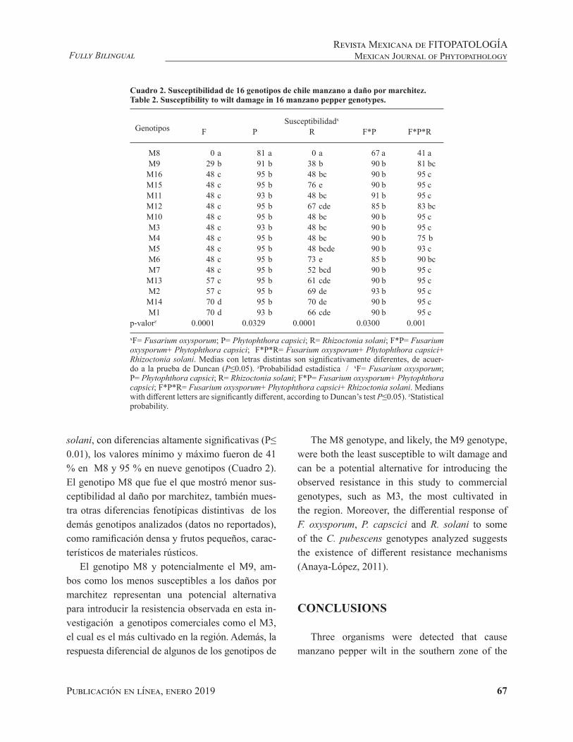
In infections caused by F. oxysporum, there were highly significant differences (P≤0.01) between genotypes with 0-70% variation between the lowest and the highest value, respectively, where the M8 genotype showed resistance, and the M9 genotype showed tolerance to the pathogen (Table 2). In seedlings inoculated with R. solani there were highly significant differences (P≤0.01), with minimum values of 0 % for M8 and maximum values of 76 % for M15. When inoculated with P. capsica, significant differences (P≤0.05) were observed, with 81-95 % variation in susceptibility between M8 and M7, respectively. In pathogen combinations, the intervals between maximum and minimum values were lower. In combinations of F. oxysporum + P.capsici with significant differences (P≤0.05), the maximum and minimum values were 67 % for M8 and 93% for M2; and the combination F. oxysporum + P.capsici + R. solani with highly significant differences (P≤0.01), the minimum and maximum values were 41% for M8 and 95% for nine genotypes (Table 2). The M8 genotype had the lowest level of wilt susceptibility and also showed other distinctive phenotypic differences compared to the rest of the genotypes analyzed (non-reported data), such as dense ramification and small fruits that are characteristic of rustic materials.
Publicación en línea, enero 2019 67
Fully BilingualRevista Mexicana de FITOPATOLOGÍA
Mexican Journal of Phytopathology
solani, con diferencias altamente significativas (P≤ 0.01), los valores mínimo y máximo fueron de 41 % en M8 y 95 % en nueve genotipos (Cuadro 2). El genotipo M8 que fue el que mostró menor sus-ceptibilidad al daño por marchitez, también mues-tra otras diferencias fenotípicas distintivas de los demás genotipos analizados (datos no reportados), como ramificación densa y frutos pequeños, carac-terísticos de materiales rústicos.
El genotipo M8 y potencialmente el M9, am-bos como los menos susceptibles a los daños por marchitez representan una potencial alternativa para introducir la resistencia observada en esta in-vestigación a genotipos comerciales como el M3, el cual es el más cultivado en la región. Además, la respuesta diferencial de algunos de los genotipos de
Cuadro 2. Susceptibilidad de 16 genotipos de chile manzano a daño por marchitez.Table 2. Susceptibility to wilt damage in 16 manzano pepper genotypes.
GenotiposSusceptibilidadx
F P R F*P F*P*R
M8 0 a 81 a 0 a 67 a 41 aM9 29 b 91 b 38 b 90 b 81 bcM16 48 c 95 b 48 bc 90 b 95 cM15 48 c 95 b 76 e 90 b 95 cM11 48 c 93 b 48 bc 91 b 95 cM12 48 c 95 b 67 cde 85 b 83 bcM10 48 c 95 b 48 bc 90 b 95 cM3 48 c 93 b 48 bc 90 b 95 cM4 48 c 95 b 48 bc 90 b 75 bM5 48 c 95 b 48 bcde 90 b 93 cM6 48 c 95 b 73 e 85 b 90 bcM7 48 c 95 b 52 bcd 90 b 95 cM13 57 c 95 b 61 cde 90 b 95 cM2 57 c 95 b 69 de 93 b 95 cM14 70 d 95 b 70 de 90 b 95 cM1 70 d 93 b 66 cde 90 b 95 c
p-valorz 0.0001 0.0329 0.0001 0.0300 0.001
xF= Fusarium oxysporum; P= Phytophthora capsici; R= Rhizoctonia solani; F*P= Fusarium oxysporum+ Phytophthora capsici; F*P*R= Fusarium oxysporum+ Phytophthora capsici+ Rhizoctonia solani. Medias con letras distintas son significativamente diferentes, de acuer-do a la prueba de Duncan (P≤0.05). zProbabilidad estadística / xF= Fusarium oxysporum; P= Phytophthora capsici; R= Rhizoctonia solani; F*P= Fusarium oxysporum+ Phytophthora capsici; F*P*R= Fusarium oxysporum+ Phytophthora capsici+ Rhizoctonia solani. Medians with different letters are significantly different, according to Duncan’s test P≤0.05). zStatistical probability.
The M8 genotype, and likely, the M9 genotype, were both the least susceptible to wilt damage and can be a potential alternative for introducing the observed resistance in this study to commercial genotypes, such as M3, the most cultivated in the region. Moreover, the differential response of F. oxysporum, P. capscici and R. solani to some of the C. pubescens genotypes analyzed suggests the existence of different resistance mechanisms (Anaya-López, 2011).
CONCLUSIONS
Three organisms were detected that cause manzano pepper wilt in the southern zone of the

Publicación en línea, enero 2019 68
Fully BilingualRevista Mexicana de FITOPATOLOGÍAMexican Journal of Phytopathology
C. pubescens analizados a F. oxysporum, P. capsci-ci y R. solani y sus combinaciones, sugieren varia-ción en los mecanismos de defensa (Anaya-López, 2011).
CONCLUSIONES
Se detectaron tres organismos causales de la marchitez de chile manzano del sur de México; de ellos Fusarium oxysporum fue el más frecuente, seguido por Phytophthora capsici y Rhizoctonia solani.
La identificación molecular permitió identidad de 88 % de los fragmentos amplificados de F. oxys-porum con F. oxysporum f.sp. lycopersici raza 2. P. capsici tuvo identidad de 83 % con P. capsici_ LT1534, versión 11, y R. solani alcanzó identidad de 90% con el grupo anastómico 3 (AG-3).
De los tres patógenos encontrados, Phytophtho-ra capsici es el que causa la mayor severidad en daño por marchitez en la región sur del Estado de México, aunque Fusarium oxysporum y Rhizocto-nia solani también causan daños en la planta.
El genotipo M8 fue resistente a F. oxysporum y R. solani, y el genotipo M9 podría ser tolerante a F. oxysporum. El genotipo M8 fue tolerante a P. capsici y a la inoculación individual y en mezcla de los tres patógenos, por lo que podría representar un genotipo potencial para estudios sobre fuentes de genes de tolerancia útiles en programas de mejora-miento genético.
AgradecimientosLos autores agradecen al Consejo Nacional de Ciencia y
Tecnología de México (CONACYT), por la beca escolar otor-
gada a Alma Janeth Vallejo Gutiérrez para estudios de maes-
tría.
State of Mexico, from which Fusarium oxysporum was the most frequently present, followed by Phytophthora capsici and Rhizoctonia solani.
The molecular identification showed 88% identity of the amplified fragments of F. oxysporum using F. oxysporum f.sp. lycopersici race 2. P. capsici showed 83% identity with P. capsici_ LT1534, version 11, and R. solani had 90% identity with the anastomic group 3 (AG-3).
From the three pathogens identified, Phytophthora capsici causes the highest level of wilt severity in the southern region of the State of Mexico, although Fusarium oxysporum and Rhizoctonia solani also cause plant damage.
The M8 genotype was resistant to F. oxysporum and R. solani, and the M9 genotype could be tolerant to F. oxysporum. The M8 genotype was tolerant to P. capsici and to inoculation with the three pathogens, alone and combined, so it could be a potential genotype for studies about tolerance genes that could be used in genetic improvement programs.
AcknowledgmentsThe authors wish to thank the Consejo Nacional de Ciencia
y Tecnología de México (CONACYT) for the scholarship
granted to Alma Janeth Vallejo Gutiérrez for her master’s
studies.
End of the English version
LITERATURA CITADAAbawi SG and Pastor-Corrales CMA. 1990. Root rots of beans
in Latin America and Africa: Diagnosis, research methodo-logies, and management strategies. Centro Internacional de Agricultura Tropical (CIAT), Cali, Colombia. 114p.

Publicación en línea, enero 2019 69
Fully BilingualRevista Mexicana de FITOPATOLOGÍA
Mexican Journal of Phytopathology
Abdullah AS, Moffat CS, Lopez-Ruiz FJ, Gibber MR, Hamblin J and Zerihun A. 2017. Host-Multi-Pathogen warfare: Pa-thogen interactions in co-infected plants. Frontiers in Plant Science 8:1806. https://doi.org/10.3389/fpls.2017.01806
Alizon S, de Roode JC and Michalakis Y. 2013. Multiple in-fections and the evolution of virulence. Ecology Letters 16:556-67. https://doi.org/10.1111/ele.12076
Anaya-López JL, González-Chavira MM, Pineda-Villordo E, Rodríguez-Guerra R, Rodríguez-Martínez R, Guevara-González RG, Guevara-Olvera L, Montero-Tavera V y Torres-Pacheco I. 2011. Selección de genotipos de chiles resistentes al complejo patogénico de la marchitez. Revista Mexicana de Ciencias Agrícolas 3:373-383. Disponible en línea:
http://www.scielo.org.mx/pdf/remexca/v2n3/v2n3a6.pdfApodaca-Sánchez MA, Zavaleta-Mejía E, García-Espinosa R,
Osada-Kawasoe S y Valenzuela-Ureta JG. 2001. Compara-ción de técnicas para evaluar in vitro la patogenicidad de Fusarium oxysporum f. sp. radicis-lycopersici y efecto de la temperatura. Revista Mexicana de Fitopatología 19:197-2002. Disponible en línea:
http://www.redalyc.org/pdf/612/61219210.pdfArias AB, Mejía CJ, Estrada MI, Arriaga RM y García VLM.
2017. Caracterización morfológica de híbridos de chi-le manzano. Revista Mexicana de Ciencias Agrícolas 8:825-836. Disponible en línea: http://www.redalyc.org/html/2631/263152088006/
Booth C. 1971. The genus Fusarium. Commonwealt Mycolo-gical Institute. Kew, Surrey, England. 237 p.
Carrizo GC, Barfuss HJM, Sehr EM, Barboza GE, Rosabelle S, Eduardo A and Ehrenderfer F. 2016. Phylogenetic re-lationships, diversification and expansion of chili peppers (Capsicum, Solanaceae). Annals of Botany 118:35-51. Disponible en línea:
https://www.ncbi.nlm.nih.gov/pubmed/27245634Chesson P. 2000. Mechanisms of maintenance of species
diversity. Annual Review of Ecology and Systematics 31:343–366. Disponible en línea:
https://doi.org/10.1146/annurev.ecolsys.31.1.343Di Rienzo JA, Casanoves F, Balzarini MG, Gonzalez L, Tabla-
da M, Robledo CW. InfoStat versión 2016. Grupo InfoStat, FCA, Universidad Nacional de Córdoba, Argentina. URL. http://www.infostat.com.ar
Erwin DC and Ribeiro OK. 1996. Phytophthora diseases world-wide. The Journal of Agricultural Science 131(2):245–249. https://doi.org/10.1017/S0021859698215796
Feng B, Li P, Wang H and Zhang X. 2010. Functional analy-sis of pcpme6 from oomycete plant pathogen Phytophtho-ra capsici. Microbial Pathogenesis 49:23-31. https://doi.org/10.1016/j.micpath.2010.03.004
Fernández ER, Trapero A y Domínguez J. 2010. Experimen-tación en agricultura. Junta de Andalucía, Consejería de Agricultura y Pesca. Sevilla, España. 350 p.
Gallegly ME and Honh Ch. 2008. Phytophthora capsici. In identifying species by morphology and ADN fingerprints. APS PRESS. St. Paul, Minnesota, Estados Unidos de América. 165 p.
González GM. 2008. Reseña de “Aspectos de sistemáti-ca y biología del complejo Rhizoctonia”. Fitosanidad 12:147-159. Disponible en línea: http://www.redalyc.org/pdf/2091/209115572003.pdf
González-Pérez S, Garces-Claver A, Mallor C, Sáenz de Miera LE, Fayos O, Pomar F, Merino F and Silvar C. 2014. New insights into Capsicum spp. relatedness and the diversi-fication process of Capsicum annuum in Spain. Plos one 9:e116276. Disponible en línea: https://www.ncbi.nlm.nih.gov/pmc/articles/PMC4278865/pdf/pone.0116276.pdf
Guigón-López C y González-González PA. 2001. Estu-dio regional de la enfermedades del chile (Capsicum annuum L.) y su comportamiento temporal en el sur de Chihuahua, México. Revista Mexicana de Fitopatología. 19:49-56. Disponible en línea: http://www.redalyc.org/pdf/612/61219107.pdf
Guzmán-Plazola RA, Fajardo-Franco ML, García-Espinosa R and Cadena-Hinojosa MA. 2011. Desarrollo epidémico de la cenicilla y rendimiento de tres cultivares de tomate en la comarca lagunera, Coahuila, México. Agrociencia 45:363-378. Disponible en línea: http://www.scielo.org.mx/pdf/agro/v45n3/v45n3a9.pdf
Hamon C, Baranger A, Coyne CJ, Mcgee RJ, Le Goff IL, L’anthoëne V, Esnault R and Pilet-Nayel ML. 2011. New consistent QTL in pea associated with partial resistance to Aphanomyces euteiches in multiple field and controlled environments from France and the United States. Theo-retical and Applied Genetics 123:261-281. DOI: 10.1007/s00122-011-1582-z
Herrera I y Laurentin H. 2012. Evaluación de la esporulación de Fusarium oxysporum f. sp. sesami en dos medios de cultivo y dos metodologías de inoculación en ajonjo-lí (Sesamum indicum). Revista Científica UDO Agrícola 12:639-643. Disponible en línea: http://www.bioline.org.br/pdf?cg12072
Kannwischer ME and Mitchell DJ. 1978. The Influence of a fungicide on the epidemiology of black shank of tobac-co. Ecology and Epidemiology 68: 1760-1765. Dispo-nible en línea en: https://www.apsnet.org/publications/phytopathology/backissues/Documents/1978Articles/Phyto68n12_1760.pdf
Kousik ChS, Donahoo RS and Hassell R. 2012. Resistance in watermelon rootstocks to crown rot caused by Phyto-phthora capsici. Crop Protection. 39: 18–25. Disponible en línea: https://www.sciencedirect.com/science/article/pii/S0261219412000907
Lamour HK, Stam R, Jupe J and Huitema E. 2012. The oomycete broad-host-range pathogen Phytophthora cap-sici. Molecular Plant Pathology 13:329-337. https://doi.org/10.1111/j.1364-3703.2011.00754.x
Leonian LH. 1922. Stem and fruit blight of peppers caused by Phytophthora capsici. Phytopathology 12:401-408. Disponible en línea: https://www.apsnet.org/publications/phytopathology/backissues/Documents/1972Articles/Phyto62n01_20.PDF
Leslie JF and Sumerell BA. 2006. The Fusarium Laboratory Ma-nual. First edition. State Avenue, Ames, Iowa, USA. Blac-kwell Publishing. 388 p. Disponible en línea: https://www.wiley.com/en-us/The+Fusarium+Laboratory+Manual-p-9780813819198
López AGF. 1984. Manejo de Hongos Fitopatógenos. Departa-mento de Enseñanza e Investigación en parasitología Agrí-cola. Chapingo, México. pp. 106-115.

Publicación en línea, enero 2019 70
Fully BilingualRevista Mexicana de FITOPATOLOGÍAMexican Journal of Phytopathology
Lozano AN, Guzmán-Plazola RA, Zavaleta ME, Aguilar RVH y Ayala EV. 2015. Etiología y evaluación de alternativas de control de marchitez del chile de árbol (Capsicum annuum L.) en la Vega de Metzitlán, Hidalgo, México. Revista Mexicana de Fitopatología 33:31-53. Disponible en línea: http://www.redalyc.org/pdf/612/61240687003.pdf
Martínez GE, Albarracin N, Arcia A, Subero L y Albarracin M. 1996. Pudrición basal del ajo causado por Fusarium oxys-porum. Agronomía Tropical 46:265-273. http://hdl.handle.net/20.500.11799/40647
Martínez EI. 2016. Caracterización morfológica y molecular de 15 colectas de chile manzano (Capsicum pubescens R. y P.) de la región sur del Estado de México. Tesis de Maestría. Centro Universitario UAEM Tenancingo, México. P.43-56. Disponible en línea: http://hdl.handle.net/20.500.11799/40647
Nelson PE, Tousson TA and Marasa WFO. 1983. Fusarium speies: an illistrated manual for identificaction. The Penns-ylvania State University. Pennsylvania, USA. 193 p.
Nora S, Albaladejo RG, González MSC, Robledo-Arnuncio JJ y Aparicio A. 2011. Movimiento de genes (polen y semi-llas) en poblaciones fragmentadas de plantas. Ecosistemas 20:35-45. Disponible en línea: http://www.redalyc.org/ar-ticulo.oa?id=54022121004
Pickersgill B. 2007. Domestication of plants in the Americas: Insights from Mendelian and molecular genetics. Annals of Botany 100: 925-940. http://doi:10.1093/aob/mcm193
Ruíz de GJI, Prohens J y Tierno R .2016. Las variedades loca-les en la mejora genética de plantas. Vitoria-Gasteiz: Ser-vicio Central de Publicaciones del Gobierno Vasco. País Vasco, España. 480 p.
Sanzón GD, Valdovinos PG, Rojas MRI y Zavaleta ME. 2012. Cambios morfológicos en células de chile CM334 inocula-do con Phytophthora capsici y con Fusarium oxysporum. Revista Mexicana de Fitopatología 30:66-71. Disponi-ble en línea: http://www.scielo.org.mx/pdf/rmfi/v30n1/v30n1a6.pdf
Secretaría de Agricultura, Ganadería, Desarrollo Rural, Pes-ca y Alimentación. 2017. Producción nacional de chile alcanza 2.3 millones de toneladas. https://www.gob.mx/sagarpa/prensa/produccion-nacional-de-chile-alcanza-2-3-millones-de-toneladas (consulta, junio 2018).
Segura LS, Zavala RD, Equihua CC, Andrés AJ y Yepez TE. 2009. Los recursos genéticos de frutales en Michoacán. Revista Chapingo Serie Horticultura 15(3): 297-305. Disponible en línea: http://www.scielo.org.mx/pdf/rcsh/v15n3/v15n3a11.pdf
Secretaría de Agricultura, Ganadería, Desarrollo Rural, Pesca y Alimentación. 2018. Relación de huertos de chile manza-no registrados para exportación del Estado de México a los Estados Unidos de América con tratamiento de irradiación. https://www.gob.mx/cms/uploads/attachment/file/379049/CHILE_MANZANO_EDO_MEX_08_23__2018.pdf (consulta, septiembre 2018).
Schubert S, Neubert A, Schierholt A, Sümer A and Zörb C. 2009. Development of salt-resistant maize hybrids: The combination of physiological strategies using conventio-nal breeding methods. Plant Science. 177(3): 196-202. https://doi.org/10.1016/j.plantsci.2009.05.011
Silva-Rojas HV, Fernandéz-Pavia, SP, Góngora-Canul C, Ma-cías-López BC y Ávila-Quezada GD. 2009. Distribución espacio temporal de la marchitez del chile (Capsicum An-nuum L.) en Chihuahua e identificación del agente Causal Phytophthora capsici Leo. Revista Mexicana de Fitopa-tología. 27: 134-147. Disponible en línea: http://www.re-dalyc.org/articulo.oa?id=61212195006
Singlenton L, Mihail JD and Rush MCh. 1992. Methods for research on soil borne Phytopathogenic Fungi. APS Press. St. Paul, Minesota, USA. 264 p.
Uc-Arguelles AK, Pérez-Moreno J, Ayala-Escobar V and Zavaleta-Mejía E. 2017. Antagonism of Saccharicola sp. against phytopathogens of the root of jalapeno pepper (Capsicum annuum). Revista Mexicana de Fitopatología 35:263-283. Disponible en línea: http://www.scielo.org.mx/pdf/rmfi/v35n2/2007-8080-rmfi-35-02-00263.pdf
United States Department of Agriculture Agricultural Research Service (USDA-ARS). 2011. Grin species records of Cap-sicum. Beltsville, Maryland: National Germplasm Resour-ces Laboratory. https://www.ars.usda.gov/northeast-area/beltsville-md-barc/beltsville-agricultural-research-center/national-germplasm-resources-laboratory/ (consulta, fe-brero 2018)
Vásquez LA, Tlapa BB, Yáñez MMJ, Pérez PR y Quintos EM. 2009. Etiología de la marchitez del ‘chile de agua’ (Cap-sicum annuum L.) en Oaxaca, México. Revista Fitotecnia Mexicana 32 (2): 127 – 134. Disponible en línea: http://www.redalyc.org/articulo.oa?id=61011222007
Velásquez-Valle R, Medina-Aguilar MM y Luna-Ruiz JJ. 2001. Sintomatología y géneros de patógenos asociados con las pudriciones de la raíz del chile (Capsicum annuum L.) en el Norte-Centro de México. Revista Mexicana de Fitopatología 19:175-181. Disponible en línea: http://www.redalyc.org/articulo.oa?id=61219207
Watanabe T. 2002. Pictorial atlas of soil and seed fungi. Mor-phogies of cultures fungi and key to species. Second edition. CRC Press. New York Washington, D.C. 500 p. Disponible en línea: http://www.eagriculture.biz/down-load/Soil/Pictorial%20Atlas%20of%20Soil%20and%20Seed%20Fungi%20%20Morphologies%20of%20Cultu-red%20Fungi%20and%20Key%20to%20Species.pdf
White TJ, Bruns T, Lee S and Taylor J. 1990. Amplification and direct sequencing of fungal ribosomal RNA genes for phylogenies. PCR Protocols. Academic Press. San Diego, CA, USA. P 315-322. Disponible en línea:
https://nature.berkeley.edu/brunslab/papers/white1990.pdf
Zhang YL, Jia QL, Li DW, Wang JE, Yin YX and Gong H. 2013. Characteristic of the pepper CaRGA2 gene in de-fense responses against Phytophthora capsici Leonian. In-ternational Journal of Molecular Sciences 14: 8985-9004. https://doi.org/10.3390/ijms14058985