Embed Size (px)
Citation preview

HYPOTHESIS AND THEORY ARTICLEpublished: 26 February 2015
doi: 10.3389/fcell.2015.00008
Rethinking gene regulatory networks in light of alternativesplicing, intrinsically disordered protein domains, andpost-translational modificationsKarl J. Niklas1*, Sarah E. Bondos2, A. Keith Dunker3 and Stuart A. Newman4
1 Plant Biology Section, School of Integrative Plant Science, Cornell University, Ithaca, NY, USA2 Department of Molecular and Cellular Medicine, Texas A&M Health Science Center, College Station, TX, USA3 Center for Computational Biology and Bioinformatics, School of Medicine, Indiana University, Indianapolis, IN, USA4 Department of Cell Biology and Anatomy, New York Medical College, Valhalla, NY, USA
Edited by:
David Ellard Keith Ferrier, Universityof St Andrews, UK
Reviewed by:
Markus Friedrich, Wayne StateUniversity, USAGunter P. Wagner, Yale University,USA
*Correspondence:
Karl J. Niklas, Plant Biology Section,School of Integrative Plant Science,Cornell University, Ithaca, NY14853-5908, USAe-mail: [email protected]
Models for genetic regulation and cell fate specification characteristically assume thatgene regulatory networks (GRNs) are essentially deterministic and exhibit multiplestable states specifying alternative, but pre-figured cell fates. Mounting evidence shows,however, that most eukaryotic precursor RNAs undergo alternative splicing (AS) and thatthe majority of transcription factors contain intrinsically disordered protein (IDP) domainswhose functionalities are context dependent as well as subject to post-translationalmodification (PTM). Consequently, many transcription factors do not have fixed cis-actingregulatory targets, and developmental determination by GRNs alone is untenable.Modeling these phenomena requires a multi-scale approach to explain how GRNsoperationally interact with the intra- and intercellular environments. Evidence shows thatAS, IDP, and PTM complicate gene expression and act synergistically to facilitate andpromote time- and cell-specific protein modifications involved in cell signaling and cellfate specification and thereby disrupt a strict deterministic GRN-phenotype mapping.The combined effects of AS, IDP, and PTM give proteomes physiological plasticity,adaptive responsiveness, and developmental versatility without inefficiently expandinggenome size. They also help us understand how protein functionalities can undergo majorevolutionary changes by buffering mutational consequences.
Keywords: cell fate specification, combinatorial transcription regulation, eukaryotes, evolution, development, gene
regulatory networks, multi-stable dynamical systems, protein structure
“The unpredictable and the predetermined unfold together to makeeverything the way it is. It’s how nature creates itself, on every scale,the snowflake and the snowstorm.”
–Tom Stoppard, Arcadia, Act 1, Scene 4
INTRODUCTIONA fundamental assumption of contemporary developmental biol-ogy is that gene regulatory networks (GRNs, herein defined ascircuits of interacting transcription factors and their cis-actingregulatory elements) are primary mechanisms controlling devel-opment. According to this assumption, at any time, the relativelevels of transcription factors in an extended network determinethe progress of development by regulating downstream genes(Carroll et al., 2004; Davidson and Erwin, 2006). This conceptionof gene control in multicellular organisms, which was formu-lated in several related versions a half century ago (Britten andDavidson, 1969; Kauffman, 1969; Britten, 1982; Davidson, 1982),proposes that GRNs are deterministic dynamical systems exhibit-ing multiple stable states. The theoretical foundations of thisframework can be traced to studies of the bi-stable gene regula-tory switch between the lytic and lysogenic states of the lambda
phage in Escherichia coli (see Ptashne, 2004), and have been gen-eralized and applied to the larger and more elaborate GRNs ofeukaryotes in the form of models ranging from discrete Booleannetworks to continuous systems of ordinary differential equations(Glass and Kauffman, 1973; Kauffman, 1974; Savageau, 1974;Glass, 1975; Lauffenburger, 2000; Jaeger and Monk, 2014). Acommonality among these models describing cell differentiationis the assumption that gene products (i.e., proteins, particularlytranscription factors) have specific identities and connectivityrelationships to one another in the GRNs in which they func-tion (Hasty and Collins, 2001; Forgacs and Newman, 2005).According to this view, variation in the outcome of the function ofGRNs (e.g., alternative cell types) arises from nonlinearities andstochastic effects to which such complex, deterministic systemsare subject.
This paradigm has been extended to other gene expressionmechanisms that have been characterized since the GRN dynam-ics model was first proposed. Among these mechanisms is thealternative splicing (AS) of pre-mRNA exons and introns toassemble different proteins, a process that permits variation inthe functionalities of subsets defining components of GRNs (e.g.,transcription factors) at the level of RNA processing. Although
www.frontiersin.org February 2015 | Volume 3 | Article 8 | 1
CELL AND DEVELOPMENTAL BIOLOGY

Niklas et al. Rethinking gene regulatory networks
the mixing and matching of basic system components has nodirect counterpart in a truly deterministic GRN model, the factorscontrolling AS can be viewed as having well defined function-alities, since the associated GRN dynamics permit different cellfates in a combinatorial deterministically prescribed manner.Likewise, the modulatory effects of microRNAs, riboswitches, andthe enzymes that mediate post-translational modifications (andthat can silence genes) can be viewed as adding a complicating,yet still deterministic set of regulatory mechanisms.
Here, we propose that this perspective must also contendwith evidence that the majority of eukaryotic transcription fac-tors contain intrinsically disordered protein (IDP) domains (Liuet al., 2006; Dunker et al., 2014) that comprise almost two-thirds of their sequences (Ward et al., 2004; Minezaki et al.,2006a,b; Singh and Dash, 2007; Xie et al., 2007), and with thefact that the conformations of these domains, and hence theirfunctions, are contingent on the intra-and extracellular environ-ments in which these proteins function (Ducas and Rhoades,2014; Srinivasan et al., 2014). Consequently, the specificity of thebinding of most regulatory transcription factors to cis-regulatoryelements, as well as their partnering with other factors mediatingconditional responses to cellular physiological status, are contextdependent and subject to change even in the absence of geneticor epigenetic alterations. Importantly, the functions of IDPs aremodulated further by both alternative splicing (AS) and post-translational modifications (PTMs), especially phosphorylation(Iakoucheva et al., 2004; Romero et al., 2006; Singh and Dash,2007). For example, AS, IDPs, and PTMs are known to act syn-ergistically in modulating the activities of the tumor repressingtranscription factor p53 (Dunker et al., 2008) and to underliethe functions of several other proteins crucial for the evolutionof multicellular organisms (Dunker et al., 2014; Niklas et al.,2014). The combined functional consequences of AS, IDPs, andPTMs make modeling GRN dynamics as strictly deterministicsystems incomplete at best (Kupiec, 2009; Braunschweig et al.,2013). If transcription factors do not have fixed cis-acting regu-latory element targets, but rather can alter their specific identityand network-topological status within a given GRN dependingon other proteins in the nucleus and external environmental fac-tors, it follows that GRNs can no longer be viewed as deterministicsystems in a strict physical or mathematical sense. If our concep-tualization is correct, we predict that the incorporation of AS,IDP, and PTM (designated, collectively but as operatively inde-pendent processes, as AS–IDP–PTM) and their well-documentedsynergistic interactions into an expanded (and thus more com-putationally sophisticated) approach will provide deeper insightinto recently recognized genotype-phenotype mapping anoma-lies, e.g., developmental system drift (True and Haag, 2001) andthe puzzle of missing heritability (Zaitlen and Kraft, 2012).
Toward this goal, we present evidence that AS–IDP–PTM pro-motes alternative, context-dependent GRN states, and thus servesa critical role in a broad range of cellular responses, includ-ing cell fate specification. We also present evidence that thesethree components are ancient in eukaryotic GRNs, a speculationdriven by the observation that early divergent unicellular eukary-otes achieve temporally alternative physiological and reproduc-tive states and respond adaptively to contingent environmental
conditions by virtue of AS–IDP–PTM. Further, we provide sug-gestions for how the determinate outcomes of plant and animaldevelopment are realized despite the indeterminacy of isolatedGRNs. Our conclusion is that we face an “incompleteness theo-rem” when we attempt to reduce development to a single causallevel (Niklas and Kutschera, 2012).
Finally, it is important to emphasize that rather than propos-ing AS-IDP-PTM as a developmental mechanism in its own right,we see its collective role as creating an adaptive plasticity that sig-nificantly diminishes the strict determinism that some attributeto GRNs. The latter framework is all-too-often treated as a bio-logical Welterklärung (theory of everything) simply because theusual experimental frames of reference make it difficult to thinkbeyond genes and their interactions. We hope to establish that thesynergistic interactions among AS, IDP, and PTM have enabledliving systems to evolve beyond the constraints that are inevitablein regulatory networks that depend on single-level dynamics.
ALTERNATIVE SPLICING (AS)Alternative splicing produces protein isoforms from the sameprecursor mRNA by retaining or excluding different exons toachieve differential translation. First observed in the infectiousadenovirus cycle (Berget et al., 1977; Chow et al., 1977) and sub-sequently in the transcripts of normal, endogenous genes (Leffand Rosenfeld, 1986), AS occurs in all eukaryotic lineages (Black,2003) and becomes more prevalent as complexity, estimated bythe number of different cell types, increases (Chen et al., 2014a).Although a number of scenarios have been advanced for the ori-gin of AS, including a role in enabling the cell to filter out aberranttranscripts (Catania and Lynch, 2008), we suggest that the con-nection between cell type number and AS (given the associationwith IDP and PTM) is an inherent one that promoted occupationof new niches (see below “AS-IDP-PTM Phylogenetic Patterns”).
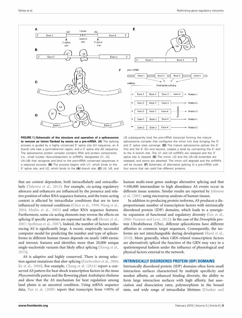
Five basic types of alternative splicing exist: alternative 3′acceptor site, alternative 5′ donor splice site, intron retention,mutually exclusive exon splicing, and exon skipping (Black, 2003).The last is the most frequent. Regulation and selection of thesplice sites are performed by trans-acting splicing activator andrepressor proteins within an RNA–protein complex, the spliceo-some, which is canonically composed of five small nuclear RNAs(i.e., U1, U2, U4–U6) and a range of assorted protein fac-tors (Figure 1). Splicing is regulated by trans-acting repressor-activator proteins and their corresponding cis-acting regulatorysilencers and enhancers on the pre-mRNA (Matera and Wang,2014). The effects of splicing factors are often position-dependent(Barash et al., 2010). A splicing factor that functions as an activa-tor when bound to an intronic enhancer element may function asa repressor when bound to its splicing element in the context ofan exon (Lim et al., 2011).
The secondary structure of the pre-mRNA transcript alsodetermines which exons and introns will be spliced, e.g., by bring-ing together splicing elements or by masking a sequence thatwould otherwise serve as a binding element for a splicing factor.Consequently, activators, repressors, and secondary pre-mRNAstructure constitute a splicing “code” that defines the protein iso-forms produced under different cellular conditions. Additionally,the elements within this code function interdependently in ways
Frontiers in Cell and Developmental Biology | Evolutionary Developmental Biology February 2015 | Volume 3 | Article 8 | 2
Niklas et al. Rethinking gene regulatory networks
FIGURE 1 | Schematic of the structure and operation of a spliceosome
to remove an intron flanked by exons on a pre-mRNA. (A) The splicingprocess is guided by a highly conserved 5′ splice site GU sequence, an Abranch site near a pyrimidine-rich region, and a 3′ splice site AG sequence.The spliceosome protein complex contains RNA and protein components(i.e., small nuclear ribonucleoprotein or snRNPs, designated U1, U2,U4–U6) that recognize and bind to the pre-mRNA conserved sequences ina stepwise process. (B) The process begins with U1, which binds to the5′ splice site, and U2, which binds to the (A) branch site. (C) U4, U6, and
U5 subsequently bind the pre-mRNA transcript forming the maturespliceosome complex that configures the intron into loop bringing the 5′and 3′ splice sites converge. (D) The mature spliceosome splices the 5′first and the 5′ GU end second, creates a lariat by connecting the 5′ endto the A branch site. The U1 and U4 snRNPs are released and the 3′splice site is cleaved. (E) The intron, U3 and the U5–U6 ensemble arereleased, and exons are attached. The intron will degrade and the snRNPswill be reused. (F) Schematic of alternative splicing of a pre-mRNA withfour exons that can yield five different proteins.
that are context dependent, both intracellularly and extracellu-larly (Talavera et al., 2013). For example, cis-acting regulatorysilencers and enhancers are influenced by the presence and rela-tive position of other RNA sequence features, and the trans-actingcontext is affected by intracellular conditions that are in turninfluenced by external conditions (Chen et al., 1999; Wang et al.,2004; Matlin et al., 2005) and other RNA sequence features.Furthermore, some cis-acting elements may reverse the effects onsplicing if specific proteins are expressed in the cell (Boutz et al.,2007; Spellman et al., 2007). Indeed, the number of factors influ-encing AS is significantly large. A recent, empirically successfulcomputer model for predicting the number and type of spliceo-forms in different human tissues depends on nearly 1400 exonicand intronic features and identifies more than 20,000 uniquesingle-nucleotide variants that likely affect splicing (Xiong et al.,2015).
AS is adaptive and highly conserved. There is strong selec-tion against mutations that alter splicing (Fairbrother et al., 2004;Ke et al., 2008). For example, Chang et al. (2014) report a con-served AS pattern for heat shock transcription factors in the mossPhyscomitrella patens and the flowering plant Arabidopsis thalianaand show that the AS mechanism for heat regulation amongland plants is an ancestral condition. Using mRNA sequencedata, Pan et al. (2008) report that transcripts from ≈95% of
human multi-exon genes undergo alternative splicing and that≈100,000 intermediate to high abundance AS events occur indifferent tissue systems. Similar results are reported by Johnsonet al. (2003) using microarray analyses of human tissues.
In addition to producing protein isoforms, AS produces a dis-proportionate number of transcription factors with intrinsicallydisordered protein (IDP) domains, which leads to a synergis-tic expansion of functional and regulatory diversity (Liu et al.,2006; Vuzman and Levy, 2012). In the case of the Drosophila pro-tein Ultrabithorax (Ubx), different spliceoforms have differentaffinities to common target sequences. Consequently, the iso-forms are not interchangeable during development (Reed et al.,2010). More generally, when GRN-related transcription factorsare alternatively spliced the function of the GRN may vary in aspatiotemporal fashion under the influence of physiological andphysical factors external to the network.
INTRINSICALLY DISORDERED PROTEIN (IDP) DOMAINSIntrinsically disordered protein (IDP) domains often form smallinteraction surfaces characterized by multiple specificity andmodest affinity, an enhanced binding diversity, the ability toform large interaction surfaces with high affinity, fast asso-ciation and dissociation rates, polymorphism in the boundstate, and wide range of intracellular lifetimes (Dunker and
www.frontiersin.org February 2015 | Volume 3 | Article 8 | 3

Niklas et al. Rethinking gene regulatory networks
Uversky, 2010; Oldfield and Dunker, 2014). These traits makeIDPs versatile signaling and regulatory molecules. Studies haveidentified intrinsically disordered domains as enriched in thenon-constitutive exons, indicating that protein isoforms may dis-play functional diversity due to the alteration of tissue-specificmodules within these regions (Buljan et al., 2012). IDP domainscan exist as molten globules with defined secondary structureor as unfolded chains that can function through transitionsamong different folded states. Their functional conformationscan change by binding to other proteins and nucleic acids(Uversky, 2002; Oldfield et al., 2008; Hsu et al., 2013). IDPsalso contribute to the process of alternative splicing: the RS-repeat domains of the conserved SR family of metazoan splicingfactors are intrinsically disordered (Braunschweig et al., 2013).Post-translational modifications can also alter IDP functionalities(Iakoucheva et al., 2004; Dyson and Wright, 2005; Oldfield et al.,2008).
Numerous examples of IDP domains involved in transcrip-tional regulation are known (Campbell et al., 2000; Haynes andIakoucheva, 2006; Liu et al., 2006; Sun et al., 2013). The C-terminal activation domain of the bZIP proto-oncoprotein c-Fos,which effectively suppresses transcription in vitro, is intrinsicallydisordered and highly mobile (Campbell et al., 2000). The C-terminal domain of the transcriptional co-repressor CtBP, whichfacilitates gene targeting and coordinated histone modificationsin the multi-protein complex, is intrinsically disordered (Bhallaet al., 2006; Haynes and Iakoucheva, 2006; Sun et al., 2010).The unbound N-terminal domains of the DELLA proteins, whichare central to the integration of plant developmental and envi-ronmental signaling, undergo disorder-order transitions uponbinding to interacting proteins (Sun et al., 2010). The DELLAproteins are similar in their domain structures to the GRASprotein family, whose N-domains are intrinsically disordered(Sun et al., 2011) and are extensively involved in plant signal-ing by virtue of undergoing disorder-order transformations ininteractions with a variety of molecular partners involved indevelopment, light signaling, nodulation, and auxin signaling andtranscription regulation to biotic and abiotic stresses.
Metazoans also carry out intercellular signaling via smallmolecules, called nuclear hormone receptors (NHRs), that bindto their cognate proteins. Following ligand binding, NHRstranslocate to the nucleus where they act as transcription factors.In addition to the structured ligand and DNA binding domains,these NHRs have flanking and linking IDP domains that bind tolarge numbers of partners. These domains may be responsible forthe variable or context dependent responses following hormonesignaling (Simons and Kumar, 2013). Thus, NHRs use disorderto bind to many partners, and many partners use disorder tobind to structured docking sites on NHR ligand binding domains(Mohan et al., 2006; Dunker et al., 2014).
Another important example is provided by the Wnt pathway.This key signaling pathway, which is utilized in development fromsponges to flies to mammals (Cadigan and Nusse, 1997; Nusseand Varmus, 2012), employs both IDPs and PTMs in fundamen-tal ways. Briefly, β-catenin, a dual-function cofactor for adhesionand transcription, is phosphorylated at four nearby sites in a dis-ordered tail by the destruction complex. This complex is held
together by the disordered scaffold protein axin, which uses along disordered region to flexibly tether β-catenin to two kinases,GSK3β and CK1α, thus speeding up the phosphorylation reac-tions by colocalization (Xue et al., 2012b; Dunker et al., 2014).These multiple phosphorylation events regulate both nuclearlocalization and proteasomal digestion of β-catenin. The activ-ity of adenomatous polyposis coli (APC), a massively disorderedmember of the β-catenin destruction complex, is also regulatedby phosphorylation (Minde et al., 2013). Thus, β-catenin accu-mulates, translocates to the nucleus, and turns on several genesthat activate cell proliferation and polarity.
These examples illustrate that intrinsically disordered tran-scription factor domains are central to plant and animal devel-opment and homeostasis. They are by no means exceptional. Liuet al. (2006) found that 82.6–93.1% of the transcription factors inthree databases contain extended regions of intrinsic disorder, incontrast to 18.6–54.5% of the proteins in two control datasets.Focusing on human transcription factors and using a disorderpredictor and Hidden Markov Models to search for regions thatare homologous to structured protein domains, Minezaki et al.(2006b) report that only 31% of the transcription factor residuesalign with known structured domains, which is only half of the62% structurally aligned residues for E. coli proteins that regulatetranscription.
Since protein-DNA recognition and protein-protein recogni-tion are central transcription factor functionalities, these andother studies illustrate the extent to which eukaryotic tran-scription factors manifest extensive flexibility as a consequenceof disorder-associated signaling and transcriptional regulation(Dunker et al., 2014). This permits them to bind to a greaterarray of partners that in turn can induce conformational changesin bound protein and DNA substrates (Oldfield et al., 2005). Awell-studied example of this is the isoforms of the DrosophilaUbx transcription factor described above. Here, two intrinsi-cally disordered domains modulate the binding affinity of thestructured DNA-binding homeodomain to its target sequence(Liu et al., 2008) and to other transcription factors (Johnsonet al., 1995; Bondos et al., 2006; Hsiao et al., 2014). The C-terminal IDP region, which is alternatively spliced, alters therelative affinity of Ubx for different DNA sequences (Liu et al.,2009).
POST-TRANSLATIONAL MODIFICATIONS (PTMs)Post-translational modifications (PTMs) alter the regulatoryinterfaces of proteins so as to induce gain, loss, or exchange ofbinding partners, thereby affecting function at many levels (VanRoey et al., 2013). Significantly, the structure of chromatin, themechanochemical medium within which eukaryotic transcrip-tion occurs, is regulated by PTM of histone proteins. Mediator,a multi-protein complex involved in RNA Pol II-regulated tran-scription, is both positively and negatively regulated by phos-phorylation (Gonzalez et al., 2014). Combinatorial PTMs ofthe C-terminal domain of RNA Polymerase II regulate multi-ple stages of transcription initiation and coordinate transcriptionwith mRNA processing (Yogesha et al., 2014).
However, our focus here is on the effect of PTMs on spe-cific transcription factors. In transcriptional regulation, each
Frontiers in Cell and Developmental Biology | Evolutionary Developmental Biology February 2015 | Volume 3 | Article 8 | 4

Niklas et al. Rethinking gene regulatory networks
transcription factor must participate in many different macro-molecular recognition/binding events (Bondos and Tan, 2001;Sun et al., 2013; Abdel-Hafiz and Horwitz, 2014). Transcriptionfactor binding to DNA often occurs in conjunction with otherspecific transcription factors, requiring tissue-specific protein-protein interactions as well. Transcription factors must interactwith Mediator or other components of the general transcriptionapparatus to elicit their function. Many transcription factors thatare active in developmental processes also bind histone acetylasesand de-acetylases. Phosphorylation can regulate each of theseevents. For example, DNA binding by the transcription factorEts-1 is allosterically coupled to a serine-rich region (Lee et al.,2008; Mooney et al., 2014). Ca2+ signaling induces phosphory-lation of this region, which modulates DNA binding by Ets-1.Phosphorylation of the intrinsically disordered PAGE4 protein(as part of the stress-response pathway) causes PAGE4 to releasethe transcription factor c-Jun, enabling its activity in transcrip-tion regulation (Mooney et al., 2014). Phosphorylation can alsoincrease interactions among cofactors. For example, the cytokinesTNF and IL-1 induce phosphorylation of the p65 subunit of NF-κB, which in turn induces a conformational change that allowsp65 ubiquitination and interaction with transcriptional cofactors(Milanovic et al., 2014). Association of Elk-1 and ETS domaintranscription factors with Mediator and histone acetyltransferasesis dependent on Elk-1 phosphorylation (Galbraith et al., 2013).
As a final example, we again turn to the Drosophila Hox proteinUbx (Ronshaugen et al., 2002). This transcription factor is multi-ply phosphorylated (Gavis and Hogness, 1991), including at siteswithin its intrinsically disordered domain, which regulates DNAbinding, protein-protein interaction and transcription activation(Tan et al., 2002; Bondos et al., 2006; Liu et al., 2008, 2009). Giventhat phosphorylation has the potential to regulate as well as coor-dinate multiple transcription factor functions, it is not surprisingthat this mechanism is widely used. Indeed, transcription factorsare disproportionately phosphorylated compared to other classesof proteins (Kaganovich and Snyder, 2012). Furthermore, thedivergence of the sequence and function of transcription factorparalogs created by whole genome duplication events correlatespositively with the extent to which the transcription factor isphosphorylated (Kaganovich and Snyder, 2012).
SYNERGISTIC EFFECTS OF AS-IDP-PTMImportantly, although AS, IDP, and PTM can operate indepen-dently of one other, they are more often co-localized to operatesynergistically. The co-localization of AS, IDP, and PTM is appar-ent in many ways. For example, pre-mRNA segments undergoingAS are far more likely to code for IDP domains than for struc-tured domains. These AS-associated IDP domains also frequentlycontain binding sites for protein or nucleic acid partners suchthat they operate together to “rewire” GRNs (Romero et al., 2006;Dunker et al., 2014). The AS–IDP collaboration to rewire GRNsis commonly observed at the tissue-specific level and is well con-served over evolutionary time (Buljan et al., 2012, 2013; Ellis et al.,2012; Colak et al., 2013).
IDP domains are also far more likely than structured regionsto undergo PTMs, especially the phosphorylation of serines andthreonines (Iakoucheva et al., 2004; Gao et al., 2010; Gao and
Xu, 2012). These IDP-associated PTMs are often observed toalter partner choice for IDP-based protein-protein interactions(Oldfield et al., 2008; Hsu et al., 2013), which can further rewireGRNs. In addition, different patterns of multiple PTMs in local-ized protein regions have been shown to signal different down-stream results, leading to their designation as a histone or PTM“code” (Strahl and Allis, 2000; Lothrop et al., 2013). Finally, “con-stellations” of multiple PTMs generally occur in IDP regions,(Pejaver et al., 2014), some examples of which have been shownto be further modified by AS (Dunker et al., 2008).
AS–IDP–PTM PHYLOGENETIC PATTERNSEvidence drawn from phylogenetically different lineages indi-cates that AS–IDP–PTM is ancient and has undergone significantamplifications during the prokaryotic-to-eukaryotic transition.For example, analyses of the Viridiplantae (the green and charo-phycean algae, and the land plants) show that early divergentunicellular chlorophytes employ AS extensively and that the fre-quency of AS in the unicellular green alga Chlamydomonas rein-hardtii is comparable to that of the flowering plant A. thaliana(Labadorf et al., 2010). Many of the ancient physiological pro-cesses in the Viridiplantae rely on IDPs, e.g., the extensivelydisordered N-terminal region of the CP12 protein regulates twocritical (and extremely ancient) enzymes in the Calvin cycle(glyceraldehyde-3-phosphate dehydrogenase and phosphoribu-lokinase) (Mileo et al., 2013). More generally, in a comprehensivestudy using over 39 million expressed sequence tags available for47 eukaryotic species with fully sequenced genomes, Chen et al.(2014b) found that the occurrence of AS has increased steadilyover the last 1.4 billion years of eukaryotic evolution. The fre-quency of AS is not due to covariance with other factors proposedto account for organismic complexity, e.g., genome size, proteininteractivity, and proteome disorder. These authors conclude thatorganismic complexity, as gauged by the number of different celltypes, has increased as a result of AS driven transcript diversifi-cation that has increased the information content of cells (Chenet al., 2014b).
Less is known about the extent to which IDP–PTM haschanged over evolutionary history. Quantitative measures of pro-teome intrinsic disorder are only recently becoming available.However, a positive relationship between a large number of pro-teins with intrinsically disordered domains and the extent towhich species are evolutionarily derived has been noted. This rela-tionship appears to be step-wise rather than continuous, whichlikely reflects major evolutionary transitions. Xue et al. (2012a)examined 3484 viral, bacterial, and eukaryotic proteomes andfound that the largest variance of intrinsically disordered con-tent occurred among the viruses (i.e., 7.3–77.3%), whereas onlya weak correlation between complexity as gauged by the num-ber of different cell types and overall ID domain content wasobserved within the eukaryotes. These authors also report thatthe ID domain content is generally independent of proteomesize for both the prokaryotes and eukaryotes, but that it is sig-nificantly higher for eukaryotic compared to prokaryotic speciesand possibly correlated with the more elaborate signaling sys-tems eukaryotes use to coordinate their intracellular functions(Xue et al., 2012a). Schad et al. (2011) report that complexity (as
www.frontiersin.org February 2015 | Volume 3 | Article 8 | 5

Niklas et al. Rethinking gene regulatory networks
gauged by the number of cell types) and proteome size (mea-sured as the total number of amino acids) correlate positivelyacross diverse organisms, and that the fraction of ID domainsincreases significantly from prokaryotes to eukaryotes, but doesnot increase further within the eukaryotes.
However, in contrast to the aforementioned study, which didnot delve into a species-level analysis of the data, Niklas et al.(2014) have uncovered a statistically robust (r2 = 0.721, P <
0.0001, F = 44.0) log-log linear relationship between the num-ber of different cell types and the fraction of ID residues inthe proteomes reported for a diverse group of unicellular andmulticellular algae, land plants, invertebrates, and vertebrates(spanning genera such as Chlamydomonas, Volvox, Arabidopsis,Hydra, Caenorhabditis, Drosophila, and Homo sapiens). Perhapsmore significant, the slope of this log-log linear relationshipnumerically significantly exceeds unity, which indicates that asmall increase in the fraction of proteomic ID residues is corre-lated with disproportionately large increases in the diversity ofcell types. As in the Schad et al. (2011) study, Niklas et al. (2014)found that the slope for the log-log linear relationship betweenthe number of different cell types and genome size (as gauged bybase-pair numbers) is less than unity, which is consistent with the
so-called C paradox. Clearly, as noted by many workers, statisti-cally robust correlations between any two variables of interest arenot evidence for cause-effect relationships. Nevertheless, strongcorrelations can be taken as evidence for consistency betweenempirical observations and theoretical expectations.
RESOLVING STOCHASTIC DEVELOPMENTAL EFFECTSMany developmental processes appear initially disorganized butsubsequently produce an ordered, patterned structure. Liveimages of Drosophila embryos provide striking examples of thisphenomenology (Bothma et al., 2014). For example, fluorescentimaging has been used to monitor the genesis of the second stripeof eve expression in Drosophila, a critical step in the segmentationstage of development (Figures 2A–C). Eve is initially activatedin a broad stripe, in which cells expressing eve are mixed withcells lacking eve expression. This observation reflects an initialrandomness of the initial decision to transcribe (or not) the evegene. However, over time, the eve expression domain is refined toa narrow stripe of homogeneous eve-expressing cells. The initialdynamic behavior is due in part to short bursts of eve transcrip-tion characterized by a range of Pol II loading rates, indicativeof non-deterministic behavior. Although deterministic systems
FIGURE 2 | Diagrams of a section of Drosophila embryo expressing
the eve>MS2 reporter at three different times in nc14 centered at
≈37% embryo length (A–C) and PONDR scores for order vs. disorder
in four transcription factors (D). Nuclei that show foci of activetranscription are depicted in green. (A–C) Redrawn from Bothma et al.(2014, see their Figures 2A–B).
Frontiers in Cell and Developmental Biology | Evolutionary Developmental Biology February 2015 | Volume 3 | Article 8 | 6

Niklas et al. Rethinking gene regulatory networks
often exhibit transient behavior en route to achieving their stablestates, the eve patterning mechanism is not conventionally deter-ministic. Whereas the maternally deposited transcription factorBicoid is essential for the correct spatial positioning of eve stripe2, and the latter’s target CREs in the stripe 2 eve enhancer arewell-characterized (Ludwig et al., 2011), studies have shown thatdisrupted, unstable, and highly abnormal Bicoid gradients fail todisturb the precision of this process (Lucchetta et al., 2008).
We propose that the initial heterogeneity in the eve patterningsystem is related to the effects of AS-IDP-PTM on the transcrip-tion factors regulating this gene, which is activated by Hunchbackand repressed by Giant and Krüppel. According to a recentcomputational analysis (Ilsley et al., 2013), Bicoid has a dualcontext-dependent activator/inhibitor role in eve 2 expression,although the mechanism for this is unknown. All four tran-scription factors contain large intrinsically disordered domains(Figure 2D). In addition, all four transcription factors are phos-phorylated and bicoid is alternatively spliced (Ollo and Maniatis,1987; Capovilla et al., 1992). The variation in transcription lev-els during these burst phases could be the result of regulation bydifferent spliceoforms or phosphoforms of these proteins.
These issues have been explored in greater detail for Ubx,where the nature of intrinsically disordered domains provides abasis for both this early stochastic behavior, and mechanisms toresolve such behavior into an ordered response. The alternativesplicing and phosphorylation of Ubx are tissue-specific, creatingdifferent dominant forms of the transcription factor in each tissuewith tissue-specific capacities for protein interactions, DNA bind-ing, and transcription regulation (Gavis and Hogness, 1991; Liuet al., 2008; Kim et al., 2010; Reed et al., 2010; Fuxreiter et al.,2011; de Navas et al., 2011). However, in any given cell minorforms created by splicing and phosphorylation are also present,yielding a mixture of Ubx functional states (O’Connor et al.,1988; Gavis and Hogness, 1991; Lopez and Hogness, 1991). Inour model, the form of the Ubx protein that first binds a newlyavailable gene target is expected to determine the initial response,creating an initial variation in transcription levels and stochasticphenotypes.
As in the case of Bicoid (Ilsley et al., 2013), Ubx has context-dependent dual activator/inhibitor roles and its “collaboration”with other transcription factors, which is regulated in a spa-tiotemporal fashion, can determine the “sign” (positive or neg-ative) of its regulatory role (Walsh and Carroll, 2007) and thusprofoundly influence GRN logic. Such modulation, canalization,and refinement of the Ubx response is likely to depend on post-translational modification or protein interactions mediated bythe intrinsically disordered regions of this protein. Like mosttranscription factors in development, Ubx (i) regulates genesencoding cell signaling proteins (Pearson et al., 2005; Bondoset al., 2006), (ii) is regulated (phosphorylated) by cell signalingproteins (Gavis and Hogness, 1991; Taghli-Lamallem et al., 2008),and (iii) binds cell signaling proteins and cell signaling-regulatedtranscription factors (Liu et al., 2008). These mechanisms enablethe community of cells to make a collective decision regardinggene regulation. Binding by the form of Ubx that is supposed toregulate a specific target gene enhancer will be supported by thepresence of other factors that cooperatively regulate this gene in
conjunction with Ubx. Downstream cell-signaling events couldfurther reinforce this decision within a neighboring group of cells.In contrast, binding by the incorrect form of Ubx may lack therequisite co-factors and signaling to stabilize the bound complex,ultimately resulting in dissociation of the protein and provid-ing a second opportunity for the correct Ubx form to bind. Inthis paradigm, AS–IDP–PTM–protein interactions (i) generatethe initial stochastic behavior, (ii) are required to reinforce thecorrect cell decisions, and (iii) mediate the rectifying response(Figure 3).
The described behaviors appear to differ from the transientsexhibited by deterministic dynamical systems (such as GRNs inthe standard model), as they evolve toward their “attractors,”i.e., stationary points and orbits, or in the case of Turing-typereaction-diffusion systems (reviewed in Forgacs and Newman,2005), stationary non-uniform spatial patterns. The temporalevolution, and the distribution and stability of attractors, are
FIGURE 3 | Model for the role of alternative splicing, intrinsically
disordered protein domains, and post-translational modification (e.g.,
phosphorylation) in cell-specific DNA target site selection by Hox
proteins. (A) The structured DNA binding homeodomain of Hox proteinsbinds to a variety of DNA sequences with extremely high affinity (Liu et al.,2009). Most Hox protein sequences are intrinsically disordered and thuscan adopt a variety of conformations (represented by different polygons),which rapidly interconvert. (B) Specific spliceoforms and phosphoforms of aHox protein are produced in each tissue. The variants can reinforce a subsetof Hox conformations or enable access to new conformations. Specificconformations may enhance or inhibit affinity for particular DNA sequences(denoted by different colored rectangles). (C) When a Hox protein binds a“correct” DNA sequence, additional copies of the same Hox proteins, oradditional other transcription factors (represented by different newpolygons). These proteins bind both the Hox protein and neighboring DNAbinding sites, thus reinforcing Hox-DNA binding. Alternately, the Hoxprotein isoform can bind other proteins first, followed by DNA binding bythe protein complex. (D) When a Hox protein binds a DNA sequence that isnot appropriate for this Hox variant, an incorrect transcriptional readout istransiently produced. Both the lower intrinsic binding affinity for this siteand the lack of reinforcing interactions with other transcription factorseventually cause the Hox protein to dissociate. The released Hox proteinthen has an opportunity to bind to a high affinity site to produce theappropriate response for this tissue.
www.frontiersin.org February 2015 | Volume 3 | Article 8 | 7
Niklas et al. Rethinking gene regulatory networks
strongly dependent on the network topology of such systems.In contrast, the rewiring of Bicoid and Ubx regulatory circuitsen route to the biologically functional patterns in which theyfunction suggests that, rather than determining the end-statesof the respective systems, the GRNs are actually subordinateto them.
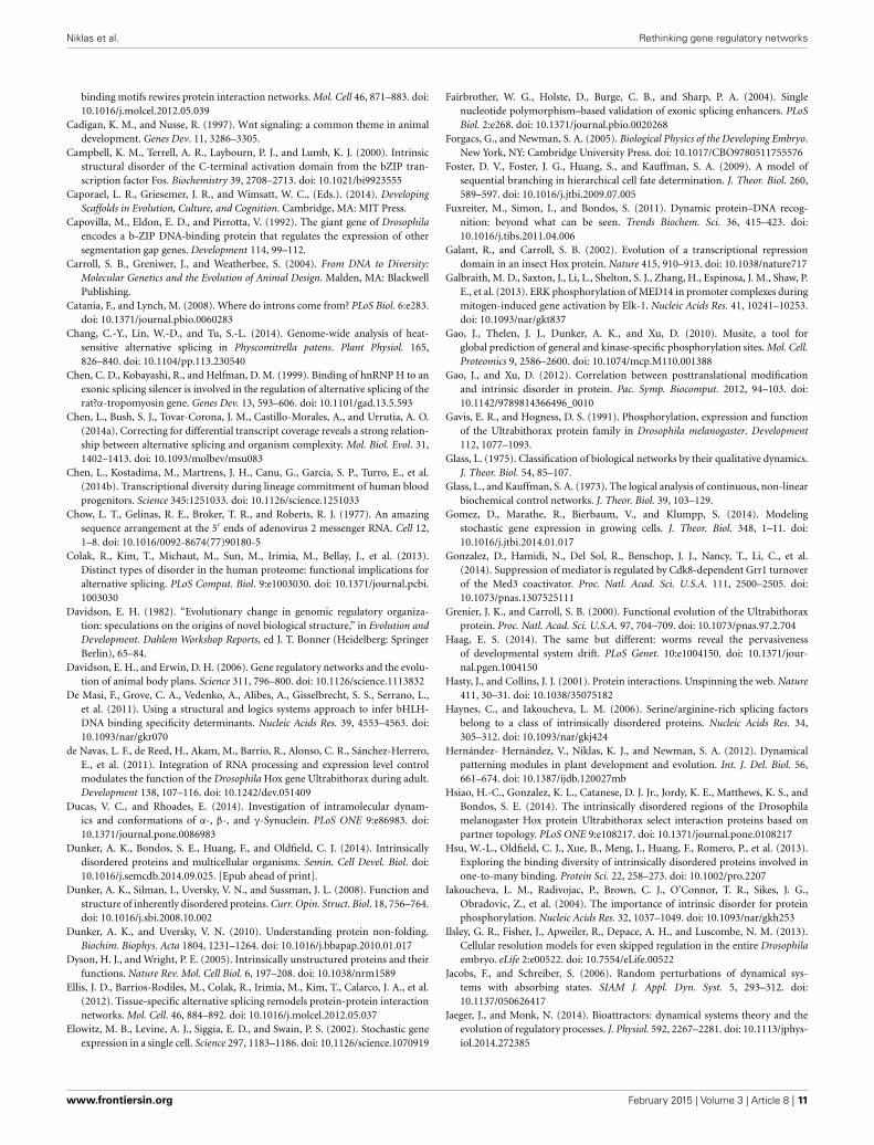
DISORDER FROM ORDERAccording to the most mathematically sophisticated determinis-tic GRN dynamics models (e.g., Foster et al., 2009; Jaeger andMonk, 2014), each cell type is an attractor. That is, if a cell’s stateat a given time is represented by a point in a multidimensional“state space” whose axes are the concentration ranges of key tran-scription factors, the point’s position will change until it settlesstably at one of a finite number of discrete sub-regions withinthe space (Figure 4). These sub-regions (i.e., system attractors)can be stationary points, periodic orbits, or a mixture of thesebehaviors, depending on the subset of the components involvedin the system. Deterministic systems of sufficient complexity canalso exhibit the so-called “butterfly effect,” in which an infinites-imal displacement of the system point can take it along widelydivergent trajectories, as well as chaotic behaviors, characterizedby “strange attractors,” i.e., regions within the state space in whicha point remains bounded but wanders in an unpredictable fashion(Strogatz, 2001; Kaneko, 2006). Each attractor in a determinis-tic dynamical system is surrounded by a “basin of attraction”
toward which a system point gravitates. Importantly, the num-ber of attractors within a deterministic dynamical system is, inprinciple, a predictable function of its network topology and rateconstants, and is always much smaller than the number of basicinteracting components. The rationale for applying this mathe-matical formalism to GRNs and cell differentiation thus arisesfrom observations like the fact that the human genome specifiesmore than 1300 transcription factors (Vaquerizas et al., 2009) butthe human body contains only about 244 cell types (Niklas et al.,2014).
If a system is less than fully deterministic, the dynamicsbecome much more complicated. One example is “noisy sys-tems” in which conventional network topologies and interactionsare in place, but the values of the variables (such as concen-trations of transcription factors) are perturbed by extrinsic orintrinsic factors, as a consequence of some key proteins and otherbiomolecules being present in small numbers in individual cellsand thus varying in a stochastic fashion (Elowitz et al., 2002;Bhalla, 2004; Gomez et al., 2014). Indeed, mathematics has shownthat the notion of an “attractor” still applies, but that their proper-ties and thus behavior are less predictable than in systems withoutnoise (Jacobs and Schreiber, 2006; Zhao and Li, 2011).
The issue of noise is well recognized to confound the biologi-cal effects of even those gene regulatory systems that are formallydeterministic. For example, Rosenfeld and coworkers examinedthe bacteriophage lambda promoter PR in E. coli and found
FIGURE 4 | Schematics of cell fate specification viewed from the
standard deterministic GRN perspective (A) and the non-deterministic
GRN perspective described in the text (B). (A) In the standard view,pre-mRNAs undergo alternative splicing (AS), and transcription factorsspecified by the variant mRNAs undergo post-translational modifications(PTMs) to form a cadre of proteins involved in cell-fate specificationnetworks (GRNs, represented as irregular shapes) via their cis-actingtargets. Discrete cell types result from the deterministic properties of
these GRNs. (B) In the proposed non-deterministic view, transcriptionfactors are generated by AS and PTM operating in the context ofintrinsically disordered protein (IDP) domains. Cell-fate determination in thiscase (represented by interactions among components of variable,context-dependent identity and specificity), is a consequence of the time-and spatial-context dependency of each of the levels shown in thisschematic, which depend on internal and external cellular conditions in afashion that eludes deterministic description at the level of GRNs.
Frontiers in Cell and Developmental Biology | Evolutionary Developmental Biology February 2015 | Volume 3 | Article 8 | 8

Niklas et al. Rethinking gene regulatory networks
that protein production rates fluctuated over the time scale ofone cell cycle, with intrinsic noise levels of the circuit decayingrapidly within each cycle (Rosenfeld et al., 2005). Nonetheless,the aggregate effect of fluctuations in other cellular componentsundermined accuracy in transcriptional responses for time-scaleslonger than a single cell cycle. Thus, although individual cir-cuits can be demonstrated to behave according to a deterministicGRN dynamics model over short time intervals (Rosenfeld et al.,2007), GRN–level determinism breaks down when embedded inthe wider network of real complexity and temporal scale.
It is our contention that the gene regulatory indetermin-ism produced by AS-IDP-PTM is different in kind from that ofdeterministic dynamical systems operating (as described above)under nonlinear, chaotic, or stochastic regimes. GRNs acted uponby AS-IDP-PTM are inherently non-deterministic, since networklogic (i.e., connectivity relationships), strengths of interactions,and even the identities of the transcription factors as regulatorymolecules are constantly subject to change due to internal fluctu-ations and external influences. As a consequence, the propertiesof the transcription factor components of GRNs that have beenthought to make them suitable to be represented as nodes indiscrete networks or variables in systems of ordinary differentialequations are actually subject to change from point to point andmoment to moment during development. Although GRNs havebeen modeled as Boolean networks acting “at the border betweenorder and chaos” (Shmulevich et al., 2005), the presence of AS-IDP-PTM in actual GRNs raises questions about whether anystrictly deterministic models can adequately capture the behaviorof these systems (Figure 4).
ORDER OUT OF DISORDERAs noted earlier, GRNs are frequently not deterministic due tothe independent and interdependent actions of AS–IDP–PTM.Nevertheless, this is in no way equivalent to the counterfactualassertion that embryonic development is itself non-deterministic.Rather, it is our hypothesis that deterministic GRN dynamicsare not a sufficient causal basis for developmental regularities.Although a GRN might provide a rough template for a cellularfunction (particularly if the GRN was established concurrentlywith the evolutionary origination of that function), remodelingof the GRN by AS–IDP–PTM will have rendered cell phenotypeidentity increasingly dependent on internal (i.e., cell physiologi-cal) and external (e.g., microenvironmental and extraorganismal)conditionalities beyond the GRN itself. This assertion is consis-tent with, if not confirmed by, somatic stem cell production andsubsequent differentiation as well as examples of dedifferentiation(e.g., Sprecher and Desplan, 2008).
The conservation of a useful cell function or morpholog-ical phenotype over the course of evolution accompanied byan unmooring from its originating GRN appears to be a com-mon scenario in the history of multicellular plants and animals,reflected in what has been termed “developmental system drift”(True and Haag, 2001; Haag, 2014). The inability to consistentlypin heritable variation in diseases and other traits to particulargenes (Zaitlen and Kraft, 2012), may also plausibly be a man-ifestation of the operation of AS-IDP-PTM over evolutionarytime.
AS-IDP-PTM may also provide flexibility and adaptabilityeven within a single tissue type. For example, in a study oflineage commitment among the eight progenitor populations ofthe major myeloid and lymphoid elements of human blood, Chenet al. (2014b) identified cell type-specific expression changes dur-ing early differentiation stages encompassing 6711 genes and10,724 transcripts. They also detected 7881 novel splice junc-tions and 2301 differentially used AS events, enriched in genesinvolved in regulatory processes. Although only AS was consid-ered, the authors concluded that “a previously undetected layer ofregulation affecting cell fating... involves transcriptional isoformsswitching without noticeable changes at the gene level” (Chenet al., 2014b).
Finally, AS–IDP–PTM and its synergies provide a contextfor understanding how the functionalities of ancient proteinsand regulatory networks can be stably modified over the courseof evolution to adapt to changing external conditions. Targetsequence recognition and selectivity by a transcription factorare subtle properties of the latter’s structure (De Masi et al.,2011). It is well documented, for example, that novel relation-ships between protein structure and PTM educed by mutationcan lead to altered protein-protein interactions resulting in dra-matic changes in transcription factor function (Brayer et al., 2011;Lynch et al., 2011). However, synergy with AS and IDP pro-vides an even greater multiplicity of functional states that can beexplored ecologically and physiologically ahead of any mutationalchange.
Furthermore, nascent potentially adaptive mutations can beretained within (and subsequently integrated into) GRNs byvirtue of AS–IDP–PTM modifications that can buffer GRNsfrom the immediate consequences of such mutations. In thisscenario, a mutated GRN could survive by virtue of AS–IDP–PTM adaptive modifications that would permit the GRN timeto adaptively reorganize. In this way, evolutionary changes wouldinvolve an interactive “genome ⇔AS–IDP–PTM” feedback loop.Consider the transcription factor AkUbx, a homolog of Ubx inthe velvet worm Acanthokara kaputensis, an invertebrate with asimple body plan. AkUbx has very little intrinsic disorder andis not alternatively spliced (Grenier and Carroll, 2000; Galantand Carroll, 2002). Ubx, in contrast, which participates in thedevelopment of the later-evolving, more complex body plansof Drosophila melanogaster has considerable disorder content aswell as undergoing AS and PTM (Gavis and Hogness, 1991;Liu et al., 2008; Reed et al., 2010). The synergistic effects ofAS-IDP-PTM ensured that once it had arisen in the earliest mul-ticellular GRNs it would have promoted its own elaboration, aswell as generation of new developmental contexts that wouldeventually be reflected in greater anatomical and physiologicalcomplexity.
CONCLUSIONSThe association of alternative splicing (AS) with intrinsicallydisordered protein (IDP) domains and post-transcriptional mod-ifications (PTMs) is a core functional complex that mediatesthe modifications of protein functionalities required for con-text dependent cell signaling, regulation, and differentiation. Thecombined effects of AS-IDP-PTM also likely buffer genomes
www.frontiersin.org February 2015 | Volume 3 | Article 8 | 9

Niklas et al. Rethinking gene regulatory networks
from mutations (some of which can subsequently become adap-tive to new conditions) and contributes to the evolvability ofGRNs (see for example, Masel and Trotter, 2010; Steiner, 2012;Albergante et al., 2014). AS–IDP–PTM is ancient and likely pro-moted variability and thus adaptive evolution to support morecomplex intracellular signaling processes coordinating the activ-ities of functionally interdependent discretized organelles, cells,tissues, and organs.
Unlike promoter activity, which primarily regulates theamount of transcripts, AS changes the structure of transcripts andtheir encoded proteins. The ability of IDP domains to assumedifferent conformations expands the functional repertoire ofproteins assembled by AS from a pre-mRNA to diversify thephenotypic domain that a single genome can provide. This reper-toire is yet again increased by PTMs, which generate additionalfunctionalities. Thus, AS–IDP–PTM can yield virtually limitlesscombinatorial possibilities, which can be adaptively sifted over thecourse of evolution.
Consequently, GRNs are inherently plastic and therefore adap-tive. Moreover, they function in a noisy cellular milieu owing tothe operation of AS–IDP–PTM in a multitude of other biochem-ical pathways as well as the effects of mutations and variations ingene and protein copy number (Richard and Yvert, 2014). (Notethat this noisiness is over and above the described intrinsic inde-terminacy.) The evolution of cell differentiation may indeed havedepended on such stochastic effects (Kupiec, 2009). However,heterogeneity at both the molecular and cell phenotypic levelsmust be suppressed for reliable development to occur. This isaccomplished by a variety of “scaffolding” effects (Caporael et al.,2014) at multiple scales, including consistency of external cuesfrom neighboring cells and the physical environment (Braendleand Félix, 2008), and the stabilizing effects of natural selection(Richard and Yvert, 2014).
The multiscale nature of developmental processes is increas-ingly acknowledged (see, for example, Schnell et al., 2008). Inparticular, tissue morphogenesis and cellular pattern formationinvolves the mobilization, by key gene products of the devel-opmental “toolkit,” of mechanical, electrical and other physicalphenomena external to the genome (Forgacs and Newman, 2005;Newman and Bhat, 2009; Hernández- Hernández et al., 2012).It is therefore unsurprising that the determination of cell typeidentity does not reside at the single scale occupied by GRNs,but rather draws on factors at several causal levels, as describedabove, among the most important of which are the mechani-cal aspects of chromatin reorganization associated with changesin gene expression (Amendola and van Steensel, 2014; Lavelle,2014).
We do not suggest that deterministic mathematical and com-putational modeling of GRNs has nothing to contribute to under-standing cell fate determination. However, this perspective mustacknowledge and integrate the ubiquitous effects of AS–IDP–PTM. Just as genes per se have long been rejected as the exclusiveor privileged level of determination of phenotype and evolu-tionary change, new understanding of the complexities of geneexpression and the conditional identities of its protein productscall into question a deterministic GRN-based reductionism indevelopmental and evolutionary biology.
ACKNOWLEDGMENTSThe authors thank the two reviewers for their constructive andthoughtful comments during the review process. Funding fromthe College of Agriculture and Life Sciences, Cornell University(to K. J. N.) and from NSF CAREER 1151394 and the TedNash Long Life Foundation M1500779 (to S. E. B.) is gratefullyacknowledged.
REFERENCESAbdel-Hafiz, H. A., and Horwitz, K. B. (2014). Post-translational modifications
of the progesterone receptors. J. Steroid Biochem. Mol. Biol. 140, 80–89. doi:10.1016/j.jsbmb.2013.12.008
Albergante, L., Blow, J. J., and Newman, T. L. (2014). Buffered Qualitative Stabilityexplains the robustness and evolvability of transcriptional networks. eLife3:e02863. doi: 10.7554/eLife.02863
Amendola, M., and van Steensel, B. (2014). Mechanisms and dynamics ofnuclear lamina-genome interactions. Curr. Opin. Cell Biol. 28, 61–68. doi:10.1016/j.ceb.2014.03.003
Barash, Y., Calarco, J. A., Gao, W., Pan, Q., Wang, X., Shai, O., et al.(2010). Deciphering the splicing code. Nature 465, 53–59. doi: 10.1038/nature09000
Berget, S. M., Moore, C., and Sharp, P. A. (1977). Spliced segments at the 5′ termi-nus of adenovirus 2 late mRNA. Proc. Natl. Acad. Sci. U.S.A. 74, 3171–3175. doi:10.1073/pnas.74.8.3171
Bhalla, J., Storchan, G. B., MacCarthy, C. M., Uversky, V. N., and Tcherkasskaya, O.(2006). Local flexibility in molecular function paradigm. Mol. Cell. Proteomics5, 1212–1223. doi: 10.1074/mcp.M500315-MCP200
Bhalla, U. S. (2004). Signaling in small subcellular volumes. I. Stochastic and dif-fusion effects on individual pathways. Biophys. J. 87, 733–744. doi: 10.1529/bio-physj.104.040469
Black, D. L. (2003). Mechanisms of alternative pre-messenger RNA splicing. Annu.Rev. Biochem. 72, 291–336. doi: 10.1146/annurev.biochem.72.121801.161720
Bondos, S. E., and Tan, X. X. (2001). Combinatorial transcription regulation: Theinteraction of transcription factors and cell signaling molecules with home-odomain proteins in Drosophila development. Crit. Rev. Eukaryot. Gene Expr.11, 145–171. doi: 10.1615/CritRevEukarGeneExpr.v11.i1-3.80
Bondos, S. E., Tan, X.-X., and Matthews, K. S. (2006). Physical and genetic inter-actions link Hox function with diverse transcription factors and cell signalingproteins. Mol. Cell. Proteomics 5, 824–834. doi: 10.1074/mcp.M500256-MCP200
Bothma, J. P., Garcia, H. G., Esposito, E., Schlissel, G., Gregor, T., and Levine,M. (2014). Dynamic regulation of eve stripe 2 expression reveals transcrip-tional bursts in living Drosophila embryos. Proc. Natl. Acad. Sci. U.S.A. 111,10598–10603. doi: 10.1073/pnas.1410022111
Boutz, P. L., Stoilov, P., Li, Q., Lin, C.-H., Chawla, G., Ostrow, K., et al. (2007).A post- transcriptional regulatory switch in polypyrimidine tract-binding pro-teins reprograms alternative splicing in developing neurons. Genes Dev. 21,1636–1652. doi: 10.1101/gad.1558107
Braendle, C., and Félix, M. A. (2008). Plasticity and errors of a robust developmen-tal system in different environments. Developmental Cell 15, 714–724.
Braunschweig, U., Gueroussov, S., Plocik, A. M., Graveley, B. R., and Blencowe, B.J. (2013). Dynamic integration of splicing within gene regulatory pathways. Cell152, 1252–1269. doi: 10.1016/j.cell.2013.02.034
Brayer, K. J., Lynch, V. J., and Wagner, G. P. (2011). Evolution of a derivedprotein-protein interaction between HoxA11 and Foxo1a in mammals causedby changes in intramolecular regulation. Proc. Natl. Acad. Sci. U.S.A. 108,E414–E420. doi: 10.1073/pnas.1100990108
Britten, R. J. (1982). “Genomic alterations in evolution,” in Evolution andDevelopment Dahlem Workshop Reports, ed J. T. Bonner (Berlin Heidelberg:Springer), 41–64.
Britten, R. J., and Davidson, E. H. (1969). Gene regulation for higher cells: a theory.Science 165, 349–357. doi: 10.1126/science.165.3891.349
Buljan, M., Chalancon, G., Dunker, A. K., Bateman, A., Balaji, S., Fuxreiter,M., et al. (2013). Alternative splicing of intrinsically disordered regions andrewiring of protein interactions. Curr. Opin. Struct. Biol. 23, 443–450. doi:10.1016/j.sbi.2013.03.006
Buljan, M., Chalancon, G., Eustermann, S., Wagner, G. P., Fuxreiter, M., Bateman,A., et al. (2012). Tissue-specific splicing of disordered segments that embed
Frontiers in Cell and Developmental Biology | Evolutionary Developmental Biology February 2015 | Volume 3 | Article 8 | 10
Niklas et al. Rethinking gene regulatory networks
binding motifs rewires protein interaction networks. Mol. Cell 46, 871–883. doi:10.1016/j.molcel.2012.05.039
Cadigan, K. M., and Nusse, R. (1997). Wnt signaling: a common theme in animaldevelopment. Genes Dev. 11, 3286–3305.
Campbell, K. M., Terrell, A. R., Laybourn, P. J., and Lumb, K. J. (2000). Intrinsicstructural disorder of the C-terminal activation domain from the bZIP tran-scription factor Fos. Biochemistry 39, 2708–2713. doi: 10.1021/bi9923555
Caporael, L. R., Griesemer, J. R., and Wimsatt, W. C., (Eds.). (2014). DevelopingScaffolds in Evolution, Culture, and Cognition. Cambridge, MA: MIT Press.
Capovilla, M., Eldon, E. D., and Pirrotta, V. (1992). The giant gene of Drosophilaencodes a b-ZIP DNA-binding protein that regulates the expression of othersegmentation gap genes. Development 114, 99–112.
Carroll, S. B., Greniwer, J., and Weatherbee, S. (2004). From DNA to Diversity:Molecular Genetics and the Evolution of Animal Design. Malden, MA: BlackwellPublishing.
Catania, F., and Lynch, M. (2008). Where do introns come from? PLoS Biol. 6:e283.doi: 10.1371/journal.pbio.0060283
Chang, C.-Y., Lin, W.-D., and Tu, S.-L. (2014). Genome-wide analysis of heat-sensitive alternative splicing in Physcomitrella patens. Plant Physiol. 165,826–840. doi: 10.1104/pp.113.230540
Chen, C. D., Kobayashi, R., and Helfman, D. M. (1999). Binding of hnRNP H to anexonic splicing silencer is involved in the regulation of alternative splicing of therat?α-tropomyosin gene. Genes Dev. 13, 593–606. doi: 10.1101/gad.13.5.593
Chen, L., Bush, S. J., Tovar-Corona, J. M., Castillo-Morales, A., and Urrutia, A. O.(2014a). Correcting for differential transcript coverage reveals a strong relation-ship between alternative splicing and organism complexity. Mol. Biol. Evol. 31,1402–1413. doi: 10.1093/molbev/msu083
Chen, L., Kostadima, M., Martrens, J. H., Canu, G., Garcia, S. P., Turro, E., et al.(2014b). Transcriptional diversity during lineage commitment of human bloodprogenitors. Science 345:1251033. doi: 10.1126/science.1251033
Chow, L. T., Gelinas, R. E., Broker, T. R., and Roberts, R. J. (1977). An amazingsequence arrangement at the 5′ ends of adenovirus 2 messenger RNA. Cell 12,1–8. doi: 10.1016/0092-8674(77)90180-5
Colak, R., Kim, T., Michaut, M., Sun, M., Irimia, M., Bellay, J., et al. (2013).Distinct types of disorder in the human proteome: functional implications foralternative splicing. PLoS Comput. Biol. 9:e1003030. doi: 10.1371/journal.pcbi.1003030
Davidson, E. H. (1982). “Evolutionary change in genomic regulatory organiza-tion: speculations on the origins of novel biological structure,” in Evolution andDevelopment. Dahlem Workshop Reports, ed J. T. Bonner (Heidelberg: SpringerBerlin), 65–84.
Davidson, E. H., and Erwin, D. H. (2006). Gene regulatory networks and the evolu-tion of animal body plans. Science 311, 796–800. doi: 10.1126/science.1113832
De Masi, F., Grove, C. A., Vedenko, A., Alibes, A., Gisselbrecht, S. S., Serrano, L.,et al. (2011). Using a structural and logics systems approach to infer bHLH-DNA binding specificity determinants. Nucleic Acids Res. 39, 4553–4563. doi:10.1093/nar/gkr070
de Navas, L. F., de Reed, H., Akam, M., Barrio, R., Alonso, C. R., Sánchez-Herrero,E., et al. (2011). Integration of RNA processing and expression level controlmodulates the function of the Drosophila Hox gene Ultrabithorax during adult.Development 138, 107–116. doi: 10.1242/dev.051409
Ducas, V. C., and Rhoades, E. (2014). Investigation of intramolecular dynam-ics and conformations of α-, β-, and γ-Synuclein. PLoS ONE 9:e86983. doi:10.1371/journal.pone.0086983
Dunker, A. K., Bondos, S. E., Huang, F., and Oldfield, C. J. (2014). Intrinsicallydisordered proteins and multicellular organisms. Semin. Cell Devel. Biol. doi:10.1016/j.semcdb.2014.09.025. [Epub ahead of print].
Dunker, A. K., Silman, I., Uversky, V. N., and Sussman, J. L. (2008). Function andstructure of inherently disordered proteins. Curr. Opin. Struct. Biol. 18, 756–764.doi: 10.1016/j.sbi.2008.10.002
Dunker, A. K., and Uversky, V. N. (2010). Understanding protein non-folding.Biochim. Biophys. Acta 1804, 1231–1264. doi: 10.1016/j.bbapap.2010.01.017
Dyson, H. J., and Wright, P. E. (2005). Intrinsically unstructured proteins and theirfunctions. Nature Rev. Mol. Cell Biol. 6, 197–208. doi: 10.1038/nrm1589
Ellis, J. D., Barrios-Rodiles, M., Colak, R., Irimia, M., Kim, T., Calarco, J. A., et al.(2012). Tissue-specific alternative splicing remodels protein-protein interactionnetworks. Mol. Cell. 46, 884–892. doi: 10.1016/j.molcel.2012.05.037
Elowitz, M. B., Levine, A. J., Siggia, E. D., and Swain, P. S. (2002). Stochastic geneexpression in a single cell. Science 297, 1183–1186. doi: 10.1126/science.1070919
Fairbrother, W. G., Holste, D., Burge, C. B., and Sharp, P. A. (2004). Singlenucleotide polymorphism–based validation of exonic splicing enhancers. PLoSBiol. 2:e268. doi: 10.1371/journal.pbio.0020268
Forgacs, G., and Newman, S. A. (2005). Biological Physics of the Developing Embryo.New York, NY: Cambridge University Press. doi: 10.1017/CBO9780511755576
Foster, D. V., Foster, J. G., Huang, S., and Kauffman, S. A. (2009). A model ofsequential branching in hierarchical cell fate determination. J. Theor. Biol. 260,589–597. doi: 10.1016/j.jtbi.2009.07.005
Fuxreiter, M., Simon, I., and Bondos, S. (2011). Dynamic protein–DNA recog-nition: beyond what can be seen. Trends Biochem. Sci. 36, 415–423. doi:10.1016/j.tibs.2011.04.006
Galant, R., and Carroll, S. B. (2002). Evolution of a transcriptional repressiondomain in an insect Hox protein. Nature 415, 910–913. doi: 10.1038/nature717
Galbraith, M. D., Saxton, J., Li, L., Shelton, S. J., Zhang, H., Espinosa, J. M., Shaw, P.E., et al. (2013). ERK phosphorylation of MED14 in promoter complexes duringmitogen-induced gene activation by Elk-1. Nucleic Acids Res. 41, 10241–10253.doi: 10.1093/nar/gkt837
Gao, J., Thelen, J. J., Dunker, A. K., and Xu, D. (2010). Musite, a tool forglobal prediction of general and kinase-specific phosphorylation sites. Mol. Cell.Proteomics 9, 2586–2600. doi: 10.1074/mcp.M110.001388
Gao, J., and Xu, D. (2012). Correlation between posttranslational modificationand intrinsic disorder in protein. Pac. Symp. Biocomput. 2012, 94–103. doi:10.1142/9789814366496_0010
Gavis, E. R., and Hogness, D. S. (1991). Phosphorylation, expression and functionof the Ultrabithorax protein family in Drosophila melanogaster. Development112, 1077–1093.
Glass, L. (1975). Classification of biological networks by their qualitative dynamics.J. Theor. Biol. 54, 85–107.
Glass, L., and Kauffman, S. A. (1973). The logical analysis of continuous, non-linearbiochemical control networks. J. Theor. Biol. 39, 103–129.
Gomez, D., Marathe, R., Bierbaum, V., and Klumpp, S. (2014). Modelingstochastic gene expression in growing cells. J. Theor. Biol. 348, 1–11. doi:10.1016/j.jtbi.2014.01.017
Gonzalez, D., Hamidi, N., Del Sol, R., Benschop, J. J., Nancy, T., Li, C., et al.(2014). Suppression of mediator is regulated by Cdk8-dependent Grr1 turnoverof the Med3 coactivator. Proc. Natl. Acad. Sci. U.S.A. 111, 2500–2505. doi:10.1073/pnas.1307525111
Grenier, J. K., and Carroll, S. B. (2000). Functional evolution of the Ultrabithoraxprotein. Proc. Natl. Acad. Sci. U.S.A. 97, 704–709. doi: 10.1073/pnas.97.2.704
Haag, E. S. (2014). The same but different: worms reveal the pervasivenessof developmental system drift. PLoS Genet. 10:e1004150. doi: 10.1371/jour-nal.pgen.1004150
Hasty, J., and Collins, J. J. (2001). Protein interactions. Unspinning the web. Nature411, 30–31. doi: 10.1038/35075182
Haynes, C., and Iakoucheva, L. M. (2006). Serine/arginine-rich splicing factorsbelong to a class of intrinsically disordered proteins. Nucleic Acids Res. 34,305–312. doi: 10.1093/nar/gkj424
Hernández- Hernández, V., Niklas, K. J., and Newman, S. A. (2012). Dynamicalpatterning modules in plant development and evolution. Int. J. Del. Biol. 56,661–674. doi: 10.1387/ijdb.120027mb
Hsiao, H.-C., Gonzalez, K. L., Catanese, D. J. Jr., Jordy, K. E., Matthews, K. S., andBondos, S. E. (2014). The intrinsically disordered regions of the Drosophilamelanogaster Hox protein Ultrabithorax select interaction proteins based onpartner topology. PLoS ONE 9:e108217. doi: 10.1371/journal.pone.0108217
Hsu, W.-L., Oldfield, C. J., Xue, B., Meng, J., Huang, F., Romero, P., et al. (2013).Exploring the binding diversity of intrinsically disordered proteins involved inone-to-many binding. Protein Sci. 22, 258–273. doi: 10.1002/pro.2207
Iakoucheva, L. M., Radivojac, P., Brown, C. J., O’Connor, T. R., Sikes, J. G.,Obradovic, Z., et al. (2004). The importance of intrinsic disorder for proteinphosphorylation. Nucleic Acids Res. 32, 1037–1049. doi: 10.1093/nar/gkh253
Ilsley, G. R., Fisher, J., Apweiler, R., Depace, A. H., and Luscombe, N. M. (2013).Cellular resolution models for even skipped regulation in the entire Drosophilaembryo. eLife 2:e00522. doi: 10.7554/eLife.00522
Jacobs, F., and Schreiber, S. (2006). Random perturbations of dynamical sys-tems with absorbing states. SIAM J. Appl. Dyn. Syst. 5, 293–312. doi:10.1137/050626417
Jaeger, J., and Monk, N. (2014). Bioattractors: dynamical systems theory and theevolution of regulatory processes. J. Physiol. 592, 2267–2281. doi: 10.1113/jphys-iol.2014.272385
www.frontiersin.org February 2015 | Volume 3 | Article 8 | 11

Niklas et al. Rethinking gene regulatory networks
Johnson, F. B., Parker, E., and Krasnow, M. A. (1995). Extradenticle protein is aselective cofactor for the Drosophila homeotics: role of the homeodomain andYPWM amino acid motif in the interaction. Proc. Natl. Acad. Sci. U.S.A. 92,739–743. doi: 10.1073/pnas.92.3.739
Johnson, J. M., Castle, J., Garrett-Engele, P., Kan, Z., Loerch, P. M., Armour, C. D.,et al. (2003). Genome-wide survey of human alternative pre-mRNA splicingwith exon junction microarrays. Science 302, 2141–2144. doi: 10.1126/sci-ence.1090100
Kaganovich, M., and Snyder, M. (2012). Phosphorylation of yeast transcription fac-tors correlates with the evolution of novel sequence and function. J. ProteomeRes. 11, 261–268. doi: 10.1021/pr201065k
Kaneko, K. (2006). Life: an Introduction to Complex Systems Biology. New York, NY:Springer.
Kauffman, S. (1969). Metabolic stability and epigenesis in randomly constructedgenetic nets. J. Theor. Biol. 22, 437–467.
Kauffman, S. (1974). The large scale structure and dynamics of gene controlcircuits: An ensemble approach. J. Theor. Biol. 44, 167–190.
Ke, S., Zhang, X. H., and Chasin, L. A. (2008). Positive selection acting on splic-ing motifs reflects compensatory evolution. Genome Res. 18, 533–543. doi:10.1101/gr.070268.107
Kim, Y., Coppey, M., Grossman, R., Ajuria, L., Jiménez, G., Paroush, Z., et al.(2010). MAPK substrate competition integrates patterning signals in theDrosophila embryo. Curr. Biol. CB 20, 446–451. doi: 10.1016/j.cub.2010.01
Kupiec, J. (2009). The Origin of Individuals. New Jersey, NJ: World ScientificPublishing Company.
Labadorf, A., Link, A., Rogers, M. F., Thomas, J., Reddy, A. S., Ben-Hur, A.,et al. (2010). Genome- wide analysis of alternative splicing in Chlamydomonasreinhardtii. BMC Genomics 11:114. doi: 10.1186/1471-2164-11-114
Lauffenburger, D. A. (2000). Cell signaling pathways as control modules: com-plexity for simplicity? Proc. Natl. Acad. Sci. U.S.A. 97, 5031–5033. doi:10.1073/pnas.97.10.5031
Lavelle, C. (2014). Pack, unpack, bend, twist, pull, push: the physical side of geneexpression. Curr. Opin. Genet. Dev. 25, 74–84. doi: 10.1016/j.gde.2014.01.001
Lee, G. M., Pufall, M. A., Meeker, C. A., Kang, H.-S., Graves, B. J., and McIntosh,L. P. (2008). The affinity of Ets-1 for DNA is modulated by phosphorylationthrough transient interactions of an unstructured region. J. Mol. Biol. 382,1014–1030. doi: 10.1016/j.jmb.2008.07.064
Leff, S. E., and Rosenfeld, M. G. (1986). Complex transcriptional units: Diversityin gene expression by alternative RNA processing. Annu. Rev. Biochem. 55,1091–1117.
Lim, K. H., Ferraris, L., Filloux, M. E., Raphael, B. J., and Fairbrother, W. G.(2011). Using positional distribution to identify splicing elements and predictpre-mRNA processing defects in human genes. Proc. Natl. Acad. Sci. U.S.A. 108,11093–11098. doi: 10.1073/pnas.1101135108
Liu, J., Perumal, N. B., Oldfield, C. J., Su, E. W., Uversky, V. N., and Dunker, A. K.(2006). Intrinsic disorder in transcription factors. Biochemistry 45, 6873–6888.doi: 10.1021/bi0602718
Liu, Y., Matthews, K. S., and Bondos, S. E. (2008). Multiple intrinsicallydisordered sequences alter DNA binding by the homeodomain of theDrosophila Hox protein Ultrabithorax. J. Biol. Chem. 283, 20874–20887. doi:10.1074/jbc.M800375200
Liu, Y., Matthews, K. S., and Bondos, S. E. (2009). Internal regulatory interactionsdetermine DNA binding specificity by a Hox transcription factor. J. Mol. Biol.390, 760–774. doi: 10.1016/j.jmb.2009.05.059
Lopez, A. J., and Hogness, D. S. (1991). Immunochemical dissection of theUltrabithorax homeoprotein family in Drosophila melanogaster. Proc. Natl.Acad. Sci. U.S.A. 88, 9924–9928.
Lothrop, A. P., Torres, M. P., and Fuchs, S. M. (2013). Decipheringpost-translational modification codes. FEBS Lett. 587, 1247–1257. doi:10.1016/j.febslet.2013.01.047
Lucchetta, E. M., Vincent, M. E., and Ismagilov, R. F. (2008). A precise Bicoid gra-dient is nonessential during cycles 11-13 for precise patterning in the Drosophilablastoderm. PLoS ONE 3:e3651. doi: 10.1371/journal.pone.0003651
Ludwig, M. Z., Manu, Kittler, R., White, K. P., and Kreitman, M. (2011).Consequences of eukaryotic enhancer architecture for gene expression dynam-ics, development, and fitness. PLoS Genet. 7:e1002364. doi: 10.1371/jour-nal.pgen.1002364
Lynch, V. J., May, G., and Wagner, G. P. (2011). Regulatory evolution throughdivergence of a phosphoswitch in the transcription factor CEBPB. Nature 480,383–386. doi: 10.1038/nature10595
Masel, J., and Trotter, M. V. (2010). Robustness and evolvability. Trends Genet. 26,406–14. doi: 10.1016/j.tig.2010.06.002
Matera, A. G., and Wang, Z. (2014). A day in the life of the spliceosome. NatureRev. Mol. Cell Biol. 15, 108–121. doi: 10.1038/nrm3742
Matlin, A. J., Clark, F., and Smith, C. W. J. (2005). Understanding alternativesplicing: towards a cellular code. Nature Rev. Mol. Cell Biol. 6, 386–398. doi:10.1038/nrm1645
Milanovic, M., Kracht, M., and Schmitz, M. L. (2014). The cytokine-inducedconformational switch of nuclear factor κB p65 is mediated by p65 phospho-rylation. Biochem. J. 457, 401–413. doi: 10.1042/BJ20130780
Mileo, E., Lorenzi, M., Erales, J., Lignon, S., Puppo, C., Le Breton, N., et al. (2013).Dynamics of the intrinsically disordered protein CP12 in its association withGAPDH in the green alga Chlamydomonas reinhardtii: a fuzzy complex. Mol.Biosyst. 9, 2869–2876. doi: 10.1039/c3mb70190e
Minde, D. P., Radi, M., Forneris, F., Maurice, M. M., and Rüdiger, S. G. D. (2013).Large extent of disorder in Adenomatous Polyposis Coli offers a strategy forWnt signaling against point mutations. PLoS ONE 8:e77257. doi: 10.1371/jour-nal.pone.0077257
Minezaki, Y., Homma, K., Kinjo, A. R., and Nishikawa, K. (2006b). Human tran-scription factors contain a high fraction of intrinsically disordered regionsessential for transcriptional regulation. J. Mol. Biol. 359, 1137–1149. doi:10.1016/j.jmb.2006.04.016
Minezaki, Y., Homma, K., and Nishikawa, K. (2006a). Genome-wide survey oftranscription factors in prokaryotes reveals many bacteria-specific families notfound in archaea. DNA Res. 12, 269–280. doi: 10.1093/dnares/dsi016
Mohan, A., Oldfield, C. J., Radivojac, P., Vacic, V., Cortese, M. S., Dunker, A. K.,et al. (2006). Analysis of molecular recognition features (MoRFs). J. Mol. Biol.362, 1043–1059. doi: 10.1016/j.jmb.2006.07.087
Mooney, S. M., Qiu, R., Kim, J. J., Sacho, E. J., Rajagopalan, K., Johng, D., et al.(2014). Cancer/testis antigen PAGE4, a regulator of c-Jun transactivation, isphosphorylated by homeodomain-interacting protein kinase 1, a componentof the stress-response pathway. Biochemistry 53, 1670–1679. doi: 10.1021/bi500013w
Newman, S. A., and Bhat, R. (2009). Dynamical patterning modules: a “patternlanguage” for development and evolution of multicellular form. Int. J. Dev. Biol.53, 693–705. doi: 10.1387/ijdb.072481sn
Niklas, K. J., Cobb, E. D., and Dunker, A. K. (2014). The number of cell types, infor-mation content, and the evolution of multicellularity. Acta Soc. Bot. Poloniae 83,337 –347. doi: 10.5586/asbp.2014.034
Niklas, K. J., and Kutschera, U. (2012). Plant development, auxin, and thesubsystem incompleteness theorem. Front. Plant Evol. Develop. 3:37. doi:10.3389/fpls.2012.00037
Nusse, R., and Varmus, H. (2012). Three decades of Wnts: a personal per-spective on how a scientific field developed. EMBO J. 31, 2670–2684. doi:10.1038/emboj.2012.146
O’Connor, M. B., Binari, R., Perkins, L. A., and Bender, W. (1988). Alternative RNAproducts from the Ultrabithorax domain of the bithorax complex. EMBO J. 7,435–445.
Oldfield, C. J., Cheng, Y., Cortese, M. S., Romero, P., Uversky, V. N., and Dunker,A. K. (2005). Coupled folding and binding with α-helix-forming molecularrecognition elements. Biochemistry 44, 12454–12470. doi: 10.1021/bi050736e
Oldfield, C. J., and Dunker, A. K. (2014). Intrinsically disordered protein andintrinsically disordered protein regions. Annu. Rev. Biochem. 83, 553–584. doi:10.1146/annurev-biochem-072711-164947
Oldfield, C. J., Meng, J., Yang, J. Y., Yang, M. Q., Uversky, V. N., Dunker, A. K., et al.(2008). Flexible nets: disorder and induced fit in the associations of p53 and 14-3-3 with their partners. BMC Genomics 9:S1. doi: 10.1186/1471-2164-9-S1-S1
Ollo, R., and Maniatis, T. (1987). Drosophila Kruppel gene product produced in abaculovirus expression system is a nuclear phosphoprotein that binds to DNA.Proc. Natl. Acad. Sci. U.S.A. 84, 5700–5704.
Pan, Q., Shai, O., Lee, L. J., Frey, B. J., and Blencowe, B. J. (2008). Deep surveying ofalternative splicing complexity in the human transcriptome by high-throughputsequencing. Nat. Genet. 40, 1413–1415. doi: 10.1038/ng.259
Pearson, J. C., Lemons, D., and McGinnis, W. (2005). Modulating Hox genefunctions during animal body patterning. Nature Rev. Genet. 6, 893–904. doi:10.1038/nrg1726
Pejaver, V., Hsu, W. L., Xin, F., Dunker, A. K., Uversky, V. N., and Radivojac, P.(2014). The structural and functional signatures of proteins that undergo mul-tiple events of post-translational modification. Protein Sci. 23,1077–1093. doi:10.1002/pro.2494
Frontiers in Cell and Developmental Biology | Evolutionary Developmental Biology February 2015 | Volume 3 | Article 8 | 12

Niklas et al. Rethinking gene regulatory networks
Ptashne, M. (2004). A Genetic Switch: Phage Lambda Revisited. New York, NY:CSHL Press.
Reed, H. C., Hoare, T., Thomsen, S., Weaver, T. A., White, R. A. H., Akam, M.,et al. (2010). Alternative splicing modulates Ubx protein function in Drosophilamelanogaster. Genetics 84, 745–758. doi: 10.1534/genetics.109.112086
Richard, M., and Yvert, G. (2014). How does evolution tune biological noise? Front.Genet. 5:374. doi: 10.3389/fgene.2014.00374
Romero, P. R., Zaidi, S., Fang, Y. Y., Uversky, V. N., Radivojac, P., Cortese, M., et al.(2006). Alternative splicing in concert with protein intrinsic disorder enablesincreased functional diversity in multicellular organisms. Proc. Natl. Acad. Sci.U.S.A. 103, 8390–8395. doi: 10.1073/pnas.0507916103
Ronshaugen, M., McGinnis, N., and McGinnis, W. (2002). Hox protein muta-tion and macroevolution of the insect body plan. Nature 415, 914–917. doi:10.1038/nature716
Rosenfeld, N., Young, J. W., Alon, U., Swain, P. S., and Elowitz, M. B. (2005).Gene regulation at the single-cell level. Science 307, 1962–1965. doi: 10.1126/sci-ence.1106914
Rosenfeld, N., Young, J. W., Alon, U., Swain, P. S., and Elowitz, M. B. (2007).Accurate prediction of gene feedback circuit behavior from component prop-erties. Mol.Syst. Biol. 3, 143. doi: 10.1038/msb4100185
Savageau, M. A. (1974). Comparison of classical and autogenous systems ofregulation in inducible operons. Nature 252, 546–549. doi: 10.1038/252546a0
Schad, E., Tompa, P., and Hegyi, H. (2011). The relationship between proteomesize, structural disorder and organism complexity. Genome Biol. 12:R120. doi:10.1186/gb-2011-12-12-r120
Schnell, S., Maini, P. K., Newman, T. J., and Newman, S. A. (eds.). (2008). MultiscaleModels of Developmental Systems. San Diego, CA: Academic Press/Elsevier.
Shmulevich, I., Kauffman, S. A., and Aldana, M. (2005). Eukaryotic cells aredynamically ordered or critical but not chaotic. Proc. Natl. Acad. Sci. U.S.A. 102,13439–13444. doi: 10.1073/pnas.0506771102
Simons, S. S., and Kumar, R. (2013). Variable steroid receptor responses:Intrinsically disordered AF1 is the key. Mol. Cell Endocrinol. 376, 81–84. doi:10.1016/j.mce.2013.06.007
Singh, G. P., and Dash, D. (2007). Intrinsic disorder in yeast transcriptionalregulatory network. Proteins 68, 602–605. doi: 10.1002/prot.21497
Spellman, R., Llorian, M., and Smith, C. W. J. (2007). Crossregulation and func-tional redundancy between the splicing regulator PTB and its paralogs nPTBand ROD1. Mol. Cell 27, 420–434. doi: 10.1016/j.molcel.2007.06.016
Sprecher, S. G., and Desplan, C. (2008). Switch of rhodopsin expression in ter-minally differentiated Drosophila sensory neurons. Nature 454, 533–537. doi:10.1038/nature07062
Srinivasan, N., Bhagawati, M., Ananthanarayanan, B., and Kumar, S. (2014).Stimuli-sensitive intrinsically disordered protein brushes. Nat. Commun.5, 5145. doi: 10.1038/ncomms6145
Steiner, C. F. (2012). Environmental noise, genetic diversity and the evolution ofevolvability and robustness in model gene networks. PLoS ONE 7:e52204. doi:10.1371/journal.pone.0052204
Strahl, B. D., and Allis, C. D. (2000). The language of covalent histone modifica-tions. Nature 403, 41–45. doi: 10.1038/47412
Strogatz, S. H. (2001). Nonlinear Dynamics and Chaos: With Applications To Physics,Biology, Chemistry, and Engineering. Cambridge: Westview Press.
Sun, X., Jones, W. T., Harvey, D., Edwards, P. J. B., Pascal, S. M., Kirk, C., et al.(2010). N-terminal domains of DELLA proteins are intrinsically unstructuredin the absence of interaction with GID1/gibberellic acid receptors. J. Biol. Chem.285, 11557–11571. doi: 10.1074/jbc.M109.027011
Sun, X., Rikkerink, E. H. A., Jones, W. T., and Uversky, V. N. (2013). Multifariousroles of intrinsic disorder in proteins illustrate its broad impact on plant biology.Plant Cell 25, 38–55. doi: 10.1105/tpc.112.106062
Sun, X., Xue, B., Jones, W. T., Rikkerink, E., Dunker, A. K., and Uversky, V. N.(2011). A functionally required unfoldome from the plant kingdom: intrinsi-cally disordered N-terminal domains of GRAS proteins are involved in molec-ular recognition during plant development. Plant Mol. Biol. 77, 205–223. doi:10.1007/s11103-011-9803-z
Taghli-Lamallem, O., Hsia, C., Ronshaugen, M., and McGinnis, W. (2008).Context-dependent regulation of Hox protein functions by CK2 phosphoryla-tion sites. Dev. Genes Evol. 218, 321–332. doi: 10.1007/s00427-008-0224-1
Talavera, D., Robertson, D. L., and Lovell, S. C. (2013). Alternative splicingand protein interaction data sets. Nat. Biotechnol. 31, 292–293. doi: 10.1038/nbt.2540
Tan, X.-X., Bondos, S., Li, L., and Matthews, K. S. (2002). Transcription acti-vation by ultrabithorax Ib protein requires a predicted alpha-helical region.Biochemistry 41, 2774–2785. doi: 10.1021/bi011967y
True, J. R., and Haag, E. S. (2001). Developmental system drift and flexibil-ity in evolutionary trajectories. Evol. Dev. 3, 109–119. doi: 10.1046/j.1525-142x.2001.003002109.x
Uversky, V. N. (2002). Natively unfolded proteins: a point where biology waitsfor a physics. Protein Sci. Publ. Protein Soc. 11, 739–756. doi: 10.1110/ps.4210102
Van Roey, K., Dinkel, H., Weatheritt, R. J., Gibson, T. J., and Davey, N. E.(2013). The switches.ELM resource: a compendium of conditional regulatoryinteraction interfaces. Sci Signal. 6, rs7. doi: 10.1126/scisignal.2003345
Vaquerizas, J. M., Kummerfeld, S. K., Teichmann, S. A., and Luscombe, N. M.(2009). A census of human transcription factors: function, expression andevolution. Nature Rev. Genet. 10, 252–263. doi: 10.1038/nrg2538
Vuzman, D., and Levy, Y. (2012). Intrinsically disordered regions as affinitytuners in protein-DNA interactions. Mol. Biosyst. 8, 47–57. doi: 10.1039/c1mb05273j
Walsh, C. M., and Carroll, S. B. (2007). Collaboration between Smads and aHox protein in target gene repression. Development 134, 3585–3592. doi:10.1242/dev.009522
Wang, Z., Rolish, M. E., Yeo, G., Tung, V., Mawson, M., et al. (2004). Systematicidentification and analysis of exonic splicing silencers. Cell 119, 831–845.
Ward, J. J., Sodhi, J. S., McGuffin, L. J., Buxton, B. F., and Jones, D. T. (2004).Prediction and functional analysis of native disorder in proteins from thethree kingdoms of life. J. Mol. Biol. 337, 635–645. doi: 10.1016/j.jmb.2004.02.002
Xie, H., Vucetic, S., Iakoucheva, L. M., Oldfield, C. J., Dunker, A. K., Uversky, V.N., et al. (2007). Functional anthology of intrinsic disorder. 1. Biological pro-cesses and functions of proteins with long disordered regions. J. Proteome Res.6, 1882–1898. doi: 10.1021/pr060392u
Xiong, H. Y., Alipanahi, B., Lee, L. J., Bretschneider, H., Merico, D., Yuen, R.K., et al. (2015). The human splicing code reveals new insights into thegenetic determinants of disease. Science 347:1254806. doi: 10.1126/science.1254806
Xue, B., Dunker, A. K., and Uversky, V. N. (2012a). Orderly order in pro-tein intrinsic disorder distribution: disorder in 3500 proteomes from virusesand the three domains of life. J. Biomol. Struct. Dyn. 30, 137–149. doi:10.1080/07391102.2012.675145
Xue, B., Dunker, A. K., and Uversky, V. N. (2012b). The roles of disorder inorchestrating the Wnt pathway. J. Biomol. Struct. Dyn. 29, 843–861. doi:10.1080/073911012010525024
Yogesha, S. D., Mayfield, J. E., and Zhang, Y. (2014). Cross-talk of phosphoryla-tion and prolyl isomerization of the C-terminal domain of RNA polymerase II.Molecules 19, 1481–1511. doi: 10.3390/molecules19021481
Zaitlen, N., and Kraft, P. (2012). Heritability in the genome-wide association era.Hum.Genet. 131, 1655–1664. doi: 10.1007/s00439-012-1199-6
Zhao, W., and Li, Y. (2011). Existence of random attractors for a p-Laplacian-type equation with additive noise. Abstr. Appl. Anal. 2011:e616451. doi:10.1155/2011/616451
Conflict of Interest Statement: The authors declare that the research was con-ducted in the absence of any commercial or financial relationships that could beconstrued as a potential conflict of interest.
Received: 02 December 2014; accepted: 26 January 2015; published online: 26 February2015.Citation: Niklas KJ, Bondos SE, Dunker AK and Newman SA (2015) Rethinking generegulatory networks in light of alternative splicing, intrinsically disordered proteindomains, and post-translational modifications. Front. Cell Dev. Biol. 3:8. doi: 10.3389/fcell.2015.00008This article was submitted to Evolutionary Developmental Biology, a section of thejournal Frontiers in Cell and Developmental Biology.Copyright © 2015 Niklas, Bondos, Dunker and Newman. This is an open-accessarticle distributed under the terms of the Creative Commons Attribution License(CC BY). The use, distribution or reproduction in other forums is permitted, providedthe original author(s) or licensor are credited and that the original publication in thisjournal is cited, in accordance with accepted academic practice. No use, distribution orreproduction is permitted which does not comply with these terms.
www.frontiersin.org February 2015 | Volume 3 | Article 8 | 13