Embed Size (px)
Citation preview
Histol Histopathol (1998) 13: 1037-1048
001: 10.14670/HH-13.1037
http://www.hh.um.es
Histology and Histopathology
From Cell Biology to Tissue Engineering
Retinal and lenticular ultrastructure in the aestivating salamanderfish, Lepidogalaxias salamandroides (Galaxiidae, Teleostei) with special reference to a new type of photoreceptor mosaic S.P. Collin1 and H. B. Collin2 1Marine Neurobiology Laboratory, Department of Zoology, University of Western Australia, Nedlands,Western Australia, Australia and
2Department of Optometry and Visual Science, University of Melbourne, Parkville, Victoria, Australia
Summary. The salamanderfish, Lep idogalaxia s slilamalldroides (Galaxiidae, Teleostei) is endemic to southwestern Australia and inhabits shallow, freshwater pools which evaporate during the hot summer months. Burrowing into the substrate in response to falling water levels allows these fish to aestivate for extended periods of time while encapsulated in a mucous cocoon even when the pools contain no water. Only a few minutes after a major rainfall , these fish emerge into rel atively clear water which subsequently becomes laden with tannin, turning the water black and reducing the pH to approximately 4.3. As part of a large study of the visual adaptations of this unique species , the retinal and lenticular morphology of the aestivating salamanderfish is examined at the level of the light and electron microscopes. The inner retina is highly vascularised by a complex system of vitreal blood vessels , while the outer retina receive s a blood supply by diffusion from a choriocapillaris. This increased retinal blood supply may be an adaptation for reducing the oxygen tension during critical periods of aestivation. Large numbers of Miiller cells traverse the thickness of the retina from the inner to the outer limiting membranes. The ganglion cells are arranged in two ill-defined layers, separated from a thick inner nuclear layer containing two layers of horizontal cells by a soma-free inner plexiform layer. The photoreceptors can be divided into three types typical of many early actinopterygian representatives ; equal double cones. small single cones and large rods (2:1:1). These photoreceptors arc arranged into a unique regular square mosaic comprising a large rod hordered by four equal double cones with a small single cone located at the corner of each repeating unit. The double cones may optimise perception of mobile prey which it tracks by flexion of its head and "neck" and the large rods may
Offprin t requests to: Dr . Shaun P. Collin. Marine Neu robiology
Laboratory. Department of Zoology, Un iversity of Western Australia,
Nedlands 6907, Western Australia, Austral ia. Fax: (08) 9380 1029. emai l: [email protected]
increase sensItIvity in the dark tannin-rich waters in which it lives. Each single cone also possesses a dense collection of polysomes and glycogen (a paraboloid) beneath its ellipsoid, the first such finding in teleosts. The retinal pigment epithelium possesses melanosomes, phagocytes and a large number of mitochondria . The anatomy of the retina and the photoreceptor mosaic is discussed in relation to the primitive phylogeny of this species and its unique life history.
Key words: Fish, Retina, Vision , Photoreceptors, Mosaic, Retinal pigment epithelium
Introduction
The Australian salamanderfish, Lepidogalaxiwi salamalldroides is a freshwater fish endemic to the southwest region of Western Australia. After its initial description by Mees in 1961, the phylogeny of this peculiar fish has been debated (see review by Collin and Collin, 1996). However, despite the controversy, it is presently agreed that this species is highly specialised and belongs to an endemic monotypic family, the Lepidogalaxiidae, lying firmly within the galaxioids as a sister group of the osmeroids (Williams, 19~7; Begle, 1991). Frankenberg (1968) regards L. salamandroides as a " living fossil" , ancestral to the galaxiids making it as primitive as any living teleost. Ilence, the salamanderfish and the Australian lungfish, Neoceratodus !orsteri may comprise an ancient cohort dating back to the "fragmentation of Pangaea" (Rosen, 1973, 1974).
The life history of this species is particularly interesting. Surviving in shallow, freshwater ponds which contain large amounts of tannin (reducing the pH from 7.0 to 4.3) (Christensen, 1982), these small fish burrow into the substrate and aestivate for many months during periods of drought (Pusey, 1981; Berra and Allen, 1989). Covered by mud and leaf litter (to a depth of up to 60 cm) in a pond completely devoid of water, L.

1038
Retinal morphology of the salamanderfish
salamandroides is thought to utilise cutaneous respiration during these periods given that it possesses an avascular swimbladder incapable of aerial gas exchange (Berra et aI., 1989; Martin et aI., 1993). A robust wedge-shaped skull (Frankenberg, 1969) and the absence of long ribs (Berra and Allen , 1989) enable the salamanderfish to penetrate the substrate and construct either a pear- or a U-shaped burrow connected to the surface by a thin tube (McDowall, 1981; Pusey, 1981). It also secretes a mucous sheath from goblet cells and mucus-secreting cells located in the epithelium of the skin and cornea which may aid in burrowing and inhibit desiccation during aestivation (Collin and Collin, 1996).
During aestivation, L. salamandroides survives on stores of fat and by reducing its metabolic rate. However, within minutes of a major rainfall, it actively emerges from its burrow (Berra and Allen, 1989; Berra et aI., 1989) to go in search of food. Initially, prey capture is aided by the clarity of the water but this is impeded by the tannin-rich substrate which stains the water black, placing enormous constraints on the visual system. Thus far, little is known of the visual capabilities of this species and how the eye has adapted to changes in the physical constraints of temperature , pH, light levels and desiccation.
A detailed ultrastructural description of the cornea has shown that, in addition to mucus-secreting cells, sutural fibres, which link stromal collagen lamellae, may provide a physical constraint to combat the extreme changes in pH during the aestivation phase and/or the ionic changes in the water following emergence from the burrow (Collin and Collin, 1996). However, a number of other findings suggest that the eyes are not only functional but optimised for vision in bright light. These include two types of pigment granules in the dermal stroma of the cornea which suggests that coloured filters may be effective in eliminating light scatter, the reduction of chromatic aberration and possibly the improvement of visual acuity (Kondrashev et aI., 1986) especially when the tannin concentrations are lowest after heavy rain (Collin and Collin, 1996). A corneal iridescent layer may also playa role as a selective coloured filter (Collin and Collin , 1996) to take advantage of the transmitted light not absorbed by the dark tannin-rich water it inhabits as it sits at the entrance of its burrow, perched on its pelvic fins in search of its prey of chironomid larvae (90% of the total stomach contents) and crustaceans (Collin S .P. and Gill H., unpublished observations). Not an active predator, L. salamandroides chooses to lie-in-wait for its benthic mobile prey. With only limited eye mobility (Collin and Collin, 1996), prey are tracked with the aid of a flexible spine or "neck" , made possible by a number of spaces between the anterior vertebrae (Frankenberg, 1969; Berra and Allen, 1989), which allows both side-to-side and up-and-down movements of the h.:!ad.
As part of a continuing study of the visual capabilities of L. salamandroides, the retina is examined at the levels of the light and electron microscopes. Our
findings show that the retina is thin, optimised for low oxygen tensions and possesses three types of photoreceptor adapted for optimising vision in low light levels. A unique regular photoreceptor mosaic may also increase the perception of movement without the advantage of an area centralis or fovea. Retinal features in common with other primitive gnathostomes also indicate that the current phylogenetic placement of this species can be confirmed.
Materials and methods
Twelve individuals (30-58 mm in length) of the Australian salamanderfish , Lepidogalaxias salamal1-droides (Galaxiidae, Teleostei) were collected from three small freshwater pools, 10 km south of Northcliffe and approximately 300 km south of Perth, Western Australia. Specimens were collected by either a fine-mesh handnet or a seine net (0.5 cm mesh size) but only in winter and spring since most of these pools are dry during the summer months. Fish were transferred to aerated tanks and transported to the University of Western Australia, where they were maintained in a constant 12: 12 hr. day/night cycle in aquaria housed in the Department of Zoology.
All specimens were sacrificed in the light with an overdose of tricaine methane sulphonate (MS222, 1:2,000) under the ethical guidelines of the National Health and Medical Research Council of Australia. Three heads, 12 whole eyes and 6 eyecups (with their corneae and lenses removed) were immersion-fixed in a mixture of fresh 2.5 % glutaraldehyde and 2% paraformaldehyde in O.lM sodium cacodylate buffer (pH , 7.4) for between 12 and 24 hours, and later stored in O.lM sodium cacodylate buffer (pH 7.4). The whole heads and 6 whole eyes were dehydrated and embedded in Historesin (Reichert-Jung) and cut in transverse section (1-2 ,um) on an American Optical rotary microtome using a glass knife. The eyecups were embedded in araldite and cut in both the transverse and tangential planes (1-2 ,urn) using an LKB Nova motorised ultramicrotome. Semi-thin sections were stained with either Toluidine blue or Richardson's stain and viewed using an Olympus compound light microscope (BH-2).
The six remaining eyes were prepared for electron microscopy by post-fixing in 2% osmium tetroxide with 1.5% potassium ferrocyanide in 0.1 M sodium cacodylate buffer (the reduced osmium method of Collin and Allansmith, 1977). Tissue was then dehydrated in acetone and embedded in resin (Polybed/812, Polysciences Inc). Thin sections were stained with lead citrate and uranyl acetate and examined on a Siemens Elmiskop lA or an Hitachi H500 transmission electron microscope.
All measurements were made on enlargements of electron micrographs using a magnifier and graticule . Photographs were taken on either 35 mm Kodak Technical Pan film (rated at 50 ASA, light microscopy) or Kodak 4489 electron microscope film.
1039
Retinal morphology of the salamanderfish
Results
The eyes of the salamanderfish, Lepidogalaxias salamandroides occupy a lateral position in the head with a binocular overlap of approximately 45° (Collin and Collin, 1996). The horizontally-elongated eyes comprise approximately 23% of the total head length (McDowall and Pusey, 1983, Fig. 1A). The lens, which fails to fill a rost rally-tapered pupil, is sp herical. Examination of the lens using transmission electron microscopy shows that its s tructure is typical of other teleosts and comprises a granular lens capsule (0.95 .urn in thickness), an anterior layer of epithelial cells (2.3.um in thickness) and a concentric arrangement of cells surrounding an ill-defined lens nucleus (Fig. 1C). The lens is suspended dorsally by a suspensory ligament and ventrally by a ligament attached to the retractor lentis mu scle which contains smooth muscle fibres and mitochondria.
The retina of L. salamandroides is approximately 135.um thick and is covered by a pattern of vitreal blood vessels closely apposed to the inner limiting membrane (ILM, Fig. IB,D). The inn e r surface of the ILM is almost entirely covered by the endfeet of Muller cell processes, which divide the ganglion cell axons into bundles, traversing the retina to the level of the outer limiting membrane (OLM; Fig. 1D). The optic nerve head is si tuated in the centro-temporal region of the retina, at the dorsa l end of a falciform process which ex tends toward the ventro-nasal retinal margin. A large proportion of the optic axons entering the retina at the optic nerv e head are my e linated. However, the myelinated axons are rare in other regions of the retinal nerve fibre layer.
The ganglion cell somata (up to 5 .urn in diameter) Jie in two ill-defined layer s and possess rough endoplasmic reticula organised in Ni ss l-bodies , mitochondria, Golgi apparati and lysosomes. The inner plexiform layer is approximately 35 .urn in thickness and is identified by its complex arrangement of dendritic processes and synaptic connections (Fig. 1B). Amacrine and bipolar cells and two layers of horizontal cells (7-9 .urn in diameter), together with scattered Muller cell nuclei , comprise the inner nuclear layer (INL, 25.um in thickness ).
The outer plexiform layer is 5.5 .urn thick and has a complex arrangement of synaptic connections between horizontal and bipolar cell processes and rod and cone terminals. The terminals of the rod (spherules) and cone (pedicles) photo receptors are tightly-packed and make many contacts with other cell processes by way of surface contacts and synaptic ribbons located at the end of sclerad-directed invaginations. The synaptic ribbons a re coated with a n amorphous s ubstance and are surrounded by vesicles which are always aligned along the length of the ribbon. Rod spherules are consistently found to have a single synaptic ribbon (up to 3 .urn in length and 60 nm in width) adjacent to an arciform density, in contrast to cone pedicles, which have multiple
(two to four) sy naptic ribbons of similar dimensions (Fig. IE). Due to the complex arrangement of processes, it is difficult to asce rtain how many invaginating processes penetrate either the rod or cone terminal , but in rods and in most cones, the synaptic ribbon projects from the juncture of two lateral processes and a single central process to form a triad. In the centre of the ribbon there is an electron lucent material bordered on each side by a dark band . The rod sy naptic vesicles are more concentrated thro ughout the rod terminal than the vesicles in the cone pedicle, although the size of the vesicles in both types appears similar (approximately 65 nm in diameter). Desmosomal and gap-like junctions appear to join the apical processes of the Muller cells and the myoid region of the photoreceptors at the OLM. Numerous processes or microvilli of the Muller cells project sclerad from the OLM and surround the inner segments of the photoreceptors.
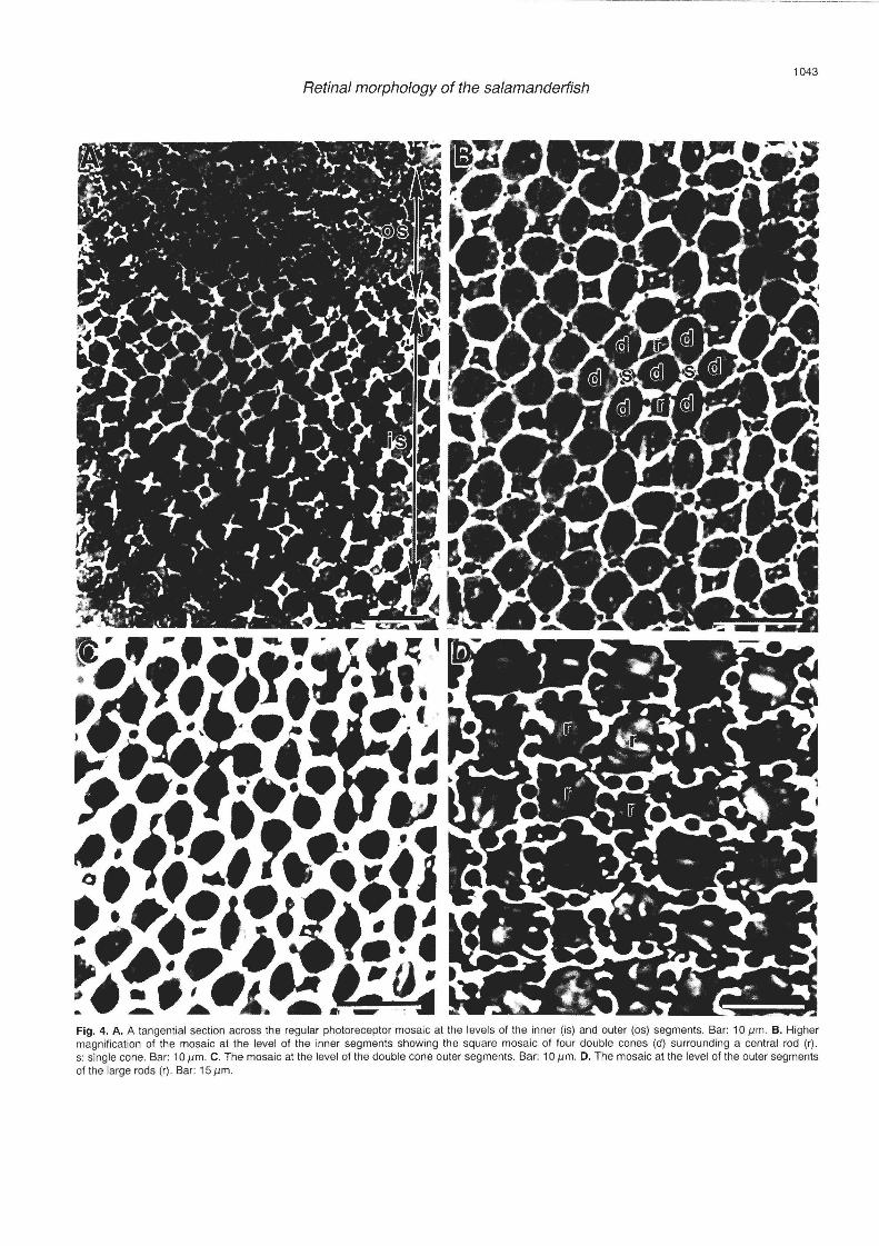
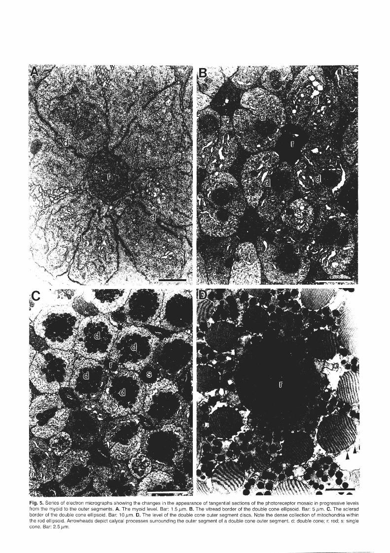
The photoreceptors can be differentiated into 3 types; equal double (twin) cones, small single cones and large rods (with a ratio of 2:1:1 Figs. 2A,B, 3A,B, 6A). These photo receptors are arranged in a regular square mosaic. Within each re pea ting unit, a large rod is bordered by four equal double cones with the small s ingle cone located at each corner (Figs. 4, 5). Units overlap where each double cone forms part of two units. The common surface between the two components of each doubl e cone is oriented perpendicular to it s neighbouring double cone (Figs. 2B-D, 5B,C).
Each doubl e cone is comprised of two cones of equal size (up to 30.um in length and 3.0 .urn in width measured at the base of the outer segment). On e component of the double cone lies slightly sclerad of the other. The double cones are closely apposed along their inner segment s urface s (Fig. 2A,B), immediately adjacent to which lies a complex series of membranes. These subsurface cisternae consist of membrane-bound structures (approximately 20 nm in diameter) which run longitudinally for the length of each inner segment. The cristae of the mitochondria located adjacent to the inner segment borders of each apposing cone in the doublet are often oriented parallel to the subsurface cisternae (Fig. 2C, D). The tapered outer segments of the equal double cones are up to 12 .urn in length and contain a series of di scs comprising two membranes, both approximately 5 nm in thickness, with an intradisc space of 5 nm. Each disc is between 14 and 28 nm from its neares t neighbouring disc . A thin triple membrane surrounds the outer segments of the cones. Each outer segment is surrounded by 12 to 14 calycal processes which do not appear to indent the disc membrane. The double cones possess a short myoid and their lightlystaining nuclei, which lie in the sclerad region of the outer nuclear layer, are often invaginated.
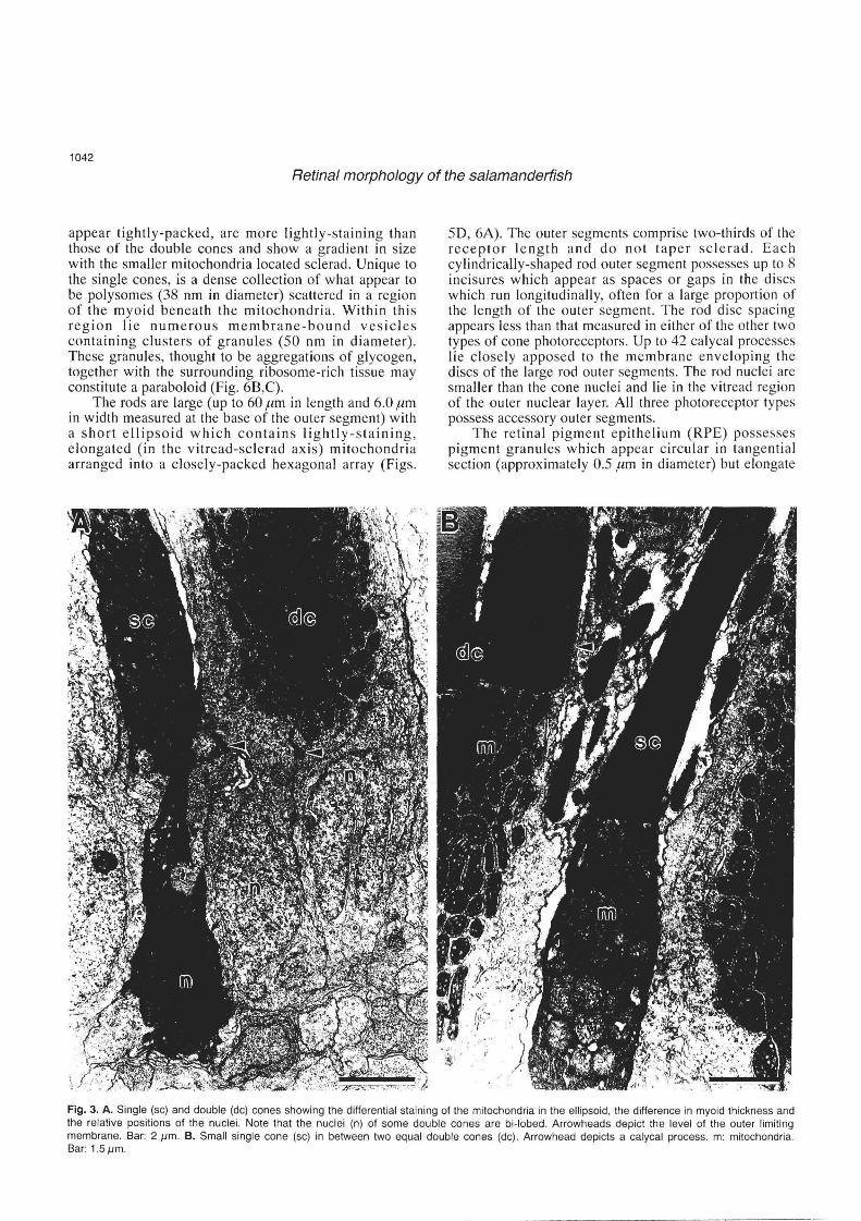
The single cones are smaller (up to 27.um in length and 1.5 .urn in width measured at the base of the outer segment) than the double cones and possess a tapering outer segment surrounded by 10 to 12 calycal processes (Figs. 3A,B, 6A). The mitochondria within the ellipsoid

B _ .. = 1 ,_
Fig. 1. A. Lateral view of the head of Lepidogalaxias salamandroides showing the large, lateral eyes and ventral mouth . Bar: 1.5 mm. B. Transverse section of the light-adapted retina showing the thick inner nuclear layer and the large row of rods (r) sclerad to the equal double cones (de) . gel, ganglion cell layer; inl: inner nuclear layer; ipl: inner plexiform layer; onl: outer nuclear layer: rpe: retinal pigment epithelium. Bar: 15 11m. C. Electron micrograph of the anterior lens showing the granular lens capsule (gc) surrounding a thicker epithelial cell layer of interdigitating cells (ec) and a concentric arrangement of inner lenticular cells. Bar: 1.5I1m . D. Electron micrograph of a vitreal blood vessel (vb) shown in transverse section indented and apposed to the inner limiting membrane (ilm, arrowheads) . Note that the endfeet of the Muller cells (m) line the ilm, dividing the ganglion cell axons (a) into fascicles . Bar: 2.5 11m. E. Electron micrograph of the outer plexiform layer showing the synaptic ribbons (arrowheads) within the rod spherules (rs) and cone pedicles (cp). Bar: 111m.

----------- -----------
Fig. 2. A. Transverse section of three equal double cones (dc) lying in the light-adapted position . Bar: 2.5 11m . B. Higher magnification of an equal double cone showing the disc membranes and mitochondria within the outer (as) and inner (is) segments, respectively. Bar: 111m. C. Tangential section of an equal double cone at the level of the ellipsoid. m: mitochondria. Bar: 111m. D. Higher magnification of the complex of subsurface cisternae lying internal to the apposing inner segment membranes (small arrowheads) of each component of a double cone. The membranes of the inner and outer cisternae are depicted by large arrowheads and arrows, respectively. m: mitochondria. Bar: 0.2 11m.
1042
Retinal morphology of the salamanderfish
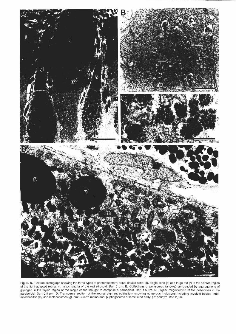
appear tightly-packed, are more lightly-staining than those of the double cones and show a gradient in size with the smaller mitochondria located sclerad. Unique to the single cones, is a dense collection of what appear to be polysomes (38 nm in diameter) scattered in a region of the myoid beneath the mitochondria . Within this region lie numerous membrane-bound vesicles containing clusters of granules (50 nm in diameter). These granules, thought to be aggregations of glycogen, together with the surrounding ribosome-rich tissue may constitute a paraboloid (Fig. 6B,C).
The rods are large (up to 60 11m in length and 6.0 !1m in width measured at the base of the outer segment) with a short ellipsoid which contains lightly-staining, elongated (in the vitread-sclerad axis) mitochondria arranged into a closely-packed hexagonal array (Figs.
50, 6A). The outer segments comprise two-thirds of the receptor length and do not taper sclerad. Each cylindrically-shaped rod outer segment possesses up to 8 incisures which appear as spaces or gaps in the discs which run longitudinally, often for a large proportion of the length of the outer segment. The rod disc spacing appears less than that measured in either of the other two types of cone photoreceptors. Up to 42 calycal processes lie closely apposed to the membrane enveloping the discs of the large rod outer segments. The rod nuclei are smaller than the cone nuclei and lie in the vitread region of the outer nuclear layer. All three photoreceptor types possess accessory outer segments.
The retinal pigment epithelium (RPE) possesses pigment granules which appear circular in tangential section (approximately 0.5 !1m in diameter) but elongate
Fig. 3. A. Single (sc) and double (dc) cones showing the differential staining of the mitochondria in the ellipsoid, the difference in myoid thickness and the relative positions of the nuclei. Note that the nuclei (n) of some double cones are bi-Iobed . Arrowheads depict the level of the outer limiting membrane. Bar: 2 J.lm. B. Small single cone (sc) in between two equal double cones (dc). Arrowhead depicts a calycal process. m: mitochondria. Bar: 1.5J.lm.
------------------
1043
Retinal morphology of the salamanderfish
Fig. 4. A. A tangential section across the regular photoreceptor mosaic at the levels of the inner (is) and outer (os) segments. Bar: 10 11m. B. Higher magnification of the mosaic at the level of the inner segments showing the square mosaic of four double cones (d) surrounding a central rod (r). s: single cone. Bar: 10 11m. C. The mosaic at the level of the double cone outer segments. Bar: 10 11m. D. The mosaic at the level of the outer segments of the large rods (r). Bar: 15 11m .
f i .
Fig. 5. Series of electron micrographs showing the changes in the appearance of tangential sections of the photoreceptor mosaic in progressive levels from the myoid to the outer segments. A. The myoid level. Bar: 1.5 11m. B. The vitread border of the double cone ellipsoid. Bar: 5 11m . C. The sclerad border of the double cone ellipsoid. Bar: 10 11m . D. The level of the double cone outer segment discs. Note the dense collection of mitochondria within the rod ellipsoid . Arrowheads depict calycal processes surrounding the outer segment of a double cone outer segment. d: double cone; r: rod; s: single cone. Bar: 2.5 11m .
-----~---~---~------
Fig. 6. A. Electron micrograph showing the three types of pholoreceptors: equal double cone (d), single cone (s) and large rod (r) in the scierad region of the light·adapted retina. m: mitochondria of the rod ell ipsoid . Bar: 311m . B. Collections of polysomes (arrows) surrounded by aggregations of glycogen in the myoid region of the single cones thought to comprise a paraboloid . Bar: 1.5 11m. C. Higher magnification of the polysomes in the paraboloid. Bar: 0.5 11m . D. Transverse section of the retinal pigment epi thelium showing numerous inclusions including myeloid bodies (mb), mitochondria (m) and melanosomes (g). bm: Bruch's membrane; p: phagosome or lameilated body; pe: pericyte. Bar: 211m.
1046
Retinal morphology of the salamanderfish
in transverse section (up to 3.0 ,urn in length) and surround the outer segments of the photoreceptors in the light-adapted state (Fig. 6A, D). Each mononucleate RPE cell is joined to its neighbour by various types of junctions including zonula adhaerens and is rich in mitochondria, Golgi apparati and phagosomes. Bruch's membrane is 0.33 ,urn in thickness and is pentalaminate. Its vitread and sclerad borders comprise the basement membranes of the retinal pigment epithelium and the choriocapillaris, respectively, with a layer of collagen fibrils separating a broken layer of elastic tissue.
The choriocapillaris possesses two to three layers of melanocytes containing oval and circular-shaped melanosomes (up to 1.3 ,urn in diameter) interspersed between large and small thin-walled capillaries (Fig. 60). The endothelial cells of the choriocapillaris are moderately fenestrated.
Discussion
The presence of vitreal blood vessels, a choriocapillaris and an extensive system of Muller cell processes indicates that the retina of Lepidogalaxias salamandroides is either metabolically-active and/or may be under extreme oxygen tension. Vitreal vessels normally provide an effective transport mechanism for metabolic exchange in retinae containing high densities of retinal neurons often concentrated into a specialised zone of acute vision e.g. in some marine (Hanyu, 1962; Collin, 1989) and freshwater (Kohbara et ai., 1987; Collin et ai., 1996a,b) teleosts. The retina in L. salamandroides is thin (135 ,urn) when compared to other teleosts, for example 265 ,urn in the cutlips minnow, Exoglossum maxillingua (Collin et ai., 1996a), 330,um in the sandlance, Limnichthyes fascia/us (Collin and Collin, 1988) and 570 ,urn in the snake mackerel, Gempylus serpens (Munk, 1985), and both the photoreceptor and ganglion cell layers possess relatively low densities of neurons (Collin S . P. and Gill H., unpublished data). Therefore, the increased blood supply to the retina of L. salamandroides may be an adaptation for reducing the oxygen tension during critical periods of aestivation when metabolic rate, and therefore oxygen exchange, may be low.
The presence of at least two morphological types of photoreceptor terminals with single (rod) and multiple (cone) synaptic ribbons provides evidence for the differentiation of photoreceptor types in L. salamalldroides, although double and single cone terminals could not be differentiated. As in other species of teleosts, the cone terminals in the salamanderfish are larger and contain more synaptic sites (ribbon synapses and surface contacts) than the rod terminals (Cohen, 1972; Braekevelt , 1982) with the lateral and central invaginations corresponding to the horizontal and bipolar cell processes , respectively (Stell, 1965; Dowling , 1968). Similarly, the sclerad and vitread location of the cone and rod nuclei within the outer nuclear layer has been noted as a differential criterion in
teleosts (Braekevelt, 1985; Collin et ai., 1996a) and mammals (Cohen, 1972; BraekeveIt, 1983).
The confirmation of three photoreceptor types in L. salamandroides has been found in a number of early actinopterygian representatives such as the reedfi s h, Calamoichthys calabaricus (Munk, 1969), the bowfin, Amia calva (Munk, 1968) and the garfish, Lepisosteus sp. (Munk, 1968; Collin and Collin, 1993) which all possess double cones, single cones and large rods. Similarly, although not closely related to the teleostean fishes, the Australian (Neoceratodus forsl eri) and African (Protopterus aethiopicus) lungfishes both possess double cones, single cones and large rods. Therefore, in support of the phylogenetic inferences proposed by Frankenberg (1968) and Rosen (1973, 1974) on osteological criteria, who both agree that L. salaman-droides is as primitive as any living teleost and together with the Australian lungfish are " living fossils", morphological features of the photoreceptors of the salamanderfish may also provide a basis for phylogenetic comparisons.
Double cones are common components of the photoreceptor mosaic in a number of teleost species. The advantage of the cone doublet over single cones is unknown but sensitivity may be raised due to its increased retinaL coverage and therefore enhanced quantal capture in low light environments (Boehlert, 1979; Lythgoe, 1979). The a rrangement of the double cones into a regular mosaic may also increase the detection of moving objects without the advantage of an area centralis or fovea (Collin S.P. and Gill H., unpublished data) and provide a means of chromatically sampling the same part of the visual field with two receptors of different spectral sensitivity. Increasing sensitivity would be of particular importance for L. salamandroides since it survives in darkly-stained water and needs to optimise its perception of small mobile prey which it tracks by flexion of its head and " neck" (Berra and Allen, 1989).
The presence of a paraboloid situated below the ellipsoid in the single cones of L. sa lamandroides emphasises the primitive origin of the photoreceptors in this species. The existence of a paraboloid appears to be a plesiomorphic (primitive) character in the phylogeny of the Actinopterygii due to its presence in all the living representativ e outgroups i.e. the Polypteriformes (bichirs, Munk, 1969), Acipenseridae (sturgeons, although lost in the rods, Munk, 1969), Polyodontidae (paddlefishes, Munk, 1969), the Ginglymodi (garfishes, Collin and Collin , 1993) and the Halecomorphi (bowfins, Munk, 1969). Until now, there have been no known representatives of the Teleostei to have retained a paraboloid in their retinal photoreceptors. Alternatively, the presence of a paraboloid may be an apomorphic (derived) trait for gnathostomes, since it has not been found in the photoreceptors of agnathans or chondrichthyans and persists in the Dipnoi (lungfishes, Pfeiffer, 1968; Munk, 1969; Locket, 1970), Amphibia, Reptilia and Aves (Walls, 1942; Detwiler, 1943). The two types of granules found in the paraboloid of L.
- - -- _._._-.------
1047
Retinal morphology of the salamanderfish
salamandroides putatively contain glycogen. Although the site of this glycogen synthesis and its metabolic pathway are not well understood , it may serve as an energy store for the myoid (Rodieck, 1973).
The high proportion of large rods and their unique position within the square mosaic in the retina of L. salamandroides is based on the assumption that the morphological characteristics traditionally used to classify rods and cones are appropriate. The features that separate the large rods from the other two cone types in the salamanderfish include the close position to the retinal pigment epithelium, a long, non-tapering outer segment, longitudinal incisures, a long myoid region, a wider spacing between the disc membranes (Cohen, 1969), the enclosure of the outer segment discs by an external membrane and a mitochondrial size gradient in the inner segment (vitreo-sclerad in the single cones and centro-peripheral in the double cones but absent in the rods). Although, some features described here for rods may be common to cones in other species of teleosts, the full complement of criteria suggest our classification is correct. However, physiological and microspectrophotometric analyses are planned to further characterise these photoreceptor types.
The presence of a large number of rods is usually associated with a nocturnal habitat or life in deep-water. However, for species living in turbid (labrids , Fineran and Nicol, 1974) or darkly-stained water (lepidogalax ids, this study) where the light levels may be constantly changing and often low, a rod-rich retina would be a distinct advantage. The rods of L. salamal1-droides are also very large in comparison to other vertebrates. In the light-adapted state, these rods are 60 .urn in length and 6.0.um in width. This compares to 30 !tm in length and 2 !tm in width in the mature European eel, Anguilla anguilla (Braekevelt, 1988), 40 .urn in length and 4 to 5 !tm in width in both the short-tailed stingray, Dasyalis brevicaudata (Braekevelt, 1994) and the northern pike, Esox lucius (Braekevelt, 1975) but falls short of the long rods of the goldeye, Hiodol1 alosoides (135 .urn, Braekevelt, 1982) and some deep-sea teleosts (340 .urn in Scopelarchus analis, Collin et aI., 1998). The environmental influences which have induced the evolution of a large rod in L. salamandroides is not understood but due to the increase in optical density of the outer segment, these large rods would effectively lower the threshold of visual sensitivity (Bassi and Powers, 1990) and may act as a macroreceptor.
The function of the rod incisures in L. salamandroides is unknown, but there may be some advantage to increasing the length of the disk perimeter and decreasing its surface area, thereby aiding in the diffusion of substances to and from the disk membrane (Rodieck, 1973). In some amphibians, the incisures are particularly numerous and deep (Nilsson, 1965; Cohen, 1972), while in some reptiles (Dunn, 1966), birds (Cohen, 1963) and mammals (Kroll and Machemer, 1968), the disks show a single deep incisure or a series
of scallops. The calycal processes that emanate from the edge of
the inner segment and sit in the grooves of the rod incisures in L. salamandroides are also little understood. These processes are widely distributed in all vertebrate classes and may have a supportive role in preventing the outer segment from rotating about the eccentrically situated connecting cilium (Rodieck, 1973).
This is the first detailed report of a rod that lies at the centre of a square mosaic. This position is usually reserved for a single cone which adopts the central position of the repeating unit where the orientation of the contiguous membrane of each double cone radiates from the centre like a four-spoked wheel. The closest description is one by Karsten (1923) who describes the photoreceptor mosaic in another amphibious species, the mudskipper, Periophthalmus koelreut eri, as a "quadrangular" mosaic with four double cones bordering a rod. Further research is necessary to establish whether this photoreceptor pattern is common to an amphibious lifestyle, as may be predicted for the Australian lungfish, or simply a developmental phenomenon designed to maximise chromatic sampling using this complement of spectrally-sensitive receptors.
Acknowledgments. We wish to thank Brian Pirie of the School of
Optometry, The University of New South Wales and Michael Archer of
the Department of Zoology, University of Western Australia for their
expert technical assistance. We also thank Kevin Smith and Barry
Hutchins from the West Australian Museum in Perth and Howard Gill
from the Department of Environmental Sciences at Murdoch University
for help in collecting specimens. Financial support was provided by the
Australian Research Council (ARC)(SPC) and the National Health and
Medical Research Council of Australia (SPC) and a National Science
Foundation Grant to Prof. R.G. Northcutt whom we thank for his
support. S.P. Collin is currently an ARC QE II Research Fellow.
References
Bassi C.J. and Powers M.K. (1990). Rod outer segment length and
visual sensitivity. Invest. Ophthalmol. Vis. Sci. 31 , 2320-2325.
Begle D.P. (1991). Relationships of the osmeroid fishes and the use of
reductive characters in phylogenetic analysis. Syst. Zoo I. 40, 33-53 .
Berra T.M. and Allen G.R. (1989). Burrowing , emergence, behaviour
and functional morphology of the Australian salamanderfish, Lepidogalaxias salamandroides. Fisheries 14, 2-10.
Berra T.M., Sever D.M . and Allen G.R. (1989). Gross and histological
morphology of the swimbladder and lack of accessory respiratory
structures in Lepidogalaxias salamandroides, an aestivating fish
from Western Australia. Copeia 1989, 850-856.
Boehlert G. (1979). Retinal development of postlarval through juvenile
Sebastes diploproa adaptations to a changing photic environment.
Rev. Can . BioI. 37, 265-280.
Braekevelt C.R. (1975). Photoreceptor fine structure in the northern pike
(Esox lucius ). J. Fish . Res. Bd. Can. 32, 1711 ·1721.
Braekevelt C.R. (1982) . Photoreceptor fine structure in the gold eye
(Hiodon alosoides) (Teleostei). Anat. Embryol. 165, 177-192.
Braekevelt C.R. (1983). Photoreceptor fine structure in the domestic

1048
Retinal morphology of the salamanderfish
ferret. Anat. Anz. 153, 33-44 .
Braekevelt C.R. (1985). Photoreceptor fine structure in the archerfish
(Toxotes iaculatrix). Am. J. Anat. 173, 89-98.
Braekevelt C.R. (1988). Retinal fine structure in the European eel
Anguilla anguilla . VIII. Photo receptors of the sexually mature silver
eel stage. Anat. Anz. 167, 1-10.
Braekevelt C.R. (1994). Retinal photoreceptor fine structure in the shorttailed stingray (Oasyatis brevicaudata). Histo!. Histopatho !. 9, 507-514 .
Christensen P. (1982). The distribution of Lepidogalaxias salaman
droides and other small freshwater fishes in the lower south-west of
Western Australia. J. Roy. Soc. West Aust. 65, 131-141.
Cohen A.1. (1963). Vertebrate retinal cells and their organization. Bio!. Rev. 38, 427-459.
Cohen A. I. (1969) . Rods and cones and the problem of visual excitation.
In: The retina: morphology , function and clinical characteristics
(Jules Stein Symposium 1966) . U.C.L.A. Forum in Medical Sci.
Cohen A.1. (1972). Rods and cones . In: Physiology of photoreceptor
organs. Fuortes M.G.F. (ed). Springer-Verlag. Berlin. pp 63-110.
Collin H.B. and Allansmith J.R. (1977). Basophils in vernal conjunctivitis
in humans: an electron microscopic study. Invest. Ophthalmo!. Vis . Sci. 16,858-864.
Collin H.B. and Collin S.P. (1996). Fine structure of the cornea and lens
in the freshwater salamanderfish , Lepidogalaxias salamandroides . Cornea 15, 414-426.
Collin S.P. (1989) . Topographic organization of the ganglion cell layer
and intraocular vascularization in the retinae of two reef teleosts .
Vision Res. 29, 765-775.
Collin S.P. and Collin H.B. (1988). The morphology of the retina and
lens of the sandlance Limnichthyes fasciatus (Creediidae) . Exp. Bio!. 47, 208-218.
Collin S.P. and Collin H.B. (1993). The visual system of the Florida
garfish , Lepisosteus platyrhincus. (Ginglymodi) : I. Retina. Brain Behav. Evo!. 42, 77-97.
Collin S.P. , Collin H.B. and Al i M.A. (1996a) . Ultrastructure and
organisation of the retina and pigment epithelium in the cutlips
minnow, Exoglossum maxiltingua (Cyprinidae, Teleostei) . Histol. Histopatho!. 11, 55-69.
Collin S.P., Coll in H.B. and Ali M.A. (1996b) . Fine structure of the retina
and pigment epithelium in the creek chub, Semotilus atromaculatus (Cyprinidae, Teleoste i) . Histo!. Histopatho!. 11 , 41 -53.
Collin S.P., Hoskins R.V. and Partridge J.C . (1998). Seven retinal
specialisations in the tubular eye of the deep-sea pearl eye ,
Scopelarchus michae/sarsi: A case study in visual optimisation. Brain Behav. Evo!. 51, 291-314.
Detwiler S.R. (1943) . Vertebrate photoreceptors. Macmillan. New York .
Dowling J.E. (1968). Synaptic organization of the frog retina: an electron
microscopic analysis comparing the retinas of frogs and primates. Proc. Roy. Soc. Lond. B 170, 205-228.
Dunn R.F. (1966). Studies on the retina of the gecko Co/eonyx variegatus . I. The visual cell classification. J. Ultrastruct. Res . 16, 651-671.
Fineran B.A. and Nicol JAC. (1974). Studies on the eyes of New
Zealand parrot-fishes (Labridae). Proc. R. Soc. Lond. B 186, 217-247.
Frankenberg R.S. (1968) . Lepidogalaxias salamandroides, a 'living
fossi l' fish. News Aust. Soc. Limno!. 6, 8-9.
Frankenberg R.S. (1969). Studies of the evolution of galaxiid fishes with
particular reference to the Australian fauna. PhD Thesis. University of Melbourne.
Hanyu I. (1962). Intraocular vascularisation in some fishes. Can. J. Zoo I.
40,87-106.
Karsten H. (1923). Das Auge von Periophthalmus k6lreuteri . Jenaische
Zeitschr. Naturwiss. L1X 1, 115-154.
Kohbara J. , Murachi S. and Nanba K. (1987). Vascular pattern of
hyaloid vessels in carp eye. Nippon Suisan Gakkaishi 53, 219-222.
Kondrashev S.L., Gamburtzeva A.G., Gnjubkina V.P ., Orlov O.J. and
My P.T. (1986). Coloration of corneas in fish . A list of species. Vision
Res. 26, 287-290.
Kroll A.J . and Machemer R. (1968). Experimental retinal detachment in
the owl monkey. III. Electron microscopy of retina and pigment
epithelium. Am. J. Ophthalmol. 66, 410-427.
Locket N .A. (1970). Landolt's club in the retina of the African lungfish,
Protopterus aethiopicus Heckel. Vision Res. 10, 299-306.
Lythgoe J .N. (1979) . The ecology of vision . Oxfo rd Sc ient i f ic
Publications. Oxford.
Mart in K.L.M ., Berra T.M. and Allen G.R. (1993). Cutaneous aerial
respirat ion during forced emergence in the Austra lian sala
manderfish, Lepidogalaxias salamandroides. Copeia 1993, 875-879.
McDowall R.M. (1981) . The relationships of Aust ra lian fishes . In :
Ecological biogeography of Australia. Keast R.A. (ed). Monogr. BioI.
41 , 1-2182.
McDowall R.M. and Pusey B.J. (1983) . Lepidogalaxias salamandroides Mees - A redescription, with natural history notes. Rec. West Aust . Mus. 11 , 11-23.
Mees G.D. (1961) . Description of a new fish of the family Galaxiidae
from Western Australia. J. Roy. Soc. West Aust. 44, 33-38.
Munk O. (1968). The eyes of Amia and Lepisosteus (Pisces, Holostei)
compared with the Brachiopterygian and teleostean eyes. Vidensk.
Meddr. dansk Naturh. Foren . 131 , 109-127.
Munk O. (1969). On the visual cells of some primitive fishes with
particular regard to the classification of rods and cones . Vidensk .
Meddr. dansk Naturh. Foren . 132, 25-30.
Munk O. (1985) . Retinal cones of the snake mackerel, Gempylus serpens Cuvier, 1829. Vidensk. Meddr. Naturh. Foren . 146,7-20.
Nilsson S.E.G. (1965) . The ultrastructure of the receptor outer segment
in the retina of the leopard frog (Rana pipiens). J. Ultrastruct. Res. 12, 207-231.
Pfeiffer W . (1968) . Ret ina und Retinomotor ik der Dipno i und
Brachiopterygii. Z. Zellforsch. Mikrosk. Anat. 89, 62-72.
Pusey B.J. (1981). The life history of the Shannon mud minnow
Lepidogalaxias salamandroides (Mees) with special reference to
aestivation. Hons Thesis. University of Western Australia.
Rodieck R.W. (1973). The vertebrate retina, principles of structure and
function. W. H. Freeman and Company. San Fransisco.
Rosen D.E. (1973). Interrelationships of higher euteleostean fishes. In:
Interrelationships of fishes . Greenwood R.H., Miles R.S. and
Patterson C. (eds) . Zoo I. J. Linn. Soc. Lond . 53 (Supp!.) , 397-513. Rosen D.E. (1974). Phylogeny and zoogeography of salmoniform fishes
and relationships of Lepidogalaxias salamandroides. BUll. Am. Mus. Nat. Hist. 153, 265-326.
Stell W.K. (1965). Correlation of retina l cytoarchitecture and ultra
structure in Golgi preparations. Anat. Rec. 153, 389-397.
Walls G.L. (1942) . The vertebrate eye and its adaptive radiation. Hafner.
New York.
Wi ll iams R.R.G. (1987) . The phylogeneti c relationships of the
salmoniform fishes based on the suspensorium and its muscles .
PhD Thesis. University of Alberta. Edmonton.
Accepted May 13, 1998