Embed Size (px)
Citation preview

Retinal Glia 225
Author's personal copy
Retinal Glia
E A Newman, University of Minnesota, Minneapolis,MN, USA
ã 2009 Elsevier Ltd. All rights reserved.
Introduction
Glial cells outnumber neurons in the central nervoussystem (CNS) by �10 to 1. Traditionally, glia werebelieved to provide only passive structural and meta-bolic support for neurons. Recent work has demon-strated, however, that glial cells in the retina as well asin the brain interact actively with neurons and havemany essential functions.There are three principal types of glial cells in
the mammalian retina: Muller cells, astrocytes, andmicroglial cells. Muller cells are the most prominentretinal glial cell. They are a specialized form of radialglia which span nearly the entire depth of the retina.Astrocytes, the second type of retinal macroglial cell,are present only in species having a retinal circulationand, in these species, are restricted largely to the nervefiber layer at the inner boundary of the retina. Micro-glia, the third type of retinal glial cell, are present in thenerve fiber layer and the inner and outer plexiformlayers of the retina. Oligodendrocytes, the glial cellsthat form the insulating sheath of myelinated axons inthe CNS, are completely absent from the retina, exceptin those species, including rabbit and guinea pig, pos-sessing myelinated axons in the nerve fiber layer.
Retinal Glial Cell Development andMorphology
Muller Cells
Muller cells differentiate from progenitor cells at theouter margin of the retinal neuroepithelium duringthe second phase of cell differentiation, following thebirth of ganglion cells, horizontal cells, and cones.The developing Muller cells migrate toward the vitre-ous humor and extend processes towards the innerand outer retinal margins. Their radial processesserve to guide migrating neurons during subsequentretinal development.In the mature retina, Muller cell somata lie in the
middle region of the inner nuclear layer (Figure 1).Single or multiple radial processes extend inwardstoward the vitreous and end in enlarged structurestermed endfeet. A second cell process extends outwardto the outer limitingmembrane,where apicalmicrovilliproject into the subretinal space. Secondary Mullercell processes project from the main trunk of the cell,
Encyclopedia of Neuroscien
surrounding neuronal somata and processes in all reti-nal layers. Muller cell endfeet terminate at the vitrealborder of the retina as well as onto blood vessels, bothat the vitreal surface and within the retina.
Muller cells contain numerous glycogen granuleswhich represent the main retinal glucose store. Theyalso contain numerous mitochondria and intermedi-ate filaments, composed both of vimentin and glialfibrillary acidic protein (GFAP). GFAP distribution isnormally sparse in Muller cells but is dramaticallyupregulated following retinal injury.
Astrocytes
Astrocytes migrate into the developing retina fromthe optic nerve and advance across the retinal surfaceas the retina matures. The distribution of astrocytesover the surface of the retina is determined by a‘contact-spacing’ interaction between cells.
In themature retina, astrocytes are restricted largelyto the nerve fiber layer (Figure 2). They are closelyassociated with blood vessels and are only present inspecies having a retinal circulation. Their processesare confined largely to the plane parallel to the retinalsurface, although processes sometimes follow bloodvessels into deeper retinal layers. Astrocyte processesare terminated by endfeet which contact superficialretinal blood vessels. Astrocytes contain glycogengranules and dense bundles of intermediate filamentscomposed of GFAP.
Microglia
Microglia are the resident macrophages of the retina.They are derived from blood monocytes and enter theretina along with blood vessels during development.Microglia are normally present in the retina in theirdormant state and are found principally in the nervefiber layer and the inner and outer plexiform layers.They have small somata and short, irregular pro-cesses. The cells are normally flat in appearance andhave slender hair-like extensions protruding fromtheir processes.
Microglial cells proliferate following retinal injuryand differentiate into macrophage-like cells thatremove degenerating retinal neurons by phagocyto-sis. Microglia also phagocytose neurons that die dur-ing the course of normal retinal development.
Glial Induction and Guidance of RetinalBlood Vessels
Glial cells induce the formation of blood vessels andguide their growth into the retina. During develop-ment, astrocytes migrate into the retina from the
ce (2009), vol. 8, pp. 225-232

Figure 2 Fluorescence micrograph of astrocytes within the
nerve fiber layer of the cat retina. The tissue was labeled with
fluorescently tagged antibodies against glial fibrillary acidic protein
(GFAP) which forms the intermediate filaments expressed in
astrocytes. From Karschin A, Wassle H, and Schnitzer J (1986)
Shape and distribution of astrocyte in the cat retina. Investigative
Ophthalmology and Visual Science 27: 828–831.
Frog
b
cba ad
e
fg
a
Carp Lizard Chicken Ox
Figure 1 Drawings of Golgi-stained Muller cells from the retinas of several species. Retinal layers, indicated to the right of the frog
Muller cells, are (a) outer nuclear layer; (b) outer plexiform layer; (c) inner nuclear layer; (e) inner plexiform layer; (f) ganglion cell layer;
(g) nerve fiber layer. The basal endfeet of the Muller cells are seen as enlargements of the processes in the nerve fiber layer. From
Ramon y Cajal S (1972) The Structure of the Retina. Springfield, IL: Thomas, C.C.
226 Retinal Glia
Author's personal copy
optic nerve, first appearing at the optic disk and thenadvancing outwards across the retinal surface. Reti-nal blood vessels, which also originate from the opticnerve, grow along newly formed astrocyte processes,which function as templates for angiogenesis. Vesselgrowth is stimulated by the secretion of vascularendothelial growth factor (VEGF), which is releasedby glial cells in response to hypoxic conditions gener-ated by neuronal activity. Astrocytes serve as guidesfor developing vessels as they grow across the innersurface of the retina while Muller cells serve as tem-plates for vessel growth into the retina and across theinner nuclear layer.
Retinal Glial Cell Membrane Properties
Retinal glial cells possess many of the same voltage-gated ion channels and neurotransmitter receptors
Encyclopedia of Neuroscienc
that are expressed in neurons. These glial channelsand receptors play a key role in mediating glia–neuron interactions in the retina. Most research onretinal glial cell properties has been conducted onMuller cells and the following discussion will focuson the properties of these specialized glial cells.
Ion Channels
Muller cells have a highmembrane permeability to Kþ,which results in a negative resting membrane potentialof –80 to –90mV, near the Kþ equilibrium potential ofthe cell. Expression of Kir4.1, an inwardly rectifyingKþ channel, is largely responsible for this Kþ mem-brane permeability. In Kir4.1 knockout animals,which lackKir4.1 channels,Muller cell input resistanceincreases from a wild-type level of 25 to 310MO,demonstrating that Kir4.1 channels account for over90% of the cell membrane conductance. Kir4.1 chan-nels are not distributed uniformly over the cell surfacebut rather are localized to cell endfeet, which terminateat the vitreal surface of the retina and on retinal bloodvessels. The dystrophin–dystroglycan complex plays acritical role in the clustering of Kþ channels to cellendfeet. As discussed below, clustering of Kþ channelscontributes to Muller cell clearance of excess Kþ fromthe retina.
In addition to Kir4.1, Muller cells express a num-ber of other voltage-gated ion channels, includingCa2þ-activated Kþ channels, fast inactivating Kþ
channels, Ca2þ channels (which share some of theproperties of L-type Ca2þ channels), and Naþ chan-nels. Muller cells also express aquaporin-4 (AQP4).These water-permeable channels are co-localizedwith Kir4.1 at Muller cell endfeet. Together, Kir4.1and AQP4 make up a macromolecular complexthat can be visualized in freeze-fracture electron
e (2009), vol. 8, pp. 225-232

Retinal Glia 227
Author's personal copy
micrographs of endfoot membranes as orthogonalarrays of particles (OAPs).Muller cells and astrocytes are extensively coupled
to each other by gap junctions, composed of con-nexin43 and 45 in Muller cells and connexin30 and43 in astrocytes. Gap junctional coupling impartsboth electrical and chemical tracer coupling to thesenetworks of glial cells. Coupling is stronger betweenastrocytes than between astrocytes and Muller cells.Coupling is very weak (or may not exist) betweenMuller cells, except in amphibians. Interestingly, cou-pling between astrocytes and Muller cells, as assessedby tracer spread, is asymmetric, with tracer passingfrom astrocytes to Muller cells more freely thanfrom Muller cells to astrocytes. In rabbit, astrocytesare coupled to oligodendrocytes as well as toMuller cells.
Neurotransmitter Receptors
Retinal Muller cells and astrocytes express a numberof neurotransmitter and neuromodulator receptors.The most prominent among these are P2Y purinergicreceptors. AmphibianMuller cells express P2Y1, P2Y2,P2Y6, and P2Y11 purinergic receptors. Muller cells ofdifferent species also express a number of glutamatereceptors, including AMPA (GluR4), NMDA, andmetabotropic types. GABAA, acetylcholine, dopamine,noradrenaline, adenosine (A2B), thrombin, and lyso-phosphatidic acid receptors as well as several neuroac-tive peptides receptors are expressed. Activation ofmany of these receptors evokes glial Ca2þ increases(see below). Interestingly, in the mammalian retina,glutamate is largely ineffective in evoking Ca2þ
increases in Muller cells or astrocytes. In contrast, glu-tamate evokes large Ca2þ increases in astrocytes inbrain slices and in culture. The insensitivity of retinalglial cells to glutamate may be a specialization toconditions in the retina, where glutamate is releasedcontinuously from neurons.Receptors to a number of growth factors, including
basic fibroblast growth factor (bFGF), epidermalgrowth factor (EGF), platelet-derived growth factor(PDGF), and nerve growth factor (NGF) are expressed.The effects of these factors are varied, and includethe stimulation of DNA synthesis, mitosis, and cellproliferation, expression of cytoskeletal filaments,and themodulation of ion channels. EndothelinB recep-tors are expressed in Muller cells and are upregulatedfollowing retinal injury.
Transporter systems
The high-affinity glutamate/aspartate transporter(GLAST, also named EAAT1) is present in Mullercells and serves to maintain a low concentration(<1 mM) of glutamate in extracellular space. It utilizes
Encyclopedia of Neuroscien
the gradients of Naþ, Kþ, and Hþ across the plasmamembrane to transport glutamate into cells. A high-affinity GABA transporter of the GAT-3 type is alsopresent in Muller cells.
Several acid/base transport systems are expressedinMuller cells. The transporters regulate intracellularpH (pHi) and can influence extracellular pH. Anelectrogenic Naþ/HCO3
� co-transporter is preferen-tially localized to the basal endfoot of Muller cells.A Naþ/Hþ exchanger and a Cl�/HCO3
� anion ex-changer are also present in Muller cells. The anionexchanger is of the AE3 type and is localized to thecell endfoot. Intracellular as well as membrane-boundforms of the enzyme carbonic anhydrase (CA) are alsoexpressed in Muller cells at high activity levels. Theenzyme contributes to pH and CO2 regulation in theretina (see below).
Calcium Signaling in Retinal Glial Cells
Calcium Waves
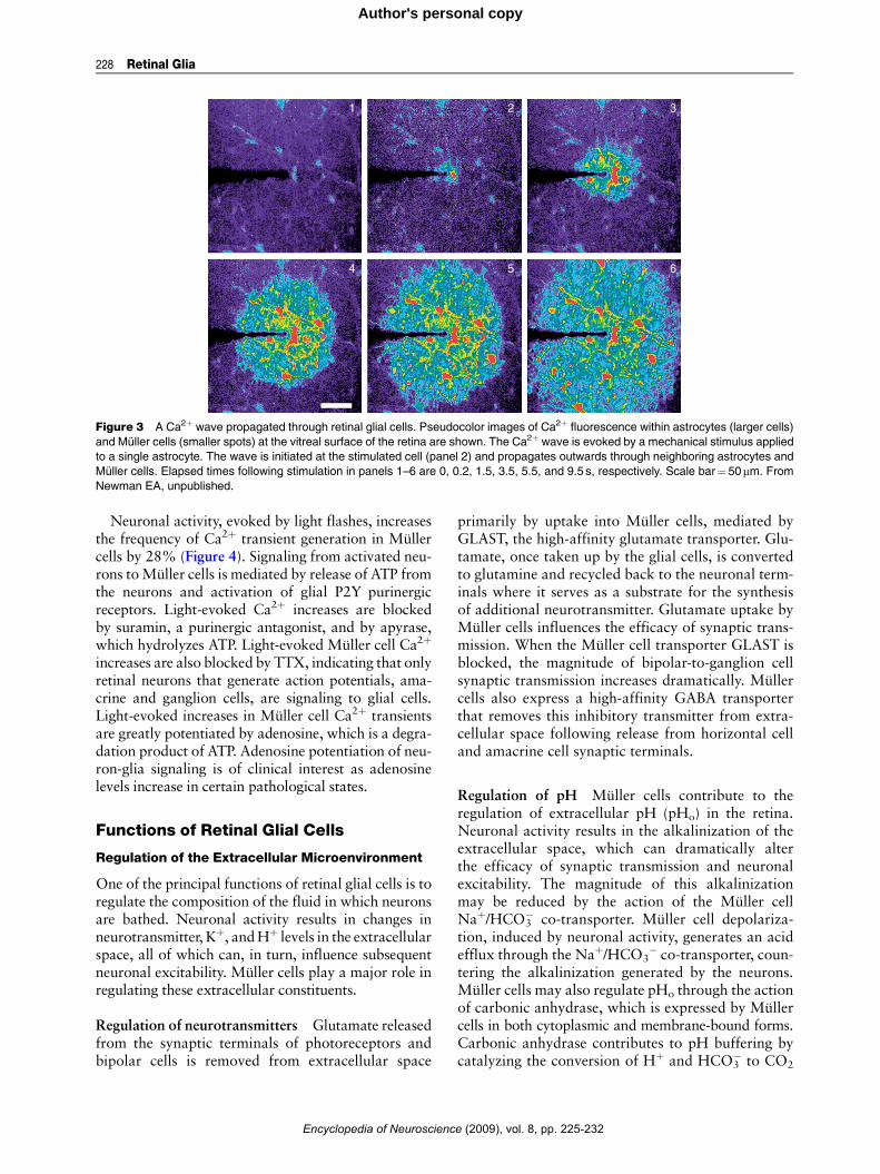
Activation of P2Y and other metabotropic receptorson Muller cells and astrocytes results in the release ofCa2þ from internal stores and to increases in intra-cellular Ca2þ. Calcium increases can also be evokedby mechanical or electrical stimulation of glial cells.Stimulation of a single glial cell often leads to thegeneration of propagated Ca2þ waves that travelthrough many astrocytes and Muller cells (Figure 3).Propagated Ca2þwaves travel at a velocity of approx-imately 23 mm s�1 and can extend outwards for dis-tances as great as 180 mm.
Glial cell-to-glial cell signaling during Ca2þ wavepropagation is mediated by two mechanisms: bymovement of the intracellular messenger IP3, whichdiffuses through gap junctions coupling glial cellstogether; and by release of ATP, which functions asan extracellular messenger. Calcium wave propaga-tion between astrocytes is mediated by both IP3 diffu-sion and by ATP release while signaling betweenastrocytes and Muller cells and between Muller cellsoccurs solely by ATP release. The component of glialCa2þ waves mediated by ATP release is blocked bypurinergic antagonists.
Light-Evoked Ca2þ Signaling
In addition to agonist-evoked Ca2þ signals, Mullercells generate spontaneous, transient increases inCa2þ in the absence of any applied stimulus. Theseendogenously generated Ca2þ increases occur ata frequency of 4.6 transients per cell per 1000 sin mammalian Muller cells. The transients rangefrom 2.5 to 6 s in duration and are similar to thoseobserved in astrocytes in the brain.
ce (2009), vol. 8, pp. 225-232
1
4 5 6
2 3
Figure 3 A Ca2þ wave propagated through retinal glial cells. Pseudocolor images of Ca2þ fluorescence within astrocytes (larger cells)
and Muller cells (smaller spots) at the vitreal surface of the retina are shown. The Ca2þ wave is evoked by a mechanical stimulus applied
to a single astrocyte. The wave is initiated at the stimulated cell (panel 2) and propagates outwards through neighboring astrocytes and
Muller cells. Elapsed times following stimulation in panels 1–6 are 0, 0.2, 1.5, 3.5, 5.5, and 9.5 s, respectively. Scale bar¼50mm. From
Newman EA, unpublished.
228 Retinal Glia
Author's personal copy
Neuronal activity, evoked by light flashes, increasesthe frequency of Ca2þ transient generation in Mullercells by 28% (Figure 4). Signaling from activated neu-rons toMuller cells is mediated by release of ATP fromthe neurons and activation of glial P2Y purinergicreceptors. Light-evoked Ca2þ increases are blockedby suramin, a purinergic antagonist, and by apyrase,which hydrolyzes ATP. Light-evoked Muller cell Ca2þ
increases are also blocked by TTX, indicating that onlyretinal neurons that generate action potentials, ama-crine and ganglion cells, are signaling to glial cells.Light-evoked increases in Muller cell Ca2þ transientsare greatly potentiated by adenosine, which is a degra-dation product of ATP. Adenosine potentiation of neu-ron-glia signaling is of clinical interest as adenosinelevels increase in certain pathological states.
Functions of Retinal Glial Cells
Regulation of the Extracellular Microenvironment
One of the principal functions of retinal glial cells is toregulate the composition of the fluid in which neuronsare bathed. Neuronal activity results in changes inneurotransmitter, Kþ, andHþ levels in the extracellularspace, all of which can, in turn, influence subsequentneuronal excitability. Muller cells play a major role inregulating these extracellular constituents.
Regulation of neurotransmitters Glutamate releasedfrom the synaptic terminals of photoreceptors andbipolar cells is removed from extracellular space
Encyclopedia of Neuroscienc
primarily by uptake into Muller cells, mediated byGLAST, the high-affinity glutamate transporter. Glu-tamate, once taken up by the glial cells, is convertedto glutamine and recycled back to the neuronal term-inals where it serves as a substrate for the synthesisof additional neurotransmitter. Glutamate uptake byMuller cells influences the efficacy of synaptic trans-mission. When the Muller cell transporter GLAST isblocked, the magnitude of bipolar-to-ganglion cellsynaptic transmission increases dramatically. Mullercells also express a high-affinity GABA transporterthat removes this inhibitory transmitter from extra-cellular space following release from horizontal celland amacrine cell synaptic terminals.
Regulation of pH Muller cells contribute to theregulation of extracellular pH (pHo) in the retina.Neuronal activity results in the alkalinization of theextracellular space, which can dramatically alterthe efficacy of synaptic transmission and neuronalexcitability. The magnitude of this alkalinizationmay be reduced by the action of the Muller cellNaþ/HCO3
� co-transporter. Muller cell depolariza-tion, induced by neuronal activity, generates an acidefflux through the Naþ/HCO3
� co-transporter, coun-tering the alkalinization generated by the neurons.Muller cells may also regulate pHo through the actionof carbonic anhydrase, which is expressed by Mullercells in both cytoplasmic and membrane-bound forms.Carbonic anhydrase contributes to pH buffering bycatalyzing the conversion of Hþ and HCO3
� to CO2
e (2009), vol. 8, pp. 225-232

a
b
10 s
10 s
ΔF/F2.5%
ΔF/F100%
Figure 4 Light-evoked Ca2þ signaling in Muller cells. (a) Cal-
cium fluorescence measured simultaneously in eight Muller cells.
The retina was exposed sequentially to a uniform dim light, a
bright flickering light, and a dim light (the light stimulus protocol
is shown at the bottom in (a) and (b)). Calcium transients are more
likely to be generated during the flickering light stimulus than
during periods of constant illumination. (b) Mean Ca2þ fluores-
cence averaged over 84 trials. The flickering light evokes both a
transient and a sustained increase in Ca2þ in Muller cells. From
Newman EA (2005) Calcium increases in retinal glial cells evoked
by light-induced neuronal activity. Journal of Neuroscience 25:
5502–5510.
2 mV
4 s
Light-elicited[K+]o decrease
a
b
Light-elicited[K+]o increase
End-foot
Vitreous humor
SRS
Bloodvessel
Müllercell
Vm
NeuronK+
K+ IPL
Figure 5 Muller cell regulation of Kþ. (a) Light-evoked depolar-
izations recorded from a Muller cell of the mudpuppy. A 4 s light
flash (bottom trace) evokes increases in extracellular Kþ concen-
tration at light ON and OFF, resulting in Kþ influx into Muller cells
and to cell depolarization. (b) Potassium siphoning in Muller cells.
Active neurons release Kþ into the inner plexiform layer (IPL). The
increase in extracellular Kþ concentration evokes a Kþ influx into
Muller cells. The resulting cell depolarization generates an equal
efflux of Kþ from other cell regions, into the vitreous humor, the
subretinal space (SRS) and onto blood vessels. This Kþ current
flowing through Muller cells reduces the initial Kþ increase in the
IPL. (a) From Karwoski and Proenza (1977) Relationship between
Muller cell responses, a local transretinal potential and potassium
flux. Journal of Neurophysiology 40: 244–259. (b) From Newman
(1966) Neuroscientist 2: 110–119.
Retinal Glia 229
Author's personal copy
and H2O. Blocking the action of the enzyme leads toincreases in the light-evoked pHo alkalinization.
Regulation of potassium Muller cells play a keyrole in regulating extracellular Kþ ([Kþ]o) within theretina. Light-evoked neuronal activity results inincreases in [Kþ]o in the inner and outer plexiform layersand to a [Kþ]o decrease in the distal retina. These [Kþ]ovariations will change themembrane potential of retinalneurons, altering their excitability. The [Kþ]o variationsare buffered by Kþ currents flowing through Mullercells, a process termed ‘Kþ siphoning,’ which is a spe-cialized formofKþ spatial buffering. Potassium releasedfrom active neurons results in an influx of Kþ intoMuller cells. The Kþ influx depolarizes Muller cells(Figure 5(a)) and drives out an equal quantity of Kþ
from other cell regions, primarily from cell endfeet,
Encyclopedia of Neuroscien
which have a high Kþ conductance. Potassium is rel-eased from basal endfeet into the vitreous humor, whichacts as a large Kþ sink, and from apical processes intothe subretinal space, where it counters the light-evoked[Kþ]o decrease generated in this region (Figure 5(b)).
ce (2009), vol. 8, pp. 225-232

230 Retinal Glia
Author's personal copy
The influx and efflux of Kþ in Muller cells duringthe Kþ siphoning process occurs largely throughKir4.1 inwardly rectifying Kþ channels. The voltage-and Kþ-dependent properties of these channelsenhance the Kþ siphoning process; channel conduc-tance increases with increased [Kþ]o, resulting ingreater siphoning currents and a more rapid transferof Kþ to the vitreous humor and the subretinal space.When Muller cell Kir4.1 channels are blocked byBa2þ, the siphoning current is interrupted and [Kþ]oregulation in the retina is compromised.
Generation of the electroretinogram A consequenceof Kþ current flow through Muller cells is the genera-tion of extracellular field potentials within the retina.These light-evoked potentials can be recorded with anelectrode on the cornea as components of the electro-retinogram (ERG). For many years, the b-wave, themost prominent component of the ERG, was believedto be generated by Kþ current flow through Mullercells.However, if Kþ siphoning is prevented by blockingMuller cell Kir channels with Ba2þ, or if the channelsare eliminated by employing Kir4.1 knockout animals,the b-wave is not reduced. These experiments demon-strate conclusively that Muller cells do not generatethe ERG b-wave. The b-wave is generated, instead,by bipolar cells, as originally suggested by Tomita.Other components of the ERG are generated by
light-evoked Kþ current flow through Muller cells,however. The slow PIII response, the retinal compo-nent of the ERG c-wave, is generated by Mullercell Kþ current flow established by a decrease in[Kþ]o in the distal retina. Muller cells also generate
Neuronvoltage
Neuroncurrent
GlialCa2+
ATPgS
5 s10% ΔF/F
20 pA
a b
2 mV
Figure 6 Glial cell inhibition of action potential generation in ganglion
increase in glial cells (glial Ca2þ). This Ca2þ increase is associated w
(neuron current) in a neighboring ganglion cell. (b) ATPgS stimulation o
of action potentials in the cell. From Newman EA (2003) Glial cell i
23: 1659–1666.
Encyclopedia of Neuroscienc
the scotopic threshold response, a rod-drivenresponse generated at the ON of light in mammals,and the M-wave, a negative response with prominentON and OFF components. Both responses are gener-ated by Kþ current flow through Muller cells estab-lished by [Kþ]o increases in the inner plexiform layer.
Modulation of Neuronal Activity
In recent years, it has become clear that glial cells inthe CNS modulate neuronal activity by releasing glio-transmitters. In the retina, stimulation of Muller cellsresults in changes in light-evoked spiking of ganglioncells. Spike activity can either be potentiated ordepressed following glial cell stimulation and the ini-tiation of Ca2þ waves in retinal glial cells. It is likelythat Muller cells, in particular, modulate neuronalactivity by several different mechanisms, two ofwhich are described below.
Inhibition of ganglion cells Selective stimulation ofMuller cells by agonist ejection evokes large Ca2þ
increases in these cells and subsequent hyperpolari-zation of adjacent ganglion cells (Figure 6(a)). Thehyperpolarization can be sufficiently strong tocompletely block the generation of action potentials(Figure 6(b)). The ganglion cell hyperpolarization ismediated by ATP release from the Muller cells.Released ATP is rapidly converted to adenosine byecto-ATPases and ecto-nucleotidases. Adenosine, inturn, activates A1 adenosine receptors on the ganglioncells, leading to the opening of Kþ channels and to cellhyperpolarization.
ATPgS
5 s
10 mV
cells. (a) Ejection of ATPgS onto the retinal surface evokes a Ca2þ
ith a hyperpolarization (neuron voltage) and an outward current
f glial cells hyperpolarizes a ganglion cell and blocks the generation
nhibition of neurons by release of ATP. Journal of Neuroscience
e (2009), vol. 8, pp. 225-232

a
d e f
b c0s 1s 10s
Figure 7 Glial cell-evoked dilation of a neighboring blood vessel. (a–c) Fluorescence images showing Ca2þ concentration within retinal
glial cells. (d–f) Infrared differential interference contrast (IR-DIC) images showing the arteriole within the region indicated in
(a). Photolysis of caged-Ca2þ in the glial cell indicated by the circle in (a) evokes a Ca2þ increase that propagates from the stimulated
cell into neighboring glial cells surrounding the blood vessel (a–c). Glial cell stimulation results in the dilation of the adjacent arteriole (d–f).
From Metea MR and Newman EA (2006) Glial cells both dilate and constrict blood vessels. A mechanism of neurovascular coupling.
Journal of Neuroscience 26: 2862–2870.
Retinal Glia 231
Author's personal copy
D-serine regulation of NMDA receptors Muller cellstimulation may also lead to the release of D-serineand to potentiation of NMDA receptor neurotrans-mission in the retina. Muller cells contain D-serine, anendogenous NMDA receptor co-agonist, and serineracemase, the synthetic enzyme for D-serine. Stimula-tion of brain astrocytes results in D-serine release anda similar release of D-serine may occur in retinalMuller cells.
Regulation of Blood Flow
Light stimulation results in an increase in blood flowin retinal arteries, bringing increased oxygen andnutrients to activated neurons. Muller cells mediateneuron to blood vessel signaling, a process termedneurovascular coupling. When Muller cells are selec-tively stimulated, neighboring arterioles either dilateor constrict (Figure 7). Vasodilation is mediatedby production of EETs while vasoconstriction ismediated by production of 20-HETE. Both vaso-active agents are metabolites of arachidonic acid.Light stimulation also evokes either vasodilationor constriction by generation of the same two ara-chidonic acid metabolites. When neuron to Mullercell signaling is blocked by a purinergic antagonist,light-evoked vasomotor responses are also blocked,indicating that Muller cells mediate neurovascularcoupling.
Encyclopedia of Neuroscien
Metabolic and Trophic Support of Neurons
Muller cells play an active role in supportingneuronal metabolism. Glucose is metabolized inMuller cells to lactate, which is then transferredto neurons where it serves as a primary energysource for oxidative metabolism. Muller cells alsocontain large reserves of glycogen, which serve as asource of glucose during times of high metabolicactivity.
Retinal glial cells secrete a number of growthfactors which have trophic effects on retinal neuronsand blood vessels. Basic fibroblast growth factor(bFGF), which promotes photoreceptor survival, issynthesized by Muller cells and retinal astrocytes.Muller cells also secrete a number of other growthfactors including brain-derived neurotrophic factor,NGF, neurotrophin-3, neurotrophin-4, and glial cellline-derived neurotrophic factor. Both Mullercells and astrocytes synthesize VEGF, which playsan essential role in the development of the retinalvasculature.
See also: Astrocyte: Calcium Signaling; Fovea: Primate;
Retina: An Overview; Retinal Color Mechanisms; Retinal
Development: An Overview; Retinal Development: Cell
Type Specification; Retinal Pharmacology: Inner Retinal
Layers.
ce (2009), vol. 8, pp. 225-232
232 Retinal Glia
Author's personal copy
Further Reading
Brew H and Attwell D (1987) Electrogenic glutamate uptake is a
major current carrier in the membrane of axolotl retinal glial
cells. Nature 327: 707–709.Brew H, Gray PTA, Mobbs P, and Attwell D (1986) Endfeet of
retinal glial cells have higher densities of ion channels that
mediate Kþ buffering. Nature 324: 466–468.Karschin A, Wassle H, and Schnitzer J (1986) Shape and distribu-
tion of astrocyte in the cat retina. Investigative Ophthalmologyand Visual Science 27: 828–831.
Karwoski CJ, Lu H-K, and Newman EA (1989) Spatial buffering of
light-evoked potassium increases by retinal Muller (glial) cells.Science 244: 578–580.
Kofuji P, Ceelen PW, Zahs KR, Surbeck LW, Lester HA, and
Newman EA (2000) Genetic inactivation of an inwardly rectify-
ing potassium channel (Kir4.1 subunit) in mice: Phenotypicimpact in retina. Journal of Neuroscience 20: 5733–5740.
Metea MR and Newman EA (2006) Glial cells both dilate and
constrict blood vessels: A mechanism of neurovascular cou-
pling. Journal of Neuroscience 26: 2862–2870.Newman EA (1984) Regional specialization of retinal glial cell
membrane. Nature 309: 155–157.Newman EA (2001) Glia of the retina. In: Ryan SJ (ed.) Retina, pp.
89–103. St. Louis: Mosby.
Encyclopedia of Neuroscienc
Newman EA (2003) Glial cell inhibition of neurons by release of
ATP. Journal of Neuroscience 23: 1659–1666.Newman EA (2005) Calcium increases in retinal glial cells evoked
by light-induced neuronal activity. Journal of Neuroscience 25:5502–5510.
Newman EA and Zahs KR (1997) Calcium waves in retinal glial
cells. Science 275: 844–847.Newman EA and Zahs KR (1998) Modulation of neuronal activity
by glial cells in the retina. Journal of Neuroscience 18:
4022–4028.
Poitry-Yamate CL, Poitry S, and Tsacopoulos M (1995) Lactatereleased by Muller glial cells is metabolized by photorecep-
tors from mammalian retina. Journal of Neuroscience 15:
5179–5191.
Ramon y Cajal S (1972) The Structure of the Retina. Springfield,IL: Thomas, C.C.
Sarthy V and Ripps H (2001) The Retinal Muller Cell. Kluwer
Academic/Plenum Publishers.
Stevens ER, Esquerra M, Kim P, et al. (2003) d-serine and serineracemase are present in the vertebrate retina and contribute to
the functional expression of NMDA receptors. Proceedings ofthe National Academy of Sciences of United States of America100: 6789–6794.
e (2009), vol. 8, pp. 225-232


![Activated Microglia in the Brain: Mitochondrial and Cell ...Microglia accounts for 5% - 10% of total glia cells of the human brain[10] [16] [17] and up to 20% in other mammalian species[18]](https://img.pdfslide.net/doc/110x75/60ff85e84df5fc1561629cb9/activated-microglia-in-the-brain-mitochondrial-and-cell-microglia-accounts.jpg)