Embed Size (px)
Citation preview
RETINAL TOXICITY OF INTRAVITREALTRIAMCINOLONE ACETONIDEA Morphological Study
SEUNG-YOUNG YU, MD, FRANCISCO MAX DAMICO, MD,FRANCESCO VIOLA, MD, DONALD J. D’AMICO, MD,LUCY H. YOUNG, MD, PHD
Purpose: To evaluate the morphologic effects of intravitreal triamcinolone acetonide(TA) on rabbit retina.
Methods: Intravitreal injections of 0.5 mg, 1 mg, 4 mg, 8 mg, and 20 mg of TA(Kenalog-40; Bristol-Myers Squibb, Princeton, NJ) in 0.1 mL were given to pigmentedrabbits. For control, 0.1 mL of TA vehicle and saline were injected. Animals were killed onday 14, and retinas were analyzed by light as well as electron microscopy.
Results: No ophthalmoscopic change was found. Eyes injected with 0.5 mg and 1 mgof TA did not have morphologic abnormality. Eyes injected with 4 mg, 8 mg, and 20 mgshowed destruction of photoreceptor outer segments and migration of macrophage-likecells in the subretinal space. Eyes injected with 20 mg showed more extensive damageand increased pigment granules in the retinal pigment epithelium cells with large oildroplets in the cytoplasm. Electron microscopy also showed loss of photoreceptor/retinalpigment epithelium interdigitations. Eyes injected with vehicle or saline did not showmorphologic changes.
Conclusion: Single intravitreal injection of 0.5 mg or 1 mg of TA did not producemorphologic retinal changes in pigmented rabbits. However, injections of 4 mg, 8 mg, and20 mg of TA produced outer retina toxic effects. These findings suggest that long-termretinal toxicity studies should be carried out, using single and repeated injections beforethis therapy becomes more widely used.
RETINA 26:531–536, 2006
Intravitreal injection of triamcinolone acetonide(TA) has been largely used as an alternative in the
treatment of macular edema secondary to diabetes,
retinal vascular occlusion, uveitis, and Irvine–Gasssyndrome.1–5 In addition, TA has been proposed asadjunctive treatment for exudative age-related macu-lar degeneration, proliferative vitreoretinopathy, anduveitis because of its antiangiogenic, antiproliferative,and antiinflammatory properties.6–9
Drug delivery into the posterior segment of the eyealleviates some issues, such as intraocular penetrationand bioavailability, and can lead to improved thera-peutic effect by increasing the intraocular drug con-centration.10,11 However, it can be associated with ahigher risk of local toxicity. The ocular side effects oftopical steroids are well known and include posterior
From the Retina Service, Massachusetts Eye and Ear Infirmary,Harvard Medical School, Boston, Massachusetts.
Supported in part by the Vitreoretinal Research Fund (to D.J.D’Amico).
Presented in part at the 2004 Association for Research in Visionand Ophthalmology Meeting, Ft. Lauderdale, FL, and the 2004Retina Society Annual Meeting, Baltimore, MD, Sept. 30 to Oct. 3,2004.
The authors do not have any proprietary interest in this study.Reprint requests: Lucy H. Young, MD, PhD, Massachusetts Eye
and Ear Infirmary, 243 Charles Street, Boston, MA 02114; e-mail:[email protected]
531

subcapsular cataract and elevation of ocular pres-sure.12 More recently, several case series on compli-cations after intravitreal injection of TA have beenreported, including ocular hypertension and infectiousor sterile endophthalmitis.13–17 However, there is littleinformation on the effect of TA on the retina.
Previous animal studies did not show retinal toxic-ity after intravitreal injection of up to 4 mg of TA,18–21
but even higher doses have been used clinically.1–4,6–9
The purpose of this study was to evaluate morphologicchanges on pigmented rabbit retina caused by intrav-itreal injection of TA (Kenalog-40; Bristol-MyersSquibb, Princeton, NJ) at concentrations used in clin-ical practice.
Methods
Animals
Twenty-four Dutch Belted rabbits (weight, 2–3 kg)were used in this study. All animals were treated inaccordance with the Association for Research in Vi-sion and Ophthalmology Statement for the Use ofAnimals in Ophthalmic and Vision Research, and theexperiments were approved by the Animal Care Com-mittee of the Massachusetts Eye and Ear Infirmary(Boston, MA). Animals were anesthetized with intra-muscular injection of 50 mg/kg ketamine hydrochlo-ride and 5 mg/kg xylazine hydrochloride, and pupilswere dilated with topical 2.5% phenylephrine. Topicalanesthesia was achieved with 0.5% proparacaine. An-imals were divided in 7 groups: 0.5 mg of TA (3 eyes),1 mg of TA (3 eyes), 4 mg of TA (4 eyes), 8 mg of TA(4 eyes), 20 mg of TA (4 eyes), vehicle (2 eyes), andsaline (4 eyes). Contralateral eyes were left un-touched.
TA Preparation
TA (Kenalog-40 [40 mg/mL TA, 0.99% (w/v) ben-zyl alcohol, 0.75% carboxymethylcellulose sodium,and 0.04% polysorbate 80]; Bristol-Myers Squibb)was used. The drug was prepared in a 1-mL tuberculinsyringe. A volume of 0.1 mL was injected in the eye.
For 0.5-mg and 1-mg injections, 0.1 mL of theoriginal suspension was diluted with 0.7 mL and 0.3mL of sterile saline for injection, respectively, and 0.1mL of the new suspension was injected into the vit-reous. For 4-mg injections, 0.1 mL of the originalsuspension was injected. For 8-mg and 20-mg injec-tions, 1 mL of the original suspension was drawn upinto the syringe and kept in a vertical position with theneedle turned up for 30 minutes, until TA settleddown. Then, 0.5 mL and 0.8 mL of the supernatant(vehicle), respectively, was discarded. The remaining
suspension was mixed, and 0.1 mL was injected intothe vitreous. Vehicle was prepared as described abovefor 8-mg and 20-mg injections.
Intravitreal Injection
Before injections, eyes were dilated, and paracen-tesis with removal of 0.1 mL of aqueous humor wasperformed using a 1-mL syringe with a 30-gaugeneedle, to avoid an increase in intraocular pressure.Intravitreal injection of 0.1 mL of solution was per-formed with a 1-mL syringe with a 27-gauge needle,slowly and under direct observation with a stereo-scopic microscope, directed to the center of the vitre-ous cavity.
Ocular and Histologic Examination
Animals underwent slit-lamp biomicroscopy, indi-rect ophthalmoscopy, and fundus photography beforeand immediately after injection and on days 1, 3, 7,and 14. All animals were killed on day 14 with intra-venous injection of phenobarbital sodium solution(Fatal-Plus; Vortech Pharmaceuticals, Dearborn, MI).Immediately after death, eyes were enucleated, theanterior segment with the lens was removed, and theposterior segment was fixed in 50% Karnovsky (Elec-tron Microscopy Sciences, Fort Washington, PA) for24 hours. After fixation, 5 ! 5-mm pieces of theinferior midperipheral posterior segment (retina/cho-roid/sclera) were processed for light microscopy (BHSSystem; Olympus Optical, Tokyo, Japan) and trans-mission electron microscopy (Philips 410 EM; PhilipsElectron Optics, Eindhoven, Holland). Light micros-copy sections were stained with phenylenediamine,and transmission electron microscopy sections werestained with lead and uranyl acetate.
Results
In all TA-injected eyes, the drug crystals settledwithin the cortical vitreous overlying the inferior mid-peripheral retina as a white mass. Its volume becameprogressively smaller, but the shrinkage was slow,remaining visible on day 14. Two eyes (4 mg and 8mg) developed a mild anterior chamber reaction thatresolved within 1 week. Another eye (8 mg) devel-oped cataract due to accidental touch of the anteriorlens capsule with the needle during paracentesis. Novitreous cells or retinal lesions such as retinal whiten-ing, hemorrhage, or pigmentary changes were noted.
Eyes injected with 0.5 mg and 1 mg of TA did notshow any morphologic change during light micros-copy, as well as eyes injected with vehicle (Fig. 1A)and saline. However, eyes injected with 4 mg, 8 mg,
532 RETINA, THE JOURNAL OF RETINAL AND VITREOUS DISEASES ● 2006 ● VOLUME 26 ● NUMBER 5

and 20 mg of TA had dose-dependent changes in theouter retina, such as loss of photoreceptor outer seg-ments and disorganization of the outer segment layer,with macrophage-like cells in the subretinal space,
particularly in areas with increased outer segmentdisorganization (Fig. 1, B and C). Retinal pigmentepithelium (RPE) cells appeared hypertrophic, withincreased pigment granules clustered in the cyto-plasm. Amorphous material was in the subretinalspace (Fig. 1, B and C). These findings were moreevident in eyes injected with 20 mg of TA. In addition,RPE cells of 20-mg injected eyes displayed abundantlarge oil droplets (Fig. 1C).
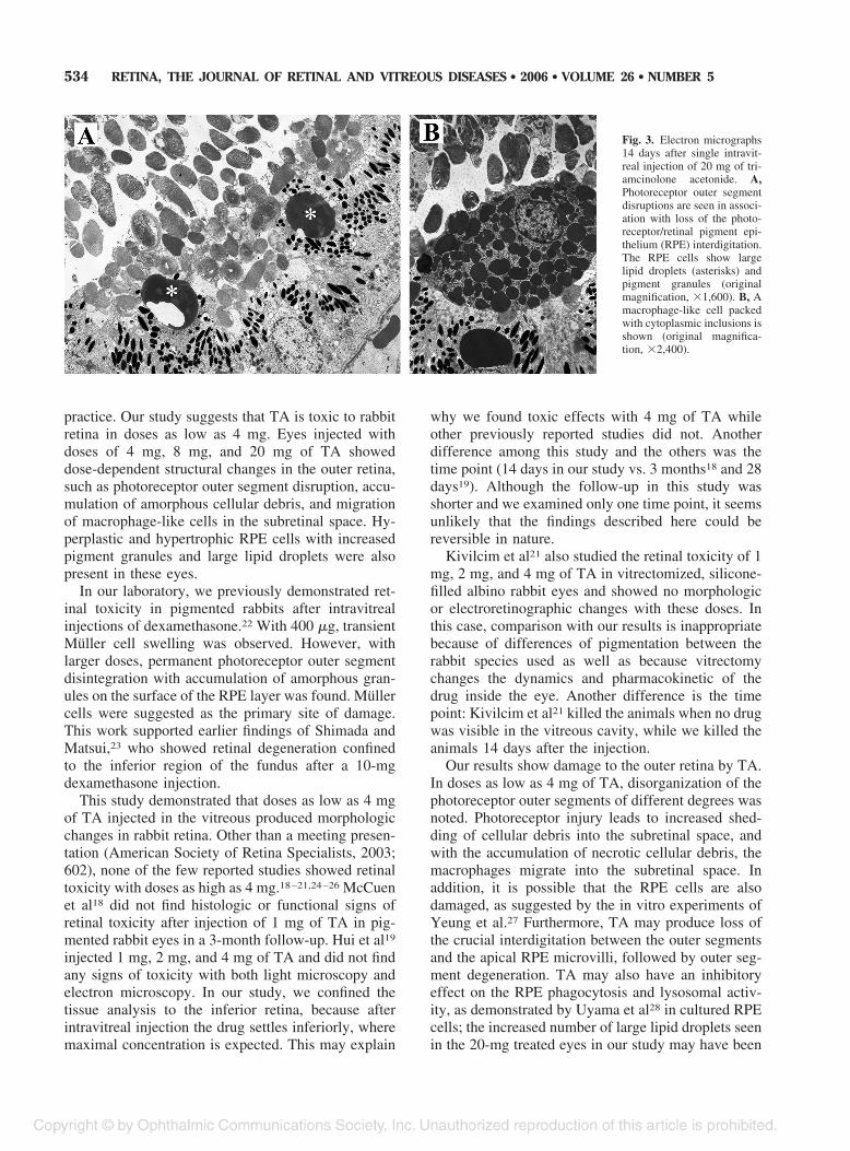
Transmission electron microscopy confirmed thesefindings. Ultrastructures of the eyes injected with 0.5mg and 1 mg of TA, vehicle, and saline were normal.However, 4-mg-, 8-mg-, and 20-mg-injected eyes haddisruption of photoreceptor outer segments with ne-crotic cellular debris within the subretinal space. Mac-rophages in the subretinal space had lamellar andhomogeneous inclusions (Fig. 2), and large lipid drop-lets were seen in the RPE cells. The inner retinaappeared normal. In eyes injected with 20 mg of TA,photoreceptor outer segment disruption with loss ofthe photoreceptor/RPE interdigitation was prominent;clusters of hyperplastic and hypertrophic RPE cellspacked with pigment granules and large oil dropletswere seen (Fig. 3A). Macrophages displayed in-creased cytoplasmic inclusions (Fig. 3B).
Discussion
Recent reports and presentations have shown wide-spread use of intravitreal TA injections despite lack ofconvincing safety data as well as randomized clinicaltrials of long-term efficacy. In this study, we analyzedthe morphologic effects of single intravitreal injec-tions of Kenalog-40 (the TA preparation used in theUnited States) at doses commonly used in clinical
Fig. 1. Light micrographs 14 days after single intravitreal injection ofvehicle (A) and of 4 mg (B) and 20 mg (C) of triamcinolone acetonide.A, Well preserved retinal morphology. B, Macrophage-like cell (arrow)on the retinal pigment epithelium (RPE) surface. C, Increased numberof macrophage-like cells (arrows), prominent clustering of pigmentgranules, and enlarged lipid droplets in the RPE cells (asterisks) (stain,phenylenediamine; original magnification, !400).
Fig. 2. Electron micrograph 14 days after single intravitreal injectionof 4 mg of triamcinolone acetonide. Disruption of photoreceptor outersegments with necrotic cellular debris within the subretinal space anda macrophage in the subretinal space with lamellar inclusions (originalmagnification, !1,600).
533RETINAL TOXICITY OF INTRAVITREAL TA ● YU ET AL
practice. Our study suggests that TA is toxic to rabbitretina in doses as low as 4 mg. Eyes injected withdoses of 4 mg, 8 mg, and 20 mg of TA showeddose-dependent structural changes in the outer retina,such as photoreceptor outer segment disruption, accu-mulation of amorphous cellular debris, and migrationof macrophage-like cells in the subretinal space. Hy-perplastic and hypertrophic RPE cells with increasedpigment granules and large lipid droplets were alsopresent in these eyes.
In our laboratory, we previously demonstrated ret-inal toxicity in pigmented rabbits after intravitrealinjections of dexamethasone.22 With 400 !g, transientMuller cell swelling was observed. However, withlarger doses, permanent photoreceptor outer segmentdisintegration with accumulation of amorphous gran-ules on the surface of the RPE layer was found. Mullercells were suggested as the primary site of damage.This work supported earlier findings of Shimada andMatsui,23 who showed retinal degeneration confinedto the inferior region of the fundus after a 10-mgdexamethasone injection.
This study demonstrated that doses as low as 4 mgof TA injected in the vitreous produced morphologicchanges in rabbit retina. Other than a meeting presen-tation (American Society of Retina Specialists, 2003;602), none of the few reported studies showed retinaltoxicity with doses as high as 4 mg.18–21,24–26 McCuenet al18 did not find histologic or functional signs ofretinal toxicity after injection of 1 mg of TA in pig-mented rabbit eyes in a 3-month follow-up. Hui et al19
injected 1 mg, 2 mg, and 4 mg of TA and did not findany signs of toxicity with both light microscopy andelectron microscopy. In our study, we confined thetissue analysis to the inferior retina, because afterintravitreal injection the drug settles inferiorly, wheremaximal concentration is expected. This may explain
why we found toxic effects with 4 mg of TA whileother previously reported studies did not. Anotherdifference among this study and the others was thetime point (14 days in our study vs. 3 months18 and 28days19). Although the follow-up in this study wasshorter and we examined only one time point, it seemsunlikely that the findings described here could bereversible in nature.
Kivilcim et al21 also studied the retinal toxicity of 1mg, 2 mg, and 4 mg of TA in vitrectomized, silicone-filled albino rabbit eyes and showed no morphologicor electroretinographic changes with these doses. Inthis case, comparison with our results is inappropriatebecause of differences of pigmentation between therabbit species used as well as because vitrectomychanges the dynamics and pharmacokinetic of thedrug inside the eye. Another difference is the timepoint: Kivilcim et al21 killed the animals when no drugwas visible in the vitreous cavity, while we killed theanimals 14 days after the injection.
Our results show damage to the outer retina by TA.In doses as low as 4 mg of TA, disorganization of thephotoreceptor outer segments of different degrees wasnoted. Photoreceptor injury leads to increased shed-ding of cellular debris into the subretinal space, andwith the accumulation of necrotic cellular debris, themacrophages migrate into the subretinal space. Inaddition, it is possible that the RPE cells are alsodamaged, as suggested by the in vitro experiments ofYeung et al.27 Furthermore, TA may produce loss ofthe crucial interdigitation between the outer segmentsand the apical RPE microvilli, followed by outer seg-ment degeneration. TA may also have an inhibitoryeffect on the RPE phagocytosis and lysosomal activ-ity, as demonstrated by Uyama et al28 in cultured RPEcells; the increased number of large lipid droplets seenin the 20-mg treated eyes in our study may have been
Fig. 3. Electron micrographs14 days after single intravit-real injection of 20 mg of tri-amcinolone acetonide. A,Photoreceptor outer segmentdisruptions are seen in associ-ation with loss of the photo-receptor/retinal pigment epi-thelium (RPE) interdigitation.The RPE cells show largelipid droplets (asterisks) andpigment granules (originalmagnification, !1,600). B, Amacrophage-like cell packedwith cytoplasmic inclusions isshown (original magnifica-tion, !2,400).
534 RETINA, THE JOURNAL OF RETINAL AND VITREOUS DISEASES ● 2006 ● VOLUME 26 ● NUMBER 5

caused by the decreased lysosomal activity in the RPEcells.
Some investigators believe that retinal toxicity as-sociated with TA may be due to its vehicle, benzylalcohol.18,29 In our model, eyes injected with vehiclealone did not show any toxicity. Similarly, Hida et al30
and Morrison et al (Association for Research in Visionand Ophthalmology, 2004; 1917) demonstrated noretinal toxicity of benzyl alcohol at the concentrationused in clinical practice, reinforcing our findings. Hidaet al30 also showed no associated toxicity with twicethe concentration of benzyl alcohol found in the Ke-nalog suspension, and Morrison et al (Association forResearch in Vision and Ophthalmology, 2004; 1917)showed toxicity only with higher concentrations("3.3 times). In addition, Yeung et al27 did not detectany in vitro toxic effect of benzyl alcohol on RPEcells. Interestingly, in a recent correspondence byRodriguez-Coleman et al,31 benzyl alcohol was re-ported to partition with triamcinolone crystals as aresult of its preference for a lipophilic environment.Thus, a better control to separate the effects of benzylalcohol from TA would have been a preparation con-taining the exact concentration of benzyl alcohol asdetermined by high-performance liquid chromatogra-phy from the sedimented suspensions of TA, the pro-cedure used in our study to prepare 8-mg and 20-mginjections.
This study was designed bearing two importantissues. First, we used pigmented rabbits because it isknown that melanin granules have affinity for certainlipophilic drugs.32 A drug dose that is safe in an albinoeye may show detrimental effects when applied to apigmented eye. Therefore, the use of pigmented rab-bits for drug toxicity studies may be more relevant tothe human eye. Second, we confined our tissue anal-ysis to the inferior retina, where maximal drug con-centration is expected after intravitreal injection. Wewere not able to affirm that toxic changes are diffuseon the retina, because we only analyzed a small part ofthe inferior retina where the drug was confined, butthe changes we found were throughout the 5 ! 5-mmfragments.
A possible critique of this study may be that we didnot use full-field electroretinography in our study.Although electroretinography is a useful test in ocularpharmacology, our personal experience is that thisexamination presents variable results when trying todetect subtle toxicity changes or localized retinalchanges confined to the drug depot. Our focus was toshow morphologic changes.
Another limitation of our study is that the actual TAconcentration was not measured before injection, andwhen dealing with a suspension of crystals that settle
rapidly, the actual injected doses may not be exactlythe targeted doses. In addition, when preparing the8-mg and 20-mg doses, we did not account for thesmall loss of steroid crystals in the ejected superna-tants. In our study, it was our goal to use a preparationmethod favored in clinical practice, and we were alsoaware from the report by Rodriguez-Coleman et al31
that the method used in our study, sedimentation with-out filtration, produced the closest final dose to thetargeted dose. The more sophisticated filtration meth-ods actually resulted in variable and consistentlylower doses than the predicted range. This was alsoconfirmed by Kreissig et al (Association for Researchin Vision and Ophthalmology, 2005; 4731).
This study showed dose-related toxic effects of TAin rabbit outer retina with doses as low as 4 mg, whilelower doses, such as 0.5 mg and 1 mg, did not causemorphologic retinal changes. Needless to say, thereare many differences, such as species differences,ocular volume differences, and presence of intraocularinflammation, to be taken into consideration before wecan apply these data to clinical practice. The rabbitvitreous volume is one half to one third of that ofhumans, and it is likely that a higher localized con-centration is further achieved with the drug pocketedwithin the overlying formed cortical vitreous in therabbit eyes. Currently, 4 mg is the most commonlyused dose of TA in intravitreal injections, but clini-cians have used higher doses. It is possible that 4 mgin the vitreous cavity of an inflamed human eye isequivalent to 0.5 mg or 1 mg in the rabbit eyes and isthus safe. Our data should caution use of higher dosesof TA and frequent repeated injections until furtherlong-term retinal toxicity studies are performed.
Key words: antiinflammatory agents, corticoste-roids, intravitreal drug delivery.
References
1. Martidis M, Duker JS, Greenberg PB, et al. Intravitrealtriamcinolone for refractory diabetic macular edema. Oph-thalmology 2002;109:920–927.
2. Massin P, Audren F, Haouchine B, et al. Intravitreal triam-cinolone acetonide for diabetic diffuse macular edema: pre-liminary results of a prospective controlled trial. Ophthalmol-ogy 2004;111:218–225.
3. Park CH, Jaffe GJ, Fekrat S. Intravitreal triamcinolone ace-tonide in eyes with cystoid macular edema associated withcentral retinal vein occlusion. Am J Ophthalmol 2003;136:419–425.
4. Conway MD, Canakis C, Livir-Rallatos C, Peyman GA.Intravitreal triamcinolone acetonide for refractory chronicpseudophakic cystoid macular edema. J Cataract Refract Surg2003;29:27–33.
5. Antcliff RJ, Spalton DJ, Stanford MR, et al. Intravitrealtriamcinolone for uveitic cystoid macular edema: an optical
535RETINAL TOXICITY OF INTRAVITREAL TA ● YU ET AL

coherence tomography study. Ophthalmology 2001;108:765–772.
6. Challa JK, Gillies MC, Penfold PL, et al. Exudative maculardegeneration and intravitreal triamcinolone: 18 months fol-low-up. Aust N Z J Ophthalmol 1998;26:277–281.
7. Gillies MC, Simpson JM, Luo W, et al. A randomized clin-ical trial of a single dose of intravitreal triamcinolone ace-tonide for neovascular age-related macular degeneration:one-year results. Arch Ophthalmol 2003;121:667–673.
8. Jonas JB, Hayler JK, Panda-Jona S. Intravitreal injection ofcrystalline cortisone as adjunctive treatment of proliferativevitreoretinopathy. Br J Ophthalmol 2000;84:1064–1067.
9. Martidis A, Duker JS, Puliafito CA. Intravitreal triamcino-lone for refractory cystoid macular edema secondary to bird-shot retinochoroidopathy. Arch Ophthalmol 2001;119:1380–1383.
10. Jonas JB. Intraocular availability of triamcinolone acetonideafter intravitreal injection. Am J Ophthalmol 2004;137:560–562.
11. Ziada G, el-Haddad S, Fatouh M, et al. Radionuclide study ofthe blood ocular barrier. Eur J Drug Metab Pharmacokinet1985;10:325–328.
12. McGhee CN, Dean S, Danesh-Meyer H. Locally adminis-tered ocular corticosteroids: benefits and risks. Drug Saf2000;25:33–55.
13. Gillies MC, Simpson JM, Billson FA, et al. Safety of anintravitreal injection of triamcinolone: results from a random-ized clinical trial. Arch Ophthalmol 2004;122:336–340.
14. Kaushik S, Gupta V, Gupta A, et al. Intractable glaucomafollowing intravitreal triamcinolone in central retinal veinocclusion. Am J Ophthalmol 2004;137:758–760.
15. Moshfeghi DM, Kaiser PK, Scott IU, et al. Acute endoph-thalmitis following intravitreal triamcinolone acetonide injec-tion. Am J Ophthalmol 2003;136:791–796.
16. Nelson ML, Tennant MT, Sivalingam A, et al. Infectious andpresumed noninfectious endophthalmitis after intravitreal tri-amcinolone acetonide injection. Retina 2003;23:686–691.
17. Jonas JB, Kreissig I, Degenring RF. Retinal complication ofintravitreal injections of triamcinolone acetonide. GraefesArch Clin Exp Ophthalmol 2004;242:184–185.
18. McCuen BW 2nd, Bessler M, Tano Y, et al. The lack oftoxicity of intravitreally administered triamcinolone ace-tonide. Am J Ophthalmol 1981;91:785–788.
19. Hui YN, Liang HC, Cai YS, et al. Corticosteroids and dauno-mycin in the prevention of experimental proliferative vitreo-retinopathy induced by macrophages. Graefes Arch Clin Exp
Ophthalmol 1993;231:109–114.20. Danis PR, Bingaman DP, Yang Y, Ladd B. Inhibition of
preretinal and optic nerve head neovascularization in pigs byintravitreal triamcinolone acetonide. Ophthalmology 1996;103:2099–2104.
21. Kivilcim M, Peyman GA, El-Dessouky ES, et al. Retinaltoxicity of triamcinolone acetonide in silicone-filled eyes.Ophthalmic Surg Lasers 2000;31:474–478.
22. Kwak HW, D’Amico DJ. Evaluation of the retinal toxicityand pharmacokinetics of dexamethasone after intravitreal in-jection. Arch Ophthalmol 1992;110:259–266.
23. Shimada H, Matsui M. Effect of intravitreal steroid injectionon rabbit eye. Acta Soc Ophthalmol Jpn 1989;93:501–510.
24. Tano Y, Chandler D, Machemer R. Treatment of intraocularproliferation with intravitreal injection of triamcinolone ace-tonide. Am J Ophthalmol 1980;90:810–816.
25. Chandler DB, Rozakis G, de Juan E Jr, Machemer R. Theeffect of triamcinolone acetonide on a refined experimentalmodel of proliferative vitreoretinopathy. Am J Ophthalmol1985;99:686–690.
26. Ishibashi T, Miki K, Sorgente N, et al. Effects of intravitrealadministration of steroids on experimental subretinal neovas-cularization in the subhuman primate. Arch Ophthalmol1985;103:708–711.
27. Yeung CK, Chan KP, Chiang SW, et al. The toxic and stressresponses of cultured human retinal pigment epithelium(ARPE19) and human glial cells (SVG) in the presence oftriamcinolone. Invest Ophthalmol Vis Sci 2003;44:5293–5300.
28. Uyama M, Sugasawa K, Kishimoto N, Kawahara S. Effect ofcorticosteroid on porcine retinal pigment epithelial cells inculture-2. Effects on phagocytosis and lysosomal activity. JJpn Ophthalmol Soc 2000;104:86–90.
29. Roth DB, Cheieh J, Spirn MJ, et al. Noninfectious endoph-thalmitis associated with intravitreal triamcinolone injection.Arch Ophthalmol 2003;121:1279–1282.
30. Hida T, Chandler D, Arena JE, Machemer R. Experimentaland clinical observations of the intraocular toxicity of com-mercial corticosteroid preparations. Am J Ophthalmol 1986;101:190–195.
31. Rodriguez-Coleman H, Peng Y, Kim H, et al. Intravitrealinjection of triamcinolone for diffuse macular edema. ArchOphthalmol 2004;122:1085–1086.
32. Dayhaw-Barker P. Retinal pigment epithelium melanin andocular toxicity. Int J Toxicol 2002;21:451–454.
536 RETINA, THE JOURNAL OF RETINAL AND VITREOUS DISEASES ● 2006 ● VOLUME 26 ● NUMBER 5


![Clinical Study Comparison between Intravitreal ...downloads.hindawi.com/journals/isrn/2013/141279.pdfcommon cause of retinal vascular disease following diabetic retinopathy [ ]. Among](https://img.pdfslide.net/doc/110x75/5e881482d74bac340b063934/clinical-study-comparison-between-intravitreal-common-cause-of-retinal-vascular.jpg)