Embed Size (px)
Citation preview
F8V W
Brit. Y. Ophthal. (1976) 6o, 504
Retinal vascular changes in bull's eye maculopathy
AMRESH CHOPDARFrom the Fundus Clinic, Moorfields Eye Hospital, London
Chloroquine toxicity produces a typical maculardegeneration commonly described as 'bull's eye'retinopathy (Hobbs, Sorsby, and Freedman, I959;Kearns and Hollenhorst, I966). Since its recognition,similar macular appearances have been describedin various other conditions without ingestion ofchloroquine (Krill and Deutman, 1972; Deutman,I974; Weise and Yannuzzi, 1974). One such caseis described in this paper and the possible natureof the aetiology is discussed.
Case report
A 51-year-old White woman was seen in the fundusclinic of Moorfields Eye Hospital. She had a history ofslow progressive loss of central vision in the right eye
during the previous I2 months, but had not found itdifficult to adjust to the dim light. Her only son has alsobeen examined and found to have no ocular disease. Inthe past she had taken fenfluramine to control obesityand at the time of presentation and for the previous fouryears was taking methaqualone.
OCULAR FINDINGS
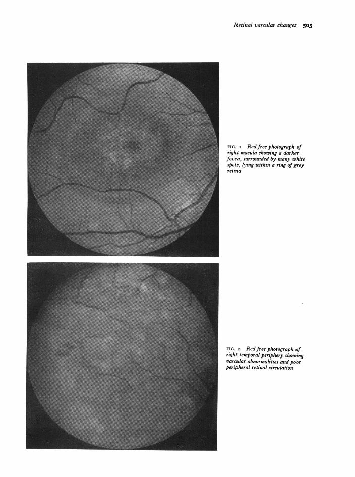
The visual acuity was counting fingers in the right and6/9 in the left eye. The external slit-lamp examination,including ocular motility, were normal. The fundishowed identical involvement at each macula. Thecentral macular area looked a deeper red than normaland surrounding it a halo-like zone could be seen withmany white, shiny, dot-like particles, giving the typical'bull's eye' phenomenon usually seen in chloroquineretinopathy (Fig. i). These white dots were found tobe retinal pigment epithelial defects when examinedwith a fundus viewing contact lens. The circle ofpunctate epithelial defect was in turn encircled by a
ring of grey retina through which the underlyingchoroidal vessels could be seen with little difficulty.The peripheral retina in the temporal part of the righteye showed sheathing of vessels with tortuosity andarterio-venous anastomosis. Peripheral to the area ofanastomosis there was an abrupt end to the retinalcirculation; this was evident from the change in colourbetween the two zones. The poorly perfused peripheralretina showed several areas of round atrophic patcheswith pigmented edges (Fig. 2). The left peripheral retinashowed sheathing and tortuosity, but no arterio-venousanastomosis.
Address for reprints: A. Chopdar, FRCS, Fundus Clinic, MoorfieldsEye Hospital, High Holborn, London WCIV 7AN
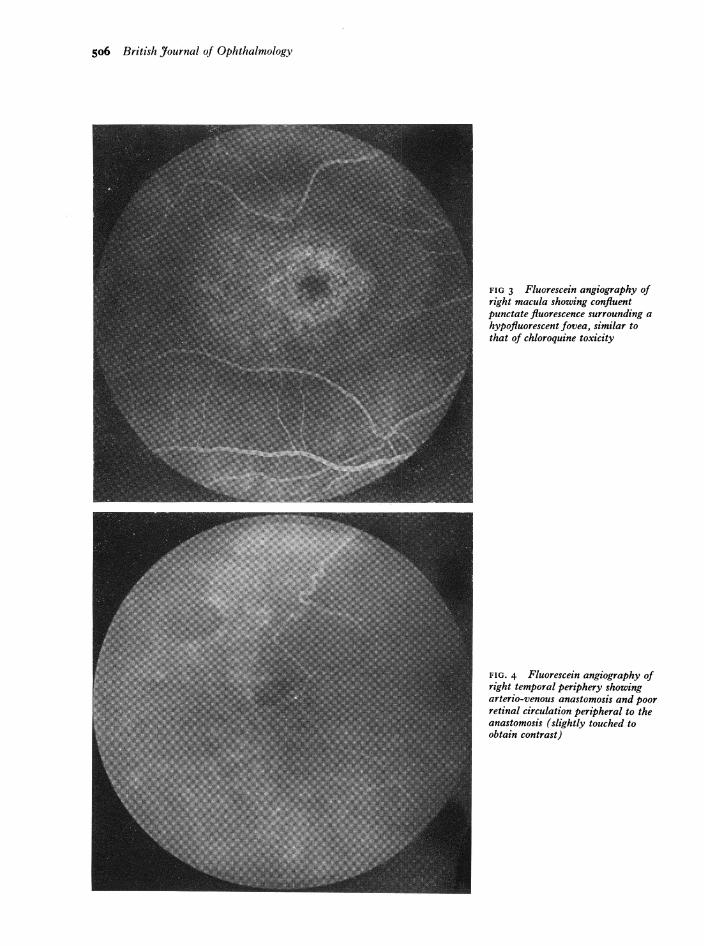
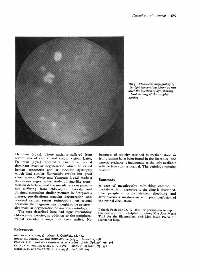
Fluorescein angiography showed a confluent punctatefluorescence surrounding a hypofluorescent macula.Around this complex was a faint fluorescent ring cor-responding with the grey circle seen clinically (Fig. 3).The peripheral retina in the right eye showed arterio-venous anastomosis and demonstrated the abruptcessation of the retinal circulation. The round atrophicpatches showed transmission defects during the earlystages and scleral staining during the later frames(Figs 4, 5).
RETINAL FUNCTION TESTS
The peripheral field showed some constriction in eacheye. There was a central scotoma in the right eye butno such defect was seen in the left. The colour visionwas normal in the left eye but could not be testedsatisfactorily in the right owing to gross reduction ofvision. The electroretinogram showed a scotopic B waveof I30 mV in the right and 200 mV in the left eye.The photopic response of the right eye was presentwith a good flicker response but the amplitude wassignificantly lower than in the left eye. Similarly theelectro-oculogram was only I55 per cent in the rightand i8o per cent in the left eye.
GENERAL PHYSICAL EXAMINATION
The patient was moderately obese with a blood pressureof I30/80 mmHg. The cardiovascular, respiratory,abdominal, and central nervous systems were all normal.
LABORATORY INVESTIGATIONS
The haematological examination showed haemoglobin15 g/Ioo ml, packed cell volume 42 per cent, mean cellhaemoglobin concentration 36 per cent, total white cellcount gooo/mm3, neutrophils 57 per cent, eosinophils3 per cent, lymphocytes 33 per cent, monocytes 7 percent, erythrocyte sedimentation rate 5 mm in I hour(Westergren), and no gross abnormality in the red bloodcells. The biochemical estimation of total serum proteinwas 7-2 g/Ioo ml, albumin 4-2 g/ioo ml, globulin 3-0 g/IOO ml with a normal electrophoretic pattern. Theimmunoglobulin assay showed a slight increase of IgMat 325 IU/ml but normal values for IgG, IgA, and IgD.Serological tests for syphilis and Reiter's syndromewere negative.
DiscussionMaculopathy mimicking chloroquine toxicity incases of cone dystrophy was described by Krill and
Retinal vascular changes 505
FIG. i Redfree photograph ofright macula showing a darkerfovea, surrounded by many whitespots, lying within a ring of greyretina
FIG. 2 Red free photograph ofright temporal periphery showingvascular abnormalities and poorperipheral retinal circulation
506 British Journal of Ophthalmology
FIG 3 Fluorescein angiography ofright macula showing confluentpunctate fluorescence surrounding ahypofluorescent fovea, similar tothat of chloroquine toxicity
FIG. 4 Fluorescein angiography ofright temporal periphery showingarterio-venous anastomosis and poorretinal circulation peripheral to theanastomosis (slightly touched toobtain contrast)
Retinal vascular changes 507
FIG 5 Fluorescein angiography ofthe right temporal periphery i o minafter the injection of dye, showingscleral staining of the atrophicpatches
Deutman (I 972). These patients suffered fromsevere loss of central and colour vision. Later,Deutman (I 974) reported a case of autosomaldominant macular degeneration which he calledbenign concentric annular macular dystrophywhich had similar fluorescein results but goodvisual acuity. Weise and Yannuzzi (1974) made afluorescein angiographic study of ring-like trans-mission defects around the macular area in patientsnot suffering from chloroquine toxicity andobtained somewhat similar pictures in Stargardt'sdisease, pre-disciform macular degeneration, andresolved central serous retinopathy; on severaloccasions the diagnosis was thought to be progres-sive macular degeneration of unknown aetiology.The case described here had signs resembling
chloroquine toxicity, in addition to the peripheralretinal vascular changes not seen earlier. No
instances of toxicity ascribed to methaqualone orfenfluramine have been found in the literature, andgenetic evidence is inadequate as the only availablerelative (the son) is normal. The aetiology remainsobscure.
SummaryA case of maculopathy mimicking chloroquinetoxicity without exposure to the drug is described.The peripheral retina showed sheathing andarterio-venous anastomosis with poor perfusion ofthe retinal circulation.
I thank Professor D. W. Hill for permission to reportthis case and for his helpful criticism; Mrs Ann MarieTurk for the illustrations, and Mrs Joyce Preen forsecretarial help.
References
DEUTMAN, A. F. (1974) Amer. J3. Ophthal., 78, 384HOBBS, H., SORSBY, A., and FREEDMAN, A. (1959) Lancet, 2, 478KEARNS, T. P., and HOLLENHORST, R. W. (I966) Arch. Ophthal., 76, 378KRILL, A. E., and DEUTMAN, A. F. (I972) Amer. J. Ophthal., 73, 352WEISE, E. E., and YANNUZZI, L. A. (1974) Ibid., 78, 204