Embed Size (px)
Citation preview
Retinoic acid induces Sertoli cell paracrine signals forspermatogonia differentiation but cell autonomouslydrives spermatocyte meiosisMathilde Raverdeaua, Aurore Gely-Pernota, Betty Féreta, Christine Dennefelda, Gérard Benoitb, Irwin Davidsona,Pierre Chambona,1, Manuel Marka,c, and Norbert B. Ghyselincka,1
aInstitut de Génétique et de Biologie Moléculaire et Cellulaire, Centre National de la Recherche Scientifique Unité Mixte de Recherche 7104, Institut Nationalde la Santé et de la Recherche Médicale Unité 964, Université de Strasbourg, F-67404 Illkirch Cedex, France; bCentre de Génétique et de PhysiologieMoléculaire et Cellulaire, Centre National de la Recherche Scientifique Unité Mixte de Recherche 5534, Université de Lyon 1, F-69622 Villeurbanne Cedex,France; and cHôpitaux Universitaires de Strasbourg, F-67091 Strasbourg Cedex, France
Contributed by Pierre Chambon, August 29, 2012 (sent for review July 26, 2012)
Direct evidence for a role of endogenous retinoic acid (RA), the activemetabolite of vitamin A in the initial differentiation andmeiotic entryof spermatogonia, and thus in the initiation of spermatogenesis is stilllacking. RA is synthesized by dedicated enzymes, the retinaldehydedehydrogenases (RALDH), and binds to and activates nuclear RAreceptors (RARA, RARB, and RARG) either within the RA-synthesizingcells or in the neighboring cells. In the present study, we have useda combination of somatic genetic ablations and pharmacological ap-proaches in vivo to show that during the first, prepubertal, spermato-genic cycle (i) RALDH-dependent synthesis of RA by Sertoli cells (SC),the supporting cells of the germ cell (GC) lineage, is indispensable toinitiate differentiation of A aligned into A1 spermatogonia; (ii) RARAin SC mediates the effects of RA, possibly through activating Mafbexpression, a gene whose Drosophila homolog is mandatory to GCdifferentiation; (iii) RA synthesized by premeiotic spermatocytes cellautonomously induces meiotic initiation through controlling the RAR-dependent expression of Stra8. Furthermore, we show that RA of SCorigin is no longer necessary for the subsequent spermatogenic cyclesbut essential to spermiation. Altogether, our data establish that theeffects of RA in vivo on spermatogonia differentiation are indirect, viaSC, but direct on meiotic initiation in spermatocytes, supportingthereby the notion that, contrary to the situation in the female, RAis necessary to induce meiosis in the male.
mouse | mutagenesis | retinoid antagonist | RAR/RXR heterodimer
Spermatogenesis is a complex and tightly regulated cell dif-ferentiation process, yielding mature spermatozoa from
spermatogonia stem cells. Spermatogonia in the single-cell state,known as A single (As) spermatogonia, have traditionally beenconsidered as spermatogonia stem cells in the mouse. Upon di-vision, As spermatogonia give rise to two paired A (Ap) sper-matogonia, then to a chain of 4–32 aligned (Aal) spermatogonia.As, Ap, and Aal are referred to as “undifferentiated spermato-gonia” because they all retain stem cell properties (1). Sub-sequently Aal cells differentiate into A1 spermatogonia, whichare irreversibly committed toward gamete production. They un-dergo a series of divisions generating B spermatogonia, whichdivide once more to yield premeiotic (i.e., preleptotene) sper-matocytes (2). The entire process of spermatogenesis relies onfunctional interactions between germ cells (GC) and somatic,supporting cells, called Sertoli cells (SC), involving a complexassortment of hormones and cytokines, among which retinoic acid(RA), the biologically active form of vitamin A (retinol). In fact,vitamin A-deficient (VAD) mice are infertile because of an arrestof spermatogonia differentiation at the Aal–A1 transition, andtreating them with either retinol or RA results in the completerecovery of spermatogenesis (3). As RA can trigger meiosis infemale GC through initiating Stra8 expression (4, 5) and canadditionally induce Stra8 expression inmale GC (3), the paradigmhas become that RA is required for meiotic initiation. However,
a recent study showing that female GC can enter meiosis in a fetalovary devoid of RA has challenged this model (6).During embryonic development RA usually acts in a paracrine
manner, one cell type controlling its synthesis, whereas a neigh-bor cell type responds to the signal (7). In cells synthesizing RA,conversion of retinol to its active metabolite depends upon ret-inaldehyde dehydrogenases (RALDH1 to RALDH3 encoded byAldh1a1 to Aldh1a3 genes). In responding cells, RA binds to andactivates nuclear RA receptors (RARA, RARB, and RARGisotypes), which are ligand-dependent transcriptional regulators.They usually function in the form of heterodimers with rexinoidreceptors (RXRA, RXRB, and RXRG isotypes) and controlexpression of target genes through binding to RA response ele-ments (RARE) located in the vicinity of the promoter (8). At theonset of spermatogenesis in the developing testis, Aldh1a1 andAldh1a2 are expressed in SC, whereas Rara and Rarg areexpressed in SC and Aal spermatogonia, respectively (9, 10).These expression patterns raise the possibility that RA triggeringthe differentiation of spermatogonia could be synthesized by SCand acts either in an autocrine manner through cell autono-mously activating RARA-dependent events in SC or in a para-crine manner through activating RARG in Aal spermatogonia,which then become committed toward meiosis.Thus, although RA seems to play a central role in the differ-
entiation of spermatogonia, the mechanisms driving RA avail-ability in these cells remain largely unknown. Moreover, it has notyet been established whether the effects of RA on spermatogoniadifferentiation are cell autonomous or mediated by SC, and theRA-controlled genetic cascades need to be clarified. In addition,direct evidence supporting a role for RA in meiotic entry of maleGC during puberty is still lacking. In the present study, we provideanswers to these questions through the phenotypic analysis ofmice lacking all RALDH activities specifically in SC.
Results and DiscussionAblation of RALDH in SC Impairs Differentiation of A1 Spermatogonia.To generate Aldh1a1-3Ser−/− mutants, in which the RALDHgenes were excised only in SC, mice carrying loxP-flanked allelesof the Aldh1a1, Aldh1a2, and Aldh1a3 genes (11–13) werecrossed with mice bearing the Amh-Cre transgene, which isexpressed from embryonic day 15 onward (14) and therefore
Author contributions: M.R., A.G.-P., M.M., and N.B.G. designed research; M.R., A.G.-P., B.F.,and C.D. performed research; G.B. and I.D. contributed new reagents/analytic tools; M.R.,A.G.-P., G.B., I.D., M.M., and N.B.G. analyzed data; and M.R., P.C., M.M., and N.B.G. wrotethe paper.
The authors declare no conflict of interest.1To whom correspondence may be addressed. E-mail: [email protected] or [email protected].
This article contains supporting information online at www.pnas.org/lookup/suppl/doi:10.1073/pnas.1214936109/-/DCSupplemental.
16582–16587 | PNAS | October 9, 2012 | vol. 109 | no. 41 www.pnas.org/cgi/doi/10.1073/pnas.1214936109
Dow
nloa
ded
by g
uest
on
June
6, 2
020
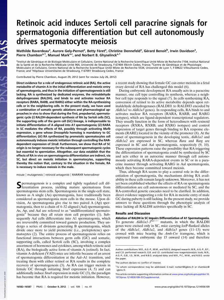
excises RALDH coding sequence before the onset of spermato-genesis (2). At postnatal day 5 (PN5), no histological defects weredetected in Aldh1a1-3Ser−/− mutant testes (n = 3) that containedspermatogonia and SC as their wild-type (WT) counterparts (Fig.1 A and B). However, the expression of Sohlh1, Sohlh2, and Kitgenes normally present in differentiated spermatogonia (15, 16)was significantly decreased in Aldh1a1-3Ser−/− testes, whereasexpression of Ngn3, Nanos3, Ret, Zbtb16, and Rarg normallypresent in undifferentiated spermatogonia (17–20) was normal(Fig. S1A), indicating a delay in the process of spermatogoniadifferentiation in the absence of RALDH in SC. At PN15 andPN40 (n = 3 testes for each age and genotype), the seminiferoustubules of Aldh1a1-3Ser−/− mutants still contained only sper-matogonia and SC (Fig. 1 D and F), whereas those of WT con-trols displayed normal spermatogenesis (Fig. 1 C and E). Theabsence of meiotic cells in Aldh1a1-3Ser−/− mutant testes wasfurther assessed by the lack of Rec8 expression (Fig. 1 G and H).To characterize the spermatogonia in Aldh1a1-3Ser−/− testes atPN40 (n = 3 for each genotype), we analyzed the expressionpattern of genes that are characteristic of given stages of differ-entiation, by in situ hybridization (ISH) analyses (Fig. S1) andwhole-mount immunohistochemistry (IHC) (Fig. 1 I–P). BothWT and Aldh1a1-3Ser−/− seminiferous tubules contained Ret-(Fig. S1) and GFRA1-positive (Fig. 1 I and J) spermatogonia,which are characteristic of As and Ap stages (19). A largenumber of spermatogonia in Aldh1a1-3Ser−/− seminiferoustubules expressed ZBTB16 and RARG (Fig. 1 K–N; Fig. S1),which are markers of Aal spermatogonia (10, 21). In contrast,KIT, which is expressed in A1 to B spermatogonia, was never
detected in the seminiferous tubules of Aldh1a1-3Ser−/− mutants(Fig. 1 O and P; Fig. S1). Thus, similarly to the situation found inVAD (3), spermatogonia in Aldh1a1-3Ser−/− mutants do notprogress beyond the Aal to A1 transition, and the apparent highnumber of Aal spermatogonia (Fig. 1 L and N) likely reflectsinhibition of their differentiation into A1 spermatogonia. Theseresults indicate that ablation of RALDH in SC blocks sper-matogonia differentiation at the Aal stage, thereby preventingthe initiation of the first wave of spermatogenesis. We concludethat, if RA is actually the product generated by RALDH and isrequired to initiate spermatogonia differentiation, it must besynthesized by SC.
RA-Activated RARA in SC Drives the Initial Transition from Aal to A1Spermatogonia, Possibly Through Cell Autonomously InducingExpression of Mafb. To show that impairment of spermatogoniadifferentiation was caused by a lack of RA in Aldh1a1-3Ser−/−
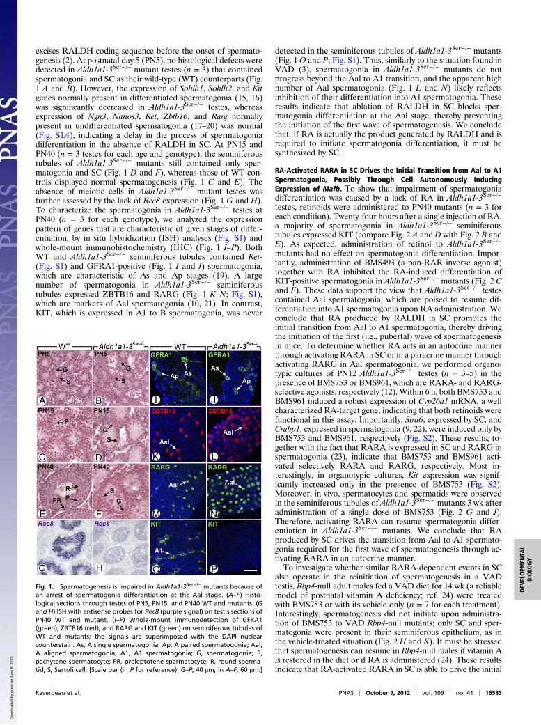
testes, retinoids were administered to PN40 mutants (n = 3 foreach condition). Twenty-four hours after a single injection of RA,a majority of spermatogonia in Aldh1a1-3Ser−/− seminiferoustubules expressed KIT (compare Fig. 2 A and D with Fig. 2 B andE). As expected, administration of retinol to Aldh1a1-3Ser−/−
mutants had no effect on spermatogonia differentiation. Impor-tantly, administration of BMS493 (a pan-RAR inverse agonist)together with RA inhibited the RA-induced differentiation ofKIT-positive spermatogonia in Aldh1a1-3Ser−/− mutants (Fig. 2 Cand F). These data support the view that Aldh1a1-3Ser−/− testescontained Aal spermatogonia, which are poised to resume dif-ferentiation into A1 spermatogonia upon RA administration. Weconclude that RA produced by RALDH in SC promotes theinitial transition from Aal to A1 spermatogonia, thereby drivingthe initiation of the first (i.e., pubertal) wave of spermatogenesisin mice. To determine whether RA acts in an autocrine mannerthrough activating RARA in SC or in a paracrine manner throughactivating RARG in Aal spermatogonia, we performed organo-typic cultures of PN12 Aldh1a1-3Ser−/− testes (n = 3–5) in thepresence of BMS753 or BMS961, which are RARA- and RARG-selective agonists, respectively (12).Within 6 h, both BMS753 andBMS961 induced a robust expression of Cyp26a1 mRNA, a wellcharacterized RA-target gene, indicating that both retinoids werefunctional in this assay. Importantly, Stra6, expressed by SC, andCrabp1, expressed in spermatogonia (9, 22), were induced only byBMS753 and BMS961, respectively (Fig. S2). These results, to-gether with the fact that RARA is expressed in SC and RARG inspermatogonia (23), indicate that BMS753 and BMS961 acti-vated selectively RARA and RARG, respectively. Most in-terestingly, in organotypic cultures, Kit expression was signif-icantly increased only in the presence of BMS753 (Fig. S2).Moreover, in vivo, spermatocytes and spermatids were observedin the seminiferous tubules of Aldh1a1-3Ser−/− mutants 3 wk afteradministration of a single dose of BMS753 (Fig. 2 G and J).Therefore, activating RARA can resume spermatogonia differ-entiation in Aldh1a1-3Ser−/− mutants. We conclude that RAproduced by SC drives the transition from Aal to A1 spermato-gonia required for the first wave of spermatogenesis through ac-tivating RARA in an autocrine manner.To investigate whether similar RARA-dependent events in SC
also operate in the reinitiation of spermatogenesis in a VADtestis, Rbp4-null adult males fed a VAD diet for 14 wk (a reliablemodel of postnatal vitamin A deficiency; ref. 24) were treatedwith BMS753 or with its vehicle only (n = 7 for each treatment).Interestingly, spermatogenesis did not initiate upon administra-tion of BMS753 to VAD Rbp4-null mutants; only SC and sper-matogonia were present in their seminiferous epithelium, as inthe vehicle-treated situation (Fig. 2 H and K). It must be stressedthat spermatogenesis can resume in Rbp4-null males if vitamin Ais restored in the diet or if RA is administered (24). These resultsindicate that RA-activated RARA in SC is able to drive the initial
Fig. 1. Spermatogenesis is impaired in Aldh1a1-3Ser−/− mutants because ofan arrest of spermatogonia differentiation at the Aal stage. (A–F) Histo-logical sections through testes of PN5, PN15, and PN40 WT and mutants. (Gand H) ISH with antisense probes for Rec8 (purple signal) on testis sections ofPN40 WT and mutant. (I–P) Whole-mount immunodetection of GFRA1(green), ZBTB16 (red), and RARG and KIT (green) on seminiferous tubules ofWT and mutants; the signals are superimposed with the DAPI nuclearcounterstain. As, A single spermatogonia; Ap, A paired spermatogonia; Aal,A aligned spermatogonia; A1, A1 spermatogonia; G, spermatogonia; P,pachytene spermatocyte; PR, preleptotene spermatocyte; R, round sperma-tid; S, Sertoli cell. [Scale bar (in P for reference): G–P, 40 μm; in A–F, 60 μm.]
Raverdeau et al. PNAS | October 9, 2012 | vol. 109 | no. 41 | 16583
DEV
ELOPM
ENTA
LBIOLO
GY
Dow
nloa
ded
by g
uest
on
June
6, 2
020
transition from Aal to A1 spermatogonia, but not the followingones and suggest that RA needs to act elsewhere in the seminif-erous epithelium to induce subsequent cycles of spermatogenesis.The neonatal testis at PN6 contains two distinct populations of
undifferentiated GC: gonocyte-derived undifferentiated sper-
matogonia and gonocytes that are able to differentiate directlyinto A1 spermatogonia to initiate the first prepubertal wave ofspermatogenesis (2, 25). The signals underlying the fate of thesetwo subpopulations were thought to be under the control ofsomatic cells but yet unidentified (25). According to our study, itis now conceivable that the signal committing gonocytes to dif-ferentiation as A1 spermatogonia relies on RA-activated RARAin SC. However, undifferentiated spermatogonia also rely on RAas attested by the requirement of retinol (or RA) to resumespermatogenesis in VAD adult males (3, 24). The Aal-to-A1transitions during all but the first wave of spermatogenesis maydepend on RAR-dependent signals distinct from those con-trolled by RARA in SC for commitment of gonocytes.To gain insights into the genetic cascade controlled by RARA
in SC, we treated organotypic cultures (n = 4) of PN12 Aldh1a1-3Ser−/− testes with BMS753 for 2 h in the presence of thetranslation inhibitor cycloheximide, thereby allowing changes inthe expression of only direct target genes. Microarray expressionprofiling identified only a few genes that were deregulated two-fold or more upon BMS753 treatment, among which was Mafb(formerly Kreisler). We confirmed by reverse transcription–quantitative PCR (RT-qPCR) that expression of Mafb mRNAwas strongly increased upon short-term activation of RARA(Fig. 2M). In addition, MAFB was detected by IHC in the SCnuclei of Aldh1a1-3Ser−/− mutants treated with BMS753 for 8 h(n = 5), but not in nuclei from testes exposed to vehicle (Fig. 2 Iand L). Importantly, chromatin immunoprecipitation assays witha pan-RAR antibody performed on cultured MSC-1 SC (26)treated for 2 h with RA revealed a robust RAR-binding site atthe end of the Mafb coding region, indicating that this gene isa direct target of RA-activated RARA in SC (Fig. 2N). Becauseno consensus RARE was found in the sequence, the motif onwhich RARA binds remains unknown. This motif is unlikely torecruit RARA/RXR heterodimers because RARA acts in SCthrough a noncanonical pathway, independently of RXR (9).Control of Mafb expression by RA-activated RARA in SC seemsparticularly relevant to spermatogonia differentiation becausethe MAFB transcription factor often instructs cell lineage com-mitment (27) and because its Drosophila ortholog Traffic Jam isrequired, in somatic cells of the fly gonad for GC differentiation(28). The involvement of MAFB in the RA-dependent differ-entiation of spermatogonia is, however, not testable througha genetic approach in the mouse as null mutants are not viable(29, 30), conditional alleles of Mafb are not available, and micebearing the chemically induced Kr mutation (31) are deficient inMAFB only in the hindbrain region (32).
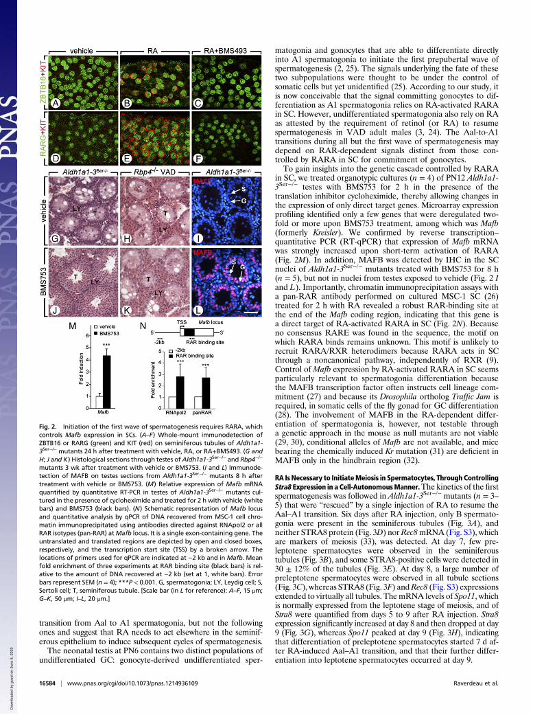
RA Is Necessary to InitiateMeiosis in Spermatocytes, ThroughControllingStra8 Expression in a Cell-AutonomousManner.The kinetics of the firstspermatogenesis was followed in Aldh1a1-3Ser−/− mutants (n = 3–5) that were “rescued” by a single injection of RA to resume theAal–A1 transition. Six days after RA injection, only B spermato-gonia were present in the seminiferous tubules (Fig. 3A), andneither STRA8 protein (Fig. 3D) nor Rec8mRNA (Fig. S3), whichare markers of meiosis (33), was detected. At day 7, few pre-leptotene spermatocytes were observed in the seminiferoustubules (Fig. 3B), and some STRA8-positive cells were detected in30 ± 12% of the tubules (Fig. 3E). At day 8, a large number ofpreleptotene spermatocytes were observed in all tubule sections(Fig. 3C), whereas STRA8 (Fig. 3F) andRec8 (Fig. S3) expressionsextended to virtually all tubules. ThemRNA levels of Spo11, whichis normally expressed from the leptotene stage of meiosis, and ofStra8 were quantified from days 5 to 9 after RA injection. Stra8expression significantly increased at day 8 and then dropped at day9 (Fig. 3G), whereas Spo11 peaked at day 9 (Fig. 3H), indicatingthat differentiation of preleptotene spermatocytes started 7 d af-ter RA-induced Aal–A1 transition, and that their further differ-entiation into leptotene spermatocytes occurred at day 9.
Fig. 2. Initiation of the first wave of spermatogenesis requires RARA, whichcontrols Mafb expression in SCs. (A–F) Whole-mount immunodetection ofZBTB16 or RARG (green) and KIT (red) on seminiferous tubules of Aldh1a1-3Ser−/− mutants 24 h after treatment with vehicle, RA, or RA+BMS493. (G andH; J and K) Histological sections through testes of Aldh1a1-3Ser−/− and Rbp4−/−
mutants 3 wk after treatment with vehicle or BMS753. (I and L) Immunode-tection of MAFB on testes sections from Aldh1a1-3Ser−/− mutants 8 h aftertreatment with vehicle or BMS753. (M) Relative expression of Mafb mRNAquantified by quantitative RT-PCR in testes of Aldh1a1-3Ser−/− mutants cul-tured in the presence of cycloheximide and treated for 2 h with vehicle (whitebars) and BMS753 (black bars). (N) Schematic representation of Mafb locusand quantitative analysis by qPCR of DNA recovered from MSC-1 cell chro-matin immunoprecipitated using antibodies directed against RNApol2 or allRAR isotypes (pan-RAR) atMafb locus. It is a single exon-containing gene. Theuntranslated and translated regions are depicted by open and closed boxes,respectively, and the transcription start site (TSS) by a broken arrow. Thelocations of primers used for qPCR are indicated at −2 kb and in Mafb. Meanfold enrichment of three experiments at RAR binding site (black bars) is rel-ative to the amount of DNA recovered at −2 kb (set at 1, white bars). Errorbars represent SEM (n = 4); ***P < 0.001. G, spermatogonia; LY, Leydig cell; S,Sertoli cell; T, seminiferous tubule. [Scale bar (in L for reference): A–F, 15 μm;G–K, 50 μm; I–L, 20 μm.]
16584 | www.pnas.org/cgi/doi/10.1073/pnas.1214936109 Raverdeau et al.
Dow
nloa
ded
by g
uest
on
June
6, 2
020
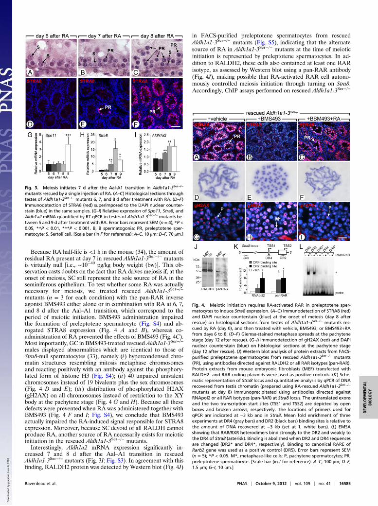
Because RA half-life is <1 h in the mouse (34), the amount ofresidual RA present at day 7 in rescued Aldh1a1-3Ser−/− mutantsis virtually null [i.e., ∼10−40 pg/kg body weight (bw)]. This ob-servation casts doubts on the fact that RA drives meiosis if, at theonset of meiosis, SC still represent the sole source of RA in theseminiferous epithelium. To test whether some RA was actuallynecessary for meiosis, we treated rescued Aldh1a1-3Ser−/−
mutants (n = 3 for each condition) with the pan-RAR inverseagonist BMS493 either alone or in combination with RA at 6, 7,and 8 d after the Aal–A1 transition, which correspond to theperiod of meiotic initiation. BMS493 administration impairedthe formation of preleptotene spermatocyte (Fig. S4) and ab-rogated STRA8 expression (Fig. 4 A and B), whereas co-administration of RA prevented the effects of BMS493 (Fig. 4C).Most importantly, GC in BMS493-treated rescued Aldh1a1-3Ser−/−
males displayed abnormalities which are identical to those ofStra8-null spermatocytes (33), namely (i) hypercondensed chro-matin structures resembling mitosis metaphase chromosomesand reacting positively with an antibody against the phosphory-lated form of histone H3 (Fig. S4); (ii) 40 unpaired univalentchromosomes instead of 19 bivalents plus the sex chromosomes(Fig. 4 D and E); (iii) distribution of phosphorylated H2AX(gH2AX) on all chromosomes instead of restriction to the XYbody at the pachytene stage (Fig. 4 G and H). Because all thesedefects were prevented when RA was administered together withBMS493 (Fig. 4 F and I; Fig. S4), we conclude that BMS493actually impaired the RA-induced signal responsible for STRA8expression. Moreover, because SC devoid of all RALDH cannotproduce RA, another source of RA necessarily exists for meioticinitiation in the rescued Aldh1a1-3Ser−/− mutants.Interestingly, Aldh1a2 mRNA expression significantly in-
creased 7 and 8 d after the Aal–A1 transition in rescuedAldh1a1-3Ser−/− mutants (Fig. 3I; Fig. S3). In agreement with thisfinding, RALDH2 protein was detected by Western blot (Fig. 4J)
in FACS-purified preleptotene spermatocytes from rescuedAldh1a1-3Ser−/− mutants (Fig. S5), indicating that the alternatesource of RA in Aldh1a1-3Ser−/− mutants at the time of meioticinitiation is represented by preleptotene spermatocytes. In ad-dition to RALDH2, these cells also contained at least one RARisotype, as assessed by Western blot using a pan-RAR antibody(Fig. 4J), making possible that RA-activated RAR cell autono-mously controlled meiosis initiation through turning on Stra8.Accordingly, ChIP assays performed on rescued Aldh1a1-3Ser−/−
Fig. 3. Meiosis initiates 7 d after the Aal-A1 transition in Aldh1a1-3Ser−/−
mutants rescued by a single injection of RA. (A–C) Histological sections throughtestes of Aldh1a1-3Ser−/− mutants 6, 7, and 8 d after treatment with RA. (D–F)Immunodetection of STRA8 (red) superimposed to the DAPI nuclear counter-stain (blue) in the same samples. (G–I) Relative expression of Spo11, Stra8, andAldh1a2 mRNA quantified by RT-qPCR in testes of Aldh1a1-3Ser−/− mutants be-tween 5 and 9 d after treatment with RA. Error bars represent SEM (n = 4); *P <0.05, **P < 0.01, ***P < 0.001. B, B spermatogonia; PR, preleptotene sper-matocyte; S, Sertoli cell. [Scale bar (in F for reference): A–C, 10 μm; D–F, 70 μm.]
Fig. 4. Meiotic initiation requires RA-activated RAR in preleptotene sper-matocytes to induce Stra8 expression. (A–C) Immunodetection of STRA8 (red)and DAPI nuclear counterstain (blue) at the onset of meiosis (day 8 afterrescue) on histological sections from testes of Aldh1a1-3Ser−/− mutants res-cued by RA (day 0), and then treated with vehicle, BMS493, or BMS493+RAfrom days 6 to 8. (D–F) Giemsa-stained metaphase spreads at the pachytenestage (day 12 after rescue). (G–I) Immunodetection of gH2AX (red) and DAPInuclear counterstain (blue) on histological sections at the pachytene stage(day 12 after rescue). (J) Western blot analysis of protein extracts from FACS-purified preleptotene spermatocytes from rescued Aldh1a1-3Ser−/− mutants(PR), using antibodies directed against RALDH2 or all RAR isotypes (pan-RAR).Protein extracts from mouse embryonic fibroblasts (MEF) transfected withRALDH2- and RAR-coding plasmids were used as positive controls. (K) Sche-matic representation of Stra8 locus and quantitative analysis by qPCR of DNArecovered from testis chromatin (prepared using RA-rescued Aldh1a1-3Ser−/−
mutants at day 8) immunoprecipitated using antibodies directed againstRNApol2 or all RAR isotypes (pan-RAR) at Stra8 locus. The untranslated exonsand the two transcription start sites (TSS1 and TSS2) are depicted by openboxes and broken arrows, respectively. The locations of primers used forqPCR are indicated at −3 kb and in Stra8. Mean fold enrichment of threeexperiments at DR4 (gray bars) and DR2 (black bars) binding sites is relative tothe amount of DNA recovered at −3 kb (set at 1, white bars). (L) EMSAshowing that RAR/RXR heterodimers bind strongly to the DR2 and weakly tothe DR4 of Stra8 (asterisk). Binding is abolished when DR2 and DR4 sequencesare changed (DR2* and DR4*, respectively). Binding to canonical RARE ofRarb2 gene was used as a positive control (DR5). Error bars represent SEM(n = 5); *P < 0.05. M*, metaphase-like cells; P, pachytene spermatocytes; PR,preleptotene spermatocyte. [Scale bar (in I for reference): A–C, 100 μm; D–F,1.5 μm; G–I, 10 μm.]
Raverdeau et al. PNAS | October 9, 2012 | vol. 109 | no. 41 | 16585
DEV
ELOPM
ENTA
LBIOLO
GY
Dow
nloa
ded
by g
uest
on
June
6, 2
020
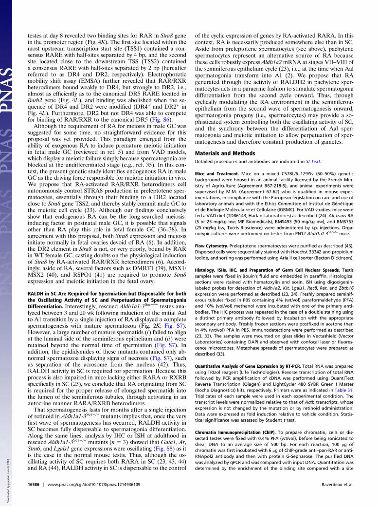
testes at day 8 revealed two binding sites for RAR in Stra8 genein the promoter region (Fig. 4K). The first site located within themost upstream transcription start site (TSS1) contained a con-sensus RARE with half-sites separated by 4 bp, and the secondsite located close to the downstream TSS (TSS2) containeda consensus RARE with half-sites separated by 2 bp (hereafterreferred to as DR4 and DR2, respectively). Electrophoreticmobility shift assay (EMSA) further revealed that RAR/RXRheterodimers bound weakly to DR4, but strongly to DR2, i.e.,almost as efficiently as to the canonical DR5 RARE located inRarb2 gene (Fig. 4L), and binding was abolished when the se-quence of DR4 and DR2 were modified (DR4* and DR2* inFig. 4L). Furthermore, DR2 but not DR4 was able to competefor binding of RAR/RXR to the canonical DR5 (Fig. S6).Although the requirement of RA for meiosis in male GC was
suggested for some time, no straightforward evidence for thisproposal was yet provided. This paradigm emerged from theability of exogenous RA to induce premature meiotic initiationin fetal male GC (reviewed in ref. 5) and from VAD models,which display a meiotic failure simply because spermatogonia areblocked at the undifferentiated stage (e.g., ref. 35). In this con-text, the present genetic study identifies endogenous RA in maleGC as the driving force responsible for meiotic initiation in vivo.We propose that RA-activated RAR/RXR heterodimers cellautonomously control STRA8 production in preleptotene sper-matocytes, essentially through their binding to a DR2 locatedclose to Stra8 gene TSS2, and thereby stably commit male GC tothe meiotic cell cycle (33). Although our findings conclusivelyshow that endogenous RA can be the long-searched meiosis-inducing factor in postnatal male GC, it is possible that signalsother than RA play this role in fetal female GC (36–38). Inagreement with this proposal, both Stra8 expression and meiosisinitiate normally in fetal ovaries devoid of RA (6). In addition,the DR2 element in Stra8 is not, or very poorly, bound by RARin WT female GC, casting doubts on the physiological inductionof Stra8 by RA-activated RAR/RXR heterodimers (6). Accord-ingly, aside of RA, several factors such as DMRT1 (39), MSX1/MSX2 (40), and RSPO1 (41) are required to promote Stra8expression and meiotic initiation in the fetal ovary.
RALDH in SC Are Required for Spermiation but Dispensable for boththe Oscillating Activity of SC and Perpetuation of SpermatogoniaDifferentiation. Interestingly, rescued Aldh1a1-3Ser−/− testes ana-lyzed between 3 and 20 wk following induction of the initial Aalto A1 transition by a single injection of RA displayed a completespermatogenesis with mature spermatozoa (Fig. 2K; Fig. S7).However, a large number of mature spermatids (i) failed to alignat the luminal side of the seminiferous epithelium and (ii) wereretained beyond the normal time of spermiation (Fig. S7). Inaddition, the epididymides of these mutants contained only ab-normal spermatozoa displaying signs of necrosis (Fig. S7), suchas separation of the acrosome from the nucleus (42). Thus,RALDH activity in SC is required for spermiation. Because thisprocess is also impaired in mice lacking either RARA or RXRBspecifically in SC (23), we conclude that RA originating from SCis required for the proper release of elongated spermatids intothe lumen of the seminiferous tubules, through activating in anautocrine manner RARA/RXRB heterodimers.That spermatogenesis lasts for months after a single injection
of retinoid in Aldh1a1-3Ser−/− mutants implies that, once the veryfirst wave of spermatogenesis has occurred, RALDH activity inSC becomes fully dispensable to spermatogonia differentiation.Along the same lines, analysis by IHC or ISH at adulthood inrescued Aldh1a1-3Ser−/− mutants (n = 3) showed that Gata1, Ar,Stra6, and Lgals1 gene expressions were oscillating (Fig. S8) as itis the case in the normal mouse testis. Thus, although the os-cillating activity of SC requires both RARA in SC (23, 43, 44)and RA (44), RALDH activity in SC is dispensable to the control
of the cyclic expression of genes by RA-activated RARA. In thiscontext, RA is necessarily produced somewhere else than in SC.Aside from preleptotene spermatocytes (see above), pachytenespermatocytes represent an alternative source of RA becausethese cells robustly express Aldh1a2mRNA at stages VII–VIII ofthe seminiferous epithelium cycle (23), i.e., at the time when Aalspermatogonia transform into A1 (2). We propose that RAgenerated through the activity of RALDH2 in pachytene sper-matocytes acts in a paracrine fashion to stimulate spermatogoniadifferentiation from the second cycle onward. Thus, throughcyclically modulating the RA environment in the seminiferousepithelium from the second wave of spermatogenesis onward,spermatogonia progeny (i.e., spermatocytes) may provide a so-phisticated system controlling both the oscillating activity of SC,and the synchrony between the differentiation of Aal sper-matogonia and meiotic initiation to allow perpetuation of sper-matogenesis and therefore constant production of gametes.
Materials and MethodsDetailed procedures and antibodies are indicated in SI Text.
Mice and Treatment. Mice on a mixed C57BL/6–129/Sv (50–50%) geneticbackground were housed in an animal facility licensed by the French Min-istry of Agriculture (Agreement B67-218-5), and animal experiments weresupervised by M.M. (Agreement 67-62) who is qualified in mouse exper-imentations, in compliance with the European legislation on care and use oflaboratory animals and with the Ethics Committee of Institut de Génétiqueet de Biologie Moléculaire et Cellulaire (IGBMC). For VAD studies, mice werefed a VAD diet (TD86143; Harlan Laboratories) as described (24). All-trans-RA(5 or 25 mg/kg bw; MP Biomedicals), BMS493 (50 mg/kg bw), and BMS753(25 mg/kg bw, Tocris Bioscience) were administered by i.p. injections. Orga-notypic cultures were performed on testes from PN12 Aldh1a1-3Ser−/− mice.
Flow Cytometry. Preleptotene spermatocytes were purified as described (45).Dispersed cells were sequentially stained with Hoechst 33342 and propidiumiodide, and sorting was performed using Aria II cell sorter (Becton Dickinson).
Histology, ISHs, IHC, and Preparation of Germ Cell Nuclear Spreads. Testissamples were fixed in Bouin’s fluid and embedded in paraffin. Histologicalsections were stained with hematoxylin and eosin. ISH using digoxigenin-labeled probes for detection of Aldh1a2, Kit, Lgals1, Rec8, Ret, and Zbtb16expression were performed as described (23, 24). Freshly prepared seminif-erous tubules fixed in PBS containing 4% (wt/vol) paraformaldehyde (PFA)and 10% (vol/vol) methanol were incubated with one of the primary anti-bodies. The IHC process was repeated in the case of a double staining usinga distinct primary antibody followed by incubation with the appropriatesecondary antibody. Freshly frozen sections were postfixed in acetone thenin 4% (wt/vol) PFA in PBS. Immunodetections were performed as described(23, 33). The samples were mounted on glass slides in Vectashield (VectorLaboratories) containing DAPI and observed with confocal laser or fluores-cence microscopes. Metaphase spreads of spermatocytes were prepared asdescribed (33).
Quantitative Analysis of Gene Expression by RT-PCR. Total RNA was preparedusing TRIzol reagent (Life Technologies). Reverse transcription of total RNAfollowed by PCR amplification of cDNA was performed using QuantiTectReverse Transcription (Qiagen) and LightCycler 480 SYBR Green I Master(Roche Diagnostics) kits, respectively. Primers were as indicated in Table S1.Triplicates of each sample were used in each experimental condition. Thetranscript levels were normalized relative to that of Actb transcripts, whoseexpression is not changed by the mutation or by retinoid administration.Data were expressed as fold induction relative to vehicle condition. Statis-tical significance was assessed by Student t test.
Chromatin Immunoprecipitation (ChIP). To prepare chromatin, cells or dis-sected testes were fixed with 0.4% PFA (wt/vol), before being sonicated toshear DNA to an average size of 500 bp. For each reaction, 100 μg ofchromatin was first incubated with 6 μg of ChIP-grade anti–pan-RAR or anti-RNApol2 antibody and then with protein G-Sepharose. The purified DNAwas analyzed by qPCR and was compared with input DNA. Quantitation wasdetermined by the enrichment of the binding site compared with a site
16586 | www.pnas.org/cgi/doi/10.1073/pnas.1214936109 Raverdeau et al.
Dow
nloa
ded
by g
uest
on
June
6, 2
020

located upstream the TSS. The sequences of the oligonucleotides used aredescribed in Table S1.
EMSAs. They were performed as previously described (46). The sequences ofthe oligonucleotides used are described in Table S1.
ACKNOWLEDGMENTS. We thank Muriel Klopfenstein for help as well asAmin Choukrallah, Emmanuel Moutier, and Abdelali Lehraiki for sharingwith us their expertise in ChIP, EMSA, and organotypic culture, respectively.We also thank Florian Guillou and Pascal Dollé for providing us the Amh-Cre
and the loxP-flanked Aldh1a2 transgenic lines, respectively, as well as all ofthe common services and platforms of IGBMC who significantly contributedto this work, notably persons from FACS (Claudine Ebel), electron microscopy(Nadia Messaddeq), and confocal imaging (Pascal Kessler and Marc Koch)facilities. This work was supported by grants from Centre National de laRecherche Scientifique, Institut National de la Santé et de la Recherche Méd-icale, Hôpital Universitaire de Strasbourg; Fondation pour la Recherche Méd-icale (FRM) Grant DEQ20071210544; and Agence Nationale de la RechercheContracts ANR-09-BLAN-0286, ANR-10-BLAN-1239-04, and ANR-09-BLAN-0282.M.R. was supported by French Ministry of Research and Technology grantsand FRM Grant FDT20110922849.
1. Nakagawa T, Sharma M, Nabeshima Y, Braun RE, Yoshida S (2010) Functional hier-archy and reversibility within the murine spermatogenic stem cell compartment.Science 328:62–67.
2. de Rooij DG, Russell LD (2000) All you wanted to know about spermatogonia butwere afraid to ask. J Androl 21:776–798.
3. Hogarth CA, Griswold MD (2010) The key role of vitamin A in spermatogenesis. J ClinInvest 120:956–962.
4. Bowles J, Koopman P (2007) Retinoic acid, meiosis and germ cell fate in mammals.Development 134:3401–3411.
5. Griswold MD, Hogarth CA, Bowles J, Koopman P (2012) Initiating meiosis: The case forretinoic acid. Biol Reprod 86:35.
6. Kumar S, et al. (2011) Sex-specific timing of meiotic induction is regulated by Cyp26b1independent of retinoic acid signalling. Nat Commun 2:151.
7. Duester G (2008) Retinoic acid synthesis and signaling during early organogenesis.Cell 134:921–931.
8. Chambon P (2005) The nuclear receptor superfamily: A personal retrospect on the firsttwo decades. Mol Endocrinol 19:1418–1428.
9. Vernet N, et al. (2006) Retinoic acid metabolism and signaling pathways in the adultand developing mouse testis. Endocrinology 147:96–110.
10. Gely-Pernot A, et al. (2012) Spermatogonia differentiation requires retinoic acid re-ceptor γ. Endocrinology 153:438–449.
11. Dupé V, et al. (2003) A newborn lethal defect due to inactivation of retinaldehydedehydrogenase type 3 is prevented by maternal retinoic acid treatment. Proc NatlAcad Sci USA 100:14036–14041.
12. Matt N, et al. (2005) Retinoic acid-dependent eye morphogenesis is orchestrated byneural crest cells. Development 132:4789–4800.
13. Vermot J, et al. (2006) Conditional (loxP-flanked) allele for the gene encoding theretinoic acid-synthesizing enzyme retinaldehyde dehydrogenase 2 (RALDH2). Genesis44:155–158.
14. Lécureuil C, Fontaine I, Crepieux P, Guillou F (2002) Sertoli and granulosa cell-specificCre recombinase activity in transgenic mice. Genesis 33:114–118.
15. Schrans-Stassen BH, van de Kant HJ, de Rooij DG, van Pelt AM (1999) Differentialexpression of c-kit in mouse undifferentiated and differentiating type A spermato-gonia. Endocrinology 140:5894–5900.
16. Barrios F, et al. (2012) SOHLH1 and SOHLH2 control Kit expression during postnatalmale germ cell development. J Cell Sci 125:1455–1464.
17. Buaas FW, et al. (2004) Plzf is required in adult male germ cells for stem cell self-re-newal. Nat Genet 36:647–652.
18. Yoshida S, et al. (2004) Neurogenin3 delineates the earliest stages of spermatogenesisin the mouse testis. Dev Biol 269:447–458.
19. Tokuda M, Kadokawa Y, Kurahashi H, Marunouchi T (2007) CDH1 is a specific markerfor undifferentiated spermatogonia in mouse testes. Biol Reprod 76:130–141.
20. Lolicato F, et al. (2008) Potential role of Nanos3 in maintaining the undifferentiatedspermatogonia population. Dev Biol 313:725–738.
21. Filipponi D, et al. (2007) Repression of kit expression by Plzf in germ cells.Mol Cell Biol27:6770–6781.
22. Bouillet P, et al. (1997) Developmental expression pattern of Stra6, a retinoic acid-responsive gene encoding a new type of membrane protein. Mech Dev 63:173–186.
23. Vernet N, et al. (2006) Prepubertal testis development relies on retinoic acid but notrexinoid receptors in Sertoli cells. EMBO J 25:5816–5825.
24. Ghyselinck NB, et al. (2006) Retinoids and spermatogenesis: Lessons frommutant micelacking the plasma retinol binding protein. Dev Dyn 235:1608–1622.
25. Yoshida S, et al. (2006) The first round of mouse spermatogenesis is a distinctiveprogram that lacks the self-renewing spermatogonia stage. Development 133:1495–1505.
26. Peschon JJ, et al. (1992) Directed expression of an oncogene to Sertoli cells in trans-genic mice using mullerian inhibiting substance regulatory sequences.Mol Endocrinol6:1403–1411.
27. Stanley ER (2009) Lineage commitment: Cytokines instruct, at last! Cell Stem Cell 5:234–236.
28. Li MA, Alls JD, Avancini RM, Koo K, Godt D (2003) The large Maf factor Traffic Jamcontrols gonad morphogenesis in Drosophila. Nat Cell Biol 5:994–1000.
29. Blanchi B, et al. (2003) MafB deficiency causes defective respiratory rhythmogenesisand fatal central apnea at birth. Nat Neurosci 6:1091–1100.
30. Moriguchi T, et al. (2006) MafB is essential for renal development and F4/80 expres-sion in macrophages. Mol Cell Biol 26:5715–5727.
31. Cordes SP, Barsh GS (1994) The mouse segmentation gene kr encodes a novel basicdomain-leucine zipper transcription factor. Cell 79:1025–1034.
32. Eichmann A, et al. (1997) The expression pattern of the mafB/kr gene in birds andmice reveals that the kreisler phenotype does not represent a null mutant. Mech Dev65:111–122.
33. Mark M, et al. (2008) STRA8-deficient spermatocytes initiate, but fail to complete,meiosis and undergo premature chromosome condensation. J Cell Sci 121:3233–3242.
34. Nau H (1986) Species differences in pharmacokinetics and drug teratogenesis. EnvironHealth Perspect 70:113–129.
35. Li H, Palczewski K, Baehr W, Clagett-Dame M (2011) Vitamin A deficiency results inmeiotic failure and accumulation of undifferentiated spermatogonia in prepubertalmouse testis. Biol Reprod 84:336–341.
36. Best D, Sahlender DA, Walther N, Peden AA, Adams IR (2008) Sdmg1 is a conservedtransmembrane protein associated with germ cell sex determination and germline-soma interactions in mice. Development 135:1415–1425.
37. Kocer A, Reichmann J, Best D, Adams IR (2009) Germ cell sex determination inmammals. Mol Hum Reprod 15:205–213.
38. Wang N, Tilly JL (2010) Epigenetic status determines germ cell meiotic commitment inembryonic and postnatal mammalian gonads. Cell Cycle 9:339–349.
39. Krentz AD, et al. (2011) DMRT1 promotes oogenesis by transcriptional activation ofStra8 in the mammalian fetal ovary. Dev Biol 356:63–70.
40. Le Bouffant R, et al. (2011) Msx1 and Msx2 promote meiosis initiation. Development138:5393–5402.
41. Chassot AA, et al. (2011) RSPO1/β-catenin signaling pathway regulates oogonia dif-ferentiation and entry into meiosis in the mouse fetal ovary. PLoS One 6:e25641.
42. Holstein AF, Roosen-Runge EC, Schirren C (1988) Illustrated Pathology of HumanSpermatogenesis (Grosse, Berlin), p 278.
43. Parvinen M (1993) Cyclic functions of Sertoli cells. The Sertoli Cell, eds Russell LD,Griswold MD (Cache River Press, Clearwater, FL), pp 331–347.
44. Sugimoto R, Nabeshima Y, Yoshida S (2012) Retinoic acid metabolism links the peri-odical differentiation of germ cells with the cycle of Sertoli cells in mouse seminif-erous epithelium. Mech Dev 128:610–624.
45. Getun IV, Torres B, Bois PR (2011) Flow cytometry purification of mouse meiotic cells. JVis Exp 50:e2602.
46. Hwang JJ, Chambon P, Davidson I (1993) Characterization of the transcription acti-vation function and the DNA binding domain of transcriptional enhancer factor-1.EMBO J 12:2337–2348.
Raverdeau et al. PNAS | October 9, 2012 | vol. 109 | no. 41 | 16587
DEV
ELOPM
ENTA
LBIOLO
GY
Dow
nloa
ded
by g
uest
on
June
6, 2
020
![Paracrine Growth Stimulation of Androgen-responsive Shionogi … · (CANCER RESEARCH 50, 4979-4983. August 15. 1990] Paracrine Growth Stimulation of Androgen-responsive Shionogi Carcinoma](https://img.pdfslide.net/doc/110x75/5e73928322d4f1405956059e/paracrine-growth-stimulation-of-androgen-responsive-shionogi-cancer-research-50.jpg)