Embed Size (px)
Citation preview

Pergamon Leuke,,,ra Research Vol. 20, No. 6, 499-505, 1996. pp.
Copyright 0 1996 Elsevier Science Ltd. All rights reserved Printed in Great Britain
0145-2126/96 $15.00 + 0.00
01452126(95)00118-2
RETINOID-REGULATED EXPRESSION OF BCL-2 AND TISSUE TRANSGLUTAMINASE DURING THE DIFFERENTIATION AND APOPTOSIS OF HUMAN MYELOID LEUKEMIA (HL-60) CELLS
L&z16 Nagy*, Vilmos A. Thomazy*, Roshantha A. S. Chandraratnat, Richard A. HeymanS and Peter J. A. Davies
*Department of Pharmacology, University of Texas-Houston Medical School, Houston, TX 77225, U.S.A.; TRetinoid Research, Departments of Chemistry and Biology, Allergan Incorporated, Irvine, CA, U.S.A.; and
fDepartment of Cell Biology, Ligand Pharmaceuticals, San Diego, CA, U.S.A.
(Received 10 April 1995. Accepted 24 July 1995)
Abstract-Retinoids induce terminal differentiation and subsequent apoptosis in the human myeloid leukemia (HL-60) cell line. We have previously shown that in HL-60 cells, ligand activation of retinoic acid receptors (RARs) is sufficient to induce differentiation but ligand activation of retinoid X receptors (RXRs) is necessary for the retinoid-induced apoptosis of these cells. In the present studies we have characterized the effect of retinoids on the expression of two apoptosis-linked gene products, BCL-2 and tissue transglutaminase. BCL-2 is a membrane-associated protein whose expression has been linked to the suppression of apoptosis in many cells. Tissue transglutaminase is a protein cross-linking enzyme that accumulates in many cells undergoing apoptotic cell death. Our data suggest that ligand activation of RARs in HL-60 cells results in a global suppression of BCL-2 expression whereas ligand activation of both RARs and RXRs triggers the selective accumulation of tissue transglutaminase in the apoptotic HL-60 cells. Copyright 0 1996 Elsevier Science Ltd.
Introduction
Retinoids are a class of polyisoprenoid hormones that have profound effects on the regulation of cellular growth, differentiation and death (for a review see [l]). These effects are mediated via the interaction of retinoids with at least two families of nuclear receptors, the retinoic acid receptors (RARs) and the retinoid X receptors (RXRs), that function as ligand-dependent transcription factors [l]. RARs and RXRs are distinct classes of nuclear receptors, encoded by distinct genes, recognizing different DNA binding sequences and differing in their ligand specificities [l]. RARs bind both all-trans retinoic acid (ATRA) and its isomer 9-cis
retinoic acid (9-cis RA) whereas RXRs bind only 9-cis
RA [2,3]. Both receptor classes include at least three
Abbreviations: ATRA, all-trans retinoic acid; 9-cis RA, 9-h retinoic acid; RAR, retinoic acid receptor; MR, retinoid X receptor; lTNPB, (E)-4-[2-(5;6;7;8-tetrahydro-5;5;8;8;-tetra- methyl-2 -napthalenyl)-l-propenyllbenzoic acid; AGN191701, (E) - 5 - [2-(5;6;7;8;-tetrahydro-3;5;5;8;8;-pentamethylnaphtha- len-2yl)propen-1-yl]-3-thiophenecarboxylic acid.
Correspondence to: Dr Peter J. A. Davies, Department of Pharmacology, University of Texas-Houston, Medical School, P.O. Box 20708, Houston, TX 77225, U.S.A. (Tel: 713 792 5904; Fax: 713 792 5911; E-mail: pdavies@ farmrl.med.uth.tmc.edu).
distinct receptor genes (RARE, I3 and y and RXRcr, 8 and y) that can give rise to multiple receptor proteins (via alternative splicing) [l]. Both RARs and RXRs require dimerization for activity [l]. RARs heterodimerize with RXRs but RXRs can bind not only to RARs but also to a variety of other nuclear receptor proteins including themselves (homodimerization) and receptors for vita- min D, thyroid hormone and orphan receptors such as the PPAR (peroxisome proliferator activator receptor)
PI. In spite of our rapidly expanding understanding of the
molecular mechanisms of retinoid action, little is known of the contribution that the different retinoid receptor pathways make to the biology of retinoids. To address this issue we have studied the ability of receptor selective retinoids (retinoids that interact only with one or other of the classes of retinoid receptors [4-71 to replicate the biological activity of a natural retinoid such as all-trans retinoic acid. Using as a model the induction of differentiation and cell death in a human myeloid leukemia cell line (HL-60) [8,9], we have shown that ligand activation of endogenous RARs is sufficient to induce the granulocytic differentiation of the cells [7]. If differentiated HL-60 cells are exposed to ligands that activate endogenous RXRs, then the cells rapidly
undergo apoptosis [7]. These studies have suggested
499

500 L. Nagy et al.
that the induction of differentiation and the expression of the cell death pathway are separable processes in HL- 60 cells, regulated by ligand activation of different retinoid receptors.
In the studies to be presented here, we have extended our investigations of the pathways of retinoid action in HL-60 cells, characterizing the expression of proteins involved in the pathway of programmed cell death. BCL-2 is a membrane-associated protein whose expres- sion prevents the induction of apoptosis in HL-60 cells [lo, 111 and many other cell types and tissues [12-141. Tissue transglutaminase (EC 2.3.2.13) is a protein cross- linking enzyme that accumulates to high levels in cells undergoing apoptosis [l&16]. The results we have obtained suggest that ATRA is able to induce a co- ordinated down-regulation of BCL-2 and an up-regula- tion of tissue transglutaminase during HL-60 apoptosis. The suppression of BCL-2 depends on the ligand activation of RARs, whereas the induction of tissue transglutaminase is mediated by ligand activation of both RARs and RXRs.
Materials and Methods
Cell culture and evaluation of cellular morphology
The parental clone of HL-60 cells (clone CCL 240) was obtained from the American Type Culture Collec- tion, Rockville, MD, U.S.A. HL-60 CCL 240 cells were cultured in suspension in RPM1 1640 medium (Gibco BRL Life Technologies, Inc., Gaithersburg, MD, U.S.A.) supplemented with 10% fetal bovine serum (Urvine Scientific Inc., Santa Ana, CA, U.S.A.). Cells were routinely cultured at 37°C 5% COa. Retinoids, or an equivalent concentration of ethanol (co.1 vol %) were added to cells in RPM1 1640 supplemented with insulin, transferrin and Na-selenite at 2 x lo5 cells/ml in 3 ml of media in 6-well tissue culture plates (Corning Glass Works, Corning, NY, U.S.A.). After the indicated times, 0.05-0.1 ml aliquots of cells were taken to make cytospin preparations that were fixed in 100% methanol and stained with Diff-Quick stain (Dade Diagnostics, Inc., Aguada, Puerto Rico) according to the manufac- turer’s protocol or processed for immunohistochemistry. Cells were scored as differentiated if they showed features of metamyelocytes or more differentiated forms, and as apoptotic if there was evidence of nuclear pycnosis and fragmentation, cytoplasmic condensation and basophilia.
Retinoids
All-trans retinoic acid (ATRA), TTNPB, 9-cis retinoic acid (Pcis RA) and AGN191701 were obtained from Allergan Inc., Irvine, CA, U.S.A. and Ligand Pharmaceuticals, San Diego, CA, U.S.A. Their ability to bind to RARs and RXRs was assayed by a ligand
binding assay as described by Boehm et al. [4] and Nagy et al. [7].
Western-blot analysis
Expression of BCL-2 protein was determined with the use of a monoclonal antibody, DAKO-bcl-2 124 (DAK0 Corporation, Carpintera, CA, U.S.A.). Cell lysates were prepared as follows: cells were collected by centrifuga- tion at 500g for 10 min. Pellets were resuspended in phosphate buffered saline (PBS) and sonicated on ice. The protein concentration was determined by using the Bio-Rad Protein Assay (Bio-Rad Chemical Division, Richmond, CA, U.S.A.). Protein containing cell lysates were normalized for protein content and 50 ug protein/ lane were size fractionated in 10% sodium dodecyl sulfate-polyacrylamide gels prior to transfer to PVW membranes using standard protocols. The membranes were blocked overnight with 5% dry cow milk in PBS at 4°C. The monoclonal anti-BCL-2 antibody was used in 1:lOOO dilution (1 &ml) in 1% dry milk in PBS, followed by a secondary antiserum: rabbit antimouse IgG (Pel-Freez Biologicals, Rogers, AR, U.S.A.) (10 pg/ ml) diluted in 1% dry milk in PBS and finally 1251 protein A (0.99 mCi/ml; ICN Radiochemicals Irvine, CA, U.S.A.) at a concentration of 0.1 l&i/ml in 1% dry milk in PBS. Membranes were dried and exposed to film. The autoradiographs were subjected to densito- metric analysis.
Transglutaminase assay
Transglutaminase activity was assayed as described previously [17]. Briefly, cell lysates were prepared and incubated in the presence of 3H putrescine and N,N- dimethyl casein. Ca2+-dependent incorporation of the radiolabeled putrescine into N,N-dimethyl casein was determined and expressed as pmol/min/mg protein.
Immunohistochemistry
Immunolocalization of BCL-2 and tissue transgluta- minase was determined in control and ATRA treated HL-60 CCL240 cells by using the mouse monoclonal antibody against human BCL-2 (as described above) or non-immune mouse serum and an affinity-purified goat anti-guinea pig tissue transglutaminase or IgG fraction of preimmune sera. Cells were treated and cytospins were made as described above. Cells were fixed in acetone for 5 min at room temperature and blocked with a mixture of 5% dry cow milk, 10% normal rabbit serum and 0.5% gelatine in PBS (blocking solution) for 20 min at room temperature. Anti-BCL-2 or anti-transgluta- minase antibody or the appropriate control sera was diluted in blocking solution at 10 pg/ml and applied to the cytospins in a wet chamber for 2 h at room tempera- ture. The slides were washed three times for 5 min in PBS. The secondary antibody FITC-conjugated rabbit

Retinoid regulation of BCL-2 and tissue transglutaminase
Fig. 1. Comparison of BCL-2 and tissue transglutaminase expression in control and ATRA treated HL-60 cells. Cells were treated and processed as described in Materials and Methods, original magnification 500 x (A) Normal morphology of control HL-60 CCL240 cells, Diff-Quick staining. (B) ATRA (1 FM) treated HL-60 cells (day 3) characteristic changes in nuclear morphology, nuclear segmentation, Diff-Quick staining. (C) ATRA (1 PM) treated HL-60 cells (day S), cells showing the differentiated phenotype and numerous apoptotic cells with fragmented, pycnotic nuclei (arrows), Diff-Quick staining. (D) Immunolocalization of BCL-2 in control cultures and (E) in cells treated with 1 uM ATRA for 3 days; (F) control cells with nonimmune serum, indirect immunofluorescence microscopy was used as described in Materials and Methods. Immunolocalization of tissue transglutaminase in HL-60 cells, (G) control cells, cells treated with 1 pM ATRA for (H) 3 days and (I) 5 days, indirect immunofluorescence microscopy
was used as described in Materials and Methods. Arrows show transglutaminase positive apoptotic cells.
antimouse Ig (Vector Laboratories, Burlingame, CA,
U.S.A.) for BCL-2 and TRIC-conjugated rabbit antigoat
Ig (Vector Laboratories) for transglutaminase, was
diluted 1:lOO in blocking solution. Incubation with the
secondary antibody was for 45 min at room temperature
in a wet chamber followed by washing twice with PBS
containing 0.1% Triton X-100 and once with PBS.
Slides were covered with coverslips and 50% glycer-
ol-PBS containing 0.2% p-phenylene diamine. Slides
were studied on a Nikon Optiphot photomicroscope.

L. Nagy et al.
-R-TMPB
-M-AGN191701
26 kD
ATRA 9-cis RA
c -8 -7 -6 C -8 -7 6
TTNPB AGN191701
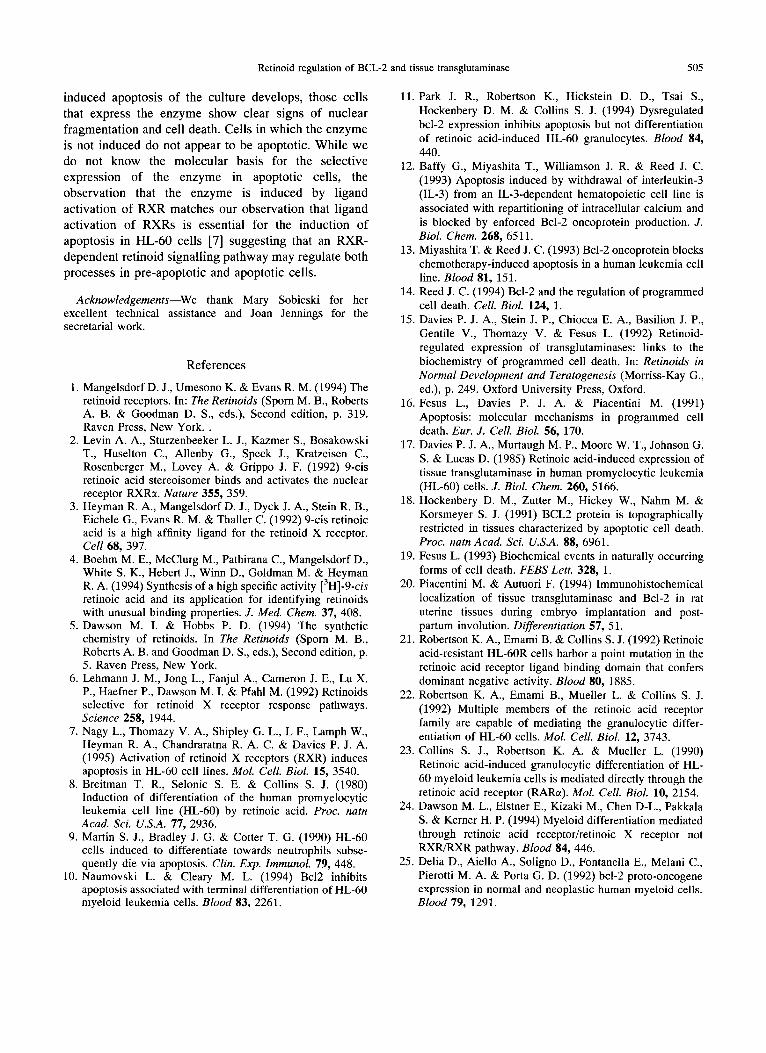
Fig. 2. (A) BCL-2 protein levels in ATRA (1 PM) treated HL-60 CCL240 cells after 3 and 5 days of treatment. Cells were treated and cell lysates were subjected to Western blot analysis as described in Materials and Methods. Blots were subjected to densitometric analysis and protein levels were expressed as a percentage of the untreated control. Insert shows a Western blot (C: control, D3: 3 day treatment, D5: 5 day treatment). (B) Level of BCL-2 protein in response to retinoids in HL-60 CCL240 cells (Western blot). HL- 60 cells treated with natural and receptor selective synthetic retinoids for 5 days, Western blot analyses were performed as described in Materials and Methods (C: control, -8, -7, -6: lo-‘, lo-‘, lop6 M, respectively, of the indicated retinoid).(C) BCL-2 protein levels in HL-60 CCL240 cells treated with the RAR selective retinoid TI’NPB and the RXR selective AGN191701 for 3 and 5 days.
BCL-2 protein levels were determined by Western blot analysis and expressed as a percentage of the control.
Results and Discussion stroyed and in others the cytoplasm is filled with
ATRA induces both the differentiation and the apoptotic death of HL-60 cells [8,9] (Fig. 1). Untreated cells have a morphology characteristic of blastic leukemia cells, large spherical nuclei surrounded by a thin shell of basophilic cytoplasm (Panel A). After 3 days of ATRA (1 PM) treatment, many cells have the condensed, lobulated and segmented nuclei character- istic of cells undergoing granulocytic differentiation (Panel B). After 5 days of ATRA treatment, there is extensive apoptosis. Many cells are completely de-
hyperchromic nuclear fragments characteristic of cells undergoing apoptotic cell death (Panel C). Panels D and E demonstrate the effect of ATRA on the expression of BCL-2 in differentiating HL-60 cells. Untreated HL-60 cells (Panel D) show a bright pattern of cytoplasmic and nuclear immunoreactivity that is much more prominent than control cells treated with a non-immune serum (Panel F). After 3 days of treatment with ATRA, however, this immunoreactivity is completely and uniformly suppressed (Panel E) suggesting a marked decrease in the level of BCL-2 expression. Western blot

Retinoid
All-trans retinoic acid*
9-k retinoic acid*
Retinoid regulation of BCL-2 and tissue transglutaminase 503
Table 1. Binding and transactivation activity of receptor-selective retinoids
Binding ICso (nM) Transactivation EC& (nM)
Structure RARC! RXRa RXRD RARa RXRc( RXRB
@-JQ=+ 15f2 >lOOO >lOOO 350f31 >lOOO >lOOO
q
7*2 32+4 12f3 191+20 100+25 200 f 30
lTNPB* @+- 36k5 >lOOO >lOOO 30+6 >lOOO >lOOO
AGN191701+ >lOOO 308 387 >lOOO 215 10.5
Binding values were determined by a competitive binding analysis. Cell extracts (50 pg of protein) containing the indicated receptor, overexpressed in a baculovirus infected SF-9 cells, were incubated at 0°C for 2 h with [3H]-9-cis retinoic acid and varying concentrations of unlabelled retinoids. Specific binding was determined by a hydroxyapatite-receptor binding assay [4]. IC,, is the concentration of unlabelled retinoid required to produce a 50% inhibition of the binding of [3H]-9-ci.s retinoic acid to the indicated receptor. Transactivation activity was assessed following the co-transfection of the indicated receptor expression vectors and either RAR- or RXR-responsive reporter constructs into CV-1 cells as described in detail previously [4,7]. The EC& value is the concentration of retinoid required to induce a 50% of maximum increase in reporter enzyme activity. Values shown are the mean f SEM of at least three determinations.
* Values described previously by Boehm et al. [4].+ Values described previously by Nagy et al. [7].
analysis of ATRA-treated HL-60 cells shows a dramatic reduction in the level of BCL-2 expression (Fig. 2A, insert). After 3 days of treatment, BCL-2 levels are reduced to 44% of control cells, by day 5 the level is reduced to 18% of control (Fig. 2A).
HL-60 cells contain both FURS and RXRs. Previously reported experiments have demonstrated the presence of RARc(, RXRcl and RXRl3 in the HL-60 cell line used in these studies [7]. RARE and RARy are neither expressed nor induced by ATRA in this HL-60 cell line. To explore the role of these receptors in the suppression of BCL-2 expression, we evaluated activity of several receptor selective retinoids. Table 1 compares the ability of these retinoids to both bind to and transcriptionally activate (transactivate) RARa and RXRs c( and IJ (the receptors expressed in HL-60 cells). 9-cis RA is a receptor panagonist since it both binds to and transacti- vates both RARs and RXRs. ATRA binds to RARs and not RXRs. It may show a low level of RXR-dependent activity in some in vivo assays due to its isomerization to 9-cis RA, a ligand for both classes of receptors. TTNPB is an RAR-specific retinoid, it is a high-affinity ligand for RARE in both binding and transactivation assays and shows no RXR agonist activity. AGN191701 is an RXR- specific retinoid binding to both RXRa and RXRl3 and transactivating them but showing no affinity for RARa. HL-60 cells were treated for 5 days with differing concentrations of each of these retinoids and the level of BCL-2 in the cells was detected by Western blot analysis (Fig. 2B). ATRA is a very potent suppressor of BCL-2 expression, even with lo-’ M ATRA the level
of BCL-2 is less than 25% of that detected in control cells. It is interesting that this residual level of BCL-2 is resistant to further suppression by higher levels of ATRA treatment suggesting either that there is a basal level of expression of the protein in RA-treated cells or there is a residual pool of cells unresponsive to ATRA. The immunochemical studies of RA-treated cells gave no evidence of heterogeniety in the response of the cells to ATRA suggesting that the former explanation is the correct one. 9-cis RA is equivalent to ATRA in its ability to suppress BCL-2 expression in these cells, even at lo-’ M the BCL-2 level is maximally suppressed. TTNPB, an RAR-specific retinoid also suppresses the expression of BCL-2 in HL-60 cells although it appears to be less potent than ATRA or 9-cis RA. The level of BCL-2 in cells treated with lo-’ M TTNPB is 46% of that found in control cells and higher levels of the drug resulted in progressive suppression in the level of the protein detected in the cells (30% and 15% of control for 10K7 M and 10K6 M, respectively). The RXR selective retinoid, AGN191701, did not produce significant
changes in BCL-2 level in these cells, lo-‘, lo-’ and lop6 M AGN191701 resulted in BCL-2 levels that were 101, 80 and 83%, respectively, of the level detected in control cells. Figure 2C compares the time course for the effect of TTNPB and AGN191701 (1 J.IM each) on the level of BCL-2 in the HL-60 cells. TTNPB produces
partial suppression in 3 days and almost complete suppression in 5 days, whereas AGN191701 resulted in only a very slight suppression in the level of the protein that was similar after either 3 or 5 days of treatment.

L. Nagy et al.
Fig. 3. Transglutaminase activity in HL-60 CCL240 cells treated with natural (ATRA and 9-cis RA) and receptor selective, synthetic (TINPB and AGN 191701) retinoids. Cells were treated for 48 h with 1 uM of the indicated retinoid. Transglutaminase assays were performed as described in Materials and Methods. The mean values of three determina- tions and S.D. are shown. The control value shows the assay background: non-enzymatic incorporation, there is no detect-
able transglutaminase activity in untreated HL-60 cells.
These studies suggest that ligand activation of endogenous RARcl in HL-60 cells is linked to the suppression of BCL-2 expression. Retinoid receptor panagonists, such as 9-cis RA or ATRA or and an RAR- specific agonist, such as TTNPB, dramatically sup- pressed the expression of the protein, whereas a ligand that activates the endogenous RXRs had no effect.
Regulation of Tissue Transglutaminase
BCL-2 is a protein whose expression is frequently suppressed in cells undergoing apoptosis [18, 191, tissue transglutaminase, on the other hand, is an enzyme that frequently accumulates to high levels in apoptotic cells [15, 16,201. We therefore compared the induction of tissue transglutaminase to the suppression of BCL-2 in apoptotic HL-60 cells. Panels G, H, and I in Fig. 1 show the levels of tissue transglutaminase in either untreated HL-60 cells (Panel G) or cells treated for 3 or 5 days with ATRA (Panels H and I). In untreated cells the level of tissue transglutaminase expression is too low to detect. The immunoreactivity in untreated cells is no different from cells treated with a pre-immune serum (data not shown). After 3 days of exposure to ATRA there is a generalized increase in the cytoplasmic tissue transglutaminase expression, but it is very variable from cell to cell (Panel H). The immunoreactive cells showed eccentric and lobulated nuclei indicative of early stages of differentiation. In the most immunoreactive cells, the nuclei are clearly segmented or even fragmented,
indicating terminal differentiation and apoptotic cell death. These changes are even more apparent after 5 days of ATRA treatment (Panel I), the apoptotic cells in the culture (recognizable by the multiple fragmented nuclear remnants) stain intensely with the anti-transglu- taminase antibodies, whereas the few remaining non- apoptotic cells are not immunoreactive. These studies suggest that there is a very selective induction of tissue transglutaminase in cells undergoing retinoid-induced apoptotic cell death.
The differences between BCL-2 suppression and the induction of tissue transglutaminase expression are also apparent when one compares the retinoid receptor specificity of the two process (Fig. 3). As reported previously, ATRA causes a dramatic increase in the level of tissue transglutaminase in HL-60 cells [17] and this induction is replicated by 9-cis RA. However, both TTNPB and AGN191701 also increase transglutaminase activity in HL-60 cells (Fig. 3). This observation suggests a significant difference between retinoid regulation of BCL-2 and transglutaminase expression in HL-60 cells. BCL-2 expression is specifically regulated by ligand activation of RARs whereas tissue transglutaminase expression can be regulated by ligand activation of either RARs or RXRs.
Differences between the retinoid receptor-dependent expression of BCL-2 and tissue transglutaminase in HL- 60 cells parallels observations we and others have made on the role of these receptors in the induction of differentiation and cell death in HL-60 cells [7,21-241. Ligand activation of the endogenous RARcr is sufficient to induce the differentiation of HL-60 cells [24] and it is likely that this differentiation is associated with a global suppression in BCL-2 expression [lo]. The time course for the disappearance of the protein parallels the appearance of cells with a differentiated phenotype. Other agents that induce HL-60 differentiation (i.e. TPA and DMSO) have also been reported to suppress BCL-2 expression [25], although neither agent is associated with the induction of apoptosis [9] or the induction of tissue transglutaminase activity ([17] and L. Nagy, unpublished observation). It is unlikely that BCL-2 suppression is a necessary component of the differentia- tion pathway in these cells since constitutive over- expression of BCL-2 in HL-60 cells does not interfere with their differentiation although it does suppress retinoid-induced apoptosis [lo, 111.
The induction of tissue transglutaminase and the expression of an apoptotic phenotype appears to be a much more selective effect of retinoids on these cells. Tissue transglutaminase appears to accumulate selec- tively in those cells that are destined to undergo apoptosis. This appears to be a relatively early event since some transglutaminase positive cells show only early signs of differentiation. However, as retinoid-
Retinoid regulation of BCL-2 and tissue transglutaminase 505
induced apoptosis of the culture develops, those cells that express the enzyme show clear signs of nuclear fragmentation and cell death. Cells in which the enzyme is not induced do not appear to be apoptotic. While we do not know the molecular basis for the selective expression of the enzyme in apoptotic cells, the observation that the enzyme is induced by ligand activation of RXR matches our observation that ligand activation of RXRs is essential for the induction of apoptosis in HL-60 cells [7] suggesting that an RXR- dependent retinoid signalling pathway may regulate both processes in pre-apoptotic and apoptotic cells.
Acknowledgements-We thank Mary Sobieski for her excellent technical assistance and Joan Jennings for the secretarial work.
References
1. Mangelsdorf D. J., Umesono K. & Evans R. M. (1994) The retinoid receptors. In: The Retinoids (Sporn M. B., Roberts A. B. & Goodman D. S., eds.), Second edition, p. 319. Raven Press, New York.
2. Levin A. A., Sturzenbeeker L. J., Kazmer S., Bosakowski T., Huselton C., Allenby G., Speck J., Kratzeisen C., Rosenberger M., Lovey A. & Grippo J. F. (1992) 9-cis retinoic acid stereoisomer binds and activates the nuclear receptor RXRc(. Nature 355, 3.59.
3. Heyman R. A., Mangelsdorf D. J., Dyck J. A., Stein R. B., Eichele G., Evans R. M. & Thaller C. (1992) 9-cis retinoic acid is a high affinity ligand for the retinoid X receptor. Cell 68, 397.
4. Boehm M. E., McClurg M., Pathirana C., Mangelsdorf D., White S. K., Hebert J., Winn D., Goldman M. & Heyman R. A. (1994) Synthesis of a high specific activity [3H]-9-cis retinoic acid and its application for identifying retinoids with unusual binding properties. J. Med. Chem. 37, 408.
5. Dawson M. I. & Hobbs P. D. (1994) The synthetic chemistry of retinoids. In The Retinoids (Spom M. B., Roberts A. B. and Goodman D. S., eds.), Second edition, p. 5. Raven Press, New York.
6. Lehmann J. M., Jong L., Fanjul A., Cameron J. E., Lu X. P., Haefner P., Dawson M. I. & Pfahl M. (1992) Retinoids selective for retinoid X receptor response pathways. Science 258, 1944.
7. Nagy L., Thomazy V. A., Shipley G. L., L F., Lamph W., Heyman R. A., Chandraratna R. A. C. & Davies P. J. A. (1995) Activation of retinoid X receptors (RXR) induces apoptosis in HL-60 cell lines. Mol. Cell. Biol. 15, 3540.
8. Breitman T. R., Selonic S. E. & Collins S. J. (1980) Induction of differentiation of the human promyelocytic leukemia cell line (HL-60) by retinoic acid. Proc. natn Acad. Sci. U.S.A. 77, 2936.
9. Martin S. J., Bradley J. G. & Cotter T. G. (1990) HL-60 cells induced to differentiate towards neutrophils subse- quently die via apoptosis. Clin. Exp. Immunol. 79, 448.
10. Naumovski L. & Cleary M. L. (1994) Bc12 inhibits apoptosis associated with terminal differentiation of HL-60 myeloid leukemia cells. Blood 83, 2261.
11. Park J. R., Robertson K., Hickstein D. D., Tsai S., Hockenbery D. M. & Collins S. J. (1994) Dysregulated bcl-2 expression inhibits apoptosis but not differentiation of retinoic acid-induced HL-60 granulocytes. Blood 84, 440.
12. Baffy G., Miyashita T., Williamson J. R. & Reed J. C. (1993) Apoptosis induced by withdrawal of interleukin-3 (IL-3) from an IL-3-dependent hematopoietic cell line is associated with repartitioning of intracellular calcium and is blocked by enforced Bcl-2 oncoprotein production. J. Biol. Chem. 268, 6.511.
13. Miyashita T. & Reed J. C. (1993) Bcl-2 oncoprotein blocks chemotherapy-induced apoptosis in a human leukemia cell line. Blood 81, 151.
14. Reed J. C. (1994) Bcl-2 and the regulation of programmed cell death. Cell. Biol. 124, 1.
1.5. Davies P. J. A., Stein J. P., Chiocca E. A., Basilion J. P., Gentile V., Thomazy V. & Fesus L. (1992) Retinoid- regulated expression of transglutaminases: links to the biochemistry of programmed cell death. In: Retinoids in Normal Development and Teratogenesis (Morriss-Kay G., ed.), p. 249. Oxford University Press, Oxford.
16. Fesus L., Davies P. J. A. & Piacentini M. (1991) Apoptosis: molecular mechanisms in programmed cell death. Eur. J. Cell. Biol. 56, 170.
17. Davies P. J. A., Murtaugh M. P., Moore W. T., Johnson G. S. & Lucas D. (1985) Retinoic acid-induced expression of tissue transglutaminase in human promyelocytic leukemia (HL-60) cells. J. Biol. Chem. 260, 5166.
18. Hockenbery D. M., Zutter M., Hickey W., Nahm M. & Korsmeyer S. J. (1991) BCL2 protein is topographically restricted in tissues characterized by apoptotic cell death. Proc. natn Acad. Sci. U.S.A. 88, 6961.
19. Fesus L. (1993) Biochemical events in naturally occurring forms of cell death. FEBS Lett. 328, 1.
20. Piacentini M. & Autuori F. (1994) Immunohistochemical localization of tissue transglutaminase and Bcl-2 in rat uterine tissues during embryo implantation and post- partum involution. Differentiation 57, 51.
21. Robertson K. A., Emami B. & Collins S. J. (1992) Retinoic acid-resistant HL-60R cells harbor a point mutation in the retinoic acid receptor ligand binding domain that confers dominant negative activity. Blood 80, 1885.
22. Robertson K. A., Emami B., Mueller L. & Collins S. J. (1992) Multiple members of the retinoic acid receptor family are capable of mediating the granulocytic differ- entiation of HL-60 cells. Mol. Cell. Biol. 12, 3743.
23. Collins S. J., Robertson K. A. & Mueller L. (1990) Retinoic acid-induced granulocytic differentiation of HL- 60 myeloid leukemia cells is mediated directly through the retinoic acid receptor (RARa). Mol. Cell. Biol. 10, 2154.
24. Dawson M. L., Elstner E., Kizaki M., Chen D-L., Pakkala S. & Kerner H. P. (1994) Myeloid differentiation mediated through retinoic acid receptor/retinoic X receptor not RXFURXR pathway. Blood 84, 446.
25. Delia D., Aiello A., Soligno D., Fontanella E., Melani C., Pierotti M. A. & Porta G. D. (1992) bcl-2 proto-oncogene expression in normal and neoplastic human myeloid cells. Blood 79, 1291.