Embed Size (px)
Citation preview
2
The Immunology Graduate Group gratefully acknowledges the financial support of all our contributors for the 25th Annual Retreat: Grant Support
• T32 AI 055428 "Immune System Development and Regulation" training grant Institutes, Centers, Departments, and Divisions
• Abramson Family Cancer Research Institute • Combined Degree and Physician Scholar Programs • Department of Medicine • Department of Microbiology • Department of Pathobiology • Department of Pathology and Laboratory Medicine • Department of Pediatrics, The Children’s Hospital of Philadelphia • Department of Surgery • Institute for Immunology • Joseph Stokes, Jr. Research Institute • The American Association of Immunologists • The Department of Pathology at Penn Dental School • The Wistar Institute • VMD-PhD Program
Corporate Sponsorship
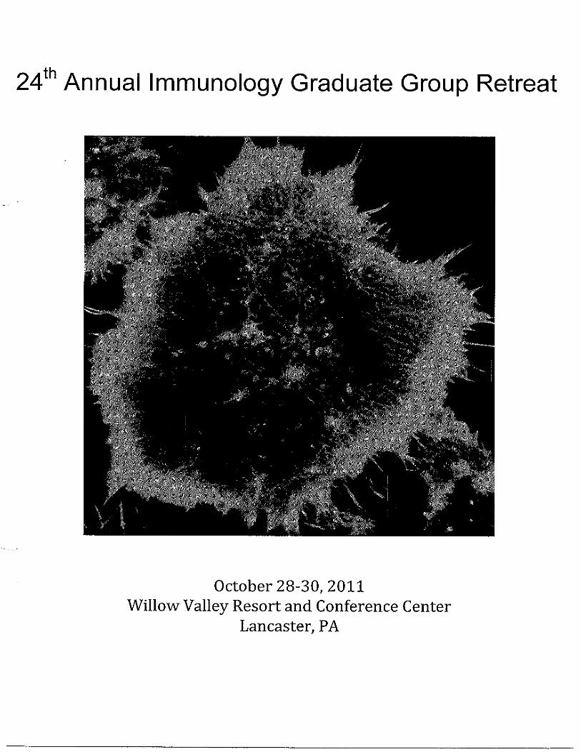
• Genetech Cover Photo Bone marrow derived dendritic cell labeled for actin (red), vinculin (green) and DAPI (blue). An ordered array of podosomes, dynamic adhesive contacts associated with cell motility, lies just behind the leading edge of the cell. From Klos Dehring DA, Clarke F, Ricart BG, Huang Y, Gomez TS, Williamson EK, Hammer DA, Billadeau DD, Argon Y, and Burkhardt JK. (2011). HS1 functions in concert with WASp to promote podosome array organization and chemotaxis in dendritic cells. J Immunol. 186:4805-18.
3
25th Annual Immunology Retreat Friday to Sunday, November 2-4, 2012
The Grand Hotel 1045 Beach Avenue, Cape May, NJ 08204
Friday, November 2, 2012 Please note: You will not be able to check into your hotel room until after 3 pm. We recommend that you leave your luggage in your vehicle until check-in at the end of Session II. All sessions and breaks to be held in the Penthouse Ballroom, 5th floor. All meals will be held in the Grand Ballroom Complex, 2nd floor. 11:00-12:00 PM Retreat registration and program pick-up, Grand Ballroom Atrium 12:00-1:20 Lunch, Deli Buffet 1:20-1:30 Welcome, John Wherry, IGG Chair 1:30-2:50 Session I: B cell biology
Session Chair: Burton Barnett 1:30-1:50 Irene Chernova
“Heterogeneity of the bone marrow plasma cell pool” 1:50-2:10 Julie Horowitz
“Role of non-core Rag1 in B cell development” 2:10-2:30 Lisa Barnett
“Dendritic cells and B cells cooperate for follicular helper T cell differentiation”
2:30-2:50 Burton Barnett
“Asymmetric division of germinal center B cells” 2:50-3:10 Break 1st and 2nd year students: Easel and posterboard set-up, Penthouse
Ballroom 3:10-4:30 Session II: Regulation of T cell responses Session Chair: Aisling O'Hara Hall 3:10-3:30 Erietta Stelekati
“Bystander chronic infection negatively impacts the development of CD8 T cell memory”
3:30-3:50 Peter Morawski
“CDK2 regulates Foxp3 stability & function” 3:50-4:10 Pamela Odorizzi
4
“PD-1 antagonizes early onset of T cell exhaustion during chronic infection”
4:10-4:30 Aisling O'Hara Hall
“The cytokines Interleukin 27 and Interferon-γ promote distinct Treg cell populations required to limit infection-induced pathology”
4:30-4:40 Break 4:40-6:00 Professional Development Session Bruce Koppelman, Ph.D. Associate Editor, Immunity Kaylene Kenyon, Ph.D. AAI Publication Director, The Journal of Immunology
and Mary Litzinger, Ph.D. Manager, AAI Educational and Career Development Programs 6:00 Grand Hotel registration and room check-in, Main Lobby Please set up posters for the remainder of the conference in
Penthouse Ballroom. 6:00-7:30 Dinner, “Little Italy” Buffet 7:45-8:50 Session IV: Keynote 7:45-7:50 Introduction to Keynote Speaker: John Wherry, Ph.D. 7:50 – 8:50 Keynote Speaker, Drew M. Pardoll, M.D., Ph.D.
Professor; Co-Director, Division of Immunology and Hematopoiesis Johns Hopkins University School of Medicine “The relationship between adaptive and innate immunity in cancer induction”
8:50-12:00 Social Saturday, November 3, 2012 8:00-9:00 AM Breakfast Buffet 9:00-10:20 Session V: Signaling in immune cells Session Chair: Sheila Rao 9:00-9:20 Ryan Moy “Antiviral autophagy against Rift Valley fever virus is conserved from flies to mammals”

5
9:20-9:40 Rohan Joshi
“DGKζ has TCR signaling and T cell developmental functions unique from DGKα”
9:40-10:00 William Comrie “Cytoskeletal constraint of ICAM-1 mobility is required for efficient T cell activation”
10:00-10:20 Scott Canna
“Hemophagocytes in TLR9-induced Macrophage Activation Syndrome are IFNγ-independent and have a regulatory phenotype”
10:20-10:40 Sheila Rao “The protein tyrosine kinase Syk mediates TNFα secretion in innate immune cells”
10:40-11:00 Break 11:00-12:20 Session VI: T cell development and differentiation Session Chair: Will Bailis 11:00-11:20 Shirley Zhang
“Progenitor homing to the thymus is reduced after bone marrow transplant”
11:20-11:40 Shaun O’Brien
“Ikaros regulates naïve CD8+ T cell autocrine IL-2 and differentiation” 11:40-12:00 Levi Rupp
“Dicer regulates CD4 and CD8 silencing during T cell development” 12:00-12:20 Will Bailis
“Notch is an unbiased regulator of Th1 and Th2 differentiation”
12:30-1:30pm Lunch, Pub Buffet 1:30-3:30 Free time to explore Cape May 3:30-5:30 Poster Session 5:30-7:00 Dinner, “The Jersey Shore” 7:00-8:30 Session VII: Faculty Talks 7:00-7:45 Claudio G. Giraudo, Ph.D. Assistant Professor of Pathology and Laboratory Medicine The Children’s Hospital of Philadelphia
"Calcium regulated exocytosis: Mechanisms to pull the trigger in target-cell killing"
6
7:45-8:30 David B. Roth, M.D., Ph.D. Simon Flexner Professor of Pathology and Laboratory Medicine
University of Pennsylvania, School of Medicine "Preserving genomic integrity during lymphocyte development 8:30 Announcement of Awards for Best Oral Presentation and Best
Poster 9:00-12:00 AM Social Sunday, November 4, 2012 8:00-11:00 AM Breakfast Buffet REMINDER: Please check out of your room by 11 AM. Please take down your
posters on Saturday night.
END OF CONFERENCE
SAVE THE DATE 26th Annual Immunology Graduate Group Retreat
October 18-20, 2013 Cape May Grand Hotel
Abstracts for Oral Presentations:
1. Irene Chernova, Alexandra Bortnick and David Allman
"Heterogeneity of the bone marrow plasma cell pool"
2. Julie E. Horowitz and Craig H. Bassing "Role of non-core Rag1 in B cell development"
3. Lisa G. Barnett, Helen M. Simkins, Radhika Goenka, Lisa L. Korn, Michael P.
Cancro, Mark J. Shlomchik, Gregory F. Wu, and Terri M. Laufer "Dendritic cells and B cells cooperate for follicular helper T cell differentiation"
4. Burton E. Barnett, Maria L. Ciocca, Radhika Goenka, Lisa G. Barnett, Junmin Wu,
Janis K. Burkardt, Terri M. Laufer, Michael P. Cancro, E. John Wherry, and Steven L. Reiner "Asymmetric division of germinal center B cells"

7
5. Erietta Stelekati, Haina Shin, Travis A. Doering, Douglas Dolfi, Carly G. Zeigler, Daniel Beiting, Jennifer Liboon, David Wolski, Peter D. Katsikis, Hao Shen, David S. Roos, W. Nicholas Haining, Georg Lauer, and E. John Wherry "Bystander chronic infection negatively impacts the development of CD8 T cell memory"
6. Peter Morawski, Parul Mehra, and Andrew D. Wells "CDK2 regulates Foxp3 stability & function"
7. Pamela Odorizzi and E. John Wherry
"PD-1 antagonizes early onset of T cell exhaustion during chronic infection"
8. Aisling O'Hara Hall, Daniel P. Beiting, Cristina M. Tato, Gretchen Harms Pritchard, Sara Cherry, Steven L. Reiner, Daniel Cua, Yasmine Belkaid, M. Merle Elloso, and Christopher A. Hunter "The cytokines Interleukin 27 and Interferon-y promote distinct Treg cell populations required to limit infection-induced pathology"
9. Ryan Moy, Daniel Schieffer, Jerome Molleston, Sheri Hanna, Beth Gold, Veronica
Schad, Ari Yasunaga and Sara Cherry "Antiviral autophagy against Rift Valley fever virus is conserved from flies to mammals"
10. Rohan Joshi, Jashanpreet Grewal, Matt Riese, and Gary A. Koretzky
"DGKζ has TCR signaling and T cell developmental functions unique from DGKα"
11. William A. Comrie, Sarah Boyle, and Janis K. Burkhardt "Cytoskeletal constraint of ICAM-1 mobility is required for efficient T cell activation"
12. Scott Canna, Julia Wrobel, Portia Kreiger, Michele Paessler, and Edward M. Behrens "Hemophagocytes in TLR9-induced Macrophage Activation Syndrome are IFNγ-independent and have a regulatory phenotype"
13. Sheila Rao, Katharine Slade, and Edward M. Behrens
"The protein tyrosine kinase Syk mediates TNF secretion in innate immune cells"
14. Shirley L. Zhang, Sugata Manna, Daniel A. Zlotoff, Scott Tiffin, and Avinash Bhandoola "Progenitor homing to the thymus is reduced after bone marrow transplant"
15. Shaun O’Brien, Rajan Thomas, and Andrew D. Wells
"Ikaros regulates naïve CD8+ T cell autocrine IL-2 and differentiation"
16. Levi J. Rupp, Brenna L. Brady, and Craig H. Bassing "Dicer regulates CD4 and CD8 silencing during T cell development"

8
17. Will Bailis, Yumi Ohtani, Terry Fang, Casey T. Weaver, David Artis, and Warren S. Pear
"Notch is an unbiased regulator of Th1 and Th2 differentiation"
Abstracts for Posters:
P1. Michael H. Askenase, John R. Grainger, Andrea C. Carpenter, Melanie S. Vacchio, Remy Bosselut, and Yasmine Belkaid "Runx3 regulates dendritic cell function to inhibit intestinal inflammation"
P2. Lauren Banks, Jiyeon Kim, Martha Jordan, and Gary A. Koretzky “Akt2 isoform-specific regulation of iTh17 and iTreg development”
P3. Sheena Baratono, Niansheng Chu, and Edward M. Behrens
"IFN and TLR9 signaling block lymphocytic differentiation of CLPs in the repeated TLR9 stimulation model of Macrophage Activation Syndrome"
P4. Jonathan R. Brestoff, Steven A. Saenz, David A. Hill, Elia D. Tait Wojno, Mark C.
Siracusa, Michael C. Abt, Lisa C. Osborne, Mira G. Nair, and David Artis “Commensal microbiota regulate the adipokine resistin-like molecule-α (RELM-α)”
P5. Irene Bukh, Roberto Calcedo, Soumitra Roy, Diane G Carnathan, Rebecca Grant,
Sarah J. Ratcliffe, James M. Wilson, and Michael R. Betts "Increased mucosal CD4+ T-cell activation following vaccination with an Adenoviral vector in rhesus macaques"
P6. Emily J.H. Chen, Meredith H. Shaffer, Edward K. Williamson, Yangping Huang, and
Janis K. Burkhardt "Ezrin and moesin are required for efficient T cell adhesion and homing to lymphoid
organs"
P7. Alan Copenhaver and Sunny Shin "Bystander cells aid immunity to L. pneumophila" P8. Erika Crosby, E. John Wherry, and Phillip Scott
"Bystander CD8 T cells influence disease development in leishmaniasis"
P9. Ellen De Obaldia, Jeremiah Bell, Dan Zlotoff, Dil Afroz Sultana, and Avinash Bhandoola "Hes1-mediated constraint of C/EBP is essential for in vivo T-cell development"
P10. Douglas V. Dolfi, Kenneth E. Schmader, and E. John Wherry
"Relationship between Tbet, CD57, and PD-1 expressed by Influenza Virus-specific CD8+ T cells in young adults versus aged individuals"
9
P11. Gretchen Harms Pritchard, Aisling O’Hara Hall, Christopher Dupont, Steven Reiner, and Christopher A. Hunter
"The role of the T-box transcription factor T-bet during the immune response to Toxoplasma gondii"
P12. Ramin Herati, Douglas Dolfi, Kate Mansfield, Andy Johnson, Jonathan Johnnidis,
and E. John Wherry "Characterization of the antigen-specific CD4 memory T cell response"
P13. Kaycie Hopkins, Laura McLane, Ari Yasunaga, Beth Gordesky-Gold and Sara
Cherry "mRNA decapping restricts bunyaviruses by competing for RNA targets in P bodies"
P14. Andy L. Johnson, Alison Crawford, Jill Angelosanto, and E. John Wherry "Metabolic regulation of CD8 T cell exhaustion"
P15. Won-keun Kim, Deepika Jain, Karla Tapia, and Carolina B. López
"Modulation of immune responses to respiratory viruses by MDA5 in vivo"
P16. Lisa L. Korn, Hannah L. Thomas, Harper Hubbeling, Sean P. Spencer, Rohini Sinha, Gregory Ditzler, Gail Rosen, Nita H. Salzman, Frederic D. Bushman, and Terri M. Laufer
"Adaptive immune regulation of the intestinal microbiome and bacterial sensing"
P17. Theresa Leichner, Atsushi Satake, and Taku Kambayashi "TCR stimulation of CD4+ T cells is required for maintenance of Foxp3+ Regulatory T cells"
P18. Susanne L. Linderman, Colleen B. Sullivan, and Scott E. Hensley
"Prime-boost vaccination with heterologous influenza strains focuses antibody responses to conserved epitopes"
P19. Laura M. McLane, Gabriela L. Cosma, Pinaki P. Banerjee, Jordan S. Orange, and
Michael H. Betts "Differential localization of T-bet and Eomes within human CD8 T-cell memory populations"
P20. Xiomara Mercado-Lopez and Carolina López
“Impact of sequence modifications on the immunostimulatory activity of defective interfering viral genomes”
P21. Laurel A. Monticelli, Gregory F. Sonnenberg, Michael C. Abt, Lisa C. Osborne, Elia
D. Tait Wojno, Theresa Alenghat, Carly G.K. Ziegler, E. John Wherry, and David Artis
"Regulation of lung tissue homeostasis by innate lymphoid cells"
P22. Claire E. O'Leary, Erin Dekleva, and Paula Oliver
"Nedd4-family interacting proteins limit T cell function by regulating E3 ligase activity"

10
P23. Olivia A. Perng, Malinda Aitken, Victoria Garcia, Liz Kropf, and Andrew J. Caton
"CD4+ T cell autoantigen recognition can direct pathways of inflammatory arthritis development"
P24. Naomi H. Philip, Christopher P. Dillon, Annelise Snyder, Meghan Wynosky-Dolfi,
Patrick Fitzgerald, Douglas R. Green, and Igor E. Brodsky
"Role of Caspase-8 in Yersinia-induced caspase-1 processing"
P25. Natalia Ramos-Hernández, Hilda E. Ramon, Allison M. Beal, Ami Laroche, Erin A. Nowelsky, and Paula M. Oliver
"The adaptor protein Ndfip1 limits IL-2 production to restrict T cell activation"
P26. Morgan A. Reuter, Lamorris Loftin, Nicholas T. Hogan, Nelson D. Glennie, and Michael R. Betts
"Understanding the CD200/CD200R pathway: Implications for HIV-1 pathogenesis"
P27. Amanda Schmidt, Tao Zou, Gary Koretzky, and Taku Kambayashi "Diacylglycerol-mediated signals promote natural regulatory T cell generation"
P28. Tammarah Sklarz, Jiyeon S. Kim, Gary A. Koretzky, and Martha S. Jordan
"The role of external antigen in natural and inducible Th17 cell development"
P29. Sean Spencer, John Grainger, and Yasmine Belkaid "Gastrointestinal eosinophils regulate mucosal CD4+ T cell responses and are controlled by the dietary metabolite retinoic acid"
P30. Natalie C. Steinel, Katherine S. Yang-Iott, Megan Fisher, and Craig H. Bassing
"The Ataxia Telangiectasia mutated protein enforces TCR and IgH allelic exclusion"
P31. Maura C. Strauman, Amaya I. Wolf, Krystyna Mozdzanowska, Katie L. Williams, Arlene Sharpe, Hao Shen, and Jan Erikson
"T cell dependent B cell response to respiratory infection with Streptococcus pneumoniae"
P32. Vesselin Tomov, Lisa Osborne, Douglas Dolfi, Gregory Sonnenberg, Laurel
Monticelli, David Artis, and E. John Wherry "Epitope-specific T cell responses to Enteric Murine Norovirus (MNV) Infection"
P33. Sagie Wagage, Louise M. Randall, Beena John, and Christopher A. Hunter
"The aryl hydrocarbon receptor promotes IL-10 production by natural killer cells"
P34. Katherine Weissler, Felipe Bedoya, Elizabeth Kropf, Victoria Garcia, and Andrew J. Caton
"Peripheral Foxp3+ regulatory T cell development in response to self-peptides"
P35. R. Paul Wilson, Skye A. Geherin, Amanda M. Schmidt, Malissa C. Diehl, Michael H. Lee, and Gudrun F. Debes
"Migration of skin antibody secreting cells"
11
P36. Meghan Wynosky-Dolfi, Annelise Snyder, Naomi H. Philip, and Igor E. Brodsky “Core TCA enzymes prevent NLRP3 inflammasome activation by intracellular salmonella”
P37. EnJun Yang, Tao Zou, MacLean Hall, and Taku Kambayashi
"Recirculation and retention contribute to a population of mature Tregs in the thymus"
P38. Rena Zheng, Boris Rebolledo Jaramillo, Ross C. Hardison, and Gerd A. Blobel
"The role of GATA and FOG proteins in the adult liver"

12
Abstracts for Oral Presentations 1. "Heterogeneity of the bone marrow plasma cell pool" Irene Chernova, Alexandra Bortnick and David Allman Immunology Graduate Group, University of Pennsylvania Long-lived plasma cells (PCs) are responsible for maintaining antibody titers and are believed to populate unique survival niches in the bone marrow (BM). Current models predict that BM PCs consist chiefly of long-lived, slowly renewing cells. However, we find that more than 50% of BM PCs exhibit characteristics of recently formed PCs. These characteristics include surface expression of the canonical naïve B cell surface protein B220, and a 50% renewal rate of less than 3 days. Surprisingly, despite the rapid turnover rate exhibited by B220+ BM PCs, antigen-induced antibody secreting cells are found within this population for more than 100 days post-immunization. Together these data offer new insights into the cellular basis of antibody titer maintenance, and suggest that BM niches are continuously repopulated by newly generated plasma cells well after antigenic exposure. 2. "Role of non-core Rag1 in B cell development" Julie E. Horowitz and Craig H. Bassing Immunology Graduate Group, University of Pennsylvania In developing lymphocytes, the RAG1/RAG2 endonuclease catalyzes V(D)J recombination along antigen receptor (AgR) loci through DNA double strand break (DSB) intermediates. RAG-mediated DSBs alter chromatin structure along AgR loci and signal changes in the expression of proteins involved in cellular survival and AgR selection, each of which may be required to prevent immunodeficiency and suppress autoimmunity. To date, RAG1/RAG2 functions have been studied using truncated “core” enzymes—the minimal forms capable of cleaving DNA in vitro. The RAG1 “non-core” region contains a RING domain with E3 ligase activity. Naturally occurring RAG1 mutations that disrupt this E3 ligase activity cause Omenn syndrome, a fatal human immunodeficiency with oligoclonal lymphocyte populations, autoimmunity, and atopy. To elucidate mechanisms by which the RAG1 E3 ligase regulates AgR repertoire, we have begun to analyze mice expressing truncated “core” Rag1 proteins (Rag1C/C mice). We show that Rag1C/C mice exhibit impaired late B cell development with pronounced loss of Igl+ B cells, identical to the phenotype of mice lacking pro-survival Pim2 kinase. We show that Rag1C/C pre-B cells contain lower Jl germline transcripts and do not induce Pim2 expression in response to RAG breaks. We also show that expression of the pro-survival BCL2 protein in Rag1C/C mice rescues Igl+ cells, but not Jl germline transcripts, and that BCL2 expression uncovers lower Jk germline transcripts in Rag1C/C pre-B cells. These data are consistent with the requirement of RAG1 E3 ligase activity for normal AgR selection, and possibly for the prevention of autoimmunity and immunodeficiency. Ongoing studies will determine the specific role of non-core Rag1 in promoting accessibility of AgR genes prior to recombination and in regulating transcription following DSBs to foster cell survival and the formation of a normal immune repertoire. 3. "Dendritic cells and B cells cooperate for follicular helper T cell differentiation" Lisa G. Barnett1, Helen M. Simkins1, Radhika Goenka2, Lisa L. Korn1, Michael P Cancro2, Mark J. Shlomchik3, Gregory F. Wu4, Terri M. Laufer1

13
1Department of Medicine, 2 Department of Pathology and Laboratory Medicine, Perelman School of Medicine, University of Pennsylvania. 3 Department of Laboratory Medicine, Yale University. 4Department of Neurology, Washington University in St. Louis. CD4+ T cells make a crucial contribution to the development of inflammatory arthritis in both in humans and in mouse models. However, how variations in the affinity with which T cells recognize target antigens might shape disease development and influence treatment modalities is poorly understood. We have examined these phenomena in mouse models of autoimmune arthritis: TS1xHACII and TS1(SW)xHACII mice express influenza hemagglutinin (HA) as a neo-self peptide and co-express transgenic TCRs that have either high affinity (TS1xHACII mice) or low affinity (TS1(SW)xHACII mice) for the HA-derived MHC class II determinant S1. Despite extensive deletion of the autoreactive HA-specific TCRs, autoimmune arthritis spontaneously develops in both strains, and in each case arthritis can be prevented by IL-17 blockade. Notably, mice expressing the lower affinity TCR display less severe extra-articular disease manifestations, and a prominent female sex bias emerges among arthritic individuals. In addition, B cells are required for arthritis development in the low affinity setting; by contrast, there is no such B cell requirement in the high affinity setting, and in this case the disease is accompanied by higher levels of systemic pro-inflammatory cytokines. These studies demonstrate that the overall affinity of the CD4+ T cell response to an autoantigen can play a prominent role in guiding the pathways that can lead to inflammatory arthritis development. They also provide a basis for the gender basis and/or extra-articular manifestations that can accompany inflammatory arthritis, and may explain why treatment modalities targeting particular pathways (e.g. cytokines vs. B cells) can exhibit different efficacies in arthritis patients. 4. "Asymmetric division of germinal center B cells" Burton E. Barnett1,2, Maria L. Ciocca1,2, Radhika Goenka3, Lisa G. Barnett2, Junmin Wu1,2, Janis K. Burkardt3,5, Terri M. Laufer2,4, Michael P. Cancro3, E. John Wherry2, Steven L. Reiner1,2,6
1Abramson Family Cancer Research Institute, 2Department of Medicine, 3Pathology and Laboratory Medicine, Perelman School of Medicine at the University of Pennsylvania, 4Philadelphia Veterans Affairs Medical Center, 5Children’s Hospital of Philadelphia, Philadelphia, PA 19104. 6Department of Microbiology and Immunology, and the Department of Pediatrics of Columbia University's College of Physicians and Surgeons. B cells that encode high-affinity, protective antibodies are generated in the germinal center (GC) reaction, a microanatomical structure that includes GC B cells and follicular helper T cells (TFH). The selection of GC B cells to proliferate and differentiate into plasma cells and memory B cells relies on contacts with TFH. In other instances where cells undergo external contacts, polarity cues are imparted that lead to asymmetric division. We observed that GC B cells asymmetrically segregate and unequally inherit the receptor for interleukin 21 (IL-21R) and the transcription factor Bcl6, which are responsible for initiating and maintaining the GC B cell fate, respectively. GC B cells deficient in ICAM-1 do not divide asymmetrically and are impaired in their ability to generate plasma cells, suggesting that cell-cell contacts give B cells polarity cues. We have observed that the polarity regulators Scribble and atypical PKC polarize during mitosis, suggesting that evolutionarily conserved mechanisms may regulate asymmetric B cell division. These observations have led to current studies using atypical PKC and Scribble deficient mice to examine the requirement for these polarity network proteins in asymmetric B cell division and the GC B cell response. Together, these data support a model where, in addition to canonical signals, GC B cells receive polarity
14
cues from TFH that result in the unequal inheritance of fate determinants by daughter B cells, leading to divergent differentiation. 5. "Bystander chronic infection negatively impacts the development of CD8 T cell memory" Erietta Stelekati1,2, Haina Shin1,2, Travis A. Doering1,2, Douglas Dolfi1,2, Carly G. Zeigler1,2, Daniel Beiting3, Jennifer Liboon1, David Wolski4, Peter D. Katsikis5, Hao Shen1, David S. Roos3, W. Nicholas Haining6, Georg Lauer4, and E. John Wherry1,2,*
1Department of Microbiology and 2Institute for Immunology, University of Pennsylvania Perelman School Medicine, Philadelphia, PA 3Department of Biology, University of Pennsylvania, Philadelphia, PA 4Gastrointestinal Unit, Massachusetts General Hospital, Harvard Medical School, Boston, MA 5Department of Microbiology and Immunology, Drexel University College of Medicine, Philadelphia, PA 6 Department of Pediatric Oncology, Dana-Farber Cancer Institute and Division of Hematology/Oncology, Children’s Hospital, Harvard Medical School, Boston, MA Eosinophils comprise a sizeable portion of resident immune cells within the healthy GI tract of both mice (>15%) and humans (10-20eos/hpf). Despite their prominence, the role of eosinophils in the regulation of GI immune responses remains unclear. Thus we investigated the role of eosinophils in promoting mucosal immune responses. Administration of vaccine to mice selectively lacking eosinophils (dblGATA1), resulted in a decreased accumulation of antigen specific CD4+ T cells in the small intestine as compared to cohoused controls while responses in the draining lymph nodes were largely unaffected. This suggests that eosinophils are important for the orchestration of tissue immune responses after vaccination. Microarray gene analysis of tissue resident gastrointestinal eosinophils revealed a signature of activation with increased levels of chemokines (MIP1-α, MIP1-β) and cytokines (IL-1α, IL-1β, and IL-6) that have been shown to be important for mounting effective immune responses in the gut. Although eosinophils are recruited to the small intestine, it is unclear what factors are regulating this process. We found that eosinophil frequencies and IL-1β production were independent of commensal organisms. On the other hand, we find a 3-fold reduction in eosinophils and absent IL-1β production in Vitamin A deficiency. A short treatment (4 days) with the Vitamin A metabolite retinoic acid partially restores both eosinophil numbers and IL-1β production. All together, our data suggest that tissue resident eosinophils have an unexpected role as central regulators of vaccine induced mucosal immunity. 6. "TITLE CDK2 regulates Foxp3 stability & function" Peter Morawski, Parul Mehra, and Andrew D. Wells Children’s Hospital of Philadelphia, University of Pennsylvania Foxp3 is a transcription factor that is necessary for the development of regulatory T cells (Tregs). Without Foxp3+ Tregs, mice (Scurfy) and humans (IPEX) develop uncontrolled inflammation and autoimmunity. While it is clear that expression of Foxp3 is required for T cells to acquire suppressive function, the signals that regulate the activity of the Foxp3 protein remain unclear. The primary structure of Foxp3 contains multiple potential kinase substrate motifs. In particular, four cyclin-dependent kinase (CDK) motifs (Ser/Thr-Pro) are present in the N-terminal ‘repressor' domain of the protein. We determined that CDK2 can partner with cyclin E to phosphorylate Foxp3 at these sites in vitro, and we mutated

15
the serine or threonine of each motif to alanine. When ectopically expressed in CD4+ T cells, the S/T>A mutant exhibited enhanced protein stability and was expressed at higher levels than wild-type Foxp3. T cells expressing STA(ble) Foxp3 showed enhanced induction (CD25) and repression (IL-2) of Foxp3-dependent genes, increased capacity to suppress conventional T cell proliferation in vitro, and were more effective in ameliorating colitis in an in vivo model of inflammatory bowel disease. Regulation of Foxp3 activity by these motifs likely involves CDK2-mediated phosphorylation, because Tregs from mice genetically deficient for CDK2 exhibit the same gain of suppressive function as cells expressing the STA(ble) mutant of Foxp3. Finally, Tregs with a gain of CDK2 activity due to genetic deletion of the CDK inhibitor p27kip1 fail to maintain normal Foxp3 protein levels. These results indicate that the p27kip1-CDK2 axis influences Foxp3 protein stability and expression levels through a phosphorylation-dependent degradation event, thereby regulating the function of Foxp3+ Tregs. 7. "PD-1 antagonizes early onset of T cell exhaustion during chronic infection" Pamela Odorizzi and E. John Wherry Department of Microbiology and Institute for Immunology, The University of Pennsylvania Perelman School of Medicine, USA Chronic viral infections, such as HIV, HBV and HCV, are a major public health threat and cause significant morbidity and mortality worldwide. Lack of immune control during these infections is associated with CD8 T cell exhaustion. A major feature of exhaustion is the expression of multiple inhibitory receptors, notably PD-1, on virus-specific CD8 T cells. Importantly, blocking inhibitory receptor pathways during the chronic phase of infection improves CD8 T cell responses and reduces viral burden. These findings emphasize the importance of inhibitory receptors in antiviral immune responses and suggest the exciting possibility of targeting these pathways in vaccinations and therapeutics. However, advancements in this area have been hindered by a lack of basic mechanistic insight into inhibitory receptor pathways and how these receptors shape the CD8 T cell response to chronic infection. To address this question, we generated LCMV-specific CD8 T cells (P14 cells) deficient in PD-1. Upon co-transfer with WT P14 cells, PD-1-/- P14 cells expanded to a greater degree than WT P14 cells during the early stages of chronic infection but contracted dramatically 14 days post-infection. Importantly, PD-1-/- P14 cells were functionally exhausted by 8 days post-infection, displaying elevated inhibitory receptor expression, reduced proliferation, and impaired cytokine production. Furthermore, we found significant dysregulation of multiple transcription factors involved in CD8 T cell differentiation, including T-bet and Eomes, in PD -1-/- P14 cells. These studies suggest a critical role for PD-1 in tempering and sustaining early CD8 T cell responses during chronic infection and have important implications in the design of vaccines and therapeutics targeting inhibitory receptor pathways. 8. "The cytokines Interleukin 27 and Interferon-γ promote distinct Treg cell populations required to limit infection-induced pathology" Aisling O'Hara Hall, Daniel P. Beiting, Cristina M. Tato, Gretchen Harms Pritchard, Sara Cherry, Steven L. Reiner, Daniel Cua, Yasmine Belkaid, M. Merle Elloso, and Christopher A. Hunter Department of Pathobiology, School of Veterinary Medicine, University of Pennsylvania Interferon-γ (IFN-γ) promotes a population of T-bet+ CXCR3+ regulatory T (Treg) cells that

16
limit T helper 1 (Th1) cell-mediated pathology. Our studies demonstrate that interleukin-27 (IL-27) also promoted expression of T-bet and CXCR3 in Treg cells. During infection with Toxoplasma gondii a similar population emerged which limited T cell responses and were dependent on IFN-γ in the periphery but IL-27 at mucosal sites. Transfer of Treg cells ameliorated the infection-induced pathology observed in Il27-/- mice and this was dependent on their ability to produce IL-10. Microarray analysis revealed that Treg cells exposed to either IFN-γ or IL-27 have distinct transcriptional profiles. Thus, IFN-γ and IL-27 have different roles in Treg cell biology and IL-27 is a key cytokine that promotes the development of Treg cells specialized to control Th1 cell-mediated immunity at local sites of inflammation. 9. "Antiviral autophagy against Rift Valley fever virus is conserved from flies to mammals" Ryan Moy, Daniel Schieffer, Jerome Molleston, Sheri Hanna, Beth Gold, Veronica Schad, Ari Yasunaga and Sara Cherry Department of Microbiology, Penn Genome Frontiers Institute, Perelman School of Medicine at the University of Pennsylvania The innate immune system is the first line of defense against infection and depends on the use of pattern recognition receptors (PRRs) for the detection of pathogens. Activation of PRRs elicits a number of effector responses such as autophagy, an ancient cytoplasmic degradative pathway. We previously found that an uncharacterized Drosophila Toll receptor, Toll-7, binds vesicular stomatitis virus (VSV) to restrict infection via autophagy. However, whether autophagy and Toll receptors restrict other viruses in flies remains unexplored. We have identified a novel role for Toll-7 in restricting the Bunyavirus Rift Valley fever virus (RVFV), which is transmitted by mosquitoes and causes pathology in both humans and livestock. Toll-7 deficient flies exhibit enhanced viral replication and mortality after RVFV challenge. Conversely, Toll-7 overexpression reduces viral RNA levels and enhances survival. RVFV induces autophagy in a Toll-7-dependent manner, and autophagy-defective flies rapidly succumb to infection. Moreover, pharmacological activation of autophagy using the drug rapamycin protects against infection and rescues the uncontrolled viral replication found in Toll-7 mutant flies. The autophagic response to RVFV may depend on noncanonical signaling pathways, as loss of the TIR adapter SARM but not MyD88 confers susceptibility to RVFV in vivo. Remarkably, autophagy also restricts RVFV in both human cells and mouse embryonic fibroblasts. Taken together, these data demonstrate that autophagy is a critical antiviral response that has been evolutionary conserved between flies and mammals. 10. "DGKζ has TCR signaling and T cell developmental functions unique from DGKα" Rohan Joshi, Jashanpreet Grewal, Matt Riese, and Gary A. Koretzky Abramson Family Cancer Research Institute, Department of Medicine Diacylglycerol (DAG) is a critical second messenger of T cell receptor (TCR) signaling. Its activity is negatively regulated through phosphorylation by diacylglycerol kinases (DGKs). Deletion of DGKα or DGKζ, the primary DGK isoforms expressed in T cells, leads to a grossly similar T cell phenotype. However, to date DGKα and DGKζ function in T cells has not been directly compared and, in fact, DGKα and DGKζ have distinct domain architectures. I therefore hypothesized that DGKα and DGKζ have different roles in T cells. After TCR engagement, DAG activates the pathways leading to the phosphorylation of extracellular signal-regulated kinase (ERK). I found that 1) deletion of DGKζ results in a 3-4

17
fold increase in ERK phosphorylation compared to deletion of DGKα and 2) DGKα and DGKζ act independently of each other to suppress ERK phosphorylation. Deletion of DGKζ but not DGKα also results in increased phosphorylation of AKT and the ribosomal protein S6 following TCR engagement, confirming that DGKζ suppresses TCR signaling differently than DGKα. TCR signaling strength is critical for natural regulatory T cell (nTreg) development. I found that deletion of DGKα alone does not affect nTreg development, while deletion of DGKζ significantly increases nTreg percentages. The differences in DGK function in T cells may be due to differences in protein expression, localization, and/or catalytic activity. Overexpression of DGKα in DGKζKO T cells did not suppress of ERK phosphorylation, suggesting that deficient endogenous DGKα expression does not explain its lesser role in TCR signaling. I found that DGKζ localizes to the immunological synapse (IS) – the site of DAG synthesis – in primary T cell-APC conjugates, while DGKα does not. In cell lines, phosphorylation of the MARCKS domain, which is not present in DGKα, controls DGKζ localization to the IS. I found that re-expression of a non-phosphorylatable MARCKS domain mutant DGKζ in DGKζ deficient T cells did not result in suppression of ERK phosphorylation after TCR engagement. I am currently investigating the role of the MARCKS domain in DGK localization and the catalytic activity of DGKα and DGKζ. 11. "Cytoskeletal constraint of ICAM-1 mobility is required for efficient T cell activation" William A Comrie, Sarah Boyle and Janis K. Burkhardt Department of Pathology and Laboratory Medicine, Children's Hospital of Philadelphia and Perelman School of Medicine at the University of Pennsylvania
LFA-1 is a critical cell adhesion and co-stimulatory molecule on the surface of T lymphocytes. Its cognate ligand ICAM-1 is expressed on the surface of dendritic cells (DCs), and upregulated during an immune response. Current models of LFA-1 activation posit that force on the α and β cytoplasmic tails causes tail separation, leading to adoption of the high affinity confirmation and induction of outside-in signaling. According to this model, ligand mobility is critical, since immobile ligands would oppose T cell cytoskeletal forces on the cytoplasmic tail of LFA-1. Thus, regulation of ICAM-1 mobility represents a potential mechanism for modulating LFA-1 affinity and outside-in signaling. To test this idea, we measured the lateral mobility of ICAM-1 on the surface of DCs, asked how maturation-associated changes in lateral mobility are controlled, and tested whether these changes have measurable effects on T cell activation. We found that ICAM-1 showed low lateral mobility on the DC membrane, and became significantly less mobile upon DC maturation. Constrained mobility of ICAM-1 was conferred by interactions with the underlying actin cytoskeleton. Interestingly, the decrease in ICAM-1 mobility upon maturation correlated with enhanced expression moesin and α-actinin, actin binding proteins known to interact with basic residues in the cytoplasmic tail of ICAM-1. Suppression of these proteins or mutation of the ICAM-1 cytoplasmic tail resulted in a release of ICAM-1 from its mobility constraints. Finally, ICAM-1 deficient DCs reconstituted with mobile ICAM-1 mutants exhibited diminished ability to stimulate T cells, demonstrating that cytoskeletal constraint of ICAM-1 mobility is essential for efficient T cell priming. 12. "Hemophagocytes in TLR9-induced Macrophage Activation Syndrome are IFN-independent and have a regulatory phenotype" Scott Canna1, Julia Wrobel1, Portia Kreiger2, Michele Paessler1, Edward Behrens1
1-The Children’s Hospital of Philadelphia, 2-Pathology, A.I. DuPont Hospital for Children
18
Macrophage Activation Syndrome (MAS) is a potentially fatal cytokine storm syndrome associated with multisystem inflammation and the development of hemophagocytes (HPCs, activated macrophages engulfing other hematopoietic cells). Animal models of MAS underscore the importance of IFN for driving pathology, particularly anemia & HPCs. Controversy exists regarding whether HPCs serve a pathogenic, regulatory, or bystander role in MAS. We have previously shown that repeated CpG treatment results in an IFN-dependent MAS-like disease in mice. We have also shown that CpG in the context of IL-10R blockade results in fulminant MAS, including HPCs. Serum cytokines in this model show massive elevations of IFN and other pro-inflammatory cytokines. We hypothesized that fulminant MAS was the result of enhanced IFN activity, but were surprised to see that IFNKO mice develop fulminant MAS with copious HPCs, but do not become anemic. These data support the IFN-dependence of anemia in this model, but show that HPCs are neither necessary nor sufficient for TLR9-induced anemia. Further experiments showed that the generation of HPCs was not dependent on IL-12, TFN, or Type I IFN. This suggested that hemophagocytes may be a non-specific regulatory response to inflammation. To evaluate this, we laser-capture microdissected splenic HPCs from mice with fulminant MAS and compared the transcriptional profiles of HPCs to resting macrophages. Interestingly, gene-set enrichment analysis demonstrated upregulation of an M2 or regulatory program in HPCs, while the M1 program was not differentially regulated. Concordantly, immunohistochemistry of bone marrow from patients known to have HPCs demonstrated staining for markers of alternative (CD206) but not classical (CD64) activation. 13. "The protein tyrosine kinase Syk mediates TNF secretion in innate immune cells" Sheila Rao, Katharine Slade, and Edward M. Berhens Immunology Graduate Group, University of Pennsylvania Cells of the innate immune system, such as macrophages and dendritic cells (DCs) secrete a variety of proinflammatory cytokines, including TNFα and IL-6 in response to various stimuli. While they are essential for protective immunity against infection, inappropriate cytokine responses can contribute to acute and chronic inflammation. Therefore, understanding the signaling events involved in this pathway is crucial for treating disease. Membrane-bound TNFα is packaged in the Golgi complex from where it is transported to the recycling endosome. From there, it is delivered to the cell surface at the site of phagocytic cups for its cleavage by TNFα-converting enzyme prior to its release into the intercellular space. The protein tyrosine kinase Syk has been reported to be involved in regulation of cytokine release; however, its exact role is unclear. Using cell lines in which we knocked down Syk and mice harboring CD11c+ DCs genetically engineered to delete Syk, we have found that Syk-specific deletion results in decreased secretion of TNFα following stimulation with the TLR9 agonist CpG DNA, while IL-6 secretion occurs normally. However, CpG-induced TNFα mRNA and intracellular protein levels are normal in the absence of Syk. Interestingly, Syk-deficient cells show a decreased level of plasma membrane TNFα, suggesting that Syk is important for trafficking of this cytokine. A known downstream effector of Syk signaling is calcium mobilization. We find that stimulation of Syk-deficient cells with the calcium ionophore ionomycin in addition to CpG rescues the TNFα secretion defect, suggesting that calcium signaling is impaired in the absence of Syk. Additionally, mice containing Syk-deficient DCs show decreased secretion of TNFα into the serum following injection with CpG, indicating an in vivo function for Syk in the recognition of viral DNA. These data suggest that Syk has a previously unappreciated role in the trafficking of TNFα and that calcium signaling is important for this event. Elucidating the exact function of
19
this kinase in the cytokine secretion pathway may be helpful for designing therapies aimed at inhibiting its activity during inflammatory disease. 14. "Progenitor homing to the thymus is reduced after bone marrow transplant" Shirley L. Zhang, Sugata Manna, Daniel A. Zlotoff, Scott Tiffin, Avinash Bhandoola Department of Pathology and Laboratory Medicine, Perelman School of Medicine, University of Pennsylvania After bone marrow transplantation (BMT), T cells are among the last of the hematopoietic lineages to recover, but the reasons for this delay are poorly understood. Previous work on T lineage reconstitution after BMT focused on intrathymic defects as potentially underlying delayed T cell recovery. In addition to any intrathymic defects, we have found that the supply of progenitors to the thymus is limiting for T cell reconstitution after BMT. We have developed an assay that directly measures homing of intravenously injected progenitors within the thymus. Using this short-term settling assay, we have found that irradiated mice exhibit dramatically reduced thymic settling by intravenously injected progenitors acutely after irradiation. This phenomenon holds true even at low doses of irradiation. By shielding the bone marrow and/or the thymus, we have demonstrated that the damage to thymic settling is a result of direct injury to the thymus. We have some evidence suggesting that irradiation-induced apoptosis of thymic stromal cell populations is responsible for the reduction in thymic settling. 15. "Ikaros regulates naïve CD8+ T cell autocrine IL-2 and differentiation" Shaun O’Brien, Rajan Thomas, Andrew D. Wells Children's Hospital of Philadelphia Naive CD8+ T cell differentiation is regulated by signals from antigen, co-stimulatory molecules, and cytokines such as IL-2. CD8 T cells produce minimal autocrine IL-2 after activation, and become dependent upon CD4+ T cells for their differentiation. The mechanism by which CD8 cells extinguish autocrine IL-2 production is unclear. We find that Ikaros, a chromatin remodeling factor that negatively regulates IL-2 production in CD4+ T cells, also controls autocrine IL-2 production by CD8+ T cells. In an in vitro model devoid of CD4 T cells, co-ligation of TCR and CD28 led to initial activation of naive CD8+ T cells, but these cells failed to differentiate into IFN-γ-producing CTL unless exogenous IL-2 was added. Increased TCR and CD28 co-ligation failed to increase their differentiation or autocrine IL-2 production. Conversely, naive CD8+ T cells with only one copy of the gene encoding Ikaros exhibited sustained autocrine IL-2 secretion and could differentiate into CD25hi IFN-γ-producing CTL in the absence of CD4+ T cells or exogenous cytokines. Enhanced autocrine IL-2 production resulted in up-regulation of Granzyme B and increased cytolytic ability-a situation that wild-type naive CD8s exhibited only upon addition of exogenous IL-2. Furthermore, IL-2 produced by Ikaros+/- CD8 cells could act in a paracrine fashion IL-2 to induce differentiation of wild-type CD8+ T cells. These data suggest that sustained autocrine IL-2 production removes the requirement for CD4-derived IL-2 in the in vitro differentiation of CD8 cells. Ikaros actively opposes CD8 autocrine IL-2 production and differentiation, and therefore may represent a novel therapeutic target in tumor immunity and autoimmunity.
20
16. "Dicer regulates CD4 and CD8 silencing during T cell development" Levi J. Rupp, Brenna L. Brady, Craig H. Bassing Perelman School of Medicine at the University of Pennsylvania, Institute for Immunology, Children’s Hospital of Philadelphia Following selection in the thymus, CD4+CD8+ double-positive (DP) thymocytes undergo fate commitment to either the CD4 or CD8 lineage. Concomitant with lineage commitment is heritable silencing of the reciprocal co-receptor to generate CD4+CD8- or CD4-CD8+ single-positive (SP) thymocytes that subsequently emigrate from the thymus as naive CD4+ or CD8+ αβ T-lymphocytes. We have recently observed that transgenic expression of the pro-survival molecule BCL-2 in mice with T cell specific deficiency of the RNA processing enzyme Dicer (EμBCL2;LckCre;Dicerflox/flox mice) results in impaired CD4/CD8 silencing during T cell development. I(IWe found that ~20% of splenic αβ T-cells of EμBCL2;LckCre;Dicerflox/flox mice exhibit CD4+CD8int-hi or CD4int-hiCD8+ (“DP”) phenotypes indicative of incomplete initiation and/or maintenance of CD4/CD8 silencing. Adoptive transfer of sorted, CFSE labeled CD4+CD8- or CD4-CD8+ splenic T cells from EμBCL2;LckCre;Dicerflox/flox mice to Rag1-/- recipients resulted in stable maintenance of CD4 and CD8 silencing following proliferation. Consistent with this observation, Dicer deletion in DP thymocytes expressing BCL2 (EμBCL2;CD4-Cre;Dicerflox/flox mice) did not result in accumulation of peripheral “DP” T cells. Moreover, thymic analysis of EμBCL2;LckCre;Dicerflox/flox mice identified aberrant CD4+CD8int-hi and CD4int-hiCD8+ cells among mature thymocytes (HSAloTCRβhi). Bone marrow chimera studies in MHCII-/- recipients indicate that some fraction of the peripheral “DP” cells in EμBCL2;LckCre;Dicerflox/flox are MHCI-restricted. We are currently investigating if the peripheral “DP” pool also contains MHCII-restricted cells. We conclude that Dicer is required for normal initiation of CD4/CD8 silencing in MHCI-restricted, and possibly MHCII-restricted T cells. Additional studies aim to elucidate the mechanisms by which Dicer controls these processes. 17. "Notch is an unbiased regulator of Th1 and Th2 differentiation" Will Bailis, Yumi Ohtani, Terry Fang, Casey T. Weaver, David Artis, and Warren S. Pear University of Pennsylvania Notch signaling is a critical regulator of T cell fate decisions throughout their development. During helper T cell differentiation, Notch signaling plays a key role in influencing activated T cell functions, where it has been suggested to provide important instructive cues. Here we demonstrate that rather than initiating a specific helper T cell differentiation program, Notch signaling maintains a permissive environment for both Th1 and Th2 cell programs. Notch does not selectively promote one functional outcome over the other, but rather concurrently supports both Th1 and Th2 programs in the same cell population, regardless of differentiating conditions. In addition to the previously reported roles for Notch signaling in regulating Il4 and Gata3-1a in T cells, we show that endogenous Notch binds to and directly regulates Tbx21 in primary murine CD4+ T cells and identify Ifng as a direct Notch target, independent of T-bet. We go on to demonstrate that Notch regulates Ifng through binding at the Ifng CNS-22 and synergizes with Tbet activity at the Ifng promoter. Unlike previously identified roles for Notch as a driver of lineage commitment in the lymphoid compartment, these data reveal a novel role for Notch as an unbiased regulator of multiple fate outcomes,

21
mediating specification through its interaction with other signaling pathways to maintain lineage transcriptional programs. Abstracts for Posters: P1. "Runx3 regulates dendritic cell function to inhibit intestinal inflammation" Michael H. Askenase1,2, John R. Grainger2, Andrea C. Carpenter3, Melanie S. Vacchio3, Remy Bosselut1,3, and Yasmine Belkaid1,2. 1Immunology Graduate Group, University of Pennsylvania 2Mucosal Immunology Unit, Laboratory of Parasitic Diseases, National Institute of Allergy and Infectious Disease 3Laboratory of Immune Cell Biology, Center for Cancer Research, National Cancer Institute Due to constant stimulus from commensal bacteria, food, and environmental antigens, the intestinal immune compartment has evolved mechanisms for immune tolerance and regulation of inflammation. Disruption of these regulatory systems can lead to chronic inflammation and disease. Specialized populations of dendritic cells (DCs) are conditioned by the intestinal milieu to shape the adaptive immune system and maintain tolerance in this tissue. However, the transcriptional programs induced in DCs by these environmental signals and the factors directing the unique function of these cells are not well characterized. Notably, mucosal DCs express Runx3, a transcription factor known to play critical role in development of T cells. TGF-beta signaling activates Runx3, and Runx3 has been shown to be required for TGF-beta induced production of IgA by B cells. The function of Runx3 in dendritic cells and its role in TGF-beta signaling in these cells is unknown. To examine the role of Runx3 in murine intestinal DCs, we deleted this transcription factor specifically from CD11c+ cells (predominantly DCs) using a Cre-LoxP system. We find that the absence of Runx3 in DCs leads to a substantial increase in the number of activated T cells in the small intestine and the draining lymph nodes as well as alteration to mucosal DC subsets. In light of these results, we believe that Runx3, in addition to its roles in T cell development and B cell class switching, may also participate in the establishment of regulatory function in intestinal dendritic cells. P2. "Akt2 isoform-specific regulation of iTh17 and iTreg development" Lauren Banks1, Jiyeon Kim2, Martha Jordan3, and Gary A. Koretzky4
1Cell and Molecular Biology Graduate Group of the University of Pennsylvania, 2 Immunology Graduate Group of the University of Pennsylvania, 3Department of Pathology and Laboratory Medicine, University of Pennsylvania, and 4Department of Medicine, University of Pennsylvania, Philadelphia, PA, USA Implicated in both autoimmune disease, and host defense from extracellular pathogens, the homeostasis of induced Th17 (iTh17) cells is of increasingly appreciated importance. Countering iTh17 effector responses are the suppressive functions of induced regulatory T (iTreg), which protect from autoimmune disease but can also hinder immune responses directed at pathogens. Though both iTh17 and iTregs are found and generated in the gut,

22
their development is mutually antagonistic, a dichotomy extending to the metabolic programing of both cell types: while iTh17s rely on glycolytic processes, iTregs depend on β-oxidation for the cell’s energetic needs. Akt is a serine-threonine kinase important for regulating cellular metabolism. In our efforts to dissect the signaling pathways important for iTh17 versus iTreg development, we recently found an isoform-specific requirement for Akt2, but not Akt1, in the in vitro induction of iTh17 cells. Furthermore, without Akt2, there are reduced numbers of iTh17s in the small intestine lamina propria, a major site of in vivo iTh17 differentiation. Conversely, the absence of Akt2 enhances iTreg development of CD4+ T cells cultured in iTreg conditions. Based on these findings, we hypothesize that discrete structural domains of Akt2 are required for proper iTh17 versus iTreg differentiation and that the mechanism of action is linked, in part, to cellular metabolism. In support of this hypothesis, we have found that without Akt2, iTh17 cells have decreased expression of the transcription factor HIF1α. HIF1α has been found to be important for in vitro differentiation of iTh17 cells and activated by mTORC1, a well-known downstream target of Akt2. Consistent with this finding, we also find decreased mRNA of GLUT1, the glucose transporter in T cells transcriptionally regulated by HIF1α, and Hk2, the rate-limiting enzyme in glycolysis. Future experiments will include a structure-function analysis of Akt2 to identify structural domains of Akt2 required for iTh17 but not for iTreg development. We will extend these observations to address the role of Akt2 in gut immune homeostasis using models of infection (Citrobacter rodentium) and colitis (Inflammatory Bowel Disease). P3. "IFN and TLR9 signaling block lymphocytic differentiation of CLPs in the repeated TLR9 stimulation model of Macrophage Activation Syndrome" Sheena Baratono1, Niansheng Chu2, and Edward M. Behrens2 1Immunology Graduate Group of the University of Pennsylvania and the 2Department of Pediatric Rheumatology, Children’s Hospital of Philadelphia Macrophage activation syndrome (MAS) is a clinical disorder that falls on a spectrum of cytokine storm illnesses. It is characterized by overwhelming systemic inflammation, peripheral cytopenias, splenomegaly, hepatitis, and disseminated intravascular coagulopathy with a strong dependence on IFNg to generate much of the pathology. B cell lymphopenia and the resultant hypogammaglobulinemia is also a well-recognized complication, sometimes severe enough to require IVIG replacement. However, the mechanism behind the B cell pathology is unknown. We explored the mechanism of the B cell lymphopenia narrowing it down to a possible block in development at the stage of the common lymphoid progenitor (CLP) to Pro B cell. To investigate MAS we used a previously published protocol using mice receiving repeated doses of CpG, a TLR9 agonist. In both the MAS model we found that immature B cell subsets were significantly deceased in the bone marrow compared to control mice yet CLP’s were increased. Using IFN -/- mice and IFN neutralizing antibody, we found that the block in B cell development is partially dependent on IFN. Utilizing bone marrow chimeras we show that the IFN mediated marrow toxicity is intrinsic to the hematopoietic cell compartment but extrinsic to the IFN receptor itself. By administration of recombinant IFN and/or CpG in our MAS model we show that the block in development requires concomitant TLR9 and IFN signaling. In summary, we show a B-cell development toxicity in a mouse model of MAS. Furthermore, we demonstrate a synergistic effect of IFN and TLR9 stimulation in driving the block in lymphopoiesis., These studies demonstrate a novel role for IFNg in B-cell lymphopoiesis. Ultimately, this model will provide insights into understanding the decreased B cell counts and hypogammaglobulinemia in patients with MAS.

23
P4. "Commensal microbiota regulate the adipokine resistin-like molecule-α (RELM-α)" Jonathan R. Brestoff, Steven A. Saenz, David A. Hill, Elia D. Tait Wojno, Mark C. Siracusa, Michael C. Abt, Lisa C. Osborne, Mira G. Nair, and David Artis Department of Microbiology, Perelman School of Medicine, University of Pennsylvania Allergic diseases are increasingly prevalent worldwide and are characterized by an exacerbated type 2 immune response. Accumulating evidence indicates that perturbations of commensal microbiota are a key factor in the development of allergic diseases by promoting type 2 inflammation. However, the mechanisms by which commensal microbiota regulate type 2 immunity remain unclear. Since commensal microbiota affect global nutrient status, we hypothesized that commensal microbiota regulate the nutrient-sensitive adipokine resistin-like molecule-α (RELM-α), a molecule that limits type 2 inflammation. To test this hypothesis, we used models of altered commensal microbiota, germ-free (GF) and oral broad-spectrum antibiotic-treated (ABX) mice. In agreement with our hypothesis, GF and ABX mice had reduced serum RELM-α. Examination of the sources of RELM-α in conventionally-reared (CNV) mice indicated that alternatively activated macrophages (AAM) in white adipose tissue (WAT) are the predominant source of RELM-α. In settings of altered commensal microbiota, the frequencies and total numbers of WAT AAM were reduced, and this cell type had markedly lower RELM-α expression on a per cell basis compared to CNV mice. To further test whether commensal microbiota regulate these phenotypes, we colonized GF mice with microbiota from CNV mice (GF-CNV). Indeed, serum RELM-α was fully restored in GF-CNV mice, as were total WAT AAM numbers and RELM-α expression. Collectively, these results indicate that commensal microbiota regulate the adipokine RELM-α and suggest that commensal-driven RELM-α production may help to limit type 2 inflammation in the context of allergic diseases. P5. "Increased mucosal CD4+ T-cell activation following vaccination with an Adenoviral vector in rhesus macaques" Irene Bukh1, Roberto Calcedo2, Soumitra Roy2, Diane G Carnathan1, Rebecca Grant2, Sarah J. Ratcliffe3, James M. Wilson2, and Michael R. Betts1 Departments of Microbiology1, Pathology2, and Biostatistics3 University of Pennsylvania School of Medicine The possibility that vaccination with Adenoviral vectors increased mucosal T-cell activation remains a central hypothesis to explain the potential enhancement of HIV acquisition within the STEP trial. Modeling this within rhesus macaques is complicated because human Adenoviruses, including Adenovirus type 5 (HAdV5), are not naturally harbored in macaques. Here, we tested whether vaccination with a rhesus macaque-specific Adenoviral vector (SAdV7) enhances mucosal T-cell activation within rhesus macaques. Following vaccination with SAdV7 or control HAdV5, we assessed changes in mucosal and peripheral CD4+ T-cell activation and Ad-specific T-cell frequency. Following intramuscular SAdV7 vaccination, rectal SAdV7-specific CD4+ T-cell responses expanded above baseline to levels ranging from 0.1-16.8% at week 5 and subsequently contracted. Heightened activation was observed on total rectal memory CD4+ T-cells in SAdV7 and HAdV5-vaccinated animals after the prime but returned to slightly above baseline after the boost vaccination. No change in CD4+ T-cell activation was observed in the blood throughout the entire study. These results indicate that peripheral vaccination with an Adenovirus vector can increase the activation of mucosal CD4+ T-cells providing an experimental model to
24
further evaluate the role of host-vector interactions on increased HIV acquisition after Ad vector vaccination. P6. "Ezrin and moesin are required for efficient T cell adhesion and homing to lymphoid organs" Emily J.H. Chen, Meredith H. Shaffer, Edward K. Williamson, Yangping Huang and Janis K. Burkhardt University of Pennsylvania and The Children’s Hospital of Pennsylvania T cell trafficking between the blood and lymphoid organs is a complex, multistep process that requires several highly dynamic and coordinated changes in cyto-architecture. Members of the ezrin, radixin and moesin (ERM) family of actin-binding proteins have been implicated in several aspects of this process, but studies have yielded conflicting results. Using mice with a conditional deletion of ezrin in CD4+ cells and moesin-specific siRNA, we generated T cells lacking ERM proteins, and investigated the effect on specific events required for T cell trafficking. ERM-deficient T cells migrated normally in multiple in vitro and in vivo assays, and could undergo efficient diapedesis in vitro. However, these cells were impaired in their ability to adhere to the β1 integrin ligand fibronectin, and to polarize appropriately in response to fibronectin binding. This defect was specific for β1 integrins, as adhesion and polarization in response to ICAM-1 were normal. In vivo, ERM-deficient T cells showed defects in homing to lymphoid organs. Taken together, these results show that ERM proteins are largely dispensable for T cell chemotaxis, but are important for β1 integrin function and homing to lymphoid organs. P7. "Bystander cells aid immunity to L. pneumophila." Alan Copenhaver and Sunny Shin University of Pennsylvania Many intracellular pathogens use secretion systems to inject virulent factors that interfere with host cells processes, including vesicle trafficking and host protein synthesis, which would be expected to limit cytokine and chemokine production. It is important then to understand how the innate immune system can overcome virulence activities and mount a successful immune response. Legionella pneumophila, the etiological agent of Legionnaire’s disease, is an excellent model intracellular bacterium, as it has not evolved to evade the mammalian immune response. Legionella uses a type IV secretion system (T4SS) to inject effector proteins into host cells that allows it to modulate host cell processes and create for itself an intracellular, replicative niche. Previous work performed by our lab has found that the innate immune system can distinguish between virulent and avirulent Legionella, as within 24 hours post infection, the immune system mounts an increased pro-inflammatory cytokine response to wild type (WT) Legionella and not to mutant bacteria lacking a T4SS during in vitro and in vivo infection. These cytokines include IL-6, TNF, and IL-12, which are critical for immune control of bacterial infection. However, it is not known whether cells injected by the T4SS or un-injected cells contribute to cytokine production. We have developed a fluorescence-based assay that allows us to detect injection by the T4SS of Legionella into host cells and the single cell level. Interestingly, our data indicate that un-injected, bystander cells produce IL-6, TNF, and IL-12 in response to virulent Legionella, but not injected cells during in vitro and in vivo infection. Our results suggest that bystander cells contribute to innate immunity against Legionella. Current research is focused on how
25
injected and un-injected cells communicate with each other to coordinate anti-Legionella immunity. P8. “Bystander CD8 T cells influence disease development in Leishmaniasis” Erika Crosby, E. John Wherry, and Phillip Scott Department of Pathobiology, University of Pennsylvania One of the hallmarks of adaptive immunity is the development of a pathogen specific immune response that is maintained long term. Despite this specificity, an important question is whether these persistent memory cells can influence the immune response to an unrelated infection. Here we utilized lymphocytic choriomeningitis virus (LCMV) and Leishmania major (L. major) as our model infections to investigate the influence of viral specific memory cells on L. major infection. We asked whether viral CD8 memory T cells could influence the course of L. major infection. Mice were infected with LCMV and 30 days later infected with L. major. From 2 weeks post infection on, there was a significant increase in the immunopathology of the LCMV immune mice compared to mice infected with L. major alone. Corresponding with the larger lesion size was an increase in the number of CD8 T cells making granzyme, but not IFN-g, within the lesions of coinfected mice. In addition to increased intracellular granzyme, there were higher levels of granzyme circulating in the serum of LCMV immune mice. The increased number of CD8 T cells in the lesion was followed by an expansion in the number of inflammatory cells present, specifically monocytes and neutrophils. Ongoing studies are focused on determining whether the CD8 memory cells are necessary and sufficient to cause the increase in pathology associated with L. major infection. P9. "Hes1-mediated constraint of C/EBP is essential for in vivo T-cell development" Ellen De Obaldia, Jeremiah Bell, Dan Zlotoff, Dil Afroz Sultana, Avinash Bhandoola Dept. of Pathology and Laboratory Medicine, Perelman School of Medicine at the University of Pennsylvania Notch signaling in the thymus induces expression of T-lineage genes and discourages progenitors from adopting alternative fates. Recent work has suggested that T-cell progenitors arrive at the thymus with T- and non-T (B, NK) lymphoid potential as well as dendritic cell potential and a degree of myeloid potential; however, the importance of actively repressing alternative gene expression programs in T-cell development has not been adequately addressed. To examine the role of Notch-mediated gene repression in T-cell development, we used mice deficient for the Notch target and bHLH transcriptional repressor Hes1. Mice with a germline deletion of Hes1 have severely hypoplastic thymi, in addition to neural tube defects that cause perinatal lethality. We found that Hes1-deficient fetal liver progenitors cultured with Notch ligands failed to downregulate the myeloid transcription factor C/EBP. To test whether the primary role of Hes1 in T-cell development is to repress expression of the transcription factor C/EBPα, we generated Hes1+/-mice with a floxed allele of C/EBPα and the fetal liver expressed Vav-Cre recombinase. We intercrossed these mice to obtain Hes1-/-;C/EBPαF/F;Vav-Cre+ fetal liver. We transplanted this fetal liver into congenic hosts and found that deletion of C/EBPα restored in vivo T-cell development from Hes1-deficient progenitors. These results establish that the essential function of Hes1 is to constrain T-cell progenitors from activating myeloid lineage programs early in T-cell development.
26
P10. "Relationship between Tbet, CD57, and PD-1 expressed by Influenza Virus-specific CD8+ T cells in young adults versus aged individuals" Douglas V. Dolfi1, Kenneth E. Schmader2, E. John Wherry1
1 The University of Pennsylvania, Philadelphia, PA 19104 2 Duke University Medical Center, Durham, NC 27705 Poor protective immune responses in aged individuals lead to significant mortality during yearly influenza epidemics. Understanding this immune dysfunction is essential to our knowledge of protective immunological memory. To address these issues we examined the differentiation state of influenza-specific T cell responses in young and elderly subjects. Although minor differences were observed in the frequency of influenza-specific T cells in young and aged individuals, distinct differences between young and elderly subjects suggest underlying problems with the quality of influenza virus-specific T cell responses in the elderly. First, we investigated terminal differentiation and senescence by examining expression of CD57 and the transcription factor Tbet. High Tbet expression drives terminal differentiation of effector T cells and these Tbethi CD8+ T cells are less able to form long-term memory. Increases in Tbet expression were observed in both total and influenza-specific CD8+ T cells in elderly subjects. In addition, there was a positive correlation between higher Tbet in CD57+ influenza-specific CD8+ T cells in aged individuals. Second, we investigated expression of inhibitory receptors on T cells from young and aged individuals. PD-1, 2B4, and Lag3 were all increased on total CD8+ T cells from aged individuals. Influenza-specific CD8+ T cells from aged subjects showed increased 2B4, but less of a difference in PD-1, Lag3 and CD160. However, higher Tbet appeared to be correlated with lower PD-1 expression. Thus, Tbet was positively associated with senescence in aged individuals and the relationship between Tbet and CD57 or PD-1 suggest that pathways regulating senescence and PD-1 expression with age are distinct. P11. "The role of the T-box transcription factor T-bet during the immune response to Toxoplasma gondii" Gretchen Harms Pritchard, Aisling O’Hara Hall, Christopher Dupont, Steven Reiner, and Christopher A. Hunter Department of Pathobiology, School of Veterinary Medicine The intracellular parasite Toxoplasma gondii induces a rapid T helper cell 1 (TH1) response in mice, characterized by production of the cytokines interferon- and IL-12, which are required for resistance to the parasite. Mice deficient in the hallmark TH1 transcription factor T-bet succumb to acute infection, while their wild-type counterparts survive. Unexpectedly, the T-bet-/- mice do not display lower levels of interferon- in the serum after infection, or a defect in the development of an antigen specific CD8+ T cell response. The role of T-bet during the immune response to a replication deficient (carbamoyl phosphate synthetase II deficient (CPS)) vaccine strain of T. gondii was also examined. In contrast to infection with replication competent strains, the CD8+ T cell antigen-specific response is significantly impaired in T-bet-/- mice following immunization with CPS. These T-bet-/- antigen-specific CD8+ T cells have reduced expansion, and the remaining antigen-specific CD8+ T cells display a memory-precursor phenotype (KLRG-1lo), whereas antigen-specific CD8+ T cells from wild type mice displayed an effector phenotype (KLRG-1hi). Moreover, this impairment is intrinsic to the CD8+ T cells, as adoptively transferred T-bet-sufficient CD8+ T cells were able to expand in a T-bet-deficient environment. These data implicate that T-bet is

27
necessary for the optimal expansion of the antigen-specific effector CD8+ T cell population during vaccination. P12. "Characterization of the antigen-specific CD4 memory T cell response" Ramin Herati, Douglas Dolfi, Kate Mansfield, Andy Johnson, Jonathan Johnnidis, E. John Wherry Division of Infectious Diseases, Hospital of the University of Pennsylvania Division of Microbiology, University of Pennsylvania Immunological memory is critical for protecting against repeated infections by pathogens. Significant immunological responses are thought to result in the generation of memory T cells. Among these, a variety of markers have been used to identify memory CD4 cells, including CXCR5 and CD45R0. However, the role of memory CD4 cells in the immunological response to antigen re-challenge has not been well-studied, nor are we able to qualitatively or quantitatively assess the immunological protection. We hypothesize that peripheral blood contains circulating, quiescent memory cells which can reactivate upon contact with antigen, and if needed, traffic to tissue to aid in the immune response. The initial goal of this project is to characterize the memory CD4 response to flu vaccination in human peripheral blood with respect to surface marker phenotype, transcription factors, and effector functions. This characterization will be accomplished using flow cytometry and in vitro culture of sorted memory populations. A flow cytometry panel to characterize CD4 memory cells has been developed. Preliminary findings indicate reliable detection of an antigen-specific response to Staphylococcal enterotoxin F and influenza peptide stimulation. The assay will be used to study the generation of CD4 memory over time in adults who receive influenza vaccination, which will be correlated to other facets of the immune response. Tracking the development of influenza-specific CD4 memory may yield a deeper understanding of immunological responses to infection and vaccination. P13. "mRNA decapping restricts bunyaviruses by competing for RNA targets in P bodies" Kaycie Hopkins, Laura McLane, Ari Yasunaga, Beth Gordesky-Gold and Sara Cherry Department of Microbiology, University of Pennsylvania Rift Valley Fever virus (RVFV) is an emerging mosquito-transmitted bunyavirus that can cause hemorrhagic and encephalitic symptoms in infected humans, and our current understanding of this virus is limited. To identify host factors impacting the replication of RVFV, we performed a genome-wide RNAi screen in Drosophila, validating 85 genes. Among this set were two genes, DCP2 and DDX6, with previously established roles in mRNA decapping, that are anti-viral against RVFV and a distantly related bunyavirus, La Crosse virus. All segmented negative strand RNA viruses, including bunyaviruses, use cap-snatching to transcribe their mRNA; in this non-canonical capping method, the virus hijacks 5’ caps from cellular mRNAs. We found cap-snatching occurs in Processing (P) bodies, concentrated depots of mRNAs, and that the cellular decapping machinery interferes with RVFV cap-snatching through metabolic turnover of cellular RNAs, thereby limiting the amount of target RNAs available for viral transcription. Additionally, we found that cell cycle related mRNAs are an enriched substrate for RVFV cap-snatching; examining cell cycle arrest lead to the discovery that bunyaviral replication is enhanced during S/G2, a time when P body size and number are increased as cell cycle mRNAs are targeted for degradation. We hypothesize that this over-abundance of targets accounts for the increase we see in viral replication during S/G2, bypassing a rate-limiting step in viral replication. Additionally,
28
we find that DCP2 overexpression restricts viral replication by decreasing target mRNA availability. Taken together, our results indicate that cap-snatching is a bottleneck in bunyaviral replication, and that the modulation of mRNA targets through distinct mechanisms serves to modulate viral infection. Targeted activation of mRNA decapping enzymes, like DCP2, may be a therapeutically viable approach to combat bunyavirus infection or transmission. P14. "Metabolic regulation of CD8 T cell exhaustion" Andy L. Johnson, Alison Crawford, Jill Angelosanto, and E. John Wherry Department of Microbiology, Institute for Immunology, Perelman School of Medicine, University of Pennsylvania Cancer and chronic viral infections such as HIV and HCV are highly prevalent diseases that are associated with the generation of dysfunctional, or exhausted, T cells. Exhausted T cells are characterized by poor effector function, proliferation, and survival compared to memory T cells generated following acute infections or vaccination. Two hallmarks of T cell exhaustion are the expression of inhibitory receptors that can negatively regulate T cell function and a transcriptional signature of altered metabolism. T cells expressing multiple inhibitory receptors are more impaired than those expressing a single inhibitory receptor, and blockade of inhibitory receptors, individually or simultaneously, improves T cell function in both animal models and human studies of chronic infection or cancer. Inhibitory receptors including PD-1, CTLA-4, and CD160 can regulate PI3K/AKT activation, which is upstream of the major metabolic regulator mTOR. Our data indicate that exhausted CD4 and CD8 T cells have delayed mTOR activation and altered levels of multiple mTOR-dependent metabolic processes including mitochondrial mass, glucose uptake, and nutrient receptor expression. We are currently i) manipulating mTOR signaling to attempt to restore functionality to exhausted T cells, ii) seeking to identify altered mitochondria-dependent metabolic processes in exhausted T cells, and iii) extending observations of mTOR-dependent signaling to both mouse aging models and chronically infected humans. The goal of these studies is to define the interplay between metabolism and effector functions in exhausted T cells and to translate this knowledge into approaches to reverse T cell exhaustion by manipulating their metabolic status. P15. "Modulation of immune responses to respiratory viruses by MDA5 in vivo" Won-keun Kim, Deepika Jain, Karla Tapia, and Carolina B. López Department of Pathobiology, University of Pennsylvania School of Veterinary Medicine, Philadelphia, PA 19104 Viral recognition by specialized cellular sensors is critical for the initiation of immune responses to virus infection. Detection of viral components by these sensors triggers production of pro-inflammatory cytokines as well as type I interferon (IFN), which plays an essential role in inducing innate and adaptive immune responses. We have shown that MDA5, one of the sensors, is involved in detection of respiratory Sendai virus (SeV). MDA5 deficient (MDA5 KO) mice are more susceptible to SeV infection in vivo. These mice show greater weight loss vs. wild-type (WT) mice for first 7 days post infection (PI); followed by delayed recovery to baseline weights (10d in WT vs. 14d in MDA5 KO). We also observed significantly higher protein concentration and total cell number in the brochoalveolar lavage in MDA5 KO compared to WT mice. At 10 days PI, lungs of MDA5 KO mice exhibit substantial focal infiltration and solidification whereas WT lungs are normal. Interestingly, we

29
found reduced induction of pro-inflammatory cytokines and chemokines in MDA5 KO mice at 3 days PI suggesting that the morbidity and lung pathology are independent of cytokine induction. In conclusion, our data suggest that MDA5 modulates expression of pro-inflammatory cytokines and chemokines, other than type I IFN, after infection with SeV in vivo. Work funded by NIAID AI080917 and AI083284 to CBL. P16. "Adaptive immune regulation of the intestinal microbiome and bacterial sensing" Lisa L. Korn*1, Hannah L. Thomas*1, Harper Hubbeling1, Sean P. Spencer1, Rohini Sinha2, Gregory Ditzler3, Gail Rosen3, Nita H. Salzman4, Frederic D. Bushman2, and Terri M. Laufer1 1Dept. of Medicine, 2Dept. of Microbiology, UPenn; 3Dept. of Electrical and Computer Engineering, Drexel University; 4Dept. of Pediatrics, The Medical College of Wisconsin Studies of intestinal homeostasis have begun to explain ways in which commensal microbiota can regulate the innate and adaptive immune systems, and to explore how innate and adaptive immunity may work in tandem to maintain the integrity of the intestinal barrier against microbes. We asked if the adaptive immune system could play a regulatory role, first, in the composition of the microbiome, and second, in innate immune sensing and response to intestinal microbiota. We first defined the alterations in the intestinal microbiome that occur with defects in adaptive immunity. Using adaptive immune deficient mice and their littermate controls, we performed quantitative real-time PCR and 454 sequencing of 16S bacterial DNA and found that the species composition of the gut microbiome was minimally altered in RAG KO mice that have a total absence of adaptive immunity. However, the microbiome was unchanged in T cell receptor alpha KO or MHCII KO mice, which lack T-dependent B cell responses and all αβ T cells or CD4 T cells, respectively. Second, we examined expression of the innate antimicrobial peptides (AMPs) REGIIIγ and REGIIIβ and their regulatory cytokine IL-22. These were increased not only in RAG KO mice, but also in the mice with less severe adaptive immune deficiencies. The IL-22/REG pathway is important in bacterial sensing and response to bacterial invasion, and notably RAG KO had increased bacterial penetration in the colonic mucosa. Strikingly, CD4 T cells transferred to RAG KO mice were sufficient to lower expression of the REG proteins and IL-22. Reconstitution of the CD4 compartment was further associated with a reduction in innate lymphoid cells (ILCs), the predominant IL-22 producers in the intestine, and their IL-22 production. Therefore, we have defined a novel function for CD4 T cells in regulating bacterial sensing that is independent of adaptive immune regulation of microbial composition. P17. "TCR stimulation of CD4+ T cells is required for maintenance of Foxp3+ Regulatory T cells" Theresa Leichner, Atsushi Satake, and Taku Kambayashi Pathology Department, University of Pennsylvania, Immunology Graduate Group Development of immunological tolerance to self is an essential process, which is dependent on a subset of T cells with suppressive function termed regulatory T cells (Tregs). Maintenance of this cell type is highly dependent on their proliferation in the periphery. Two
30
factors are known to play a major role in Treg proliferation; the cytokine Interleukin-2 (IL-2) as well as T cell receptor (TCR) stimulation through major histocompatibility complex class II (MHC-II) molecules. Recent work from our laboratory has suggested that the MHC-II requirement can be bypassed if exogenous IL-2 is provided, suggesting that Treg themselves do not require TCR stimulation for their proliferation. Rather, these data suggested that the production of IL-2 by non-regulatory, or conventional, T cells (Tconvs) required MHC-II/TCR interactions. We therefore hypothesized that adequate signaling through the TCR on Tconvs is required for maintaining Tregs. Hence, we predicted that attenuated TCR signaling in Tconvs would lead to decreased Treg numbers and development of autoimmunity. We tested this hypothesis by placing T cells into MHC-II KO bone marrow chimeric mice, where MHC-II molecules were partially missing. We found that this restricted MHC-II expression leads to partially attenuated IL-2 mRNA expression by Tconvs, which was associated with decreased Treg numbers and the subsequent development of severe dermatitis and weight loss. The decrease in Treg numbers and the onset of dermatitis was delayed by the injection of IL-2, suggesting that the disease is caused by decreased IL-2 production by Tconvs. These results suggest that continuous TCR stimulation through self MHC-II complexes by Tconvs is critical in the prevention of a widespread inflammatory syndrome, pointing to a crucial role in positive selection of T cells bearing TCRs with adequate affinity to self MHC-II complexes. P18. "Prime-boost vaccination with heterologous influenza strains focuses antibody responses to conserved epitopes" Susanne L. Linderman1,2, Colleen B. Sullivan2, Scott E. Hensley1,2,*
1The Wistar Institute and 2Institute for Immunology, University of Pennsylvania, Philadelphia, PA 19104 Seasonal influenza vaccines predominantly induce antibodies that bind to variable regions of the hemagglutinin (HA) glycoprotein. However, in humans, the 2009 pandemic H1N1 vaccine disproportionately induced antibodies that recognize conserved regions of HA. Most antibodies isolated from pandemic H1N1 vaccinated humans bind to seasonal H1N1 viruses and possess many somatic mutations indicating that they likely originated from cells previously primed by seasonal H1N1 viruses. This raises the possibility that antibody immunodominance hierarchies can potentially be manipulated by strategic prime-boost vaccinations using heterologous influenza strains. To address this, we analyzed a historic collection of monoclonal antibodies derived from mice receiving homologous PR8/PR8 and heterologous WSN/PR8 vaccinations. Heterologous vaccination elicited a higher proportion of antibodies recognizing conserved regions of HA compared to homologous vaccination. These studies support the idea that heterologous prime-boost regimens focus antibody responses towards conserved regions of influenza viruses. P19. "Differential localization of T-bet and Eomes within human CD8 T-cell memory populations" Laura M. McLane, Gabriela L. Cosma, Pinaki P. Banerjee, Jordan S. Orange, and Michael H. Betts In mice, two T-box transcription factors, T-bet and Eomes, drive the differentiation of CD8 T-cell lineages; however, little is known regarding their role in human CD8 T-cell differentiation. Here, we characterized T-bet and Eomes expression and localization within
31
human CD8 memory T-cell populations. We find T-bet and Eomes are broadly expressed in human memory CD8 T cells, with increasing levels of T-bet and Eomes strongly correlating with differentiation from central memory to effector memory and effector subpopulations. In resting T-cells, T-bet levels directly correlate to subcellular localization, with a higher propensity for nuclear expression of T-bet within T-bethi cells and predominately cytoplasmic expression in T-betlo cells. Additionally, Eomes is also localized to either the nucleus or cytoplasm. Upon T-cell receptor stimulation, the percentage of T-cells that express T-bet dramatically increases, while the percentage of cells expressing Eomes remains largely unchanged, across all memory populations. Interestingly, despite stable Eomes levels, both factors relocalize to the nucleus in the majority of cells across all populations within the first 24 hours post-stimulation. These data suggest that T-bet and Eomes could be regulated at the level of subcellular localization and that they are likely regulated via different mechanisms. Additionally, these data suggest a novel model for CD8 T-cell differentiation in humans based on the localization of T-bet and Eomes, and reveal the likely presence of previously unknown regulation mechanisms for these critical lineage defining transcription factors. P20. “Impact of sequence modifications on the immunostimulatory activity of defective interfering viral genomes” Xiomara Mercado-Lopez and Carolina López Defective interfering viral genomes (DIVGs) are generated during RNA viral replication. They are strong inducers of DC maturation and cellular immune responses. Virus stocks rich in DIVGs induce the expression of high levels of IFN-β, IL-12, IL-6 and other cytokines in cell cultures. More importantly, these byproducts of viral replication are also generated in vivo and modulate the outcome of infection. Previous studies suggest that DIVGs induce cytokine expression through the action of cell pattern recognition receptors, specifically the members of the RIG-I like receptors, MDA5 and RIG-I. Whether DIVG recognition by these viral sensors and the resulting high cytokine expression depends on the DIVG sequence, structure, or length has not been addressed. The goal of this study is to identify genomic sequences and structures that provide DIVGs with their strong immunostimulatory activity. We have developed an efficiently reverse genetics protocol to rescue recombinant DIVGs from Sendai virus. Using this system, we have generated DIVGs carrying genomes of different lengths and compositions that we then tested for their stimulatory ability. In addition, we addressed the contribution of dsRNA structures and 5’PPP to the immunostimulatory ability of DIVGs transcribed in vitro. Our results suggest that specific structures of the DIVGs are essential for their ability to trigger high cytokine expression. This technology will allow us to determine the minimal molecular stimulatory motif of SeV DIVGs and explore its use as novel adjuvant. P21. "Regulation of lung tissue homeostasis by innate lymphoid cells" Laurel A. Monticelli1,2, Gregory F. Sonnenberg1,2, Michael C. Abt1,2, Lisa C. Osborne1,2, Elia D. Tait Wojno1,2, Theresa Alenghat1,2, Carly G.K. Ziegler1, E. John Wherry1 and David Artis1,2 1Department of Microbiology and Institute for Immunology, Perelman School of Medicine, University of Pennsylvania, Philadelphia, PA 19104, USA. 2Department of Pathobiology, School of Veterinary Medicine, University of Pennsylvania, Philadelphia, PA 19104, USA. Innate lymphoid cells (ILCs) are a recently described group of innate immune cells that can regulate immunity, inflammation and tissue repair at barrier surfaces including the skin,
32
airways and intestine. The mechanisms by which ILCs promote epithelial cell repair and tissue homeostasis at barrier surfaces remains poorly characterized. We identified a population of murine ILCs expressing CD90, IL-2Rα, IL-7Rα and IL-33R that accumulated in the lung following influenza virus infection. Strikingly, depletion of ILCs during influenza virus infection resulted in loss of airway epithelial barrier integrity, decreased lung function and impaired airway remodeling, suggesting an essential role for ILCs in regulating lung tissue repair. To investigate the mechanisms by which lung ILCs influence epithelial tissue homeostasis we performed genome-wide transcriptional profiling of lung ILCs. Multiple genes associated with wound healing pathways, including the epidermal growth factor (EGF) family member amphiregulin were identified in lung ILCs. Stimulation with the epithelial cell-derived cytokine IL-33 promoted expression of amphiregulin by ILCs and administration of amphiregulin effectively restored airway tissue remodeling in ILC-depleted mice, suggesting that ILC-intrinsic amphiregulin may be a central mediator of lung epithelial repair. Supporting this, chemical or genetic inhibition of the EGFR-amphiregulin pathway resulted in severely impaired lung function and a failure to restore airway tissue homeostasis. Taken together, these data demonstrate a critical role for ILCs in mediating respiratory tissue homeostasis though the amphiregulin-EGFR pathway. P22. "Nedd4-family interacting proteins limit T cell function by regulating E3 ligase activity" Claire E. O'Leary1, Erin Dekleva2, and Paula Oliver1,2
1Cell and Molecular Biology Graduate Group, University of Pennsylvania 2Department of Pathology and Lab Medicine, The Children’s Hospital of Philadelphia When T cells are activated, ubiquitin ligases tune transcriptional networks that regulate effector T cell differentiation. Several Nedd4-family E3 ubiquitin ligases have known roles in this process. We have found that Nedd4-family interacting protein 1 (Ndfip1) and Ndfip2 are upregulated following T cell activation and promote the catalytic activity of Nedd4-family E3 ligases in vitro. Previous work from our lab has shown that Ndfip1 negatively regulates T cell activation and TH2 cytokine production. As in vitro studies indicated functional redundancy of Ndfip1 and Ndfip2, we tested whether these two proteins perform similar functions in T cells in vivo by generating mice lacking Ndfip2. Comparing the phenotypes of young mice lacking either Ndfip1 or Ndfip2 revealed that, in contrast to Ndfip1, Ndfip2 does not play a prominent role as a negative regulator of T cell activation. However, our studies on mice that lack both Ndfip1 and Ndfip2 in T cells suggest that, like Ndfip1, Ndfip2 limits T cell-mediated pathogenicity following activation. Thus, we propose that both Ndfip1 and Ndfip2 limit T cell function. Supporting this, using the naïve CD4+ T cell transfer model of colitis, we found that Ndfip2-/- CD4+ T cells cause equivalent disease burden as transfer of Ndfip1-/- CD4+ T cells, and transfer of doubly deficient CD4+ T cells causes more severe disease. Interestingly, while the transfer of both Ndfip1-/- and double deficient T cells leads to TH2-mediated disease, transfer of wild-type or Ndfip2-/- T cells leads to TH17-driven colitis. Although both wild-type and Ndfip2-/- T cells differentiate into TH17 cells following transfer, transfer of Ndfip2-/- T cells results in an elevated percentage and total number of IL-17 producing T cells. While Ndfip2 does not appear to directly regulate T cell activation and early TH lineage differentiation, our data indicate that Ndfip2 acts as a negative regulator of effector T cell number and/or function. Future experiments will distinguish between these possibilities and explore the underlying mechanisms of how Ndfip2 limits T cell function to dampen the adaptive immune response.

33
P23. "CD4+ T cell autoantigen recognition can direct pathways of inflammatory arthritis development" Olivia A Perng, Malinda Aitken, Victoria Garcia, Liz Kropf and Andrew J. Caton The University of Pennsylvania and The Wistar Institute CD4+ T cells make a crucial contribution to the development of inflammatory arthritis both in humans and in mouse models. However, how the affinity with which T cells recognize target antigens might shape disease development and influence treatment modalities is poorly understood. We have examined these phenomena in mouse models of autoimmune arthritis: TS1xHACII and TS1(SW)xHACII mice express influenza hemagglutinin (HA) as a neo-self peptide and co-express transgenic TCRs that have either high affinity (TS1xHACII) or low affinity (TS1(SW)xHACII) for the HA-derived MHCII determinant, S1. Despite extensive deletion of autoreactive TCRs, arthritis spontaneously develops in both strains and in each case, arthritis can be prevented by blocking IL-17 activity. Arthritis develops in TS1xHACII mice with high incidence and is accompanied by extra-articular manifestations. Conversely, disease develops with a female bias in TS1(SW)xHACII mice and inflammation is largely joint-targeted. Moreover, both Th17 cells and B cells are required for arthritis development in the low affinity setting, and we have developed some lines of evidence suggesting that they appear to play a reciprocal role in supporting each others’ development. By contrast, B cells are not required in the high affinity setting where the disease is accompanied by higher levels of pro-inflammatory cytokines. These studies demonstrate that the overall affinity of the CD4+ T cell response to an autoantigen can play a prominent role in guiding the pathways to inflammatory arthritis development and presentation. These studies may also explain why treatment modalities targeting particular pathways (cytokines vs B cells) can exhibit different efficacies in arthritis patients. P24. "Role of Caspase-8 in Yersinia-induced Caspase-1 processing" Naomi H. Philip1,2, Christopher P. Dillon4, Annelise Snyder2, Meghan Wynosky-Dolfi2, Patrick Fitzgerald, Douglas R. Green4, Igor E. Brodsky1,2,3 1Immunology Graduate Group, 2Department of Pathobiology, 3Institute for Immunology, University of Pennsylvania, Philadelphia, PA, 4St. Jude Children’s Research Hospital, Memphis, TN Cell death plays a central role in host-pathogen interactions, as it can eliminate the pathogen’s replicative niche and provide pro-inflammatory signals necessary for an effective immune response; conversely, cell death can serve as a virulence strategy to allow pathogens to eliminate immune cells and evade host anti-microbial functions. Cell death by apoptosis is mediated by the executioner caspases-3 and -7. A proinflammatory form of cell death, termed pyroptosis, requires caspase-1, which is recruited to inflammasome complexes by host sensors that detect microbial products within the cytosol or disruption of cellular membranes by microbial pathogens. Pathogenic Yersiniae cause cell death in infected macrophages, through pathways that have features of both apoptosis and pyroptosis. Both cell death and caspase-1 processing in Yersinia-infected cells involves the Yersinia effector protein YopJ, which is a potent inhibitor of the NF-κB and MAPK signaling pathways. However, the mechanisms that mediate YopJ-induced cell death, the relative contributions of different cell death pathways to Yersinia-induced cell death, and the role of these pathways in anti-Yersinia immune responses are currently unknown. In contrast to infection by other pathogens, caspase-1 is dispensable for YopJ-induced cell death and Yersinia-induced caspase-1 activation does not require known inflammasome components.

34
We therefore investigated the possibility that other death pathways may play a role in YopJ-dependent caspase-1 and cell death during infection by Yersinia. Intriguingly, we find that cell extrinsic death signals involving caspase-8 and the kinases RIP1 and RIP3 are required for Yersinia-induced cell death and caspase-1 processing. Our data provide the first evidence of the role of caspase-8 in caspase-1 processing. Activation of these pathways during Yersinia infection may induce specific pro-inflammatory signals that shape innate and adaptive responses and promote microbial clearance. P25. "The adaptor protein Ndfip1 limits IL-2 production to restrict T cell activation" Natalia Ramos-Hernández1, Hilda E. Ramon1,2, Allison M. Beal3, Ami Laroche3, Erin A. Nowelsky3 and Paula M. Oliver1, 3 1 Perelman School of Medicine, University of Pennsylvania, 2 Current address, Pfizer Pharmaceuticals, 3 The Children’s Hospital of Philadelphia, Cell Pathology Division Department of Pathology and Laboratory Medicine Ndfip1 is an adaptor protein for the Nedd4-family of E3 ubiquitin ligases. Our lab has shown that in CD4 T cells, Ndfip1 is required for Itch-mediated degradation of JunB. Thus, activated CD4 T cells lacking Ndfip1 have increased levels of JunB and are biased towards Th2-cytokine production. Here we present data suggesting that the functions of Ndfip1 in CD4 T cells go beyond promoting Itch-mediated degradation of JunB. T cells lacking Ndfip1 produce IL-2, upregulate IL-2 receptor alpha (IL-2Ra), and proliferate, in the absence of CD28 co-stimulation. Furthermore, T cells in mice lacking both Ndfip1 and CD28 become activated, produce IL-4, and drive inflammation at barrier surfaces much like those lacking only Ndfip1. Interestingly, this defect is not dependent on the E3 ubiquitin ligase Itch or on IL4 production. Our most recent data suggest that Ndfip1 prevents T cell activation by directly controling IL2 production in the absence of CD28 co-stimulatory signals. Together, our data support a role for Ndfip1 in limiting IL-2 production, thus restricting T cell activation. P26. "Understanding the CD200/CD200R pathway: Implications for HIV-1 pathogenesis" Morgan A. Reuter1*, Lamorris Loftin1*, Nicholas T. Hogan2, Nelson D. Glennie3, Michael R. Betts1 1Department of Microbiology, Perelman School of Medicine, University of Pennsylvania 2Department of Biology, School of Arts and Sciences, University of Pennsylvania 3CAMB-MVP Graduate Program, University of Pennsylvania *Co-first author HIV pathogenesis is linked to chronic immune activation, which is a better predictor of HIV disease progression than viral set point. The goal of this study was to identify and characterize new markers of cellular activation that may contribute to disease progression. Microarray analysis of activated human T cells showed upregulation of CD200 mRNA, suggesting the CD200/CD200R pathway may play a role in immune activation. To date, the majority of CD200/CD200R studies have been performed in mouse model systems, largely in the field of cancer biology. Our study extends the field’s understanding of the human CD200/CD200R pathway. Further, our work suggests how immunoregulation by CD200 and CD200R may play a role during HIV pathogenesis. This pathway has been linked to reduced cellular activation but this is the first study of CD200/CD200R in the context of HIV infection. Disregulation of this immunosuppressive pathway may explain increased cellular activation
35
during HIV pathogenesis. CD200, expressed on activated T cells, interacts with its ligand CD200R, expressed on myeloid cells. In healthy, HIV-negative donors, SEB-activated T cells upregulated CD200 mRNA as early as 6 hours post-stimulation, with further increases by 18 hours. Protein expression was minimally observed by flow cytometry after 6 hours, peaking 24 to 48 hours post-stimulation, and CD4+ T cells expressed substantially more CD200 than CD8+ T cells. Crosslinking CD200R decreased cytokine production and CSFE-monitored cellular proliferation. We are currently extending our studies into HIV-infected cohorts to address the hypothesis that depression of the CD200/CD200R pathway may contribute to immune activation during HIV pathogenesis. P27. "Diacylglycerol-mediated signals promote natural regulatory T cell generation" Amanda Schmidt, Tao Zou, Gary A. Koretzky, and Taku Kambayashi Department of Pathology Development of natural regulatory T cells (nTregs) is requisite for the maintenance of central tolerance. nTregs are thought to develop as the result of strong T cell receptor (TCR)-mediated signals perceived during T cell maturation in the thymus. This notion comes from experimental systems in which T cells bearing a fixed TCR adopt the Treg fate only if their cognate antigen is present in the thymus during development. However, whether strong TCR signals drive nTreg development in a polyclonal setting and which specific signaling pathways downstream of the TCR contribute to Treg development are currently unknown. In this study, we focused on a critical signaling pathway activated by the TCR, which involves PLCg-mediated formation of a key lipid second messenger diacylglycerol (DAG). Using mice that lack diacylglycerol kinase-ζ (DGKζ), a negative regulator of DAG, we demonstrate that enhanced DAG signals result in a significant increase in nTreg development in a cell-autonomous manner. DGKζ-deficient T cells exhibited increased NF-κB and ERK signaling, suggesting that these downstream pathways might contribute to Treg development. Indeed, the absence of c-Rel almost completely abrogated the increase in Treg development observed in DGKζ-deficient mice. Moreover, inhibition of ERK also prevented Treg development in the absence of DGKζ, suggesting that both NF-κB and ERK signaling contribute to Treg development. Our findings are the first to show that selective enhancement of DAG-mediated signaling results in augmented Treg development in a polyclonal setting, potentially through increased ERK and NF-κB signaling. P28. "The role of external antigen in natural and inducible Th17 cell development" Tammarah Sklarz*, Jiyeon S. Kim*1, Gary A. Koretzky*2, Martha S. Jordan*3 *Abramson Family Cancer Research Institute, 1Perelman School of Medicine, 2Department of Medicine, 3Department of Pathology and Laboratory Medicine, University of Pennsylvania, Philadelphia, PA 19104, USA CD4+ T helper 17 (Th17) cells play a critical role in protective immune responses at mucosal sites; however, if not properly regulated, Th17 cells can contribute to autoimmunity. Recent studies have shown that there are two populations of Th17 cells, “inducible Th17 cells” (iTh17), that require peripheral antigen exposure to acquire effector function, and “natural Th17 cells” (nTh17), which acquire effector function during development in the thymus, prior to exposure to foreign antigens. It has been reported that the development of iTh17 cells in the small intestinal lamina propria (siLP) is dependent upon commensal bacteria; however,

36
the role of commensals in nTh17 development in not known. Additionally, the contribution of non-self antigen to CD4+ IL-17 expressing T cells in other organs has not been well documented. To determine the role of foreign antigens in Th17 cell development, we analyzed the thymus, small intestine, spleen, lung, and skin from germ-free mice. Germ-free mice, which are devoid of microbiota, are useful tools for investigating the effect foreign antigens on Th17 cell development. Here we demonstrate that the nTh17 cell compartment remains intact in the absence of external antigen exposure. In contrast, and consistent with previous reports, we find that in the siLP, there is a complete lack of iTh17 cells in the germ-free mice. Our analysis of other peripheral organs reveals that CD4+ IL-17+ T cells are also intact in the lung and spleen of germ-free mice. Moreover, the dermis and epidermis of germ-free mice contain CD4+ gamma-delta T cell receptor (TCRγδ) cells expressing IL-17 at frequencies comparable to their conventional counterparts. Taken together, our findings show that nTh17 cells in the thymus develop independently of commensal microbiota, while this stimulation is critical to iTh17 cell development in the small intestine. The origin of IL-17 producing T cells in other peripheral organs of germ-free mice is still unknown; however, ongoing studies will help determine whether these cells arise from the thymus or differentiate into IL-17 producing cells following stimulation by self-antigens. P29. "Gastrointestinal eosinophils regulate mucosal CD4+ T cell responses and are controlled by the dietary metabolite retinoic acid" Sean Spencer12, John Grainger2, Yasmine Belkaid2
1: Immunology Graduate Group, University of Pennsylvania 2: Mucosal Immunology Section, Laboratory of Parasitic Diseases, NIH Eosinophils comprise a sizeable portion of resident immune cells within the healthy GI tract of both mice (>15%) and humans (10-20eos/hpf). Despite their prominence, the role of eosinophils in the regulation of GI immune responses remains unclear. Thus we investigated the role of eosinophils in promoting mucosal immune responses. Administration of vaccine to mice selectively lacking eosinophils (dblGATA1), resulted in a decreased accumulation of antigen specific CD4+ T cells in the small intestine as compared to cohoused controls while responses in the draining lymph nodes were largely unaffected. This suggests that eosinophils are important for the orchestration of tissue immune responses after vaccination. Microarray gene analysis of tissue resident gastrointestinal eosinophils revealed a signature of activation with increased levels of chemokines (MIP1-α, MIP1-β) and cytokines (IL-1α, IL-1β, and IL-6) that have been shown to be important for mounting effective immune responses in the gut. Although eosinophils are recruited to the small intestine, it is unclear what factors are regulating this process. We found that eosinophil frequencies and IL-1β production were independent of commensal organisms. On the other hand, we find a 3-fold reduction in eosinophils and absent IL-1β production in Vitamin A deficiency. A short treatment (4 days) with the Vitamin A metabolite retinoic acid partially restores both eosinophil numbers and IL-1β production. All together, our data suggest that tissue resident eosinophils have an unexpected role as central regulators of vaccine induced mucosal immunity. P30. "The ataxia telangiectasia mutated protein enforces TCR and IgH allelic exclusion" Natalie C.Steinel, Katherine S. Yang-Iott, Megan Fisher, and Craig H. Bassing Immunology Graduate Group, Perelman School of Medicine at the University of Pennsylvania, Philadelphia, PA
37
Antigen receptor allelic exclusion occurs through the asynchronous initiation and feedback inhibition of V-to-(D)J recombination of antigen receptor gene segments on homologous chromosomes. Recently, we have identified that soluble Ataxia Telangiectasia mutated (ATM) kinase participates in allelic exclusion at Ig by preventing additional V-to-J recombination events. Here, we show that ATM enforces both TCR and IgH allelic exclusion. ATM prevents the degradation of unrepaired coding ends (CEs). We find that reducing the potential for CE degradation, via the addition of a prearranged D1J1.1 in ATM-deficient mice, further increased bi-allelic V-to-DJ rearrangements and TCR allelic inclusion. V(D)J cleavage and repair is restricted to the G1 phase of the cell cycle. DNA damage that persists into S phase can result in chromosome breaks, translocations, and deletions; outcomes which reduce the frequency of in-frame, bi-allelic rearrangements and allelic inclusion. We observe that inactivation of cyclin D3, which is required for cell cycle entry following TCR and IgH assembly, increases the frequency of TCR and IgH chains expressed from both alleles, and suggests that cyclin D3 enforces allelic exclusion. Moreover, we find that deletion of both ATM and cyclin D3 has a synergistic effect and results in a significant increase in TCR and IgH allelic inclusion, above that observed with ATM or D3 inactivation alone. This study, taken together with our prior analysis of Ig, suggests that ATM-mediated enforcement of allelic exclusion is common to both T and B lineages, and multiple antigen receptor loci. P31. "T cell dependent B cell response to respiratory infection with Streptococcus pneumoniae " Maura C. Strauman1, Amaya I. Wolf1, Krystyna Mozdzanowska1, Katie L. Williams1, Arlene Sharpe2, Hao Shen3, and Jan Erikson1 The Wistar Institute1, Harvard University, Department of Pathology2, University of Pennsylvania, Department of Microbiology3
Infection with Streptococcus pneumoniae affects all demographics worldwide, and causes invasive and deadly infections such as pneumonia, meningitis and bacteremia. S. pneumoniae also asymptomatically colonizes the human nasopharynx, and its presence in a significant proportion of the human population contributes to its ability to spread to new hosts. Although the introduction of polyvalent vaccines has considerably decreased the incidence of invasive diseases, there has been a rise in incidence of strains of S. pneumoniae not included in the vaccines. This necessitates further vaccine development. Infection or vaccination will induce B cells to produce antibodies (Abs) against S. pneumoniae antigens, such as pneumococcal surface protein A (PspA) or pneumococcal polysaccharide (PnPS), with or without the help of T cells. Thus far, most studies of the T cell-dependent (TD) or -independent (TI) nature of the B cell response to S. pneumoniae have been performed using either heat-killed bacteria, components of bacteria, or live bacteria injected at a non-respiratory tract (RT) site. To investigate the B cell response in the more physiologically relevant setting of the RT, mice were infected via the intranasal route with live S. pneumoniae strain P1121. P1121-specific Abs from mouse serum and bronchoalveolar lavage fluid were measured and anti-bacterial antibody (Ab) secreting cells from the nose and the bone marrow were enumerated. To determine if the B cell responses to PspA and PnPS were TD or TI, infected mice deficient in Inducible Costimulator (ICOS) and mice treated with anti-CD40L Ab were assessed. Ab responses to RT infection with live P1121 revealed that the IgG and IgA RT responses were TD and directed toward PspA but not PnPS. Understanding the nature of Ab response to S. pneumoniae protein and polysaccharide antigens could help inform future vaccination strategies.

38
P32. "Epitope-specific T cell responses to Enteric Murine Norovirus (MNV) Infection" Vesselin Tomov1, Lisa Osborne2,3, Douglas Dolfi2,3, Gregory Sonnenberg2,3, Laurel Monticelli2,3, David Artis2,3, and E. John Wherry2,3 1 Department of Medicine, Division of Gastroenterology; 2 Department of Microbiology; 3Institute for Immunology, University of Pennsylvania Perelman School of Medicine, Philadelphia, PA Norovirus (NV) gastroenteritis is a major contributor to global morbidity and mortality, yet no effective vaccine exists and little is known about immune mechanisms leading to NV control. The recent discovery of murine noroviruses (MNV), a new genogroup of natural enteric mouse pathogens capable of growth in tissue culture, has helped establish a key role for T cells in NV clearance. Despite these advances, important questions remain regarding the magnitude, location, and dynamics of the T cell response. To address these questions we conducted a T cell activation screen using a peptide library spanning all MNV open reading frames (ORFs), and identified MHC class I and class II immunodominant epitopes. One of these epitopes, derived from the C-terminus of the capsid protein (ORF2519-527), was highly conserved among all NV genogroups, including human strains. Using novel MHC class I-peptide tetramer reagents, we tracked MNV-specific CD8 T cells in mucosal and peripheral tissues during acute (MNV-CW3) and persistent (MNV-CR6) infection. We show that enteric MNV infection elicits robust T cell responses primarily in the intestinal mucosa, and that MNV-specific T cells dynamically regulate the expression of a number of surface molecules associated with activation, differentiation, and homing. Furthermore, infection with MNV-CW3 vs. MNV-CR6 resulted in quantitative and phenotypic differences between epitope-specific CD8 T cell subsets. Finally, we show that adoptive transfer of MNV-specific CD8 T cells from immune mice into persistently-infected Rag1-/- animals results in significant reduction in intestinal viral load and shedding. P33. “Differential requirement of SLP-76 signaling in regulatory T cell development and function” Sagie Wagage, Louise M. Randall*, Beena John and Christopher A. Hunter Department of Pathobiology, School of Veterinary Medicine, University of Pennsylvania, Philadelphia, PA 19104 *Current address: Department of Medicine (RMH), University of Melbourne, Post Office Royal Melbourne Hospital, Melbourne, Victoria, Australia The cytokine IL-10 has an important role in limiting infection-induced pathology in a number of settings, including toxoplasmosis. Infection of an IL-10 reporter mouse with Toxoplasma gondii revealed multiple cell types that expressed the reporter but natural killer cells were a predominant early source of this cytokine. These infection-induced IL-10+ NK cells expressed high levels of the IL-12 target genes T-bet, KLRG-1, and IFN- and IL-12 depletion abrogated NK cell IL-10 reporter expression. However IL-12 signaling alone was not sufficient to promote NK cell production of IL-10 and activation of the aryl hydrocarbon receptor (AHR) was also required for optimal IL-10 expression. NK cells basally expressed the AHR as well as the chaperone proteins that associate with the AHR. IL-12 signaling promoted AHR expression. Treatment with an AHR agonist enhanced NK cell IL-10 secretion, while AHR antagonists inhibited IL-10 production. NK cells isolated from T. gondii-infected Ahr-/- mice had impaired expression of IL-10, suggesting that Ahr signaling contributes to NK cell IL-10 production in vivo. Together these data identify the AHR as a critical cofactor involved in the ability of IL-12 to promote NK cell production of IL-10.
39
P34. "Peripheral Foxp3+ regulatory T cell development in response to self-peptides" Katherine Weissler1,2, Felipe Bedoya2, Elizabeth Kropf2, Victoria Garcia2, Andrew J Caton2
1. Immunology Graduate Group, University of Pennsylvania, Philadelphia, PA 2. Department of Immunology, Wistar Institute, Philadelphia, PA It is generally accepted that developing thymocytes can become Foxp3+ regulatory T cells (Tregs) through recognition of self-antigens during thymic selection. Tregs can also develop from conventional CD4+ T cells responding to exogenously administered peptides (including food antigens) in vivo, and via TGF-β signaling during activation in vitro, but the processes by which Treg induction may occur in response to self-peptides in the periphery remain unclear. We are examining this question by transferring conventional CD4+CD25-Foxp3- T cells specific for influenza virus hemagglutinin (HA) into mice that express varying amounts of the HA molecule as a self-antigen (HA Tg mice). We find that conventional CD4+ T cells can convert into Foxp3+ Tregs upon recognition of a self-antigen in the lymph nodes and spleens, and that accumulation of HA-specific Tregs is greater in mice expressing HA at lower levels than it is in mice expressing relatively higher levels of HA. Conversion occurs most efficiently at sites draining mucosal surfaces, in agreement with published studies demonstrating the existence of specialized subsets of dendritic cells at these locations that promote Treg induction. These studies are defining parameters that determine the ability of self-peptides to induce the formation of Foxp3+ Tregs in the periphery. P35. "Migration of skin antibody secreting cells" R. Paul Wilson, Skye A. Geherin, Amanda M. Schmidt, Malissa C. Diehl, Michael H. Lee and Gudrun F. Debes School of Veterinary Medicine The skin protects the body from environmental threats by acting as a physical and immunological barrier. Lymphocytes continuously recirculate through the skin providing immunosurveillance while limiting overt immune responses. Antibodies produced by antibody secreting cells (ASCs) are not only crucial effector molecules in host defense but also in autoimmunity and allergy. It is currently believed that ASCs are generated in secondary lymph organs before they become sessile at their final destination, such as the bone marrow or mucosa. In contrast to this view, we found that ASCs are present in the afferent lymph draining the uninflamed skin, suggesting that they provide mobile surveillance of the skin. ASCs significantly increase in the skin during chronic inflammation, which correlates with higher local antibody titers. Taken together, this data support a model in which the local tissue antibody titers are regulated by the recruitment and retention of ASCs at the effector site. P36. "Core TCA enzymes prevent NLRP3 Inflammasome activation by intracellular Salmonella" Meghan Wynosky-Dolfi, Annelise Snyder, Naomi H. Philip, and Igor E. Brodsky University of Pennsylvania School of Veterinary Medicine Microbial infection triggers assembly of multiprotein inflammasome complexes that induce activation of caspase-1 and subsequent caspase-1 dependent antimicrobial responses that control pathogen replication through IL-1- and IL-18-dependent recruitment and activation of

40
lymphocytes, and a pro-infammatory cell death termed pyroptosis. Nod-like Receptors (NLRs) initiate inflammasome assembly in response to diverse stimuli that indicate the presence of bacterial infection. NLRC4 is required for rapid inflammasome assembly in response to Salmonella flagellin or the Salmonella Type III secretion system inner rod subunit, which are injected into the infected cells during the initial invasion of cells by the bacteria. In the absence of NLRC4 or Salmonella flagellin, a delayed inflammasome activation that requires NLRP3 occurs in response to bacterial RNA. In contrast to inflammasome-activating signals, the mechanisms used by bacterial pathogens to evade inflammasome activation are poorly understood. Salmonella evades NLRC4 inflammasome activation by downregulating flagellin expression during systemic infection. Whether Salmonella evades NLRP3 inflammasome activation is unknown. To identify Salmonella factors that might prevent NLRP3 inflammasome activation, we generated and screened a Salmonella Typhimurium transposon library under conditions that eliminate NLRC4-activating signals. Surprisingly, lack of specific Salmonella core TCA cycle enzymes induced rapid NLRP3 inflammasome activation in infected cells. This NLRP3 inflammasome activation required mitochondrial ROS but not caspase-11, suggesting a distinct pathway of inflammasome activation in response to intracellular Salmonella with altered metabolic functions. Notably, although these core components were dispensable for Salmonella replication in macrophages or acute infection of C57BL/6 mice, they contributed to virulence in a persistent infection model of Salmonella infection, suggesting that inflammasome evasion promotes long-term Salmonella persistence. P37. "Recirculation and retention contribute to a population of mature Tregs in the thymus" EnJun Yang, Tao Zou, MacLean Hall, and Taku Kambayashi Immunology Graduate Group. Department of Pathology and Laboratory Medicine, University of Pennsylvania Regulatory T cells (Tregs) are a subset of T cells that are important in maintaining immunological tolerance. Like other T cells, the thymus is a critical site for the formation of Tregs. T cells leave the thymus soon after development and populate peripheral secondary lymphoid organs. Consistent with previous reports, we used RAG2-GFP reporter mice to distinguish newly developed T cells from older T cells (>2 weeks after development) and found that the majority of conventional T cells in the thymus are newly developed. In contrast, we unexpectedly found that a substantial fraction (>50%) of Tregs in the thymus were Tregs that had developed more than 2 weeks ago. The proportion of these older Tregs continued to increase with age within the thymi of these reporter mice. We hypothesized that this pool of mature Tregs could be formed either by recirculation of Tregs from the periphery or by Tregs that never leave the thymus. Adoptive transfer studies suggested that Tregs have the ability to recirculate back to the thymus. In addition, Tregs of donor origin were detected in donor thymi transplanted into recipient mice 6 weeks later, suggesting that Tregs can also be retained in the thymus for a prolonged period of time. Furthermore, using a combination of adoptive transfers and bone marrow transplantation of RAG2-GFP reporter hematopoietic stem cells, we estimate that both recirculation and thymic retention of Tregs contribute equally to the establishment of the mature Treg pool in the thymus. We propose that this mature population of Tregs affects the development of new Tregs, in order to either control the antigen specificity or the numbers of Tregs. Experiments to test the function of thymic resident Tregs are currently underway.
41
P38. "The role of GATA and FOG proteins in the adult liver" Rena Zheng1,2, Boris Rebolledo Jaramillo3, Ross C. Hardison4, Gerd A. Blobel1,2 1Perelman School of Medicine, University of Pennsylvania, Philadelphia, PA 2Division of Hematology, The Children’s Hospital of Philadelphia, Philadelphia, PA 3Huck Institutes of the Life Sciences, Pennsylvania State University, University Park, PA 4Department of Biochemistry and Molecular Biology, Pennsylvania State University, University Park, PA GATA transcription factors and Friend of GATA (FOG) cofactors interact to regulate the development of many tissues. GATA4 and GATA6 are essential for liver bud outgrowth in embryonic development. However, in the adult liver, the expression patterns and functions of GATA4, GATA6 and their FOG cofactors remain unclear. We examined the expression of GATA and FOG mRNA by qPCR in the adult mouse liver. GATA4, GATA6 and FOG1 are the predominant GATA and FOG mRNAs expressed. Expression of GATA4 and FOG1 proteins were confirmed by western blot. Previous reports on the expression patterns of GATA factors in the adult liver disagreed on the cell type(s) in which GATA factors are expressed. To address this issue we analyzed purified hepatocytes and found that they express GATA4, GATA6, and FOG1 mRNA and GATA4 and FOG1 proteins. Anti-GATA4 and anti-FOG1 ChIP revealed the occupancy at liver-specific genes further supporting a role of GATA4 and FOG1 in directly regulating these genes. To identify the liver transcription programs controlled by GATA4 we carried out anti-GATA4 ChIP-seq. 21583 peaks were identified in this manner. ChIP qPCR on fifteen of these peaks confirmed the validity of the approach. HOMER motif analysis identified the GATA consensus sequence WGATAR as the most enriched motif. Analysis of the ChIP-seq peaks by Genome Region Enrichment of Annotations Tool (GREAT) revealed an enrichment of liver metabolism and disease gene ontologies. In summary, we provide for the first time a broad analysis of GATA and FOG proteins in the adult liver. Current work focuses on understanding in vivo function through hepatocyte specific deletion of liver expressed GATA factors.