Embed Size (px)
Citation preview
POSTER PRESENTATION Open Access
Retrospective slice prescription compensationimproves coronary cross-sectional areameasurement by MRITravis Smith*, Krishna Nayak
From 2011 SCMR/Euro CMR Joint Scientific SessionsNice, France. 3-6 February 2011
ObjectiveTo determine if vessel orientation can be estimated ret-rospectively, and if this information improves measure-ments of coronary luminal area.
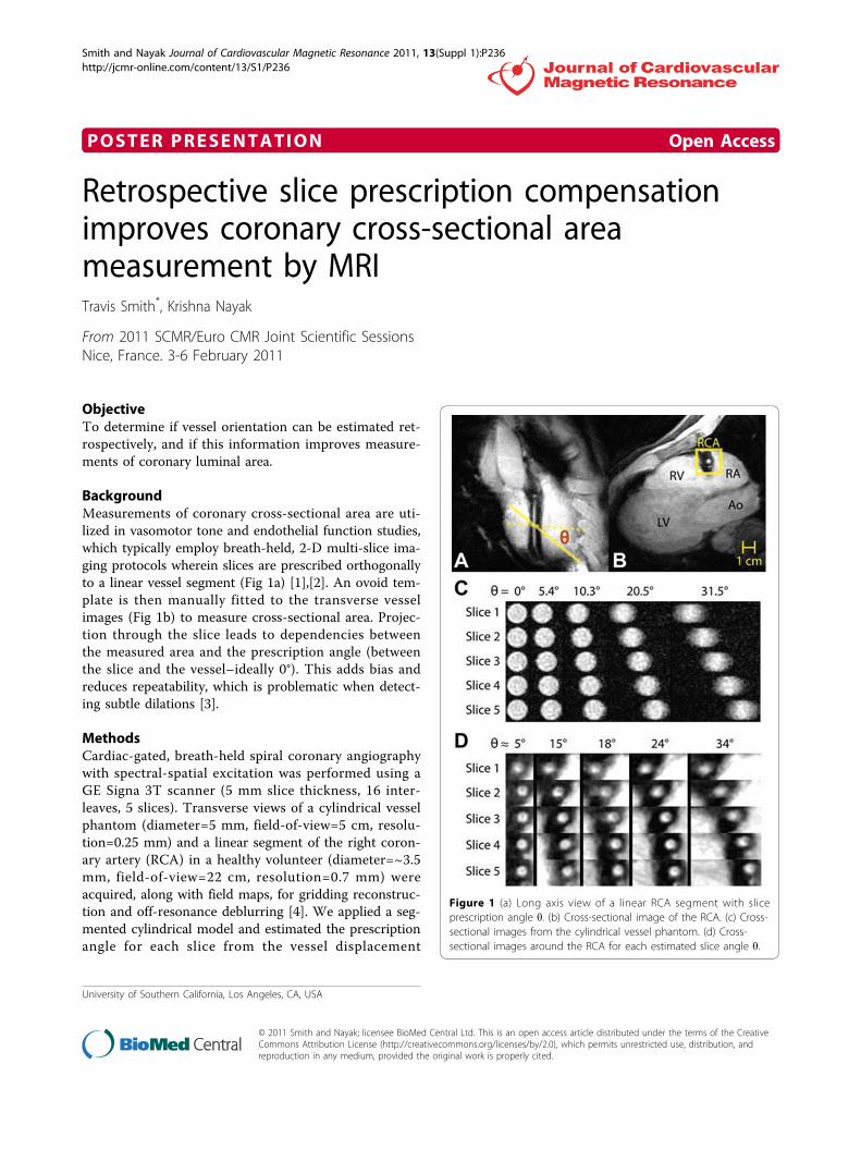
BackgroundMeasurements of coronary cross-sectional area are uti-lized in vasomotor tone and endothelial function studies,which typically employ breath-held, 2-D multi-slice ima-ging protocols wherein slices are prescribed orthogonallyto a linear vessel segment (Fig 1a) [1],[2]. An ovoid tem-plate is then manually fitted to the transverse vesselimages (Fig 1b) to measure cross-sectional area. Projec-tion through the slice leads to dependencies betweenthe measured area and the prescription angle (betweenthe slice and the vessel–ideally 0°). This adds bias andreduces repeatability, which is problematic when detect-ing subtle dilations [3].
MethodsCardiac-gated, breath-held spiral coronary angiographywith spectral-spatial excitation was performed using aGE Signa 3T scanner (5 mm slice thickness, 16 inter-leaves, 5 slices). Transverse views of a cylindrical vesselphantom (diameter=5 mm, field-of-view=5 cm, resolu-tion=0.25 mm) and a linear segment of the right coron-ary artery (RCA) in a healthy volunteer (diameter=~3.5mm, field-of-view=22 cm, resolution=0.7 mm) wereacquired, along with field maps, for gridding reconstruc-tion and off-resonance deblurring [4]. We applied a seg-mented cylindrical model and estimated the prescriptionangle for each slice from the vessel displacement
University of Southern California, Los Angeles, CA, USA
Figure 1 (a) Long axis view of a linear RCA segment with sliceprescription angle θ. (b) Cross-sectional image of the RCA. (c) Cross-sectional images from the cylindrical vessel phantom. (d) Cross-sectional images around the RCA for each estimated slice angle θ.
Smith and Nayak Journal of Cardiovascular Magnetic Resonance 2011, 13(Suppl 1):P236http://jcmr-online.com/content/13/S1/P236
© 2011 Smith and Nayak; licensee BioMed Central Ltd. This is an open access article distributed under the terms of the CreativeCommons Attribution License (http://creativecommons.org/licenses/by/2.0), which permits unrestricted use, distribution, andreproduction in any medium, provided the original work is properly cited.
through the two neighboring slices. The measuredcross-sectional areas were then geometrically compen-sated to correct any apparent ellipticity from non-ortho-gonal prescriptions. This process was repeated withseveral prescription angles ranging from 0° to approxi-mately 30°.
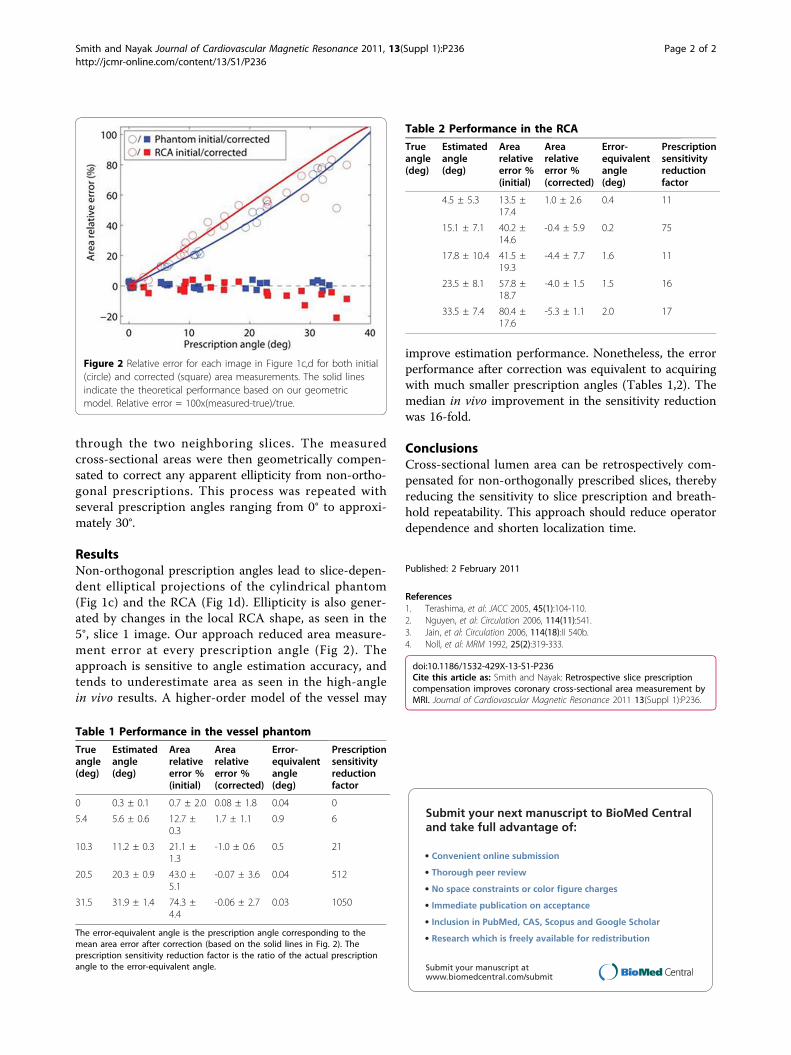
ResultsNon-orthogonal prescription angles lead to slice-depen-dent elliptical projections of the cylindrical phantom(Fig 1c) and the RCA (Fig 1d). Ellipticity is also gener-ated by changes in the local RCA shape, as seen in the5°, slice 1 image. Our approach reduced area measure-ment error at every prescription angle (Fig 2). Theapproach is sensitive to angle estimation accuracy, andtends to underestimate area as seen in the high-anglein vivo results. A higher-order model of the vessel may
improve estimation performance. Nonetheless, the errorperformance after correction was equivalent to acquiringwith much smaller prescription angles (Tables 1,2). Themedian in vivo improvement in the sensitivity reductionwas 16-fold.
ConclusionsCross-sectional lumen area can be retrospectively com-pensated for non-orthogonally prescribed slices, therebyreducing the sensitivity to slice prescription and breath-hold repeatability. This approach should reduce operatordependence and shorten localization time.
Published: 2 February 2011
References1. Terashima, et al: JACC 2005, 45(1):104-110.2. Nguyen, et al: Circulation 2006, 114(11):541.3. Jain, et al: Circulation 2006, 114(18):II 540b.4. Noll, et al: MRM 1992, 25(2):319-333.
doi:10.1186/1532-429X-13-S1-P236Cite this article as: Smith and Nayak: Retrospective slice prescriptioncompensation improves coronary cross-sectional area measurement byMRI. Journal of Cardiovascular Magnetic Resonance 2011 13(Suppl 1):P236.
Submit your next manuscript to BioMed Centraland take full advantage of:
• Convenient online submission
• Thorough peer review
• No space constraints or color figure charges
• Immediate publication on acceptance
• Inclusion in PubMed, CAS, Scopus and Google Scholar
• Research which is freely available for redistribution
Submit your manuscript at www.biomedcentral.com/submit
Figure 2 Relative error for each image in Figure 1c,d for both initial(circle) and corrected (square) area measurements. The solid linesindicate the theoretical performance based on our geometricmodel. Relative error = 100x(measured-true)/true.
Table 1 Performance in the vessel phantom
Trueangle(deg)
Estimatedangle(deg)
Arearelativeerror %(initial)
Arearelativeerror %(corrected)
Error-equivalentangle(deg)
Prescriptionsensitivityreductionfactor
0 0.3 ± 0.1 0.7 ± 2.0 0.08 ± 1.8 0.04 0
5.4 5.6 ± 0.6 12.7 ±0.3
1.7 ± 1.1 0.9 6
10.3 11.2 ± 0.3 21.1 ±1.3
-1.0 ± 0.6 0.5 21
20.5 20.3 ± 0.9 43.0 ±5.1
-0.07 ± 3.6 0.04 512
31.5 31.9 ± 1.4 74.3 ±4.4
-0.06 ± 2.7 0.03 1050
The error-equivalent angle is the prescription angle corresponding to themean area error after correction (based on the solid lines in Fig. 2). Theprescription sensitivity reduction factor is the ratio of the actual prescriptionangle to the error-equivalent angle.
Table 2 Performance in the RCA
Trueangle(deg)
Estimatedangle(deg)
Arearelativeerror %(initial)
Arearelativeerror %(corrected)
Error-equivalentangle(deg)
Prescriptionsensitivityreductionfactor
4.5 ± 5.3 13.5 ±17.4
1.0 ± 2.6 0.4 11
15.1 ± 7.1 40.2 ±14.6
-0.4 ± 5.9 0.2 75
17.8 ± 10.4 41.5 ±19.3
-4.4 ± 7.7 1.6 11
23.5 ± 8.1 57.8 ±18.7
-4.0 ± 1.5 1.5 16
33.5 ± 7.4 80.4 ±17.6
-5.3 ± 1.1 2.0 17
Smith and Nayak Journal of Cardiovascular Magnetic Resonance 2011, 13(Suppl 1):P236http://jcmr-online.com/content/13/S1/P236
Page 2 of 2