Embed Size (px)
Citation preview

Review began 07/08/2021 Review ended 07/09/2021 Published 07/18/2021
© Copyright 2021Madkhali. This is an open access articledistributed under the terms of theCreative Commons Attribution LicenseCC-BY 4.0., which permits unrestricteduse, distribution, and reproduction in anymedium, provided the original author andsource are credited.
Reversible Takotsubo Syndrome ComplicatedWith Acute Thromboembolic Stroke Co-OccurringWith Cola-Induced Severe HypokalemiaMohammed A. Madkhali
1. Internal Medicine, Jazan University Faculty of Medicine, Jizan, SAU 2. Internal Medicine, The University of ToledoMedical Center, Toledo, USA
Corresponding author: Mohammed A. Madkhali, [email protected]
AbstractTakotsubo syndrome (TTS) is a poorly understood phenomenon that manifests as reversible myocardialdamage. Despite the overall benign course of TTS, acute complications and significant mortality andmorbidity are frequently encountered. However, the risk factors for developing such complications are notwell determined. We present a case of reversible TTS within 48 hours after correction of cola-inducedhypokalemia complicated by thromboembolic stroke. We present the patient's clinical presentation withimaging studies and discuss the case in light of related literature. A 62-year-old woman with a longstandinghistory of inadequate oral intake and excessively sweetened cola soft drinks consumption. Presented withprogressive symmetrical quadriparesis and found to have severe hypokalemia with rhabdomyolysis, elevatedtroponin, and acute left ventricular dysfunction. The patient was diagnosed with TTS and hypokalemicparalysis. Her hospital course was complicated with an acute thromboembolic stroke of bilateral occipitallobes and left cerebellum. Takotsubo cardiomyopathy was reversible 48 hours after correction of severehypokalemia. Cardiac ischemic workup was negative for any evidence of myocardial ischemia. In thispresented case, TTS was revisable within 48 hours after the correction of cola-induced hypokalemia.However, patients with similar presentations might have a higher risk for in-hospital thromboemboliccomplications of TTS.
Categories: Cardiology, Internal Medicine, NephrologyKeywords: cola-induced hypokalemia, takotsubo syndrome, hypokalemic paralysis, thromboembolic stroke,rhabdomyolysis.
IntroductionTakotsubo syndrome (TTS) is a reversible myocardial damage that mimics the clinical manifestation of acutecoronary artery disease (ACS). TTS is diagnosed in about 2% of patients presenting to the hospital withsuspected ACS [1]. The precise pathophysiology of TTS is still unknown. However, multiple predisposingphysical and mental risk factors have been proposed in retrospective studies to trigger TTS development [2].Nevertheless, further longitudinal studies are required to prove the association between those risk factorsand TTS.
The overall natural history of TTS is benign; however, some of the affected patients developed acutecomplications with subsequent mortality and morbidity [3]. In this case, we present a patient withcomplicated clinical course consisting of TTS, hypokalemic paralysis, and rhabdomyolysis in the setting ofcola-induced severe hypokalemia.
Case PresentationA 62-year-old Caucasian woman presented to the emergency department with a history of progressivemuscular weakness associated with nausea, vomiting, and diarrhea. At baseline, the patient wasindependently ambulating with a walker. However, she started to have a gradual bilateral upper and lowerextremities weakness for two weeks before her presentation. As a result, she became dependent on herroommate to perform daily living activities (i.e., bathing, dressing, toileting, transferring, and feeding). Shealso reports an associated history of diarrhea three to four times a day since the onset of her symptoms. Shereports a history of weight loss, poor appetite, polyuria, polydipsia, memory impairment, decreased visualacuity, and hearing loss on review of systems. She denied any history of shortness of breath or chest pain.The patient stated that she has been drinking 8-10 cans of 16 oz of Pepsi cola every day. The past medicalhistory is remarkable for chronic obstructive pulmonary disease (COPD), history of abdominal trauma withsubsequent splenectomy, gastroesophageal reflux with esophagitis and hiatal hernia status post failedNissan fundoplication, alcohol abuse, and longstanding multiple psychiatric disorders treated with differentantipsychotics and antidepressants. The patient stated that she had not taken any of her home medicationsfor more than two weeks before the presentation. In addition, she reports a 20 pack-years history of cigarettesmoking and lived with a roommate who has been busy lately and did not provide sufficient support to thepatient.
1, 2
Open Access CaseReport DOI: 10.7759/cureus.16470
How to cite this articleMadkhali M A (July 18, 2021) Reversible Takotsubo Syndrome Complicated With Acute Thromboembolic Stroke Co-Occurring With Cola-InducedSevere Hypokalemia. Cureus 13(7): e16470. DOI 10.7759/cureus.16470

On physical examination, vital signs were normal; the patient looked cachexic and dehydrated with dry skinand mucous membranes. She was also confused with impaired remote memory and appeared in poor hygieneas she could not shower or sanitize her excreta for three to four days due to weakness. Neurologicalexamination showed normal cranial nerves, a decreased strength in the upper and lower extremities (3/5 and2/5, respectively), and normal sensation to soft touch. On abdominal examination, the patient was noted tohave a midline surgical incision with reducible umbilical hernia, no abdominal tenderness was elicited, andshe had normal bowel sounds.
Initial laboratory workup was remarkable for profound hypokalemia; serum potassium was 1.6 mmol/L (refrange: 3.5-5.0) with an elevated troponin of 2.63 ng/mL, brain natriuretic peptide (BNP) of 570 pg/mL, andtotal creatine kinase (CK) 1,391 U/L. Other electrolytes were normal, she had a serum sodium of 144mmol/L, magnesium of 2.2 mg/dl, phosphorous of 2.7 mg/dl and calcium of 8.9 mg/dl. Furthermore, bloodtests for complete blood count (CBC), creatinine, blood urea nitrogen (BUN), thyroid-stimulating hormone(TSH), free T4, lipid and coagulation profiles were unremarkable for any abnormalities. Liver functionshowed an elevated aspartate transaminase (AST) of 55 U/L (ref range: 0-41) but normal alanineaminotransferase (ALT) and alkaline phosphatase. Electrocardiogram on presentation showed a prolongedQT-interval (QTc 583 ms) with U-wave and ST-segment depression on lateral leads (Figure 1).
FIGURE 1: Initial EKG, serum potassium of 1.6 mmol/L.
Therefore, an echocardiogram was done on the day of admission, and it showed a moderate to a severereduction in the left ventricular ejection fraction (estimated LVEF of 30%-35%) associated with apical andanterior wall akinesia. It also showed moderate mitral and mild tricuspid valve regurgitations (Video 1). Thepatient was started on IV heparin infusion for presumed non-ST elevation myocardial infarction.
VIDEO 1: TTE on presentation showing reduced LVEF with apical andanterior walls akinesia.LVEF: left ventricular ejection fraction.
View video here: https://youtu.be/HJ9jW5mXAeE
Hypokalemia was corrected with intravenous and oral potassium chloride replacements. The serumpotassium level returned to a normal value of 4.0 mmol/L on the third day of the hospital stay. Aftercorrecting hypokalemia, the follow-up EKG showed resolutions of ST-depression, improved QT-prolongation(QTc of 463 ms) and disappearance of U-wave (Figure 2).
2021 Madkhali et al. Cureus 13(7): e16470. DOI 10.7759/cureus.16470 2 of 6
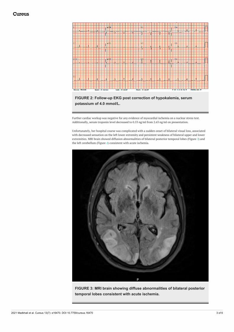
FIGURE 2: Follow-up EKG post correction of hypokalemia, serumpotassium of 4.0 mmol/L.
Further cardiac workup was negative for any evidence of myocardial ischemia on a nuclear stress test.Additionally, serum troponin level decreased to 0.53 ng/ml from 2.63 ng/ml on presentation.
Unfortunately, her hospital course was complicated with a sudden onset of bilateral visual loss, associatedwith decreased sensation on the left lower extremity and persistent weakness of bilateral upper and lowerextremities. MRI brain showed diffusion abnormalities of bilateral posterior temporal lobes (Figure 3) andthe left cerebellum (Figure 4) consistent with acute ischemia.
FIGURE 3: MRI brain showing diffuse abnormalities of bilateral posteriortemporal lobes consistent with acute ischemia.
2021 Madkhali et al. Cureus 13(7): e16470. DOI 10.7759/cureus.16470 3 of 6

FIGURE 4: MRI brain showing left cerebellum diffusion abnormalityconsistent with acute ischemia.
Therefore, she underwent trans-esophageal echocardiogram to rule out left ventricular thrombusformation. It showed resolutions of left ventricular wall motion abnormalities with moderate to severedilatation of the left atrium and no visualized thrombus in the left atrial appendage. Also, it showed anintact atrial septum, and no interatrial shunt on agitated saline bubble study (Video 2).
VIDEO 2: TEE showing resolution of left ventricular wall motionabnormalities.TEE: transesophageal echocardiography.
View video here: https://youtu.be/sSQ-bQmRjpI
Hence, the patient was diagnosed with reversible TTS associated with in-hospital complication ofthromboembolic stroke.
2021 Madkhali et al. Cureus 13(7): e16470. DOI 10.7759/cureus.16470 4 of 6

The patient was treated with anticoagulation and aspirin, IV heparin infusion was switched to oralrivaroxaban (xarelto) 20 mg daily, in addition to aspirin 324 mg daily for outpatient anticoagulation. Ondischarge, the serum level of potassium remained at a normal range with dietary replacement. She wasdischarged to a skilled nursing facility followed by cardiology and neurology clinics for further evaluationand management.
DiscussionThis patient was presented with severe hypokalemia associated with asymptomatic left ventriculardysfunction and elevated cardiac markers suggestive of myocardial injury. Her complicated hospital courseprecludes the planned coronary arteries angiogram and LV angiography to rule out the obstructive coronaryartery disease. Nevertheless, post potassium replacement, this patient had quick reversibility ofcardiomyopathy as shown in the trans-esophageal echocardiogram with no evidence of ischemia on thenuclear stress test. Therefore, the other possibilities of myocarditis or coronary artery diseases (CAD)including vasospastic CAD are very unlikely in this case. Furthermore, this patient has multiple known riskfactors for the development of TTS (i.e., female, postmenopausal, and history of psychiatric disorders) [2].
Low serum potassium is regarded as the most commonly encountered electrolyte abnormality in the clinicalsetting. Most patients with hypokalemia are found to have a mild to a moderate decrease of serumpotassium below 3.6 mmol/L but more than 2.5 mmol/L. Less frequently, patients with severe hypokalemia(i.e., serum potassium of < 2.5 mmol/L) are predisposed to the risks of cardiac arrhythmias and musclenecrosis with subsequent rhabdomyolysis and ascending paralysis [4]. Hypokalemia can result fromdecreased potassium intake, extracellular to intracellular shift, and uncompensated gastrointestinal or renalloss of serum potassium. There are multiple reports of cola-induced severe hypokalemia complicated withmyopathy, rhabdomyolysis, acute kidney injury, and atrial fibrillation following the excessive consumptionof those drinks [5-8]. The proposed mechanism of cola-induced hypokalemia was discussed by Tsimihodimoset al.; they indicated that, when consumed excessively, the unabsorbed fructose content of cola soft drinkscan cause osmotic diarrhea and subsequent gastrointestinal loss of potassium. Additionally, the high glucosecontent in those drinks could result in transient hyperinsulinemia and osmotic diuresis with a consequentintracellular shift of potassium and renal loss, respectively. Furthermore, the caffeine content of cola softdrinks could cause hypokalemia through intracellular potassium redistribution and its known diureticeffect [9]. In addition to excessive consumption of Pepsi cola drinks, this presented patient had other factorscontributing to the severity of hypokalemia. She has been dependent upon sweeten soft drinks as a source ofdaily calories with decreased oral intake of potassium; additionally, the patient has chronic intermittentnausea with vomiting secondary to large hiatal hernia status post failed Nissan fundoplication. Apart fromthose factors, no other potential etiologies for hypokalemia could be considered in this case, including drug-induced hypokalemia since the patient has not been taking home medications - including proton pumpinhibitor (PPI) and antipsychotics - for more than two weeks before her presentation.
In a different clinical setting, severe hypokalemia has been proposed as a potential triggering factor for thedevelopment of TTS. For instance, TTS has been associated with severe hypokalemia secondary to primaryaldosteronism and decreased oral intake [10,11]. Furthermore, excessive caffeine consumption from energydrinks and weight loss supplements has been linked to the occurrence of TTS [12,13]. However, to the best ofour knowledge, there have been no reports of coexistent TTS with cola-induced severe hypokalemia.Therefore, the excessive consumption of caffeinated cola drinks in this patient could represent an unknowntriggering factor for TTS.
The prognosis of TTS is primarily benign in the majority of patients. However, there has been growingevidence highlighting the acute consequential mortality and morbidly associated with this disease.According to the international consensus review of TTS, the acute clinical course of TTS was complicated byatrial fibrillation in 5%-15% and LV thrombus formation in 2%-8% of the affected patients [3]. Nevertheless,the risk factors for developing those acute complications are still elusive. Cervellin et al. (2014) described anassociation between hypokalemia and elevated serum levels of D-dimer among patients with acute atrialfibrillation. Accordingly, this association could imply a higher risk of thromboembolism in the setting ofhypokalemia [14]. The diagnosis of atrial fibrillation was neither evident during the presentation of our casenor detected through cardiac telemetry throughout her hospital stay. However, this does not preclude thepossibility of paroxysmal atrial fibrillation with consequent LV thrombus formation before the patient’spresentation. As a matter of fact, in a recent retrospective study, the left ventricular thrombus was onlydetected in three out of 17 cases with ischemic stroke and TTS. Additionally, only 11% of patients withthromboembolic events were found to have in-hospital complication of atrial fibrillation. This study wasconducted on 1,676 registered patients in the international Takotsubo database [15]. To sum up, thecoexistence of TTS with cola-induced severe hypokalemia could carry a higher risk for majorthromboembolic complications as described in this case.
ConclusionsIn this presented case, TTS was reversible within 48 hours after the correction of cola-induced hypokalemia.However, patients with similar presentations might have a higher risk for in-hospital thromboemboliccomplications of TTS.
2021 Madkhali et al. Cureus 13(7): e16470. DOI 10.7759/cureus.16470 5 of 6
Additional InformationDisclosuresHuman subjects: Consent was obtained or waived by all participants in this study. Conflicts of interest: Incompliance with the ICMJE uniform disclosure form, all authors declare the following: Payment/servicesinfo: All authors have declared that no financial support was received from any organization for thesubmitted work. Financial relationships: All authors have declared that they have no financialrelationships at present or within the previous three years with any organizations that might have aninterest in the submitted work. Other relationships: All authors have declared that there are no otherrelationships or activities that could appear to have influenced the submitted work.
AcknowledgementsThanks to the University of Toledo for unrestricted support and Dr.Steven Zook for his supervision inwriting this paper.
References1. Akashi YJ, Nef HM, Lyon AR: Epidemiology and pathophysiology of Takotsubo syndrome. Nat Rev Cardiol.
2015, 12:387-97. 10.1038/nrcardio.2015.392. Ghadri JR, Wittstein IS, Prasad A, et al.: International Expert Consensus Document on Takotsubo Syndrome
(Part I): Clinical Characteristics, Diagnostic Criteria, and Pathophysiology. Eur Heart J. 2018, 39:2032-46.10.1093/eurheartj/ehy076
3. Ghadri JR, Wittstein IS, Prasad A, et al.: International Expert Consensus Document on Takotsubo Syndrome(Part II): Diagnostic Workup, Outcome, and Management. Eur Heart J. 2018, 39:2047-62.10.1093/eurheartj/ehy077
4. Gennari FJ: Hypokalemia. N Engl J Med. 1998, 339:451-8. 10.1056/NEJM1998081333907075. Sharma R, Guber HA: Cola-induced hypokalemia-a case report and review of the literature . Endocr Pract.
2013, 19:e21-3. 10.4158/EP12241.CR6. Ferrazzoli D, Sabetta A, Palamara G, Caremani L, Capobianco M, Balbi P, Frazzitta G: A cola-induced
hypokalemic rhabdomyolysis with electromyographic evaluation: a case report. SAGE Open Med Case Rep.2017, 5:2050313X17695717. 10.1177/2050313X17695717
7. Lee TW, Bae E, Hwang K, et al.: Severe hypokalemic paralysis and rhabdomyolysis occurring after bingeeating in a young bodybuilder: case report. Medicine. 2017, 96:e8251. 10.1097/MD.0000000000008251
8. Zakharova E: Severe hypokalemia due to excessive coca-cola consumption, presenting with tetraparesis,rhabdomyolysis, atrial fibrillation and hypokalemic nephropathy - case report. J Urol Nephrol Open Access.2016, 2:1-4. 10.15226/2473-6430/2/2/00114
9. Tsimihodimos V, Kakaidi V, Elisaf M: Cola-induced hypokalaemia: pathophysiological mechanisms andclinical implications. Int J Clin Pract. 2009, 63:900-2. 10.1111/j.1742-1241.2009.02051.x
10. Ou Y, Zhao Z, Tsauo J, Jiang L, Yang Y, Chen M: An unusual case of takotsubo syndrome withhyperaldosteronism as the potential cause. J Clin Endocrinol Metab. 2018, 103:12-5. 10.1210/jc.2017-01117
11. Andreozzi F, Cuminetti G, Karmali R, Kamgang P: Electrolyte disorders as triggers for takotsubocardiomyopathy. Eur J Case Rep Intern Med. 2018, 5:000760. 10.12890/2018_000760
12. Kaoukis A, Panagopoulou V, Mojibian HR, Jacoby D: Reverse Takotsubo cardiomyopathy associated with theconsumption of an energy drink. Circulation. 2012, 125:1584-5. 10.1161/CIRCULATIONAHA.111.057505
13. Dai Z, Fukuda T, Kinoshita K, Komiyama N: Inverted takotsubo cardiomyopathy associated with theconsumption of a weight management supplement. CJC Open. 2020, 2:26-9. 10.1016/j.cjco.2019.11.002
14. Cervellin G, Bonfanti L, Picanza A, Lippi G: Serum potassium levels inversely correlate with D-dimer inpatients with acute-onset atrial fibrillation. Arq Bras Cardiol. 2015, 104:181-4. 10.5935/abc.20140187
15. Ding KJ, Cammann VL, Szawan KA, et al.: Intraventricular thrombus formation and embolism in takotsubosyndrome: insights from the International Takotsubo Registry. Arterioscler Thromb Vasc Biol. 2020, 40:279-87. 10.1161/ATVBAHA.119.313491
2021 Madkhali et al. Cureus 13(7): e16470. DOI 10.7759/cureus.16470 6 of 6