Embed Size (px)
Citation preview
Reversing the Outcome of Synapse Elimination atDeveloping Neuromuscular Junctions In Vivo: Evidencefor Synaptic Competition and Its MechanismStephen G. Turney*, Jeff W. Lichtman*
Center for Brain Science and Department of Molecular and Cellular Biology, Harvard University, Cambridge, Massachusetts, United States of America
Abstract
During mammalian development, neuromuscular junctions and some other postsynaptic cells transition from multiple- tosingle-innervation as synaptic sites are exchanged between different axons. It is unclear whether one axon invades synapticsites to drive off other inputs or alternatively axons expand their territory in response to sites vacated by other axons. Herewe show that soon-to-be-eliminated axons rapidly reverse fate and grow to occupy vacant sites at a neuromuscular junctionafter laser removal of a stronger input. This reversal supports the idea that axons take over sites that were previouslyvacated. Indeed, during normal development we observed withdrawal followed by takeover. The stimulus for axon growthis not postsynaptic cell inactivity because axons grow into unoccupied sites even when target cells are functionallyinnervated. These results demonstrate competition at the synaptic level and enable us to provide a conceptual frameworkfor understanding this form of synaptic plasticity.
Citation: Turney SG, Lichtman JW (2012) Reversing the Outcome of Synapse Elimination at Developing Neuromuscular Junctions In Vivo: Evidence for SynapticCompetition and Its Mechanism. PLoS Biol 10(6): e1001352. doi:10.1371/journal.pbio.1001352
Academic Editor: William A. Harris, University of Cambridge, United Kingdom
Received January 25, 2012; Accepted May 15, 2012; Published June 26, 2012
Copyright: � 2012 Turney, Lichtman. This is an open-access article distributed under the terms of the Creative Commons Attribution License, which permitsunrestricted use, distribution, and reproduction in any medium, provided the original author and source are credited.
Funding: This work was supported by the Gatsby Charitable Foundation and by US National Institutes of Health (NIH) R01 grant NS020364-23 to J.W.L. Thefunders had no role in study design, data collection and analysis, decision to publish, or preparation of the manuscript.
Competing Interests: The authors have declared that no competing interests exist.
Abbreviations: AChR, acetylcholine receptor; CFP, cyan fluorescent protein; GFP, green fluorescent protein; SIT, silicon-intensified target; YFP, yellow fluorescentprotein
* E-mail: [email protected] (SGT); [email protected] (JWL)
Introduction
Physiological evidence that axons completely lose connections
with some postsynaptic cells as part of naturally occurring
development was first observed at the neuromuscular junction in
mammals more than 40 years ago [1]. Since then analogous axonal
loss has been seen in many parts of the central and peripheral
nervous systems [2,3]. While the underlying mechanism is still
unclear anywhere, evidence suggests that in the neuromuscular
system local events at or near the synapse regulate the process.
Evidence for local regulation includes the following: (1) the axonal
inputs that are eliminated from neuromuscular junctions do so by
gradually vacating their synaptic contact sites [4] rather than
suddenly undergoing degeneration, as occurs when axons are
damaged [5]; (2) the axon that ultimately is maintained increases its
synaptic contact area by gradually occupying many of the synaptic
sites that were previously occupied by other motor axons [4]; (3) the
loss and acquisition of synaptic sites is paralleled by a local reduction
and strengthening in synaptic efficacy [6]; (4) the loss of axonal
branches from one axon that projects to many muscle fibers occurs
asynchronously, suggesting that the timing of elimination is not set
by a signal from the cell soma but regulated independently at each
neuromuscular junction site [7]; (5) local differences between the
synaptic activity of axons converging at the same neuromuscular
junction have the ability to cause synapses to be eliminated [8,9]; (6)
local changes in target cell signaling can affect synapse maintenance
[10]; and (7) once an axon has vacated all of its synaptic territory at a
neuromuscular junction, it locally sheds cytoplasm that is internal-
ized by glia associated with the neuromuscular junction entry zone
[4,11]. Collectively, these data argue that the ultimate identity of the
one permanent presynaptic input to a muscle fiber is determined by
events occurring at the level of individual neuromuscular junctions.
Other data suggest that neuronal properties (as opposed to synaptic
properties) such as an axon’s biochemical identity or its firing
pattern play a role in determining the outcome of synapse
elimination, but even these may operate through local synaptic
mechanisms [12].
Several different local mechanisms have been proposed to
explain what drives this process forward. One idea is that
individual axon branch removal occurs randomly from a motor
unit and is related to an intrinsic requirement that neurons scale
back their initially exuberant arbors [13]. A second idea is that the
fate of axons is predetermined by positional or perhaps other
molecular cues that specify which axon is the best match for each
muscle fiber [14–16]. A third possibility is that axons converging at
a neuromuscular junction compete with each other causing all but
the ultimate victor to be removed. It is also possible that some
combination of these forces is at play. The idea that synapse
elimination is primarily the result of a competitive interaction
between the innervating axons was originally proposed because in
many muscles the loss of inputs results in exactly one axon
remaining at each junction [17].
PLoS Biology | www.plosbiology.org 1 June 2012 | Volume 10 | Issue 6 | e1001352
A competitive mechanism is also suggested by the fact that
increases in the size and strength of one input are related to the
shrinkage and weakening of other axons [4,6]. But there is no
direct evidence supporting such a mechanism at the synaptic level,
and while a number of studies have suggested inter-axonal
competition as the likely mechanism, to our knowledge none have
shown a direct reciprocal causal relationship between the fates of
the surviving and eliminated axons during developmental synapse
elimination [8,18–21]. Moreover, in some circumstances multiple
axons can remain at the same neuromuscular junction [22–24]
indicating that in some circumstances either competition can be
overridden by other factors or that the whole process is not
competitive in the first place. Understanding what drives the
process forward is important because this mechanism seems to be
one of the strategies at play more generally in the developing
mammalian nervous system to help shape it to the particular
environment in which it finds itself.
Thus we felt that it would be worthwhile to directly test whether
or not synapse elimination is driven by interaxonal competition.
We reasoned that if competition between two axons vying for the
same postsynaptic site was causing the elimination of one of them,
then that axon should not ever be removed if its putative
competitor was no longer present. We therefore ablated the axon
that had the greatest likelihood of being maintained at a
neuromuscular junction to see if the weaker input would have a
reversal of fate and now be maintained. If this outcome did occur,
we were interested to know when the decision for an axon to be
eliminated finally becomes irreversible. For example, it was
possible that axons compete and set into motion a program of
elimination that is irreversible even many days before the axonal
loss finally occurs. The cascade that leads to neuronal cell death
has such points of no return [25], which imply that the
downstream events are irreversible. Might the same be true for
the program leading to synapse elimination? If conversely the
synapse elimination program were readily reversible, even at late
stages, it would argue that axons remain viable in an ongoing
effort to maintain access to the target muscle fiber. In this latter
case, the synaptic reorganization events might be played out with
little lag between the competitive actions and their consequences
on axonal growth or retraction. For example, if one input
‘‘pushed’’ another off a synaptic site, axon withdrawal would be
temporally coordinated with near simultaneous axonal takeover,
allowing for a highly dynamic process where axonal territory
might wax and wane on timescales of minutes or hours. Indeed
time-lapse imaging shows that an axon’s synaptic territory can be
increased and decreased in a dynamic manner [4]. Despite the
relatively high temporal and spatial resolution images in many
previous studies, however, the motive force for growth and
retraction and the details of these behaviors for interacting axons
remain obscure.
The experiments reported here allowed us to examine how
developing axons respond to vacated synaptic sites. We developed
a laser-based technique with which we could remove one of two
closely spaced axons that innervated the same neuromuscular
junction. This technique showed that axons readily grew to occupy
vacant sites even when they appeared to be in the process of
withdrawing at the time the sites were vacated. In addition we
observed that axons were stimulated to grow even in situations
when the muscle fiber was still active. This combination of
synaptic vacancy and the axonal takeover it induces allows us to
explain a range of complex phenomena associated with synapse
elimination.
Results
Damaging Axonal Branches in vivo with an UltrafastPulsed Laser
In order to selectively remove one axonal branch without
damaging any neighboring axons in vivo, we used a diode-pumped
mode-locked Ti:Sapphire laser oscillator to cause localized
phototoxicity in fluorescent protein containing motor axons in
living mice. By taking advantage of non-linear aspects of multi-
photon excitation, we could damage one axon and leave
immediately adjacent axons unscathed. The focused laser spot
was positioned over an axon branch using a modified scanning
microscope system (see Materials and Methods and Figure S1),
and the axon’s fluorescence was bleached at one location. One
hundred and seventy-three axons were irradiated (71 in adult
neuromuscular junctions and 102 in 1-wk-old neonates).
Damage to axons typically evolved over 30–45 min and the
whole process of axon removal required many hours. Even though
we observed bleaching of the axon segment at the time of
irradiation, evidence for structural damage only became apparent
within 10–20 min (see Figures 1–3). Signs of axon damage
included dramatic swelling of the axon distal to the site of laser
focus and a progressive widening of the region of non-fluorescence
both distal and proximal to the laser irradiation site. Presumably
this loss of fluorescence is secondary to leakage of proteins from the
cytoplasm at the damage site. This phase which typically lasted up
to several hours was followed by the complete disappearance of the
distal axon save for occasionally a few small disconnected
fluorescent fragments that ultimately all disappeared by 10 h. In
the proximal direction the damage initiated a die-back that was
reminiscent both in time course and scale of ‘‘acute axonal
degeneration’’ of damaged central axons [26]. Typically, the die-
back stopped at the proximal branch point (Figure 2), although
sometimes it extended anterogradely from the branch point to
cause the disappearance of other terminal branches. If the
fluorescence at the laser spot recovered after several minutes, that
was an indication that the fluorescence in the axonal branch had
been bleached but the axon was not seriously damaged because no
subsequent changes were noted over the next half hour to hour, or
the following day (see Materials and Methods for details).
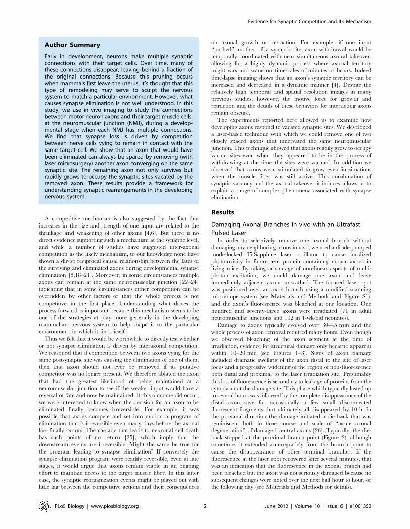
Author Summary
Early in development, neurons make multiple synapticconnections with their target cells. Over time, many ofthese connections disappear, leaving behind a fraction ofthe original connections. Because this pruning occurswhen mammals first leave the uterus, it’s thought that thistype of remodeling may serve to sculpt the nervoussystem to match a particular environment. However, whatcauses synapse elimination is not well understood. In thisstudy, we use in vivo imaging to study the connectionsbetween motor neuron axons and their target muscle cells,at the neuromuscular junction (NMJ), during a develop-mental stage when each NMJ has multiple connections.We find that synapse loss is driven by competitionbetween nerve cells vying to remain in contact with thesame target cell. We show that an axon that would havebeen eliminated can always be spared by removing (withlaser microsurgery) another axon converging on the samesynaptic site. The remaining axon not only survives butrapidly grows to occupy the synaptic sites vacated by theremoved axon. These results provide a framework forunderstanding synaptic rearrangements in the developingnervous system.
Evidence for Synaptic Competition and Its Mechanism
PLoS Biology | www.plosbiology.org 2 June 2012 | Volume 10 | Issue 6 | e1001352
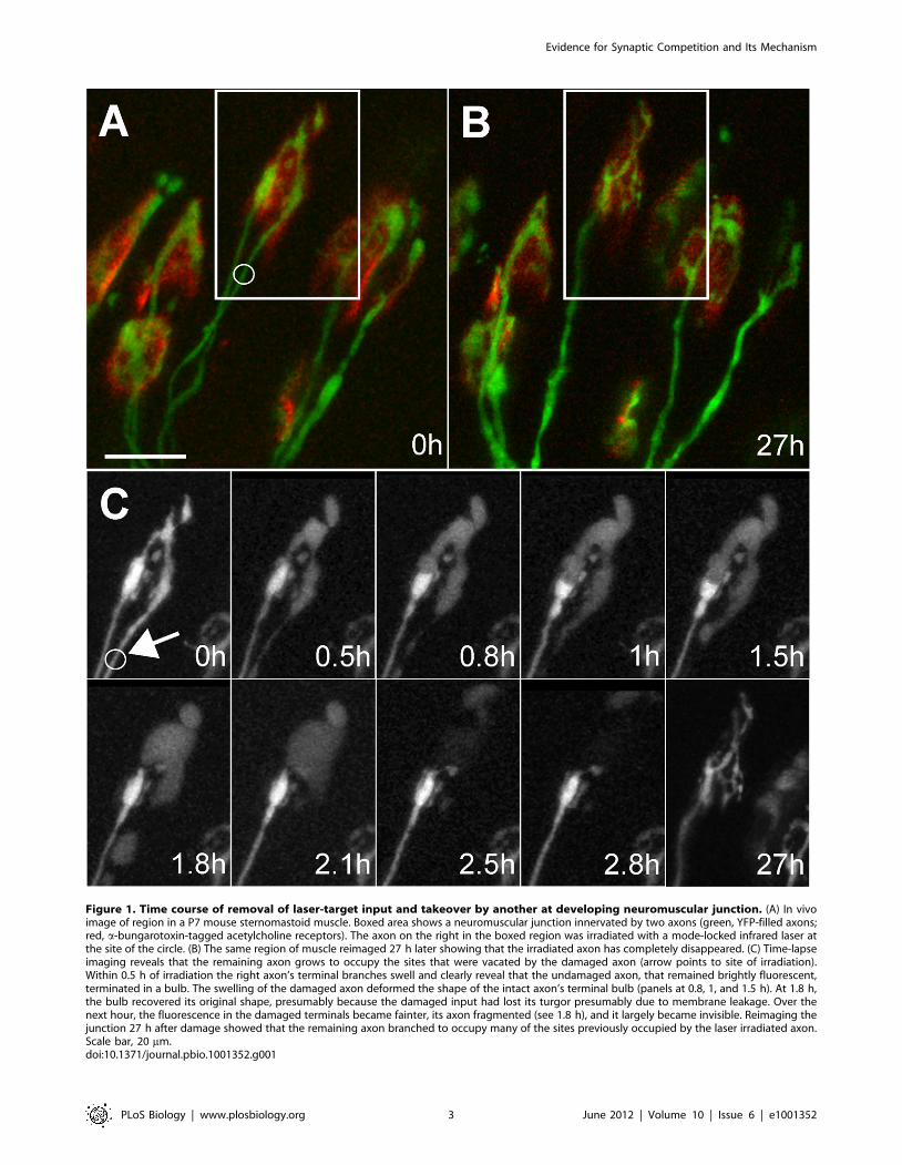
Figure 1. Time course of removal of laser-target input and takeover by another at developing neuromuscular junction. (A) In vivoimage of region in a P7 mouse sternomastoid muscle. Boxed area shows a neuromuscular junction innervated by two axons (green, YFP-filled axons;red, a-bungarotoxin-tagged acetylcholine receptors). The axon on the right in the boxed region was irradiated with a mode-locked infrared laser atthe site of the circle. (B) The same region of muscle reimaged 27 h later showing that the irradiated axon has completely disappeared. (C) Time-lapseimaging reveals that the remaining axon grows to occupy the sites that were vacated by the damaged axon (arrow points to site of irradiation).Within 0.5 h of irradiation the right axon’s terminal branches swell and clearly reveal that the undamaged axon, that remained brightly fluorescent,terminated in a bulb. The swelling of the damaged axon deformed the shape of the intact axon’s terminal bulb (panels at 0.8, 1, and 1.5 h). At 1.8 h,the bulb recovered its original shape, presumably because the damaged input had lost its turgor presumably due to membrane leakage. Over thenext hour, the fluorescence in the damaged terminals became fainter, its axon fragmented (see 1.8 h), and it largely became invisible. Reimaging thejunction 27 h after damage showed that the remaining axon branched to occupy many of the sites previously occupied by the laser irradiated axon.Scale bar, 20 mm.doi:10.1371/journal.pbio.1001352.g001
Evidence for Synaptic Competition and Its Mechanism
PLoS Biology | www.plosbiology.org 3 June 2012 | Volume 10 | Issue 6 | e1001352
Laser Removal of One Axonal Input to a MultiplyInnervated Neuromuscular Junction Invariably Leads toTakeover of the Synaptic Site by the Remaining Input
We attempted to remove one of the axons converging at
multiply innervated neuromuscular junction in early postnatal life.
In anesthetized mice that were 7–8 d old we located neuromus-
cular junctions in the superficial (ventral) surface of the
sternomastoid muscle that were innervated by two axons. In the
sternomastoid muscle about half the neuromuscular junctions are
multiply innervated at 1 wk of age, whereas at 2 wk the number of
multiply innervated neuromuscular junctions is very small
(,0.1%) [8]. At 1 wk nearly all of the multiply innervated
junctions are contacted by only two axons [4,7], indicating that
each of these junctions will lose one input over the next several
days. Using multi-photon laser irradiation we successfully removed
one axon from each of 15 multiply innervated neuromuscular
junctions. At an additional 87 neonatal muscle fibers, axons were
damaged but the experiment failed for other reasons including
connective tissue buildup and muscle fiber rotation that obscured
details when we returned to the muscle the next day; inadvertent
muscle, nerve, or blood vessel damage; or occasional animal
mortality post-surgery. After confirming that the axon was
damaged during the imaging session (up to 3 h) we sutured the
neck wound and allowed the animals to recover. In most cases
(10/15) we intentionally irradiated the axon with the larger
caliber. In 4/15 junctions the two axons had nearly the same
caliber, but based on their appearance at the site of entry into the
junction, we could identify the axon that we thought had less
territory; we then irradiated the larger input. In the remaining case
we intentionally laser irradiated the axon with the smaller caliber.
In all of these junctions the selectivity of the laser damage was
apparent because whereas the damaged axon disappeared, in no
case did the non-targeted axon show any swelling, fragmentation,
bleaching, or loss (Figures 1–3). Because in this experiment the two
axons were labeled with the same fluorescent protein and in most
cases their territories coalesced at light microscopic resolutions, it
was not possible to know precisely how much territory the
remaining axon occupied except retrospectively. Once the laser
irradiated axon had disappeared, it was easy to see the extent of
the territory occupied by the remaining axon (Figure 3B). In the 14
cases where we attempted to remove the stronger input, the
remaining axon occupied half or less of the junctional acetylcho-
line receptor (AChR) sites (mean 12%), and in the one case where
we irradiated the thinner axon, the remaining axon occupied 80%
of junction (Table 1). The territories occupied by these axons were
consistent with previous work showing that terminal axon caliber
correlated with synaptic territory [7]. Because the axon occupying
the majority of the territory at postnatal day 7 or 8 (P7 or P8) was
more than twice as likely to remain at a junction than the axon
occupying the minority of the territory [4], we would anticipate
that in most of the 14 cases where we targeted the stronger axon,
the remaining undamaged axon would have been eliminated had
we not perturbed the system.
Nonetheless, when we re-anesthetized the mice a day later
and returned to the same muscle fibers, in none of the 14 cases
had the remaining axon withdrawn. Nor in any case did we see
any evidence of regrowth of the damaged axon. We were
certain that the axon remaining at the junction was the axon
that was not irradiated on the previous day because its site of
entry into the junction was in each case the same as the site
where the thin axon was situated on the day of laser irradiation
(see Figure 3).
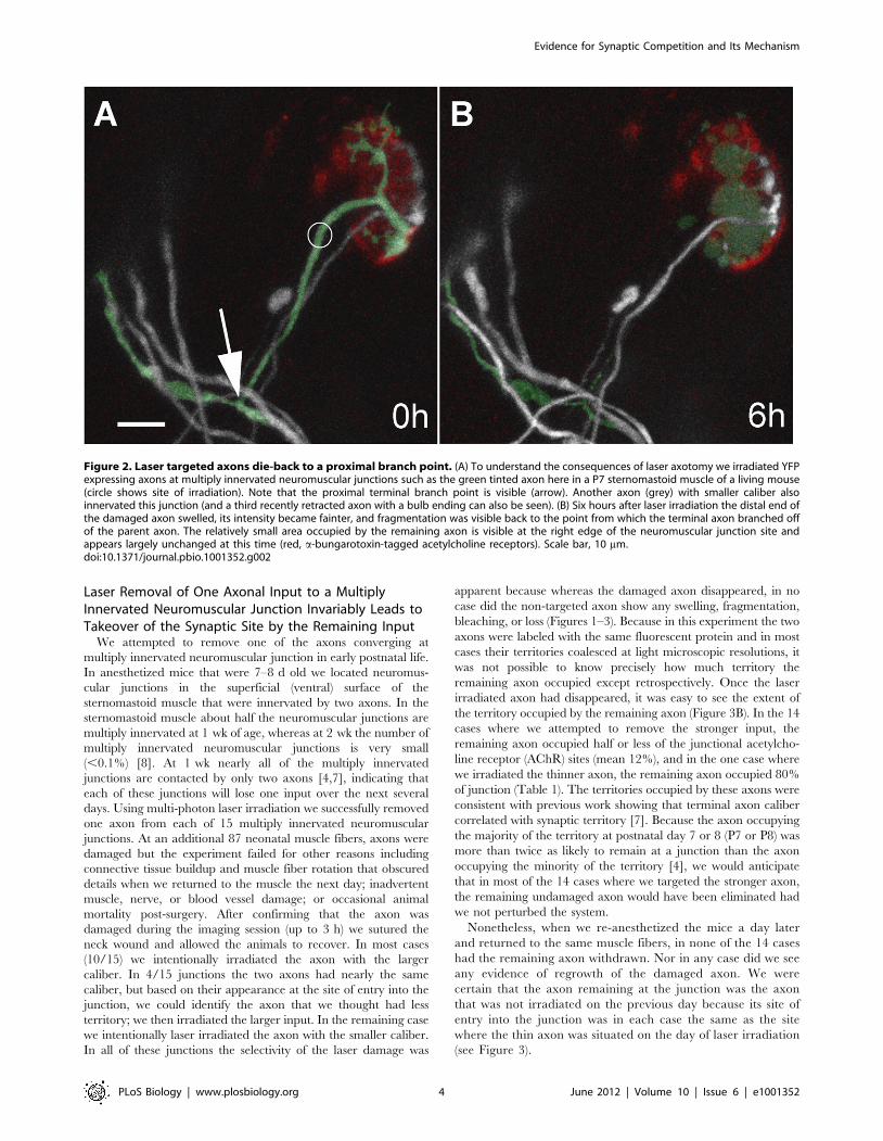
Figure 2. Laser targeted axons die-back to a proximal branch point. (A) To understand the consequences of laser axotomy we irradiated YFPexpressing axons at multiply innervated neuromuscular junctions such as the green tinted axon here in a P7 sternomastoid muscle of a living mouse(circle shows site of irradiation). Note that the proximal terminal branch point is visible (arrow). Another axon (grey) with smaller caliber alsoinnervated this junction (and a third recently retracted axon with a bulb ending can also be seen). (B) Six hours after laser irradiation the distal end ofthe damaged axon swelled, its intensity became fainter, and fragmentation was visible back to the point from which the terminal axon branched offof the parent axon. The relatively small area occupied by the remaining axon is visible at the right edge of the neuromuscular junction site andappears largely unchanged at this time (red, a-bungarotoxin-tagged acetylcholine receptors). Scale bar, 10 mm.doi:10.1371/journal.pbio.1001352.g002
Evidence for Synaptic Competition and Its Mechanism
PLoS Biology | www.plosbiology.org 4 June 2012 | Volume 10 | Issue 6 | e1001352
In each case, however, the axon that remained changed in a
striking way. Each of these thin axons now extended branches
throughout the postsynaptic area to fully occupy the territory
formerly overlain by the laser irradiated axon. In all but one case
the growth response appeared complete within the first 24 h after
laser exposure. The axonal expansion of territory was reminiscent
of the ‘‘takeover’’ seen during normal synapse elimination at the
neuromuscular junction: the advancing axon specifically enlarged
its coverage of the adjacent postsynaptic AChR sites without
extending sprouts to new sites [4]. Because ordinarily smaller
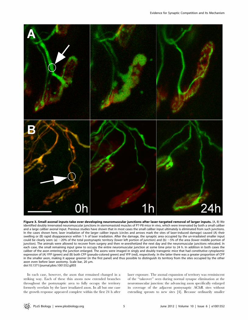
Figure 3. Small axonal inputs take over developing neuromuscular junctions after laser-targeted removal of larger inputs. (A, B) Weidentified doubly innervated neuromuscular junctions in sternomastoid muscles of P7-P8 mice in vivo, which were innervated by both a small caliberand a large caliber axonal input. Previous studies have shown that in most cases the small caliber input ultimately is eliminated from such junctions.In the cases shown here, laser irradiation of the larger caliber inputs (circles and arrows mark the sites of laser-induced damage) caused (A) theirswelling or (B) rapid disappearance within 1 h of laser irradiation. After the damage, the synaptic area occupied by the un-irradiated smaller inputcould be clearly seen: (a) ,20% of the total postsynaptic territory (lower left portion of junction) and (b) ,5% of the area (lower middle portion ofjunction). The animals were allowed to recover from surgery and then re-anesthetized the next day and the neuromuscular junctions relocated. Ineach case, the small remaining input grew to occupy the entire neuromuscular junction at some time prior to 24 h. In addition in both cases thecaliber of the axon entering the junction enlarged. The axons were imaged in singly and doubly transgenic mice that had constitutive cytoplasmicexpression of (A) YFP (green) and (B) both CFP (pseudo-colored green) and YFP (red), respectively. In the latter there was a greater proportion of CFPin the smaller axon, making it appear greener (in the first panel) and thus possible to distinguish its territory from the sites occupied by the otheraxon even before laser axotomy. Scale bar, 20 mm.doi:10.1371/journal.pbio.1001352.g003
Evidence for Synaptic Competition and Its Mechanism
PLoS Biology | www.plosbiology.org 5 June 2012 | Volume 10 | Issue 6 | e1001352
axons were twice as likely to leave a multiply innervated junction
than larger ones [4], the probability that none of them (14 cases)
would have withdrawn is very low (probability ,0.0000003).
Therefore, the fate of the weaker axon was changed by removing
the stronger input, a result that argues that competition is the
cause of the elimination of the weaker input.
We were interested to know what occurred in the immediate
aftermath of removing the other input. In particular, did the
remaining axon continue to be eliminated for some time,
suggesting, for example, that competitive effects have some
momentum and a certain amount of time is required before an
axon can change its fate? We found, however, that at no time after
axon removal did the remaining axon show any evidence of
continued elimination. In three cases we reimaged junctions less
than 24 h after the initial view (one at 6 h, one at 12 h, and one at
17 h). At the 6 h and 12 h views, the remaining axons had not lost
any territory (see Figure 2). The junction viewed at 17 h had
already shown signs of expansion. Unfortunately, it was not
possible for us to anesthetize the same animal for imaging more
than once per day and have it survive, so the exact time axons
began to grow following laser damage of the other synaptic
occupant remains unclear.
We do know, however, that at some point after a period of
quiescence that lasted up to 12 h, the territory occupied by the
remaining axon changed rapidly. In 13/13 junctions imaged at
24 h after laser induced removal of the stronger input, the
remaining axon had expanded dramatically. In all but one case the
axon occupied the entire postsynaptic site, and in the one other
case, it occupied 75% of it (Table 1A, Figures 1 and 3). The
increase in territory by the remaining axon was often matched by
a thickening of the caliber of its preterminal branch (Table 1A,
Figures 1 and 3). In the one junction we studied in which the
weaker input was intentionally eliminated, we also noted complete
takeover of its territory by the stronger axon at 24 h (Table 1A).
Thus by 1 d after the laser removal of one axon at dually
innervated neuromuscular junctions, the remaining input had
grown and now appeared identical to the axons that survive
naturally occurring synapse elimination and singly innervate
neuromuscular junctions. In each case the undamaged axon
now occupied all or nearly all the postsynaptic territory and
possessed a thick preterminal axon. Importantly, in 6 of the 15
cases, the axon that remained had occupied less than 5% of the
junction at the time of axon removal. These axons were as
effective in taking over the remaining territory as axons that had a
larger footprint at the time of axon removal (Figure 3B and
Table 1A). We thus conclude that once a competing axon is
removed the remaining axon, within hours, and irrespective of the
contact area of its terminal arbor, changes its fate to take the
position and characteristics of the dominant axon.
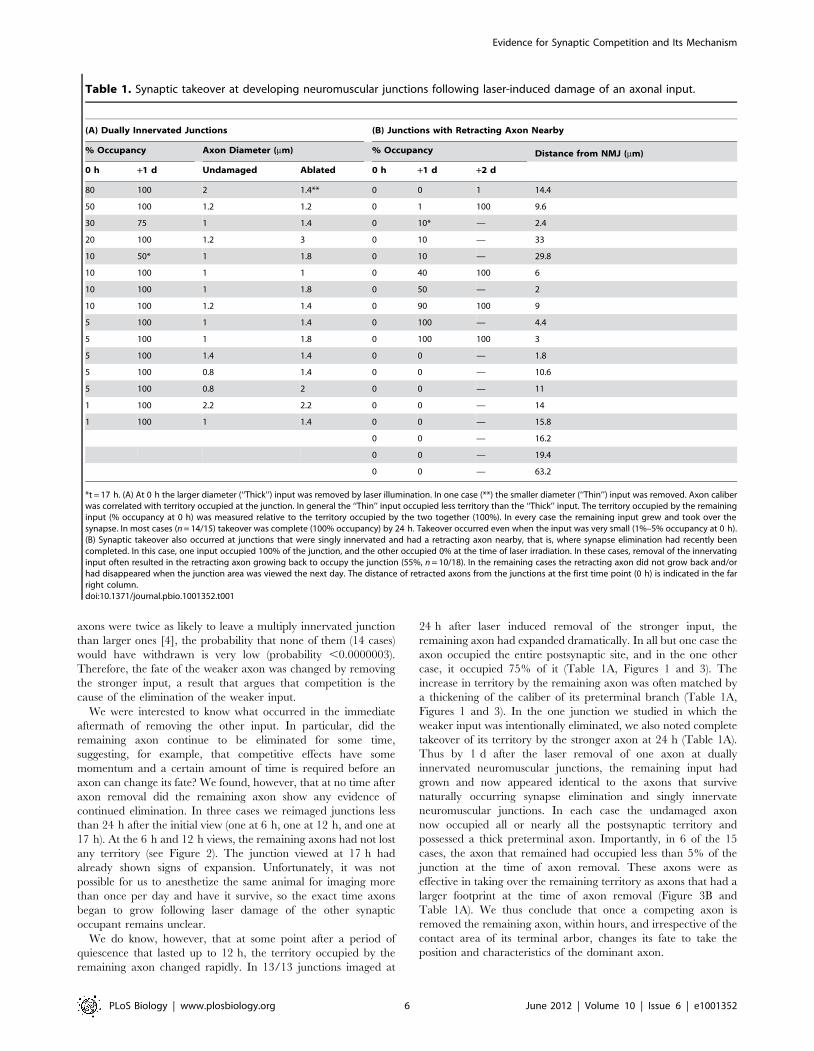
Table 1. Synaptic takeover at developing neuromuscular junctions following laser-induced damage of an axonal input.
(A) Dually Innervated Junctions (B) Junctions with Retracting Axon Nearby
% Occupancy Axon Diameter (mm) % Occupancy Distance from NMJ (mm)
0 h +1 d Undamaged Ablated 0 h +1 d +2 d
80 100 2 1.4** 0 0 1 14.4
50 100 1.2 1.2 0 1 100 9.6
30 75 1 1.4 0 10* — 2.4
20 100 1.2 3 0 10 — 33
10 50* 1 1.8 0 10 — 29.8
10 100 1 1 0 40 100 6
10 100 1 1.8 0 50 — 2
10 100 1.2 1.4 0 90 100 9
5 100 1 1.4 0 100 — 4.4
5 100 1 1.8 0 100 100 3
5 100 1.4 1.4 0 0 — 1.8
5 100 0.8 1.4 0 0 — 10.6
5 100 0.8 2 0 0 — 11
1 100 2.2 2.2 0 0 — 14
1 100 1 1.4 0 0 — 15.8
0 0 — 16.2
0 0 — 19.4
0 0 — 63.2
*t = 17 h. (A) At 0 h the larger diameter (‘‘Thick’’) input was removed by laser illumination. In one case (**) the smaller diameter (‘‘Thin’’) input was removed. Axon caliberwas correlated with territory occupied at the junction. In general the ‘‘Thin’’ input occupied less territory than the ‘‘Thick’’ input. The territory occupied by the remaininginput (% occupancy at 0 h) was measured relative to the territory occupied by the two together (100%). In every case the remaining input grew and took over thesynapse. In most cases (n = 14/15) takeover was complete (100% occupancy) by 24 h. Takeover occurred even when the input was very small (1%–5% occupancy at 0 h).(B) Synaptic takeover also occurred at junctions that were singly innervated and had a retracting axon nearby, that is, where synapse elimination had recently beencompleted. In this case, one input occupied 100% of the junction, and the other occupied 0% at the time of laser irradiation. In these cases, removal of the innervatinginput often resulted in the retracting axon growing back to occupy the junction (55%, n = 10/18). In the remaining cases the retracting axon did not grow back and/orhad disappeared when the junction area was viewed the next day. The distance of retracted axons from the junctions at the first time point (0 h) is indicated in the farright column.doi:10.1371/journal.pbio.1001352.t001
Evidence for Synaptic Competition and Its Mechanism
PLoS Biology | www.plosbiology.org 6 June 2012 | Volume 10 | Issue 6 | e1001352
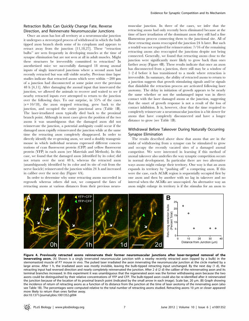
Retraction Bulbs Can Quickly Change Fate, ReverseDirection, and Reinnervate Neuromuscular Junctions
Once an axon has lost all territory at a neuromuscular junction
it undergoes a stereotyped process of withdrawal in which the bulb
tipped axon branch sheds some of its cytoplasm and appears to
retract away from the junction [11,18,27]. These ‘‘retraction
bulbs’’ are seen frequently in developing muscles at the time of
synapse elimination but are not seen at all in adult muscles. Might
these structures be irreversibly committed to retraction? In
anesthetized mice we successfully damaged 18 strong axonal
inputs of singly innervated junctions where a second axon had
recently retracted but was still visible nearby. Previous time lapse
studies indicate that retracted axons which were within ,200 mm
of a junction had disconnected at some point over the previous
48 h [4,11]. After damaging the axonal input that innervated the
junction, we allowed the animals to recover and waited to see if
nearby retracted inputs ever attempted to return to the junction
over the following days. To our surprise, in 55% of the cases
(n = 10/18), the axon stopped retracting, grew back to the
junction, and occupied the entire junctional area (Table 1B).
The laser-irradiated axon typically died back to the proximal
branch point. Although in most cases given the position of the two
axons it was unambiguous that the damaged axon did not
reinnervate the junction, a potential ambiguity could occur if the
damaged axon rapidly reinnervated the junction while at the same
time the retracting axon completely disappeared. In order to
directly identify the re-growing axon, we used a doubly transgenic
mouse in which individual neurons expressed different concen-
trations of cyan fluorescent protein (CFP) and yellow fluorescent
protein (YFP) in each axon (see Materials and Methods). In this
case, we found that the damaged axon (identified by its color) did
not return over the next 48 h, whereas the retracted axon
(unambiguously identified by its color and its site of exit from the
nerve fascicle) reinnervated the junction within 24 h and increased
in caliber over the next day (Figure 4A).
In order to determine why some retracting axons succeeded in
regrowth whereas others did not, we compared the fates of
retracting axons at various distances from their previous neuro-
muscular junction. In three of the cases, we infer that the
retracting axons had only recently been eliminated because at the
time of laser irradiation of the dominant axon they still had a fine
filamentous process connecting them to the junctional site. All of
these retracting axons reoccupied the junction 24 h later. But such
a tendril was not required for reinnervation: 7/16 of the remaining
retracting axons also reoccupied the junctions despite not being
connected. Generally, we found that retracting axons close to the
junction were significantly more likely to grow back than ones
farther away (Figure 4B). These results indicate that once an axon
has disconnected from a junction, there still may be a window of
1–2 d before it has transitioned to a mode where retraction is
irreversible. In summary, the ability of retracted axons to return to
a junction suggests that growth stimulating signals and/or signals
that disinhibit the retraction process are activated following laser
axotomy. The delay in initiation of growth appears to be nearly
the same whether or not the undamaged input was in direct
contact with the laser damaged axons at the junction, suggesting
that the onset of growth response is not a result of the loss of
contact inhibition. It is, however, clear that the time required to
completely reinnervate a neuromuscular junction is a bit slower for
axons that have completely disconnected and have a longer
distance to grow (see Table 1B).
Withdrawal Before Takeover During Naturally OccurringSynapse Elimination
The results described above show that axons that are in the
midst of withdrawing from a synapse can be stimulated to grow
and occupy the recently vacated sites of a damaged axonal
competitor. We were interested in learning if this method of
axonal takeover also underlies the way synaptic competition occurs
in normal development. In particular there are two alternative
ways axons might enlarge their territory. One way is that an axon
expands its territory by ‘‘pushing off’’ a competing axon. If this
were the case, each AChR region is sequentially occupied first by
one axon and then by another with no lag in takeover and no
interval when the AChRs are unoccupied. An alternative way an
axon might enlarge its territory is if the stimulus for an axon to
Figure 4. Previously retracted axons reinnervate their former neuromuscular junctions after laser-targeted removal of theinnervating axon. (A) Shown is a singly innervated neuromuscular junction with a nearby recently retracted axon (tipped by a bulb) in thesternomastoid muscle of P7 mouse in vivo. The pulsed laser irradiated the axon innervating the neuromuscular junction at the circle marked by alarge arrow. After 1 h, the irradiated axon was mostly invisible, leaving the bulb-tipped retracting input unchanged. By the next day (1 d), theretracting input had reversed direction and nearly completely reinnervated the junction. After 2 d (2 d) the caliber of the reinnervating axon and itsterminal branches increased. In this experiment it was unambiguous that the regenerated axon was the former withdrawing axon because the twoaxons could be distinguished by their relative concentrations of YFP and CFP. The bulb-tipped axon could also be re-identified after it reinnervatedthe junction because of the location of its proximal branch point (indicated by the small arrow in each image). Scale bar, 20 mm. (B) Graph showingthe incidence of return of retracting axons as a function of its distance from the junction at the time of laser axotomy of the innervating axon (alsosee Table 1B). The percentages were computed relative to the total number of retracting axons studied. Retracting axons 10 mm or closer appearedmore likely to return than ones farther away.doi:10.1371/journal.pbio.1001352.g004
Evidence for Synaptic Competition and Its Mechanism
PLoS Biology | www.plosbiology.org 7 June 2012 | Volume 10 | Issue 6 | e1001352
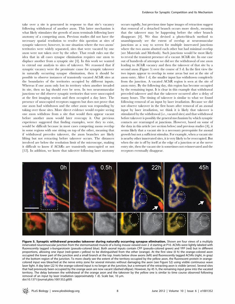
take over a site is generated in response to that site’s vacancy
following withdrawal of another axon. This latter mechanism is
what likely stimulates the growth of axon terminals following laser
axotomy of a competing axon. Previous studies did not have the
necessary spatial resolution to resolve this question at sites of
synaptic takeover; however, in one situation where the two axons’
territories were widely separated, sites that were vacated by one
axon were not taken over by the other. That result rules out the
idea that in all cases synapse elimination requires one axon to
displace another from a synaptic site [4]. In this work we wanted
to extend our analysis to sites of takeover. We reasoned that if
synaptic vacancy were the proximate cause for synaptic takeover
in naturally occurring synapse elimination, then it should be
possible to observe instances of transiently vacated AChR sites at
the boundaries of the territories occupied by different inputs.
Whereas if one axon only lost its territory when another invaded
its site, then no lag should ever be seen. In two neuromuscular
junctions we did observe synaptic territories that were unoccupied
at the first imaging session and then occupied a day later. The
presence of unoccupied receptors suggests but does not prove that
one axon had withdrawn and the other axon was responding by
taking over those sites. More direct evidence would require seeing
one axon withdraw from a site that would then appear vacant
before another axon would later reoccupy it. Our previous
experience suggested that finding examples, were they to exist,
would be difficult because in most cases competing axons overlap
in some regions with one sitting on top of the other, meaning that
if withdrawal precedes takeover, the axon branches are likely
lifting but not retracting before takeover occurs. The distances
involved are below the resolution limit of the microscope, making
it difficult to know if AChRs are transiently unoccupied or not
[12]. In addition, we know that takeover following laser axotomy
occurs rapidly, but previous time lapse images of retraction suggest
that removal of a detached branch occurs more slowly, meaning
that the takeover may be happening before the other branch
disappears [4]. We thus devised a photo-bleach method to
unambiguously see the extent of overlap at neuromuscular
junctions as a way to screen for multiple innervated junctions
where the two axons abutted each other but had minimal overlap
(see Materials and Methods). Such junctions would be most likely
to reveal the transient presence of a vacant AChR site. In one case
out of hundreds of attempts we did see the withdrawal of one axon
leading to AChR vacancy and then the takeover of that site by a
second axon (Figure 5) over the course of 3 d. In the first view the
two inputs appear to overlap in some areas but not at the site of
axon entry. After 1 d, the smaller input has withdrawn completely
from the junction. A vacated AChR region is seen at the site of
axon entry. By the following day, this region has become occupied
by the remaining input. It is clear in this example that withdrawal
preceded takeover and that the takeover occurred after a delay of
many hours. The timing of takeover is similar to what we found
following removal of an input by laser irradiation. Because we did
not observe takeover in the first hours after removal of an axonal
input by laser irradiation, we think it is likely that takeover is
stimulated by the withdrawal (i.e., vacated sites) and that withdrawal
before takeover is possibly the general mechanism by which synaptic
contacts are rearranged at junctions. However, based on some of
the data in this article (see section below) and previous studies [4], it
seems likely that a vacant site is a necessary prerequisite for axonal
growth but not a sufficient stimulus. For example, when a vacant site
is nearby other innervated sites, it is very likely to be reoccupied. But
when the site is off by itself at the edge of a junction or at the nerve
entry site, then the vacant site is sometimes not reinnervated and the
receptors eventually disappear.
Figure 5. Synaptic withdrawal precedes takeover during naturally occurring synapse elimination. Shown are four views of a multiplyinnervated neuromuscular junction from the sternomastoid muscle of a living mouse viewed over 2 d starting at P10. AChRs were lightly labeled withfluorescently tagged a-bungarotoxin (pseudo-colored blue). Both axonal inputs contain CFP (pseudo-colored green) and YFP (red) but in differentproportions, allowing one input (red+green = yellow) to be distinguished from the other (orange). At the first view (0 h) the orange-colored axonoccupied the lower part of the junction and a small branch at the top. Insets below show axons (left) and fluorescently tagged AChRs (right, in gray)of the bottom region of the junction. To more clearly see the extent of the territory occupied by the yellow axon, the fluorescent protein in orange-colored input was bleached at the nerve entry zone for several minutes without damaging the axon (see Figure S2) using visible continuous wavelaser light. A day later (22 h) the orange-colored input is no longer at the junction, but a remnant of the retracting axon is visible (arrow). Several sitesthat had previously been occupied by the orange axon are now vacant (dashed ellipse). However, by 45 h, the remaining input grew into the vacatedterritory. The delay between the withdrawal of the orange axon and the takeover by the yellow one is similar to time course observed followingremoval of an input by laser irradiation (approximately 1 d). Scale bar, 10 mm.doi:10.1371/journal.pbio.1001352.g005
Evidence for Synaptic Competition and Its Mechanism
PLoS Biology | www.plosbiology.org 8 June 2012 | Volume 10 | Issue 6 | e1001352
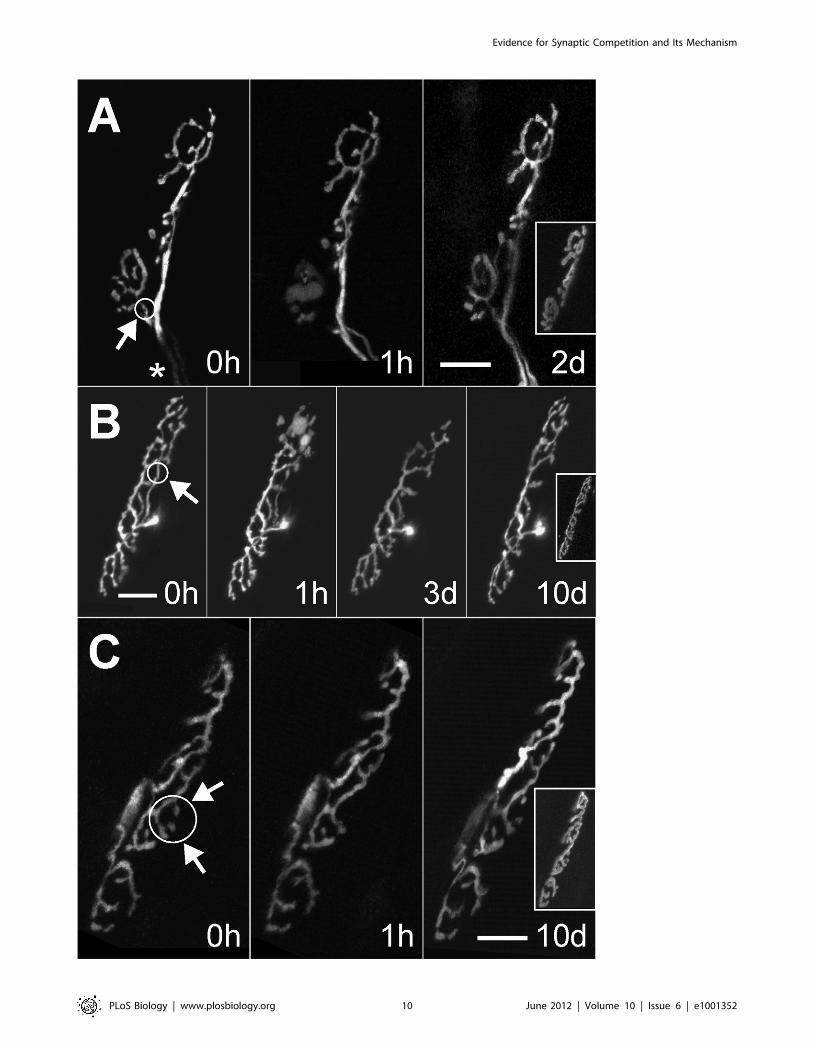
Axon Growth Induced by Small Unoccupied SynapticSites within Innervated Neuromuscular Junctions
The results above suggest that a signal from a vacant site may
stimulate the growth response during naturally occurring synapse
elimination. In many situations axonal growth is thought to be
stimulated by signals that emanate from denervated, and thus
inactive, target cells [28]. However, in the cases of normal
takeover in development and in the case where we targeted the
laser to a weak input, the loss of an axon was unlikely to give rise to
target inactivity because we had denervated a minuscule portion of
a large synapse. We were thus interested to know if small partial
denervations of target cells are generally sufficient to activate an
axonal regeneration response. In particular if only a small synaptic
bouton is removed and the muscle fiber is not functionally
denervated, will sprouting be stimulated? As described above we
found one case in development where a small site became
unoccupied and then later reoccupied by the remaining axon (see
Figure 5), but we wanted to see if this was a general trend that
occurred if vacant sites were present at any age. We were also
interested to know whether the proximate cause for the sprouting
within a neuromuscular junction could be explained by release of
contact inhibition. We thus did focal laser axotomies in adult
neuromuscular junctions to denervate small isolated synaptic
boutons while retaining innervation to the rest of the junction.
Surprisingly in adult animals more than half (55%, n = 12/22) of
these small laser-targeted axonal surgeries which denervated
between 5% and 30% of the AChR sites still induced reinnerva-
tion (Figure 6). The reinnervation started after a delay of at least
1 d following laser exposure and was typically complete by 2 d but
sometimes longer (see Figure 6B). In all cases reinnervation
occurred by sprouting from adjacent branches in the terminal.
The sprouts appeared to be directed specifically to unoccupied
AChR sites and not elsewhere, yet the original branching pattern
(pre-irradiation) was not necessarily preserved, suggesting that
regrowth was not necessarily guided by preexisting glial sheaths
but by highly localized signaling originating at the vacated sites.
Discussion
This study was undertaken to better understand the sequence of
events that occur during development underlying the transition
from multiple to single innervation in skeletal muscle. This
phenomenon, which has analogs in other parts of the developing
nervous system, occurs by one axon’s takeover of most of the
postsynaptic sites that were earlier occupied by other axons.
However, a number of questions about the underlying mechanism
remain unanswered. First, what drives the exchange of territory
such that when one axon loses sites another typically gains those
sites [4]? Second, what determines the identity of the eventual
surviving input given that an axon that loses territory at one time
point sometimes gains it back at a later time [4]? And third, why
do the contacts of an axon within a neuromuscular junction tend
over time to cluster to occupy a contiguous segregated territory
[29]? In this work we focused on answering the first question. In so
doing we think we have also uncovered explanations for the other
questions and believe we now have a framework to interpret many
aspects of this form of synaptic plasticity.
We show that axons rapidly respond to vacant synaptic sites by
growth. In multiply innervated neuromuscular junctions an axon
whose elimination appears imminent will, within 1 d, occupy all
the sites of an axon that was experimentally removed. Moreover,
axons that have recently withdrawn completely from a neuro-
muscular junction will reverse their fate and reoccupy it if the
innervating axon is caused to disappear. These results strongly
support the idea that the process leading to single innervation is
competitive: an axon destined for elimination always survives if the
other innervating axon is removed.
This growth response of one terminal axon branch to the
damage of another terminal branch is in some ways reminiscent of
the reinnervation response following partial denervation of a
muscle where an axon that is undamaged sprouts to occupy
neuromuscular junctions on denervated muscle fibers [30].
However, these two phenomena seem to be dissimilar in several
important respects and may have different underlying mecha-
nisms. First, a number of studies support the idea that sprouting
following partial denervation is stimulated by muscle fiber
inactivity [30,31]; however, several of our results show axons
growing into vacated synaptic sites even when the muscle fiber is
functionally innervated. It is also clear that in naturally occurring
synapse elimination, an axon continues to take over vacated sites
even when it already occupies the vast majority of the terminal
area so that its growth is not being stimulated by inactivity of the
muscle [4]. A second difference between partial denervation of
muscle and the growth response described here is that the
following partial denervation axons grow through vacated
Schwann cell tubes [32] or extend along new Schwann cell
processes [33]. Neither of these paths is available within
neuromuscular junctions. Third, the growth response following
laser axotomy in neonatal animals is fast compared to the response
of axons in adults to partial denervation. Another difference is that
many of the axons undergoing branch loss in development were
atrophic and had to transition from a withdrawing state to a
growing state, whereas the axons responding by growth following
partial denervation in adults are in a healthy quiescent state before
being induced to grow. These differences suggest that the local
growth response to synaptic vacancy within a neuromuscular
junction is different from the growth response of axons to the loss
of all innervation to a subset of muscle fibers (i.e., partial
denervation).
Because muscle fiber inactivity is unlikely to be the stimulus that
induces axonal growth into vacant synaptic sites in our studies,
what then is the signal? One idea is that Schwann cell processes
that no longer are associated with an axon become activated.
Previous studies have shown that Schwann cell activation
following nerve damage is a potent stimulus for axon growth
[34,35]. Thus, it is possible that focal loss of nerve-glial contact
leads to the release of a glial-derived signal that causes axons to
grow. Interestingly glial cell-derived neurotrophic factor (GDNF),
a glial based growth factor, is one of the strongest known stimuli
for mammalian motor nerve growth [36,37]. Growth based on loss
of nerve-glial contact is an attractive idea because it does not
require muscle inactivity as a stimulus. Such a mechanism could
be the same as the one that promotes axon regrowth along
nerveless Schwann cell tubes to distant muscle targets following
nerve damage far from muscle end organs (such as the sciatic
nerve) [38]. Another possibility is that Schwann cell activation is
downstream of a signal originating in the postsynaptic cell or that
the vacant postsynaptic site signals axon growth directly. Ongoing
experiments are aimed at deciding between these alternatives.
The results presented here suggest the primacy of the
withdrawal (or loss of maintenance of synaptic contacts) as the
initiating event leading to synaptic takeover and ultimately single
innervation of neuromuscular junctions. Interestingly, signals from
presynaptic, postsynaptic, and glial cells all seem to be able to
regulate synaptic maintenance. For example, at the mammalian
neuromuscular junction, synaptic loss can be initiated by
postsynaptic protein synthesis inhibition [10], focal blockade of
neurotransmission at a synaptic site [8], exuberant branching of
Evidence for Synaptic Competition and Its Mechanism
PLoS Biology | www.plosbiology.org 9 June 2012 | Volume 10 | Issue 6 | e1001352
Evidence for Synaptic Competition and Its Mechanism
PLoS Biology | www.plosbiology.org 10 June 2012 | Volume 10 | Issue 6 | e1001352
motor axons in development [13], terminal sprouting [35], and
glial loss [39,40]. A molecular understanding of developmental
synaptic reorganization may therefore require understanding the
relative roles of these diverse signals and cells in causing synapse
loss. The important point from our perspective is that by focusing
on synapse loss (as opposed to synaptic addition) it may be possible
to get to root causes for the rearrangements.
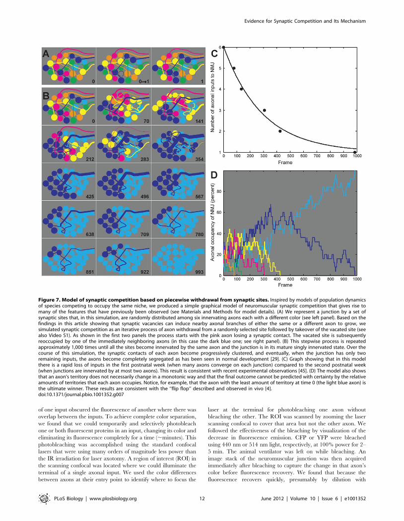
Based on these results we developed a surprisingly robust
graphical model simulating synaptic competition (Figure 7). In this
model we posit that maintenance of synaptic contacts is imperfect
and that at regular intervals an axon withdraws from an individual
synaptic site at random. However, in each case the consequence of
the vacated site is the same: as shown in this work, when an axon
withdraws from a synaptic site, signals stimulate nearby axons to
grow and attempt to occupy that site. In this model the
reoccupation favors axons that have the largest number of nearby
synaptic contacts (Figure 7A and see Materials and Methods for
details). This proposed mechanism is inspired by models used in
evolutionary biology for understanding the survival and extinction
of different populations within the same niche [41]. In this case the
different populations that are in competition are the several axons
converging on the same neuromuscular junction, with each
synaptic contact being equivalent to an individual member of
one or another population. As shown this process will lead
eventually to single innervation (Figure 7B). This simple model
also provides insight into the cause of several other features of
synaptic competition such as synaptic segregation [29], flip-flop
[4], and the slowing pace of input loss in the second compared to
the first postnatal week (Figure 7B and C). Thus, loss-initiated
synaptic takeover suggests a useful conceptual framework for
understanding competitive synaptic rearrangements at the neuro-
muscular junction.
An important question is whether the model proposed here also
has relevance to synaptic rearrangements occurring in other parts
of the nervous system. It is well known that axons are lost from
neurons at the same developmental stage that axons are removed
from muscle fibers. In several cases it is also clear that a remaining
input to a neuron (such as the climbing fiber on a Purkinje cell or a
preganglionic axon on a submandibular ganglion cell) elaborates
new synaptic connections at the time other axons are being
eliminated [42]. The complementary nature of the loss and gain of
connections by withdrawing and remaining inputs, respectively,
raises the possibility that a remaining input is induced to grow and
innervate synaptic sites that have become vacant because of axon
loss. In the climbing fiber system, the final area occupied by the
surviving axon is far greater than the area occupied by the multiple
axons initially innervating the Purkinje cell soma. This large
increase in area is a consequence of the Purkinje cell elaborating its
dendritic tree at the same time climbing fiber axons are being lost
from the soma [43]. In this case new (and hence vacant)
postsynaptic sites on the expanding dendritic arbor may stimulate
the climbing fiber to ‘‘climb’’ from the soma and grow out along
the dendrites. In autonomic ganglia it is also clear that axons
elaborate synapses on the soma and then grow out to innervate
sites on the dendrites [44]. Thus in both of these cases
rearrangement of synaptic connections has two parts: (1) loss of
some axonal inputs and (2) concomitant elaboration of additional
connections by the axons that survive. Hence the mechanism by
which motor axons grow locally in response to vacated sites at the
neuromuscular junction may inform on and be analogous to the
mechanisms underlying the establishment of both the correct
number of innervating axons and their total synaptic drive on
neurons (see also discussion in Tapia et al. [45] elaborating the
idea set out above that the mechanism for competitive synaptic
rearrangements is consistent with a net increase in synaptic
numbers and an important general role for elimination during
neural development). Moreover, if our evidence of continuing
plasticity in adult muscles is any guide (see Figure 6), a kind of local
structural plasticity might continue on multiply innervated
neurons even in the adult brain. As such ‘‘evolution’’ of neural
network connectivity from a dynamic competitive state to one
marked by long-term functional stability could be the basis of
indelible alterations in brain function such that occur with
learning.
Materials and Methods
Transgenic MiceMice that expressed either cytoplasmic GFP (line GFP-I+/+),
YFP (line YFP-16+/+), or both CFP and YFP (lines CFP-5+/+ or
CFP-23+/+ crossbred with YFP-16+/+) were used for all experi-
ments (protocols approved by Washington University Animal
Studies Committee and Harvard University’s Institutional Animal
Care and Use Committee) [46]. The GFP line was used for studies
in adult mice. The YFP and CFP lines were used for neonatal
mice. The GFP line could not be used for imaging in neonatal
mice because the onset of GFP expression in motor neurons was
delayed until after the first postnatal week.
Multicolor Axon Labeling and PhotobleachingTo be able to visually distinguish one input from another we
developed a method of labeling axons in multiple colors using very
bright thy1-driven XFP transgenic mouse lines. By crossing two
lines in which all motor neurons constitutively express a
fluorescent protein, we found that in young animals intrinsic
variation in expression level of each fluorescent protein between
neurons was sufficient for us to distinguish axons by color. An
advantage of this approach was that every multiply innervated
junction on the muscle surface was a potential candidate for
imaging. A challenge was that the spectral separation of the axons
was incomplete when imaged using conventional confocal
microscopy. Because every neuron expressed the same two
fluorescent markers (varying only in linear combination), all the
axons appeared in both fluorescence channels. Thus fluorescence
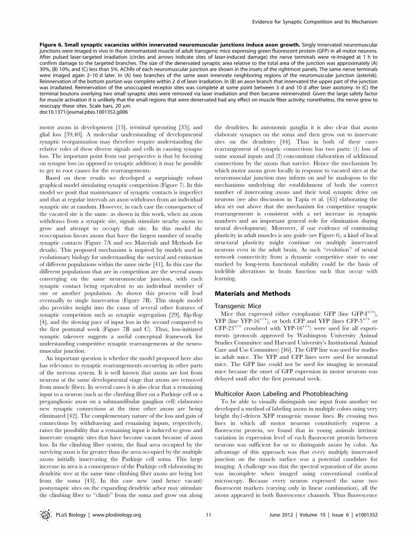
Figure 6. Small synaptic vacancies within innervated neuromuscular junctions induce axon growth. Singly innervated neuromuscularjunctions were imaged in vivo in the sternomastoid muscle of adult transgenic mice expressing green fluorescent protein (GFP) in all motor neurons.After pulsed laser-targeted irradiation (circles and arrows indicate sites of laser-induced damage) the nerve terminals were re-imaged at 1 h toconfirm damage to the targeted branches. The size of the denervated synaptic area relative to the total area of the junction was approximately (A)30%, (B) 10%, and (C) less than 5%. AChRs of each neuromuscular junction are shown in the insets of the rightmost panels. The same nerve terminalswere imaged again 2–10 d later. In (A) two branches of the same axon innervate neighboring regions of the neuromuscular junction (asterisk).Reinnervation of the bottom portion was complete within 2 d of laser irradiation. In (B) an axon branch that innervated the upper part of the junctionwas irradiated. Reinnervation of the unoccupied receptor sites was complete at some point between 3 d and 10 d after laser axotomy. In (C) theterminal boutons overlying two small synaptic sites were removed via laser irradiation and then became reinnervated. Given the large safety factorfor muscle activation it is unlikely that the small regions that were denervated had any effect on muscle fiber activity; nonetheless, the nerve grew toreoccupy these sites. Scale bars, 20 mm.doi:10.1371/journal.pbio.1001352.g006
Evidence for Synaptic Competition and Its Mechanism
PLoS Biology | www.plosbiology.org 11 June 2012 | Volume 10 | Issue 6 | e1001352
of one input obscured the fluorescence of another where there was
overlap between the inputs. To achieve complete color separation,
we found that we could temporarily and selectively photobleach
one or both fluorescent proteins in an input, changing its color and
eliminating its fluorescence completely for a time (,minutes). This
photobleaching was accomplished using the standard confocal
lasers that were using many orders of magnitude less power than
the IR irradiation for laser axotomy. A region of interest (ROI) in
the scanning confocal was located where we could illuminate the
terminal of a single axonal input. We used the color differences
between axons at their entry point to identify where to focus the
laser at the terminal for photobleaching one axon without
bleaching the other. The ROI was scanned by zooming the laser
scanning confocal to cover that area but not the other axon. We
followed the effectiveness of the bleaching by visualization of the
decrease in fluorescence emission. CFP or YFP were bleached
using 440 nm or 514 nm light, respectively, at 100% power for 2–
5 min. The animal ventilator was left on while bleaching. An
image stack of the neuromuscular junction was then acquired
immediately after bleaching to capture the change in that axon’s
color before fluorescence recovery. We found that because the
fluorescence recovers quickly, presumably by dilution with
Figure 7. Model of synaptic competition based on piecewise withdrawal from synaptic sites. Inspired by models of population dynamicsof species competing to occupy the same niche, we produced a simple graphical model of neuromuscular synaptic competition that gives rise tomany of the features that have previously been observed (see Materials and Methods for model details). (A) We represent a junction by a set ofsynaptic sites that, in this simulation, are randomly distributed among six innervating axons each with a different color (see left panel). Based on thefindings in this article showing that synaptic vacancies can induce nearby axonal branches of either the same or a different axon to grow, wesimulated synaptic competition as an iterative process of axon withdrawal from a randomly selected site followed by takeover of the vacated site (seealso Video S1). As shown in the first two panels the process starts with the pink axon losing a synaptic contact. The vacated site is subsequentlyreoccupied by one of the immediately neighboring axons (in this case the dark blue one; see right panel). (B) This stepwise process is repeatedapproximately 1,000 times until all the sites become innervated by the same axon and the junction is in its mature singly innervated state. Over thecourse of this simulation, the synaptic contacts of each axon become progressively clustered, and eventually, when the junction has only tworemaining inputs, the axons become completely segregated as has been seen in normal development [29]. (C) Graph showing that in this modelthere is a rapid loss of inputs in the first postnatal week (when many axons converge on each junction) compared to the second postnatal week(when junctions are innervated by at most two axons). This result is consistent with recent experimental observations [45]. (D) The model also showsthat an axon’s territory does not necessarily change in a monotonic way and that the final outcome cannot be predicted with certainty by the relativeamounts of territories that each axon occupies. Notice, for example, that the axon with the least amount of territory at time 0 (the light blue axon) isthe ultimate winner. These results are consistent with the ‘‘flip flop’’ described and observed in vivo [4].doi:10.1371/journal.pbio.1001352.g007
Evidence for Synaptic Competition and Its Mechanism
PLoS Biology | www.plosbiology.org 12 June 2012 | Volume 10 | Issue 6 | e1001352
unbleached fluorescent protein in the more proximal axon, the
same region could be photobleached repeatedly and without any
apparent phototoxic effects (see [47] for control experiments). We
assume that because the fluorescent proteins were freely diffusible
in the cytoplasm and not tethered to any important organelles or
membranes, we noted no immediate (minutes to hours) or delayed
(days) toxic effects on the irradiated neuron (see Figure S2). Once
bleached, the areas where the two axons overlapped in the
junction were disambiguated by taking images of the junction with
each laser line.
ImagingAdult and neonatal mice were anesthetized, intubated, and
respirated as previously described [4,47,48]. The ventral neck skin
was incised and retracted laterally to expose the right sternomas-
toid muscle, which was gently lifted on a flat steel platform. Care
was taken not to stretch the muscle in order to prevent damage to
the muscle, blood supply, or innervating nerve bundle. The wound
was filled with sterile saline and a glass coverslip was placed over
the muscle, making a meniscus with the saline to help keep the
muscle from drying. The coverslip did not touch the muscle.
Axons innervating the central band of neuromuscular junctions
were visualized using standard epifluorescence optics (YFP filter
cube) at high magnification (606 0.9NA water immersion
objective). Once a particular nerve terminal was selected, images
were taken using a confocal microscope (Bio-Rad MRC1024MP
or Olympus FV-1000). GFP-filled axons in adult animals were
illuminated using 488 nm light. In neonates, YFP-filled axons
were illuminated using either 488 nm light or 514 nm light, and
axons that were filled with both CFP and YFP were illuminated
using 458 nm and 514 nm light simultaneously. The ventilator
was turned off temporarily (30–60 s) to acquire a stack of images.
Axons and acetylcholine receptors were imaged sequentially. To
avoid damage to the muscle and synapse, acetylcholine receptors
were labeled only after laser ablation of axons. Receptors were
lightly labeled with alexa-647 conjugated a-bungarotoxin (5 mg/
ml in PBS for 40 s), and the muscle was then rinsed well with PBS.
This dosage did not paralyze the muscle, as greater than 70% of
the receptors remained unlabeled [4]. Movement artifacts were
removed from stacks using special alignment software (Autoquant;
Media Cybernetics, Inc). A 2-D image of each junction was then
obtained by a maximum intensity projection. The same junction
was imaged multiple times, before and after laser ablation, and in
most cases at 1-d intervals thereafter. The mice were resuscitated
after each imaging session as previously described [4,47]. In the
time lapse views the color balance of the image from different time
points was sometimes corrected using the background autofluo-
rescence as a reference.
Laser AblationLaser ablation was performed using a Spectra Physics Tsunami
Ti:S laser oscillator and pumped by a Millenia 5W solid state laser
using scanning mirrors from a laser scanning microscope. The
pulsed laser output was tuned to 815 nm. The laser power was
approximately 120 mW at the back aperture of the objective. An
IR-corrected water dipping cone objective (LUMPlan W-IR2 6060.9NA Olympus) was used to focus the laser onto an axon branch.
The scanner’s mirrors were parked (i.e., maximal zoom) to
position the laser onto a ,0.5 mm spot (diffraction limited). In this
way all of the laser’s power was focused to the waist of an
hourglass-shaped beam giving a football-shaped spot (long axis
,2.4 mm). The damage required 30 s to 1 min of mode-locked
operation of the laser (approximately 1nJ energy per 80 fs pulse).
This laser axotomy approach differs from a previous technique
that used a regenerative optical amplifier to provide pulse powers of
10–40 nJ and oil immersion, high NA short working distance
objectives to break down the optical transparency and cause rapid
heating and vaporization at the focal spot [49]. That previous
approach had the advantage that the damage could be accom-
plished in less than a second with relatively few pulses of laser
irradiation and showed immediate effects. Our approach required
many seconds with far more pulses and required many minutes to
discern the damage (although photobleaching was evident quickly).
However, because we were damaging axons by virtue of their ability
to absorb the multiphoton excitation, we may have achieved a
degree of selectivity that would not be possible otherwise. We
believe this damage depends on absorbance of the two photon
excitation because there was no damage at the same power level
when the laser was not mode-locked. Fluorescence excitation thus
appears critical for damage at the laser intensities we used. At the
powers used, no scarring or collateral damage to muscles fibers or
nearby axons was observed, nor was there a visible plasma bubble.
To visualize the excitation spot and its position in the optical field
simultaneously on a Bio-Rad MRC1024MP microscope system, we
replaced a mirror above the objective in the excitation and return
light path with a dichroic mirror (Chroma 700DCSPXR), which
reflected the exciting infrared light and transmitted the fluorescent
emission to the eyepieces or a camera (Figure S1). A YFP filter cube
(Chroma) was used in the epifluorescence light path, above the
specially installed dichroic mirror. The fluorescent axons and site of
laser excitation were visualized using a low-light SIT video camera
(Dage-MTI Series 68). Filters were installed to block reflection of the
IR laser to the camera and eyepieces. In adults, damage was easier
to generate in GFP-expressing than YFP-expressing axons. We
think the lower susceptibility of YFP axons to damage may be due to
lower absorption of multiphoton light by YFP at 815 nm. At longer
wavelengths where YFP might be a better absorber, the laser pulse
energy available in our system was substantially less than 1 nJ. In
pups interestingly, axons were more easily damaged than in adults.
YFP expressing axons in pups were just as susceptible to damage as
GFP labeled axons in adults, and the threshold intensity for damage
was roughly the same for both fluors.
ModelingOur model of synaptic rearrangements at a neuromuscular
junction is based on evolutionary graph theory [41]. The vertices
of the graph represent the individual synaptic sites of the junction.
In our simulation, each site is randomly assigned to one of six
axons. The axons are assumed to have equal fitness; therefore, the
probability of axon withdrawal is the same at all synaptic sites. The
axon growth rate is assumed to be constant for all axons. A site is
selected at random for axon withdrawal. We then randomly select
a neighboring synaptic site. The axon innervating this neighboring
site grows to take over the vacated site. Thus an axon that occupies
multiple neighboring sites has a higher probability of taking over
the vacated site than an axon that innervates only one neighboring
site. This process of withdrawal from a randomly selected site and
takeover by a randomly selected neighboring site is repeated until
all the synaptic sites of the junction are innervated by the same
axon. To perform our simulations we used a macro written for
Matlab (The Mathworks, Inc.; Natick, MA) based on a Potts
model with non-periodic boundary conditions.
Supporting Information
Figure S1 Modified light path of a multiphoton microscope
system for laser axotomy. In order to optically target single axons
for removal in living mice, the sternomastoid muscle was exposed
Evidence for Synaptic Competition and Its Mechanism
PLoS Biology | www.plosbiology.org 13 June 2012 | Volume 10 | Issue 6 | e1001352
in anesthetized mice (ventral side up) on an upright microscope
stage. The microscope was equipped with standard epifluores-
cence optics and a low-light silicon-intensified target (SIT) video
camera. A short pass (,700 nm) extended reflection dichroic
mirror was placed in the light path between the objective turret
and the YFP (or GFP) fluorescence filter cube to prevent any of the
reflected infrared light from contaminating the visible light
wavelengths seen by the video camera. This allowed for
simultaneous pulsed infrared laser excitation and epifluorescence
visible light illumination. Scanhead zoom mirrors were used to
position a diffraction limited spot of the infrared laser to a fixed
point in the specimen focal plane whose position was marked on a
video monitor receiving the output of the SIT camera. Using the
lateral and vertical movements of the stage, the axon of interest
was then moved into position. When the shutter for the pulsed
laser was opened, a bright fluorescence spot due to multiphoton
excitation was visible in the wide field fluorescence image but only
when the pulsed laser was striking a fluorescent object—that is, a
GFP- or YFP-filled axon. The laser was allowed to dwell on the
axon until the fluorescence intensity disappeared and did not
rapidly recover (,0.5–1 min). As described in the text, the axon
subsequently swelled and then disintegrated. The same neuro-
muscular junction was reimaged with confocal microscopy after
re-anesthetizing the mouse one or more times over the next several
days.
(TIF)
Figure S2 Wavelength selective photobleaching of cytoplasmi-
cally expressed fluorescent protein does not damage motor
neurons in vivo. (A) Natural color variation between axons in
the sternomastoid muscle of a double transgenic P7 mouse pup
constitutively expressing both CFP (green) and YFP (red) in all
motor neurons. (B) The color difference between inputs to the
topmost neuromuscular junction was enhanced by scanning with
high intensity 514 nm laser light at high scan zoom at the site of
the circle to photobleach the YFP in left input to this junction. (C)
Both CFP and YFP were then photobleached by exciting with
both 440 nm and 514 nm laser light, allowing the territory
occupied by the non-bleached axon to be observed unambigu-
ously. (D) Fluorescence recovery occurred within a few minutes
with no apparent immediate damage to the irradiated input. (E)
On the following day, the non-irradiated input had lost synaptic
territory and the previously irradiated input has gained synaptic
territory. (F) The YFP in the now-dominant input was photo-
bleached again using 514 nm light to reveal the synaptic territory
occupied by the non-irradiated input, which confirmed its loss of
territory (arrow). Scale bar, 20 mm.
(TIF)
Video S1 Approximately 1,000 frame simulation showing the
synaptic rearrangement process at a single neuromuscular
junction. There are initially six axons. Each axon is labeled a
different color. The postsynaptic receptors are not shown and
assumed to be unchanging during the course of the simulation. At
each frame a synaptic contact is randomly selected for withdrawal
and one of the axons adjacent to the vacated site is randomly
selected to grow and occupy the vacated site. Note the rapid loss of
inputs early in the simulation process (first half) and the prolonged
period of dual innervation (second half). There is also evidence of
progressive segregation of contacts [29], flip-flop [4], and
ultimately the transition to single innervation.
(MOV)
Author Contributions
The author(s) have made the following declarations about their
contributions: Conceived and designed the experiments: ST JL. Performed
the experiments: ST JL. Analyzed the data: ST JL. Contributed reagents/
materials/analysis tools: ST JL. Wrote the paper: ST JL.
References
1. Redfern PA (1970) Neuromuscular transmission in new-born rats. J Physiol 209:
701–709.
2. Purves D, Lichtman JW (1980) Elimination of synapses in the developing
nervous system. Science 210: 153–157.
3. Luo L, O’Leary DD (2005) Axon retraction and degeneration in development
and disease. Annu Rev Neurosci 28: 127–156.
4. Walsh MK, Lichtman JW (2003) In vivo time-lapse imaging of synaptic takeover
associated with naturally occurring synapse elimination. Neuron 37: 67–73.
5. Beirowski B, Adalbert R, Wagner D, Grumme DS, Addicks K, et al. (2005) The
progressive nature of Wallerian degeneration in wild-type and slow Wallerian
degeneration (WldS) nerves. BMC Neurosci 6: 6.
6. Colman H, Nabekura J, Lichtman JW (1997) Alterations in synaptic strength
preceding axon withdrawal. Science 275: 356–361.
7. Keller-Peck CR, Walsh MK, Gan WB, Feng G, Sanes JR, et al. (2001)
Asynchronous synapse elimination in neonatal motor units: studies using GFP
transgenic mice. Neuron 31: 381–394.
8. Balice-Gordon RJ, Lichtman JW (1994) Long-term synapse loss induced by focal
blockade of postsynaptic receptors. Nature 372: 519–524.
9. Buffelli M, Burgess RW, Feng G, Lobe CG, Lichtman JW, et al. (2003) Genetic
evidence that relative synaptic efficacy biases the outcome of synaptic
competition. Nature 424: 430–434.
10. McCann CM, Nguyen QT, Santo Neto H, Lichtman JW (2007) Rapid synapse
elimination after postsynaptic protein synthesis inhibition in vivo. J Neurosci 27:
6064–6067.
11. Bishop DL, Misgeld T, Walsh MK, Gan WB, Lichtman JW (2004) Axon branch
removal at developing synapses by axosome shedding. Neuron 44: 651–661.
12. Kasthuri N, Lichtman JW (2003) The role of neuronal identity in synaptic
competition. Nature 424: 426–430.
13. Thompson W, Jansen JK (1977) The extent of sprouting of remaining motor units in
partly denervated immature and adult rat soleus muscle. Neuroscience 2: 523–535.
14. Laskowski MB, Sanes JR (1987) Topographic mapping of motor pools onto
skeletal muscles. J Neurosci 7: 252–260.
15. Laskowski MB, Colman H, Nelson C, Lichtman JW (1998) Synaptic competition
during the reformation of a neuromuscular map. J Neurosci 18: 7328–7335.
16. Thompson WJ, Sutton LA, Riley DA (1984) Fibre type composition of single
motor units during synapse elimination in neonatal rat soleus muscle. Nature
309: 709–711.
17. Brown MC, Jansen JK, Van Essen D (1976) Polyneuronal innervation of skeletal
muscle in new-born rats and its elimination during maturation. J Physiol 261:
387–422.
18. O’Brien RA, Ostberg AJ, Vrbova G (1978) Observations on the elimination of
polyneuronal innervation in developing mammalian skeletal muscle. J Physiol
282: 571–582.
19. Snider WD, Lichtman JW (1996) Are neurotrophins synaptotrophins? Mol Cell
Neurosci 7: 433–442.
20. Colman H, Lichtman JW (1993) Interactions between nerve and muscle:
synapse elimination at the developing neuromuscular junction. Dev Biol 156: 1–
10.
21. Fladby T, Jansen JK (1987) Postnatal loss of synaptic terminals in the partially
denervated mouse soleus muscle. Acta Physiol Scand 129: 239–246.
22. Barry JA, Ribchester RR (1995) Persistent polyneuronal innervation in partially
denervated rat muscle after reinnervation and recovery from prolonged nerve
conduction block. J Neurosci 15: 6327–6339.
23. Lichtman JW, Wilkinson RS, Rich MM (1985) Multiple innervation of tonic
endplates revealed by activity-dependent uptake of fluorescent probes. Nature
314: 357–359.
24. Keller-Peck CR, Feng G, Sanes JR, Yan Q, Lichtman JW, et al. (2001) Glial cell
line-derived neurotrophic factor administration in postnatal life results in motor
unit enlargement and continuous synaptic remodeling at the neuromuscular
junction. J Neurosci 21: 6136–6146.
25. Chang LK, Putcha GV, Deshmukh M, Johnson EM, Jr. (2002) Mitochondrial
involvement in the point of no return in neuronal apoptosis. Biochimie 84: 223–
231.
26. Kerschensteiner M, Schwab ME, Lichtman JW, Misgeld T (2005) In vivo
imaging of axonal degeneration and regeneration in the injured spinal cord. Nat
Med 11: 572–577.
27. Bixby JL (1981) Ultrastructural observations on synapse elimination in neonatal
rabbit skeletal muscle. J Neurocytol 10: 81–100.
Evidence for Synaptic Competition and Its Mechanism
PLoS Biology | www.plosbiology.org 14 June 2012 | Volume 10 | Issue 6 | e1001352
28. Sanes JR, Lichtman JW (1999) Development of the vertebrate neuromuscular
junction. Annu Rev Neurosci 22: 389–442.29. Gan WB, Lichtman JW (1998) Synaptic segregation at the developing
neuromuscular junction. Science 282: 1508–1511.
30. Brown MC, Holland RL, Hopkins WG (1981) Motor nerve sprouting. AnnualReview of Neuroscience 4: 17–42.
31. Keynes RJ, Hopkins WG, Brown MC (1983) Sprouting of mammalian motorneurones at nodes of Ranvier: the role of the denervated motor endplate. Brain
Research 264: 209–213.
32. Nguyen QT, Sanes JR, Lichtman JW (2002) Pre-existing pathways promoteprecise projection patterns. Nat Neurosci 5: 861–867.
33. Son YJ, Thompson WJ (1995) Nerve sprouting in muscle is induced and guidedby processes extended by Schwann cells. Neuron 14: 133–141.
34. Taniuchi M, Clark HB, Johnson EM, Jr. (1986) Induction of nerve growth factorreceptor in Schwann cells after axotomy. Proceedings of the National Academy
of Sciences of the United States of America 83: 4094–4098.
35. Kang H, Tian L, Thompson W (2003) Terminal Schwann cells guide thereinnervation of muscle after nerve injury. J Neurocytol 32: 975–985.
36. Henderson CE, Phillips HS, Pollock RA, Davies AM, Lemeulle C, et al. (1994)GDNF: a potent survival factor for motoneurons present in peripheral nerve and
muscle. Science 266: 1062–1064.
37. Yan Q, Matheson C, Lopez OT (1995) In vivo neurotrophic effects of GDNF onneonatal and adult facial motor neurons. Nature 373: 341–344.
38. Taniuchi M, Clark HB, Schweitzer JB, Johnson EM, Jr. (1988) Expression ofnerve growth factor receptors by Schwann cells of axotomized peripheral nerves:
ultrastructural location, suppression by axonal contact, and binding properties.The Journal of Neuroscience: The Official Journal of the Society for
Neuroscience 8: 664–681.
39. Trachtenberg JT, Thompson WJ (1996) Schwann cell apoptosis at developingneuromuscular junctions is regulated by glial growth factor. Nature 379: 174–
177.
40. Reddy LV, Koirala S, Sugiura Y, Herrera AA, Ko CP (2003) Glial cells
maintain synaptic structure and function and promote development of the
neuromuscular junction in vivo. Neuron 40: 563–580.
41. Lieberman E, Hauert C, Nowak MA (2005) Evolutionary dynamics on graphs.
Nature 433: 312–316.
42. Lichtman JW (1977) The reorganization of synaptic connexions in the rat
submandibular ganglion during post-natal development. The Journal of
Physiology 273: 155–177.
43. Hashimoto K, Ichikawa R, Kitamura K, Watanabe M, Kano M (2009)
Translocation of a ‘‘winner’’ climbing fiber to the Purkinje cell dendrite and
subsequent elimination of ‘‘losers’’ from the soma in developing cerebellum.
Neuron 63: 106–118.
44. Forehand CJ, Purves D (1984) Regional innervation of rabbit ciliary ganglion
cells by the terminals of preganglionic axons. The Journal of Neuroscience: The
Official Journal of the Society for Neuroscience 4: 1–12.
45. Tapia JCW, Kasthuri N, Hayworth K, Schalek R, Berger D, Guatimosim C,
Seung HS, Lichtman JW (2012) Pervasive synaptic branch removal in the
mammalian neuromuscular system at birth. Neuron 74: 816–829.
46. Feng G, Mellor RH, Bernstein M, Keller-Peck C, Nguyen QT, et al. (2000)
Imaging neuronal subsets in transgenic mice expressing multiple spectral
variants of GFP. Neuron 28: 41–51.
47. Turney SG, Lichtman JW (2008) Chapter 11: imaging fluorescent mice in vivo
using confocal microscopy. Methods Cell Biol 89: 309–327.
48. van Mier P, Lichtman JW (1994) Regenerating muscle fibers induce directional
sprouting from nearby nerve terminals: studies in living mice. J Neurosci 14:
5672–5686.
49. Yanik MF, Cinar H, Cinar HN, Chisholm AD, Jin Y, et al. (2004) Neurosurgery:
functional regeneration after laser axotomy. Nature 432: 822.
Evidence for Synaptic Competition and Its Mechanism
PLoS Biology | www.plosbiology.org 15 June 2012 | Volume 10 | Issue 6 | e1001352