Embed Size (px)
Citation preview

Review ArticleAntibodies in the Pathogenesis of Hypertension
Christopher T. Chan,1 Maggie Lieu,1 Ban-Hock Toh,2 Tin S. Kyaw,3
Alexander Bobik,3 Christopher G. Sobey,1 and Grant R. Drummond1
1 Vascular Biology & Immunopharmacology Group, Department of Pharmacology, Monash University, Clayton, VIC 3800, Australia2 Centre for Inflammatory Diseases, Department of Medicine, Southern Clinical School, Monash University, Clayton, VIC, Australia3 Vascular Biology & Atherosclerosis Laboratory, Baker IDI Heart and Diabetes Institute, Melbourne, VIC, Australia
Correspondence should be addressed to Grant R. Drummond; [email protected]
Received 8 April 2014; Revised 21 May 2014; Accepted 4 June 2014; Published 23 June 2014
Academic Editor: Tomasz Guzik
Copyright © 2014 Christopher T. Chan et al. This is an open access article distributed under the Creative Commons AttributionLicense, which permits unrestricted use, distribution, and reproduction in any medium, provided the original work is properlycited.
It has long been known that circulating levels of IgG and IgM antibodies are elevated in patients with essential and pregnancy-related hypertension. Recent studies indicate these antibodies target, and in many cases activate, G-protein coupled receptors andion channels. Prominent among these protein targets are AT
1receptors, 𝛼
1-adrenoceptors, 𝛽
1-adrenoceptors, and L-type voltage
operated Ca2+ channels, all of which are known to play key roles in the regulation of blood pressure throughmodulation of vasculartone, cardiac output, and/orNa+/water reabsorption in the kidneys.This suggests that elevated antibody productionmay be a causalmechanism in at least some cases of hypertension. In this brief review, we will further describe the protein targets of the antibodiesthat are elevated in individuals with essential and pregnancy-related hypertension and the likely pathophysiological consequencesof antibody binding to these targets. We will speculate on the potential mechanisms that underlie elevated antibody levels inhypertensive individuals and, finally, we will outline the therapeutic opportunities that could arise with a better understandingof how and why antibodies are produced in hypertension.
1. Introduction
Hypertension is defined as chronically elevated blood pres-sures of >140/90mmHg. Two of the most common forms ofthe condition are essential hypertension, where the under-lying cause is unknown, and preeclampsia or pregnancy-related hypertension. For several decades it has been knownthat both essential and pregnancy-related hypertension areassociated with elevated serum levels of antibodies [1–4].More recently, studies in humans and animal models of eachcondition have begun to identify the protein targets of theseantibodies as receptors and ion channels with key roles inthe regulation of blood pressure. Such studies not only offerinsights into the mechanisms by which antibodies mightcontribute to hypertension but they also highlight potentialnew avenues for the clinical management of hypertension. Inthis brief review we will summarise the evidence in supportof a role for antibodies in the pathophysiology of essentialhypertension and preeclampsia. We will discuss the protein
targets of the antibodies that have been identified in hyper-tensive individuals and provide some potential explanationsfor why the production of these antibodies may be elevated.Finally, we will speculate on how such findings may translateinto improved clinical management of hypertension.
2. Antibodies as Causes of Disease
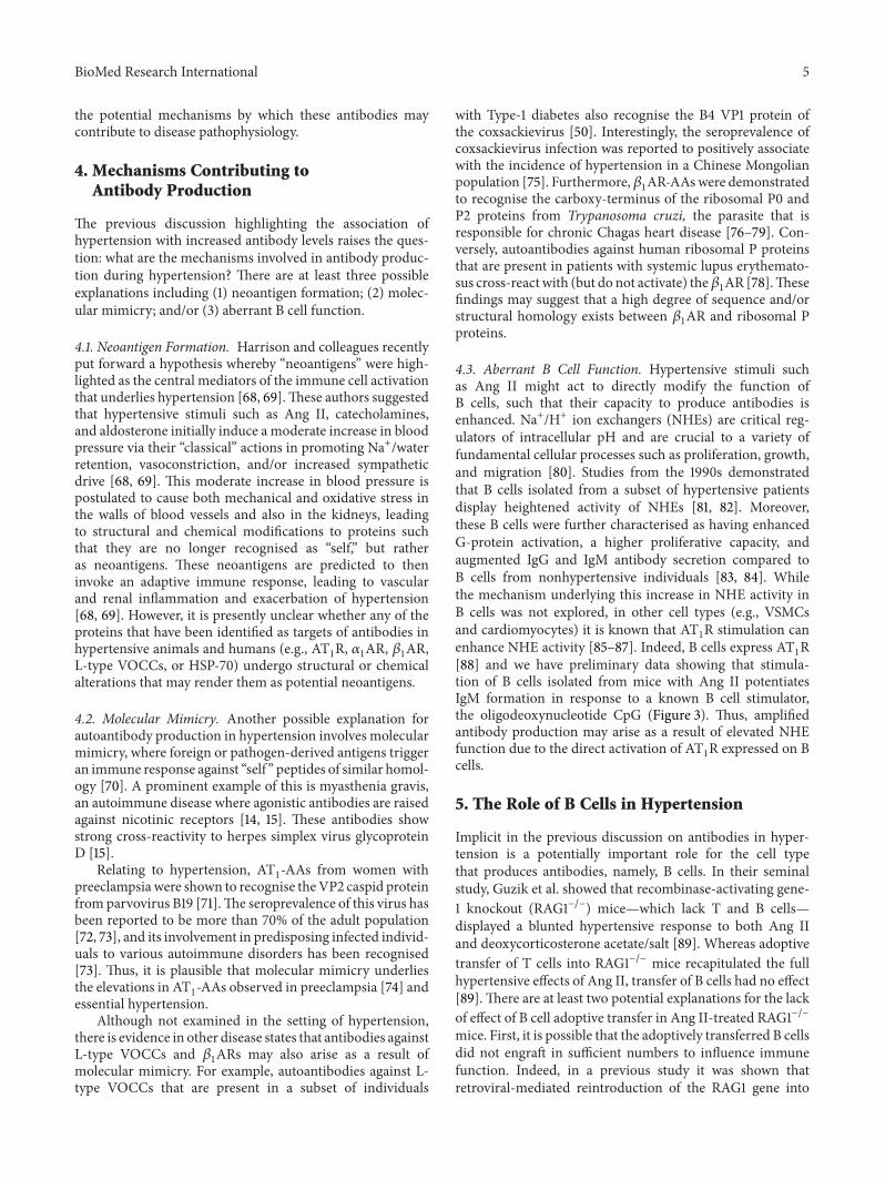
Antibodies, or immunoglobulins (Ig), are produced exclu-sively by B cells as part of the mammalian adaptive immuneresponse [5]. Antibodies play a crucial role in adaptiveimmunity through their ability to bind antigens, which arenormally toxic substances or fragments of pathogen-derivedproteins. Such binding results in either neutralisation of theantigen itself or, when the antigen is bound to a cell (e.g.,bacteria), destruction of that cell via activation of the comple-ment system, neutrophil degranulation, or phagocytosis bymacrophages (Figure 1).
Hindawi Publishing CorporationBioMed Research InternationalVolume 2014, Article ID 504045, 9 pageshttp://dx.doi.org/10.1155/2014/504045

2 BioMed Research International
Signal transduction leading to
receptor activation
Formation of proinflammatoryimmune complexes
Neutralisation of freeantigens (i.e., virions)
Phagocytosis of opsonisedpathogens
Antibody-dependent
cell-mediated cytotoxicity
Cytokine release
Antibody-mediated complementopsonisation
Antibodies
Direct cell lysis by complement viaformation of membrane attack complex
Figure 1: Schematic diagram showing the various types of antibody-mediated autoimmune responses.
For B cells to generate and secrete antibodies, theymust first undergo differentiation into plasma cells. NaıveB cells detect antigens via their B cell receptors, whichare membrane-bound immunoglobulins (IgM) with uniqueand randomly-generated antigen-binding sites [6]. Followingbinding, the antigen is internalised, processed, and displayedby major histocompatibility class II (MHC II) proteins onthe extracellular surface of the B cell [6]. The next step inB cell differentiation involves the detection of the MHC II-presented antigen by the T cell receptor (TCR) of an activatedT helper (TH) cell that has previously encountered the sameantigen. The TH cell also provides essential costimulatorysignals in the form of interactions between its CD40 ligandand the CD40 receptor on the B cell [7], as well as cytokinessuch as interferon-gamma and interleukin-4 [8]. Together,these signals ultimately promote the differentiation of the Bcell into an antibody-secreting plasma cell. Such signals alsoplay a crucial role in isotype switching (e.g. IgM → IgG)[9], which allows the generated antibodies to interact withdifferent effector molecules and thereby direct the type ofimmune response that is mounted.
Although antibodies normally target foreign molecules,under some circumstances they may be raised against host-derived molecules. Such a loss of recognition of “self ”is the basis for autoimmune diseases. There are severalmechanisms by which aberrant antibody production canlead to autoimmune pathologies (Figure 1). For example, thebinding of antibodies to antigens expressed on the surfaceof endogenous cells may lead to the destruction of these
cells via complement- or leukocyte-dependent interactions.This type of response is termed a “Type II Hypersensitivityreaction” and is the cause of the loss of erythrocytes inautoimmune haemolytic anaemia [10]. Alternatively, “TypeIII Hypersensitivity reactions” involve the recognition ofsoluble antigens in the host by antibodies and the subsequentformation of “immune complexes.” Immune complexes arecross-linked aggregations of antibodies and antigens that canbe deposited in various tissues to cause local inflammatoryresponses [11]. Immune complexes are a hallmark of severalautoimmune disorders including vasculitis and systemiclupus erythematosus where deposition of such complexes inthe kidneys gives rise to glomerulonephritis [12, 13]. Finally,some autoimmune diseases are associated with the formationof nonimmunogenic, agonistic antibodies to receptors [14].These types of diseases are often classified as “Type VHypersensitivity Reactions.” Agonistic antibodies stimulatereceptors in a similar fashion to their cognate ligands andthus lead to overstimulation of the specific system involved.Myasthenia gravis is an example of an autoimmune diseasecaused by the generation of agonistic antibodies againstnicotinic receptors [14, 15].
3. Protein Targets ofHypertension-Related Antibodies
Studies dating back to the 1970s demonstrated that essentialhypertension in humans is associated with elevated IgGand IgM titres [1–4]. However, these early studies did not

BioMed Research International 3
identify the targets of these antibodies and thus providedno indication of whether they were important to the patho-physiology of hypertension. Studies from around the sametime on animal models confirmed that hypertension wasassociated with an increase in antibody production and evenwent some way towards implicating a possible causativerole for these antibodies. For example, Ba et al. identifiedautoantibodies in the serum of spontaneously hypertensiverats (SHRs) that were cytotoxic to T cells. Although theauthors did not establish a precise mechanism by whichthese antibodies contribute to hypertension, they impliedthat the antibodies might induce apoptosis of “suppressor” Tcells that normally prevent damage to the vascular wall andthus protect against cardiovascular disease [16]. In a separatestudy, it was shown that rats with hypertension induced byrenal infarction had high serum levels of antibodies thatbound to arteries, glomeruli, and basementmembranes of thekidneys [17].Given the key roles of the kidney and vasculaturein the regulation of hemodynamic parameters, this latterstudy provided an indication that elevated antibody pro-duction may actually contribute to the chronic elevation inblood pressure that defines hypertension. And indeed, morerecent work identifying the specific molecular targets of theantibodies present in individuals with essential hypertensionand preeclampsia not only supports this idea, but also beginsto shed light on how elevated antibody production mightcontribute to elevated blood pressure.
3.1. Angiotensin II Type-1 Receptors. The angiotensin II type-1receptor (AT
1R) plays a crucial role in the regulation of blood
pressure [18]. Stimulation of the AT1R by its cognate ligand,
angiotensin II, results in vascular smoothmuscle cell (VSMC)contraction and proliferation, release of aldosterone from theadrenal glands, and activation of the sympathetic nervoussystem [18, 19]. Furthermore, it has recently been discoveredthat AT
1receptor activation on T lymphocytes promotes a
proinflammatory phenotype that contributes to hypertension[20].
AT1R-activating IgG autoantibodies (AT
1-AAs) directed
against the second extracellular loop of the AT1R are preva-
lent in over 95% of patients with pregnancy-associatedhypertension, and antibody titres correlate positively withdisease severity [21, 22]. AT
1-AAs appear to activate a cascade
of proinflammatory cytokines that contribute directly tohypertension in preeclampsia [23]. In vivo administrationof AT
1-AAs isolated from preeclamptic humans to pregnant
mice was shown to induce hypertension in those animals[24]. Furthermore, AT
1-AA treatment causes an increase
in the circulating levels of tumour necrosis factor-𝛼 andinterleukin-6 in pregnant mice and inhibition of thesecytokines with neutralising antibodies blunts hypertension[24, 25].
AT1-AAs have also been identified in a subset of indi-
viduals with essential hypertension [26–28]. These AT1-AAs
appear to be similar in function and specificity as thoseidentified in preeclamptic patients as they also bind to thesecond extracellular loop of the AT
1R [26, 28]. The fact
that essential hypertensive patients with AT1-AAs respond
with greater blood pressure reductions to AT1R blockade by
candesartan than hypertensive individuals without AT1-AA
[29, 30] suggests a causal role for AT1-AAs in at least some
cases of hypertension.
3.2. Alpha-1 Adrenergic Receptors. The alpha-1 adrenergicreceptor (𝛼
1AR) is a G-protein coupled receptor that is
primarily expressed on VSMCs and proximal renal tubules[31]. Activation of the 𝛼
1AR by its endogenous ligands,
noradrenaline and adrenaline, or synthetic compounds suchas phenylephrine, results in VSMC contraction and increasedtotal peripheral resistance, as well as increased Na+ reab-sorption in the kidney causing elevated blood pressure [32].IgG receptor-activating autoantibodies against the 𝛼
1AR
(𝛼1AR-AA) have been described in patients with essential
hypertension [28, 33–35]. Unlike AT1-AAs, which appear
to bind to a similar domain of the AT1R (i.e., the second
extracellular loop) irrespective of the patient from whichthey were derived, 𝛼
1AR-AA from different patients display
selectivity towards separate regions of the receptor, with anti-bodies from some individuals targeting the first extracellularloop, and antibodies from other individuals targeting thesecond extracellular loop [33, 34]. It is unclear whether thesevarying binding properties have implications for the abilityof a specific antibody to modulate receptor function. It isalso unclear whether 𝛼
1AR-AAs are present or elevated in
individuals with preeclampsia.
3.3. Beta-1 Adrenergic Receptors. The 𝛽-1 adrenergic receptor(𝛽1AR) shares the same endogenous agonists with 𝛼
1ARs
but differs in ligand affinity, tissue distribution, and func-tional outcomes following stimulation. 𝛽
1ARs are localised
predominately in cardiac tissue where activation results inincreased heart rate and contractility and an overall increasein cardiac output [36]. Cardiac output is a major determinantof blood pressure and thus 𝛽
1AR blockers have long been
used as antihypertensive agents [37]. 𝛽1AR agonistic IgG
autoantibodies (𝛽1AR-AA) against the second extracellular
loop of the receptors were detected in the serum of spon-taneously hypertensive rats [38]. Furthermore, injection of𝛽1AR-AAs into healthy Lewis rats promoted cardiomyopathy
and increases in systolic blood pressure [39]. Althoughevidence for the presence of 𝛽
1AR-AAs in human essential
hypertension and preeclampsia is lacking, these antibodieshave been identified in patients with idiopathic dilatedcardiomyopathy [40].
3.4. L-Type Voltage Gated Calcium Channels. L-type volt-age gated calcium channels (L-type VOCCs) are expressedon VSMCs in resistance vessels and, in their open state,directly contribute to vascular tone and blood pressure byfacilitating the influx of extracellular Ca2+ [41]. It is wellestablished that L-type VOCC expression in the vasculatureis upregulated in experimental hypertension and that thiscontributes to increased Ca2+ levels in VSMCs and thuselevated vascular resistance [42–47]. The importance of L-typeVOCCs in humanhypertension is highlighted by the factthat inhibitors of these channels (e.g., nifedipine) continue to

4 BioMed Research International
Antibodies
Hypertension
reabsorptioninflammation
HSP-70L-typeVOCCs
Vasoconstriction,Increased cardiac Renal or vascular
outputelevated totalperipheral resistance
Enhanced Na+/H2O
AT1R 𝛼1AR 𝛽1AR 𝛼1AR
Figure 2: Schematic diagram showing the mechanism by which autoantibodies may promote increases in vascular tone, cardiac output,Na+/water reabsorption, and renal and vascular inflammation, and thereby contribute to hypertension.
represent one of the most effective and widely-used classesof antihypertensive medications [48]. Thus, it is noteworthythat autoantibodies against L-type VOCCs were identifiedin some patients with essential hypertension [49]. Althoughthe authors did not examine the effect of these antibodieson L-type VOCC function, a separate study demonstratedincreased intracellular Ca2+ influx in pancreatic islet cells fol-lowing the binding of analogous IgG and IgA autoantibodiesto L-type VOCCs in the setting of Type-1 diabetes [50]. Thisimplies that antibodies against L-type VOCCs are likely tobe agonistic in nature and could thus contribute to increasedVSMC Ca2+ influx in hypertension.
3.5. Heat Shock Proteins. Heat shock proteins (HSPs) are afamily of highly-conserved proteins that provide protectionagainst danger-related signals by acting as molecular chap-erones to assist in the folding and trafficking of proteinsduring cellular stress [51]. Among the multitude of knownmammalian HSPs, HSP-70 has received most attention inthe field of hypertension research. First, in vitro exposure ofcultured VSMCs, endothelial cells or isolated aortic rings tohypertension-relevant stimuli such as oxidative stress, cyclicstrain, and angiotensin II, induces the expression of HSP-70[52–54]. Second, levels of HSP-70 and HSP-70-reactive CD4T cells are elevated in the kidneys in several rat models ofhypertension [55–57]. Finally, HSP-70 serum concentrationsare elevated in pregnancy-associated hypertension and arepositively correlated with blood pressure in affected women[58].
Elevated levels of IgG and IgA antibody titres againstHSP-70 have been identified in essential hypertensive indi-viduals [59, 60]. Surprisingly, elevated anti-HSP-70 antibodylevels in essential hypertension were not associated withchanges in serum HSP-70 in these patients [60]. Thus, thefunction of anti-HSP-70 antibodies in essential hypertensionremains unclear. It is possible that anti-HSP-70 antibodiescould either promote inflammation via formation of cir-culating immune complexes or, alternatively, alleviate theproinflammatory actions of these proteins via neutralisation.A more recent study by Molvarec et al. was unable todemonstrate any changes in circulating levels of anti-HSP-70antibodies in women with preeclampsia [61].
3.6. Miscellaneous. A study in borderline hypertensivepatients described a reduction in circulating levels of anti-oxidised LDL IgG antibodies [62]. However, a follow-upinvestigation failed to detect any difference in levels ofthese antibodies between patients with clinical hypertensionand normotensive controls [63], and thus the significanceof anti-oxidised LDL antibodies in the pathophysiology ofhypertension is unclear. Other studies in borderline hyper-tensive individuals detected elevations in circulating anti-endothelial cell IgG and IgM antibodies [64–66]. Whiledata in the setting of human essential hypertension is stillmissing, these antibodies have been identified in womenwithsevere preeclampsia and have been proposed to contribute toendothelial dysfunction [67].
Figure 2 provides a summary of the targets of antibodiesthat have been shown to be elevated in hypertension and
BioMed Research International 5
the potential mechanisms by which these antibodies maycontribute to disease pathophysiology.
4. Mechanisms Contributing toAntibody Production
The previous discussion highlighting the association ofhypertension with increased antibody levels raises the ques-tion: what are the mechanisms involved in antibody produc-tion during hypertension? There are at least three possibleexplanations including (1) neoantigen formation; (2) molec-ular mimicry; and/or (3) aberrant B cell function.
4.1. Neoantigen Formation. Harrison and colleagues recentlyput forward a hypothesis whereby “neoantigens” were high-lighted as the central mediators of the immune cell activationthat underlies hypertension [68, 69].These authors suggestedthat hypertensive stimuli such as Ang II, catecholamines,and aldosterone initially induce a moderate increase in bloodpressure via their “classical” actions in promoting Na+/waterretention, vasoconstriction, and/or increased sympatheticdrive [68, 69]. This moderate increase in blood pressure ispostulated to cause both mechanical and oxidative stress inthe walls of blood vessels and also in the kidneys, leadingto structural and chemical modifications to proteins suchthat they are no longer recognised as “self,” but ratheras neoantigens. These neoantigens are predicted to theninvoke an adaptive immune response, leading to vascularand renal inflammation and exacerbation of hypertension[68, 69]. However, it is presently unclear whether any of theproteins that have been identified as targets of antibodies inhypertensive animals and humans (e.g., AT
1R, 𝛼1AR, 𝛽
1AR,
L-type VOCCs, or HSP-70) undergo structural or chemicalalterations that may render them as potential neoantigens.
4.2. Molecular Mimicry. Another possible explanation forautoantibody production in hypertension involves molecularmimicry, where foreign or pathogen-derived antigens triggeran immune response against “self ” peptides of similar homol-ogy [70]. A prominent example of this is myasthenia gravis,an autoimmune disease where agonistic antibodies are raisedagainst nicotinic receptors [14, 15]. These antibodies showstrong cross-reactivity to herpes simplex virus glycoproteinD [15].
Relating to hypertension, AT1-AAs from women with
preeclampsia were shown to recognise theVP2 caspid proteinfrom parvovirus B19 [71].The seroprevalence of this virus hasbeen reported to be more than 70% of the adult population[72, 73], and its involvement in predisposing infected individ-uals to various autoimmune disorders has been recognised[73]. Thus, it is plausible that molecular mimicry underliesthe elevations in AT
1-AAs observed in preeclampsia [74] and
essential hypertension.Although not examined in the setting of hypertension,
there is evidence in other disease states that antibodies againstL-type VOCCs and 𝛽
1ARs may also arise as a result of
molecular mimicry. For example, autoantibodies against L-type VOCCs that are present in a subset of individuals
with Type-1 diabetes also recognise the B4 VP1 protein ofthe coxsackievirus [50]. Interestingly, the seroprevalence ofcoxsackievirus infection was reported to positively associatewith the incidence of hypertension in a Chinese Mongolianpopulation [75]. Furthermore,𝛽
1AR-AAswere demonstrated
to recognise the carboxy-terminus of the ribosomal P0 andP2 proteins from Trypanosoma cruzi, the parasite that isresponsible for chronic Chagas heart disease [76–79]. Con-versely, autoantibodies against human ribosomal P proteinsthat are present in patients with systemic lupus erythemato-sus cross-react with (but do not activate) the𝛽
1AR [78].These
findings may suggest that a high degree of sequence and/orstructural homology exists between 𝛽
1AR and ribosomal P
proteins.
4.3. Aberrant B Cell Function. Hypertensive stimuli suchas Ang II might act to directly modify the function ofB cells, such that their capacity to produce antibodies isenhanced. Na+/H+ ion exchangers (NHEs) are critical reg-ulators of intracellular pH and are crucial to a variety offundamental cellular processes such as proliferation, growth,and migration [80]. Studies from the 1990s demonstratedthat B cells isolated from a subset of hypertensive patientsdisplay heightened activity of NHEs [81, 82]. Moreover,these B cells were further characterised as having enhancedG-protein activation, a higher proliferative capacity, andaugmented IgG and IgM antibody secretion compared toB cells from nonhypertensive individuals [83, 84]. Whilethe mechanism underlying this increase in NHE activity inB cells was not explored, in other cell types (e.g., VSMCsand cardiomyocytes) it is known that AT
1R stimulation can
enhance NHE activity [85–87]. Indeed, B cells express AT1R
[88] and we have preliminary data showing that stimula-tion of B cells isolated from mice with Ang II potentiatesIgM formation in response to a known B cell stimulator,the oligodeoxynucleotide CpG (Figure 3). Thus, amplifiedantibody production may arise as a result of elevated NHEfunction due to the direct activation of AT
1R expressed on B
cells.
5. The Role of B Cells in Hypertension
Implicit in the previous discussion on antibodies in hyper-tension is a potentially important role for the cell typethat produces antibodies, namely, B cells. In their seminalstudy, Guzik et al. showed that recombinase-activating gene-1 knockout (RAG1−/−) mice—which lack T and B cells—displayed a blunted hypertensive response to both Ang IIand deoxycorticosterone acetate/salt [89]. Whereas adoptivetransfer of T cells into RAG1−/− mice recapitulated the fullhypertensive effects of Ang II, transfer of B cells had no effect[89]. There are at least two potential explanations for the lackof effect of B cell adoptive transfer in Ang II-treated RAG1−/−mice. First, it is possible that the adoptively transferred B cellsdid not engraft in sufficient numbers to influence immunefunction. Indeed, in a previous study it was shown thatretroviral-mediated reintroduction of the RAG1 gene into

6 BioMed Research International
8000
6000
4000
2000
0
Control Ang ll CpG CpG + Ang ll
lgM
conc
entr
atio
n (n
g/m
L)
∗
Figure 3: Effect of angiotensin II (Ang II; 0.1𝜇M, 48 h) or CpGoligodeoxynucleotides (CpG; 5𝜇g/mL, 48 h) alone or in combina-tion on IgM antibody secretion from primary cultures of mouseB cells. Values represent mean ± S.E.M. of 𝑛 = 14 experiments.∗
𝑃 < 0.05 for Bonferroni’s post-hoc test after one-way ANOVA.
RAG1−/−mice restored T cells numbers back to levels in wild-type mice, while B cell numbers only increased marginally[90]. This suggests that the immunological environment inRAG1−/− mice, while being favourable to the survival andfunction of T cells, may be incompatible with the growthand function of B cells. An alternative explanation maylie in the different mechanisms that activate T cells and Bcells during an immune response. Whilst activation of Tcells relies primarily on antigen presentation from innateimmune cells such as dendritic cells (which are relativelyunaffected in RAG1−/− mice), as discussed previously, Bcell activation and differentiation into an effector phenotypenormally requires interactions with TH cells [6]. Hence, thelack of T cells in RAG1−/− mice may have precluded thepossibility of any adoptively transferred B cells becomingactivated. Indeed, a critical role of TH cells in B cell activationduring hypertension was suggested in a recent study showingthat adoptive transfer of TH cells from preeclamptic miceinto normal pregnant mice induced AT
1-AA production and
elevated blood pressure [91]. Importantly, a B cell depletingagent ameliorated both of these effects [91].
6. Therapeutic Implications
An understanding of the role of B cells and antibody pro-duction during hypertension could aid in the refinementof current treatment approaches and also in developmentof novel antihypertensive therapies. For example, by iden-tifying the autoantibodies that are specifically elevated in agiven hypertensive patient, it might be possible to “tailor”the way their disease is subsequently managed for betterclinical outcomes; that is, individuals with AT
1-AAs would
favourably respond to AT1R blockers over patients with L-
type VOCC autoantibodies, where calcium channel blockerssuch as nifedipine would be preferred.
In terms of new therapeutic approaches, identificationof the specific pathogen-derived or neoantigens that lead to
elevated antibody generation in hypertension could lead toimmunisation strategies aimed at neutralising such antigensor steering the immune response away from one that pro-motes hypertension. Indeed, two vaccines against Ang II havebeen developed and showed some early promise in reducingblood pressure in hypertensive patients [92]. However, due totheir lower efficacy compared to conventional inhibitors ofthe renin-angiotensin system, the vaccines did not proceedinto Phase III clinical trials [93, 94], and thus further workis needed to determine if alternative immunisation strategies(i.e., involving different adjuvants and/or immunogens) willbe more effective.
B cell-depleting agents, which include antibodies againstthe B cell specific surface receptorCD20 and theB cell activat-ing factor BAFF, are already in clinical use for the treatment ofautoimmune diseases such as lupus erythematosus [95] andcould potentially be used to treat hypertension. Of course,these drugs have the potential for causing immunosuppres-sion and hence their use might be best reserved for thosehypertensive patients that do not respond to conventionaltherapies. Until recently, one of the main therapeutic optionsfor individuals with resistant hypertension was surgical den-ervation of the renal artery [96]; however, the effectivenessof this procedure has recently been called into question [97].Thus, B cell-modulating drugs might yet be a safer and moreefficacious therapeutic option for such patients.
7. Conclusion
There is evidence that circulating antibody levels are elevatedin both essential and pregnancy-related hypertension. Manyof these antibodies appear to target receptors and ion chan-nels known to be involved in the regulation of blood pressure.Further studies are required to characterise the precise impactthat antibody binding has on the function of these proteinsand to uncover the mechanisms responsible for aberrantantibody production in hypertension. Such studies shouldnot only allow us to evaluate the significance of elevatedantibody production in the pathophysiology of hypertension,but theymay also lead to the development of new therapeuticapproaches and/or the refinement of current approaches,to improve the management of clinical hypertension in thefuture.
Conflict of Interests
The authors declare that there is no conflict of interestsregarding the publication of this paper.
Acknowledgments
Christopher T. Chan is supported by an Australian Post-graduate Award (ID no. 5131432). Grant R. Drummond andChristopher G. Sobey are supported by Senior ResearchFellowships from the National Health and Medical ResearchCouncil of Australia (NHMRC; ID nos. APP1006017 and350327, resp.). None of these funding sources had any role in

BioMed Research International 7
thewriting of the report or in the decision to submit the paperfor publication.
References
[1] A. Ebringer and A. E. Doyle, “Raised serum IgG levels inhypertension,” British Medical Journal, vol. 2, no. 5702, pp. 146–148, 1970.
[2] P. Suryaprahba, T. Padma, and U. Brahmaji Rao, “Increasedserum IgG levels in essential hypertension,” Immunology Let-ters, vol. 8, no. 3, pp. 143–145, 1984.
[3] E. Hilme, H. Herlitz, T. Soderstrom, and L. Hansson, “Increasedsecretion of immunoglobulins in malignant hypertension,”Journal of Hypertension, vol. 7, no. 2, pp. 91–95, 1989.
[4] T. Gudbrandsson, L. Hansson, H. Herlitz, L. Lindholm, and L.A. Nilsson, “Immunological changes in patients with previousmalignant essential hypertension,” Lancet, vol. 1, no. 8217, pp.406–408, 1981.
[5] T. W. Lebien and T. F. Tedder, “B lymphocytes: How theydevelop and function,”Blood, vol. 112, no. 5, pp. 1570–1580, 2008.
[6] D. C. Parker, “T cell-dependent B cell activation,” AnnualReview of Immunology, vol. 11, pp. 331–360, 1993.
[7] I. S. Grewal and R. A. Flavell, “CD40 and CD154 in cell-mediated immunity,”Annual Review of Immunology, vol. 16, pp.111–135, 1998.
[8] J. Purkerson and P. Isakson, “A two-signal model for regulationof immunoglobulin isotype switching,”The FASEB Journal, vol.6, no. 14, pp. 3245–3252, 1992.
[9] S. G. Tangye, A. Ferguson, D. T. Avery, C. S. Ma, and P. D.Hodgkin, “Isotype switching by human B cells is division-associated and regulated by cytokines,” Journal of Immunology,vol. 169, no. 8, pp. 4298–4306, 2002.
[10] B. C. Gehrs and R. C. Friedberg, “Autoimmune hemolyticanemia,”TheAmerican Journal of Hematology, vol. 69, no. 4, pp.258–271, 2002.
[11] A. N. Theofilopoulos and F. J. Dixon, “Immune complexes inhuman diseases,” The American Journal of Pathology, vol. 100,no. 2, pp. 529–594, 1980.
[12] D. Koffler, V. Agnello, R.Thoburn, and H. G. Kunkel, “Systemiclupus erythematosus: prototype of immune complex nephritisin man,” Journal of Experimental Medicine, vol. 134, supplement3, pp. 169S–179S, 1971.
[13] W. L. Gross, A. Trabandt, and E. Reinhold-Keller, “Diagnosisand evaluation of vasculitis,” Rheumatology, vol. 39, no. 3, pp.245–252, 2000.
[14] J. M. Lindstrom, M. E. Seybold, V. A. Lennon, S. Whittingham,and D. D. Duane, “Antibody to acetylcholine receptor in myas-thenia gravis: prevalence, clinical correlates, and diagnosticvalue,” Neurology, vol. 51, no. 4, p. 933, 1998.
[15] P. L. Schwimmbeck, T. Dyrberg, D. B. Drachman, and M. B.A. Oldstone, “Molecular mimicry and myasthenia gravis. Anautoantigenic site of the acetylcholine receptor 𝛼-subunit thathas biologic activity and reacts immunochemically with herpessimplex virus,” Journal of Clinical Investigation, vol. 84, no. 4, pp.1174–1180, 1989.
[16] D. Ba, N. Takeichi, T. Kodama, and H. Kobayashi, “Restorationof T cell depression and suppression of blood pressure inspontaneously hypertensive rats (SHR) by thymus grafts orthymus extracts,” Journal of Immunology, vol. 128, no. 3, pp. 1211–1216, 1982.
[17] F. N. White and A. Grollman, “Autoimmune factors associatedwith infarction of the kidney,” Nephron, vol. 204, pp. 93–102,1964.
[18] M. J. Peach, “Renin angiotensin system: biochemistry andmechanisms of action,” Physiological Reviews, vol. 57, no. 2, pp.313–370, 1977.
[19] L. Hunyady and K. J. Catt, “Pleiotropic AT1 receptor signalingpathways mediating physiological and pathogenic actions ofangiotensin II,”Molecular Endocrinology, vol. 20, no. 5, pp. 953–970, 2006.
[20] J. L. Silva-Filho, M. C. Souza, M. D. G. Henriques et al., “AT1receptor-mediated angiotensin II activation and chemotaxis ofT lymphocytes,” Molecular Immunology, vol. 48, no. 15-16, pp.1835–1843, 2011.
[21] G. Wallukat, V. Homuth, T. Fischer et al., “Patients withpreeclampsia develop agonistic autoantibodies against theangiotensin AT1 receptor,” Journal of Clinical Investigation, vol.103, no. 7, pp. 945–952, 1999.
[22] A. H. Siddiqui, R. A. Irani, S. C. Blackwell, S. M. Ramin,R. E. Kellems, and Y. Xia, “Angiotensin receptor agonisticautoantibody is highly prevalent in preeclampsia: correlationwith disease severity,” Hypertension, vol. 55, no. 2, pp. 386–393,2010.
[23] Y. Xia and R. E. Kellems, “Angiotensin receptor agonisticautoantibodies and hypertension : preeclampsia and beyond,”Circulation Research, vol. 113, no. 1, pp. 78–87, 2013.
[24] C. C. Zhou, R. A. Irani, Y. Dai et al., “Autoantibody-mediatedIL-6-dependent endothelin-1 elevation underlies pathogenesisin a mouse model of preeclampsia,” Journal of Immunology, vol.186, no. 10, pp. 6024–6034, 2011.
[25] R. A. Irani, Y. Zhang, C. C. Zhou et al., “Autoantibody-mediatedangiotensin receptor activation contributes to preeclampsiathrough tumor necrosis factor-𝛼 signaling,” Hypertension, vol.55, no. 5, pp. 1246–1253, 2010.
[26] M. L. Fu, H. Herlitz, W. Schulze et al., “Autoantibodies againstthe angiotensin receptor (AT1) in patients with hypertension,”Journal of Hypertension, vol. 18, no. 7, pp. 945–953, 2000.
[27] F. Zhu, Y. Sun, M. Wang et al., “Correlation between HLA-DRB1, HLA-DQB1 polymorphism and autoantibodies againstangiotensin AT(1) receptors in Chinese patients with essentialhypertension,” Clinical Cardiology, vol. 34, no. 5, pp. 302–308,2011.
[28] Y. H. Liao, Y. M. Wei, M. Wang, Z. H. Wang, H. T. Yuan,and L. X. Cheng, “Autoantibodies against AT1-receptor and 𝛼1-adrenergic receptor in patients with hypertension,” Hyperten-sion Research, vol. 25, no. 4, pp. 641–646, 2002.
[29] Y. Sun, Y. Liao, Y. Yuan et al., “Influence of autoantibod-ies against AT1 receptor and AGTR1 polymorphisms oncandesartan-based antihypertensive regimen: results from theStudy ofOptimal Treatment inHypertensive PatientswithAnti-AT1-Receptor Autoantibodies trial,” Journal of the AmericanSociety of Hypertension, vol. 8, no. 1, pp. 21–27, 2014.
[30] F. Wei, X. J. Jia, S. Q. Yu et al., “Candesartan versus imidapril inhypertension: a randomised study to assess effects of anti-AT1receptor autoantibodies,”Heart, vol. 97, no. 6, pp. 479–484, 2011.
[31] J. L. Reid, “Alpha-adrenergic receptors and blood pressurecontrol,” The American Journal of Cardiology, vol. 57, no. 9, pp.6E–12E, 1986.
[32] M. T. Piascik and D. M. Perez, “𝛼1-adrenergic receptors: newinsights and directions,” Journal of Pharmacology and Experi-mental Therapeutics, vol. 298, no. 2, pp. 403–410, 2001.

8 BioMed Research International
[33] H. P. Luther, V. Homuth, and G. Wallukat, “𝛼1-Adrenergicreceptor antibodies in patients with primary hypertension,”Hypertension, vol. 29, no. 2, pp. 678–682, 1997.
[34] M. L. Fu,H.Herlitz, G.Wallukat et al., “Functional autoimmuneepitope on 𝛼1-adrenergic receptors in patients with malignanthypertension,” The Lancet, vol. 344, no. 8938, pp. 1660–1663,1994.
[35] K. Wenzel, H. Haase, G. Wallukat et al., “Potential relevance of𝛼1-Adrenergic receptor autoantibodies in refractory hyperten-sion,” PLoS ONE, vol. 3, no. 11, Article ID e3742, 2008.
[36] K. P. Minneman, R. N. Pittman, and P. B. Molinoff, “Beta-adrenergic receptor subtypes: properties, distribution, and reg-ulation,” Annual Review of Neuroscience, vol. 4, pp. 419–461,1981.
[37] M. A. van Baak, H. A. J. Struyker Boudier, and J. F. M. Smits,“Antihypertensive mechanisms of beta-adrenoceptor blockade:a review,” Clinical and Experimental Hypertension ATheory andPractice, vol. 7, no. 1, pp. 1–72, 1985.
[38] G. Wallukat, I. E. Blasig, R. Morwinski, H. J. Herrmann, andE. Rohde, “The sera of spontaneously hypertensive rats containagonistic auto-antibodies against the 𝛽1-adrenoceptor,” Journalof Hypertension, vol. 13, no. 9, pp. 1031–1036, 1995.
[39] R. Jahns, V. Boivin, L. Hein et al., “Direct evidence for a 𝛽1-adrenergic receptor-directed autoimmune attack as a cause ofidiopathic dilated cardiomyopathy,” Journal of Clinical Investi-gation, vol. 113, no. 10, pp. 1419–1429, 2004.
[40] Y. Magnusson, G. Wallukat, F. Waagstein, A. Hjalmarson, andJ. Hoebeke, “Autoimmunity in idiopathic dilated cardiomyopa-thy: characterization of antibodies against the 𝛽1-adrenoceptorwith positive chronotropic effect,” Circulation, vol. 89, no. 6, pp.2760–2767, 1994.
[41] W. A. Catterall, “Voltage-gated calcium channels,” Cold SpringHarbor perspectives in biology, vol. 3, no. 8, Article ID a003947,2011.
[42] D. W. Wilde, P. B. Furspan, and J. F. Szocik, “Calcium currentin smooth muscle cells from normotensive and geneticallyhypertensive rats,” Hypertension, vol. 24, no. 6, pp. 739–746,1994.
[43] R. H. Cox and I. M. Lozinskaya, “Augmented calcium currentsin mesenteric artery branches of the spontaneously hyperten-sive rats,” Hypertension, vol. 26, no. 6, pp. 1060–1064, 1995.
[44] Y. Ohya, T. Tsuchihashi, S. Kagiyama, I. Abe, andM. Fujishima,“Single L-type calcium channels in smooth muscle cells fromresistance arteries of spontaneously hypertensive rats,” Hyper-tension, vol. 31, no. 5, pp. 1125–1129, 1998.
[45] J. M. Simard, X. Li, and K. Tewari, “Increase in functional Ca2+channels in cerebral smooth muscle with renal hypertension,”Circulation Research, vol. 82, no. 12, pp. 1330–1337, 1998.
[46] A. Pesic, J. A. Madden, M. Pesic, and N. J. Rusch, “Highblood pressure upregulates arterial L-type Ca2+ channels: ismembrane depolarization the signal?” Circulation research, vol.94, no. 10, pp. e97–e104, 2004.
[47] P. F. Pratt, S. Bonnet, L. M. Ludwig, P. Bonnet, and N. J.Rusch, “Upregulation of L-type Ca2+ channels in mesentericand skeletal arteries of SHR,” Hypertension, vol. 40, no. 2, pp.214–219, 2002.
[48] D. J. Triggle, “L-type calcium channels,”Current PharmaceuticalDesign, vol. 12, no. 4, pp. 443–457, 2006.
[49] Z. H. Zhou, J. Wang, H. Xiao et al., “A novel autoantibody inpatients with primary hypertension: antibody against L-typeCa2+ channel,” Chinese Medical Journal, vol. 121, no. 16, pp.1513–1517, 2008.
[50] C. Bason, R. Lorini, C. Lunardi et al., “In type 1 diabetes a subsetof anti-coxsackievirus B4 antibodies recognize autoantigensand induce apoptosis of pancreatic beta cells,” PLoS ONE, vol.8, no. 2, Article ID e57729, 2013.
[51] M. J. Schlesinger, “Heat shock proteins,” Journal of BiologicalChemistry, vol. 265, no. 21, pp. 12111–12114, 1990.
[52] Q. Xu, G. Schett, C. Li, Y. Hu, and G. Wick, “Mechanical stress-induced heat shock protein 70 expression in vascular smoothmuscle cells is regulated by rac and ras small G proteins but notmitogen-activated protein kinases,” Circulation Research, vol.86, no. 11, pp. 1122–1128, 2000.
[53] Q. Xu, D. G. Li, N. J. Holbrook, and R. Udelsman, “Acutehypertension induces heat-shock protein 70 gene expression inrat aorta,” Circulation, vol. 92, no. 5, pp. 1223–1229, 1995.
[54] D. F. Liao, Z. G. Jin, A. S. Baas et al., “Purification andidentification of secreted oxidative stress-induced factors fromvascular smooth muscle cells,” Journal of Biological Chemistry,vol. 275, no. 1, pp. 189–196, 2000.
[55] G. Parra, Y. Quiroz, J. Salazar et al., “Experimental inductionof salt-sensitive hypertension is associated with lymphocyteproliferative response to HSP70,” Kidney International, vol. 74,no. 111, pp. S55–S59, 2008.
[56] N. Ishizaka, T. Aizawa, M. Ohno et al., “Regulation and local-ization of HSP70 and HSP25 in the kidney of rats undergoinglong-term administration of Angiotensin II,”Hypertension, vol.39, no. 1, pp. 122–128, 2002.
[57] H. Pons, A. Ferrebuz, Y. Quiroz et al., “Immune reactivity toheat shock protein 70 expressed in the kidney is cause of salt-sensitive hypertension,” The American Journal of Physiology—Renal Physiology, vol. 304, no. 3, pp. F289–F299, 2013.
[58] A. Molvarec, Z. Prohaszka, B. Nagy et al., “Association of ele-vated serumheat-shock protein 70 concentrationwith transienthypertension of pregnancy, preeclampsia and superimposedpreeclampsia: a case-control study,” Journal of Human Hyper-tension, vol. 20, no. 10, pp. 780–786, 2006.
[59] T. Wu, J. Ma, S. Chen et al., “Association of plasma antibodiesagainst the inducible Hsp70 with hypertension and harshworking conditions,” Cell Stress & Chaperones, vol. 6, no. 4, pp.394–401, 2001.
[60] A. G. Pockley, U. de Faire, R. Kiessling, C. Lemne, T.Thulin, andJ. Frostegard, “Circulating heat shock protein and heat shockprotein antibody levels in established hypertension,” Journal ofHypertension, vol. 20, no. 9, pp. 1815–1820, 2002.
[61] A. Molvarec, Z. Derzsy, J. Kocsis et al., “Circulating anti-heat-shock-protein antibodies in normal pregnancy and preeclamp-sia,” Cell Stress and Chaperones, vol. 14, no. 5, pp. 491–498, 2009.
[62] R. Wu, U. de Faire, C. Lemne, J. L. Witztum, and J. Frostegard,“Autoantibodies to OxLDL are decreased in individuals withborderline hypertension,” Hypertension, vol. 33, no. 1 I, pp. 53–59, 1999.
[63] J. Frostegard, R. Wu, C. Lemne et al., “Circulating oxidizedlow-density lipoprotein is increased in hypertension,” ClinicalScience, vol. 105, no. 5, pp. 615–620, 2003.
[64] D. P. Papadopoulos, T. K. Makris, P. Krespi et al., “Antiendothe-lial cell antibody levels in healthy normotensives with high nor-mal blood pressure,” Clinical and Experimental Hypertension,vol. 28, no. 8, pp. 663–667, 2006.
[65] D. P. Papadopoulos, T. K.Makris, U. Papazachou,M. Daskalaki,E. Sanidas, and V. E. Votteas, “Antiendothelial cell antibodylevels in patients with masked hypertension,” InternationalJournal of Cardiology, vol. 130, no. 3, pp. 405–408, 2008.

BioMed Research International 9
[66] J. Frostegard, R. Wu, C. Gillis-Haegerstrand, C. Lemne, andU. de Faire, “Antibodies to endothelial cells in borderlinehypertension,” Circulation, vol. 98, no. 11, pp. 1092–1098, 1998.
[67] V. J. Rappaport, G. Hirata, H. K. Yap, and S. C. Jordan, “Anti-vascular endothelial cell antibodies in severe preeclampsia,”TheAmerican Journal of Obstetrics and Gynecology, vol. 162, no. 1,pp. 138–146, 1990.
[68] D. G. Harrison, T. J. Guzik, H. E. Lob et al., “Inflammation,immunity, and hypertension,” Hypertension, vol. 57, no. 2, pp.132–140, 2011.
[69] D. G. Harrison, A. Vinh, H. Lob, andM. S.Madhur, “Role of theadaptive immune system in hypertension,” Current Opinion inPharmacology, vol. 10, no. 2, pp. 203–207, 2010.
[70] M. B. Oldstone, “Molecular mimicry and immune-mediateddiseases,”TheFASEB Journal, vol. 12, no. 13, pp. 1255–1265, 1998.
[71] F. Herse, S. Verlohren, K. Wenzel et al., “Prevalence of agonisticautoantibodies against the angiotensin II type 1 receptor andsoluble fms-like tyrosine kinase 1 in a gestational age-matchedcase study,” Hypertension, vol. 53, no. 2, pp. 393–398, 2009.
[72] J. R. Kerr, “Parvovirus B19 infection,” European Journal ofClinical Microbiology and Infectious Diseases, vol. 15, no. 1, pp.10–29, 1996.
[73] C. Lunardi, E. Tinazzi, C. Bason, M. Dolcino, R. Corrocher, andA. Puccetti, “Human parvovirus B19 infection and autoimmu-nity,” Autoimmunity Reviews, vol. 8, no. 2, pp. 116–120, 2008.
[74] B. Lamarca, K. Wallace, and J. Granger, “Role of angiotensinII type i receptor agonistic autoantibodies (AT1-AA) inpreeclampsia,” Current Opinion in Pharmacology, vol. 11, no. 2,pp. 175–179, 2011.
[75] L. Liu, Y. Liu, W. Tong et al., “Pathogen burden in essentialhypertension,” Circulation Journal, vol. 71, no. 11, pp. 1761–1764,2007.
[76] I. Ferrari, M. J. Levin, G. Wallukat et al., “Molecular mimicrybetween the immunodominant ribosomal protein P0 of Try-panosoma cruzi and a functional epitope on the human 𝛽1-adrenergic receptor,” Journal of Experimental Medicine, vol. 182,no. 1, pp. 59–65, 1995.
[77] P. Lopez Bergami, J. Scaglione, and M. J. Levin, “Antibodiesagainst the carboxyl-terminal end of the Trypanosoma cruziribosomal P proteins are pathogenic,” The FASEB Journal, vol.15, no. 14, pp. 2602–2612, 2001.
[78] D. Kaplan, I. Ferrari, P. L. Bergami et al., “Antibodies toribosomal P proteins of Trypanosoma cruzi in Chagas diseasepossess functional autoreactivity with heart tissue and differfromanti-P autoantibodies in lupus,”Proceedings of theNationalAcademy of Sciences of the United States of America, vol. 94, no.19, pp. 10301–10306, 1997.
[79] A. Mijares, L. Verdot, N. Peineau, B. Vray, J. Hoebeke, and J.Argibay, “Antibodies from Trypanosoma cruzi infected micerecognize the second extracellular loop of the 𝛽1-adrenergicand M2-muscarinic receptors and regulate calcium channels inisolated cardiomyocytes,” Molecular and Cellular Biochemistry,vol. 163-164, pp. 107–112, 1996.
[80] M. E. Malo and L. Fliegel, “Physiological role and regulationof the Na+/H+ exchanger,” Canadian Journal of Physiology andPharmacology, vol. 84, no. 11, pp. 1081–1095, 2006.
[81] D. Rosskopf, R. Dusing, and W. Siffert, “Membrane sodium-proton exchange and primary hypertension,”Hypertension, vol.21, no. 5, pp. 607–617, 1993.
[82] D. Rosskopf, E. Fromter, andW. Siffert, “Hypertensive sodium-proton exchanger phenotype persists in immortalized lym-phoblasts from essential hypertensive patients. A cell culture
model for human hypertension,” Journal of Clinical Investiga-tion, vol. 92, no. 5, pp. 2553–2559, 1993.
[83] D. Rosskopf, K. Hartung, J. Hense, and W. Siffert, “Enhancedimmunoglobulin formation of immortalized B cells fromhypertensive patients,”Hypertension, vol. 26, no. 3, pp. 432–435,1995.
[84] W. Siffert, D. Rosskopf, A. Moritz et al., “Enhanced G proteinactivation in immortalized lymphoblasts from patients withessential hypertension,” Journal of Clinical Investigation, vol. 96,no. 2, pp. 759–766, 1995.
[85] V. N. Phan, M. Kusuhara, P. A. Lucchesi, and B. C. Berk, “A90-kD NA+-H+ exchanger kinase has increased activity inspontaneously hypertensive rat vascular smooth muscle cells,”Hypertension, vol. 29, no. 6, pp. 1265–1272, 1997.
[86] E. Takahashi, J. I. Abe, and B. C. Berk, “Angiotensin II stimulatesp90(rsk) in vascular smooth muscle cells: a potential Na+-H+exchanger kinase,” Circulation Research, vol. 81, no. 2, pp. 268–273, 1997.
[87] S. Gunasegaram, R. S. Haworth, D. J. Hearse, and M. Avkiran,“Regulation of sarcolemmal Na+/H+ exchanger activity byangiotensin II in adult rat ventricular myocytes opposingactions via AT1 versus AT2 receptors,”Circulation Research, vol.85, no. 10, pp. 919–930, 1999.
[88] J. D. Zhang, M. B. Patel, Y. S. Song et al., “A Novel role for type1 angiotensin receptors on T lymphocytes to limit target organdamage in hypertension,” Circulation Research, vol. 110, no. 12,pp. 1604–1617, 2012.
[89] T. J. Guzik, N. E. Hoch, K. A. Brown et al., “Role of the T cell inthe genesis of angiotensin II-induced hypertension and vasculardysfunction,” Journal of Experimental Medicine, vol. 204, no. 10,pp. 2449–2460, 2007.
[90] C. Lagresle-Peyrou, F. Yates, M. Malassis-Seris et al., “Long-term immune reconstitution in RAG-1-deficient mice treatedby retroviral gene therapy: a balance between efficiency andtoxicity,” Blood, vol. 107, no. 1, pp. 63–72, 2006.
[91] S. R. Novotny, K. Wallace, J. Heath et al., “Activating autoanti-bodies to the angiotensin II type I receptor play an importantrole in mediating hypertension in response to adoptive transferof CD4+ T lymphocytes from placental ischemic rats,” TheAmerican Journal of Physiology—Regulatory Integrative andComparative Physiology, vol. 302, no. 10, pp. R1197–R1201, 2012.
[92] P. Maurer and M. F. Bachmann, “Immunization againstangiotensins for the treatment of hypertension,” ClinicalImmunology, vol. 134, no. 1, pp. 89–95, 2010.
[93] T. H. Do, Y. Chen, V. T. Nguyen, and S. Phisitkul, “Vaccines inthe management of hypertension,” Expert Opinion on BiologicalTherapy, vol. 10, no. 7, pp. 1077–1087, 2010.
[94] M. J. Brown, “Success and failure of vaccines against renin-angiotensin system components,” Nature Reviews Cardiology,vol. 6, no. 10, pp. 639–647, 2009.
[95] M. Ramos-Casals, I. Sanz, X. Bosch, J. H. Stone, and M.A. Khamashta, “B-cell-depleting therapy in systemic lupuserythematosus,”The American Journal of Medicine, vol. 125, no.4, pp. 327–336, 2012.
[96] M. D. Esler, H. Krum, M. Schlaich, R. E. Schmieder, M.Bohm, and P. A. Sobotka, “Renal sympathetic denervation fortreatment of drug-resistant hypertension: one-year results fromthe symplicity htn-2 randomized, controlled trial,” Circulation,vol. 126, no. 25, pp. 2976–2982, 2012.
[97] D. L. Bhatt, D. E. Kandzari, W. W. O’Neill et al., “A controlledtrial of renal denervation for resistant hypertension,” The NewEngland Journal ofMedicine, vol. 370, no. 15, pp. 1393–1401, 2014.

Submit your manuscripts athttp://www.hindawi.com
Stem CellsInternational
Hindawi Publishing Corporationhttp://www.hindawi.com Volume 2014
Hindawi Publishing Corporationhttp://www.hindawi.com Volume 2014
MEDIATORSINFLAMMATION
of
Hindawi Publishing Corporationhttp://www.hindawi.com Volume 2014
Behavioural Neurology
EndocrinologyInternational Journal of
Hindawi Publishing Corporationhttp://www.hindawi.com Volume 2014
Hindawi Publishing Corporationhttp://www.hindawi.com Volume 2014
Disease Markers
Hindawi Publishing Corporationhttp://www.hindawi.com Volume 2014
BioMed Research International
OncologyJournal of
Hindawi Publishing Corporationhttp://www.hindawi.com Volume 2014
Hindawi Publishing Corporationhttp://www.hindawi.com Volume 2014
Oxidative Medicine and Cellular Longevity
Hindawi Publishing Corporationhttp://www.hindawi.com Volume 2014
PPAR Research
The Scientific World JournalHindawi Publishing Corporation http://www.hindawi.com Volume 2014
Immunology ResearchHindawi Publishing Corporationhttp://www.hindawi.com Volume 2014
Journal of
ObesityJournal of
Hindawi Publishing Corporationhttp://www.hindawi.com Volume 2014
Hindawi Publishing Corporationhttp://www.hindawi.com Volume 2014
Computational and Mathematical Methods in Medicine
OphthalmologyJournal of
Hindawi Publishing Corporationhttp://www.hindawi.com Volume 2014
Diabetes ResearchJournal of
Hindawi Publishing Corporationhttp://www.hindawi.com Volume 2014
Hindawi Publishing Corporationhttp://www.hindawi.com Volume 2014
Research and TreatmentAIDS
Hindawi Publishing Corporationhttp://www.hindawi.com Volume 2014
Gastroenterology Research and Practice
Hindawi Publishing Corporationhttp://www.hindawi.com Volume 2014
Parkinson’s Disease
Evidence-Based Complementary and Alternative Medicine
Volume 2014Hindawi Publishing Corporationhttp://www.hindawi.com


![Endogenous antibodies contribute to macrophage-mediated ......to putative molecular similarities between Wallerian degeneration and pathogenesis of CMT [5], we investi-gated the role](https://img.pdfslide.net/doc/110x75/60f39e47decd75786f37873a/endogenous-antibodies-contribute-to-macrophage-mediated-to-putative-molecular.jpg)


![Pulmonary Hypertensiondownloads.hindawi.com/journals/specialissues/272713.pdf[3] V.V.McLaughlin,S.L.Archer,D.B.Badeschetal.,“ACCF/AHA 2009 expert consensus document on pulmonary](https://img.pdfslide.net/doc/110x75/5f7c1cc8d3c85f36ee0a21fc/pulmonary-h-3-vvmclaughlinslarcherdbbadeschetalaoeaccfaha-2009-expert.jpg)


![[Micro] pathogenesis](https://img.pdfslide.net/doc/110x75/55d6fc34bb61eb0d2b8b47a6/micro-pathogenesis-55d98896d0eb8.jpg)