Embed Size (px)
Citation preview

Review ArticleBiological Effects of Medicinal Plants on Induced Periodontitis:A Systematic Review
Jefferson Soares de Oliveira,1 Moara e Silva Conceição Pinto,2
Lucas de Araújo de Bastos Santana,1 Antonione Santos Bezerra Pinto,3
David di Lenardo,1 and Daniel Fernando Pereira Vasconcelos2
1Laboratory of Biology and Biochemistry Plants (BIOqPLANT), Federal University of Piaui, Parnaiba, PI, Brazil2Laboratory of Histological Analysis and Prepare (LAPHIS), Federal University of Piaui, Parnaiba, PI, Brazil3Department of Morphology, LABICONTE, Federal University of Ceara, Fortaleza, CE, Brazil
Correspondence should be addressed to Daniel Fernando Pereira Vasconcelos; [email protected]
Received 1 June 2016; Revised 22 June 2016; Accepted 23 June 2016
Academic Editor: Gul Atilla
Copyright © 2016 Jefferson Soares de Oliveira et al. This is an open access article distributed under the Creative CommonsAttribution License, which permits unrestricted use, distribution, and reproduction in any medium, provided the original work isproperly cited.
Objective. The aim of this systematic review was to investigate the advances in the study of medicinal plants and their biologiceffects on periodontitis in animal models. Study Design. A systematic search was conducted by three independent researchers,who screened articles published up to March/2016, to identify the studies that contained sufficient and clear information on theassociation of the medicinal plants and periodontitis in murine models. The searches were performed using PubMed, Cochrane,and Science Direct databases. Results. After a critical analysis of titles and abstracts, 30 studies were finally eligible for analysis. Thestudies presented a great diversity of the experiment designed regarding the methods of induced periodontitis and the evaluationof the medicinal plants efficacy. None of the studies described the possible toxic effects associated with the administration of theplant material to animals and whether they could prevent damage to organs caused by systemic effect of induced periodontitis.Gel-based formulations containing plant substances are seen as an interesting strategy to treat periodontitis. Conclusions. In thissystematic review, the state-of-the-art knowledge on the medicinal plants and the induced periodontitis was critically evaluatedand discussed from the experiment designed to the possible clinical application.
1. Introduction
Periodontitis is one of the most extensive oral problems thataffect human population, resulting from an inflammatoryresponse against microorganisms involved with plaque accu-mulation on the subgingival dental surface.The developmentof the process depends on the interaction between thebacteria in the site of infection. The presence of an oralbiofilm composed by bacteria and their products includesalso lipopolysaccharides and proteinases that are responsiblefor the progression of periodontitis. Bacteria stimulate hostimmunopathological and inflammatory mechanisms thatresult in the destruction of the periodontal tissue [1].
Different strategies have been used to treat periodon-tal diseases. Mechanical therapy and surgical proceduresreduce microbial burden, being effective in the control of
the periodontitis progression. Nevertheless, this regulationis not always satisfactory, possibly due to the prominentrole of immunogenetic response on periodontal destruction.In some cases, adjunctive therapies may be required [2].Thus, the discovery and development of potential therapeuticdrugs with the ability to regulate the host immune andbacteria-mediated inflammatory interactions are a valuableapproach for the prevention and treatment of the peri-odontal disease [2–4]. In this context, plants can representan interesting source of molecules with a potential activ-ity against periodontal disease progression. The growingincidence of periodontitis, the increased resistance of oralbacteria to antibiotics, and the adverse effects of somedrugs used in dentistry all motivate the search for safeand effective molecules to treat and prevent the disease[5].
Hindawi Publishing CorporationInternational Journal of DentistryVolume 2016, Article ID 3719879, 10 pageshttp://dx.doi.org/10.1155/2016/3719879
2 International Journal of Dentistry
Medicinal plants have been fundamental for thousands ofyears to provide bioactive molecules used to treat differenttypes of human infirmities, such as inflammation, pain, andtumors. Also, they can be a source of compounds to be testedin the treatment of periodontal diseases. Nowadays, thereis an increasing number of scientific investigation exploringplant extracts or purified molecules in periodontal diseases[3]. Several of these studies were performed using animalmodels since biochemical, histological, and anatomical fea-tures are similar to humans [3, 4, 6, 7].
The aim of this systematic review was to investigate theadvances in the study of medicinal plants and the develop-ment of induced periodontitis in animal models. Data weredescribed and discussed in order to evaluate the limitationand also the perspectives of the application of these agents inthe treatment of the periodontal disease.
2. Material and Methods
A systematic search was conducted by three indepen-dent researchers who screened articles published up toMarch/2016 in order to identify the studies that containedsufficient and clear information on the association of themedicinal plants and periodontitis in murine models. Thesearches were performed using PubMed (http://www.ncbi.nlm.nih.gov/pubmed), Cochrane (http://www.cochraneli-brary.com/), and Science Direct (http://www.sciencedirect.com/) databases using the key words plant, periodontitis, andrats and search details “plant” [All Fields] AND “periodonti-tis” [All Fields] AND “rats” [All Fields]. The articles selectedby each researcher were compared to remove duplicaterecords and 109 different articles were initially evaluated.After a critical analysis of titles and abstracts, 79 articles wereexcluded such as articles that were not in English, articlesthat were not fully available, or those that did not report anassociation between medicinal products and murine modelof periodontitis. Hence, 30 studies were finally eligible for aqualitative analysis for this review (Figure 1).
3. Results and Discussion
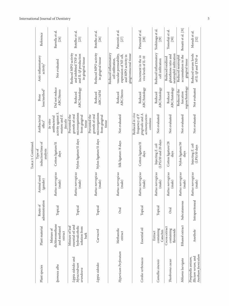
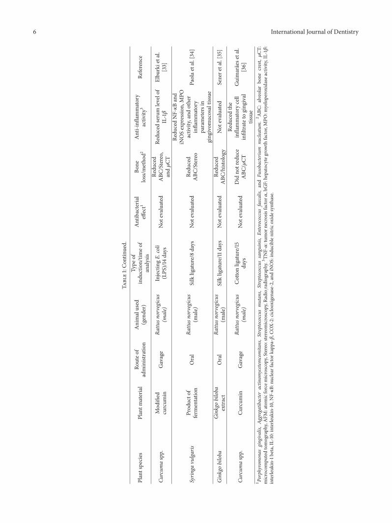
Table 1 summarize plant species, plant material, route ofadministration, animal used, type of induction and time ofanalysis, and the ability of the medicinal plant to reducealveolar bone loss related to 30 selected articles.
The majority of articles (43.3%) described studies con-ductedwith plant extracts, 40%with purified compounds and16.6% with a mixture of two or more plant extracts. Amongthem, six articles described experiments developed using acommercial product [8–13], containing purified compoundsor amixture of plant extracts. None of authorsmentioned theprocess of obtaining selected plant material as an importantlimitation for development of the research.
In general, studies were preferentially conducted usingRattus norvegicus (90%) of different strains Wistar, Lewis, orSprague-Dawley. This specie is one of the mostly used forin vivo experimental models, including the pathogenesis ofperiodontal disease because they offer some advantages suchas price, easy handling, and the possibility ofmicrobiological,
Articles from PubMed, Cochrane, and Science Direct
(keywords: plant, periodontitis,and rats)
Articles excluded
and included
after a critical
analysis of titles
and abstracts
n = 109
n = 79
Final inclusion of articles in the systematic review
n = 30
Figure 1: Search flowchart and selection of articles for the review ofthe literature.
macroscopic, and histological evaluation [14]. Other stud-ies were conducted using Mus musculus Balb/c (6.6%) orC57BL/6. Rodents were preferred as the animal model sincethey present biochemical, histological, and anatomical fea-tures similar to humans [14].
Taking into consideration the sex of the animal, mostauthors chose to use males (86.6%). Two articles describedthe use of females [12, 15] and two studies were conductedusingmales and females [4, 8]. One study did not describe thesex of the animals [16]. Most in vivo studies which investigatethe biological effect of plant substances favor the use of maleanimals.This choice is based on the fact that female hormonesshould interfere in the development and progression of thedisease [17]. None of the articles analyzed was related to thehormonal influence on the biological activities investigated.However, studies conducted with females could clarify theeffect of their hormones on the specific pathways involved inthe periodontitis progression.
Regarding the common type of periodontitis-induction,in 66.6% of the articles, the process was caused by ligature(silk: 23.3%, nylon: 23.3%, and cotton: 20%). This method iswidely used because it facilitates the accumulation of biofilm.This procedure increases the infiltration of inflammatory cellsand the production of chemical mediators that lead to thedegradation of the tissues around the teeth contributing tothe destruction of the periodontal tissues [1]. In addition toperiodontitis induced by ligature, 16.6% of articles describedthe gingival injection of bacteria, such as P. gingivalis and16.6% described the administration of E. coli endotoxin (LPS)or S. griseus (proteases) to promote the periodontal disease.The inoculation of bacteria or their subproducts leads to

International Journal of Dentistry 3
Table1:Listof
medicinalplants,
experim
entalm
etho
ds,and
theirb
iologicaleffectso
nindu
cedperio
dontitis.
Plantspecies
Plantm
aterial
Routeo
fadministratio
nAnimalused
(gender)
Type
ofindu
ction/tim
eof
analysis
Antibacteria
leffect1
Bone
loss/m
etho
d2Anti-infl
ammatory
activ
ity3
Reference
Pana
xnotoginsengand
Rehm
anniaradix
Mixture
ofextracts(9
:1weight)
Oral
Rattu
snorvegicus
(male)
InjectingE.
coli
endo
toxin(LPS
)/28
days
Not
evaluated
Redu
ced
ABC
/𝜇CT
Redu
cedtheinvitro
releaseo
fTNF-𝛼fro
mhu
man
mon
ocyticcells
andhG
Fcells
Alm
eida
etal.
[2]
Ocim
umsanctum
Tulsi
extract
Topical
Rattu
snorvegicus
(malea
ndfemale)
Silkligature/9days
Not
evaluated
Not
redu
ced
ABC
/Stereo
Presented
anti-inflammatory
activ
ityin
another
experim
entalm
odel
Hosadurga
etal.
[4]
Scutellariabaica
lensis
Baicalin
Oral
Rattu
snorvegicus
(male)
Nylon
ligature/7days
Not
evaluated
Redu
ced
ABC
/histolog
yNot
evaluated
Caietal.[6]
Rhizom
acoptidis,
Hydrastiscanadensis,
andCo
rtex
phellodendri
Berberine
Oral
Rattu
snorvegicus
(male)
Silkligature/8days
Not
evaluated
Redu
ced
ABC
/𝜇CT
Not
evaluated
Tuetal.[7]
Cucurbita
pepo,M
entha
piperita,Crataegusspp
.,Ro
smarinus
officin
alis,
Capsicu
mannu
um,and
Achilleamillefo
lium
LongoV
ital�
Oral
Rattu
snorvegicus
(malea
ndfemale)
InjectingA.
viscosus
andP.gingivalis/63
days
Not
evaluated
Redu
ced
ABC
/Radio
Not
evaluated
Klausen
etal.[8]
Mangifer
aindica
Mangiferin
(Sigma-Aldric
hCo.)
Oral
Rattu
snorvegicus
(male)
Cottonligature/1,4,
and7days
Not
evaluated
Redu
ced
ABC
/histolog
y
InhibitedCO
X-2
expressio
n,ther
ollin
g,andadhesio
nof
leuk
ocytes
inthe
perio
dontaltissue
Carvalho
etal.
[9]
Curcum
alonga
Curcum
in(Sigma-Aldric
hCo.)
Gavage
Rattu
snorvegicus
(male)
Nylon
ligature/30
days
Not
evaluated
Redu
ced
ABC
/histolog
y
Redu
cedthee
xpression
ofTN
F-𝛼in
ging
ival
tissues
Zhou
etal.[10]
Magnolia
officin
alis
Magno
lol
(Sigma-Aldric
hCo.)
Oral
Rattu
snorvegicus
(male)
Silkligature/9days
Invitro
activ
ityagainstP.gingivalis
andA.
actin
o-mycetem
comita
n
Redu
ced
ABC
/𝜇CT
Inhibitedneutroph
ilmigratio
n,MPO
activ
ity,and
COX-
2andiN
OSexpressio
nin
ging
ivaltissues
Luetal.[11]
Rhizom
adrynariaea
ndRehm
anniaglu
tinosa
Bu-Shen-Gu-Ch
i-Wan
(JiuZ
hiTang
Pharmaceutical)
Oral
Rattu
snorvegicus
(female)
InjectingP.gingivalis
andligature/28
days
Not
evaluated
Improved
the
mineraldensity
oftheb
one/𝜇CT
Redu
cedlevelsof
IL-1𝛽,T
NF-𝛼,and
inflammatorycell
infiltrationin
the
perio
dontaltissues
Yang
etal.[12]
4 International Journal of Dentistry
Table1:Con
tinued.
Plantspecies
Plantm
aterial
Routeo
fadministratio
nAnimalused
(gender)
Type
ofindu
ction/tim
eof
analysis
Antibacteria
leffect1
Bone
loss/m
etho
d2Anti-infl
ammatory
activ
ity3
Reference
Pinu
spinaster
Pycnogenol�
(TradepiaC
o.)
Oral
Balb/c(m
ale)
InjectingP.
gingivalis/34
days
Antibacteria
lactiv
ityagainstP.
gingivalis
Redu
ced
ABC
/Stereo
Not
evaluated
Sugimotoetal.
[13]
Vaccinium
macrocarpon
Aqueou
sextract
containing
tann
inandph
enolic
compo
unds
Oral
Mus
musculus
(female)
InjectingP.gingivalis
andF.nu
cleatum
/42
days
Anti-a
dhesive
prop
ertie
sagainst
P.gingivalisandF.
nucle
atum
.Increasedthe
phagocytosisof
P.gingivalis
Not
evaluated
Redu
cedin
vivo
levels
ofTN
F-𝛼
Polaketal.[15]
Curcum
alonga
Curcum
inTo
pical
Rattu
snorvegicus
Silkligature/28
days
Not
evaluated
Did
notreduce
ABC
/Stereo
Exhibited
anti-inflammatory
activ
ityin
another
experim
entalm
odel
Hosadurga
etal.
[16]
Camelliasin
ensis
Extract
containing
Catechin
Topical
Rattu
snorvegicus
(male)
InjectingE.
coli(LPS
)andS.grise
us(proteases)/56
days
Not
evaluated
Did
notreduce
ABC
/histolog
y
Redu
cedinflammatory
cellinfiltrationand
levelsof
TNF-𝛼
inthep
eriodo
ntal
lesio
n
Maruyam
aetal.
[19]
Spatholobu
ssub
erectus
Aqueou
sextract
Oral
Mus
musculus
(male)
InjectingP.gingivalis
/42days
Invitro
antib
acteria
lactiv
ityagainstP.
gingivalis
Redu
ced
ABC
/Stereo
Not
evaluated
Toyamae
tal.
[20]
Carapa
guianensis
And
iroba
oil
Oral
Rattu
snorvegicus
(male)
Cottonligature/50
days
Not
evaluated
Did
notreduce
ABC
/histolog
y
Redu
cedtheq
uantity
ofinflammatorycells
inhisto
logy
Carm
onae
tal.
[21]
Protium
heptaphyllu
m𝛼-amyrin
and
𝛽-amyrin
Oral
Rattu
snorvegicus
(male)
Nylon
ligature/1d
ayNot
evaluated
Not
evaluated
Redu
cedging
ival
TNF-𝛼levelsandMPO
activ
ity
Holanda
Pinto
etal.[22]
Camelliasin
ensis
Extract
containing
Catechin
Oral
Rattu
snorvegicus
(male)
Nylon
ligature/7,14,
and28
days
Not
evaluated
Redu
ced
ABC
/histolog
yRe
ducedin
vivo
levels
ofTN
F-𝛼
Choetal.[23]
International Journal of Dentistry 5
Table1:Con
tinued.
Plantspecies
Plantm
aterial
Routeo
fadministratio
nAnimalused
(gender)
Type
ofindu
ction/tim
eof
analysis
Antibacteria
leffect1
Bone
loss/m
etho
d2Anti-infl
ammatory
activ
ity3
Reference
Ipom
oeaalba
Mixture
ofdichloromethane
andmethano
lextract
Topical
Rattu
snorvegicus
(male)
Cottonligature/11
days
Invitro
antib
acteria
lactiv
ityagainstS.
mutan
sand
E.faecalis
Did
notreduce
ABC
/Stereo
Not
evaluated
Barrellaetal.
[24]
Lippiasid
oidesand
Myracrodruon
urun
deuva
Mixture
ofleaf
essentialoiland
hydroalcoh
olic
solutio
nfro
mbark
Topical
Rattu
snorvegicus
(male)
Nylon
ligature/11days
Preventedthe
grow
thof
oral
microorganism
sfro
mging
ival
tissue
Redu
ced
ABC
/histolog
y
Redu
cedMPO
activ
ityandinhibitedTN
F-𝛼
andIL-1𝛽prod
uctio
nin
ging
ivaltissue
Botelhoetal.
[25]
Lippiasid
oides
Carvacrol
Topical
Rattu
snorvegicus
(male)
Nylon
ligature/11days
Preventedthe
grow
thof
oral
microorganism
sfro
mging
ival
tissue
Redu
ced
ABC
/AFM
Redu
cedMPO
activ
ityin
ging
ivaltissue
Botelhoetal.
[26]
Hypericu
mPerfo
ratum
Methano
licextract
Oral
Rattu
snorvegicus
(male)
Silkligature/8days
Not
evaluated
Redu
ced
ABC
/Stereo
Redu
cedinflammatory
cellinfiltration,
vascular
perm
eability,
expressio
nof
NF-𝜅B,
andMPO
activ
ityin
ging
ivom
ucosaltissue
Paterniti
etal.
[27]
Cordiaverbenacea
Essentialoil
Topical
Rattu
snorvegicus
(male)
Cottonligature/11
days
Redu
cedin
vivo
frequ
ency
ofP.
gingivalisandA.
actin
omycetem
-comita
ns
Redu
ced
ABC
/histolog
yIncrem
entinthein
vivo
levelsof
IL-10
Pimenteletal.
[28]
Camelliasin
ensis
Extract
containing
Catechin
Topical
Rattu
snorvegicus
(male)
InjectingE.
coli
(LPS
)/10
and20
days
Not
evaluated
Redu
ced
ABC
/histolog
yRe
ducedinflammatory
cellinfiltration
Yoshinagae
tal.
[29]
Theobrom
acacao
Cocoa
extract
containing
flavono
ids
Oral
Rattu
snorvegicus
(male)
Cottonligature/28
days
Not
evaluated
Redu
ced
ABC
/histolog
y
Redu
ced/oxidized
glutathion
eratio
and
neutroph
ilinfiltration
Tomofujietal.
[30]
Mikanialaevigata
Ethano
lextract
Subcutaneous
Rattu
snorvegicus
(male)
Nylon
ligature/30
days
Not
evaluated
Redu
cedthe
furcation
region
/histolog
y
Redu
cedneutroph
ilaccumulationin
the
ging
ivaltissue
Benatti
etal.[31]
Pimpinella
anisu
m,
Illicium
verum,and
Anethu
mfoenicu
lum
Anethole
Intraperito
neal
Rattu
snorvegicus
(male)
InjectingE.
coli
(LPS
)/10
days
Not
evaluated
Not
evaluated
Redu
cedserum
levels
ofIL-1𝛽andTN
F-𝛼
Moradietal.
[32]
6 International Journal of Dentistry
Table1:Con
tinued.
Plantspecies
Plantm
aterial
Routeo
fadministratio
nAnimalused
(gender)
Type
ofindu
ction/tim
eof
analysis
Antibacteria
leffect1
Bone
loss/m
etho
d2Anti-infl
ammatory
activ
ity3
Reference
Curcum
aspp.
Mod
ified
curcum
inGavage
Rattu
snorvegicus
(male)
InjectingE.
coli
(LPS
)/14
days
Not
evaluated
Redu
ced
ABC
/Stereo,
and𝜇CT
Redu
cedserum
levelof
IL-1𝛽
Elbu
rkietal.
[33]
Syrin
gavulga
risProd
ucto
fferm
entatio
nOral
Rattu
snorvegicus
(male)
Silkligature/8days
Not
evaluated
Redu
ced
ABC
/Stereo
Redu
cedNF-𝜅Band
iNOSexpressio
n,MPO
activ
ity,and
other
inflammatory
parametersin
ging
ivom
ucosaltissue
Paolae
tal.[34]
Ginkgo
biloba
Ginkgo
biloba
extract
Oral
Rattu
snorvegicus
(male)
Silkligature/11days
Not
evaluated
Redu
ced
ABC
/histolog
yNot
evaluated
Sezere
tal.[35]
Curcum
aspp.
Curcum
inGavage
Rattu
snorvegicus
(male)
Cottonligature/15
days
Not
evaluated
Did
notreduce
ABC
/𝜇CT
Redu
cedthe
inflammatorycell
infiltratetoging
ival
tissue
Guimaraese
tal.
[36]
1Porphyromonas
gingivalis,
Aggregatibacteractin
omycetem
comita
ns,Streptococcusmutan
s,Streptococcussanguinis,
Enterococcus
faecalis,
and
Fusobacterium
nucle
atum
.2ABC
:alveolar
bone
crest,𝜇CT
:microcompu
tedtomograph
y,AFM
:atomicforcem
icroscop
y,Stereo:stereom
icroscop
y,Ra
dio :radiograph
y.3TN
F-𝛼:tum
ornecrosisfactor𝛼,hGF:hepatocytegrow
thfactor,M
PO:m
yeloperoxidase
activ
ity,IL-1𝛽:
interle
ukin-1beta,IL-10:interleuk
in-10,NF-𝜅B:
nucle
arfactor
kapp
a-𝛽,C
OX-
2:ciclo
oxigenase-2,andiN
OS:indu
ciblen
itricoxides
ynthase.

International Journal of Dentistry 7
an inflammatory response different from that promoted byperiodontitis induced through induction with ligature [18].
Some of the studies analyzed described that long periodsof experimental design were utilized to properly investigatethe severity of tissue destruction during periodontal diseasetreatment: 63 days [8]; 56 days [19]; 42 days [20]; and 50days [21]. However, the time of analysis changed substantially.More than 55% of the studies performed experiments duringone to two weeks, 26.6% for three to four weeks, and 13.3%during six weeks or more. One study was conducted withan acute periodontitis rat model in 24 hours [22]. Thesevariations in the experimental periods may be due to factorssuch as the type and location of the ligature, the bacteriaspecies or their sub-products injected for the induction ofthe disease and the specie (strain), and age and weight of theanimal used. These factors are generally associated with theobjective of the study and the expected results.
More than 66%of studies chose the oral administration toevaluate the efficacy of the plant material.The topical admin-istration was described by 26.6% of the studies and 6.9%treated animals through subcutaneous or intraperitonealcavity injections.The route of administration is an importantparameter to influence the efficacy of the material since it caninterfere with the sufficient amount of substance available topromote the biological effects. As observed, different resultswere seen for alveolar bone loss of animals submitted tothe treatment with an extract containing Catechin obtainedfrom Camellia sinensis. Although the route of administrationhad not influenced the anti-inflammatory activity of thematerial, a significant reduction of bone loss was observedwhen given orally [23] and no difference was seen after itstopical treatment [19].
Periodontal disease initiation and progression occur asa consequence of the host response to microorganisms ofthe dental biofilm [1]. Therefore, the antibacterial effect is animportant factor in the periodontal therapy. Only 26.6% ofthe studies investigated the capacity of thematerial to presentantibacterial activity. In the study developed by Barrella et al.[24] the organic extract obtained from Ipomoea alba showedsignificant in vitro activity against Streptococcus mutans, S.sanguinis, and Enterococcus faecalis. The commercial prod-uct Magnolol obtained from Magnolia officinalis exhibitedintense inhibition of Porphyromonas gingivalis and Aggregat-ibacter actinomycetemcomitans growth in a dependent dose[11] and the aqueous extract from Vaccinium macrocarponcontaining tannin and phenolic compounds inhibited theadhesion of Fusobacterium nucleatum and Porphyromonasgingivalis [15].Study developed by Botelho et al. [25] andBotelho et al. [26] with Lippia sidoides observed the decreaseof salivary bacterial levels and this event was followed byan increment in clinical scores of gingival bleeding. Exceptfor the work published by Klausen et al. [8] which did notevaluate the alveolar bone loss, all the articles that describedthe identification of antibacterial activity also demonstratedthat plant material treatment induced a significant reductionof alveolar bone loss.
The alveolar bone loss is one of the most importantparameters evaluated in the induced periodontal disease.From 30 selected articles, 27 evaluated the ability of the
plant material to promote a significant reduction of the boneloss. Carvacrol purified from Lippia sidoides, the extractcontaining Catechin obtained from Camellia sinensis, theessential oil of Cordia verbenacea, and the methanolic extractof Hypericum perforatum are examples of 77% of studiesthat observed a significant reduction of the bone loss [23,26–28]. On the other hand, in 23% of studies such asandiroba oil from Carapa guianensis, as well as the mixtureof dichloromethane and methanol extracts from Ipomoeaalba and Longo Vital, the authors did not observe a sig-nificant effect [8, 21, 24]. Moreover, it was interesting tonote that different studies using the same plant materialhave reached different results regarding alveolar bone loss.According to Yoshinaga et al. [29] the topical treatmentof animals with the extract containing Catechin (Camelliasinensis) promoted a significant reduction of bone loss inperiodontitis induced by E. coli (LPS). On the other hand,the same material did not present a significant reductionin the alveolar bone loss against periodontitis induced byinjection of E. coli (LPS) and S. griseus (proteases) [19].Different efficacy was also observed in studies developedwithcurcumin (Curcuma longa). The topical administration ofcurcumin did not reduce the alveolar bone loss [16] while itsoral administration reduced this parameter significantly [10].The methodologies used to evaluate the alveolar bone losswere histology (44.0%), stereomicroscopy (25.9%), micro-computed tomography (22.2%), atomic force microscopy(3.7%), and radiography (3.7%). Histology, stereomicroscopy,andmicrocomputed tomography are favorablemethods oncethey yield precise information concerning the evaluation ofperiodontal tissue destruction.
It is noteworthy that the reduction of the inflammatoryprocess may be associated with a reduced bone loss [12, 30,31]. Because of this, a reduction of inflammatory processis usually investigated in periodontal disease. Taking theselected articles into account, 76.6% of them evaluatedinflammatory parameters associated with induced periodon-titis. The common strategies used to observe the abilityof the plant material to inhibit the inflammatory processin periodontal tissue were the evaluation of migration ofinflammatory cells (30%), measurement of proinflammatorycytokines (TNF-𝛼: 30% and IL1-𝛽: 13.3%) [2, 10, 12, 15, 19, 22,23, 25, 32, 33], respectively, and dosage of myeloperoxidase(MPO) activity (20%) [11, 22, 25–27, 34]. Other molecularmarkers of inflammatory process, such as iNOS (13.0%)[11, 34], NF-𝜅B (8.7%) [27, 34], COX-2 (8.7%) [9, 11],and IL-10 (4.3%) [28], were also investigated. Although thepresence of the anti-inflammatory effect reflects a possibleefficacy against periodontitis, 13% of the authors who foundanti-inflammatory properties did not observe a significantreduction of alveolar bone loss in the periodontitis assays.The anti-inflammatory activity was not analyzed in the 23.3%[6–8, 13, 20, 24, 35] of the studies. The andiroba oil (Carapaguianensis), the extract from Camellia sinensis (containingCatechin), and Curcumin (Curcuma spp.) were able to reducethe number of inflammatory cells in the gingival tissue.However, they were not effective to protect from destructiveperiodontal process [19, 21, 36]. According to the authors,these negative results may be related to the time delay to

8 International Journal of Dentistry
reach levels that are high enough for the biological effectsof administered substances in the experimental modelsadopted. Further researches are necessary to observe howmuch time is appropriate to manage the substances for therecovery of bone loss.
Although there is a close relation between the bonereabsorption and the inflammatory response, we suggestthat the negative results of some of substances analyzedshould be attributed to the fact that they do not act in theosteoclastogenesis process. This information is supported bythe personal observation conducted using sulphated polysac-charides recovered from redmarine algaeGracilaria caudata.Our data revealed that the treatment of experimental ani-mals with sulphated polysaccharides improved clinical andinflammatory parameters. However, no significant effect wasobserved in the reduction of alveolar bone loss (unpublisheddata).
It is important to mention that, although many plantsubstances may have deleterious effects to different animalorgans [37], none of the evaluated studies has investigated thepossible toxicological effects associated with the administra-tion of the medicinal plants. In addition, several studies havedocumented that induced periodontitis is followed by signif-icant changes of morphological structures and biochemicalfunctions of different organs [38, 39]. Yet again, none ofstudies investigated the ability of these medicinal plants toprevent changes in organs promoted by a systemic action ofthe induced periodontitis.
Finally, 8 of the 30 selected articles conducted theirexperiments with a gel-based formulation containing theplant material investigated. In these experiments, the authorsadministered the gel topically, generally three times a day.The main idea behind these investigations is to reveal thefurther potential of the combined gel preparation to combatperiodontal disease in closer than clinical situations.This wasthe outlook investigated in the studies published by [40, 41].The authors evaluated the efficiency of a green tea Catechingel as an adjunct on human periodontal therapy. In firstwork [40], the authors observed a significant reduction onpockets and inflammation during the 4 weeks of the clinicaltrial. In the second work [41], it was demonstrated that whenused as an adjunct to periodontal treatment, green tea gelcould provide benefit in reducing bleeding on probing andgingival inflammation at 1st and 3rd months of evaluation.What makes these studies even more relevant is the fact thatthe findings on complementary products for the treatment ofhuman periodontitis still can be enhanced.
4. Conclusion
In conclusion, the selected studies presented a large diversityof experimental designs, concerning the type of induction,time of analysis, and methods used for the evaluation ofalveolar bone loss, anti-inflammatory, and antibacterial activ-ities. None of the studies evaluated the possible toxic effectsassociated with the administration of the material analyzedor their ability to prevent damages to organs caused bysystemic effects of induced periodontitis. Gel-based formu-lations present an interesting strategy to treat periodontitis;
however, further studies are necessary to clarify its usefulnessin the clinical situation.
Competing Interests
The authors declare that they have no conflict of interests.
Acknowledgments
This research is supported by the Federal University of Piauı(UFPI-Edital PIBIC 2014/2015 and BIAMA 03/2014), CNPq(455104/2014-0).The authors thank teacher Abilio Borghi forthe grammar review of the paper.
References
[1] A. Bascones-Martınez, M. Munoz-Corcuera, S. Noronha, P.Mota, C. Bascones-Ilundain, and J. Campo-Trapero, “Hostdefencemechanisms against bacterial aggression in periodontaldisease: basic mechanisms,” Medicina Oral, Patologia Oral yCirugia Bucal, vol. 14, no. 12, pp. e680–e685, 2009.
[2] J. De Almeida, E. Ervolino, L. H. Bonfietti et al., “Adjuvant ther-apy with sodium alendronate for the treatment of experimentalperiodontitis in rats,” Journal of Periodontology, vol. 86, no. 10,pp. 1166–1175, 2015.
[3] R.-Y. Huang, S.-H. Lu, K.-W. Su et al., “Diacerein: a potentialtherapeutic drug for periodontal disease,” Medical Hypotheses,vol. 79, no. 2, pp. 165–167, 2012.
[4] R. R. Hosadurga, S. N. Rao, R. Edavanputhalath et al., “Eval-uation of the efficacy of 2% Ocimum sanctum gel in thetreatment of experimentalperiodontitis,” International Journalof Pharmaceutical Investigation, vol. 5, no. 1, pp. 35–42, 2015.
[5] C. M. Ardila, M. A. Lopez, and I. C. Guzman, “High resistanceagainst clindamycin, metronidazole and amoxicillin in Porphy-romonas gingivalis and Aggregatibacter actinomycetemcomitansisolates of periodontal disease,”Medicina Oral, Patologia Oral yCirugia Bucal, vol. 15, no. 6, pp. e947–e951, 2010.
[6] X. Cai, C. Li, G. Du, and Z. Cao, “Protective effects of baicalinon ligature-induced periodontitis in rats,” Journal of PeriodontalResearch, vol. 43, no. 1, pp. 14–21, 2008.
[7] H.-P. Tu, M. M. J. Fu, P.-J. Kuo et al., “Berberine’s effect onperiodontal tissue degradation bymatrixmetalloproteinases: anin vitro and in vivo experiment,” Phytomedicine, vol. 20, no. 13,pp. 1203–1210, 2013.
[8] B. Klausen, A. Apostolopoulos, K. Stoltze, and F. Norgaard,“Effect of LongoVital treatment on development of periodontaldisease in rats,” Scandinavian Journal of Dental Research, vol.101, no. 1, pp. 33–36, 1993.
[9] R. R. Carvalho, C. H. Pellizzon, L. Justulin Jr. et al., “Effectof mangiferin on the development of periodontal disease:involvement of lipoxin A4, anti-chemotaxic action in leukocyterolling,” Chemico-Biological Interactions, vol. 179, no. 2-3, pp.344–350, 2009.
[10] T. Zhou, D. Chen, Q. Li, X. Sun, Y. Song, and C. Wang,“Curcumin inhibits inflammatory response and bone lossduring experimental periodontitis in rats,” Acta OdontologicaScandinavica, vol. 71, no. 2, pp. 349–356, 2013.
[11] S.-H. Lu, R.-Y. Huang, and T.-C. Chou, “Magnolol amelioratesligature-induced periodontitis in rats and osteoclastogenesis: invivo and in vitro study,” Evidence-Based Complementary and

International Journal of Dentistry 9
Alternative Medicine, vol. 2013, Article ID 634095, 12 pages,2013.
[12] H. Yang, Q. Wen, J. Xue, and Y. Ding, “Alveolar bone regenera-tion potential of a traditional Chinese medicine, Bu-Shen-Gu-Chi-Wan, in experimental periodontitis,” Journal of PeriodontalResearch, vol. 49, no. 3, pp. 382–389, 2014.
[13] H. Sugimoto, K. Watanabe, T. Toyama et al., “Inhibitory effectsof French pine bark extract, pycnogenol�, on alveolar boneresorption and on the osteoclast differentiation,” PhytotherapyResearch, vol. 29, no. 2, pp. 251–259, 2015.
[14] X. Struillou, H. Boutigny, A. Soueidan, and P. Layrolle, “Exper-imental animal models in periodontology: a review,”The OpenDentistry Journal, vol. 4, no. 1, pp. 33–47, 2010.
[15] D. Polak, R. Naddaf, L. Shapira, E. I. Weiss, and Y. Houri-Haddad, “Protective potential of non-dialyzable material frac-tion of cranberry juice on the virulence of P. gingivalis and F.nucleatum mixed infection,” Journal of Periodontology, vol. 84,no. 7, pp. 1019–1025, 2013.
[16] R. R. Hosadurga, S. Rao, J. Jose, N. C. Rompicharla, M.Shakil, and R. Shashidhara, “Evaluation of the efficacy of 2%curcumin gel in the treatment of experimental periodontitis,”Pharmacognosy Research, vol. 6, no. 4, pp. 326–333, 2014.
[17] E. Figuero, A. Carrillo-De-Albornoz, D. Herrera, and A.Bascones-Martınez, “Gingival changes during pregnancy: I.Influence of hormonal variations on clinical and immunolog-ical parameters,” Journal of Clinical Periodontology, vol. 37, no.3, pp. 220–229, 2010.
[18] S. Garcia de Aquino, F. R. Manzolli Leite, D. R. Stach-Machado,J. A. Francisco da Silva, L. C. Spolidorio, and C. Rossa Jr.,“Signaling pathways associated with the expression of inflam-matory mediators activated during the course of two models ofexperimental periodontitis,” Life Sciences, vol. 84, no. 21-22, pp.745–754, 2009.
[19] T. Maruyama, T. Tomofuji, Y. Endo et al., “Supplementation ofgreen tea catechins in dentifrices suppresses gingival oxidativestress and periodontal inflammation,” Archives of Oral Biology,vol. 56, no. 1, pp. 48–53, 2011.
[20] T. Toyama, K. Todoki, Y. Takahashi et al., “Inhibitory effectsof Jixueteng on P. gingivalis-induced bone loss and osteoclastdifferentiation,”Archives of Oral Biology, vol. 57, no. 11, pp. 1529–1536, 2012.
[21] G. B. Carmona, R. K. C. Teixeira, M. V. H. Brito et al., “Effectof andiroba oil on periodontitis in wistar rats,” Acta CirurgicaBrasileira, vol. 28, no. 6, pp. 430–434, 2013.
[22] S. A. Holanda Pinto, L. M. S. Pinto, G. M. A. Cunha, M.H. Chaves, F. A. Santos, and V. S. Rao, “Anti-inflammatoryeffect of 𝛼, 𝛽-Amyrin, a pentacyclic triterpene from Protiumheptaphyllum in rat model of acute periodontitis,” Inflam-mopharmacology, vol. 16, no. 1, pp. 48–52, 2008.
[23] A.-R. Cho, J.-H. Kim, D.-E. Lee et al., “The effect of orallyadministered epigallocatechin-3-gallate on ligature-inducedperiodontitis in rats,” Journal of Periodontal Research, vol. 48,no. 6, pp. 781–789, 2013.
[24] G. E. Barrella, I. B. Suffredini, F. V. Ribeiro, F. R. Cirano, andS. P. Pimentel, “Evaluation of the effect of an organic extractobtained from Ipomoea alba L. on experimental periodontitis inrats,” Brazilian Oral Research, vol. 26, no. 2, pp. 158–164, 2012.
[25] M. A. Botelho, V. S. Rao, C. B. M. Carvalho et al., “Lippiasidoides and Myracrodruon urundeuva gel prevents alveolarbone resorption in experimental periodontitis in rats,” Journalof Ethnopharmacology, vol. 113, no. 3, pp. 471–478, 2007.
[26] M. A. Botelho, J. G. Martins, R. S. Ruela et al., “Protective effectof locally applied carvacrol gel on ligature-induced periodonti-tis in rats: a tapping mode AFM study,” Phytotherapy Research,vol. 23, no. 10, pp. 1439–1448, 2009.
[27] I. Paterniti, E. Briguglio, E.Mazzon et al., “Effects of HypericumPerforatum, in a rodent model of periodontitis,” BMC Comple-mentary and Alternative Medicine, vol. 10, article 73, 2010.
[28] S. P. Pimentel, G. E. Barrella, R. C. V. Casarin et al., “Protec-tive effect of topical Cordia verbenacea in a rat periodontitismodel: immune-inflammatory, antibacterial and morphomet-ric assays,” BMC Complementary and Alternative Medicine, vol.12, no. 1, article 224, pp. 1–8, 2012.
[29] Y. Yoshinaga, T. Ukai, S. Nakatsu et al., “Green tea extractinhibits the onset of periodontal destruction in rat experimentalperiodontitis,” Journal of Periodontal Research, vol. 49, no. 5, pp.652–659, 2014.
[30] T. Tomofuji, D. Ekuni, K. Irie et al., “Preventive effects of acocoa-enriched diet on gingival oxidative stress in experimentalperiodontitis,” Journal of Periodontology, vol. 80, no. 11, pp.1799–1808, 2009.
[31] B. B. Benatti, J. C. Campos-Junior, V. J. Silva-Filho et al., “Effectsof a Mikania laevigata extract on bone resorption and RANKLexpression during experimental periodontitis in rats,” Journal ofApplied Oral Science, vol. 20, no. 3, pp. 340–346, 2012.
[32] J. Moradi, F. Abbasipour, J. Zaringhalam et al., “Anethole, amedicinal plant compound, decreases the production of pro-inflammatory TNF-𝛼 and IL-1𝛽 in a rat model of LPS-inducedperiodontitis,” Iranian Journal of Pharmaceutical Research, vol.13, no. 4, pp. 1319–1325, 2014.
[33] M. S. Elburki, C. Rossa, M. R. Guimaraes et al., “A novelchemicallymodified curcumin reduces severity of experimentalperiodontal disease in rats: Initial observations,” Mediators ofInflammation, vol. 2014, Article ID 959471, 10 pages, 2014.
[34] R.D. I. Paola, G.Oteri, E.Mazzon et al., “Effects of verbascoside,biotechnologically purified by Syringa vulgaris plant cell cul-tures, in a rodent model of periodontitis,” Journal of Pharmacyand Pharmacology, vol. 63, no. 5, pp. 707–717, 2011.
[35] U. Sezer,M. I. Kara, K. Erciyas et al., “Protective effects of ginkgobiloba extract on ligature-induced periodontitis in rats,” ActaOdontologica Scandinavica, vol. 71, no. 1, pp. 38–44, 2013.
[36] M. R. Guimaraes, L. S. Coimbra, S. G. de Aquino, L. C.Spolidorio, K. L. Kirkwood, and C. Rossa Jr., “Potent anti-inflammatory effects of systemically administered curcuminmodulate periodontal disease in vivo,” Journal of PeriodontalResearch, vol. 46, no. 2, pp. 269–279, 2011.
[37] U. F. Ezuruike and J. M. Prieto, “The use of plants in the tradi-tional management of diabetes in Nigeria: pharmacological andtoxicological considerations,” Journal of Ethnopharmacology,vol. 155, no. 2, pp. 857–924, 2014.
[38] T. Tomofuji, D. Ekuni, R. Yamanaka et al., “Chronic adminis-tration of lipopolysaccharide and proteases induces periodontalinflammation and hepatic steatosis in rats,” Journal of Periodon-tology, vol. 78, no. 10, pp. 1999–2006, 2007.
[39] T. Tomofuji, T. Sanbe, D. Ekuni et al., “Oxidative damage ofrat liver induced by ligature-induced periodontitis and chronicethanol consumption,” Archives of Oral Biology, vol. 53, no. 12,pp. 1113–1118, 2008.
[40] V. K. Chava and B. D. Vedula, “Thermo-reversible green teacatechin gel for local application in chronic periodontitis: a 4-

10 International Journal of Dentistry
week clinical trial,” Journal of Periodontology, vol. 84, no. 9, pp.1290–1296, 2013.
[41] K. Rattanasuwan, S. Rassameemasmaung, V. Sangalungkarn,andC. Komoltri, “Clinical effect of locally delivered gel contain-ing green tea extract as an adjunct to non-surgical periodontaltreatment,” Odontology, vol. 104, no. 1, pp. 89–97, 2016.

Submit your manuscripts athttp://www.hindawi.com
Hindawi Publishing Corporationhttp://www.hindawi.com Volume 2014
Oral OncologyJournal of
DentistryInternational Journal of
Hindawi Publishing Corporationhttp://www.hindawi.com Volume 2014
Hindawi Publishing Corporationhttp://www.hindawi.com Volume 2014
International Journal of
Biomaterials
Hindawi Publishing Corporationhttp://www.hindawi.com Volume 2014
BioMed Research International
Hindawi Publishing Corporationhttp://www.hindawi.com Volume 2014
Case Reports in Dentistry
Hindawi Publishing Corporationhttp://www.hindawi.com Volume 2014
Oral ImplantsJournal of
Hindawi Publishing Corporationhttp://www.hindawi.com Volume 2014
Anesthesiology Research and Practice
Hindawi Publishing Corporationhttp://www.hindawi.com Volume 2014
Radiology Research and Practice
Environmental and Public Health
Journal of
Hindawi Publishing Corporationhttp://www.hindawi.com Volume 2014
The Scientific World JournalHindawi Publishing Corporation http://www.hindawi.com Volume 2014
Hindawi Publishing Corporationhttp://www.hindawi.com Volume 2014
Dental SurgeryJournal of
Drug DeliveryJournal of
Hindawi Publishing Corporationhttp://www.hindawi.com Volume 2014
Hindawi Publishing Corporationhttp://www.hindawi.com Volume 2014
Oral DiseasesJournal of
Hindawi Publishing Corporationhttp://www.hindawi.com Volume 2014
Computational and Mathematical Methods in Medicine
ScientificaHindawi Publishing Corporationhttp://www.hindawi.com Volume 2014
PainResearch and TreatmentHindawi Publishing Corporationhttp://www.hindawi.com Volume 2014
Preventive MedicineAdvances in
Hindawi Publishing Corporationhttp://www.hindawi.com Volume 2014
EndocrinologyInternational Journal of
Hindawi Publishing Corporationhttp://www.hindawi.com Volume 2014
Hindawi Publishing Corporationhttp://www.hindawi.com Volume 2014
OrthopedicsAdvances in