Embed Size (px)
Citation preview

Send Orders for Reprints to [email protected]
Current Medicinal Chemistry, 2016, 23, 1-16 1
REVIEW ARTICLE
0929-8673/16 $58.00+.00 © 2016 Bentham Science Publishers
Design and Fabrication of Magnetically Responsive Nanocarriers for Drug Delivery
Slavko Kralja,b,c,*, Tanja Potrčd, Petra Kocbekd, Silvia Marchesanb and Darko Makoveca
aDepartment for Materials Synthesis, Jožef Stefan Institute, Jamova 39, 1000 Ljubljana, Slovenia; bDepart-ment of Chemical and Pharmaceutical Sciences, University of Trieste, Via Giorgieri 1, 34127 Trieste, Italy; cNanos SCI, (Nanos Scientificae d.o.o.), Teslova 30, 1000 Ljubljana, Slovenia; dFaculty of Pharmacy, Uni-versity of Ljubljana, Aškerčeva cesta 7, 1000 Ljubljana, Slovenia
A R T I C L E H I S T O R Y
Received: April 14, 2016 Revised: June 14, 2016 Accepted: June 29, 2016 DOI: 10.2174/0929867323666160813 211736
Abstract: Magnetically-assisted delivery of therapeutic agents to the site of interest, which is referred to as magnetic drug targeting, has proven to be a promising strategy in a number of studies. One of the key advantages over other targeting strategies is the possibility to control remotely the distribution and accumulation of the nanocarriers after parenteral administration. How-ever, preparation of effective and robust magnetically responsive nanocarriers based on superparamagnetic iron oxide nanocrystals (SPIONs) still represents a great scientific challenge, since spatial guidance of individual SPIONs is ineffective despite the presence of high magnetic field gradient. A strategy to overcome this issue is the clustering of SPIONs to achieve sufficient mag-netic responsiveness. In this mini-review, we address current and future strategies for the design and fabrication of magnetically responsive nanocarriers based on SPIONs for mag-netically-targeted drug delivery, including the underlying physical requirements, the possibil-ity of drug loading, and the control of drug release at the targeted site.
Keywords: Magnetic nanoparticles, nanoparticle clusters, magnetic drug delivery, iron oxide nanocrystals, SPION clusters, magnetic targeting, magnetic nanocarriers, magnetically-assisted delivery.
1. INTRODUCTION
Nanotechnology is advancing at a fast pace and holds promise to overcome many of current therapeutic limits through the advent of nanomedicine [1, 2]. Many drug candidates never undergo translation from pre-clinical trials to market due to their specific physico-chemical properties (e.g. poor water solubility), which hinder their efficacy and/or safety when administered in traditional formulations, such as tablets, capsules, and solutions for injections [3]. Proper design and de-velopment of novel nanocarrier systems can revitalize such drug candidates and bring them back into further *Address correspondence to this author at the Department for Mate-rials Synthesis, Jožef Stefan Institute, Jamova 39, 1000 Ljubljana, Slovenia; Department of Chemical and Pharmaceutical Sciences, University of Trieste, Via Giorgieri 1, 34127 Trieste, Italy; Nanos SCI, (Nanos Scientificae d.o.o.), Teslova 30, 1000 Ljubljana, Slo-venia; Tel: 00386 1 477 36 29; Fax: 00386 1 252 93 85; E-mail: [email protected]
translational studies. Drug adverse effects can be di-minished or avoided by drug incorporation into ad-vanced nanodelivery systems, which enable passive or active drug targeting, including smart external guiding of the nanocarriers in the body, and controlled drug release at the target site [4].
Nanomaterials in the form of nanoparticles, nano-tubes, nanorods, and other self-assembled nanostruc-tures can be transformed into advanced nanocarriers, which are particularly suited for biomedical applica-tions [1]. A key feature is their nanoscale size, which correlates with the size of biological macromolecules and subcellular structures. They can be used for ad-vanced diagnostics and treatment of various diseases as well as in tissue regeneration [5]. Nanodelivery sys-tems based solely on organic materials are nowadays approaching a mature stage, meaning their entry to the market, upon a few decades of development. For ex-ample, nanomedicines in form of liposomes (Dauno-
Please provide corresponding author(s)
photographsize should be 4" x 4" inches
2 Current Medicinal Chemistry, 2016, Vol. 23, No. 30 Kralj et al.
Xome®, Myocet®, Doxil®) and albumin nanoparticles (Abraxane®) have already reached clinical use in can-cer treatment [6, 7]. Despite the extensive development of diverse nanocarriers, poor stability, low drug load-ing, and lack of external guidance for efficient target-ing, are often limiting factors for successful translation from preclinical to clinical use [8-11].
Recently, inorganic nanomaterials have attracted in-creasing attention due to their unique properties. They display notably higher thermal, chemical, and biologi-cal stability in physiological conditions relative to or-ganic materials [12]. Among inorganic nanomateri-als, which have been proven to be safe and efficient in the treatment of human pathologies, iron oxide nanoc-rystals play an elected role, being either in paramag-netic form (akaganeit; β-FeOOH) or exhibiting ferro-magnetic or superparamagnetic properties (maghemite; γ-Fe2O3 and magnetite; Fe3O4) [13-15]. Superpar-amagnetic iron oxide nanocrystals (SPIONs) have been synthesized by a variety of approaches ranging from traditional low-cost coprecipitation methods to more sophisticated techniques such as sonolysis, electrochemical methods, laser pyrolysis or chemical vapour deposition [16, 17], resulting in commercial manufacture of different magnetic particles for research and clinical use by different well-known companies and start-ups (Table 1).
A promising strategy for magnetic drug targeting typically involves the concomitant use of different nanotechnological approaches. It is useful only of the disease is localized to a specific part of the body and it is advantageous over other targeting strategies due to the ability to remotely control the distribution and ac-cumulation of the parenterally administered nanocarri-ers in the body. The most frequently used magnetic component of magnetically responsive nanocarriers (MNCs) are iron oxide nanocrystals, which are incor-porated together with a therapeutic component (drug) into a single MNC, such as liposomes, micelles, po-lymeric matrix type or inorganic core-shell nanoparti-cles [12, 18]. MNCs can also be prepared with other types of magnetic nanoparticles such as metallic Fe, Co, and FePt exhibiting superior magnetic properties but lacking biocompatibility because they can be read-ily oxidized and are intrinsically toxic. SPIONs can be manipulated by an external magnetic field gradient if properly assembled into MNCs. Their feature of remote responsiveness combined with the intrinsic penetrabil-ity of magnetic fields into the human body, opens up great opportunities for many applications involving external guidance and retention of the MNCs at a de-
sired site. Due to their unique physical properties, SPI-ONs can play a role also for other biomedical applica-tions. Key examples include: contrast agents in mag-netic resonance imaging (MRI) for medical diagnostics [19] or magnetic particle imaging [20]; heat-producing agents when exposed to radio frequency al-ternating magnetic field (AMF) in magnetic hyper-thermia for cancer treatment, or for controlled drug release from MNCs [21, 22]; building blocks of mag-neto-mechanical actuation-associated nanomedicines in low and super-low frequency AMF [23-26], and many others [27].
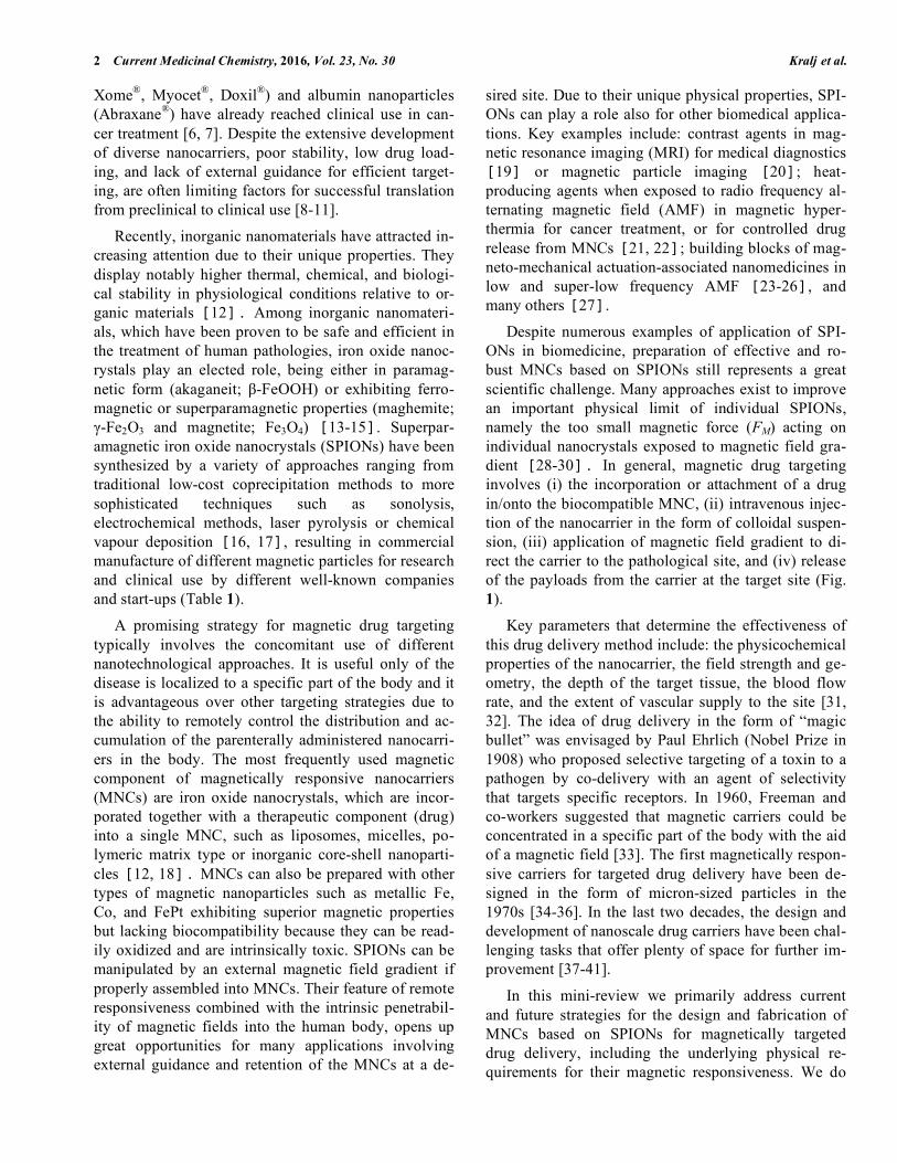
Despite numerous examples of application of SPI-ONs in biomedicine, preparation of effective and ro-bust MNCs based on SPIONs still represents a great scientific challenge. Many approaches exist to improve an important physical limit of individual SPIONs, namely the too small magnetic force (FM) acting on individual nanocrystals exposed to magnetic field gra-dient [28-30]. In general, magnetic drug targeting involves (i) the incorporation or attachment of a drug in/onto the biocompatible MNC, (ii) intravenous injec-tion of the nanocarrier in the form of colloidal suspen-sion, (iii) application of magnetic field gradient to di-rect the carrier to the pathological site, and (iv) release of the payloads from the carrier at the target site (Fig. 1).
Key parameters that determine the effectiveness of this drug delivery method include: the physicochemical properties of the nanocarrier, the field strength and ge-ometry, the depth of the target tissue, the blood flow rate, and the extent of vascular supply to the site [31, 32]. The idea of drug delivery in the form of “magic bullet” was envisaged by Paul Ehrlich (Nobel Prize in 1908) who proposed selective targeting of a toxin to a pathogen by co-delivery with an agent of selectivity that targets specific receptors. In 1960, Freeman and co-workers suggested that magnetic carriers could be concentrated in a specific part of the body with the aid of a magnetic field [33]. The first magnetically respon-sive carriers for targeted drug delivery have been de-signed in the form of micron-sized particles in the 1970s [34-36]. In the last two decades, the design and development of nanoscale drug carriers have been chal-lenging tasks that offer plenty of space for further im-provement [37-41].
In this mini-review we primarily address current and future strategies for the design and fabrication of MNCs based on SPIONs for magnetically targeted drug delivery, including the underlying physical re-quirements for their magnetic responsiveness. We do
Design and Fabrication of Magnetically Responsive Nanocarriers Current Medicinal Chemistry, 2016, Vol. 23, No. 30 3
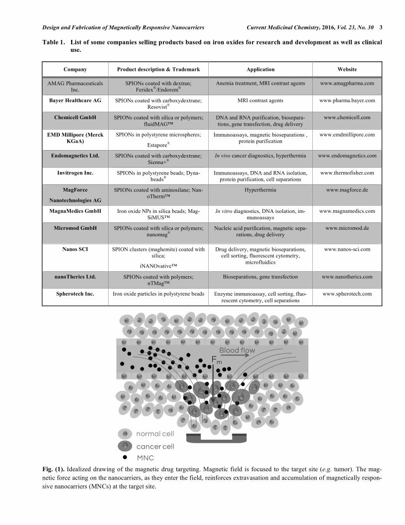
Table 1. List of some companies selling products based on iron oxides for research and development as well as clinical use.
Company Product description & Trademark Application Website
AMAG Pharmaceuticals Inc.
SPIONs coated with dextran; Feridex®/Endorem®
Anemia treatment, MRI contrast agents www.amagpharma.com
Bayer Healthcare AG SPIONs coated with carboxydextrane; Resovist®
MRI contrast agents www.pharma.bayer.com
Chemicell GmbH SPIONs coated with silica or polymers; fluidMAG™
DNA and RNA purification, biosepara-tions, gene transfection, drug delivery
www.chemicell.com
EMD Millipore (Merck KGaA)
SPIONs in polystyrene microspheres;
Estapore®
Immunoassays, magnetic bioseparations , protein purification
www.emdmillipore.com
Endomagnetics Ltd. SPIONs coated with carboxydextrane; Sienna+®
In vivo cancer diagnostics, hyperthermia www.endomagnetics.com
Invitrogen Inc. SPIONs in polystyrene beads; Dyna-beads®
Immunoassays, DNA and RNA isolation, protein purification, cell separations
www.thermofisher.com
MagForce
Nanotechnologies AG
SPIONs coated with aminosilane; Nan-oTherm™
Hyperthermia www.magforce.de
MagnaMedics GmbH Iron oxide NPs in silica beads; Mag-SiMUS™
In vitro diagnostics, DNA isolation, im-munoassays
www.magnamedics.com
Micromod GmbH SPIONs coated with silica or polymers; nanomag®
Nucleic acid purification, magnetic sepa-rations, drug delivery
www.micromod.de
Nanos SCI SPION clusters (maghemite) coated with silica;
iNANOvative™
Drug delivery, magnetic bioseparations, cell sorting, fluorescent cytometry,
microfluidics
www.nanos-sci.com
nanoTherics Ltd. SPIONs coated with polymers; nTMag™
Bioseparations, gene transfection www.nanotherics.com
Spherotech Inc. Iron oxide particles in polystyrene beads Enzyme immunoassay, cell sorting, fluo-rescent cytometry, cell separations
www.spherotech.com
Fig. (1). Idealized drawing of the magnetic drug targeting. Magnetic field is focused to the target site (e.g. tumor). The mag-netic force acting on the nanocarriers, as they enter the field, reinforces extravasation and accumulation of magnetically respon-sive nanocarriers (MNCs) at the target site.

4 Current Medicinal Chemistry, 2016, Vol. 23, No. 30 Kralj et al.
not analyse in detail other aspects such as the tech-niques for the synthesis of SPIONs, or the interactions of the nanocarrier systems with the biological envi-ronment. An important emphasis of the mini-review will also be devoted to the drug loading possibilities into a nanocarrier and the controlled release of the pay-loads at the targeted site. For an in-depth discussion of other targeting strategies, such as passive targeting based solely on enhanced permeation and retention (EPR) effect, and a number of active targeting strate-gies which rely on binding of the targeting ligands (af-finity moieties) to the nanocarrier surface and their nanotoxicity assessments, please refer to existing com-prehensive reviews [42-46].
2. PHYSICAL REQUIREMENTS FOR EFFEC-TIVE MAGNETIC TARGETING
The key feature of magnetically responsive carriers is their ability to be efficiently (i) isolated from com-plex fluids, such as blood flowing through the target region, and (ii) retained at a desired site for prolonged time, if exposed to a magnetic field gradient generated by a strong permanent magnet such as Nd-Fe-B (Neo-dymium Iron Boron alloy). The magnet can be placed outside the body over the target site or implanted inter-nally using minimally invasive surgery [32, 47, 48]. It is crucial to understand that a magnetic field gradient is required to exert magnetic force (FM) at a distance; a uniform field gives rise to a magnetic torque, but no translational action. For successful magnetic targeting, the FM should prevail over the thermal energy (kBT), causing a Brownian motion, and over the hydrody-namic drag in the blood. The FM acting on the nanoc-rystal is proportional to its magnetization (M), volume (V), and magnetic field gradient (∇H), i.e., FM = µ0 M V∇H (where µ0 is the permeability of free space), with M being mainly defined by the type of magnetic mate-rial and magnetic field strength. Particle size (d) has only a minor effect on the M of nanocrystals, while it has a much larger impact on the FM, since FM increases with d3. The size of individual SPIONs should not be increased over ∼15 nm because at that size limit the SPIONs lose their superparamagnetic properties and thus they are not suitable for the preparation of colloi-dal suspensions, due to magnetic dipole-dipole interac-tions resulting in undesired aggregation [49]. On the other hand, the individual SPIONs, unfortunately, can-not be effectively guided within the body despite the presence of high magnetic field, due to the too small FM acting on each individual SPION [50]. Thus, the most beneficial way to effectively increase FM acting on a nanocarrier in a stable suspension exposed to a
magnetic-field gradient, whilst maintaining the super-paramagnetic state, is to increase its volume [28, 51]. This objective could be achieved by assembly of nu-merous individual SPIONs into well-defined clusters that is the basis for nanocarriers’ magnetic responsive-ness [29]. The common feature of these clusters is that multiple SPIONs are physically held together in a small spatial compartment. We have recently published inno-vative nanotechnological approaches for the prepara-tion of highly magneto-responsive SPIONs clusters with a size range of 50-150 nm, optimal for further de-velopment into magnetically responsive drug delivery systems [29, 30, 52]. Indeed, a great challenge still re-mains in the efficient incorporation of a drug into a nanocarrier, especially preparation of MNCs with spa-tially separated compartments for drug and magnetic nanocrystals, with advantages in terms of improved drug stability [12, 53].
To achieve magnetic targeting, the magnetic force acting on individual magnetic carriers needs to over-come the hydrodynamic drag force (FD), which enables the carrier to evade the magnetically targeted site. The FD is linearly dependent on the viscosity of the fluid medium (e.g. blood), the diameter of the carrier, and the difference in velocities of the carrier and the fluid. The motion of the carrier towards high magnetic gradi-ent is affected also by buoyancy force, which depends on the difference between the densities of the carrier and of the fluid medium. After taking into an account all the above-mentioned forces, we come to the pa-rameter known as magnetophoretic mobility of the car-rier, i.e. a parameter that describes the carrier’s mag-netic responsiveness [49]. Since the magnetophoretic mobility depends on the carrier size, it is significantly greater in case of micron-sized particles or micro-spheres (∼ 1 µm in diameter) compared to nanoscale carriers (∼ 100 nm in diameter) and the individual nanocrystals (∼ 10 nm in diameter). This “guiding” feature of microspheres is already widely applied in bioseparations, which exploit the same physical princi-ples for magnetic manipulation [32]. Thus, such rela-tively big microspheres can be efficiently used in cell and other types of bioseparations where the bio-molecules and cells coupled with microspheres can be isolated from the complex mixtures in the shortest pos-sible time [49]. However, the magnetically responsive micro-sized carriers are too big for the intravenous or intra-arterial applications because they can cause em-bolism. Moreover, the majority of solid tumors exhibit a porous vasculature with the pore size between 380 nm and 780 nm, depending on the tumor type and its

Design and Fabrication of Magnetically Responsive Nanocarriers Current Medicinal Chemistry, 2016, Vol. 23, No. 30 5
microenvironment [54]. Therefore, the optimal particle size for preparation of magnetically responsive drug carriers should be a compromise between the biomedi-cal size restrictions, giving preference to smaller parti-cle sizes, and the particle magnetophoretic mobility parameter, being greater in case of larger particles. Some studies on magnetic targeting, where the hydro-dynamics of the magnetic carriers have been investi-gated, revealed that the magnetic field gradients should be at least 8 Tm-1 and 100 Tm-1 for effective targeting of the femoral and carotid arteries, respectively [55]. In fact, the magnetic drug targeting is likely to be most effective in body regions where the blood flow is slow and in the regions close to the permanent magnet, due to both lower FD and larger magnetic field gradients [56]. The minimum size of the magnetic carriers based on SPIONs dispersed in an aqueous suspension to be efficiently attracted in a magnetic field gradient in a reasonable time is approximately 50 nm [28]. There are various terms used in literature for such assembled par-ticles with size from 50 to 300 nm, i.e. magnetic nano-beads, multi-core nanoparticles, nanocomposite parti-cles, nanospheres, nanoclusters, nanoparticle clusters, and SPION clusters. However, we use the general term of MNCs, as the optimal size of drug delivery system based on SPIONs for intravenous application is in the “nano” range.
3. STRUCTURAL TYPES OF MAGNETICALLY RESPONSIVE NANOCARRIERS IN DRUG DE-LIVERY
The syntheses of the magnetic component as well as the design of the nanocarrier are key steps to achieve a magnetically-responsive nanodelivery system capable of efficient targeting as discussed above. Drugs may be coupled to the carrier surface by means of electrostatic or covalent bonding. Although this approach is straightforward, it has some drawbacks. Surface-bound drug molecules may impair colloidal stability of the nanodelivery system; drug release may be hampered due to strong binding; alternatively, extensive burst release can occur in case the drug is weakly bound. Drug attachment to the nanocarrier surface is also lim-ited by the specific surface area of the nanocarrier. Therefore, when the nanocarrier size is increased for improved magnetic responsiveness, there will be an overall reduced surface area, and thus a relatively smaller amount of binding sites for the drug. An inter-esting alternative to surface attachment is drug incorpo-ration into an organic and/or inorganic matrix, which physically holds the SPIONs together in a defined spa-tial compartment whilst allowing drug loading and re-
lease [12]. The drug loading capacity of such delivery systems is often enhanced compared to the drug at-tachment to the nanocarrier surface. In addition, the majority of drugs are low-molecular-weight molecules often associated with challenging physicochemical properties, such as poor water solubility. Therefore, the design and fabrication of a suitable nanocarrier system for delivery of such drugs requires additional attention.
3.1. Magnetoliposomes
Liposomes are biocompatible, biodegradable, and generally non-toxic nanodelivery systems that enable transport of both (i) hydrophilic drugs in the aqueous interior and (ii) hydrophobic/amphiphilic drugs em-bedded in the liposome membrane [57]. However, the applicability of liposomes for the delivery of poorly soluble compounds is limited, since only small drug amounts can be incorporated into the hydrophobic liposomal bilayer; besides, this approach can impair the bilayer stability. Currently, large unilamellar liposomes with a diameter ranging from 100 to 200 nm represent the most successful nanodelivery systems in clinical use. Besides surface properties, the size of the carrier is indeed a crucial parameter affecting circulation half-time in the bloodstream [58].
Magnetoliposomes can be prepared by incorpora-tion of SPIONs into the liposomes. They represent a novel and very promising multi-purpose tool for imag-ing and magnetic targeting, which can be non-invasively controlled [57]. Importantly, both liposome components and SPIONs are FDA-approved, thus their combination may proceed to the clinical practice with minimal safety concerns [59]. The magnetic respon-siveness can be achieved by incorporation of either hy-drophilic or hydrophobic SPIONs in the aqueous liposome interior or in the liposome membrane, respec-tively (Fig. 2). Several examples have been reported in the literature thus proving the versatility of this ap-proach. Nappini et al. have described incorporation of up to 30 SPIONs with the size from 10 to 16 nm per individual magnetoliposome [60]. Garnier et al. pre-pared magnetoliposomes with high iron content by in-corporation of up to 77 citrate-coated SPIONs with a size of ∼7 nm into a single liposome [58]. Thermosen-sitive magnetoliposomes containing citrate-coated SPIONs and paclitaxel for the treatment of various solid tumors were described by Kulshrestha et al. [61]. A similar system with entrapped dextran-coated SPI-ONs and a fluorescent dye was prepared by Tai et al. [59]. Bothun et al. designed folate-targeted cationic magnetoliposomes with encapsulated doxorubicin and

6 Current Medicinal Chemistry, 2016, Vol. 23, No. 30 Kralj et al.
anionic SPIONs [62]. Guo et al. developed monodis-perse dextran-coated magnetoliposomes with the size of ∼120 nm where SPIONs and doxorubicin were en-capsulated in the liposome interior. This formulation demonstrated excellent stability in serum and good magnetic responsiveness. The release of doxorubicin was triggered by exposure to the low frequency AMF [63]. Undesired interaction between drug and SPION (i.e., drug complexation with surface iron atoms) can be avoided with spatial separation of the two compo-nents; for example, magnetoliposomes may have hy-drophobic SPIONs in the liposomal bilayer, so that the aqueous liposome interior will be available for incorpo-ration of sensitive drugs. The research group of Babin-cova prepared large unilamellar magnetoliposomes with spatial separation of doxorubicin and SPIONs that were encapsulated into the liposome interior and lipid bilayer, respectively [64]. Chen et al. described MNC in the form of 100 nm-sized unilamellar magnetol-iposomes with embedded 5 nm-sized oleate-coated SPIONs in the liposome membrane, and carboxyfluo-rescein in the aqueous interior. The drug release was triggered by hyperthermia, thanks to heat generation by SPIONs exposed to AMF [65]. Bixner and Reimhult presented vesicles with hydrophobic, monodisperse 3.5 nm N-palmityl-6-nitrodopamide-coated SPIONs em-bedded in the membrane. In this study the effectiveness of SPION incorporation into the liposomes was de-pendant on the size and surface coating of SPIONs and it was significantly improved compared to other previ-ously reported methods, i.e., 26 SPIONs per 100 nm-sized vesicle. However, SPION loading decreased and the polidispersity and lamellarity of vesicles increased, when oleate-coated SPIONs were embedded in the liposome membrane [57]. Magnetoresponsive hybrid
liposomes with incorporated thermosensitive polymers and SPIONs were designed by Katagiri and co-workers. In this case, oleate-coated SPIONs were in-corporated in the lipid bilayer, while the water-soluble fluorescent dye pyranine was loaded in the liposome interior. Remote control of pyranine release was achieved by exposure to AMF; the release rate was af-fected by the amount of incorporated SPIONs and the type of thermosensitive polymers used [66].
Peiris et al. described a multi-component nanocar-rier system for doxorubicin delivery with SPIONs in the form of short nanochains being attached to the drug-loaded liposomes. This delivery system coupled with AMF-triggered drug release showed improved penetration and deposition at micrometastatic sites. As a result, a low dose of cytotoxic drug proved effective for cancer treatment, indicating great potential for ap-plication in all types of cancer that exhibit aggressive and metastatic phenotypes [67-70].
Magnetoliposomes are frequently studied MNCs for the delivery of hydrophilic biomacromolecules, such as therapeutic proteins. Ye et al. developed magnetol-iposomes loaded with recombinant human interferon-α2b and hydrophilic 10 nm-sized SPIONs. Cell ex-periments and animal studies demonstrated that magne-toliposomes delivered by magnetic targeting signifi-cantly inhibited cancer cell growth, and notably re-duced tumor size in nude mice, indicating improved therapeutic efficacy and reduced side effects to non-target organs [71]. Meier et al. developed immuno-magnetoliposomes with a high amount of entrapped hydrophilic SPIONs for MRI [72]. They showed that liposome PEGylation up to a certain degree had a posi-tive influence on both the amount of entrapped SPIONs and the stability of magnetoliposomes. Furthermore,
Fig. (2). Schematic representation of the most frequent types of drug-loaded magnetoliposomes. Hydrophilic SPIONs and drugs can be encapsulated into the inner aqueous compartment, while hydrophobic SPIONs and drugs can be incorporated into the phospholipid bilayer.

Design and Fabrication of Magnetically Responsive Nanocarriers Current Medicinal Chemistry, 2016, Vol. 23, No. 30 7
Martins et al. tested the application of surface-modified magnetoliposomes for the affinity adsorption of anti-bodies, with the antigen being either embedded in the phospholipid bilayer or covalently coupled to the sur-face of the magnetoliposome [73]. This system dis-played good properties for further development into immunodiagnostics or drug delivery formulations.
3.2. Polymeric Magnetically-Responsive Nanocarri-ers
Nanocarriers based on polymer matrices are nowa-days widely investigated for various applications in biomedicine. The polymer matrix can be either hydro-philic or hydrophobic; therefore, hydrophilic as well as hydrophobic drugs can be loaded. Drug release from the polymer matrix can be either diffusion- or erosion-controlled. Additional incorporation of SPIONs makes such nanocarriers magnetically-responsive.
In 1994, Häfeli and co-workers successfully synthe-sized biodegradable poly(lactic acid) microparticles containing magnetite nanocrystals and the beta-emitter 90Y for targeted radiotherapy of subcutaneous tumors [74]. This initial study was based on micro-sized carri-ers, while further developments mostly focused on the design of nanosized polymeric carriers. Hu and co-workers designed 200 nm-sized tamoxifen-loaded poly(L-lactic acid) nanocarrier containing 6 nm-sized SPIONs [75]. Similar polymeric nanocarriers were ob-tained from poly(lactic-co-glycolic acid) loaded with 10 nm-sized hydrophobic SPIONs for the targeting of intracellular compartments (Fig. 3) [76]. The nanopar-ticles have the potential to be evolved into magneti-cally-responsive drug nanocarriers for relatively hy-
drophobic anticancer drugs. Burnand et al. used the catechol-modified poly(vinyl alcohol) as a coating polymer for monodispersed SPION clusters. The clus-ters with size between 40 and 80 nm showed potential applicability in biomedicine [77].
Fang et al. successfully prepared SPION clusters with an average diameter of approximately 140 nm and coated with chitosan for detection and identification of low abundant glycopeptides in glycoproteome analysis. Aberrant glycosylation of some important proteins is highly associated with various disease states including cancer. The presented formulation showed great poten-tial for the design of relevant drug delivery systems [78].
There is vast literature on drug delivery applications of polymer-based nanocarriers containing SPIONs, especially for cancer treatment, but also for diagnostic use. For instance, Chertok and co-workers presented starch-coated SPIONs with a hydrodynamic diameter of 110 nm [79]. The particles were composed of mul-tiple individual SPIONs and a 5-fold increase in in vivo accumulation was seen for glioma sites that were mag-netically-targeted, relative to non-targeted tumors. Yang and co-workers developed cisplatin- and gemcit-abine-loaded 250 nm-sized magnetic poly(ethyl-2-cyanoacrylate) particles [80]. The nanocarrier was successfully loaded with hydrophobic SPIONs and cis-platin whereas incorporation efficiency of relatively hydrophilic gemcitabine was much lower. Thus, cis-platin exhibited sustained release, whereas gemcitabine displayed rapid release. Another magnetically-responsive polymeric nanocarrier was reported by Di Corato and co-workers [81]. They designed 100 nm-
Fig. (3). (A) TEM image and (B) schematic representation of a polymeric nanoparticle with the diameter of approximately 100 nm [76]. Multiple hydrophobic SPIONs were incorporated in the polymer matrix together with sparingly water-soluble drug or fluorescent dye.

8 Current Medicinal Chemistry, 2016, Vol. 23, No. 30 Kralj et al.
sized poly(maleic anhydride-alt-1-octadecene) nanoparticles loaded with multiple SPIONs and quan-tum dots. The nanosystem demonstrated good magnetic responsiveness that was confirmed by magnetic cell sorting. The magnetic particles could be loaded with hydrophobic drugs and applied in drug delivery. Li et al. developed a promising multifunctional theranostic nanodelivery system based on polyethylene glycol functionalized SPIONs, which were loaded with chlo-rin e6, a widely used photosensitizer in photodynamic therapy. Due to the greatly accelerated cellular uptake, such magnetic nanoparticles showed a much stronger cancer cell eradication in comparison to free chlorin e6, therefore indicating a good potential for application in magnetically-targeted, photodynamic cancer treatment [82]. SPION clusters coated with poly(dopamine) with an average diameter of 80 nm were developed as a magnetic field-directed theranostic agent by Wu et al. The SPION clusters enabled ultrasensitive MRI imag-ing whereas the poly(dopamine) coating induced can-cer cell death under near-infrared laser irritation, due to the photothermal conversion ability of poly(dopamine) [83]. Oka et al. described preparation of composite particles based on biodegradable polymer polyhy-droxyalkanoate and SPIONs. A model drug pyrene was incorporated into the polymer matrix and released upon biodegradation, indicating applicability of the system in magnetically-guided drug delivery [84]. Release can be triggered also via magnetic hyperthermia when the matrix is prepared from thermo-responsive polymer and loaded with SPIONs [85]. Drug release occurs through nanoscale cracks formed within the matrix, due to the local heat generated by SPIONs exposed to high frequency AMF [21, 86-88]. Another possibility is drug release triggered by formation of openings that are induced by a magneto-mechanical force [89].
Shortcomings of polymer matrix nanocarriers in-clude: relatively high polydispersity, limited mechani-cal and microbiological stability, and high cost [43], therefore inorganic nanocarriers represent an interest-ing alternative and are currently widely investigated.
3.3. Silica-Based Core-Shell Magnetically-Responsive Nanocarriers
Core-shell particles have attracted considerable at-tention in the field of drug delivery, due to many un-precedented advantages [90]. Silica-based shell struc-tures are especially interesting [91]. Research in the field has been intensively focused on the development of facile and versatile synthesis methods for the fabri-cation of mesoporous silica shells on various types of
magnetic core particles, since high surface area, high pore volume, and large pore channels are advantageous properties of mesoporous silica [92]. Furthermore, it is highly desirable that mesopore channels in the shell are open to the surface in order to achieve loading and release of various guest molecules. However, these core-shell particles can also be transformed into hollow carriers to enable enhanced drug loading in the interior and preserved magnetic responsiveness [93].
Silica is a product of sol-gel process that involves alkoxide precursors (tetraethyl orthosilicate; TEOS), which undergo hydrolysis upon reaction with water and catalyst. Subsequent condensation results in the forma-tion of either homogeneous spherical particles or de-fined layers (i.e., shell) on the surface of core particles. In the late 1960s, Stöber reported the preparation of colloidal silica particles as a result of sol-gel chemistry [94]. Since the classical Stöber process yields only non-porous silica with low surface area, new ap-proaches have been developed for the preparation of mesostructured silica [95-98]. Deng et al. have re-ported magnetically-responsive silica-coated particles with perpendicularly aligned channels inside the silica shell [99]. The highly ordered channels are formed upon addition of cetyltrimethylammonium bromide (CTAB), and subsequent refluxing in acetone. The Shi group proposed a similar carrier system composed of a non-magnetic hematite core that was transformed into a magnetic core, upon silica coating and reductive ther-mal treatment [93]. These hollow nanostructures were further evolved into drug delivery system with high-loading of ibuprofen (∼ 12 % w/w) and prolonged drug release in a simulated body fluid. Liong and co-workers have prepared multifunctional nanocarriers composed of multiple 20 nm-sized SPIONs and then coated them with a 100–200 nm-thick mesoporous and fluorescent silica shell [100]. Hydrophobic anticancer drugs such as camptothecin and paclitaxel were loaded into silica pores via incubation of the empty nanocar-rier in a concentrated DMSO solution of the anticancer drug. The authors observed that only 4 % w/w of the loaded drug was released into aqueous medium, while the total drug amount came out of the mesopores when the nanocarrier was incubated in DMSO or methanol.
Our group has recently developed novel magneti-cally-responsive SPION clusters based on chemically-directed assembly of individual silica-coated SPIONs in aqueous suspension [30]. The SPION clusters have a diameter of approximately 50 nm, and display a “raspberry-like” morphology that show high surface area and a large fraction of iron oxide (∼ 46 emu g-1).
Design and Fabrication of Magnetically Responsive Nanocarriers Current Medicinal Chemistry, 2016, Vol. 23, No. 30 9
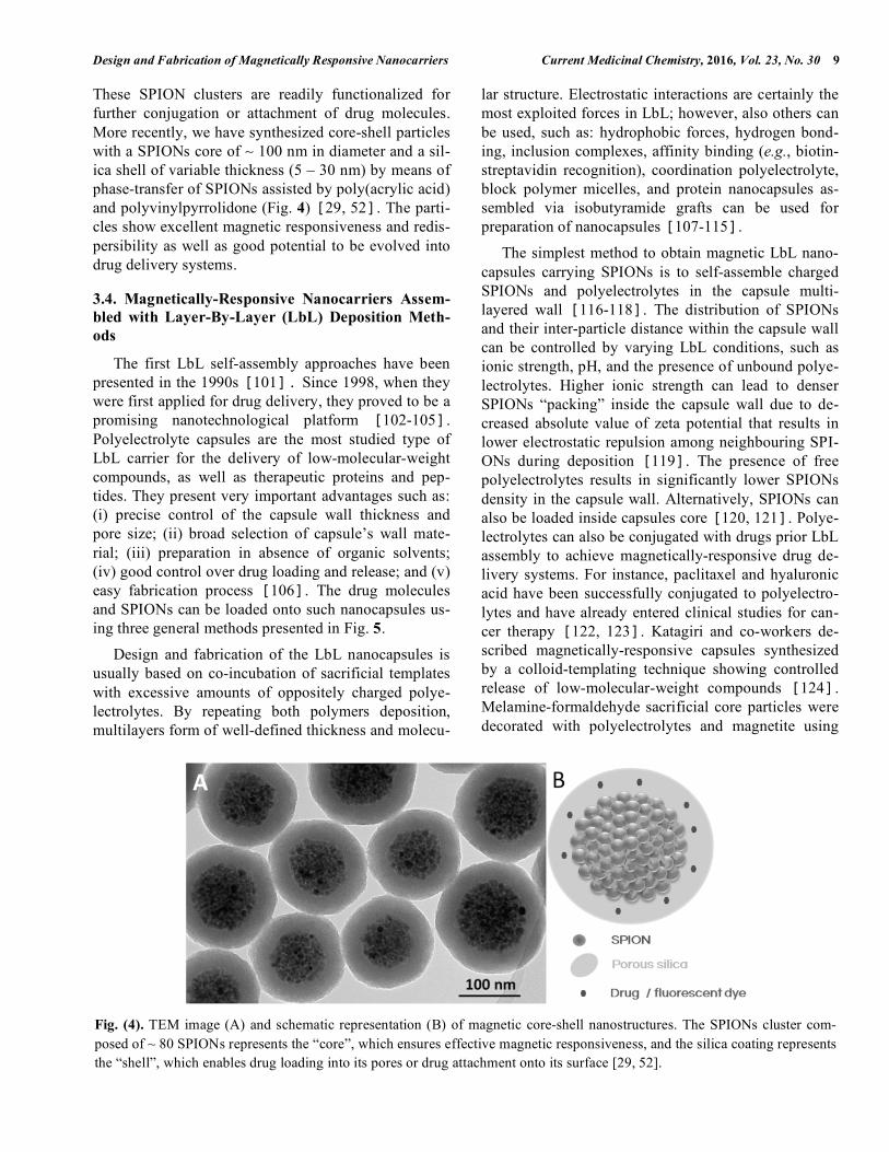
These SPION clusters are readily functionalized for further conjugation or attachment of drug molecules. More recently, we have synthesized core-shell particles with a SPIONs core of ∼ 100 nm in diameter and a sil-ica shell of variable thickness (5 – 30 nm) by means of phase-transfer of SPIONs assisted by poly(acrylic acid) and polyvinylpyrrolidone (Fig. 4) [29, 52]. The parti-cles show excellent magnetic responsiveness and redis-persibility as well as good potential to be evolved into drug delivery systems.
3.4. Magnetically-Responsive Nanocarriers Assem-bled with Layer-By-Layer (LbL) Deposition Meth-ods
The first LbL self-assembly approaches have been presented in the 1990s [101]. Since 1998, when they were first applied for drug delivery, they proved to be a promising nanotechnological platform [102-105]. Polyelectrolyte capsules are the most studied type of LbL carrier for the delivery of low-molecular-weight compounds, as well as therapeutic proteins and pep-tides. They present very important advantages such as: (i) precise control of the capsule wall thickness and pore size; (ii) broad selection of capsule’s wall mate-rial; (iii) preparation in absence of organic solvents; (iv) good control over drug loading and release; and (v) easy fabrication process [106]. The drug molecules and SPIONs can be loaded onto such nanocapsules us-ing three general methods presented in Fig. 5.
Design and fabrication of the LbL nanocapsules is usually based on co-incubation of sacrificial templates with excessive amounts of oppositely charged polye-lectrolytes. By repeating both polymers deposition, multilayers form of well-defined thickness and molecu-
lar structure. Electrostatic interactions are certainly the most exploited forces in LbL; however, also others can be used, such as: hydrophobic forces, hydrogen bond-ing, inclusion complexes, affinity binding (e.g., biotin-streptavidin recognition), coordination polyelectrolyte, block polymer micelles, and protein nanocapsules as-sembled via isobutyramide grafts can be used for preparation of nanocapsules [107-115].
The simplest method to obtain magnetic LbL nano-capsules carrying SPIONs is to self-assemble charged SPIONs and polyelectrolytes in the capsule multi-layered wall [116-118]. The distribution of SPIONs and their inter-particle distance within the capsule wall can be controlled by varying LbL conditions, such as ionic strength, pH, and the presence of unbound polye-lectrolytes. Higher ionic strength can lead to denser SPIONs “packing” inside the capsule wall due to de-creased absolute value of zeta potential that results in lower electrostatic repulsion among neighbouring SPI-ONs during deposition [119]. The presence of free polyelectrolytes results in significantly lower SPIONs density in the capsule wall. Alternatively, SPIONs can also be loaded inside capsules core [120, 121]. Polye-lectrolytes can also be conjugated with drugs prior LbL assembly to achieve magnetically-responsive drug de-livery systems. For instance, paclitaxel and hyaluronic acid have been successfully conjugated to polyelectro-lytes and have already entered clinical studies for can-cer therapy [122, 123]. Katagiri and co-workers de-scribed magnetically-responsive capsules synthesized by a colloid-templating technique showing controlled release of low-molecular-weight compounds [124]. Melamine-formaldehyde sacrificial core particles were decorated with polyelectrolytes and magnetite using
Fig. (4). TEM image (A) and schematic representation (B) of magnetic core-shell nanostructures. The SPIONs cluster com-posed of ~ 80 SPIONs represents the “core”, which ensures effective magnetic responsiveness, and the silica coating represents the “shell”, which enables drug loading into its pores or drug attachment onto its surface [29, 52].

10 Current Medicinal Chemistry, 2016, Vol. 23, No. 30 Kralj et al.
LbL assembly. After removal of the organic core, the outermost wall was decorated with an additional lipid bilayer and, at the same time, a dye was encapsulated into the capsule interior and used as a model drug. Dye release was triggered on-demand by exposing the nanocarrier to radiofrequency AMF, which caused heating of the SPIONs-containing shell, thus increasing the lipid bilayer permeability.
3.5. Colloidosome as Magnetically-Responsive Nanocarriers
Colloidosomes are a special type of assembled nanostructures where the liquid interior compartment is enclosed by a layer of relatively tightly packed nanoparticles or nanocrystals, assuring the robustness of the whole structure [125]. A number of studies de-scribe micron-sized (> 1 µm) colloidosomes composed of iron oxide nanocrystals, however, such systems are not appropriate for drug delivery due to the carrier oversize [126, 127]. Recently, Bollhorst and co-workers have described an innovative approach for the synthesis of submicron (< 1 µm) bifunctional colloi-dosomes that allowed the simultaneous incorporation of SPIONs and fluorescent silica nanoparticles in a sin-gle submicron colloidosome (Fig. 6) [128]. Such col-loidosomes represent a promising platform for their use in magnetically responsive drug delivery. Their few-hundred-nanometers large aqueous interior can be used for delivering hydrophilic cargo that can be a small molecule or even a large biomacromolecule, such as a therapeutic protein or gene. Preparation of colloi-dosomes is based on water-in-oil mini-emulsions stabi-lized by oil-soluble surfactants; therefore, hydrophilic cargo is not in contact with the surfactants. That could be advantageous for the delivery of enzymes and other therapeutic proteins that may be very sensitive to the composition of the formulation.
Drug release from colloidosomes can occur via dif-fusion through the nanopores among the packed nanoc-rystals in the colloidosome shell. Shell porosity can be affected by the heat produced due to exposure of col-loidosomes to radiofrequency AMF, or due to potential mechanical actuation generated by low-frequency AMF as proposed by Golovin et al. [26]. However, drug release from colloidosomes can also be achieved by light-triggered disassembly of the colloidosome shell as it has recently been proposed by Li et al. [129].
An innovative colloidosome-like delivery system was reported by Gong et al. [130]. The magnetic car-rier was fabricated by using a microfluidic flow-focusing approach. The liquid interior was loaded with acetylsalicylic acid, while the shell was composed of SPIONs embedded into crosslinked chitosan. Drug re-lease was mechanically-controlled by compression-extension of the magneto-elastic shell induced by AMF and was shown to be dependent on frequency and magnitude of the applied magnetic field.
Despite the rapid progress and great promise of col-loidosome systems, further research and optimization is needed to achieve optimal size for drug delivery appli-cations.
4. PERSPECTIVES AND FUTURE CHAL-LENGES
Magnetic targeting was presented as a promising strategy in a number of studies, but it was tested only in a few clinical trials to date [37, 131]. Lubbe and co-workers [132, 133] have performed the first clinical trial of magnetically-targeted drug delivery where 14 patients were treated with epidoxorubicin that was electrostatically-conjugated to the surface of a mag-netic carrier. The carrier was effectively targeted to the
Fig. (5). Scheme representing the most common approaches for preparation of magnetic drug-loaded capsules based on SPI-ONs with the use of layer-by-layer (LbL) assembly technique: (i) loading of low-molecular-weight compounds into preformed polyelectrolyte nanocapsules; (ii) direct incorporation of drug and SPIONs into capsule’s wall during deposition of oppositely charged polyelectrolytes onto sacrificial template core particles; and (iii) deposition of oppositely charged polyelectrolytes and SPIONs onto drug-based core nanoparticles.

Design and Fabrication of Magnetically Responsive Nanocarriers Current Medicinal Chemistry, 2016, Vol. 23, No. 30 11
tumor site in 6 patients. A second clinical trial was per-formed by Koda and co-workers on 32 patients with hepatocellular carcinoma [134]. The doxorubicin-coupled magnetic carrier was successfully targeted in 30 patients using an external magnetic field. In another clinical trial, Wilson and co-workers conjugated doxorubicin to the magnetic carrier, which was selec-tively delivered to hepatocellular carcinoma by using an external magnetic field [135]. The results showed that the magnetic carriers were effectively targeted to the tumor site and up to 91% of the tumor volume was affected by the drug. It is thus apparent that, despite of the slow progress in the clinical translation of magneti-cally-responsive carriers, their potential remains great for targeted drug delivery.
In general, the techniques used for the syntheses of various types of high-quality nanocrystals with well-defined magnetic properties are nowadays known and well-optimized [16]. However, strategies for the as-sembly of individual nanocrystals into multifunctional hierarchical structures such as MNCs are rather less developed, and not yet prepared for industrial scale-up. Currently available magnetic targeting is likely to be ineffective in case the target site is located deep in the body, due to insufficient magnetic force exerted on a distant MNC, thus resulting in poor magnetic capturing [49, 56, 136]. Therefore, it is an urgent need to de-sign new highly magneto-responsive nanomaterials with high drug loading. Nanoscale size, high magnetic responsiveness, and high drug loading are unfortu-nately incompatible features. Therefore, such a chal-lenge should be solved with the development of effi-cient delivery systems that find an optimal balance in terms of how much magnetic responsiveness can be sacrificed to take advantage of a smaller carrier size and higher drug loading.
Since SPIONs in the form of magnetite and maghemite are recognized as safe and biocompatible, the development of new MNCs is expected to be pri-marily based on SPIONs assemblies. The future design of nanocarriers and magnetically actuated nanomedici-nes leads to the synthesis of novel superparamagnetic structures with anisotropic shapes such as nanorods, nanodiscs, nanotubes, nanoworms, and nanochains [23, 29, 137-140]. These materials may have larger magnetic moment and better magnetic responsiveness in a magnetic field gradient compared to spherical par-ticles of the comparable cross-section. Recently, we reported a new approach for the magnetically-assisted synthesis of anisotropically shaped nanostructures, namely superparamagnetic nanochains, with the length of ∼ 500 nm and diameter of only ∼ 100 nm (Fig. 7) [29] . Such nanochains can easily be magnetically guided with low magnetic field gradients, whilst having the potential to be transformed into an effective MNC. Besides magnetic targeting, the superparamagnetic nanocarriers with anisotropic shapes also show great promise in magneto-mechanical actuation of nanomedicines by low frequency AMF. However, the major challenge remains in the advancement of scale-up approaches for the synthesis of such anisotropic nanostructures.
We shall note that a very important difference be-tween magnetic and active targeting lies in the design of the nanocarrier. In contrast with active targeting, MNCs do not need affinity ligands on their surface to be targeted to a specific site in the body [141]. Fur-thermore, such affinity ligands bound on the surface of the nanocarriers often impair the colloidal stability of the delivery system in a complex medium such as blood. Therefore, the design of MNCs allows more freedom for the optimization of the nanocarrier surface
Fig. (6). (A) SEM micrograph and (B) schematic representation of magnetic colloidosome composed of SPIONs and fluores-cent silica NPs. Adapted with permission from ref. [128]. Copyright ©2015 WILEY-VCH Verlag GmbH & Co. KGaA.

12 Current Medicinal Chemistry, 2016, Vol. 23, No. 30 Kralj et al.
to avoid undesired interactions with blood components, such as formation of protein corona leading to a prema-ture elimination of the nanocarrier by the reticuloendo-thelial system [142, 143].
The MNCs of the future should respond to magnetic guidance to perform several tasks, including: controlled release of the payload, selective targeting, and eventu-ally other actions at the disease site. Often researchers attempt to use chemical cues to trigger drug release at the desired site in the body, such as reductive intracel-lular environment, specific enzymes overexpressed by cancer cells (cathepsins), acidic pH in the tumors, and low endosomal pH [8]. Despite all such efforts, these types of triggered drug release systems lack the re-quired precision and robustness and might not be suffi-cient to achieve the desired goals by itself. The drug release from the MNCs at the target site can be trig-gered remotely by magnetic heating in a radio fre-quency AMF or with the help of low frequency AMF magneto-mechanical actuation [26, 89]. However, in the near future it can be expected that a combination of various chemical and physical cues of triggering means will be integrated into an individual MNC in order to achieve more selective drug release at the target site.
CONFLICT OF INTEREST
The author(s) confirm that this article content has no conflict of interest.
ACKNOWLEDGEMENTS
The support of the Ministry of Higher Education, Science and Technology of the Republic of Slovenia within the National Programs P2-0089 and P1-0189, and Research Project J1-7302 is gratefully acknowl-edged. SK is grateful to the European Social Found, Operational Programme 2014-2020 (Axis 3 – Educa-
tion and Training, Specific Programme n.26 – TAL-ENTS3 Fellowship Programme – “MAGIC SPY”). The authors thank Dr Tobias Bollhorst for SEM micrograph of magnetic colloidosome.
REFERENCES [1] Webber, M. J.; Appel, E. A.; Meijer, E. W.; Langer, R.,
Supramolecular biomaterials. Nat. Mater., 2016, 15(1), 13-26.
[2] Marchesan, S.; Prato, M. Nanomaterials for (Nano)medicine. ACS Med. Chem. Lett., 2013, 4(2), 147-149.
[3] Agrawal, U.; Sharma, R.; Gupta, M.; Vyas, S. P. Is nanotechnology a boon for oral drug delivery? Drug Dis-cov. Today, 2014, 19(10), 1530-1546.
[4] Wicki, A.; Witzigmann, D.; Balasubramanian, V.; Huwyler, J. Nanomedicine in cancer therapy: Challenges, opportuni-ties, and clinical applications. J. Control. Release, 2015, 200, 138-157.
[5] Gunn, J.; Zhang, M. Polyblend nanofibers for biomedical applications: perspectives and challenges. Trends Biotech-nol., 2010, 28(4), 189-197.
[6] Wagner, V.; Dullaart, A.; Bock, A.K.; Zweck, A. The emerging nanomedicine landscape. Nat. Biotech., 2006, 24(10), 1211-1217.
[7] Peer, D.; Karp, J. M.; Hong, S.; Farokhzad, O. C.; Margalit, R.; Langer, R. Nanocarriers as an emerging platform for cancer therapy. Nat. Nanotechnol., 2007, 2(12), 751-760.
[8] Mura, S.; Nicolas, J.; Couvreur, P. Stimuli-responsive nanocarriers for drug delivery. Nat. Mater., 2013, 12(11), 991-1003.
[9] Lu, C.-H.; Willner, I. Stimuli-Responsive DNA-Functionalized Nano-/Microcontainers for Switchable and Controlled Release. Angew. Chem. Int. Ed., 2015, 54(42), 12212-12235.
[10] Cheng, Z.; Al Zaki, A.; Hui, J. Z.; Muzykantov, V. R.; Tsourkas, A. Multifunctional nanoparticles: cost versus benefit of adding targeting and imaging capabilities. Sci-ence, 2012, 338(6109), 903-910.
[11] Elsabahy, M.; Wooley, K. L. Design of polymeric nanopar-ticles for biomedical delivery applications. Chem. Soc. Rev., 2012, 41(7), 2545-2561.
[12] Chen, Y.; Chen, H.; Shi, J. In Vivo Bio-Safety Evaluations and Diagnostic/Therapeutic Applications of Chemically Designed Mesoporous Silica Nanoparticles. Adv. Mater., 2013, 25(23), 3144-3176.
Fig. (7). TEM images showing the magnetic nanochains prepared by facile sol-gel synthesis in a magnetic field using SPION clusters as primary building blocks. Superparamagnetic nanochains form stable colloidal suspensions, express excellent mag-netic responsiveness, and hold great potential to be evolved towards a magnetic drug-delivery system [29].

Design and Fabrication of Magnetically Responsive Nanocarriers Current Medicinal Chemistry, 2016, Vol. 23, No. 30 13
[13] Tadic, M.; Milosevic, I.; Kralj, S.; Mbodji, M.; Motte, L. Silica-Coated and Bare Akaganeite Nanorods: Structural and Magnetic Properties. J. Phys. Chem. C, 2015, 119(24), 13868-13875.
[14] Kralj, S.; Makovec, D.; Čampelj, S.; Drofenik, M. Produc-ing ultra-thin silica coatings on iron-oxide nanoparticles to improve their surface reactivity. J. Magn. Magn. Mater., 2010, 322(13), 1847-1853.
[15] Tadic, M.; Milosevic, I.; Kralj, S.; Saboungi, M.L.; Motte, L. Ferromagnetic behavior and exchange bias effect in aka-ganeite nanorods. Appl. Phys. Lett., 2015, 106, 183706.
[16] Laurent, S.; Forge, D.; Port, M.; Roch, A.; Robic, C.; Van-der Elst, L.; Muller, R. N. Magnetic Iron Oxide Nanoparti-cles: Synthesis, Stabilization, Vectorization, Physicochemi-cal Characterizations, and Biological Applications. Chem. Rev., 2008, 108(6), 2064-2110.
[17] Lu, A.-H.; Salabas, E. L.; Schüth, F. Magnetic Nanoparti-cles: Synthesis, Protection, Functionalization, and Applica-tion. Angew. Chem. Int. Ed., 2007, 46(8), 1222-1244.
[18] Zhang, Q.; Wang, W.; Goebl, J.; Yin, Y. Self-templated synthesis of hollow nanostructures. Nano Today, 2009, 4(6), 494-507.
[19] Riehemann, K.; Schneider, S. W.; Luger, T. A.; Godin, B.; Ferrari, M.; Fuchs, H. Nanomedicine--challenge and per-spectives. Angew. Chem. Int. Ed., 2009, 48(5), 872-897.
[20] Gleich, B.; Weizenecker, J. Tomographic imaging using the nonlinear response of magnetic particles. Nature, 2005, 435(7046), 1214-1217.
[21] Kumar, C. S.; Mohammad, F. Magnetic nanomaterials for hyperthermia-based therapy and controlled drug delivery. Adv. Drug Deliv. Rev., 2011, 63(9), 789-808.
[22] Stanley, S. A.; Gagner, J. E.; Damanpour, S.; Yoshida, M.; Dordick, J. S.; Friedman, J. M. Radio-Wave Heating of Iron Oxide Nanoparticles Can Regulate Plasma Glucose in Mice. Science, 2012, 336(6081), 604-608.
[23] Kim, D.-H.; Rozhkova, E. A.; Ulasov, I. V.; Bader, S. D.; Rajh, T.; Lesniak, M. S.; Novosad, V. Biofunctionalized magnetic-vortex microdiscs for targeted cancer-cell destruc-tion. Nat. Mater., 2010, 9(2), 165-171.
[24] Dobson, J. Cancer therapy: Death by magnetism. Nat. Ma-ter., 2012, 11, 1006-1008.
[25] Dobson, J. Cancer therapy: A twist on tumour targeting. Nat. Mater., 2010, 9(2), 95-96.
[26] Golovin, Y. I.; Gribanovsky, S. L.; Golovin, D. Y.; Kly-achko, N. L.; Majouga, A. G.; Master capital A, C.; Sokol-sky, M.; Kabanov, A. V. Towards nanomedicines of the fu-ture: Remote magneto-mechanical actuation of nanomedi-cines by alternating magnetic fields. J. Control. Release, 2015, 219, 43-60.
[27] Mahmoudi, M.; Sant, S.; Wang, B.; Laurent, S.; Sen, T. Superparamagnetic iron oxide nanoparticles (SPIONs): de-velopment, surface modification and applications in chemo-therapy. Adv. Drug Deliv. Rev., 2011, 63(1-2), 24-46.
[28] Ditsch, A.; Laibinis, P. E.; Wang, D. I. C.; Hatton, T. A. Controlled Clustering and Enhanced Stability of Polymer-Coated Magnetic Nanoparticles. Langmuir, 2005, 21(13), 6006-6018.
[29] Kralj, S.; Makovec, D. Magnetic Assembly of Superpar-amagnetic Iron Oxide Nanoparticle Clusters into Nano-chains and Nanobundles. ACS Nano, 2015, 9(10), 9700-9707.
[30] Kralj, S.; Makovec, D. The chemically directed assembly of nanoparticle clusters from superparamagnetic iron-oxide nanoparticles. RSC Adv., 2014, 4(25), 13167-13171.
[31] Sun, C.; Lee, J. S.; Zhang, M. Magnetic nanoparticles in MR imaging and drug delivery. Adv. Drug Deliv. Rev., 2008, 60(11), 1252-1265.
[32] Neuberger, T.; Schöpf, B.; Hofmann, H.; Hofmann, M.; von Rechenberg, B. Superparamagnetic nanoparticles for bio-medical applications: Possibilities and limitations of a new drug delivery system. J. Magn. Magn. Mater., 2005, 293(1), 483-496.
[33] Freeman, M. W.; Arrott, A.; Watson, J. H. L. Magnetism in Medicine. J. Appl. Phys., 1960, 31, 404-405.
[34] Zimmermann, U.; Pilwat, G. Organ specific application of drugs by means of cellular capsule systems. Z. Naturforsch. C, 1976, 31(11-12), 732-736.
[35] Widder, K. J.; Senyel, A. E.; Scarpelli, G. D., Magnetic microspheres: a model system of site specific drug delivery in vivo. Proc. Soc. Exp. Biol. Med., 1978, 158(2), 141-146.
[36] Widder, K. J.; Senyei, A. E.; Ranney, D. F. Magnetically responsive microspheres and other carriers for the biophysi-cal targeting of antitumor agents. Adv. Pharmacol. Che-mother., 1979, 16, 213-271.
[37] Polyak, B.; Friedman, G. Magnetic targeting for site-specific drug delivery: applications and clinical potential. Expert Opin. Drug Deliv., 2009, 6(1), 53-70.
[38] Alexiou, C.; Arnold, W.; Klein, R. J.; Parak, F. G.; Hulin, P.; Bergemann, C.; Erhardt, W.; Wagenpfeil, S.; Lubbe, A. S. Locoregional cancer treatment with magnetic drug target-ing. Cancer Res., 2000, 60(23), 6641-6648.
[39] Lubbe, A. S.; Alexiou, C.; Bergemann, C. Clinical applica-tions of magnetic drug targeting. J. Surg. Res., 2001, 95(2), 200-206.
[40] Gallo, J. M.; Varkonyi, P.; Hassan, E. E.; Groothius, D. R. Targeting anticancer drugs to the brain: II. Physiological pharmacokinetic model of oxantrazole following intraarte-rial administration to rat glioma-2 (RG-2) bearing rats. J. Pharmacokinet. Biopharm., 1993, 21(5), 575-592.
[41] Alexiou, C.; Schmid, R. J.; Jurgons, R.; Kremer, M.; Wan-ner, G.; Bergemann, C.; Huenges, E.; Nawroth, T.; Arnold, W.; Parak, F. G. Targeting cancer cells: magnetic nanopar-ticles as drug carriers. Eur. Biophys. J., 2006, 35(5), 446-450.
[42] Veiseh, O.; Gunn, J. W.; Zhang, M. Design and fabrication of magnetic nanoparticles for targeted drug delivery and imaging. Adv. Drug Deliv. Rev., 2010, 62(3), 284-304.
[43] Arruebo, M.; Fernández-Pacheco, R.; Ibarra, M. R.; San-tamaría, J. Magnetic nanoparticles for drug delivery. Nano Today, 2007, 2 (3), 22-32.
[44] McCarthy, J. R.; Weissleder, R. Multifunctional magnetic nanoparticles for targeted imaging and therapy. Adv. Drug Deliv. Rev., 2008, 60(11), 1241-1251.
[45] Shubayev, V. I.; Pisanic, T. R., 2nd; Jin, S. Magnetic nanoparticles for theragnostics. Adv. Drug Deliv. Rev., 2009, 61(6), 467-477.
[46] Feliu, N.; Docter, D.; Heine, M.; del Pino, P.; Ashraf, S.; Kolosnjaj-Tabi, J.; Macchiarini, P.; Nielsen, P.; Alloyeau, D.; Gazeau, F.; Stauber, R. H.; Parak, W. J. In vivo degen-eration and the fate of inorganic nanoparticles. Chem. Soc. Rev., 2016.
[47] Yellen, B. B.; Forbes, Z. G.; Halverson, D. S.; Fridman, G.; Barbee, K. A.; Chorny, M.; Levy, R.; Friedman, G. Tar-geted drug delivery to magnetic implants for therapeutic applications. J. Magn. Magn. Mater., 2005, 293(1), 647-654.
[48] Rosengart, A. J.; Kaminski, M. D.; Chen, H.; Caviness, P. L.; Ebner, A. D.; Ritter, J. A. Magnetizable implants and functionalized magnetic carriers: A novel approach for non-invasive yet targeted drug delivery. J. Magn. Magn. Mater., 2005, 293(1), 633-638.
[49] Pankhurst, Q. A.; Connolly, J.; Jones, S. K.; Dobson, J. Applications of magnetic nanoparticles in biomedicine. J. Phys. D: Appl. Phys., 2003, 36(13), 167-181.

14 Current Medicinal Chemistry, 2016, Vol. 23, No. 30 Kralj et al.
[50] Kavre, I.; Kostevc, G.; Kralj, S.; Vilfan, A.; Babic, D. Fab-rication of magneto-responsive microgears based on mag-netic nanoparticle embedded PDMS. RSC Adv., 2014, 4(72), 38316-38322.
[51] Dušak, P.; Mertelj, A.; Kralj, S.; Makovec, D. Controlled heteroaggregation of two types of nanoparticles in an aque-ous suspension. J. Colloid Interface Sci., 2015, 438, 235-243.
[52] Tadic, M.; Kralj, S.; Jagodic, M.; Hanzel, D.; Makovec, D. Magnetic properties of novel superparamagnetic iron oxide nanoclusters and their peculiarity under annealing treat-ment. Appl. Surf. Sci., 2014, 322, 255-264.
[53] Hosta-Rigau, L.; Shimoni, O.; Stadler, B.; Caruso, F. Ad-vanced subcompartmentalized microreactors: polymer hy-drogel carriers encapsulating polymer capsules and liposomes. Small, 2013, 9(21), 3573-3583.
[54] Hobbs, S. K.; Monsky, W. L.; Yuan, F.; Roberts, W. G.; Griffith, L.; Torchilin, V. P.; Jain, R. K. Regulation of transport pathways in tumor vessels: role of tumor type and microenvironment. Proc. Natl. Acad. Sci. USA, 1998, 95(8), 4607-4612.
[55] Voltairas, P. A.; Fotiadis, D. I.; Michalis, L. K. Hydrody-namics of magnetic drug targeting. J. Biomech., 2002, 35(6), 813-821.
[56] Grief, A. D.; Richardson, G. Mathematical modelling of magnetically targeted drug delivery. J. Magn. Magn. Ma-ter., 2005, 293(1), 455-463.
[57] Bixner, O.; Reimhult, E. Controlled magnetosomes: Em-bedding of magnetic nanoparticles into membranes of monodisperse lipid vesicles. J. Colloid Interface Sci., 2016, 466, 62-71.
[58] Garnier, B.; Tan, S.; Miraux, S.; Bled, E.; Brisson, A. R. Optimized synthesis of 100 nm diameter magnetol-iposomes with high content of maghemite particles and high MRI effect. Contrast Media Mol. Imaging, 2012, 7(2), 231-239.
[59] Tai, L. A.; Tsai, P. J.; Wang, Y. C.; Wang, Y. J.; Lo, L. W.; Yang, C. S. Thermosensitive liposomes entrapping iron ox-ide nanoparticles for controllable drug release. Nanotech-nology, 2009, 20(13), 135101.
[60] Nappini, S.; Bonini, M.; Ridi, F.; Baglioni, P. Structure and permeability of magnetoliposomes loaded with hydrophobic magnetic nanoparticles in the presence of a low frequency magnetic field. Soft Matter, 2011, 7(10), 4801-4811.
[61] Kulshrestha, P.; Gogoi, M.; Bahadur, D.; Banerjee, R. In vitro application of paclitaxel loaded magnetoliposomes for combined chemotherapy and hyperthermia. Colloids Surf. B Biointerfaces, 2012, 96, 1-7.
[62] Bothun, G. D.; Lelis, A.; Chen, Y.; Scully, K.; Anderson, L. E.; Stoner, M. A. Multicomponent folate-targeted magne-toliposomes: design, characterization, and cellular uptake. Nanomedicine, 2011, 7(6), 797-805.
[63] Guo, H.; Chen, W.; Sun, X.; Liu, Y.N.; Li, J.; Wang, J. Theranostic magnetoliposomes coated by carboxymethyl dextran with controlled release by low-frequency alternat-ing magnetic field. Carbohydr. Polym., 2015, 118, 209-217.
[64] Babincová, M.; Čičmanec, P.; Altanerová, V.; Altaner, Č.; Babinec, P. AC-magnetic field controlled drug release from magnetoliposomes: design of a method for site-specific chemotherapy. Bioelectrochemistry, 2002, 55(1–2), 17-19.
[65] Chen, Y.; Bose, A.; Bothun, G. D. Controlled Release from Bilayer-Decorated Magnetoliposomes via Electromagnetic Heating. ACS Nano, 2010, 4(6), 3215-3221.
[66] Katagiri, K.; Imai, Y.; Koumoto, K.; Kaiden, T.; Kono, K.; Aoshima, S. Magnetoresponsive on-demand release of hy-brid liposomes formed from Fe3 O4 nanoparticles and thermosensitive block copolymers. Small, 2011, 7(12), 1683-1689.
[67] Peiris, P. M.; Bauer, L.; Toy, R.; Tran, E.; Pansky, J.; Doolittle, E.; Schmidt, E.; Hayden, E.; Mayer, A.; Keri, R. A.; Griswold, M. A.; Karathanasis, E. Enhanced delivery of chemotherapy to tumors using a multicomponent nanochain with radio-frequency-tunable drug release. ACS Nano, 2012, 6(5), 4157-68.
[68] Peiris, P. M.; Toy, R.; Abramowski, A.; Vicente, P.; Tucci, S.; Bauer, L.; Mayer, A.; Tam, M.; Doolittle, E.; Pansky, J.; Tran, E.; Lin, D.; Schiemann, W. P.; Ghaghada, K. B.; Griswold, M. A.; Karathanasis, E. Treatment of cancer mi-crometastasis using a multicomponent chain-like nanoparti-cle. J. Control. Release, 2014, 173, 51-58.
[69] Peiris, P. M.; Tam, M.; Vicente, P.; Abramowski, A.; Toy, R.; Bauer, L.; Mayer, A.; Pansky, J.; Doolittle, E.; Tucci, S.; Schmidt, E.; Shoup, C.; Rao, S.; Murray, K.; Gopalakrishnan, R.; Keri, R. A.; Basilion, J. P.; Griswold, M. A.; Karathanasis, E. On-command drug release from nanochains inhibits growth of breast tumors. Pharm. Res., 2014, 31(6), 1460-1468.
[70] Peiris, P. M.; Abramowski, A.; McGinnity, J.; Doolittle, E.; Toy, R.; Gopalakrishnan, R.; Shah, S.; Bauer, L.; Ghaghada, K. B.; Hoimes, C.; Brady-Kalnay, S. M.; Basil-ion, J. P.; Griswold, M. A.; Karathanasis, E. Treatment of Invasive Brain Tumors Using a Chain-like Nanoparticle. Cancer Res., 2015, 75(7), 1356-1365.
[71] Ye, H.; Tong, J.; Wu, J.; Xu, X.; Wu, S.; Tan, B.; Shi, M.; Wang, J.; Zhao, W.; Jiang, H.; Jin, S. Preclinical evaluation of recombinant human IFNα(2)b-containing magnetol-iposomes for treating hepatocellular carcinoma. Int. J. Nanomedicine, 2014, 9, 4533-4550.
[72] Meier, S.; Pütz, G.; Massing, U.; Hagemeyer, C. E.; von Elverfeldt, D.; Meißner, M.; Ardipradja, K.; Barnert, S.; Pe-ter, K.; Bode, C.; Schubert, R.; von zur Muhlen, C. Im-muno-magnetoliposomes targeting activated platelets as a potentially human-compatible MRI contrast agent for tar-geting atherothrombosis. Biomaterials, 2015, 53, 137-148.
[73] Martins, F.; Pinho, S. C.; Zollner, T. C. A.; Zollner, R. L.; de Cuyper, M.; Santana, M. H. A. Surface-modified mag-netic colloids for affinity adsorption of immunoglobulins. J. Magn. Magn Mater., 2008, 320(13), 1867-1870.
[74] Häfeli, U. O.; Sweeney, S. M.; Beresford, B. A.; Humm, J. L.; Macklis, R. M. Effective targeting of magnetic radioac-tive 90Y-microspheres to tumor cells by an externally ap-plied magnetic field. Preliminary in vitro and in vivo results. Nucl. Med. Biol., 1995, 22(2), 147-155.
[75] Hu, F. X.; Neoh, K. G.; Kang, E. T. Synthesis and in vitro anti-cancer evaluation of tamoxifen-loaded magnet-ite/PLLA composite nanoparticles. Biomaterials, 2006, 27(33), 5725-5733.
[76] Kocbek, P.; Kralj, S.; Kreft, M. E.; Kristl, J. Targeting in-tracellular compartments by magnetic polymeric nanoparti-cles. Eur. J. Pharm. Sci., 2013, 50(1), 130-138.
[77] Burnand, D.; Monnier, C. A.; Redjem, A.; Schaefer, M.; Rothen-Rutishauser, B.; Kilbinger, A.; Petri-Fink, A. Catechol-derivatized poly(vinyl alcohol) as a coating mole-cule for magnetic nanoclusters. J. Magn. Magn. Mater., 2015, 380, 157-162.
[78] Fang, C.; Xiong, Z.; Qin, H.; Huang, G.; Liu, J.; Ye, M.; Feng, S.; Zou, H. One-pot synthesis of magnetic colloidal nanocrystal clusters coated with chitosan for selective en-richment of glycopeptides. Anal. Chim. Acta, 2014, 841, 99-105.
[79] Chertok, B.; Moffat, B. A.; David, A. E.; Yu, F.; Berge-mann, C.; Ross, B. D.; Yang, V. C. Iron Oxide Nanoparti-cles as a Drug Delivery Vehicle for MRI Monitored Mag-netic Targeting of Brain Tumors. Biomaterials, 2008, 29(4), 487-496.

Design and Fabrication of Magnetically Responsive Nanocarriers Current Medicinal Chemistry, 2016, Vol. 23, No. 30 15
[80] Yang, J.; Lee, H.; Hyung, W.; Park, S. B.; Haam, S. Mag-netic PECA nanoparticles as drug carriers for targeted de-livery: synthesis and release characteristics. J. Microencap-sul., 2006, 23(2), 203-212.
[81] Di Corato, R.; Bigall, N. C.; Ragusa, A.; Dorfs, D.; Genovese, A.; Marotta, R.; Manna, L.; Pellegrino, T. Multi-functional Nanobeads Based on Quantum Dots and Mag-netic Nanoparticles: Synthesis and Cancer Cell Targeting and Sorting. ACS Nano, 2011, 5(2), 1109-1121.
[82] Li, Z.; Wang, C.; Cheng, L.; Gong, H.; Yin, S.; Gong, Q.; Li, Y.; Liu, Z. PEG-functionalized iron oxide nanoclusters loaded with chlorin e6 for targeted, NIR light induced, pho-todynamic therapy. Biomaterials, 2013, 34(36), 9160-9170.
[83] Wu, M.; Zhang, D.; Zeng, Y.; Wu, L.; Liu, X.; Liu, J. Nanocluster of superparamagnetic iron oxide nanoparticles coated with poly (dopamine) for magnetic field-targeting, highly sensitive MRI and photothermal cancer therapy. Nanotechnology, 2015, 26(11), 115102.
[84] Oka, C.; Ushimaru, K.; Horiishi, N.; Tsuge, T.; Kitamoto, Y. Core–shell composite particles composed of biodegrad-able polymer particles and magnetic iron oxide nanoparti-cles for targeted drug delivery. J. Magn. Magn. Mater., 2015, 381, 278-284.
[85] Regmi, R.; Bhattarai, S. R.; Sudakar, C.; Wani, A. S.; Cun-ningham, R.; Vaishnava, P. P.; Naik, R.; Oupicky, D.; Lawes, G. Hyperthermia controlled rapid drug release from thermosensitive magnetic microgels. J. Mater. Chem., 2010, 20(29), 6158-6163.
[86] Purushotham, S.; Ramanujan, R. V. Thermoresponsive magnetic composite nanomaterials for multimodal cancer therapy. Acta Biomater., 2010, 6(2), 502-510.
[87] Gaihre, B.; Khil, M. S.; Lee, D. R.; Kim, H. Y. Gelatin-coated magnetic iron oxide nanoparticles as carrier system: drug loading and in vitro drug release study. Int. J. Pharm., 2009, 365(1-2), 180-189.
[88] Satarkar, N. S.; Hilt, J. Z. Magnetic hydrogel nanocompo-sites for remote controlled pulsatile drug release. J. Control. Release, 2008, 130(3), 246-251.
[89] Brazel, C. S. Magnetothermally-responsive nanomaterials: combining magnetic nanostructures and thermally-sensitive polymers for triggered drug release. Pharm. Res., 2009, 26(3), 644-656.
[90] Li, W.; Zhao, D. Extension of the Stöber Method to Con-struct Mesoporous SiO2 and TiO2 Shells for Uniform Mul-tifunctional Core–Shell Structures. Adv. Mater., 2013, 25(1), 142-149.
[91] Wang, Y.; Gu, H. Core–Shell-Type Magnetic Mesoporous Silica Nanocomposites for Bioimaging and Therapeutic Agent Delivery. Adv. Mater., 2015, 27(3), 576-585.
[92] Chen, Y.; Chen, H.; Shi, J. In vivo bio-safety evaluations and diagnostic/therapeutic applications of chemically de-signed mesoporous silica nanoparticles. Adv. Mater., 2013, 25(23), 3144-3176.
[93] Zhao, W.; Gu, J.; Zhang, L.; Chen, H.; Shi, J. Fabrication of Uniform Magnetic Nanocomposite Spheres with a Mag-netic Core/Mesoporous Silica Shell Structure. J. Am. Chem. Soc., 2005, 127(25), 8916-8917.
[94] Stöber, W.; Fink, A.; Bohn, E. Controlled growth of mono-disperse silica spheres in the micron size range. J. Colloid Interface Sci., 1968, 26(1), 62-69.
[95] Kresge, C. T.; Leonowicz, M. E.; Roth, W. J.; Vartuli, J. C.; Beck, J. S. Ordered mesoporous molecular sieves synthe-sized by a liquid-crystal template mechanism. Nature, 1992, 359(6397), 710-712.
[96] Liu, J.; Qiao, S. Z.; Hu, Q. H.; Lu, G. Q. Magnetic nano-composites with mesoporous structures: synthesis and ap-plications. Small, 2011, 7(4), 425-443.
[97] Zhang, Q.; Zhang, T.; Ge, J.; Yin, Y. Permeable silica shell through surface-protected etching. Nano Lett., 2008, 8(9), 2867-2871.
[98] Shokouhimehr, M.; Piao, Y.; Kim, J.; Jang, Y.; Hyeon, T. A magnetically recyclable nanocomposite catalyst for olefin epoxidation. Angew. Chem. Int. Ed., 2007, 46(37), 7039-43.
[99] Deng, Y.; Qi, D.; Deng, C.; Zhang, X.; Zhao, D. Superpar-amagnetic High-Magnetization Microspheres with an Fe3O4@SiO2 Core and Perpendicularly Aligned Mesoporous SiO2 Shell for Removal of Microcystins. J. Am. Chem. Soc., 2008, 130(1), 28-29.
[100] Liong, M.; Lu, J.; Kovochich, M.; Xia, T.; Ruehm, S. G.; Nel, A. E.; Tamanoi, F.; Zink, J. I. Multifunctional Inor-ganic Nanoparticles for Imaging, Targeting, and Drug De-livery. ACS Nano, 2008, 2(5), 889-896.
[101] Decher, G. Fuzzy Nanoassemblies: Toward Layered Po-lymeric Multicomposites. Science, 1997, 277(5330), 1232-1237.
[102] Donath, E.; Sukhorukov, G. B.; Caruso, F.; Davis, S. A.; Möhwald, H. Novel Hollow Polymer Shells by Colloid-Templated Assembly of Polyelectrolytes. Angew. Chem. Int. Ed., 1998, 37(16), 2201-2205.
[103] Caruso, F.; Caruso, R. A.; Möhwald, H. Nanoengineering of Inorganic and Hybrid Hollow Spheres by Colloidal Templating. Science, 1998, 282(5391), 1111-1114.
[104] LaVan, D. A.; Lynn, D. M.; Langer, R. Moving smaller in drug discovery and delivery. Nat. Rev. Drug Discov., 2002, 1(1), 77-84.
[105] Caruso, F.; Susha, A. S.; Giersig, M.; Möhwald, H. Mag-netic Core–Shell Particles: Preparation of Magnetite Multi-layers on Polymer Latex Microspheres. Adv. Mater., 1999, 11(11), 950-953.
[106] Ai, H. Layer-by-layer capsules for magnetic resonance imaging and drug delivery. Adv. Drug Deliv. Rev., 2011, 63(9), 772-788.
[107] Mertz, D.; Cui, J.; Yan, Y.; Devlin, G.; Chaubaroux, C.; Dochter, A.; Alles, R.; Lavalle, P.; Voegel, J. C.; Blencowe, A.; Auffinger, P.; Caruso, F. Protein Capsules Assembled via Isobutyramide Grafts: Sequential Growth, Biofunction-alization, and Cellular Uptake. ACS Nano, 2012, 6(9), 7584-7594.
[108] Mertz, D.; Tan, P.; Wang, Y.; Goh, T. K.; Blencowe, A.; Caruso, F. Bromoisobutyramide as an intermolecular sur-face binder for the preparation of free-standing biopolymer assemblies. Adv. Mater., 2011, 23(47), 5668-5673.
[109] Mertz, D.; Wu, H.; Wong, J. S.; Cui, J.; Tan, P.; Alles, R.; Caruso, F. Ultrathin, bioresponsive and drug-functionalized protein capsules. J. Mater. Chem., 2012, 22(40), 21434-21442.
[110] Mertz, D.; Affolter-Zbaraszczuk, C.; Barthes, J.; Cui, J.; Caruso, F.; Baumert, T. F.; Voegel, J.-C.; Ogier, J.; Meyer, F. Templated assembly of albumin-based nanoparticles for simultaneous gene silencing and magnetic resonance imag-ing. Nanoscale, 2014, 6(20), 11676-11680.
[111] Ejima, H.; Richardson, J. J.; Liang, K.; Best, J. P.; van Koeverden, M. P.; Such, G. K.; Cui, J.; Caruso, F. One-step assembly of coordination complexes for versatile film and particle engineering. Science, 2013, 341(6142), 154-157.
[112] Kotov, N. A. Layer-by-layer self-assembly: the contribution of hydrophobic interactions. Nanostruct. Mater., 1999, 12(5), 789-796.
[113] Kharlampieva, E.; Sukhishvili, S. A. Ionization and pH Stability of Multilayers Formed by Self-Assembly of Weak Polyelectrolytes. Langmuir, 2003, 19(4), 1235-1243.
[114] Wang, L.; Wang, Z.; Zhang, X.; Shen, J.; Chi, L.; Fuchs, H. A new approach for the fabrication of an alternating multi-layer film of poly(4-vinylpyridine) and poly(acrylic acid)

16 Current Medicinal Chemistry, 2016, Vol. 23, No. 30 Kralj et al.
based on hydrogen bonding. Macromol. Rapid Commun., 1997, 18(6), 509-514.
[115] Zhang, X.; Chen, H.; Zhang, H. Layer-by-layer assembly: from conventional to unconventional methods. Chem. Commun., 2007, (14), 1395-1405.
[116] Fang, M.; Grant, P. S.; McShane, M. J.; Sukhorukov, G. B.; Golub, V. O.; Lvov, Y. M. Magnetic Bio/Nanoreactor with Multilayer Shells of Glucose Oxidase and Inorganic Nanoparticles. Langmuir, 2002, 18(16), 6338-6344.
[117] Lu, Z.; Prouty, M. D.; Guo, Z.; Golub, V. O.; Kumar, C. S. S. R.; Lvov, Y. M. Magnetic Switch of Permeability for Polyelectrolyte Microcapsules Embedded with Co@Au Nanoparticles. Langmuir, 2005, 21(5), 2042-2050.
[118] Caruso, F.; Spasova, M.; Susha, A.; Giersig, M.; Caruso, R. A. Magnetic Nanocomposite Particles and Hollow Spheres Constructed by a Sequential Layering Approach. Chem. Mater., 2001, 13(1), 109-116.
[119] Liu, G.; Wang, Z.; Lu, J.; Xia, C.; Gao, F.; Gong, Q.; Song, B.; Zhao, X.; Shuai, X.; Chen, X.; Ai, H.; Gu, Z. Low mo-lecular weight alkyl-polycation wrapped magnetite nanoparticle clusters as MRI probes for stem cell labeling and in vivo imaging. Biomaterials, 2011, 32(2), 528-537.
[120] Voigt, A.; Buske, N.; Sukhorukov, G. B.; Antipov, A. A.; Leporatti, S.; Lichtenfeld, H.; Baumler, H.; Donath, E.; Mohwald, H. Novel polyelectrolyte multilayer micro- and nanocapsules as magnetic carriers. J. Magn. Magn. Mater., 2001, 225(1), 59-66.
[121] Hu, Y.; Liu, C.; Li, D.; Long, Y.; Song, K.; Tung, C.H. Magnetic Compression of Polyelectrolyte Microcapsules for Controlled Release. Langmuir, 2015, 31(41), 11195-11199.
[122] Mita, M.; Mita, A.; Sarantopoulos, J.; Takimoto, C. H.; Rowinsky, E. K.; Romero, O.; Angiuli, P.; Allievi, C.; Eis-enfeld, A.; Verschraegen, C. F. Phase I study of paclitaxel poliglumex administered weekly for patients with advanced solid malignancies. Cancer Chemother. Pharmacol., 2009, 64(2), 287-295.
[123] Thierry, B.; Kujawa, P.; Tkaczyk, C.; Winnik, F. M.; Bilodeau, L.; Tabrizian, M. Delivery Platform for Hydro-phobic Drugs: Prodrug Approach Combined with Self-Assembled Multilayers. J. Am. Chem. Soc., 2005, 127(6), 1626-1627.
[124] Katagiri, K.; Nakamura, M.; Koumoto, K. Magnetorespon-sive smart capsules formed with polyelectrolytes, lipid bi-layers and magnetic nanoparticles. ACS Appl. Mater. Inter-faces, 2010, 2(3), 768-773.
[125] Porta, F.; Kros, A. Colloidosomes as Single Implantable Beads for the In Vivo Delivery of Hydrophobic Drugs. Part. Part. Syst. Char., 2013, 30(7), 606-613.
[126] Duan, H.; Wang, D.; Sobal, N. S.; Giersig, M.; Kurth, D. G.; Möhwald, H. Magnetic Colloidosomes Derived from Nanoparticle Interfacial Self-Assembly. Nano Lett., 2005, 5(5), 949-952.
[127] Samanta, B.; Patra, D.; Subramani, C.; Ofir, Y.; Yesilbag, G.; Sanyal, A.; Rotello, V. M. Stable Magnetic Colloi-dosomes via Click-Mediated Crosslinking of Nanoparticles at Water-Oil Interfaces. Small, 2009, 5(6), 685-688.
[128] Bollhorst, T.; Shahabi, S.; Wörz, K.; Petters, C.; Dringen, R.; Maas, M.; Rezwan, K. Bifunctional Submicron Colloi-dosomes Coassembled from Fluorescent and Superpar-amagnetic Nanoparticles. Angew. Chem. Int. Ed., 2015, 54(1), 118-123.
[129] Li, S.; Moosa, B. A.; Croissant, J. G.; Khashab, N. M. Elec-trostatic assembly/disassembly of nanoscaled colloi-
dosomes for light-triggered cargo release. Angew. Chem. Int. Ed., 2015, 54(23), 6804-6808.
[130] Gong, X.; Peng, S.; Wen, W.; Sheng, P.; Li, W. Design and Fabrication of Magnetically Functionalized Core/Shell Mi-crospheres for Smart Drug Delivery. Adv. Funct. Mater., 2009, 19(2), 292-297.
[131] Duguet, E.; Vasseur, S.; Mornet, S.; Devoisselle, J.-M. Magnetic nanoparticles and their applications in medicine. Nanomedicine, 2006, 1(2), 157-168.
[132] Lubbe, A. S.; Bergemann, C.; Huhnt, W.; Fricke, T.; Riess, H.; Brock, J. W.; Huhn, D. Preclinical experiences with magnetic drug targeting: tolerance and efficacy. Cancer Res., 1996, 56(20), 4694-4701.
[133] Lubbe, A. S.; Bergemann, C.; Riess, H.; Schriever, F.; Reichardt, P.; Possinger, K.; Matthias, M.; Dorken, B.; Herrmann, F.; Gurtler, R.; Hohenberger, P.; Haas, N.; Sohr, R.; Sander, B.; Lemke, A. J.; Ohlendorf, D.; Huhnt, W.; Huhn, D. Clinical experiences with magnetic drug target-ing: a phase I study with 4'-epidoxorubicin in 14 patients with advanced solid tumors. Cancer Res., 1996, 56(20), 4686-4693.
[134] Koda, J.; Venook, A.; Walser, E.; Goodwin, S. A multicen-ter, phase I/II trial of hepatic intra-arterial delivery of doxorubicin hydrochloride adsorbed to magnetic targeted carriers in patients with hepatocellular carcinoma. Eur. J. Cancer, 2002, 38(7), 18.
[135] Wilson, M. W.; Kerlan, R. K., Jr.; Fidelman, N. A.; Venook, A. P.; LaBerge, J. M.; Koda, J.; Gordon, R. L. He-patocellular carcinoma: regional therapy with a magnetic targeted carrier bound to doxorubicin in a dual MR imag-ing/ conventional angiography suite--initial experience with four patients. Radiology, 2004, 230(1), 287-293.
[136] Dobson, J. Magnetic nanoparticles for drug delivery. Drug Develop. Res., 2006, 67(1), 55-60.
[137] Fratila, R. M.; Rivera-Fernandez, S.; de la Fuente, J. M. Shape matters: synthesis and biomedical applications of high aspect ratio magnetic nanomaterials. Nanoscale, 2015, 7(18), 8233-8260.
[138] Giri, S.; Trewyn, B. G.; Stellmaker, M. P.; Lin, V. S. Y. Stimuli-Responsive Controlled-Release Delivery System Based on Mesoporous Silica Nanorods Capped with Mag-netic Nanoparticles. Angew. Chem. Int. Ed., 2005, 44(32), 5038-5044.
[139] Park, J.-H.; von Maltzahn, G.; Zhang, L.; Schwartz, M. P.; Ruoslahti, E.; Bhatia, S. N.; Sailor, M. J. Magnetic Iron Ox-ide Nanoworms for Tumor Targeting and Imaging. Adv. Mater., 2008, 20(9), 1630-1635.
[140] Stoffelbach, F.; Aqil, A.; Jerome, C.; Jerome, R.; Detrem-bleur, C. An easy and economically viable route for the decoration of carbon nanotubes by magnetite nanoparticles, and their orientation in a magnetic field. Chem. Commun., 2005, (36), 4532-4533.
[141] Kralj, S.; Rojnik, M.; Kos, J.; Makovec, D. Targeting EGFR-overexpressed A431 cells with EGF-labeled silica-coated magnetic nanoparticles. J. Nanopart. Res., 2013, 15(5), 1-11.
[142] Kralj, S.; Drofenik, M.; Makovec, D. Controlled surface functionalization of silica-coated magnetic nanoparticles with terminal amino and carboxyl groups. J. Nanopart. Res., 2010, 13(7), 2829-2841.
[143] Kralj, S.; Rojnik, M.; Romih, R.; Jagodič, M.; Kos, J.; Makovec, D. Effect of surface charge on the cellular uptake of fluorescent magnetic nanoparticles. J. Nanopart. Res., 2012, 14(10), 1-14.
DISCLAIMER: The above article has been published in Epub (ahead of print) on the basis of the materials provided by the author. The Editorial Department reserves the right to make minor modifications for further improvement of the manuscript.
PMID: 27528059