Embed Size (px)
Citation preview

Review ArticleHistorical ESWT Paradigms Are Overcome: A Narrative Review
Heinz Lohrer,1,2,3 Tanja Nauck,1,3 Vasileios Korakakis,4,5 and Nikos Malliaropoulos3,6,7,8,9
1European Sportscare Network (ESN), Zentrum fur Sportorthopadie, Borsigstrasse 2, 65205 Wiesbaden-Nordenstadt, Germany2Institute for Sport and Sport Sciences, Albert-Ludwigs-Universitat Freiburg im Breisgau, Schwarzwaldstraße 175,79117 Freiburg, Germany3European SportsCare, 68 Harley Street, London W1G 7HE, UK4Aspetar Orthopaedic and Sports Medicine Hospital, Sport City Street, P.O. Box 29222, Doha, Qatar5Institute for Postgraduate Studies in Manual Therapy, 111528 Athens, Greece6Thessaloniki Sports and Exercise Medicine Clinic, Asklipiou 17, 54639 Thessaloniki, Greece7National Track and Field Centre, Sports Medicine Clinic of S.E.G.A.S., Kautatzoglion Stadion, Agiou Dimitriou 100,54636 Thessaloniki, Greece8Sports Clinic, Rheumatology Department, Barts Health NHS Trust, Bancroft Road, London E1 4DG, UK9Centre for Sports and Exercise Medicine, Queen Mary, University of London, Bancroft Road, London E1 4DG, UK
Correspondence should be addressed to Heinz Lohrer; [email protected]
Received 21 February 2016; Revised 17 May 2016; Accepted 13 June 2016
Academic Editor: Giuseppe Filardo
Copyright © 2016 Heinz Lohrer et al. This is an open access article distributed under the Creative Commons Attribution License,which permits unrestricted use, distribution, and reproduction in any medium, provided the original work is properly cited.
Extracorporeal Shock Wave Therapy (ESWT) is a conservative treatment modality with still growing interest in musculoskeletaldisorders. This narrative review aims to present an overview covering 20-year development in the field of musculoskeletal ESWT.Eight historical paradigms have been identified and put under question from a current perspective: energy intensity, focus size,anesthesia, imaging, growth plates, acuteness, calcifications, and number of sessions. All paradigms as set in a historical consensusmeeting in 1995 are to be revised. First, modern musculoskeletal ESWT is divided into focused and radial technology and thephysical differences are about 100-fold with respect to the applied energy. Most lesions to be treated are easy to reach and clinicalfocusing plays a major role today. Lesion size is no longer a matter of concern. With the exception of nonunion fractures full,regional, or even local anesthesia is not helpful in musculoskeletal indications. Juvenile patients can also effectively be treatedwithout risk of epiphyseal damage. Further research is needed to answer the question about if and which acute injuries can bemanaged effectively. Treatment parameters like the number of sessions are still relying on empirical data and have to be furtherelucidated.
1. Introduction
Explosive events in nature (e.g., lightning stroke) and technics(e.g., airplanes breaking through the sound barrier) createshock waves. In principle, these acoustic waves transmitenergy “from the point of generation to remote regions.”Theprinciple of this natural phenomenon has been transferred tomedical application. “Shock and pressure waves are pulses,while ultrasound is a continuous oscillation” [1]. Shockwavesare generated extracorporeally (electrohydraulic, piezoelec-tric, or electromagnetic). The resulting energy is focusedby concentrating reflectors and is noninvasively transmitted
inside the body to induce therapeutic effects at a target area.So-called radial shockwaves have different physical charac-teristics. They are pressure waves and not real shockwaves.Different tissues possess different acoustic impedance. At theinterface between these tissues, acoustic energy is releasedand transformed into mechanical energy [1].
Starting in 1980, extracorporeal shockwaves were appliedtranscutaneously for the first time in medicine to destroy akidney stone in a human [2]. Since then, several million peo-ple have benefited from this noninvasive method. As a resultof the high energy applied in Extracorporeal Shock WaveLithotripsy, much research has been performed to investigate
Hindawi Publishing CorporationBioMed Research InternationalVolume 2016, Article ID 3850461, 7 pageshttp://dx.doi.org/10.1155/2016/3850461
2 BioMed Research International
possible side effects on tissues which are penetrated by theshockwaves on their way from the skin to the stone. By doingthis, attention was paid not only to the focus zone wherethe highest energy is delivered but also to the surroundingarea where lower energy is released. In consequence bothdestructive and regenerative effects were seen in bony tissues[3]. A dose-dependent effect was detected with high energyleading to more destructive effects and lower energy leadingto more regenerative effects on the treated tissue [4–6]. Inthe early 1990s, extracorporeal shockwave effects on boneand soft tissues have led to indicating this treatment alsofor musculoskeletal disorders [4, 6]. Consequently, specificdevices for musculoskeletal focused Extracorporeal ShockWave Therapy (fESWT) were introduced into the market.These devices focus the shock waves to a point which isapproximately 4–6 cm apart from the application to theskin. Compared with the urologic lithotripters which recom-mended immersion of the patient in a water bath, this firstgeneration of orthopedic devices had reduced and adjustableenergy release. Coupling to the patient’s body was performedby ultrasound gel and aiming was realized by ultrasound [7].In a consensus meeting in 1995, instructions were establishedfor the use of extracorporeal shock waves in musculoskeletalindications [8]: (a) high energy only, (b) small “focus,”(c) anesthesia, (d) imaging guided application, (e) avoidinggrowth plates, (f) no acute injuries, (g) soft tissue pain inthe proximity to bones (insertional tendinopathy), and (h)tendinopathies with extraosseous calcification.
In the early 2000s, devices featuring ballistic pressurewaves were introduced into the Extracorporeal Shock WaveTherapy (ESWT)market.These waves are producedmechan-ically by a compressed air driven projectile which hits theapplicator. This technology is since named radial ESWT(rESWT). The respective devices are much cheaper, smaller,and easier to handle. However, the maximum rESWT energyis delivered at the applicator to skin interface and focusedshock waves peak pressure is about 100 times higher whilethe pulse duration is 1000 times shorter [9]. The clinicaleffect of rESWT could soon be demonstrated [10] and todayrESWT is a widely accepted method with comparable resultsspecifically for superficial musculoskeletal disorders [11, 12].
This review paper updates the current knowledge withrespect to the historical paradigms as set in 1995 [8].
2. Materials and Methods
This narrative review presents eight different ESWTparadigms which were extracted from a historical Germanconsensus meeting held in 1995. We evaluated if theseparadigms are still true after 20 years of further developmentof the method.
Historically, most research related to musculoskeletalESWT literature was published in German language andin books or journals which are not referenced in Medline.Therefore, a systematic search was judged not to be areasonable approach.
The bases for the current investigation are the authors’databases, containing both historical Medline listed paperson ESWT and also historical ESWT articles which were
published in German language. The content of these articlesis further reported.
For each of the eight individual paradigms, the historicalbackground is addressed. Developments over time and recentperspectives to these topics were analysed also from theauthors’ literature databases.
3. Results
3.1. High Energy Only? Historically, the companies providedthe users with different specifications of the used energylevels, some of them used the applied energy flux density(ED), and others used the voltage (kV) led into the deviceto produce the shock waves. In particular, the description ofthe voltage is device depending and therefore a comparisonbetween different technologies (devices from different pro-ducers) is meaningless. So the convention was made to useED (mJ/mm2) as the comparable parameter. It turned out thatit is not enough to look at only one parameter. So it is nowonder that there are many conflicting publications due tothe different energy descriptions [13, 14].
Beside thewell-known shockwave effect of disintegrationof concrements, a stimulation of fibrous tissue could bedemonstrated to occur and this different biologic mechanismwas dose-dependent [15].
Consequently and already in the early 1990s musculo-skeletal ESWT was divided into “low” (0.08–0.23mJ/mm2)and “medium” (14–18 kV) energy applications [8]. Notconcordant with the former, a classification of low(<0.08mJ/mm2), medium (0.08–0.28mJ/mm2), andhigh (>0.28mJ/mm2) energy was introduced and established[5, 16]. Evidence was obtained from an experimentalstudy, demonstrating that “energy flux densities of over0.28mJ/mm should not be used clinically in the treatment oftendon disorders” [5]. Initially, low energy ESWT was called“pain therapy” and anesthesia was not considered a “conditiosine qua non” [8]. Early reports demonstrated promisingresults with low energy ESWT for soft tissue injuries liketennis elbow and plantar fasciitis [8]. Meanwhile, softtissue indications were equally established for low energyfESWT and also rESWT. Comparable results are publishedregarding plantar fasciitis [17–19], Achilles [20, 21], andpatellar tendinopathy [20, 22, 23]. A recent systematicreview, respectively, confirms that “there is no scientificevidence in favor of either rESWT or fESWT with respect totreatment outcome” [24].
3.2. Small “Focus” Only? Historically, ESWT was performedwith lithotripters and also the first generation of muscu-loskeletal ESWT devices was based on the focused tech-nology. Respectively, maximum energy was applied to asmall area 5–10 cm below the applicator and this energy wasconcentrated in an area with a diameter of 5–10mm [1].Therefore, painful syndromes originating from a larger areawere not considered as an indication for ESWT [6, 8]. Sim-ilarly, radiating or referred pain syndromes without a clearanatomic substrate were not regarded suitable for ESWT [6].
At that time, the fact that relevant energy is also measur-able peripherally to the focal zone was neglected. Accepted
BioMed Research International 3
indications were nonunion fractures, plantar fasciitis, tenniselbow, and calcific shoulder tendinopathy [8]. The “smallfocus only” statement was held until the invention of theradial technology [20], with the maximum energy deliveredat the tip of the applicator. Due to the smaller sizes and lowercosts of the devices, rESWT has increasingly been used allover the world. Even if the applied energy diminishes bysquare relative to the penetration depth, also this methodhas meanwhile clearly demonstrated its effectiveness for softtissue injuries in level 1 studies [18, 19].
In a next step, rESWT was applied to treat more complexmusculoskeletal symptoms associated with trigger points.The underlying mechanism of action is explained by theconcept of myofascial pain [25]. Recently and inspired bytraditional Chinese medicine, ESWT acupuncture has beeninvented [26].
3.3. (Local) Anesthesia? Anesthesia allows applying shock-waves with higher intensities. Derived from kidney stone andnonunion fracture experience, high energy was proposed fororthopedic ESWT indications [8, 27]. Consequently, in theearly 1990s it was suggested to adapt anesthesia (full, regional,or local) according to the applied energy level [8]. As a resultof analgesia or anesthesia, several randomized controlledstudies failed to demonstrate a significant advantage of ESWTagainst sham treatment [28, 29]. It was in 2005 when tworandomized controlled studies revealed that local anesthesiaat least reduces the effect of ESWT for plantar fasciitis [30, 31]and this effect was only partly compensated by applyinghigher energy levels under local anesthesia [31]. Comparablenegative local anesthesia effects were demonstrated for inser-tional Achilles tendinopathy [32].
Nowadays, (local) anesthesia is still regarded as helpfulfor bone indications [33] but is not recommended for softtissue ESWT [24].
Meanwhile, there is evidence from experimental researchthat the pain producing effect of ESWT is responsible forthe release of neuropeptides (like substance P) initiating bothcentral and local trophic effects to increase metabolism inbradytrophic tissues [24, 34]. It was experimentally demon-strated that “. . . ESWT dose-dependently activates and sen-sitizes primary afferent nociceptive C-fibers, and that bothactivation and sensitizationwere prevented if local anesthesiawas applied” [34].
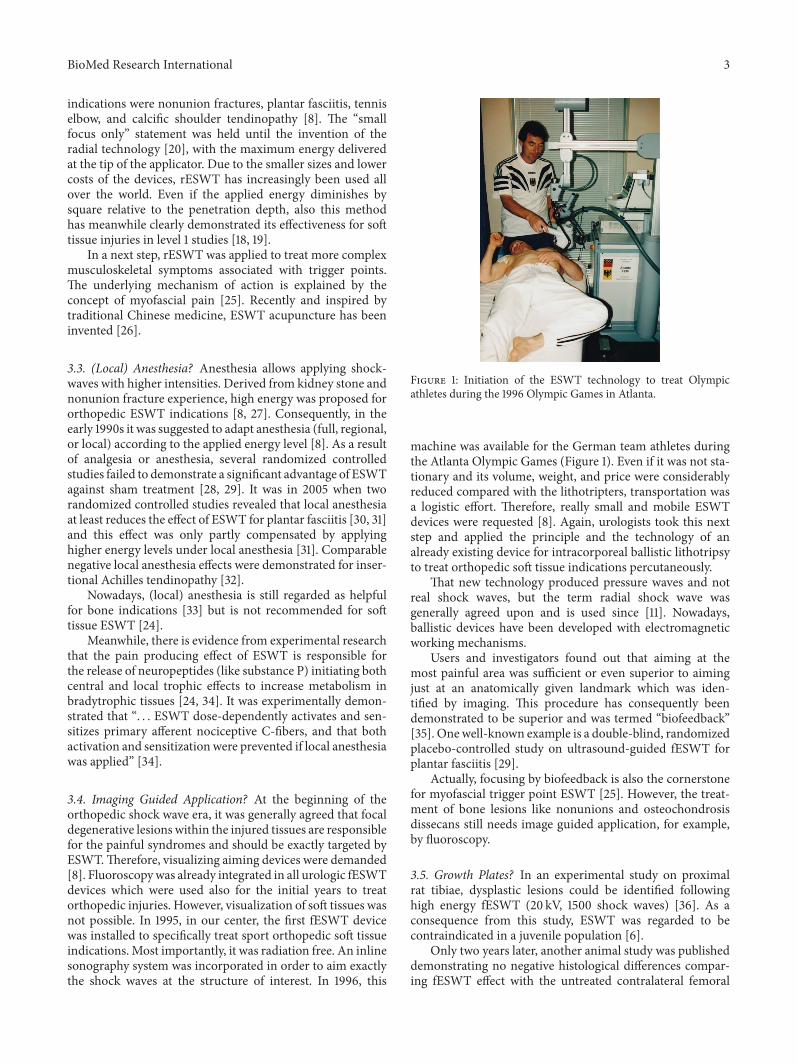
3.4. Imaging Guided Application? At the beginning of theorthopedic shock wave era, it was generally agreed that focaldegenerative lesions within the injured tissues are responsiblefor the painful syndromes and should be exactly targeted byESWT.Therefore, visualizing aiming devices were demanded[8]. Fluoroscopywas already integrated in all urologic fESWTdevices which were used also for the initial years to treatorthopedic injuries. However, visualization of soft tissues wasnot possible. In 1995, in our center, the first fESWT devicewas installed to specifically treat sport orthopedic soft tissueindications. Most importantly, it was radiation free. An inlinesonography system was incorporated in order to aim exactlythe shock waves at the structure of interest. In 1996, this
Figure 1: Initiation of the ESWT technology to treat Olympicathletes during the 1996 Olympic Games in Atlanta.
machine was available for the German team athletes duringthe Atlanta Olympic Games (Figure 1). Even if it was not sta-tionary and its volume, weight, and price were considerablyreduced compared with the lithotripters, transportation wasa logistic effort. Therefore, really small and mobile ESWTdevices were requested [8]. Again, urologists took this nextstep and applied the principle and the technology of analready existing device for intracorporeal ballistic lithotripsyto treat orthopedic soft tissue indications percutaneously.
That new technology produced pressure waves and notreal shock waves, but the term radial shock wave wasgenerally agreed upon and is used since [11]. Nowadays,ballistic devices have been developed with electromagneticworking mechanisms.
Users and investigators found out that aiming at themost painful area was sufficient or even superior to aimingjust at an anatomically given landmark which was iden-tified by imaging. This procedure has consequently beendemonstrated to be superior and was termed “biofeedback”[35]. Onewell-known example is a double-blind, randomizedplacebo-controlled study on ultrasound-guided fESWT forplantar fasciitis [29].
Actually, focusing by biofeedback is also the cornerstonefor myofascial trigger point ESWT [25]. However, the treat-ment of bone lesions like nonunions and osteochondrosisdissecans still needs image guided application, for example,by fluoroscopy.
3.5. Growth Plates? In an experimental study on proximalrat tibiae, dysplastic lesions could be identified followinghigh energy fESWT (20 kV, 1500 shock waves) [36]. As aconsequence from this study, ESWT was regarded to becontraindicated in a juvenile population [6].
Only two years later, another animal study was publisheddemonstrating no negative histological differences compar-ing fESWT effect with the untreated contralateral femoral
4 BioMed Research International
head of immature rabbits [37]. Another experimental rabbitstudy was published in German language. The investiga-tors applied 800 focused impulses (0.32mJ/mm2) which iscomparable to a high fESWT in a human bone application.Obviously, these two papers were underestimated in thescientific world [38]. For rESWT, a recently published ratexperiment could detect “no negative effects” when 1500 or3000 impulses of 4 bar were applied to the immature rat knees[39].
Even if initially mentioned anecdotally already in 1995[40] it was only recently when the first clinical case seriesreported both safety and effectiveness whenOsgood Schlatteror Sever’s diseases were treated by using rESWT [41, 42].
3.6. Acute Injuries? When introducing musculoskeletalESWT, it was declared to be indicated for chronic injuries.The reason for this was that in general a new treatmentmodality should provide evidence before being spread outto the public, and, as long as the evidence is missing, itshould be recommended only for patients, who already havebeen treated by other options. This means that three monthsof conservative treatment should have been performedwithout success before ESWT is indicated as an alternativeto operative treatment [6, 8]. Extensive technical, manpower,and time requirements have been advocated as rationales forthis limitation [8]. Additionally, economic factors limitedthe musculoskeletal ESWT application. Consequently,most research was traditionally made for conservativelypretreated injuries with a history of more than three months.International shock wave societies still consider onlynonacute pathologies (http://www.digest-ev.de/leitlinien/).With the advent of cheaper and more flexible ESWTdevices, this rule has been broken. For instance, in acuteand operatively treated long bone fractures high energyfESWT effectively reduced the number of nonunions [43].Contrary to this, in a randomized controlled study rESWTtreatment was inferior to stretching for plantar fasciitispatients when patients were not pretreated and complainedabout symptoms under six weeks [44].
If ESWT can be relevant to effectively treat acute muscu-lar or tendon strains is currently not known and respectiveresearch is needed.
3.7. Tendinopathies with Extraosseous Calcification. Histor-ically, only mechanical (and not biologic) ESWT effectswere regarded as relevant in medicine. At the transmissionthrough tissues with similar acoustic properties (soft tissue)a minor amount of energy is released. It was assumed thatthe resulting mechanical effect is negligible. In contrast, highacoustic impedance differences exist between cortical bone(6.12 × 10⁶ kg/m2s) and soft tissue (e.g., muscle = 1.66 ×10⁶ kg/m2s). ESWT consequently releases a large amount ofmechanical energy at the interface. This concept was therationale not only to treat kidney stones but also to treatsoft tissue calcifications [6]. Initially, a real destruction ofbone was not detected as a result from ESWT [6], butlater experimental research demonstrated a dose-dependentinduction of cortical fractures and periosteal detachment[45]. Relevant acoustic impedance differences exist also at the
interface between tendon and bone. Therefore, well-definedinsertional tendinopathies like tennis elbow and plantarfasciitis were thought to be also eligible for ESWT treatmentspecifically when combined with a spur [8].
These treatment principles were held until the inventionof the rESWTwith a completely different technology. Histor-ically, the main differences between fESWT and rESWT areas follows: (a) principle of generation = pneumatic rESWTversus electrohydraulic, piezoelectric, or electromagneticfESWT, (b) wavelength = 0.15 to 1.5m (rESWT) versus1.5mm (fESWT), and (c) maximum pressure = 1 (rESWT)versus 10–100 (fESWT)MPa and penetration depth = 2–5 cm(rESWT) versus 5–20 cm (fESWT) [9]. Nowadays, there arealso ballistic devices available with electromagnetic workingmechanisms accelerating the projectile to hit the applicator.Clinically most important thing is that the maximum energyin rESWT is delivered at the interface between the applicatorhead of the device and the skin and diminishes its energyinside the treated tissue by the square of the penetration depth[9].
As a result, rESWT was applied to tendon lesions, fea-tured by their immediately subcutaneous localization and bya large area of injured tissue. Midportion Achilles tendinopa-thy and patellar tendinopathy fulfil these criteria and havebeen demonstrated to be an indication for rESWT [20, 46,47]. Based on current evidence, we are unable to preferfESWT or rESWT for musculoskeletal soft tissue injuries[11, 23]. Conflicting evidence exists from the results of twostudies that directly compared fESWT to rESWT in plantarfasciitis and patellar tendinopathy patients [19, 23]. FESWTrevealed moderately superior results compared to rESWTin plantar fasciitis, while no difference was demonstratedbetween the two applications regarding patellar tendinopathy[19, 23].
3.8. Three Sessions Only? The number of required treatmentsessions is a relevant parameter in principle. Recently, system-atic research recommends “three treatment sessions at 1-weekintervals, with 2000 impulses per session and the highestenergy flux density the patient can tolerate” [24]. However,historical reports do not adequately address that detail [4,8]. Analogue to and derived from the lithotripsy nonunionfractures have been treated with high energy predominantlyin one session.The reason for this procedure ismost probablybased upon the intensive effort required by anesthesia andfluoroscopy. For the soft tissue conditions a wide range (1 to10) of treatment sessions was initially instructed [4, 6]. Theneed of standardization of treatment regimens in randomizedcontrolled trials established one to three ESWT sessions atweekly intervals as a standard clinical practice regardless ofthe underlying pathology [12, 17, 18, 23, 46, 48–53].
Recently, there have been a few reports which retrospec-tively addressed the number of rESWT sessions needed totreat soft tissue pathologies such as trigger digits, symp-tomatic calcified shoulder tendinopathy, and plantar fasciitis.These studies revealed that pretreatment symptom durationwas significantly correlated with the number of rESWTsessions applied [54]. Additionally, there is evidence thatthere is a dose-related ESWT effect with lower energy flux
BioMed Research International 5
densities [mJ/mm2] requiring more treatment sessions toobtain the same effect [55].
Discussion is still going on about which parameters orwhich combination of parameters should be used to maxi-mize the effect of ESWT treatment for a specific indication.In this context, it has to be mentioned that comparability ofstudies should not be reduced on one single parameter (e.g.,energy flux density).
In clinical practice, ESWT is rarely used as a monother-apy. Strategic loading and/or exercises are usually prescribedin addition to shock waves, a fact that in general RCTs havenot adequately addressed. An individualized interventionshould be considered depending initially on the type andcharacteristics of the pathology [56].
4. Discussion
Themost important finding of this review is that all historicalparadigms as set for musculoskeletal ESWT in 1995 did notwithstand the technical and clinical developments over thelast 20 years. The initial phase of the musculoskeletal ESWTwas driven by side effect research in context with lithotripsyinvestigation and the first orthopedic applications have beenperformed by urologists [4]. Principles which were alreadyknown from more than two million lithotripsies in men andfrom respective animal studies were transferred and adaptedto musculoskeletal indications.
At the beginning of the musculoskeletal shock wave ageit was thought that the higher the energy is, the betterthe outcome would be. For soft tissue pathologies it wasearly realized that lower ESWT intensities are able to inducetissue regeneration instead of necrotic reactions [5]. Thepain resulting from the ESWT is clearly depending on theenergy intensity [34] but clinical focusing was shown toimprove the treatment results especially when performedwithout local anesthesia [30, 34]. Specific ESWT devices formusculoskeletal conditions were produced. Further reduc-tion of the applied energy was delivered with the rESWTtechnology. So and over the years, devices becamemuchmoreflexible/mobile and had reduced volume, weight, and costs.
There are an increasing number of high quality ESWTstudies for musculoskeletal conditions published in the liter-ature. It can be summarized without exaggeration that ESWTis the best analyzed treatment modality in the orthopedicfield. This statement includes also operative interventions. Arecent systematic musculoskeletal ESWT review concludesthat there is more need for high level studies [12]. But thequestion to be answered in future is not if ESWT worksbut rather which treatment protocol and parameters are thebest for specific and well described conditions [47]. Researchfinally has to follow clinical practice, where treatment proto-cols are individualized.
Until now, clinical ESWT research is aiming exclu-sively at detecting the success of ESWT applied followinga standardized protocol. The question, however, if ESWT issimilarly effective in each stage of a given musculoskeletalindication is completely unanswered up to date. For instance,a “tendon pathology continuum model” has been described[56]. Derived from this, tendinopathy is “no longer a ‘one size
fits all’ diagnosis” [57]. It is to expect that different stages of agiven pathology will respond differently to ESWT. Moreover,monotherapies are rarely used in clinical practice. Given theformer, future randomized controlled work should focus onassessing and comparing more realistic treatment protocols.
5. Conclusion
With the exception of bone related conditions, modernmusculoskeletal ESWT is performed with energy below0.28mJ/mm2 and without anesthesia. The size of the tissuearea to be treated can be small or large. “Biofeedback”is superior to imaging guided focusing. ESWT applicationin apophyseal osteochondral lesions in patients with opengrowth plates seems to be promising and safe. ESWT pro-tocols should be adapted to the stage and chronicity of thetreated pathology.
Competing Interests
Heinz Lohrer received fees for lecturing from Storz MedicalAG, Tagerwilen, CH. Employment of Tanja Nauck waspartially paid by Storz Medical AG, Tagerwilen, CH.
Acknowledgments
The authors are grateful to Ms. Grainne Mc Ginley forher valuable help in language editing of the paper as anative English speaker. The authors are grateful to StorzMedical, Lohstampfestrasse 8, 8274 Tagerwilen, Switzerland,for funding the open access publication article processingcharge.
References
[1] P. Novak, “Physics: F-SW and R-SW. Basic information onfocused and radial shock wave physics,” in MultidisciplinaryMedical Applications, H. Lohrer and L. Gerdesmeyer, Eds.,pp. 28–49, Level 10 Buchverlag Daniela Bamberg, Heilbronn,Germany, 1st edition, 2015.
[2] C. Chaussy, W. Brendel, and E. Schmiedt, “Extracorporeallyinduced destruction of kidney stones by shock waves,” TheLancet, vol. 316, no. 8207, pp. 1265–1268, 1980.
[3] J. Graff, K.-D. Richter, and J. Pastor, “Effect of high energy shockwaves on bony tissue,” Urological Research, vol. 16, pp. 252–258,1988.
[4] G. Haupt, “Use of extracorporeal shock waves in the treatmentof pseudarthrosis, tendinopathy and other orthopedic diseases,”The Journal of Urology, vol. 158, no. 1, pp. 4–11, 1997.
[5] J. D. Rompe, C. J. Kirkpatrick, K. Kullmer,M. Schwitalle, andO.Krischek, “Dose-related effects of shock waves on rabbit tendoAchillis. A sonographic and histological study,” The Journal ofBone & Joint Surgery—British Volume, vol. 80, no. 3, pp. 546–552, 1998.
[6] G. P. Dahmen, R. Franke, V. Gonchars et al., “Die Behand-lung knochennaher Weichteilschmerzen mit ExtrakorporalerStoßwellentherapie (ESWT). Indikation. Technik und bisherigeTherapie,” in Die Stoßwelle—Forschung und Klinik, C. Chaussy,F. Eisenberger, D. Jochum, and D. Wilbert, Eds., pp. 175–186,Attempo, Tubingen, Germany, 1995.
6 BioMed Research International
[7] H. Lohrer, J. Scholl, and S. Arentz, “RSWT-schmerztherapie inder orthopadie und sportmedizin. Entwicklung und einsatz,” inExtrakorporale Stoßwellentherapie, L. Gerdesmeyer, Ed., pp. 75–88, Books on Demand, Norderstedt, Germany, 2004.
[8] G. Dahmen, G. Haupt, J. D. Rompe, M. Loew, J. Haist,and R. Schleberger, “Standortbestimmung der arbeitsgruppe‘orthopadische stoßwellenbehandlungen’,” in Die Stoßwelle—Forschung und Klinik, C. Chaussy, F. Eisenberger, D. Jocham,and D. Wilbert, Eds., pp. 137–142, Attempto Verlag, Tubingen,Germany, 2015.
[9] P. Novak, “Physics: F-SW and R-SW. Basic information onfocused and radial shock wave physics,” in MultidisciplinaryMedical Applications, H. Lohrer and L. Gerdesmeyer, Eds.,pp. 28–49, Level 10 Buchverlag Daniela Bamberg, Heilbronn,Germany, 2015.
[10] G. Haupt, R. Diesch, T. Straub et al., “Radiale stoßwell-entherapie beim fersensporn (Fasciitis plantaris),”DerNiederge-lassene Chirurg, vol. 6, no. 4, pp. 1–5, 2002.
[11] C. B. Foldager, C. Kearney, andM. Spector, “Clinical applicationof extracorporeal shock wave therapy in orthopedics: focusedversus unfocused shock waves,” Ultrasound in Medicine andBiology, vol. 38, no. 10, pp. 1673–1680, 2012.
[12] C. Speed, “A systematic review of shockwave therapies in softtissue conditions: focusing on the evidence,” British Journal ofSports Medicine, vol. 48, no. 21, pp. 1538–1542, 2014.
[13] M. Delius, F. Ueberle, and S. Gambihler, “Destruction ofgallstones and model stones by extracorporeal shock waves,”Ultrasound in Medicine and Biology, vol. 20, no. 3, pp. 251–258,1994.
[14] W. Folberth, H. Krause, and T. Reuner, “Stoßwellenmeßtechnikin der Lithotripsie: Historie und Ausblick,” in Die Stoßwelle—Forschung und Klinik, C. Chaussy, F. Eisenberger, D. Jocham,andD.Wilbert, Eds., pp. 45–50, Attempto, Tubingen, Germany,1995.
[15] G. Haupt andM. Chvapil, “Effect of shock waves on the healingof partial-thickness wounds in piglets,” Journal of SurgicalResearch, vol. 49, no. 1, pp. 45–48, 1990.
[16] L. Gerdesmeyer, M. Henne, M. Gobel, and P. Diehl, “Physicalprinciples and generation of shockwaves,” in ExtracorporealShockwaveTherapy, L. Gerdesmeyer and L. S. Weil, Eds., pp. 11–20, Data Trace Publishing Company, Towson, Md, USA, 2007.
[17] H. Gollwitzer, A. Saxena, L. A. DiDomenico et al., “Clinicallyrelevant effectiveness of focused extracorporeal shock wavetherapy in the treatment of chronic plantar fasciitis: a random-ized, controlledmulticenter study,”The Journal of Bone and JointSurgery—American Volume, vol. 97, no. 9, pp. 701–708, 2015.
[18] L. Gerdesmeyer, C. Frey, J. Vester et al., “Radial extracorporealshock wave therapy is safe and effective in the treatment ofchronic recalcitrant plantar fasciitis: results of a confirmatoryrandomized placebo-controlled multicenter study,” The Amer-ican Journal of Sports Medicine, vol. 36, no. 11, pp. 2100–2109,2008.
[19] H. Lohrer, T. Nauck, N. V. Dorn-Lange, J. Scholl, and J. C.Vester, “Comparison of radial versus focused extracorporealshock waves in plantar fasciitis using functional measures,” Footand Ankle International, vol. 31, no. 1, pp. 1–9, 2010.
[20] H. Lohrer, J. Scholl, and S. Arentz, “Achilles tendinopa-thy and patellar tendinopathy. Results of radial shockwavetherapy in patients with unsuccessfully treated tendinoses,”Sportverletzung-Sportschaden, vol. 16, no. 3, pp. 108–114, 2002.
[21] J. D. Rompe, B. Nafe, J. P. Furia, and N. Maffulli, “Eccentricloading, shock-wave treatment, or a wait-and-see policy for
tendinopathy of the main body of tendo Achillis: a randomizedcontrolled trial,” The American Journal of Sports Medicine, vol.35, no. 3, pp. 374–383, 2007.
[22] K. H. E. Peers, R. J. J. Lysens, P. Brys, and J. Bellemans, “Cross-sectional outcome analysis of athletes with chronic patellartendinopathy treated surgically and by extracorporeal shockwave therapy,” Clinical Journal of Sport Medicine, vol. 13, no. 2,pp. 79–83, 2003.
[23] H. van der Worp, J. Zwerver, M. Hamstra, I. van den Akker-Scheek, and R. L. Diercks, “No difference in effectivenessbetween focused and radial shockwave therapy for treatingpatellar tendinopathy: a randomized controlled trial,” KneeSurgery, Sports Traumatology, Arthroscopy, vol. 22, no. 9, pp.2026–2032, 2014.
[24] C. Schmitz,N. B.M.Csaszar, S.Milz et al., “Efficacy and safety ofextracorporeal shock wave therapy for orthopedic conditions:a systematic review on studies listed in the PEDro database,”British Medical Bulletin, vol. 116, no. 1, pp. 115–138, 2015.
[25] M. Gleitz and K. Hornig, “Trigger points—diagnosis andtreatment concepts with special reference to extracorporealshockwaves,” Orthopade, vol. 41, no. 2, pp. 113–125, 2012.
[26] H. Everke, “10 years of experience of acupuncture with shockwaves,” Deutsche Zeitschrift fur Akupunktur, vol. 55, no. 3, pp.20–23, 2012.
[27] R. Schultheiss, “Stoßwellen-Technologie in Orthopadie undUnfallchirurgie,” in Die Stoßwelle. Forschung und Klinik, C.Chaussy, F. Eisenberger, D. Jocham, and D. Wilbert, Eds., pp.237–245, Attempto, Tubingen, Germany, 1995.
[28] M. Haake, M. Buch, C. Schoellner et al., “Extracorporealshock wave therapy for plantar fasciitis: randomised controlledmulticentre trial,” British Medical Journal, vol. 327, no. 7406, pp.75–77, 2003.
[29] R. Buchbinder, R. Ptasznik, J. Gordon, J. Buchanan, V. Prabaha-ran, and A. Forbes, “Ultrasound-guided extracorporeal shockwave therapy for plantar fasciitis: a randomized controlled trial,”The Journal of the AmericanMedical Association, vol. 288, no. 11,pp. 1364–1372, 2002.
[30] J. D. Rompe, A. Meurer, B. Nafe, A. Hofmann, and L.Gerdesmeyer, “Repetitive low-energy shock wave applicationwithout local anesthesia is more efficient than repetitive low-energy shock wave application with local anesthesia in thetreatment of chronic plantar fasciitis,” Journal of OrthopaedicResearch, vol. 23, no. 4, pp. 931–941, 2005.
[31] G. Labek, V. Auersperg, M. Ziernhold, N. Poulios, and N.Bohler, “Influence of local anesthesia and energy level on theclinical outcome of extracorporeal shock wave-treatment ofchronic plantar fasciitis,” Zeitschrift fur Orthopadie und ihreGrenzgebiete, vol. 143, no. 2, pp. 240–246, 2005.
[32] J. P. Furia, “High-energy extracorporeal shock wave therapyas a treatment for insertional achilles tendinopathy,” AmericanJournal of Sports Medicine, vol. 34, no. 5, pp. 733–740, 2006.
[33] W. Schaden, R. Mittermayr, N. Haffner, D. Smolen, L.Gerdesmeyer, andC.-J.Wang, “Extracorporeal shockwave ther-apy (ESWT)—first choice treatment of fracture non-unions?”International Journal of Surgery, vol. 24, pp. 179–183, 2015.
[34] T. Klonschinski, S. J. Ament, T. Schlereth, J. D. Rompe, andF. Birklein, “Application of local anesthesia inhibits effects oflow-energy Extracorporeal Shock Wave Treatment (ESWT) onnociceptors,” Pain Medicine, vol. 12, no. 10, pp. 1532–1537, 2011.
[35] A. Sems, R. Dimeff, and J. P. Iannotti, “Extracorporeal shockwave therapy in the treatment of chronic tendinopathies,”
BioMed Research International 7
Journal of the American Academy of Orthopaedic Surgeons, vol.14, no. 4, pp. 195–204, 2006.
[36] L. D. Yeaman, C. P. Jerome, and D. L. McCullough, “Effects ofshock waves on the structure and growth of the immature ratepiphysis,” The Journal of Urology, vol. 141, no. 3, pp. 670–674,1989.
[37] K. N. Van Arsdalen, S. Kurzweil, J. Smith, and R. M. Levin,“Effect of lithotripsy on immature rabbit bone and kidneydevelopment,” The Journal of Urology, vol. 146, no. 1, pp. 213–216, 1991.
[38] K. Nassenstein, I. Nassenstein, and R. Schleberger, “Effectsof high-energy shock waves on the structure of the imma-ture epiphysis—a histomorphological study,” Zeitschrift furOrthopadie und ihre Grenzgebiete, vol. 143, no. 6, pp. 652–655,2005.
[39] Z.Oztemur,H.Ozturk, S. Ozyurek, C. Kaloglu, U.H.Golge, andO. Bulut, “The long-term effects of extracorporeal shock waveson the epiphysis of the adolescent rat,” Journal of OrthopaedicScience, vol. 18, no. 1, pp. 159–164, 2013.
[40] R. Schleberger, “Anwendung der extrakorporalen Stoßwelle amStutz-und Bewegungsapparat im mittelenergetischen Bereich,”in Die Stoßwelle. Forschung und Klinik, C. Chaussy, F. Eisen-berger, D. Jocham, and D. Wilbert, Eds., pp. 166–174, AttemptoVerlag, Tubingen, Germany, 1995.
[41] H. Lohrer, T.Nauck, J. Scholl, J. Zwerver, andN.Malliaropoulos,“Extracorporeal shock wave therapy for patients sufferingfrom recalcitrant osgood-schlatter disease,” Sportverletzung-Sportschaden, vol. 26, no. 4, pp. 218–222, 2012.
[42] H. Lohrer and T. Nauck, “Radial shock wave therapy forpatients with apophysitis calcanei,” Deutsche Zeitschrift furSportmedizin, vol. 66, no. 3, pp. 60–63, 2015.
[43] C.-J. Wang, H.-C. Liu, and T.-H. Fu, “The effects of extracorpo-real shockwave on acute high-energy long bone fractures of thelower extremity,” Archives of Orthopaedic and Trauma Surgery,vol. 127, no. 2, pp. 137–142, 2007.
[44] J. D. Rompe, A. Cacchio, L.Weil Jr. et al., “Plantar fascia-specificstretching versus radial shock-wave therapy as initial treatmentof plantar fasciopathy,” The Journal of Bone & Joint Surgery—American Volume, vol. 92, no. 15, pp. 2514–2522, 2010.
[45] M. Maier, J. Hausdorf, T. Tischer et al., “New bone formationby extracorporeal shock waves. Dependence of induction onenergy flux density,” Orthopade, vol. 33, no. 12, pp. 1401–1409,2004.
[46] J. D. Rompe, J. Furia, and N. Maffulli, “Eccentric loading versuseccentric loading plus shock-wave treatment for midportionachilles tendinopathy: a randomized controlled trial,”AmericanJournal of Sports Medicine, vol. 37, no. 3, pp. 463–470, 2009.
[47] S. Mani-Babu, D. Morrissey, C. Waugh, H. Screen, and C.Barton, “The effectiveness of extracorporeal shockwave therapyin lower limb tendinopathy: a systematic review,”TheAmericanJournal of Sports Medicine, vol. 43, no. 3, pp. 752–761, 2015.
[48] J. P. Furia, J. D. Rompe, A. Cacchio, A. Del Buono, and N.Maffulli, “A single application of low-energy radial extracor-poreal shock wave therapy is effective for the management ofchronic patellar tendinopathy,” Knee Surgery, Sports Traumatol-ogy, Arthroscopy, vol. 21, no. 2, pp. 346–350, 2013.
[49] J. D. Rompe, J. Furia, and N. Maffulli, “Eccentric loadingcompared with shock wave treatment for chronic insertionalachilles tendinopathy. A randomized, controlled trial,” TheJournal of Bone & Joint Surgery—American Volume, vol. 90, no.1, pp. 52–61, 2008.
[50] J. D. Rompe, A. Cacchio, J. P. Furia, and N. Maffulli, “Low-energy extracorporeal shock wave therapy as a treatment formedial tibial stress syndrome,” American Journal of SportsMedicine, vol. 38, no. 1, pp. 125–132, 2010.
[51] A. Notarnicola, V. Pesce, G. Vicenti, S. Tafuri, M. Forcignano,and B. Moretti, “SWAAT study: extracorporeal shock wavetherapy and arginine supplementation and other nutraceuticalsfor insertional achilles tendinopathy,” Advances inTherapy, vol.29, no. 9, pp. 799–814, 2012.
[52] C.-J. Wang, J.-Y. Ko, Y.-S. Chan, L.-H. Weng, and S.-L. Hsu,“Extracorporeal shockwave for chronic patellar tendinopathy,”American Journal of Sports Medicine, vol. 35, no. 6, pp. 972–978,2007.
[53] J. Zwerver, F. Dekker, and G.-J. Pepping, “Patient guided Piezo-electric extracorporeal shockwave therapy as treatment forchronic severe patellar tendinopathy: a pilot study,” Journal ofBack and Musculoskeletal Rehabilitation, vol. 23, no. 3, pp. 111–115, 2010.
[54] N.Malliaropoulos, G. Crate, M.Meke, T. Nauck, H. Lohrer, andN. Padhiar, “The recurrence rate of plantar fasciitis after RadialExtracorporeal Shock Wave Therapy (rESWT) and the successrate of the treatment: a retrospective study,” BioMed ResearchInternational, In press.
[55] S.-J. Lee, J.-H. Kang, J.-Y. Kim, J.-H. Kim, S.-R. Yoon, and K.-I.Jung, “Dose-related effect of extracorporeal shock wave therapyfor plantar fasciitis,” Annals of Rehabilitation Medicine, vol. 37,no. 3, pp. 379–388, 2013.
[56] J. L. Cook and C. R. Purdam, “Is tendon pathology a contin-uum? A pathology model to explain the clinical presentation ofload-induced tendinopathy,” British Journal of Sports Medicine,vol. 43, no. 6, pp. 409–416, 2009.
[57] J. Cook, “Tendinopathy: no longer a ‘one size fits all’ diagnosis,”British Journal of Sports Medicine, vol. 45, no. 5, article 385, 2011.

Submit your manuscripts athttp://www.hindawi.com
Stem CellsInternational
Hindawi Publishing Corporationhttp://www.hindawi.com Volume 2014
Hindawi Publishing Corporationhttp://www.hindawi.com Volume 2014
MEDIATORSINFLAMMATION
of
Hindawi Publishing Corporationhttp://www.hindawi.com Volume 2014
Behavioural Neurology
EndocrinologyInternational Journal of
Hindawi Publishing Corporationhttp://www.hindawi.com Volume 2014
Hindawi Publishing Corporationhttp://www.hindawi.com Volume 2014
Disease Markers
Hindawi Publishing Corporationhttp://www.hindawi.com Volume 2014
BioMed Research International
OncologyJournal of
Hindawi Publishing Corporationhttp://www.hindawi.com Volume 2014
Hindawi Publishing Corporationhttp://www.hindawi.com Volume 2014
Oxidative Medicine and Cellular Longevity
Hindawi Publishing Corporationhttp://www.hindawi.com Volume 2014
PPAR Research
The Scientific World JournalHindawi Publishing Corporation http://www.hindawi.com Volume 2014
Immunology ResearchHindawi Publishing Corporationhttp://www.hindawi.com Volume 2014
Journal of
ObesityJournal of
Hindawi Publishing Corporationhttp://www.hindawi.com Volume 2014
Hindawi Publishing Corporationhttp://www.hindawi.com Volume 2014
Computational and Mathematical Methods in Medicine
OphthalmologyJournal of
Hindawi Publishing Corporationhttp://www.hindawi.com Volume 2014
Diabetes ResearchJournal of
Hindawi Publishing Corporationhttp://www.hindawi.com Volume 2014
Hindawi Publishing Corporationhttp://www.hindawi.com Volume 2014
Research and TreatmentAIDS
Hindawi Publishing Corporationhttp://www.hindawi.com Volume 2014
Gastroenterology Research and Practice
Hindawi Publishing Corporationhttp://www.hindawi.com Volume 2014
Parkinson’s Disease
Evidence-Based Complementary and Alternative Medicine
Volume 2014Hindawi Publishing Corporationhttp://www.hindawi.com

![Comparative analysis of angiogenic gene expression in normal … · 2011-12-22 · bums after ESWT treatment postoperatively [22-24]. In the clinical experience, ESWT has been shown](https://img.pdfslide.net/doc/110x75/5f3290f8e41aca35325ae265/comparative-analysis-of-angiogenic-gene-expression-in-normal-2011-12-22-bums-after.jpg)

![Research Paper Shockwave Targeting on Subchondral Bone Is … · 2018-12-28 · shockwave therapy (ESWT) on OA knee is gradually being noticed [17]. ESWT has shown success in the](https://img.pdfslide.net/doc/110x75/5f3290f9e41aca35325ae26c/research-paper-shockwave-targeting-on-subchondral-bone-is-2018-12-28-shockwave.jpg)

![Clinical Use of Extracorporeal Shockwave Therapy (ESWT ... · (ESWT) may induce focal growth plate dysplasia [10]. The use of ESWT over the growth plate in 3 experimen- tal ponies](https://img.pdfslide.net/doc/110x75/5e6d0de66f07cc416f60c348/clinical-use-of-extracorporeal-shockwave-therapy-eswt-eswt-may-induce-focal.jpg)