Embed Size (px)
Citation preview

Review ArticleManagement of Atrial Fibrillation in Critically Ill Patients
Mattia Arrigo, Dominique Bettex, and Alain Rudiger
Cardiosurgical Intensive Care Unit, Institute of Anesthesiology, University Hospital Zurich, Raemistraße 100,8091 Zurich, Switzerland
Correspondence should be addressed to Alain Rudiger; [email protected]
Received 29 September 2013; Revised 24 December 2013; Accepted 24 December 2013; Published 16 January 2014
Academic Editor: Marcus J. Schultz
Copyright © 2014 Mattia Arrigo et al. This is an open access article distributed under the Creative Commons Attribution License,which permits unrestricted use, distribution, and reproduction in any medium, provided the original work is properly cited.
Atrial fibrillation (AF) is common in ICU patients and is associated with a two- to fivefold increase inmortality.This paper providesa reappraisal of the management of AF with a special focus on critically ill patients with haemodynamic instability. AF can causehypotension and heart failure with subsequent organ dysfunction. The underlying mechanisms are the loss of atrial contractionand the high ventricular rate. In unstable patients, sinus rhythm must be rapidly restored by synchronised electrical cardioversion(ECV). If pharmacological treatment is indicated, clinicians can choose between the rate control and the rhythm control strategy.The optimal substance should be selected depending on its potential adverse effects. A beta-1 antagonist with a very short half-life(e.g., esmolol) is an advantage for ICU patients because the effect of beta-blockade on cardiovascular stability is unpredictable inthose patients. Amiodarone is commonly used in the ICU setting but has potentially severe cardiac and noncardiac side effects.Digoxin controls the ventricular response at rest, but its benefit decreases in the presence of adrenergic stress. Vernakalant convertsnew-onset AF to sinus rhythm in approximately 50% of patients, but data on its efficacy and safety in critically ill patients arelacking.
1. Introduction
Atrial fibrillation (AF) is the most common arrhythmia inpatients hospitalised in intensive care units (ICUs) and isassociated with increased morbidity and mortality [1–6].In light of the improved understanding of the underlyingpathophysiology, novel therapeutic options, and recentlypublished guidelines for AF, this paper provides a reappraisalof the topic with a special focus on the management of AF incritically ill patients with haemodynamic instability.
2. Materials and Methods
A search of the PubMed database and a review of bibli-ographies from selected articles was performed to identifyoriginal data relating to this topic. Articles were scruti-nised regarding their study design, population evaluated,interventions, outcomes, and limitations. A special focuswas on the literature available from critically ill patients.However, if such information was lacking, references fromnon-ICU patients were included in this narrative review.When evidence-based recommendations were not availableat all personal recommendations were incorporated in this
report (and highlighted accordingly) to assist the cliniciansin the management of critically ill patients with AF.
3. Results and Discussion
3.1. Definition and Clinical Manifestation. AF is a supraven-tricular arrhythmia characterized by disorganized atrialdepolarisations without effective atrial contractions. If AFterminates spontaneously, it is defined as paroxysmal. WhenAF is sustained beyond seven days or is terminated withelectrical or pharmacological cardioversion it is defined aspersistent. If a conversion in sinus rhythm cannot be achieved,AF is defined as permanent [5].
In critically ill patients, untreated AF can cause hypoten-sion (mean arterial pressure < 65mmHg), myocardialischemia, and heart failure (pulmonary edema, cardiogenicshock) with subsequent tissue hypoxia (SvO2 < 65%, lactate> 2.0mmol/l) and organ dysfunction (encephalopathy, acutekidney injury with urine output < 0.5mL/kg/h and liverdysfunction).The underlyingmechanisms of these complica-tions are the loss of atrial contraction and the high ventricularrate, which both impair the ventricular filling. The loss of
Hindawi Publishing CorporationCritical Care Research and PracticeVolume 2014, Article ID 840615, 10 pageshttp://dx.doi.org/10.1155/2014/840615
2 Critical Care Research and Practice
the atrial kick is particularly detrimental in patients withdiastolic dysfunction, such as left ventricular hypertrophy ofany cause. Left atrial pressure increases, causing pulmonaryvenous hypertension and subsequent pulmonary edemawith dyspnea. When stroke volume deteriorates, cardiogenicshock develops [7]. In addition, the high heart rate and thesecondary elevation of end-diastolic ventricular pressureincrease the myocardial oxygen demands, precipitatingacute myocardial ischemia. Uncontrolled tachycardia for theduration of days to weeks may cause tachycardia-inducedmyocardial dysfunction (tachycardiomyopathy) leading tosevere systolic heart failure, which is potentially reversibleafter appropriate treatment [8, 9].
3.2. Diagnostic Evaluation. AF is diagnosed by a 12-leadelectrocardiogram (ECG), typically when a lack of 𝑃-waves,high-frequency fibrillation waves at rates of 350–600/min,and an irregular ventricular response (absolute arrhythmia)are observed. The ventricular rate in untreated patientswith normal atrioventricular conduction is typically between100 and 160 bpm, but normo- and bradycardic ventricularresponse rates are possible. The initial ECG may provideimportant additional information on myocardial ischemia,left-ventricular hypertrophy, or conduction disorders. Whenthe differentiation of narrow-complex tachycardia is chal-lenging, 6mg of adenosine pushed intravenously can termi-nate a reentry tachycardia or unmask atrial flutter and AF[10]. Of note, adenosine can precipitate ventricular tachycar-dia in preexcitation syndromes (e.g. Wolff-Parkinson-White)by rapid anterograde conduction of AF via the accessorypathway [11]. After cardiac surgery, an atrial lead ECGfrom the pacemaker wire can be helpful. The evaluation ofsymptoms and haemodynamic consequences is the next step[1]. If AF is accompanied by acute chest pain, dyspnea, arterialhypotension, and/or cardiogenic shock, immediate action isrequired (see below). Transthoracic echocardiography, chestradiography, and electrolyte and serologic tests for thyroidfunction are required to identify the underlying cause ofthe AF [12]. In cardiac surgery patients, transthoracic ortransesophageal echocardiography may be necessary to ruleout pericardial effusion, a common trigger for AF in the earlypostoperative phase.
3.3. Epidemiology. Advanced age is the biggest risk factorsfor developing AF. The incidence and prevalence rises withage (>60 years: 1%; >80 years: 5–15%) [13–18]. AF occurs inpatients with cardiac disorders (hypertensive heart disease,coronary artery disease, valvular heart disease, pericarditis,congenital heart disease and acquired cardiomyopathies) aswell as in patients with no apparent cardiac abnormalities(lone AF) [5]. Many noncardiac diseases (thyroid disorders,pulmonary diseases, and alcohol overconsumption) are alsoassociated with AF [19, 20]. Acute illness and surgery areassociated with increased rates of AF. The incidence of new-onset AF in critically ill patients is 6–20% [21–23]. In thesubgroup of patients with sepsis, the incidence of new-onsetAF correlates with the severity of sepsis; up to half of thepatients with septic shock experience new-onset AF [24].
In cases with acute coronary artery disease, AF occurs in 6–21% of patients [25]. The highest incidence is observed inpatients after open heart surgery, in particular mitral valvesurgery and coronary artery bypass graft surgery, with doc-umented rates reaching 30–40% [26, 27]. The peak incidenceof AF occurs during the first 2–4 days after cardiac surgery[26]. Overall, AF is associated with cardioembolic events andheart failure, longer hospital stays, and reduced quality of lifeaswell as a two- to fivefold increasedmortality [21–23, 26, 28].
3.4. Underlying Mechanisms. The complex pathophysiologi-cal mechanisms of AF have been reviewed extensively [1–6,29]. To better understand the various treatment options, somebasic elements of AF are summarised below. Reentry of exci-tation wavefronts has long been considered the main mech-anism of AF. However, intensive research during the recentdecades has revealed an interaction between the initiationtriggers and maintenance factors of AF. Table 1 summarisesthe promoters of AF and what specific treatments, if any, theyare amenable to [5, 26, 29–32]. Clearly, these promoters of AFare different in critically ill patients compared to outpatients.Any heart disease or cardiac surgery involving sutures on theatria can induce structural remodelling of the atria, whichresults in inflammation, myocyte alteration, and tissue fibro-sis, all of which promote AF. A few minutes after the onset ofAF, an electrical remodelling process involving ion channelfunction and intracellular calcium homeostasis is stimulated,leading to a shortening of the refractory periods of atrialcardiomyocytes and contributing to the persistence of AF[7, 33]. Within days, alterations to the intracellular calciumhomeostasis cause contractile remodelling, dysfunction, andfurther dilatation of the atria [8, 9, 33]. Increased sympathetictone and systemic inflammation also play a central role inmaintaining AF [10, 34]. Inflammation may lead to atrialmyocarditis with subsequent electrical and structural atrialchanges, resulting in the initiation andmaintenance of AF [11,35]. BecauseAF triggersAF, paroxysmalAFmight progress topersistent and permanent AF [1, 36]. Finally, an origin of AFhas been localised in themyocardial sleeves of the pulmonaryveins, opening the door to new ablation techniques [12,37–41]. However, the complex mechanisms leading to thisectopic activity with bursts of rapid discharge are not yet fullyunderstood.
3.5. Management of Patients with Haemodynamic Instability.The initial management of patients with haemodynamicinstability includes the restoration of an adequate perfusionpressure with, depending upon the aetiology of AF, admin-istration of fluids, vasopressors, and/or inotropes. Specialattention should be addressed to sedation and analgesia,which ensure patient comfort and reduce the incidence ofharmful sympathetic activation, and a sufficient oxygensupply of the myocardium must be guaranteed.
3.5.1. Electrical Cardioversion (ECV). In patients with acutechest pain, dyspnea, or haemodynamic instability, the sinusrhythm must be rapidly restored by synchronised ECV(Figure 1). Compared to the success rate of 90% in outpatients
Critical Care Research and Practice 3
Table 1: Modifiable promoters of atrial fibrillation.
Mechanism Etiology Specific treatment options
Myocardial stretch (atrialhypertension, atrial dilatation, reducedcontractility)
Fluid overloadAcute mitral insufficiencyMitral stenosis
Fluid removal (restrictive fluid administration,diuretics, renal replacement therapy)Intra-aortic balloon pump; cardiac surgeryValvuloplasty
Inappropriate oxygen delivery to themyocardium
Myocardial ischemiaHypovolemiaAnemia
RevascularizationFluid challengeTransfusion of red blood cells
Electrolyte disturbances (risk factors:diuretics, dialysis)
HypokalemiaHypomagnesemia
Substitution of potassium (goal K+ 4.5–5.5mmol/L)Substitution of magnesium (goal Mg++ >1.0mmol/L)
Systemic and local inflammationHeart-lung machineSepsisMyocarditis
Steroids; off-pump cardiosurgical techniquesAntimicrobial therapyImmunosuppression
Adrenergic overstimulation Inotropic supportStress (pain, anxiety)
Reduction of inotropesSedation; analgesia; betablockers
Endocrine disorder Elevated thyroid hormonesPheochromocytoma
Betablockers; thyreostatic drugsAlpha- and betablockers
Various Hypothermia Correction of hypothermia
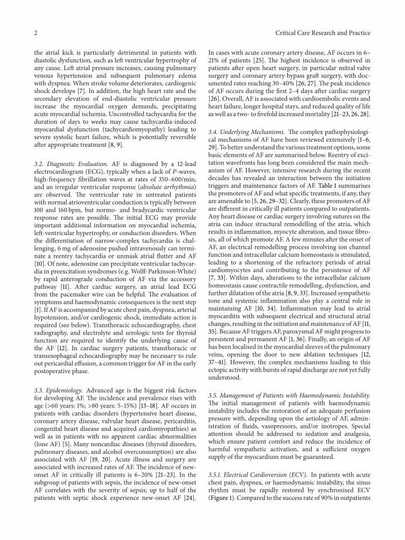
Newly diagnosed atrial fibrillation
Step 1:1st clinical assessment
Hemodynamic unstable?Severe symptoms? Yes
Yes
Yes
Yes
Yes
Yes
Urgentcardioversion
Step 2:Start rate control
Rapid conversion insinus rhythm?
sinus rhythm?
Optimization andfollowup
Optimization andfollowup
Optimization andfollowup
Optimization andfollowup
Step 3:2nd clinical assessment
Rate control achieved?Light symptoms?
Step 4:Evaluate strategy switch
AF >48h or uncertain?No anticoagulation?
Exclude atrialthrombi (TEE)
Step 5:Start rhythm control
Rapid conversion in
Step 6:3rd clinical assessment Adequate hemodynamics?
NoAdvanced ICUmanagement
Figure 1: Management algorithm. Legend: ICU intensive care unit. Algorithm modified from [2, 4].
4 Critical Care Research and Practice
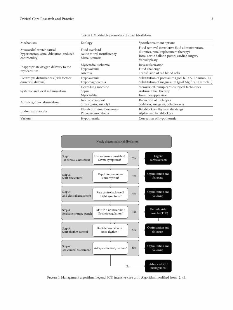
Table 2: Frequently used intravenous antiarrhythmic substances in the ICU.
Substance Dosing Half-life Commentary
Esmolol1.0mg/kg in boluses of 10–20mg iv, followed bycontinuous infusion (start with0.05mg/kg/min, increase dose every 30minutes if necessary)
7–10minGood efficacy in high adrenergic state. Positiveeffect on cardiovascular comorbidities. Considernegative inotropic effects
Diltiazem 0.25mg/kg iv over 2 minutes, followed bycontinuous infusion (10–15mg/h) if necessary 2–4 h Longer half-life as esmolol. No beta-blocking
effects. Consider negative inotropic effects
Amiodarone150–300mg iv, followed by a continuousinfusion (900–1200mg daily) up to 0.1 g/kgMaintenance dose 200mg daily
20–100 dGood efficacy, safe in patients with structuralheart disease. Extreme long half-life up to 80 days.Consider extracardiac side effects
Digoxin 0.25–0.5mg iv every 4–8 h up to 1mg, followedby maintenance dose of 0.25mg daily 20 h–6 d
Positive inotropic effect. Reduce dose in renaldysfunction. Check digoxin plasma levels to avoidtoxicity
[13–18, 42], the conversion rate is much lower in criticallyill patients undergoing urgent cardioversion, with publishedsuccess rates as low as 30% [5, 43–46]. Pretreatment withantiarrhythmic drugs facilitates ECV and reduces immediaterecurrences [19, 20, 47, 48]. Chest wall impedance, left atrialsize, and duration of AF are inversely related to successrate. Prior to ECV, patients should receive sedation andanalgesia. Endotracheal intubation is required in patients atrisk of aspiration. Anterior-posterior electrode positioningand biphasic waveforms provide higher success rates thanlateral electrode positioning and monophasic waveforms[21–23, 42]. In postoperative cardiosurgical patients, forwhom impedance is high and electrodes are often placedunfavourably due to wound dressing and chest tubes, werecommend a single shock of 200 Joules to increase thesuccess rate [24, 49]. A previous study demonstrated that ahigh initial energy reduces the incidence of tachyarrhythmiccomplications [25, 50]. Particular care must be taken topreserve the wound dressings and to avoid the nipples.If repeated ECV is applied, the synchronisation mode hasto be switched on before every use, as this mode usuallyswitches off after every discharge to allow immediate defib-rillation if necessary. In patients with pacemakers or internalcardioverter/defibrillators (ICD), internal overdrive pacingand/or cardioversion may be attempted by the cardiologiststo restore the sinus rhythm. If this is not possible, the externalelectrodes should be placed at least 8 cm from the aggregate.After cardioversion, the device should be checked to ensurenormal function.
In patients with life-threatening symptoms, ECV is indi-cated even if the presence of an atrial thrombus cannotbe excluded. In stable patients with AF lasting more than48 h, a transesophageal echocardiography to exclude an atrialthrombus is recommended [26, 27, 51]. Alternatively an ade-quate anticoagulation regimen of 3 weeks before cardiover-sion is recommended [6]. After successful cardioversion theanticoagulation should be continued for at least 4 weeks toprevent cardioembolic complications due to atrial stunning[6, 26]. If ECVwas not successful, pharmacological treatmentis indicated as described below. Similarly, an antiarrhythmictreatment is usually required temporarily to maintain sinusrhythm after successful ECV.
3.6. Management of Hemodynamic Stable Patients
3.6.1. Rate versus Rhythm Control. Clinicians can choosebetween a rate control and a rhythm control strategy. Therate control approach toleratesAFbut controls the ventricularresponse rate to improve the ventricular filling and avoida tachycardiomyopathy. It is the treatment of choice inpatients with permanent AF or in oligosymptomatic patients(Figure 1). Rate control can be accomplished with beta-blockers, calcium channel blockers (diltiazem, verapamil),digoxin, or amiodarone. 24 h telemetry should confirm thatthe target heart rate of less than 110 bpm at rest has beenachieved [21–23, 26, 28, 52]. Some patients may experienceclinical improvement only after the restoration of sinusrhythm (rhythm control strategy), which can be achieved byECV and/or drugs (see Figure 1). However, several trials inoutpatients failed to show a benefit of this strategy comparedto rate control only [53, 54] even in patients with congestiveheart failure [55]. The lack of a survival benefit in the rhythmcontrol arm was probably caused by the inefficacy of currentantiarrhythmic drugs and their adverse effects.
3.6.2. Pharmacological Options. A multitude of substancesare licensed for the pharmacologic treatment of AF, but onlya few are indicated in the ICU setting (Table 2). Because theliterature does not provide conclusive results on the optimalpharmacologic treatment of AF for ICU patients, cliniciansshould choose the optimal substance depending on its poten-tial adverse effects [56]. Before starting an antiarrhythmictreatment, clinicians should optimise all concurring factors[56–58]: electrolyte derangements (potassium, magnesium)should be corrected to upper-normal levels. Particularlymagnesium is an effective, cheap andwell-tolerated treatmentoption for AF [3, 59–63].
We recommend to start with substances with a low riskprofile and short half-life, such as betablockers (see below),and to escalate to other substance classes such as amiodaroneonly in cases of contraindications or inefficacy of the initialtreatment. Generally, intravenous substances are preferredbecause of their faster onset and more reliable action.
Critical Care Research and Practice 5
Selective beta-1 receptor antagonists have negative chron-otropic, dromotropic, and bathmotropic effects, slowingheart rate, delaying conduction in the atrioventricular node,and reducing myocardial excitability, respectively. Beta-blockers are therefore the initial treatment of choice for a ratecontrol strategy. Adverse effects include the negative inotropeactivity on themyocardium as well as vasodilatation [64] thatcan potentially worsen haemodynamics. Hence, a drug witha short half-life is recommended for ICU patients, for whomthe effect of beta-blockade on cardiovascular stability isunpredictable. Our choice is esmolol which is eliminated byunspecific esterases and hydrolases resulting in a very shorthalf-life of 7–10 minutes [65]. When esmolol treatment isinitiated, we typically repeat intravenous esmolol injectionsof 10–20mg to reach a dose of 1mg/kg within a few minutesto assess its haemodynamic effects. If the mean arterialpressure remains above 60mmHg, a continuous infusion isstarted at a rate of 0.05mg/kg/min and is further increased in30-minute intervals according to clinical needs. In patientswith oral beta-blockers, therapy should be continued as itsignificantly reduces the risk of AF up to 40%, particularlyin the postoperative phase [1, 60, 66–68].
Amiodarone is commonly used in the ICU setting forthe treatment of AF. First of all, it has less negative inotropiceffects compared to beta-blockers and calciumchannel block-ers [69]. Secondly, amiodarone is safer for patients withstructural heart disease compared to class Ic antiarrhythmicagents, such as flecainide [3]. Amiodarone is a multichannelblocker with inhibiting effects on adrenergic receptors andpotassium and calcium channels. It is a highly lipophilicsubstance with a very large distribution volume and anextremely long half-life [43, 45]. While a single dose of 150–300mg of amiodarone is enough to achieve pharmacologicalconversion to sinus rhythm in some patients, the majority ofpatients require long-term therapy. Therefore, a loading doseof 0.1 g/kg is required in the first 7–10 days, which can beadministered intravenously or orally. Thereafter, a daily oralmaintenance dose of 200mg is recommended. Importantly,amiodarone has potential severe adverse effects [70]. Prolon-gation of the QT interval is typical, but torsade de pointesare uncommon (<0.5%) [71]. Hypo- and hyperthyroidism arethe most common extracardiac side effects of amiodarone(>20%); thus, the thyrotropin (TSH) and free thyroid hor-mone (fT4, fT3) levels should be checked before treatmentand every six months thereafter. Photosensitivity, cornealdeposits, and neurological side effects are also frequent, whilepulmonary and hepatic toxicity are rare but potentially life-threatening adverse effects of amiodarone.
Digoxin inhibits the sodium-potassium pump, increasingthe calcium availability to the contractile apparatus [72].Digoxin controls the ventricular response through directaction on the atrioventricular node and by a centrallymediated vagal stimulation. Despite its efficacy in controllingresting heart rates, it is not a converter, and its benefitdecreases with adrenergic stress, limiting its efficacy in crit-ically ill patients. On the other hand, the positive inotropiceffect of digoxin may be beneficial for patients with heartfailure. The plasma half-life ranges from 20 to 50 hours inpatients with normal kidney function and increases up to
4–6 days in patients with end-stage renal disease [72]. Inaddition, drug interactions may reduce digoxin clearanceand electrolyte disturbances, such as hypokalemia, hypo-magnesemia, and hypercalcemia, and exacerbate digoxintoxicity. In critically ill patients, for whom a rapid controlof heart rate is desired, we administer 0.25mg digoxinintravenously every 4 to 8 hours up to a cumulative doseof 1.0 to 1.5mg, followed by a maintenance dose of 0.25mgonce daily. In patients with impaired kidney function, themaintenance dose must be reduced (0.125mg daily for acreatinine clearance of 60–90mL/min and 0.125 every otherday for a creatinine clearance of 30–60mL/min) [72]. Toavoid adverse events, regular surveillance of electrolytes andsigns of digitalis toxicity (see below) are recommended.Serum digoxin levels (measured at least 6 hours after the lastdose) may be helpful to corroborate the diagnosis of toxicitybut are not recommended for routine use [73]. Digoxin cancause a broad spectrum of ventricular and supraventriculararrhythmias, such as ectopic rhythms, pacemaker depression,or conduction abnormalities. Visual disturbances (blurredvision, flashing lights, halos, and green or yellow patterns),nausea, and vomiting are typical extracardiac manifestationsof digoxin toxicity. Dialysis is an ineffective treatment forintoxication, but the administration of digoxin immune Fabis highly effective in life-threatening digoxin poisoning [74].
Nondihydropyridine calcium channel blockers (e.g., dil-tiazem, verapamil) are an alternative treatment for patientswith contraindications to beta-blockers. Verapamil is morenegatively inotropic than diltiazem and must be used withcaution in patients with heart failure and after cardiac surgerybecause of the increased incidence of conduction disorders.The initial intravenous dose of diltiazem is 0.25mg/kg over2min. If the response is inadequate, a second dose of0.35mg/kg over 2min after 15min or a continuous infusionof 10–15mg/h is administered. The usual intravenous doseof verapamil is 2.5–5mg over 2min and may be followed by5–10mg after 15–30min.
Dronedarone is an oral multichannel blocker, whichcompared to amiodarone has a reduced lipophilicity and noiodine components. It showed promising efficacy in multipletrials [75, 76]; however, increased mortality in patients withheart failure and risks of severe hepatotoxicity are of concern[77–80]. Dronedarone has not been evaluated in critically illpatients and is not yet available for intravenous administra-tion, limiting its use in ICU settings.
The class Ic agents flecainide and propafenone are effi-cacious in restoring sinus rhythm but are associated withincreased mortality in patients with structural heart disease[81]. Therefore, they cannot be generally recommended inICU patients.
Vernakalant is a new antiarrhythmic agent that targetsatrial specific channels and has been approved for pharma-cological cardioversion of AF of ≤7 days duration [76, 83–85]. Vernakalant is given intravenously at an initial dose of3mg/kg over 10min. If conversion fails, a second dose of2mg/kg is given after 15min. Nausea, transient dysgeusia,and sneezing are common side effects. Vernakalant has alsobeen studied after cardiac surgery, showing a conversion rateof nearly 50%, with a low incidence of severe side effects
6 Critical Care Research and Practice
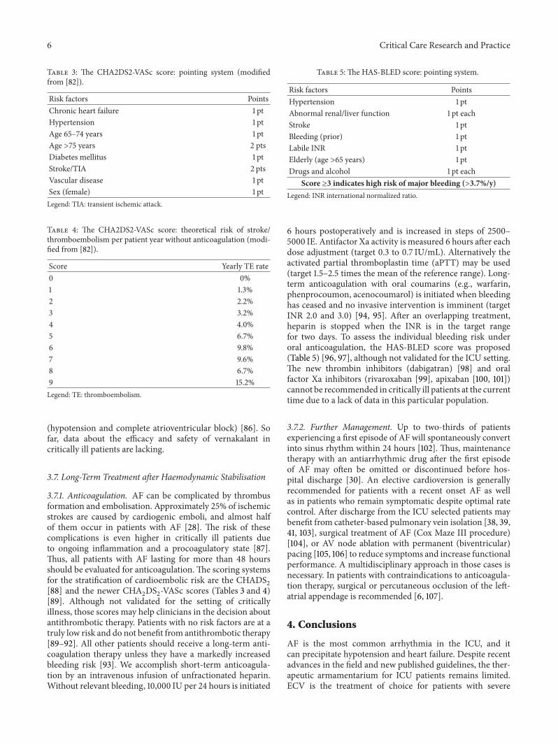
Table 3: The CHA2DS2-VASc score: pointing system (modifiedfrom [82]).
Risk factors PointsChronic heart failure 1 ptHypertension 1 ptAge 65–74 years 1 ptAge >75 years 2 ptsDiabetes mellitus 1 ptStroke/TIA 2 ptsVascular disease 1 ptSex (female) 1 ptLegend: TIA: transient ischemic attack.
Table 4: The CHA2DS2-VASc score: theoretical risk of stroke/thromboembolism per patient year without anticoagulation (modi-fied from [82]).
Score Yearly TE rate0 0%1 1.3%2 2.2%3 3.2%4 4.0%5 6.7%6 9.8%7 9.6%8 6.7%9 15.2%Legend: TE: thromboembolism.
(hypotension and complete atrioventricular block) [86]. Sofar, data about the efficacy and safety of vernakalant incritically ill patients are lacking.
3.7. Long-Term Treatment after Haemodynamic Stabilisation
3.7.1. Anticoagulation. AF can be complicated by thrombusformation and embolisation. Approximately 25% of ischemicstrokes are caused by cardiogenic emboli, and almost halfof them occur in patients with AF [28]. The risk of thesecomplications is even higher in critically ill patients dueto ongoing inflammation and a procoagulatory state [87].Thus, all patients with AF lasting for more than 48 hoursshould be evaluated for anticoagulation. The scoring systemsfor the stratification of cardioembolic risk are the CHADS
2
[88] and the newer CHA2DS2-VASc scores (Tables 3 and 4)
[89]. Although not validated for the setting of criticallyillness, those scores may help clinicians in the decision aboutantithrombotic therapy. Patients with no risk factors are at atruly low risk and do not benefit from antithrombotic therapy[89–92]. All other patients should receive a long-term anti-coagulation therapy unless they have a markedly increasedbleeding risk [93]. We accomplish short-term anticoagula-tion by an intravenous infusion of unfractionated heparin.Without relevant bleeding, 10,000 IU per 24 hours is initiated
Table 5: The HAS-BLED score: pointing system.
Risk factors PointsHypertension 1 ptAbnormal renal/liver function 1 pt eachStroke 1 ptBleeding (prior) 1 ptLabile INR 1 ptElderly (age >65 years) 1 ptDrugs and alcohol 1 pt each
Score ≥3 indicates high risk of major bleeding (>3.7%/y)Legend: INR international normalized ratio.
6 hours postoperatively and is increased in steps of 2500–5000 IE. Antifactor Xa activity is measured 6 hours after eachdose adjustment (target 0.3 to 0.7 IU/mL). Alternatively theactivated partial thromboplastin time (aPTT) may be used(target 1.5–2.5 times the mean of the reference range). Long-term anticoagulation with oral coumarins (e.g., warfarin,phenprocoumon, acenocoumarol) is initiated when bleedinghas ceased and no invasive intervention is imminent (targetINR 2.0 and 3.0) [94, 95]. After an overlapping treatment,heparin is stopped when the INR is in the target rangefor two days. To assess the individual bleeding risk underoral anticoagulation, the HAS-BLED score was proposed(Table 5) [96, 97], although not validated for the ICU setting.The new thrombin inhibitors (dabigatran) [98] and oralfactor Xa inhibitors (rivaroxaban [99], apixaban [100, 101])cannot be recommended in critically ill patients at the currenttime due to a lack of data in this particular population.
3.7.2. Further Management. Up to two-thirds of patientsexperiencing a first episode of AF will spontaneously convertinto sinus rhythm within 24 hours [102]. Thus, maintenancetherapy with an antiarrhythmic drug after the first episodeof AF may often be omitted or discontinued before hos-pital discharge [30]. An elective cardioversion is generallyrecommended for patients with a recent onset AF as wellas in patients who remain symptomatic despite optimal ratecontrol. After discharge from the ICU selected patients maybenefit from catheter-based pulmonary vein isolation [38, 39,41, 103], surgical treatment of AF (Cox Maze III procedure)[104], or AV node ablation with permanent (biventricular)pacing [105, 106] to reduce symptoms and increase functionalperformance. A multidisciplinary approach in those cases isnecessary. In patients with contraindications to anticoagula-tion therapy, surgical or percutaneous occlusion of the left-atrial appendage is recommended [6, 107].
4. Conclusions
AF is the most common arrhythmia in the ICU, and itcan precipitate hypotension and heart failure. Despite recentadvances in the field and new published guidelines, the ther-apeutic armamentarium for ICU patients remains limited.ECV is the treatment of choice for patients with severe

Critical Care Research and Practice 7
symptoms, but its efficacy is limited. Amiodarone, beta-blockers, calcium channel blockers, and digoxin are usedmost frequently, but their use is often complicated by adverseeffects. Newer drugs, such as dronedarone and vernakalant,have not been generally introduced into the ICU setting yetbecause they are not available intravenously, are contraindi-cated with structural heart disease, or are disadvised due tohaemodynamic instability. New substanceswith high efficacy,favourable haemodynamic effects, and a low risk profile areurgently needed.
Abbreviations
AF: Atrial fibrillationECV: Electrical cardioversionECG: ElectrocardiogramICD: Internal cardioverter/defibrillatorINR: International normalised ratioICU: Intensive care unitTSH: Thyroid-stimulating hormone (thyrotropin).
Conflict of Interests
Mattia Arrigo and Dominique Bettex declare that they haveno conflict of interests. Alain Rudiger received honorariafrom BAXTER and AOP ORPHAN PHARMACEUTICALS,both of which are distributing esmolol in Switzerland.
Authors’ Contribution
Mattia Arrigo, Dominique Bettex, and Alain Rudiger (1) haveall made substantial contributions to the interpretation ofthe available literature and to the conception design of thisreview paper; (2) have been involved in drafting the paperor revising it critically for important intellectual content;and (3) have given final approval of the version to bepublished. Dominique Bettex and Alain Rudiger are equallycontributing last authors.
References
[1] E. G. Daoud, “Management of atrial fibrillation in the post-cardiac surgery setting,” Cardiology Clinics, vol. 22, no. 1, pp.159–166, 2004.
[2] G. Y. Lip and H.-F. Tse, “Management of atrial fibrillation,”TheLancet, vol. 370, no. 9587, pp. 604–618, 2007.
[3] M. E. Sleeswijk, T. Van Noord, J. E. Tulleken, J. J. M. Ligtenberg,A. R. J. Girbes, and J. G. Zijlstra, “Clinical review: treatment ofnew-onset atrial fibrillation inmedical intensive care patients: aclinical framework,”Critical Care, vol. 11, no. 6, article 233, 2007.
[4] C. W. Khoo and G. Y. H. Lip, “Acute management of atrialfibrillation,” Chest, vol. 135, no. 3, pp. 849–859, 2009.
[5] EuropeanHeart RhythmAssociation, EuropeanAssociation forCardio-Thoracic Surgery, A. J. Camm et al., “Guidelines forthe management of atrial fibrillation: the Task Force for theManagement of Atrial Fibrillation of the European Society ofCardiology (ESC),” European Heart Journal, vol. 31, pp. 2369–2429, 2010.
[6] A. J. Camm, G. Y. H. Lip, R. de Caterina et al., “2012 focusedupdate of the ESC Guidelines for the management of atrialfibrillation: an update of the 2010 ESC Guidelines for themanagement of atrial fibrillation. Developed with the specialcontribution of the European Heart Rhythm Association,”European Heart Journal, vol. 33, pp. 2719–2747, 2012.
[7] A. Rudiger, V.-P. Harjola, A. Muller et al., “Acute heart failure:clinical presentation, one-year mortality and prognostic fac-tors,” European Journal of Heart Failure, vol. 7, no. 4, pp. 662–670, 2005.
[8] D. L. Packer, G. H. Bardy, and S. J. Worley, “Tachycardia-induced cardiomyopathy: a reversible form of left ventriculardysfunction,”The American Journal of Cardiology, vol. 57, no. 8,pp. 563–570, 1986.
[9] G. Fenelon, W. Wijns, E. Andries, and P. Brugada, “Tachycar-diomyopathy: mechanisms and clinical implications,” Pacingand Clinical Electrophysiology, vol. 19, no. 1, pp. 95–106, 1996.
[10] A. Rudiger and G. Niedermaier, “Unmasking atrial flutter,”Schweizerische Medizinische Wochenschrift, vol. 130, no. 31-32,Article ID 1125, 2000.
[11] A. J. Turley, S. Murray, and J. Thambyrajah, “Pre-excited atrialfibrillation triggered by intravenous adenosine: a commonlyused drug with potentially life-threatening adverse effects,”Emergency Medicine Journal, vol. 25, no. 1, pp. 46–48, 2008.
[12] R. L. Page, “Newly diagnosed atrial fibrillation,” The NewEngland Journal of Medicine, vol. 351, no. 23, pp. 2408–2416,2004.
[13] S. Stewart, C. L. Hart, D. J. Hole, and J. J. V. McMurray,“Population prevalence, incidence, and predictors of atrialfibrillation in the Renfrew/Paisley study,” Heart, vol. 86, no. 5,pp. 516–521, 2001.
[14] A. S. Go, E. M. Hylek, K. A. Phillips et al., “Prevalence ofdiagnosed atrial fibrillation in adults: national implications forrhythm management and stroke prevention: the anticoagula-tion and risk factors in atrial fibrillation (ATRIA) study,” Journalof the American Medical Association, vol. 285, no. 18, pp. 2370–2375, 2001.
[15] Y. Miyasaka, M. E. Barnes, B. J. Gersh et al., “Secular trends inincidence of atrial fibrillation in Olmsted County, Minnesota,1980 to 2000, and implications on the projections for futureprevalence,” Circulation, vol. 114, no. 2, pp. 119–125, 2006.
[16] J. Heeringa, D. A. M. van der Kuip, A. Hofman et al.,“Prevalence, incidence and lifetime risk of atrial fibrillation: theRotterdam study,” European Heart Journal, vol. 27, no. 8, pp.949–953, 2006.
[17] G. V. Naccarelli, H. Varker, J. Lin, and K. L. Schulman, “Increas-ing prevalence of atrial fibrillation and flutter in the UnitedStates,”The American Journal of Cardiology, vol. 104, no. 11, pp.1534–1539, 2009.
[18] D. M. Lloyd-Jones, T. J. Wang, E. P. Leip et al., “Lifetime risk fordevelopment of atrial fibrillation: the framingham heart study,”Circulation, vol. 110, no. 9, pp. 1042–1046, 2004.
[19] I. Klein and K. Ojamaa, “Thyroid hormone and the cardiovas-cular system,” The New England Journal of Medicine, vol. 344,no. 7, pp. 501–509, 2001.
[20] A. V. Samokhvalov, H. M. Irving, and J. Rehm, “Alcoholconsumption as a risk factor for atrial fibrillation: a systematicreview and meta-analysis,” European Journal of CardiovascularPrevention and Rehabilitation, vol. 17, no. 6, pp. 706–712, 2010.
[21] A. J. Walkey, R. S. Wiener, J. M. Ghobrial, L. H. Curtis, andE. J. Benjamin, “Incident stroke and mortality associated with
8 Critical Care Research and Practice
new-onset atrial fibrillation in patients hospitalized with severesepsis,” Journal of the American Medical Association, vol. 306,no. 20, pp. 2248–2255, 2011.
[22] S. Goodman, T. Shirov, and C. Weissman, “Supraventriculararrhythmias in intensive care unit patients: short and long-termconsequences,” Anesthesia and Analgesia, vol. 104, no. 4, pp.880–886, 2007.
[23] D. Annane, V. Sebille, D. Duboc et al., “Incidence and prognosisof sustained arrhythmias in critically III patients,”TheAmericanJournal of Respiratory and Critical Care Medicine, vol. 178, no. 1,pp. 20–25, 2008.
[24] R.Meierhenrich, E. Steinhilber, C. Eggermann et al., “Incidenceandprognostic impact of new-onset atrial fibrillation in patientswith septic shock: a prospective observational study,” CriticalCare, vol. 14, no. 3, article R108, 2010.
[25] J. Schmitt, G. Duray, B. J. Gersh, and S. H. Hohnloser, “Atrialfibrillation in acute myocardial infarction: a systematic reviewof the incidence, clinical features and prognostic implications,”European Heart Journal, vol. 30, no. 9, pp. 1038–1045, 2009.
[26] J. P. Mathew, M. L. Fontes, I. C. Tudor et al., “A multicenter riskindex for atrial fibrillation after cardiac surgery,” Journal of theAmerican Medical Association, vol. 291, no. 14, pp. 1720–1729,2004.
[27] J. Auer, T. Weber, R. Berent, C.-K. Ng, G. Lamm, and B. Eber,“Risk factors of postoperative atrial fibrillation after cardiacsurgery,” Journal of Cardiac Surgery, vol. 20, no. 5, pp. 425–431,2005.
[28] S. Stewart, C. L. Hart, D. J. Hole, and J. J. V. McMurray,“A population-based study of the long-term risks associatedwith atrial fibrillation: 20-year follow-up of the renfrew/paisleystudy,”TheAmerican Journal ofMedicine, vol. 113, no. 5, pp. 359–364, 2002.
[29] U. Schotten, S. Verheule, P. Kirchhof, and A. Goette, “Patho-physiological mechanisms of atrial fibrillation: a translationalappraisal,” Physiological Reviews, vol. 91, no. 1, pp. 265–325, 2011.
[30] M. K. Chung, “Cardiac surgery: postoperative arrhythmias,”Critical Care Medicine, vol. 28, no. 10, pp. N136–N144, 2000.
[31] C. W. Hague Jr., L. L. Creswell, D. D. Gutterman, and L.A. Fleisher, “Epidemiology, mechanisms, and risks: americanCollege of Chest Physicians guidelines for the prevention andmanagement of postoperative atrial fibrillation after cardiacsurgery,” Chest, vol. 128, no. 2, pp. 9S–S16, 2005.
[32] K. K. Christians, B. Wu, E. J. Quebbeman, and K. J. Brasel,“Postoperative atrial fibrillation in noncardiothoracic surgicalpatients,” The American Journal of Surgery, vol. 182, no. 6, pp.713–715, 2001.
[33] M. Allessie, J. Ausma, and U. Schotten, “Electrical, contractileand structural remodeling during atrial fibrillation,” Cardiovas-cular Research, vol. 54, no. 2, pp. 230–246, 2002.
[34] T. T. Issac, H. Dokainish, and N. M. Lakkis, “Role of inflam-mation in initiation and perpetuation of atrial fibrillation: asystematic reviewof the published data,” Journal of theAmericanCollege of Cardiology, vol. 50, no. 21, pp. 2021–2028, 2007.
[35] P. Korantzopoulos, T. Kolettis, K. Siogas, and J. Goudevenos,“Atrial fibrillation and electrical remodeling: the potential roleof inflammation and oxidative stress,”Medical Science Monitor,vol. 9, no. 9, pp. RA225–RA229, 2003.
[36] M. C. Wijffels, C. J. Kirchhof, R. Dorland, and M. A. Allessie,“Atrial fibrillation begets atrial fibrillation: a study in awakechronically instrumented goats,” Circulation, vol. 92, no. 7, pp.1954–1968, 1995.
[37] M. Haıssaguerre, P. Jaıs, D. C. Shah et al., “Spontaneousinitiation of atrial fibrillation by ectopic beats originating in thepulmonary veins,” The New England Journal of Medicine, vol.339, no. 10, pp. 659–666, 1998.
[38] H. Oral, C. Pappone, A. Chugh et al., “Circumferential pul-monaryvein ablation for chronic atrial fibrillation,” The NewEngland Journal of Medicine, vol. 354, no. 9, pp. 934–941, 2006.
[39] M. N. Khan, P. Jaıs, J. Cummings et al., “Pulmonary-vein iso-lation for atrial fibrillation in patients with heart failure,” TheNew England Journal of Medicine, vol. 359, no. 17, pp. 1778–1785,2008.
[40] O. M. Wazni, N. F. Marrouche, D. O. Martin et al., “Radiofre-quency ablation versus antiarrhythmic drugs as first-line treat-ment of symptomatic atrial fibrillation: a randomized trial,”Journal of the AmericanMedical Association, vol. 293, no. 21, pp.2634–2640, 2005.
[41] J. Cosedis Nielsen, A. Johannessen, P. Raatikainen et al.,“Radiofrequency ablation as initial therapy in paroxysmal atrialfibrillation,” The New England Journal of Medicine, vol. 367, pp.1587–1595, 2012.
[42] P. Kirchhof, L. Eckardt, P. Loh et al., “Anterior-posterior versusanterior-lateral electrode positions for external cardioversion ofatrial fibrillation: a randomised trial,” The Lancet, vol. 360, no.9342, pp. 1275–1279, 2002.
[43] D. W. Holt, G. T. Tucker, P. R. Jackson, and G. C. A. Storey,“Amiodarone pharmacokinetics,” The American Heart Journal,vol. 106, no. 4, pp. 840–847, 1983.
[44] S. Kanji, D. R. Williamson, B. M. Yaghchi, M. Albert, and L.McIntyre, “Epidemiology and management of atrial fibrillationin medical and noncardiac surgical adult intensive care unitpatients,” Journal of Critical Care, vol. 27, pp. 326.e1–326.e18,2012.
[45] M. Staubli, J. Bircher, R. L. Galeazzi, H. Remund, and H.Studer, “Serum concentrations of amiodarone during long termtherapy: relation to dose, efficacy and toxicity,”European Journalof Clinical Pharmacology, vol. 24, pp. 485–494, 1983.
[46] A. Mayr, N. Ritsch, H. Knotzer et al., “Effectiveness of direct-current cardioversion for treatment of supraventricular tach-yarrhythmias, in particular atrial fibrillation, in surgical inten-sive care patients,” Critical Care Medicine, vol. 31, no. 2, pp. 401–405, 2003.
[47] G. Opolski, J. Stanisawska, A. Gorecki, G. S. Wiecicka, A.Torbicki, and T. Kraska, “Amiodarone in restoration and main-tenance of sinus rhythm in patients with chronic atrial fibril-lation after unsuccessful direct-current cardioversion,” ClinicalCardiology, vol. 20, no. 4, pp. 337–340, 1997.
[48] C. Sticherling, S. Behrens, W. Kamke, A. Stahn, and M. Zabel,“Comparison of acute and long-term effects of single-doseamiodarone and verapamil for the treatment of immediaterecurrences of atrial fibrillation after transthoracic cardiover-sion,” Europace, vol. 7, no. 6, pp. 546–553, 2005.
[49] B. M. Glover, S. J. Walsh, C. J. McCann et al., “Biphasic energyselection for transthoracic cardioversion of atrial fibrillation.The BEST AF Trial,” Heart, vol. 94, no. 7, pp. 884–887, 2008.
[50] M. M. Gallagher, Y. G. Yap, M. Padula, D. E. Ward, E. Rowland,and A. J. Camm, “Arrhythmic complications of electrical car-dioversion: relationship to shock energy,” International Journalof Cardiology, vol. 123, no. 3, pp. 307–312, 2008.
[51] A. L. Klein, R. A. Grimm, R. D. Murray et al., “Use oftransesophageal echocardiography to guide cardioversion inpatients with atrial fibrillation,” The New England Journal ofMedicine, vol. 344, no. 19, pp. 1411–1420, 2001.
Critical Care Research and Practice 9
[52] I. C. Van Gelder, H. F. Groenveld, H. J. G. M. Crijns etal., “Lenient versus strict rate control in patients with atrialfibrillation,”The New England Journal of Medicine, vol. 362, no.15, pp. 1363–1373, 2010.
[53] D. G. Wyse, A. L. Waldo, J. P. DiMarco et al., “A comparisonof rate control and rhythm control in patients with atrialfibrillation,” The New England Journal of Medicine, vol. 347, no.23, pp. 1825–1833, 2002.
[54] I. C. Van Gelder, V. E. Hagens, H. A. Bosker et al., “Acomparison of rate control and rhythm control in patients withrecurrent persistent atrial fibrillation,”TheNew England Journalof Medicine, vol. 347, no. 23, pp. 1834–1840, 2002.
[55] D. Roy, M. Talajic, S. Nattel et al., “Rhythm control versus ratecontrol for atrial fibrillation and heart failure,”TheNew EnglandJournal of Medicine, vol. 358, pp. 2667–2677, 2008.
[56] J. Dunning, T. Treasure, M. Versteegh, and S. A. M. Nashef,“Guidelines on the prevention and management of de novoatrial fibrillation after cardiac and thoracic surgery,” EuropeanJournal of Cardio-thoracic Surgery, vol. 30, no. 6, pp. 852–872,2006.
[57] D. J. Tarditi and S. M. Hollenberg, “Cardiac arrhythmias in theintensive care unit,” Seminars in Respiratory and Critical CareMedicine, vol. 27, no. 3, pp. 221–229, 2006.
[58] P. Seguin and Y. Launey, “Atrial fibrillation is not just an artefactin the ICU,” Critical Care, vol. 14, no. 4, article 182, 2010.
[59] S. Miller, E. Crystal, M. Garfinkle, C. Lau, I. Lashevsky, and S.J. Connolly, “Effects of magnesium on atrial fibrillation aftercardiac surgery: a meta-analysis,” Heart, vol. 91, no. 5, pp. 618–623, 2005.
[60] D. C. Burgess,M. J. Kilborn, and A. C. Keech, “Interventions forprevention of post-operative atrial fibrillation and its compli-cations after cardiac surgery: a meta-analysis,” European HeartJournal, vol. 27, no. 23, pp. 2846–2857, 2006.
[61] M. E. Sleeswijk, J. E. Tulleken, T. Van Noord, J. H. J. M.Meertens, J. J. M. Ligtenberg, and J. G. Zijlstra, “Efficacy ofmagnesium-amiodarone step-up scheme in critically ill patientswith new-onset atrial fibrillation: a prospective observationalstudy,” Journal of Intensive Care Medicine, vol. 23, no. 1, pp. 61–66, 2008.
[62] H. V. Ganga, A. Noyes, C. M.White, and J. Kluger, “Magnesiumadjunctive therapy in atrial arrhythmias,” Pacing and ClinicalElectrophysiology, vol. 36, pp. 1308–1318, 2013.
[63] J. L. Moran, J. Gallagher, S. L. Peake, D. N. Cunningham,M. Salagaras, and P. Leppard, “Parenteral magnesium sulfateversus amiodarone in the therapy of atrial tachyarrhythmias: aprospective, randomized study,” Critical Care Medicine, vol. 23,no. 11, pp. 1816–1824, 1995.
[64] V. S. Murthy, T. F. Hwang, and M. E. Zagar, “Cardiovascularpharmacology of ASL-8052, an ultra-short acting 𝛽 blocker,”European Journal of Pharmacology, vol. 94, no. 1-2, pp. 43–51,1983.
[65] K. P. Garnock-Jones, “Esmolol: a review of its use in the short-term treatment of tachyarrhythmias and the short-term controlof tachycardia and hypertension,” Drugs, vol. 72, no. 1, pp. 109–132, 2012.
[66] S. R. Ommen, J. A. Odell, andM. S. Stanton, “Atrial arrhythmiasafter cardiothoracic surgery,” The New England Journal ofMedicine, vol. 336, no. 20, pp. 1429–1434, 1997.
[67] E. Crystal, S. J. Connolly, K. Sleik, T. J. Ginger, and S. Yusuf,“Interventions on prevention of postoperative atrial fibrillationin patients undergoing heart surgery: a meta-analysis,” Circula-tion, vol. 106, no. 1, pp. 75–80, 2002.
[68] A. N.Mooss, R. L.Wurdeman, S.M.Mohiuddin et al., “Esmololversus diltiazem in the treatment of postoperative atrial fibrilla-tion/atrial flutter after open heart surgery,”The American HeartJournal, vol. 140, no. 1, pp. 176–180, 2000.
[69] G. Delle Karth, A. Geppert, T. Neunteufl et al., “Amiodaroneversus diltiazem for rate control in critically ill patients withatrial tachyarrhythmias,” Critical Care Medicine, vol. 29, no. 6,pp. 1149–1153, 2001.
[70] P. Zimetbaum, “Amiodarone for atrial fibrillation,” The NewEngland Journal of Medicine, vol. 356, no. 9, pp. 935–941, 2007.
[71] E. S. Kaufman, P. A. Zimmermann, T. Wang et al., “Risk ofproarrhythmic events in the atrial fibrillation follow-up investi-gation of rhythmmanagement (AFFIRM) study: a multivariateanalysis,” Journal of the American College of Cardiology, vol. 44,no. 6, pp. 1276–1282, 2004.
[72] E. H. Yang, S. Shah, and J. M. Criley, “Digitalis toxicity: a fadingbut crucial complication to recognize,”The American Journal ofMedicine, vol. 125, no. 4, pp. 337–343, 2012.
[73] R. W. Jelliffe, “Some comments and suggestions concerningpopulation pharmacokinetic modeling, especially of digoxin,and its relation to clinical therapy,” Therapeutic Drug Monitor-ing, vol. 34, pp. 368–377, 2012.
[74] E. M. Antman, T. L. Wenger, V. P. Butler Jr., E. Haber, and T.W. Smith, “Treatment of 150 cases of life-threatening digitalisintoxication with digoxin-specific Fab antibody fragments.Final report of a multicenter study,” Circulation, vol. 81, no. 6,pp. 1744–1752, 1990.
[75] S. H. Hohnloser, H. J. G. M. Crijns, M. van Eickels et al., “Effectof dronedarone on cardiovascular events in atrial fibrillation,”The New England Journal of Medicine, vol. 360, no. 7, pp. 668–678, 2009.
[76] D. Dobrev and S. Nattel, “New antiarrhythmic drugs fortreatment of atrial fibrillation,” The Lancet, vol. 375, no. 9721,pp. 1212–1223, 2010.
[77] S. J. Connolly, A. J. Camm, J. L. Halperin et al., “Dronedaronein high-risk permanent atrial fibrillation,” The New EnglandJournal of Medicine, vol. 365, no. 24, pp. 2268–2276, 2011.
[78] S. Nattel, “Dronedarone in atrial fibrillation: jekyll and hyde?”TheNew England Journal of Medicine, vol. 365, no. 24, pp. 2321–2322, 2011.
[79] M.D. Ezekowitz, “Maintaining sinus rhythm:making treatmentbetter than the disease,” The New England Journal of Medicine,vol. 357, no. 10, pp. 1039–1041, 2007.
[80] L. Køber, C. Torp-Pedersen, J. J. V. McMurray et al., “Increasedmortality after dronedarone therapy for severe heart failure,”The New England Journal of Medicine, vol. 358, pp. 2678–2687,2008.
[81] D. S. Echt, P. R. Liebson, L. B. Mitchell et al., “Mortalityand morbidity in patients receiving encainide, flecainide, orplacebo: the cardiac arrhythmia suppression trial,” The NewEngland Journal of Medicine, vol. 324, no. 12, pp. 781–788, 1991.
[82] G. Y. H. Lip, L. Frison, J. L. Halperin, and D. A. Lane, “Identi-fying patients at high risk for stroke despite anticoagulation: acomparison of contemporary stroke risk stratification schemesin an anticoagulated atrial fibrillation cohort,” Stroke, vol. 41, no.12, pp. 2731–2738, 2010.
[83] D. Roy, C. M. Pratt, C. Torp-Pedersen et al., “Vernakalanthydrochloride for rapid conversion of atrial fibrillation: a phase3, randomized, placebo-controlled trial,” Circulation, vol. 117,no. 12, pp. 1518–1525, 2008.
10 Critical Care Research and Practice
[84] C. M. Pratt, D. Roy, C. Torp-Pedersen et al., “Usefulness ofvernakalant hydrochloride injection for rapid conversion ofatrial fibrillation,”The American Journal of Cardiology, vol. 106,no. 9, pp. 1277–1283, 2010.
[85] A. J. Camm, A. Capucci, S. H. Hohnloser et al., “A randomizedactive-controlled study comparing the efficacy and safety ofvernakalant to amiodarone in recent-onset atrial fibrillation,”Journal of the American College of Cardiology, vol. 57, no. 3, pp.313–321, 2011.
[86] P. R. Kowey, P. Dorian, L. B. Mitchell et al., “Vernakalanthydrochloride for the rapid conversion of atrial fibrillationafter cardiac surgery a randomized, double-blind, placebo-controlled trial,” Circulation, vol. 2, no. 6, pp. 652–659, 2009.
[87] T. Watson, E. Shantsila, and G. Y. Lip, “Mechanisms of throm-bogenesis in atrial fibrillation: virchow’s triad revisited,” TheLancet, vol. 373, no. 9658, pp. 155–166, 2009.
[88] B. F. Gage, A. D. Waterman, W. Shannon, M. Boechler, M. W.Rich, and M. J. Radford, “Validation of clinical classificationschemes for predicting stroke: results from the National Reg-istry of Atrial Fibrillation,” Journal of the American MedicalAssociation, vol. 285, no. 22, pp. 2864–2870, 2001.
[89] G. Y. H. Lip, R. Nieuwlaat, R. Pisters, D. A. Lane, and H. J.G. M. Crijns, “Refining clinical risk stratification for predictingstroke and thromboembolism in atrial fibrillation using a novelrisk factor-based approach: the Euro Heart Survey on atrialfibrillation,” Chest, vol. 137, no. 2, pp. 263–272, 2010.
[90] J. B. Olesen, C. Torp-Pedersen, M. L. Hansen, and G. Y. H.Lip, “The value of the CHA2DS2-VASc score for refining strokerisk stratification in patients with atrial fibrillation with aCHADS2 score 0-1: a nationwide cohort study,”Thrombosis andHaemostasis, vol. 107, pp. 1172–1179, 2012.
[91] T. P. van Staa, E. Setakis, G. L. di Tanna, D. A. Lane, and G. Y.H. Lip, “A comparison of risk stratification schemes for strokein 79 884 atrial fibrillation patients in general practice,” Journalof Thrombosis and Haemostasis, vol. 9, no. 1, pp. 39–48, 2011.
[92] E. Abu-Assi, F. Otero-Ravina, G. Allut Vidal et al., “Compar-ison of the reliability and validity of four contemporary riskstratification schemes to predict thromboembolism in non-anticoagulated patients with atrial fibrillation,” InternationalJournal of Cardiology, vol. 166, no. 1, pp. 205–209, 2013.
[93] J. S. Healey, S. J. Connolly, M. R. Gold et al., “Subclinical atrialfibrillation and the risk of stroke,” The New England Journal ofMedicine, vol. 366, no. 2, pp. 120–129, 2012.
[94] R. G. Hart, L. A. Pearce, and M. I. Aguilar, “Meta-analysis:antithrombotic therapy to prevent stroke in patients who havenonvalvular atrial fibrillation,” Annals of Internal Medicine, vol.146, no. 12, pp. 857–867, 2007.
[95] J. Mant, F. R. Hobbs, K. Fletcher et al., “Warfarin versus aspirinfor stroke prevention in an elderly community population withatrial fibrillation (the BirminghamAtrial Fibrillation Treatmentof the Aged Study, BAFTA): a randomised controlled trial,”TheLancet, vol. 370, no. 9586, pp. 493–503, 2007.
[96] R. Pisters, D. A. Lane, R. Nieuwlaat, C. B. de Vos, H. J. G. M.Crijns, and G. Y. H. Lip, “A novel user-friendly score (HAS-BLED) to assess 1-year risk of major bleeding in patients withatrial fibrillation: the euro heart survey,” Chest, vol. 138, no. 5,pp. 1093–1100, 2010.
[97] G. Y. H. Lip, L. Frison, J. L. Halperin, and D. A. Lane,“Comparative validation of a novel risk score for predictingbleeding risk in anticoagulated patients with atrial fibrillation:the HAS-BLED (hypertension, abnormal renal/liver function,stroke, bleeding history or predisposition, labile INR, elderly,
drugs/alcohol concomitantly) score,” Journal of the AmericanCollege of Cardiology, vol. 57, no. 2, pp. 173–180, 2011.
[98] S. J. Connolly, M. D. Ezekowitz, S. Yusuf et al., “Dabigatranversus warfarin in patients with atrial fibrillation,” The NewEngland Journal of Medicine, vol. 361, no. 12, pp. 1139–1151, 2009.
[99] M. R. Patel, K. W. Mahaffey, J. Garg et al., “Rivaroxaban versuswarfarin in nonvalvular atrial fibrillation,” The New EnglandJournal of Medicine, vol. 365, no. 10, pp. 883–891, 2011.
[100] S. J. Connolly, J. Eikelboom, C. Joyner et al., “Apixaban inpatients with atrial fibrillation,” The New England Journal ofMedicine, vol. 364, no. 9, pp. 806–817, 2011.
[101] C. B. Granger, J. H. Alexander, J. J. V.McMurray et al., “Apixabanversus warfarin in patients with atrial fibrillation,” The NewEngland Journal of Medicine, vol. 365, no. 11, pp. 981–992, 2011.
[102] P. G. Danias, T. A. Caulfield, M. J. Weigner, D. I. Silverman, andW. J. Manning, “Likelihood of spontaneous conversion of atrialfibrillation to sinus rhythm,” Journal of the American College ofCardiology, vol. 31, no. 3, pp. 588–592, 1998.
[103] O.Wazni, B.Wilkoff, andW. Saliba, “Catheter ablation for atrialfibrillation,”The New England Journal of Medicine, vol. 365, no.24, pp. 2296–2304, 2011.
[104] J. L. Cox, R. D. B. Jaquiss, R. B. Schuessler, and J. P. Boineau,“Modification of the maze procedure for atrial flutter and atrialfibrillation. II. Surgical technique of the maze III procedure,”Journal of Thoracic and Cardiovascular Surgery, vol. 110, no. 2,pp. 485–495, 1995.
[105] N. A. Chatterjee, G. A. Upadhyay, K. A. Ellenbogen, F. A.McAl-ister, N. K. Choudhry, and J. P. Singh, “Atrioventricular nodalablation in atrial fibrillation a meta-analysis and systematicreview,” Circulation, vol. 5, no. 1, pp. 68–76, 2012.
[106] N. A. Chatterjee, G. A. Upadhyay, K. A. Ellenbogen, D. L.Hayes, and J. P. Singh, “Atrioventricular nodal ablation in atrialfibrillation: a meta-analysis of biventricular vs. right ventricularpacing mode,” European Journal of Heart Failure, vol. 14, pp.661–667, 2012.
[107] D. R. Holmes, V. Y. Reddy, Z. G. Turi et al., “Percutaneousclosure of the left atrial appendage versus warfarin therapyfor prevention of stroke in patients with atrial fibrillation: arandomised non-inferiority trial,”TheLancet, vol. 374, no. 9689,pp. 534–542, 2009.

Submit your manuscripts athttp://www.hindawi.com
Stem CellsInternational
Hindawi Publishing Corporationhttp://www.hindawi.com Volume 2014
Hindawi Publishing Corporationhttp://www.hindawi.com Volume 2014
MEDIATORSINFLAMMATION
of
Hindawi Publishing Corporationhttp://www.hindawi.com Volume 2014
Behavioural Neurology
EndocrinologyInternational Journal of
Hindawi Publishing Corporationhttp://www.hindawi.com Volume 2014
Hindawi Publishing Corporationhttp://www.hindawi.com Volume 2014
Disease Markers
Hindawi Publishing Corporationhttp://www.hindawi.com Volume 2014
BioMed Research International
OncologyJournal of
Hindawi Publishing Corporationhttp://www.hindawi.com Volume 2014
Hindawi Publishing Corporationhttp://www.hindawi.com Volume 2014
Oxidative Medicine and Cellular Longevity
Hindawi Publishing Corporationhttp://www.hindawi.com Volume 2014
PPAR Research
The Scientific World JournalHindawi Publishing Corporation http://www.hindawi.com Volume 2014
Immunology ResearchHindawi Publishing Corporationhttp://www.hindawi.com Volume 2014
Journal of
ObesityJournal of
Hindawi Publishing Corporationhttp://www.hindawi.com Volume 2014
Hindawi Publishing Corporationhttp://www.hindawi.com Volume 2014
Computational and Mathematical Methods in Medicine
OphthalmologyJournal of
Hindawi Publishing Corporationhttp://www.hindawi.com Volume 2014
Diabetes ResearchJournal of
Hindawi Publishing Corporationhttp://www.hindawi.com Volume 2014
Hindawi Publishing Corporationhttp://www.hindawi.com Volume 2014
Research and TreatmentAIDS
Hindawi Publishing Corporationhttp://www.hindawi.com Volume 2014
Gastroenterology Research and Practice
Hindawi Publishing Corporationhttp://www.hindawi.com Volume 2014
Parkinson’s Disease
Evidence-Based Complementary and Alternative Medicine
Volume 2014Hindawi Publishing Corporationhttp://www.hindawi.com