Embed Size (px)
Citation preview
Int J Clin Exp Med 2015;8(9):14634-14646www.ijcem.com /ISSN:1940-5901/IJCEM0012256
Review ArticleY chromosome azoospermia factor region microdeletions and transmission characteristics in azoospermic and severe oligozoospermic patients
Xiao-Wei Yu1*, Zhen-Tong Wei2*, Yu-Ting Jiang1, Song-Ling Zhang2*
1Prenatal Diagnosis Center, The First Hospital of Jinlin University, Changchun 130021, Jinlin Province, China; 2Department of Gynecologic Tumors, The First Hospital of Jinlin University, Changchun 130021, Jinlin Province, China. *Co-first authors.
Received July 2, 2015; Accepted August 27, 2015; Epub September 15, 2015; Published September 30, 2015
Abstract: Spermatogenesis is an essential reproductive process that is regulated by many Y chromosome specific genes. Most of these genes are located in a specific region known as the azoospermia factor region (AZF) in the long arm of the human Y chromosome. AZF microdeletions are recognized as the most frequent structural chro-mosomal abnormalities and are the major cause of male infertility. Assisted reproductive techniques (ART) such as intra-cytoplasmic sperm injection (ICSI) and testicular sperm extraction (TESE) can overcome natural fertilization barriers and help a proportion of infertile couples produce children; however, these techniques increase the trans-mission risk of genetic defects. AZF microdeletions and their associated phenotypes in infertile males have been extensively studied, and different AZF microdeletion types have been identified by sequence-tagged site polymerase chain reaction (STS-PCR), suspension array technology (SAT) and array-comparative genomic hybridization (aCGH); however, each of these approaches has limitations that need to be overcome. Even though the transmission of AZF microdeletions has been reported worldwide, arguments correlating ART and the incidence of AZF microdeletions and explaining the occurrence of de novo deletions and expansion have not been resolved. Using the newest find-ings in the field, this review presents a systematic update concerning progress in understanding the functions of AZF regions and their associated genes, AZF microdeletions and their phenotypes and novel approaches for screening AZF microdeletions. Moreover, the transmission characteristics of AZF microdeletions and the future direction of research in the field will be specifically discussed.
Keywords: Male Infertility, azoospermia factor region (AZF), microdeletion, assisted reproductive techniques (ART), vertical transmission
Introduction
Infertility can be defined as the inability to con-ceive after one year of unprotected intercourse. This problem affects approximately 10% to 15% of married couples globally and male infer-tility accounts for 50% of the cases [1, 2]. Genetic factors play well-recognized roles in male infertility, and genetic alterations of the Y chromosome are especially important [3, 4]. The major genetic factors in male infertility are chromosomal abnormalities and Y chromosom-al microdeletions (YCMs) [5]. YCMs occur in approximately 10 to 15% of azoospermic patients and 5 to 10% of severe oligospermic patients, and are commonly found at the azo-
ospermia factor (AZF) locus in the q11.23 band [2, 4, 6]. Detailed molecular analyses further subdivide the AZF locus into three (possibly four) subregions: AZFa, AZFb and AZFc, along with a fourth possible AZFd region [7, 8]. Microdeletions in these regions result in differ-ent degrees of spermatogenetic failure; howev-er, AZF gene function and the exact genotype-phenotype relationship of microdeletions and infertility in the AZF locus have not been fully explored. Sequence-tagged site polymerase chain reaction (STS-PCR) is recognized as the gold-standard method for laboratory AZF micro-deletion diagnosis [9, 10]; however, multi-ana-lyte suspension array (MASA) technology also provides a rapid, sensitive and high-throughput
AZF microdeletions and transmission characteristics
14635 Int J Clin Exp Med 2015;8(9):14634-14646
method for detecting Y chromosome microde-letions [11, 12].
The rapid development of assisted reproduc-tive technologies (ART) such as in vitro fertiliza-tion, intracytoplasmic sperm injection (ICSI) and testicular sperm extraction (TESE) make reproduction possible for millions of infertile couples; however, these techniques elevate the risk of transmitting genetic defects, including vertical transmission of AZF microdeletions and the related infertility problems to their sons [13, 14]. Thus, routine screening for AZF micro-deletions before undergoing ART treatment is a critical diagnostic test.
In this review, we will systematically update the progress that has been made in AZF region identification, describe novel approaches for AZF microdeletion screening and summarize the current understanding of associated gene functions, AZF microdeletion types and AZF
microdeletion phenotypes. Furthermore, we will specifically discuss the transmission char-acteristics of AZF microdeletions and outline the future goals of research in this field.
AZFs in Y chromosome
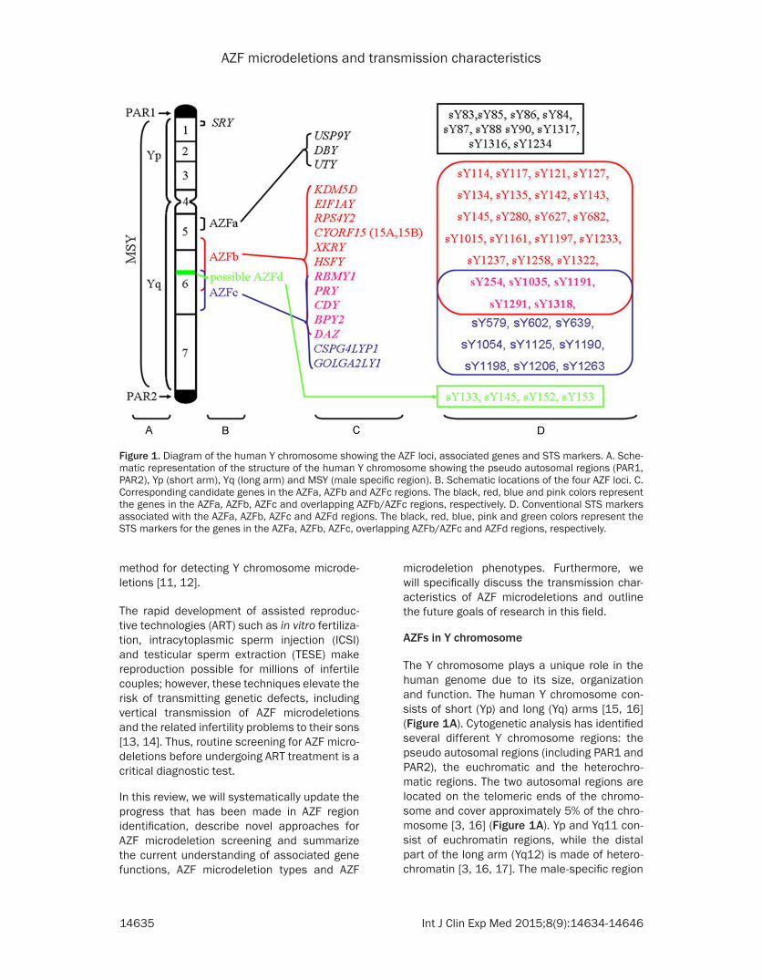
The Y chromosome plays a unique role in the human genome due to its size, organization and function. The human Y chromosome con-sists of short (Yp) and long (Yq) arms [15, 16] (Figure 1A). Cytogenetic analysis has identified several different Y chromosome regions: the pseudo autosomal regions (including PAR1 and PAR2), the euchromatic and the heterochro-matic regions. The two autosomal regions are located on the telomeric ends of the chromo-some and cover approximately 5% of the chro-mosome [3, 16] (Figure 1A). Yp and Yq11 con-sist of euchromatin regions, while the distal part of the long arm (Yq12) is made of hetero-chromatin [3, 16, 17]. The male-specific region
Figure 1. Diagram of the human Y chromosome showing the AZF loci, associated genes and STS markers. A. Sche-matic representation of the structure of the human Y chromosome showing the pseudo autosomal regions (PAR1, PAR2), Yp (short arm), Yq (long arm) and MSY (male specific region). B. Schematic locations of the four AZF loci. C. Corresponding candidate genes in the AZFa, AZFb and AZFc regions. The black, red, blue and pink colors represent the genes in the AZFa, AZFb, AZFc and overlapping AZFb/AZFc regions, respectively. D. Conventional STS markers associated with the AZFa, AZFb, AZFc and AZFd regions. The black, red, blue, pink and green colors represent the STS markers for the genes in the AZFa, AZFb, AZFc, overlapping AZFb/AZFc and AZFd regions, respectively.
AZF microdeletions and transmission characteristics
14636 Int J Clin Exp Med 2015;8(9):14634-14646
of the Y chromosome (MSY), previously called the non-recombining region of Y, harbors both euchromic and heterochromatic sequences and accounts for 95% of the Y chromosome length [3, 16] (Figure 1A). The euchromatic MSY sequences contain 8 Mb of Yp and 14.5 Mb of Yq. These sequences are subdivided into three discrete classes: X-transposed (3.4 Mb), X-degenerate (8.6 Mb) and ampliconic (10.2 Mb) [3, 4].
Characterized by eight massive palindromes, ampliconic sequences constitute a major por-tion of the MSY euchromatic sequences. Six of the eight palindromes contain testis-specific protein coding genes [3, 4, 16]. Moreover, eight of nine multicopy protein coding gene families in the MSY have members contained within the palindromes and six of those families are locat-ed exclusively within the palindromes [18]. The Y chromosome harbors many of the genes that control spermatogenesis, and most of the known chromosomal aberrations related to azoospermia or oligozoospermia have been identified in the long arm of this chromosome [3, 16]. Genetic analysis of patients with idio-pathic azoospermia or severe idiopathic oligo-zoospermia identified three AZF subregions: AZFa (approximately 800 kb); AZFb (approxi-mately 3.2 Mb); and AZFc (approximately 3.5 Mb) [19, 20] (Figure 1B). Most of the Y chromo-some specific genes are located in these AZF regions.
AZFa and candidate genes
The AZFa subregion is located in the proximal region of deletion interval 5 of Yq 11.21 (subin-terval 5C of the human Y chromosome) [21, 22]. The region spans about 800 kb and encodes single copy genes with homologues in the X chromosome [4]. The genes have been found to be necessary for normal spermatogen-esis. Candidate genes in the AZFa locus include USP9Y (Ubiquitin specific peptidase 9, Y-linked), DBY (Dead box on Y) and UTY (Ubiquitously transcribed tetratricopeptide repeat gene, Y-linked) [3, 4] (Figure 1C).
USP9Y consists of 46 exons and spans 159 kb of genomic DNA [23]. The USP9Y gene encodes a ubiquitin-specific protease and belongs to the peptidase C19 family [23]. Molecular analysis revealed USP9Y deletions or mutations in infer-tile patients [24]; however, a recent study
reported that normal spermatogenesis was observed in an individual with a complete AZFa region USP9Y deletion [25]. Thus, USP9Y is not required for normal spermatogenesis, but may exert its effects on fertility in combination with other potential candidate genes in the AZFa region. DBY consists of 17 exons spanning 16 kb [26] and it is specifically expressed in testis tissue [27]. This gene encodes an ATP depen-dent RNA helicase DEAD box protein in humans and plays a significant role in the pre-meiotic spermatogonia phase of spermatogenesis [27]. Molecular analysis revealed a high prevalence of deletions or mutations in the DBY gene in infertile patients when compared to the USP9Y and UTY genes [25, 26]. All available data sup-port the hypothesis that DBY is an important spermatogenesis gene. UTY encodes a protein that is rich in tetratricopeptide repeats [28]. This gene is mapped to the 5C interval corre-sponding to the AZFa region [28]. Deletion anal-ysis revealed the presence of a UTY deletion in one infertile male patient, but this deletion was also accompanied by a DBY deletion [26]. No infertility associated UTY specific deletion has been found yet.
AZFb and candidate genes
The AZFb locus is located in the middle region of Yq11 (approximately between the 5 M to 6B subintervals) [22]. This region spans 6.2 to 7.7 Mb of MSY sequences, but overlaps with the AZFc region by 1.5 Mb [29]. The AZFb region contains several single copy genes as well as multicopy gene families. Single copy protein coding genes found within the AZFb region include KDM5D (lysine (K)-specific demethyl-ase 5D), EIF1AY (eukaryotic translation initia-tion factor 1A, Y-linked), RPS4Y2 (Ribosomal protein S4 Y isoform 2) and CYORF15 (chromo-some Y open reading frame 15A and 15B) [3, 4] (Figure 1C).
KDM5D encodes a histone H3 lysine 4 (H3K4) demethylase that forms a protein complex with the MSH5 (MutS protein homolog 5) DNA repair factor during spermatogenesis, suggesting KDM5D involvement in male germ cell chroma-tin remodeling [30, 31]. KDM5D is located in the germ cell nucleus during prophase I, and KDM5D disruption may contribute to the AZFb deletion phenotype [3]. EIF1AY is a ubiquitously expressed Y-linked member of the EIF-1A family
AZF microdeletions and transmission characteristics
14637 Int J Clin Exp Med 2015;8(9):14634-14646
and is involved in translation initiation [32], but EIF1AY’s function in male fertility has not been well established. RPS4Y2 expression has been demonstrated to be is testis-specific, so it is reasonable to hypothesize that RPS4Y2 may potentially play a role in posttranscriptional regulation of the spermatogenic program [33]. The CYORF15A and CYORF15B sequences belong to the taxilin family and are involved in transcriptional regulation in osteoblasts [3, 34]. Despite their presence in the AZFb region, the role of the CYORF15 genes in normal sper-matogenesis is largely unknown.
Due to the presence of ampliconic sequences, the AZFb region also contains a set of seven multicopy gene families: XKRY (XK, Kell blood group complex subunit-related, Y-linked), HSFY (heat shock transcription factor, Y-linked), RBMY1A1 (RNA binding motif protein, Y-linked, family 1, member A1), PRY (PTPN13-like, Y-linked), CDY (Chromodomain Y, Y-linked), BPY2 (Basic protein Y2, Y-linked) and DAZ (deleted in azoospermia) [3, 19] (Figure 1C). Twenty members from these gene families are located in AZFb, but several genes also map to the AZFc region (Figure 1C). Only the genes that are exclusively located in the AZFb region are discussed in this section.
The XKRY gene is expressed specifically in the testis and maps to the yellow-coded amplicon family [3, 35], but no role for XKRY in spermato-genesis has been validated. HSFY encodes a member of the heat shock factor family of tran-scriptional activators and displays testis-pre-dominant expression [36]. HSFY’s functions in male fertility have been extensively studied. First, the HSFY gene is found in Sertoli cells and in spermatogenic cells up to the spermatid stage, with predominant expression in round spermatids [37, 38]. Second, HSFY protein lev-els are low in spermatogenic cell samples from patients with maturation arrest [39]. Finally, Vinci et al. detected a partial AZFb deletion that was only found to affect the functional copies of HSFY in an azoospermic man [40]. The PRY gene encodes two full-length functional units in the AZFb region (PRY1 and PRY2) and two shorter versions in the AZFc region (PRY3 and PRY4) [41]. PRY1 and PRY2 exhibit testis-spe-cific expression, but PRY expression in germ cells is irregular and is only detected in a few individual sperm and spermatids [42].
Interestingly, both gene and protein expression of PRY are higher in the defective germ cell fraction of the ejaculate and in sperm obtained from men with abnormal semen parameters. This expression pattern suggests that PRY could be a useful biomarker for defective sper-matogenesis [42]. Furthermore, it has been suggested that PRY plays a role in male germ cell apoptosis because approximately 40% of PRY-positive cells show DNA fragmentation [42]. RBMY1A1 is part of the RBM gene family and is solely expressed in male germ cells [43]. This gene has been linked to mRNA storage and transport from the nucleus during spermato-genesis [3]. Furthermore, RBMY1A1 disruption plays a significant role in the AZFb deletion phe-notype [44].
AZFc and candidate genes
The AZFc region has been extensively studied due to its important role in male infertility. The AZFc locus is located in the distal part of Yq (deletion subintervals 6C through 6E) [22]. The AZFc region spans 4.5 Mb and resides among three large palindromic sequences that are derived from six distinct amplicon families [3, 22]. The AZFc locus encodes 21 candidate genes and 11 families of transcription units that are specifically expressed in the testis [3, 4]. Seven of these families are located in the AZFc deletion interval, including GOLGA2LY1 (Golgi autoantigen, golgin subfamily a2-like, Y-linked 1) and CSPG4LYP1 (chondroitin sulfate proteoglycan 4-like, Y-linked pseudogene 1) [4] (Figure 1C). Important candidate genes in this deletion interval include DAZ, BPY2 and CDY1 (CDY1a and CDY1b, Y chromosome 1) [4, 45, 46] (Figure 1C). Interestingly, restriction map-ping has also identified three PRY and TTY2 genes proximal to the AZFc region [47]; howev-er, specific deletions of these genes have not yet been identified and their status as possible AZFc candidate genes remains unconfirmed.
DAZ was the first identified and most important candidate gene family in AZFc region. The DAZ family consists of four RNA binding protein-encoding genes (DAZ1-4) [48]. DAZ protein is localized to the innermost layer of the male germ cell epithelium and to spermatozoa tails [48]. A high incidence of DAZ gene deletion has been observed in infertile men [49], indicating that DAZ may be critically important for sper-
AZF microdeletions and transmission characteristics
14638 Int J Clin Exp Med 2015;8(9):14634-14646
matogenesis; however, other genes in the AZFc loci also play a vital role in normal spermato-genesis [50, 51]. BPY2 encodes a highly charged testis-specific protein that has been tentatively linked to cytoskeletal regulation in spermatogenesis [45, 52]. BPY2 protein exhib-its nuclear localization in male germ cells at all developmental stages [53], but the exact role of BPY2 in spermatogenesis is still unclear. CDY1 genes are specifically expressed in the testis and are involved in histone replacement during spermatogenesis [54]. Two CDY1 genes are located in the AZF region, one within the DAZ cluster and other at the distal end [55]. Removal of DAZ also removes one copy of CDY1, strongly suggesting that the gene plays a role in spermatogenesis.
Possible AZFd involvement
The AZFd locus was first identified using multi-plex PCR reactions and proposed to exist between AZFb and AZFc [8] (Figure 1B). However, the AZFd region remains controver-sial. Simoni et al. assert that the MSY sequenc-es and microdeletion mechanism clearly indi-cate that AZFd locus cannot exist between the AZFb and AZFc regions [56], while Muslu- manoglu et al. reported microdeletions in the putative AZFd loci by deletion analysis through multiplex PCR reactions using four STS (sY133, sY145, sY152 and sY153) [57]. Moreover, recent studies reporting AZFd deletions in infertile men add further credence to the exis-tence and involvement of the AZFd locus in male fertility [9, 58, 59].
AZF region microdeletions
YCMs occur in the q11.23 band and are com-monly subdivided according to microdeletion location into AZFa, AZFb, AZFc or AZFd micro-deletions [3, 7]. Complete AZFa deletion removes an estimated 792 kb DNA in the proxi-mal Yq11 region, including two candidate genes [60]. Complete AZFb deletion removes 6.23 Mb DNA spanning 32 genes and all the testis-spe-cific gene families in the AZFb region [29]. AZFc deletions are estimated to span 3.5 Mb DNA encoding 21 genes and seven gene families [45]. Furthermore, three partial AZFc deletions have been identified, including the b1/b3, b2/b3 and gr/gr deletions. These partial AZFc dele-tions have been associated with a high risk of dysfunctional spermatogenesis [3, 4, 61]. AZF
microdeletions also include combined dele-tions, including AZFab, AZFac, AZFad, AZFabc, AZFbc, AZFbd and AZFbcd [3, 4]. Each of these microdeletions may result in different degrees of spermatogenetic failure [22, 62].
Mechanisms of microdeletions
YCMs occur due to errors in homologous recombination [63]. AZFa deletions result from homologous intrachromosomal recombi-nation between two human endogenous retro-viral sequences HERV15yq1 and HERV15yq2 that are located in the proximal Yq11 region [63]. Complete AZFb deletions are caused by homologous recombination between the Palindrome P5 and the proximal arm of palin-drome P1 in the Yq arm [29]. AZFc deletions have been demonstrated to result from homol-ogous recombination between the sub-ampli-cons b2 and b4 in palindromes P3 and P1 [45]. The proximity of AZFc to the Yq12 heterochro-matic region may also trigger a high percentage of unequal intra-chromosomal recombination, increasing the chance of AZFc deletion [4, 45]. Non-homologous recombination between P5/distal-P1 or between P4/distal-P1 may also lead to AZFbc deletions [29]. The mechanisms underlying other types of microdeletions require further investigation.
Prevalence of AZF microdeletions
AZF microdeletions are observed in 10 to 15% of infertile men with azoospermia or severe oli-gozoospermia, and are very rare in fertile men or men with a sperm density greater than 5 mil-lion/ml [7]. In a large cohort of 1,738 infertile men in Northeastern China, the YCM incidence was 8.57% [2]. The most common microdele-tion observed in the 1,738 patient study was in AZFc, followed by AZFbc, AZFb, AZFabc, AZFa and AZFac [2]. Similarly, AZF microdeletions were detected in 5.06% of patients (185 cases) in another survey of 3,654 patients, including 147 patients with azoospermia and 38 patients with severe oligozoospermia [64]. The most fre-quent microdeletions in the 3,654 patient study were observed in the AZFc region, fol-lowed by the AZFbcd, AZFbc, AZFb, AZFa and AZFac regions [64]. AZFc deletion accounted for at least two thirds of all Yq deletions in all previous studies. Interestingly, a recent study reported that the prevalence of AZF microdele-tions was much higher in men from couples
AZF microdeletions and transmission characteristics
14639 Int J Clin Exp Med 2015;8(9):14634-14646
with recurrent pregnancy loss than in men from fertile couples. The result of this study sug-gests that YCM in the AZF region may be a pos-sible etiologic factor for recurrent pregnancy loss [65].
AZF microdeletion phenotypes
The partial removal of the AZFa region is asso-ciated with hypo-spermatogenesis, whereas the complete deletion of the AZFa region inhib-its the production and maturation of germ cells in the seminiferous tubules. Testicular biopsies of AZFa deletion patients reveal a Sertoli cell-only (SCO) phenotype that is characterized by the total absence of germ cells in the seminifer-ous tubules or by the presence of germ cells in a very small number of tubules [4]. Consequently, it is virtually impossible to retrieve mature sperm with the TESE technique [66]. Brown et al. [67] reported two patients with USP9Y dele-tions who presented with SCO type 1 syndrome, and a third patient with a similar deletion who had hypospermatogenesis. Similar findings were also observed in other investigations [23, 24]. Unlike AZFa microdeletions, patients with AZFb microdeletions had normal spermatogo-nia and primary spermatocytes in all tubules observed [21]. However, deletions in the AZFb region lead to pre-meiotic spermatogenic arrest or SCO syndrome and, eventually, azoospermia. It is difficult to recover mature sperm from AZFb deletion patients using TESE. Patients with AZFc microdeletions were found to have less severe spermatogenic disruption. Vogt et al. observed the presence of germ cells at differ-ent developmental stages in some tubules; however, the majority of tubules were devoid of all germ cells except Sertoli cells [22]. Four of the patients examined by Vogt et al. produced a small quantity of motile sperm, ranging from 0.1 to 2 million/ml [22]. Similar to a SCO type 2 phenotype, Vogt et al. observed germ cells at different stages in different tubules. All evi-dence supports the hypothesis that the primary cause of the spermatogenic failure in AZFc deletion patients is a post meiotic spermatid or sperm maturation defect. Partial deletions involving the DAZ genes in the AZFc region have been found to cause hypospermatogenesis [68]. Moreover, complete AZFb and AZFc dele-tions were associated with SCO syndrome and alterations in spermatocyte maturation [69]. Infertile men with deletions in the putative AZFd region may have a mildly depressed or
even normal sperm count, but abnormal sperm morphology [8]. Muslumanoglu et al. [57] reported three azoospermic men with matura-tion-arrested testicular histology. These three patients were found to carry a single locus (sY152) microdeletion confined to the putative AZFd region. Interestingly, a recent study revealed that patients with expanded or de novo AZF microdeletions had significantly high-er levels of FSH and lower levels of inhibin B when compared to fertile control men [69]. Therefore, low inhibin B and high FSH levels may be useful diagnostic markers that indicate potential causative mutations in the AZF region in infertile men.
Genetic screening methods for AZF microdele-tion diagnosis
Karyotyping was commonly used to identify macrodeletions in the long arm of the Y chro-mosome, but the technique failed to detect smaller interstitial deletions. Southern blotting was used to identify microdeletions related to azoospermia or oligozoospermia [4]. Because karyotyping and Southern blotting are both labor-intensive, time consuming, costly and complex techniques, many researchers have successfully screened for microdeletions using PCR, a relatively simple, sensitive, fast and reproducible technique that allows multiplex-ing. The STS-PCR technique has been in use for over two decades and has been developed into the gold-standard method for laboratory diag-nosis of Y chromosomal microdeletions [3, 4]. PCR-based deletion analysis is usually designed according to the following major factors: (1) Selection of the STS-PCR markers. The best and most informative markers should be highly specific, non-polymorphic, single copy markers or markers limited to a small specific region of the Y chromosome [4]. (2) Selection of DNA samples. YCMs primarily occur during the pre-fertilization stages, but they may also occur as a post-fertilization event. If microdeletions occur during pre-fertilization stages, it is highly likely that they will be transmitted to the whole body of the newborn boy. If the deletion occurs during the post-fertilization stages, the deletion will cause mosaicism characterized by normal Y chromosomes in leukocytes but deleted Y chro-mosomes in sperm or testicular DNA [4, 70]. Although lymphocyte DNA is the cheapest and most readily available sample for basic YCM screening, sperm or testicular DNA is the most
AZF microdeletions and transmission characteristics
14640 Int J Clin Exp Med 2015;8(9):14634-14646
reliable sample to screen for all kinds of YCMs [4, 70] PCR quality control. PCR amplification failures lead to the false screening results. Thus, high-quality DNA and appropriate internal and external positive and negative controls are necessary to minimize false negatives. (4) STS marker reliability. STS markers are short, known DNA sequences that are mapped to different locations in the genome. They offer high speed, convenience, reliability and low cost for genetic screening analysis. To date, about 1287 Y-specific STS biomarkers have been generat-ed, including 992 single-copy and 285 multi-copy STS markers [71]. A selection of conven-tional STS biomarkers targeting different AZF microdeletions are listed in Figure 1D. Moreover, multiplex STS-based PCR microdele-tion analysis using multiple STS markers in each AZF sub-region can detect over 95% of deletions [56].
Interestingly, Bor et al. combined fluorescent multiplex PCR with capillary electrophoresis and successfully screened for Y chromosome microdeletions in 49 azoospermic and 149 severe oligozoospermic men [72]. They con-cluded that the application of capillary electro-phoresis to the detection of PCR products pro-vides a useful, semi-automated and high throughput method for rapidly screening YCMs [72]; however, this approach has rarely been used to screen for YCMs in infertile patients.
Because of the rapid advancements in molecu-lar diagnostic techniques, it has become nec-essary to generate a new hybridization method for the rapid and efficient detection and analy-sis of large quantities of genetic data. Since chemical assays utilize a solid phase that often produces high background signals and can adversely affect reaction kinetics by immobiliz-ing a reactant [73], suspension array technolo-gy (SAT) was developed by the Luminex Corporation (Austin, TX, USA) to eliminate the need for a solid surface during clinical testing. With this technique, oligonucleotide probes are allowed to hybridize with microsphere beads bearing a unique fluorescent label. Each micro-sphere set is internally labeled with two spec-tral fluorochromes of different intensities. The unique spectral emission is recognized by a red laser, while the biotinylated amplicon bound to the surface of the microsphere is read by a green laser that quantifies the fluorescence of the reporter molecule (streptavidin R-phycoery-
thrin) [4, 11, 12]. This technology can simulta-neously analyze 100 analytes by combining 100 different sets of microspheres in a single reaction. SAT has been increasingly used to detect YCMs in infertile patients [11, 12]. The technique is effective, sensitive, rapid, repro-ducible and easily performed. However, there are a few disadvantages associated with the SAT technique, including low array size, hybrid-ization problems and difficulties optimizing a single specific annealing temperature for the entire experiment.
Array-comparative genomic hybridization (aC- GH) was developed to analyze submicroscopic Y deletions [74, 75]. Yuen et al. successfully used Agilent’s Human Genome 8 × 15 K array aCGH format as a template to create a custom Y chromosome aCGH to detect microdeletion prevalence in male infertility. They concluded that their aCGH approach was a reliable, high-resolution alternative to multiplex PCR screen-ing of pathogenic YCMs in male infertility [75]; however, additional studies are needed to com-prehensively evaluate this state-of-the-art technique.
Transmission characteristics of YCMs
Men with Y deletions are generally infertile and, therefore, deletions cannot be transmitted to their offspring. In recent decades, the ICSI and TESE techniques have successfully helped men with azoospermia or severe oligozoospermia reproduce. However, these techniques greatly increase the risk of YCM transmission from infertile fathers to their male offspring. Although many studies have reported the transmission of AZF microdeletions from father to son, the data from these studies have rarely been summarized.
Prevalence of AZF microdeletion transmission
AZF microdeletions are inherited through the paternal germ line or occur as de novo events. It has been reported by many studies that more than 80% of AZF microdeletions are of de novo origin [14, 76]. Most deletions occur during the pre-fertilization stage, while some deletions are post-fertilization events [77]. If a sperm with YCMs fertilizes an egg, it will transmit the YCMs to the male child. On the other hand, if the dele-tion occurs as a post fertilization event, it may cause mosaicism characterized by normal Y chromosomes in leukocytes and Y chromo-
AZF microdeletions and transmission characteristics
14641 Int J Clin Exp Med 2015;8(9):14634-14646
somes with the post fertilization deletion in sperm or testicular DNA [4, 77]. Since infertility is the major phenotype of men with AZF micro-deletions, the natural transmission of AZF microdeletions has rarely been reported [16, 76]. Dai et al. reported that YCMs in 7 of 10 infertile men were naturally transmitted from father to son [76]. Samli et al. also observed the natural transmission of an AZFb microdele-tion from a father to each of his three sons [14]. In contrast, vertical transmission of AZF micro-deletions from father to son through ICSI has been widely reported [78, 79]. In 1996, Kent-First et al. described the first study of the sons produced by a population of 32 couples with infertile fathers who received reproductive assistance via ICSI [80]. They found that the incidence of microdeletions in the ICSI popula-tion was about 9.4%, which was close to the incidence of AZF microdeletions in infertile men.
ART have been associated with elevated inci-dences of sexual chromosomal aberrations, de novo chromosomal abnormalities and sperm aneuploidy [81]. Thus, one of the major con-cerns regarding ART is whether ART cause AZF microdeletions. A new study by Liu et al. com-pared the YCM occurrence in 19 candidate genes from 199 fathers and their 228 sons (Chinese, Han ethnicity) that were conceived by IVF (85 sons), ICSI (73 sons) or natural concep-tion (70 sons). They observed that the YCM inci-dences of the fathers for IVF, ICSI and naturally conceived sons was 10.7%, 3.2% and 8.2%, respectively [69]. They identified one de novo YCM among the 70 naturally conceived off-spring but none among the 158 ART conceived offspring. There were no statistically significant differences in incidence among the three groups or of de novo YCMs between the natu-rally conceived and ART conceived sons [69]. Therefore, they finally concluded that ART does not significantly increase the risk of YCM in male offspring [69]. However, this conclusion remains controversial; the correlation between the incidence of AZF microdeletions and ART needs to be verified in a large, ethnically and geographically diverse cohort of infertile men and their offspring.
Expansion of AZF microdeletions in the off-spring
A significant amount of research in recent years has focused on investigating the genetic chang-
es in offspring conceived by ICSI. Some studies have reported that ICSI can only vertically transmit YCMs without the expansion or de novo occurrence of YCMs [78, 82]. Rolf et al. reported a case of what was probably an identi-cal, partial deletion of the distal part of the AZFb region over three generations [83]. A study by Minor et al. also identified an identical and partial AZFc gr/gr deletion that was verti-cally transmitted over three generations via fathers receiving reproductive assistance through ICSI [84].
However, a growing number of studies have reported that YCM can be transmitted vertically from father to son via ICSI and that ICSI can contribute to YCM expansions as well as de novo YCM [79, 80, 85]. Dai et al. examined the expansion of AZF deletions in 10 father-son pairs, and found expansion microdeletions (S1/F1, S2/F2, S6/F6, S7/F7, S8/F8, S9/F9 and S10/F10) in seven father-son pairs and de novo microdeletions (S3/F3, S4/F4 and S5/F5) in the three remaining father-son pairs [76]. Samli et al. reported an unusual family in which an azoospermic patient (proband) and three broth-ers inherited a Yq microdeletion from their father through spontaneous pregnancy [14]. The brothers and their father all carried a Yq microdeletion in the AZFb subregion involving the RBM1 loci. Additionally, an uncle carried a different deletion in the AZFc region (sY1539). The proband and one of his brothers shared an identical deletion with their father, plus addi-tional de novo deletions in the AZFa and AZFb subregions [14].
Although Liu et al. claimed that ART is not asso-ciated with the expansion or occurrence of de novo microdeletions [69], a significant number of studies have demonstrated that AZF micro-deletions are capable of transmitting them-selves and expanding over the generations through natural pregnancy or ART [79, 80, 85].
Perspectives
Human spermatogenesis is a complex process involving a series of coordinated events regu-lated and governed by key genes located in the AZF regions of the Y chromosome. Evidence indicates that DBY in AZFa; KDM5D and RBMY1A1 in AZFb; and DAZ and CDY in AZFb/c are key determinants in spermatogenesis [3, 4]. Mutations in the AZFc region are the most common genetic cause of male infertility, and
AZF microdeletions and transmission characteristics
14642 Int J Clin Exp Med 2015;8(9):14634-14646
the DAZ gene is deleted in most cases involving AZFc deletions.
Despite the tremendous breakthroughs that have been achieved in recent decades, the functions of the AZF genes are still not well understood. The lack of easily accessible ani-mal models and in vitro spermatogenic cell lines are the major obstacles investigating AZF gene functions in normal spermatogenesis. Additionally, the biological properties of the AZF regions appear to be quite complicated. This complexity is illustrated by the tremendous variability associated with the AZFb and AZFc sequences and the intricate regulation of their corresponding genetic determinants. Therefore, future research should focus on fully sequenc-ing AZF diversity across the Y chromosome and pursuing more in-depth functional character-ization of the AZF genes.
Genomic sequencing of AZF has clearly subdi-vided AZF into the AZFa, AZFb and AZFc regions, and may have identified a fourth proposed AZFd region. Homologous recombination is commonly accepted as the major cause of most AZF microdeletions; however, deletions via non-homologous recombination have also been reported. Patterns of AZF microdeletions have been verified by many specific STS mark-ers. STS-PCR has become the gold-standard method for clinically screening AZF microdele-tions, but this method is not appropriate for large sample sizes or identifying novel AZF microdeletions. New state-of-the-art tech-niques like SAT and aCGH have partially res-cued the limitations of STS-PCR techniques; however, the efficacy of these approaches needs to be verified in future studies. Because the identification of novel AZF molecular micro-deletions and their associated phenotypes is of great importance to the clinical screening of infertile patients, improvement of the current methods or generation of new approaches for laboratory and clinical screening of AZF micro-deletions in large numbers of samples is great-ly needed.
Although the prevalence of AZF microdeletions has been extensively studied, the results from various geographically and ethnically distinct populations are inconsistent and even contro-versial. Based on the available evidence, there are some clearly established points. First, AZF microdeletions are common in men with azo-
ospermia and severe oligozoospermia, as well as in the offspring of these men conceived by ART treatment. Second, although most AZF microdeletions are of de novo origin, AZF micro-deletions can be vertically transmitted to off-spring, and vertically transmitted AZF microde-letions can be transmitted identically or with expansions through both natural pregnancy and ART treatment. Finally, because this infor-mation has diagnostic, prognostic, preventive and ethical value that benefits infertile couples, it is critical to perform screening for specific AZF deletions and other chromosomal abnor-malities in infertile males. nuscript.
Acknowledgements
The work was supported by Jilin Province Science and Technology Department Project of Natural Science Foundation (20150101136JC). We thank Medjaden Bioscience Limited for assisting in the preparation of this manuscript.
Disclosure of conflict of interest
None.
Address correspondence to: Dr. Song-Ling Zhang, Department of Gynecologic Tumors, The First Hospital of Jinlin University, 71 Xinmin Street, Changchun 130021, Jilin Province, P. R. China. Tel: +8615843121678; Fax: +8615843121678; E-mail: [email protected]
References
[1] Bushnik T, Cook JL, Yuzpe AA, Tough S and Collins J. Estimating the prevalence of infertili-ty in Canada. Hum Reprod 2012; 27: 738-746.
[2] Zhang YS, Dai RL, Wang RX, Zhang HG, Chen S and Liu RZ. Analysis of Y chromosome micro-deletion in 1738 infertile men from northeast-ern China. Urology 2013; 82: 584-588.
[3] Navarro-Costa P, Plancha CE and Goncalves J. Genetic dissection of the AZF regions of the human Y chromosome: thriller or filler for male (in) fertility? J Biomed Biotechnol 2010; 2010: 936569.
[4] Suganthi R, Vijesh VV, Vandana N and Fathima Ali Benazir J. Y choromosomal microdeletion screening in the workup of male infertility and its current status in India. Int J Fertil Steril 2014; 7: 253-266.
[5] Poongothai J, Gopenath TS and Manonayaki S. Genetics of human male infertility. Singapore Med J 2009; 50: 336-347.
[6] Zhang F, Li L, Wang L, Yang L, Liang Z, Li J, Jin F and Tian Y. Clinical characteristics and treat-
AZF microdeletions and transmission characteristics
14643 Int J Clin Exp Med 2015;8(9):14634-14646
ment of azoospermia and severe oligospermia patients with Y-chromosome microdeletions. Mol Reprod Dev 2013; 80: 908-915.
[7] Vineeth VS and Malini SS. A Journey on Y Chromosomal Genes and Male Infertility. Int J Hum Genet 2011; 11: 203-215.
[8] Kent-First M, Muallem A, Shultz J, Pryor J, Roberts K, Nolten W, Meisner L, Chandley A, Gouchy G, Jorgensen L, Havighurst T and Grosch J. Defining regions of the Y-chromosome responsible for male infertility and identifica-tion of a fourth AZF region (AZFd) by Y-chromosome microdeletion detection. Mol Reprod Dev 1999; 53: 27-41.
[9] Al-Achkar W, Wafa A and Moassass F. Cyto- genetic abnormalities and Y-chromosome mi-crodeletions in infertile Syrian males. Biomed Rep 2013; 1: 275-279.
[10] Ambulkar PS, Sigh R, Reddy M, Varma PS, Gupta DO, Shende MR and Pal AK. Genetic Risk of Azoospermia Factor (AZF) Micro- deletions in Idiopathic Cases of Azoospermia and Oligozoospermia in Central Indian Population. J Clin Diagn Res 2014; 8: 88-91.
[11] Sun K, Chen XF, Zhu XB, Hu HL, Zhang W, Shao FM, Li P, Miao QL, Huang YR and Li Z. A new molecular diagnostic approach to assess Y chromosome microdeletions in infertile men. J Int Med Res 2012; 40: 237-248.
[12] Zhu YJ, Liu SY, Wang H, Wei P and Ding XP. The prevalence of azoospermia factor microdele-tion on the Y chromosome of Chinese infertile men detected by multi-analyte suspension ar-ray technology. Asian J Androl 2008; 10: 873-881.
[13] Kamischke A, Gromoll J, Simoni M, Behre HM and Nieschlag E. Transmission of a Y chromo-somal deletion involving the deleted in azo-ospermia (DAZ) and chromodomain (CDY1) genes from father to son through intracytoplas-mic sperm injection: case report. Hum Reprod 1999; 14: 2320-2322.
[14] Samli H, Murat Samli M and Solak M. Natural transmission of AZFb Y-chromosomal microde-letion from father to his three sons. Arch Androl 2006; 52: 423-426.
[15] Krausz C and McElreavey K. Y chromosome and male infertility. Frontiers Biosci 1999; 4: 1-8.
[16] Lahn BT, Pearson NM and Jegalian K. The hu-man Y chromosome, in the light of evolution. Nat Rev Genet 2001; 2: 207-216.
[17] Bachtrog D and Charlesworth B. Towards a complete sequence of the human Y chromo-some. Genome Biol 2001; 2: 1016.
[18] Skaletsky H, Kuroda-Kawaguchi T, Minx PJ, Cordum HS, Hillier L, Brown LG, Repping S, Pyntikova T, Ali J, Bieri T, Chinwalla A, Delehaunty A, Delehaunty K, Du H, Fewell G,
Fulton L, Fulton R, Graves T, Hou SF, Latrielle P, Leonard S, Mardis E, Maupin R, McPherson J, Miner T, Nash W, Nguyen C, Ozersky P, Pepin K, Rock S, Rohlfing T, Scott K, Schultz B, Strong C, Tin-Wollam A, Yang SP, Waterston RH, Wilson RK, Rozen S and Page DC. The male-specific region of the human Y chromosome is a mo-saic of discrete sequence classes. Nature 2003; 423: 825-837.
[19] Ferlin A, Moro E, Rossi A, Dallapiccola B and Foresta C. The human Y chromosome’s azo-ospermia factor b (AZFb) region: sequence, structure, and deletion analysis in infertile men. J Med Genet 2003; 40: 18-24.
[20] Lahn BT and Page DC. Functional coherence of the human Y chromosome. Science 1997; 278: 675-680.
[21] Foresta C, Moro E and Ferlin A. Y chromosome microdeletions and alterations of spermato-genesis. Endocr Rev 2001; 22: 226-239.
[22] Vogt PH, Edelmann A, Kirsch S, Henegariu O, Hirschmann P, Kiesewetter F, Kohn FM, Schill WB, Farah S, Ramos C, Hartmann M, Hartschuh W, Meschede D, Behre HM, Castel A, Nieschlag E, Weidner W, Grone HJ, Jung A, Engel W and Haidl G. Human Y chromosome azoospermia factors (AZF) mapped to different subregions in Yq11. Hum Mol Genet 1996; 5: 933-943.
[23] Sun C, Skaletsky H, Birren B, Devon K, Tang Z, Silber S, Oates R and Page DC. An azoosper-mic man with a de novo point mutation in the Y-chromosomal gene USP9Y. Nat Genet 1999; 23: 429-432.
[24] Sargent CA, Boucher CA, Kirsch S, Brown G, Weiss B, Trundley A, Burgoyne P, Saut N, Durand C, Levy N, Terriou P, Hargreave T, Cooke H, Mitchell M, Rappold GA and Affara NA. The critical region of overlap defining the AZFa male infertility interval of proximal Yq contains three transcribed sequences. J Med Genet 1999; 36: 670-677.
[25] Luddi A, Margollicci M, Gambera L, Serafini F, Cioni M, De Leo V, Balestri P and Piomboni P. Spermatogenesis in a man with complete dele-tion of USP9Y. N Engl J Med 2009; 360: 881-885.
[26] Foresta C, Ferlin A and Moro E. Deletion and expression analysis of AZFa genes on the hu-man Y chromosome revealed a major role for DBY in male infertility. Hum Mol Genet 2000; 9: 1161-1169.
[27] Ditton HJ, Zimmer J, Kamp C, Rajpert-De Meyts E and Vogt PH. The AZFa gene DBY (DDX3Y) is widely transcribed but the protein is limited to the male germ cells by translation control. Hum Mol Genet 2004; 13: 2333-2341.
[28] Greenfield A, Carrel L, Pennisi D, Philippe C, Quaderi N, Siggers P, Steiner K, Tam PP,
AZF microdeletions and transmission characteristics
14644 Int J Clin Exp Med 2015;8(9):14634-14646
Monaco AP, Willard HF and Koopman P. The UTX gene escapes X inactivation in mice and humans. Hum Mol Genet 1998; 7: 737-742.
[29] Repping S, Skaletsky H, Lange J, Silber S, Van Der Veen F, Oates RD, Page DC and Rozen S. Recombination between palindromes P5 and P1 on the human Y chromosome causes mas-sive deletions and spermatogenic failure. Am J Hum Genet 2002; 71: 906-922.
[30] Akimoto C, Kitagawa H, Matsumoto T and Kato S. Spermatogenesis-specific association of SMCY and MSH5. Genes Cells 2008; 13: 623-633.
[31] Lee MG, Norman J, Shilatifard A and Shiekhattar R. Physical and functional associ-ation of a trimethyl H3K4 demethylase and Ring6a/MBLR, a polycomb-like protein. Cell 2007; 128: 877-887.
[32] Roll-Mecak A, Shin BS, Dever TE and Burley SK. Engaging the ribosome: universal IFs of translation. Trends Biochem Sci 2001; 26: 705-709.
[33] Andres O, Kellermann T, Lopez-Giraldez F, Rozas J, Domingo-Roura X and Bosch M. RPS4Y gene family evolution in primates. BMC Evol Biol 2008; 8: 142.
[34] Yu VW, Gauthier C and St-Arnaud R. Inhibition of ATF4 transcriptional activity by FIAT/gamma-taxilin modulates bone mass accrual. Ann N Y Acad Sci 2006; 1068: 131-142.
[35] Ho M, Chelly J, Carter N, Danek A, Crocker P and Monaco AP. Isolation of the gene for McLeod syndrome that encodes a novel mem-brane transport protein. Cell 1994; 77: 869-880.
[36] Tessari A, Salata E, Ferlin A, Bartoloni L, Slongo ML and Foresta C. Characterization of HSFY, a novel AZFb gene on the Y chromosome with a possible role in human spermatogenesis. Mol Hum Reprod 2004; 10: 253-258.
[37] Shinka T, Sato Y, Chen G, Naroda T, Kinoshita K, Unemi Y, Tsuji K, Toida K, Iwamoto T and Nakahori Y. Molecular characterization of heat shock-like factor encoded on the human Y chromosome, and implications for male infer-tility. Biol Reprod 2004; 71: 297-306.
[38] Kinoshita K, Shinka T, Sato Y, Kurahashi H, Kowa H, Chen G, Umeno M, Toida K, Kiyokage E, Nakano T, Ito S and Nakahori Y. Expression analysis of a mouse orthologue of HSFY, a can-didate for the azoospermic factor on the hu-man Y chromosome. J Med Invest 2006; 53: 117-122.
[39] Sato Y, Yoshida K, Shinka T, Nozawa S, Nakahori Y and Iwamoto T. Altered expression pattern of heat shock transcription factor, Y chromosome (HSFY) may be related to altered differentiation of spermatogenic cells in testes
with deteriorated spermatogenesis. Fertil Steril 2006; 86: 612-618.
[40] Vinci G, Raicu F, Popa L, Popa O, Cocos R and McElreavey K. A deletion of a novel heat shock gene on the Y chromosome associated with azoospermia. Mol Hum Reprod 2005; 11: 295-298.
[41] Stouffs K, Lissens W, Verheyen G, Van Landuyt L, Goossens A, Tournaye H, Van Steirteghem A and Liebaers I. Expression pattern of the Y-linked PRY gene suggests a function in apop-tosis but not in spermatogenesis. Mol Hum Reprod 2004; 10: 15-21.
[42] Stouffs K, Lissens W, Van Landuyt L, Tournaye H, Van Steirteghem A and Liebaers I. Characterization of the genomic organization, localization and expression of four PRY genes (PRY1, PRY2, PRY3 and PRY4). Mol Hum Reprod 2001; 7: 603-610.
[43] Elliott DJ, Millar MR, Oghene K, Ross A, Kiesewetter F, Pryor J, McIntyre M, Hargreave TB, Saunders PT, Vogt PH, Chandley AC and Cooke H. Expression of RBM in the nuclei of human germ cells is dependent on a critical region of the Y chromosome long arm. Proc Natl Acad Sci U S A 1997; 94: 3848-3853.
[44] Elliott DJ. RBMY genes and AZFb deletions. J Endocrinol Invest 2000; 23: 652-658.
[45] Kuroda-Kawaguchi T, Skaletsky H, Brown LG, Minx PJ, Cordum HS, Waterston RH, Wilson RK, Silber S, Oates R, Rozen S and Page DC. The AZFc region of the Y chromosome features massive palindromes and uniform recurrent deletions in infertile men. Nat Genet 2001; 29: 279-286.
[46] Ravel C, Chantot-Bastaraud S, El Houate B, Rouba H, Legendre M, Lorenco D, Mandelbaum J, Siffroi JP and McElreavey K. Y-chromosome AZFc structural architecture and relationship to male fertility. Fertil Steril 2009; 92: 1924-1933.
[47] Huynh T, Mollard R and Trounson A. Selected genetic factors associated with male infertility. Hum Reprod Update 2002; 8: 183-198.
[48] Habermann B, Mi HF, Edelmann A, Bohring C, Backert IT, Kiesewetter F, Aumuller G and Vogt PH. DAZ (Deleted in AZoospermia) genes en-code proteins located in human late sperma-tids and in sperm tails. Hum Reprod 1998; 13: 363-369.
[49] Reijo R, Lee TY, Salo P, Alagappan R, Brown LG, Rosenberg M, Rozen S, Jaffe T, Straus D, Hovatta O, et al. Diverse spermatogenic de-fects in humans caused by Y chromosome de-letions encompassing a novel RNA-binding protein gene. Nat Genet 1995; 10: 383-393.
[50] Foresta C, Ferlin A, Garolla A, Rossato M, Barbaux S and De Bortoli A. Y-chromosome de-
AZF microdeletions and transmission characteristics
14645 Int J Clin Exp Med 2015;8(9):14634-14646
letions in idiopathic severe testiculopathies. J Clin Endocrinol Metab 1997; 82: 1075-1080.
[51] Stuppia L, Gatta V, Calabrese G, Guanciali Franchi P, Morizio E, Bombieri C, Mingarelli R, Sforza V, Frajese G, Tenaglia R and Palka G. A quarter of men with idiopathic oligo-azoosper-mia display chromosomal abnormalities and microdeletions of different types in interval 6 of Yq11. Hum Genet 1998; 102: 566-570.
[52] Wong EY, Tse JY, Yao KM, Lui VC, Tam PC and Yeung WS. Identification and characterization of human VCY2-interacting protein: VCY2IP-1, a microtubule-associated protein-like protein. Biol Reprod 2004; 70: 775-784.
[53] Tse JY, Wong EY, Cheung AN, O WS, Tam PC and Yeung WS. Specific expression of VCY2 in human male germ cells and its involvement in the pathogenesis of male infertility. Biol Reprod 2003; 69: 746-751.
[54] Vogt PH. Azoospermia factor (AZF) in Yq11: to-wards a molecular understanding of its func-tion for human male fertility and spermatogen-esis. Reprod Biomed Online 2005; 10: 81-93.
[55] Yen PH. A long-range restriction map of dele-tion interval 6 of the human Y chromosome: a region frequently deleted in azoospermic males. Genomics 1998; 54: 5-12.
[56] Simoni M, Bakker E and Krausz C. EAA/EMQN best practice guidelines for molecular diagno-sis of y-chromosomal microdeletions. State of the art 2004. Int J Androl 2004; 27: 240-249.
[57] Muslumanoglu MH, Turgut M, Cilingir O, Can C, Ozyurek Y and Artan S. Role of the AZFd locus in spermatogenesis. Fertil Steril 2005; 84: 519-522.
[58] Balkan M, Tekes S and Gedik A. Cytogenetic and Y chromosome microdeletion screening studies in infertile males with Oligozoospermia and Azoospermia in Southeast Turkey. J Assist Reprod Genet 2008; 25: 559-565.
[59] Hussein AA, Vasudevan R, Patimah I, Prashant N and Nora FA. Association of azoospermia factor region deletions in infertile male sub-jects among Malaysians. Andrologia 2015; 47: 168-177.
[60] Kamp C, Hirschmann P, Voss H, Huellen K and Vogt PH. Two long homologous retroviral se-quence blocks in proximal Yq11 cause AZFa microdeletions as a result of intrachromosom-al recombination events. Hum Mol Genet 2000; 9: 2563-2572.
[61] Wan L and Cai ZM. [Partial deletions in the AZFc region of the Y chromosome are associ-ated with male infertility]. Zhonghua Nan Ke Xue 2009; 15: 165-169.
[62] Cram DS, Osborne E and McLachlan RI. Y chro-mosome microdeletions: implications for as-sisted conception. Med J Aust 2006; 185: 433-434.
[63] Sun C, Skaletsky H, Rozen S, Gromoll J, Nieschlag E, Oates R and Page DC. Deletion of azoospermia factor a (AZFa) region of human Y chromosome caused by recombination be-tween HERV15 proviruses. Hum Mol Genet 2000; 9: 2291-2296.
[64] Totonchi M, Mohseni Meybodi A, Borjian Boroujeni P, Sedighi Gilani M, Almadani N and Gourabi H. Clinical data for 185 infertile Iranian men with Y-chromosome microdele-tion. J Assist Reprod Genet 2012; 29: 847-853.
[65] Karaer A, Karaer K, Ozaksit G, Ceylaner S and Percin EF. Y chromosome azoospermia factor region microdeletions and recurrent pregnan-cy loss. Am J Obstet Gynecol 2008; 199: 662, e1-5.
[66] Hopps CV, Mielnik A, Goldstein M, Palermo GD, Rosenwaks Z and Schlegel PN. Detection of sperm in men with Y chromosome microdele-tions of the AZFa, AZFb and AZFc regions. Hum Reprod 2003; 18: 1660-1665.
[67] Brown GM, Furlong RA, Sargent CA, Erickson RP, Longepied G, Mitchell M, Jones MH, Hargreave TB, Cooke HJ and Affara NA. Characterisation of the coding sequence and fine mapping of the human DFFRY gene and comparative expression analysis and mapping to the Sxrb interval of the mouse Y chromo-some of the Dffry gene. Hum Mol Genet 1998; 7: 97-107.
[68] Ferlin A, Arredi B, Speltra E, Cazzadore C, Selice R, Garolla A, Lenzi A and Foresta C. Molecular and clinical characterization of Y chromosome microdeletions in infertile men: a 10-year experience in Italy. J Clin Endocrinol Metab 2007; 92: 762-770.
[69] Liu XH, Yan LY, Lu CL, Li R, Zhu XH, Jin HY, Zhang Y, Zhang WX, Gao SH and Qiao J. ART do not increase the risk of Y-chromosome micro-deletion in 19 candidate genes at AZF regions. Reprod Fertil Dev 2014; 26: 778-786.
[70] Dada R, Kumar R, Shamsi MB, Kumar R, Kucheria K, Sharma RK, Gupta SK and Gupta NP. Higher frequency of Yq microdeletions in sperm DNA as compared to DNA isolated from blood. Asian J Androl 2007; 9: 720-722.
[71] Lange J, Skaletsky H, Bell GW and Page DC. MSY Breakpoint Mapper, a database of se-quence-tagged sites useful in defining natural-ly occurring deletions in the human Y chromo-some. Nucleic Acids Res 2008; 36: D809-814.
[72] Bor P, Hindkjaer J, Kolvraa S and Ingerslev HJ. A new approach for screening for Y microdele-tions: capillary electrophoresis combined with fluorescent multiplex PCR. J Assist Reprod Genet 2003; 20: 46-51.
[73] Peterson AW, Wolf LK and Georgiadis RM. Hybridization of mismatched or partially
AZF microdeletions and transmission characteristics
14646 Int J Clin Exp Med 2015;8(9):14634-14646
matched DNA at surfaces. J Am Chem Soc 2002; 124: 14601-14607.
[74] Patrick DF, Maher E, Sharkey F and Wilkie N. Application of array-CGH for the detection of submicroscopic chromosomal Imbalances in 400 cases of children with idiopathic mental retardation and congenital malformations.
[75] Yuen RK, Merkoulovitch A, MacDonald JR, Vlasschaert M, Lo K, Grober E, Marshall CR, Jarvi KA, Kolomietz E and Scherer SW. Development of a high-resolution Y-chromo- some microarray for improved male infertility diagnosis. Fertil Steril 2014; 101: 1079-1085, e3.
[76] Dai RL, Sun LK, Yang X, Li LL, Zhu HB and Liu RZ. Expansion and de novo occurrence of Y chromosome microdeletions occurring via nat-ural vertical transmission in northeastern China. J Int Med Res 2012; 40: 1182-1191.
[77] Edwards RG and Bishop CE. On the origin and frequency of Y chromosome deletions respon-sible for severe male infertility. Mol Hum Reprod 1997; 3: 549-554.
[78] Cram DS, Ma K, Bhasin S, Arias J, Pandjaitan M, Chu B, Audrins MS, Saunders D, Quinn F, deKretser D and McLachlan R. Y chromosome analysis of infertile men and their sons con-ceived through intracytoplasmic sperm injec-tion: vertical transmission of deletions and rar-ity of de novo deletions. Fertil Steril 2000; 74: 909-915.
[79] Page DC, Silber S and Brown LG. Men with in-fertility caused by AZFc deletion can produce sons by intracytoplasmic sperm injection, but are likely to transmit the deletion and infertili-ty. Hum Reprod 1999; 14: 1722-1726.
[80] Kent-First MG, Kol S, Muallem A, Ofir R, Manor D, Blazer S, First N and Itskovitz-Eldor J. The incidence and possible relevance of Y-linked microdeletions in babies born after intracyto-plasmic sperm injection and their infertile fa-thers. Mol Hum Reprod 1996; 2: 943-950.
[81] Bonduelle M, Camus M, De Vos A, Staessen C, Tournaye H, Van Assche E, Verheyen G, Devroey P, Liebaers I and Van Steirteghem A. Seven years of intracytoplasmic sperm injection and follow-up of 1987 subsequent children. Hum Reprod 1999; 14 Suppl 1: 243-264.
[82] Buch B, Galan JJ, Lara M, Real LM, Martinez-Moya M and Ruiz A. Absence of de novo Y-chromosome microdeletions in male children conceived through intracytoplasmic sperm in-jection. Fertil Steril 2004; 82: 1679-1680.
[83] Rolf C, Gromoll J, Simoni M and Nieschlag E. Natural transmission of a partial AZFb deletion of the Y chromosome over three generations: case report. Hum Reprod 2002; 17: 2267-2271.
[84] Minor A, Wong EC, Harmer K and Ma S. Molecular and cytogenetic investigation of Y chromosome deletions over three generations facilitated by intracytoplasmic sperm injection. Prenat Diagn 2007; 27: 743-747.
[85] Komori S, Kato H, Kobayashi S, Koyama K and Isojima S. Transmission of Y chromosomal mi-crodeletions from father to son through intra-cytoplasmic sperm injection. J Hum Genet 2002; 47: 465-468.


![Azoospermia and embryo morphokinetics: testicular sperm ... · AZF (azoospermia factor) region are clinically important due to their association with failure or disruption of spermatogen-esis[1–3].Deletionsonthe](https://img.pdfslide.net/doc/110x75/5c4596b593f3c34c377ddd20/azoospermia-and-embryo-morphokinetics-testicular-sperm-azf-azoospermia.jpg)