Embed Size (px)
Citation preview

Review
Diversity of structures and properties among catalasesP. Chelikania, I. Fitab and P. C. Loewena,*
a Department of Microbiology, University of Manitoba, Winnipeg MB, R3T 2N2 (Canada), Fax: +1 204 474 7603, e-mail: [email protected] Institut de Biología Molecular de Barcelona, Consejo Superior de Investigaciones Científicas, 18–26 Jordi-Girona,Barcelona (Spain)
Received 2 June 2003; received after revision 24 June 2003; accepted 1 July 2003
Abstract. More than 300 catalase sequences are nowavailable, divided among monofunctional catalases (> 225), bifunctional catalase-peroxidases (> 50) andmanganese-containing catalases (> 25). When combinedwith the recent appearance of crystal structures from atleast two representatives from each of these groups (ninefrom the monofunctional catalases), valuable insightsinto the catalatic reaction mechanism in its various formsand into catalase evolution have been gained. The struc-tures have revealed an unusually large number of modifi-
CMLS, Cell. Mol. Life Sci. 61 (2004) 192–2081420-682X/04/020192-17DOI 10.1007/s00018-003-3206-5© Birkhäuser Verlag, Basel, 2004
CMLS Cellular and Molecular Life Sciences
cations unique to catalases, a result of interacting with re-active oxygen species. Biochemical and physiologicalcharacterization of catalases from many different organ-isms has revealed a surprisingly wide range of catalaticefficiencies, despite similar sequences. Catalase gene ex-pression in micro-organisms generally is controlled ei-ther by sensors of reactive oxygen species or by growthphase regulons, although the detailed mechanisms varyconsiderably.
Key words. Catalase; peroxidase; crystal structure; heme; dimanganese.
Introduction
Catalases, or more correctly, hydroperoxidases, are oneof the most studied classes of enzymes. The name cata-lase was first applied, perhaps inappropriately by today’sstandards, in 1900 [1], and the protein has been the objectof study in many different organisms ever since. Theubiquity of the enzyme, its ease of assay, involving acheap, readily available substrate, H2O2, and the spectac-ular display of oxygen evolution have combined to makeit an attractive target for biochemists and molecular biol-ogists alike. A number of reviews of catalases have ap-peared over the years, and the most recent [2, 3] providea thorough background of structural and physiological in-formation up to early 2000. There has been no slowing inthe appearance of work relevant to catalase physiology
* Corresponding author.
and structure, and the focus of this review will be on de-velopments since 2000. Recent insights into the structureof catalases arising from a number of new and informa-tive crystal structures will be presented and discussed inlight of implications for the catalytic mechanism. Thestructural and mechanistic picture will be supplementedwith an overview of recent reports on the regulation ofexpression of catalases.The overall reaction catalyzed by catalases is the degra-dation of two molecules of hydrogen peroxide to waterand oxygen (reaction 1).
2H2O2 Æ 2H2O + O2 (1)
This deceptively simple overall reaction can be brokendown into two stages, but what is involved in each of thestages depends on the type of catalase. The mechanismrelevant to a particular catalase will be addressed in theappropriate section.

CMLS, Cell. Mol. Life Sci. Vol. 61, 2004 Review Article 193
Three classes of proteins unrelated on the basis of se-quence and structure exhibit significant catalase activity.The class that is most widespread in nature and which hasbeen most extensively characterized is composed ofmonofunctional, heme-containing enzymes subdividedbased on having large (> 75 kDa) or small (< 60 kDa)subunits. Phylogenetic analyses have demonstrated theexistence of two distinct clades or subgroupings of smallsubunit enzymes and one clade of large subunit enzymesamong the monofunctional catalases [4]. The second, lesswidespread class is composed of bifunctional, heme-con-taining catalase-peroxidases that are closely related by se-quence and structure to plant peroxidases. The third classincludes the nonheme or Mn-containing catalases. In ad-dition, a diverse group of proteins, all heme-containing,such as chloroperoxidase, plant peroxidases and myoglo-bin, exhibit very low levels of catalase activity, attribut-able to the presence of heme, which alone exhibits cata-latic activity [2].
Regulation of catalase gene expression
Cells usually modulate their stress response systemsthrough regulatory proteins that sense the stressor or amessenger of the stressor and cause appropriate changesin transcription and, occasionally, translation or proteoly-sis. Most bacteria produce one or more catalases that usu-ally respond to oxidative stress, either directly to H2O2
levels or to the presence of other active oxygen species.The earliest studies of catalase gene regulation were car-ried out in Escherichia coli, and these have been exten-sively reviewed [5]. The catalase-peroxidase HPI (hy-droperoxidase I), or KatG, is controlled as part of theOxyR regulon, which senses active oxygen species, andthe monofunctional catalase HPII is controlled as part ofthe sS regulon in stationary phase. Very recently, poly-amines, including putrescine, spermidine and spermine,were reported to up-regulate both the oxyR and rpoS reg-ulons in E. coli [6], but the mechanism remains unclear.The commonality of a reactive oxygen sensor and growthphase or s-transcription factor control mechanisms incatalase gene expression is illustrated in table 1, althoughthere is no consistency in type of catalase or type of reg-ulator. In addition, other regulators have been adapted tomodulate catalase levels, but the generality of their in-volvement and the regulatory protein involved has notbeen defined.Heme synthesis must be sufficiently rapid to satisfy in-duced catalase synthesis rates. E. coli successfully in-creases the rate of heme synthesis to satisfy the demandsfor it in cells expressing plasmid-encoded catalases, butwhether this is simply a response to heme demand or co-ordinate regulation of heme operons is not clear. InStaphylococcus aureus, Pseudomonas aeruginosa and
Bacillus subtilis, there is coordinate control of oxidativestress proteins and iron storage and transport proteins[7–9], but no link to the control of protoporphyrin syn-thesis has been demonstrated. There has been one reportof iron-deficient catalase from Proteus mirabilis (PMC)being produced in E. coli during its rapid expression un-der the control of an inducible T7 promoter [10]. In thisinstance, presumably the protophorphyrin was availablefor protein folding, but the availability or insertion rate ofiron was limiting.
Monofunctional, heme-containing catalases
Heme-containing catalases all have in common a two-stage mechanism for the degradation of H2O2. In the firststep, one hydrogen peroxide molecule oxidizes the heme(in heme-containing catalases) to an oxyferryl species inwhich one oxidation equivalent is removed from the ironand one from the porphyrin ring to generate a porphyrincation radical (reaction 2).
Enz (Por-FeIII) + H2O2 ÆCpd I (Por+.-FeIV = O) + H2O
(2)
The second hydrogen peroxide molecule is utilized as areductant of compound I to regenerate the resting-stateenzyme, water and oxygen (reaction 3).
Cpd I (Por+.-FeIV= O) + H2O2 ÆEnz (Por-FeIII) + H2O + O2
(3)
Despite this common reaction, there are great differencesin reactive capability among the members of this verylarge family of enzymes, and recent research is only justbeginning to provide us with clues that point to some ex-planations for the differences.
PhylogenyThree reviews of catalase phylogeny have appeared, eachsucceeding review involving an increased number of sequences, from 20 in 1993 [11] to 74 in 1997 [4] to 256in 2003 [12]. In 1997, the clear division of monofunc-tional catalases into three clades was obvious, arisingfrom a minimum of two gene duplication events. Themore exhaustive recent analysis confirms these conclu-sions but integrates a larger picture including catalase-peroxidases and nonheme catalases into a universal treeof life. Clade 1 catalases are predominantly of plant ori-gin, but with one algal representative and a subgroup ofbacterial origin. Clade 2 enzymes are all large subunitenzymes of bacterial and fungal origin. The one archae-bacterial clade 2 enzyme is postulated to have arisen in ahorizontal transfer event from a Bacillus species. Theclade 3 enzymes are all small subunit enzymes from bac-teria, archaebacteria, fungi and other eukaryotes. It is

194 P. Chelikani, I. Fita and P. C. Loewen Diversity among catalases
postulated that the archtypal monofunctional catalasewas a large subunit clade 2 enzyme from which an earlygene duplication event, accompanied by loss of the se-quence at the 5¢ and 3¢ ends, gave rise to the clade 1 en-zymes. Clade 3 enzymes are not present in older taxo-nomic groups, suggesting that they arose much later inevolution as a result of a gene duplication in bacteria thatthen spread by horizontal and lateral transfers amongbacteria to archaebacteria and eukaryotes. The divisioninto three clades is illustrated in figure 1 with a focus on the nine catalases for which structures have been deter-mined.
Kinetic propertiesMany catalases have been characterized over the centuryof study, but very few of the enzymes have been charac-terized side by side. This has resulted in many indepen-dent reports of activities and properties, but no way toproperly compare the results. Such a comparison of 16common catalases, including seven for which the struc-tures have been determined, has revealed just how great adivergence in properties there is within the catalase fam-ily [13]. Catalases do not follow Michaelis-Menten kinet-ics except at very low substrate concentrations, and dif-ferent enzymes are affected differently at higher substrate
Table 1. Regulatory responses of catalases and catalase-peroxidases in bacteria and fungi.
Organism Catalase Typea Regulator Additional response Reference
Agrobacterium tumefaciens KatA CPx OxyR [76]Emericella nidulans CatA 3 post-transcriptional control [77]Emericella nidulans CatB 2 developmental, H2O2 [78]Emericella nidulans CatC 3 stationary phase [79]Emericella nidulans CpeA CPx StuA [80]Apergillus niger CatR 2 H2O2 [81]Bacillus firmus CatII 3 H2O2/ascorbate [82]Bacillus firmus CatIII 2 stationary phase [82]Bacillus subtilis KatA 3 PerR [83, 84]Bacillus subtilis KatE 2 sB [85]Bacillus subtilis KatX 1 sF [86]Bacteroides fragilis KatB 3 OxyR [87]Brucella abortus KatE 3 OxyR [88]Dinococcus radiodurans CatE 2 H2O2 [89]Escherichia coli HPI (KatG) CPx OxyR [90]Escherichia coli HPII (KatE) 2 sS [91]Haemophilus influenzae HktE 3 H2O2 /ascorbate [92]Helicobacter pylori KatA 3 Fur [93, 94]Mycobacterium smegmatis KatG CPx FurA [95]Mycobacterium tuberculosis KatG CPx FurA [96]Neisseria gonorrhea Kat 3 OxyR [97]Erinea carotovora HPII 2 sS [98]Pseudomonas aeruginosa KatA 3 Fur [99]Pseudomonas aeruginosa KatB 1 OxyR [100]Pseudomonas putida CatA 3 log phase [101]Pseudomonas putida CatC 2 stationary phase [101] Pseudomonas syringae CatF 1 stationary phase [102]Pyrobaculum calidifontis Kat pc Mn aerobic conditions [103]Rhizobium etli KatG CPx OxyR [104]Salmonella typhimurium KatM Mn sS [105]Salmonella typhimurium HPI CPx OxyR [106]Salmonella typhimurium HPII 2 sS [107]Sinorhizobium leguminosarum Cat log phase [108]Sinorhizobium meliloti KatA 3 H2O2 [109]Sinorhizobium meliloti KatC 2 stationary phase [110] Staphylococcus simulans ACKI oxygen [111]Staphylococcus aureus KatA 3 PerR, Fur [7, 112]Streptomyces coelicolor CatA 3 CatR [113]Streptomyces coelicolor CatB 2 sB [114]Streptomyces coelicolor CatC CPx FurA [115]Streptomyces reticuli CpeB CPx FurS [116]Vibrio fischeri KatA 3 stationary phase, H2O2 [117]Xanthomonas oryzae KatX 2 sS [118]Xanthomonas campestrisi KatE 2 sS [119]
a1, clade 1; 2, clade 2; 3, clade 3; CPx, catalase-peroxidase; Mn, manganese-containing catalase.

was not affected. Boiling in buffer, or alternatively, heat-ing to 65°C in 5.6 M urea was required to dissociate thedimer. While not as stable as HPII, small subunit cata-lases such as bovine liver catalase (BLC) still exhibitedenhanced stability for an enzyme from an organism nor-mally growing at 37°C, with a Tm for loss of activity of56°C. The thermal stability of HPII has also been utilizedas a purification step [13] . Catalases, generally, are alsoresistant to treatment with an ethanol:chloroform mix-ture, another property that can be exploited as a tool forcatalase purification [13]. Unfortunately, HPII proteinheated above 60°C or treated with ethanol:chloroformdoes not readily crystallize, suggesting that some alter-ations in structure have occurred that were not reflectedin enzyme activity. Another example of the robustness of HPII lies in its re-tention of high levels of activity even after treatment witha 1:1 (w:w) ratio of proteinase K for 16 h at 37°C [17].Despite the retention of activity, a closer analysis revealedthat the protein had suffered limited cleavage but that thecore of the enzyme containing the heme active site re-mained intact. Rapid cleavage of about 75 N-terminalresidues occurred initially within one h, followed by aslower cleavage of 160 C-terminal residues. Removal ofthe C-terminal domain exposed one further protease sen-sitive sight that allowed cleavage of the subunit into twoapproximately equal-sized fragments. Despite the intro-duction of this last nick and possibly one other nearby, themonomers remained intact within the larger tetramer. Both thermal stability and resistance of the majority ofthe protein to proteolysis can be explained in terms of avery rigid, stable structure that resists unfolding, therebypreventing access to the protease active site. Resistance toproteolysis is an advantage to HPII because it is ex-pressed in stationary phase, a period of rapid proteinturnover and elevated protease levels. There would havebeen a strong selective pressure to retain this property, butit is not clear whether resistance to proteolysis evolved inE. coli and thermal stability was an inadvertent outcome.Alternatively, the catalase could have originated in a ther-mophilic organism and been horizontally transferred toE. coli, where protease resistance has been the selectivepressure for retention of thermal resistance.The enhanced thermal stability of HPII was initially as-cribed to the extended sequence of 80 residues from onesubunit being overlapped by the wrapping domain of anadjacent subunit [16]. However, proteolytic removal of allbut 5 residues of the overlapped N-terminal region didnot significantly affect the thermal stability, whereas re-moval of the C-terminal domain lowered the Tm by morethan 25°C [17]. The importance of the N-terminal se-quence in folding and oligomerization was demonstratedin HPII variants, N-terminally truncated by only 20residues, not accumulating protein when expressed frommutant genes [18], and this has been confirmed in the
CMLS, Cell. Mol. Life Sci. Vol. 61, 2004 Review Article 195
concentrations. Most small subunit enzymes begin to suffer inactivation by H2O2 at concentrations above300–500 mM and never reach the Michaelis-MentenVmax predicted by extrapolation from rates at low sub-strate concentrations. On the other hand, large subunit en-zymes start to suffer inhibition only above 3 M [H2O2], ifat all, and exceed the predicted Michaelis-Menten Vmax.Consequently, the presentation of observed data in termsof the typical constants Km and Vmax is misleading becausetrue Michaelis-Menten kinetics are not applicable. Withthis proviso in mind, the observed kcat values ranged from54,000 s–1(P. aeruginosa KatB) to 833,000 sec–1 (PMC),and the [H2O2] at Vmax/2 ranged from 38 to 599 mM. Se-quence differences among catalases must be responsiblefor the widely differing reaction rates and substrateaffinities, but providing a rationale for how is not yet pos-sible. Greater insights into the reaction mechanism mustbe gained before this goal will be realized.
Catalases are uniquely stableThe conclusion from phylogenetic reasoning that the ar-chetypal catalase was a clade 2, large subunit enzyme issupported by the unusual resistance to denaturation andproteolysis exhibited by enzymes from this clade. Resis-tance of catalase HPII to pH and thermal denaturationwas noted early in the study of catalases from E. coli andother enteric bacteria and used as a diagnostic test to dif-ferentiate among catalases in crude extracts [14, 15].Documentation of the thermal stability revealed that therewas actually a small increase in activity above 60°C andthat activity began to drop only above 80°C, with a Tm of83°C [16]. The loss of activity was accompanied bychanges in secondary structure, but the dimer association
Figure 1. Phylogenetic tree of the monofunctional catalases, focus-ing on the nine catalases for which structures have been determined.The highlighted enzymes are a subset of the 255 sequences used togenerate the tree [12]. Adjacent to each enzyme is a summary ofheme type (b or d), heme orientation and whether or not NADPHcan bound (+ or –, respectively). The heme can be oriented with theimidazole ring of the essential histidine located either above ring III(His-III) or above ring IV (His-IV) of the heme.

196 P. Chelikani, I. Fita and P. C. Loewen Diversity among catalases
catalase from Candida tropicalis, where truncation by 4or 24 residues produces aggregates of inactive proteinsmaller than tetrameric [19]. In the latter case, even the 4-residue truncation eliminates the overlap or interweavingfeature common to catalases, which would be expected tohave an effect on subunit association. Longer truncationsextending to within 3 residues of the heme active site hadthe expected effect of preventing heme binding [19].
Insights from recent structuresNew insights into catalase function have been providedby the solution of two monofunctional catalase structures,one published and the second soon to be reported, and thecomparison of the structures of a number of variants withthe earlier released native structures [see table 2 for Pro-tein Data Base (PDB) accession numbers]. The previ-ously reported seven catalase structures include the smallsubunit clade 3 enzymes from BLC [20, 21], Micrococ-cus lysodeiticus (MLC) [22], PMC [23], Saccharomycescerevisiae (CATA) [24, 25], and human erythrocytes(HEC) [26] and the large subunit clade 2 enzymes fromPenicillium vitale (PVC) [27, 28] and E. coli (HPII) [29,30]. The two new structures include Pseudomonas sy-ringae (CatF) [31, 32], which provides the first look at aclade 1 catalase, and the Helicobacter pylori catalase(HPC) with and without formic acid bound [P. C. Loewenet al., unpublished]. The typical core catalase features ofa heme-containing active site deeply buried in a beta-bar-rel structure with two or three channels providing accessto the heme has been described in many papers and re-views. Some of the similarities and differences betweensmall and large subunit catalases are evident in the struc-tures of CatF and HPII in figure 2. The CatF and HPCstructures retain these common features, but CatF pre-sents an unexpected heme orientation and lacks reduced
nicotinamide adenine dinucleotide phosphate (NADPH).In addition, the presence of four subunits in the asym-metric unit of CatF makes possible an assessment ofasymmetry, particularly in solvent location, among sub-units and in comparison with other catalases.
Heme orientationThe first catalase structures solved, BLC and MLC, had acommon heme orientation in which the active site Hiswas situated above ring III of the heme (referred to as theHis-III orientation), leading to the conclusion that thiswas the normal orientation for heme in catalases. Thiswas reinforced by the heme orientation found in PMC,CATA and HEC. The presence of the ‘flipped’ orientationwith the active site His located above ring IV of the heme(His-IV) in PVC and HPII was assumed to be unique tolarge subunit enzymes. The fact that the hemes in PVCand HPII were modified to heme d with a cis-hydrox-yspirolactone group on ring III [33] reinforced the ideathat large subunit enzymes were unique. The CatF struc-ture presents heme in the His-IV orientation, suggestingthat the ‘normal’ and, in view of probable evolutionarydevelopment, original orientation of heme in catalases isHis-IV. The residues making contact with the two methylgroups and two vinyl groups on rings I and II create a ma-trix of van der Waals interactions that favor the His-IVorientation in CatF, PVC and HPII and that would preventany attempt to flip the heme to the His-III orientation.Similarly, the matrix of van der Waals interactions in theclade 3 catalases BLC, MLC, PMC, HEC and CATA fa-vor the His-III orientation and would prevent adoption ofthe His-IV orientation. A survey of 228 catalase se-quences identified residues at the positions equivalent to301 (Ala) and 350 (Leu) in CatF that are the key deter-minants in heme orientation through their proximity to
Table 2. Protein Data Bank accession numbers for native catalases, their complexes and variants (available at www.rcsb.org/pdb).
Catalase Organism Accession numbers
A. Monofunctional catalasesPVC Penicillium vitale 4CATBLC Bos taurus (liver) 7CAT, 8CAT, 4BLCPMC Proteus mirabilis 2CAG, 2CAH, 1M85, 1MQF, 1E93CATA Saccharomyces cerevisiae 1AE4HEC Homo sapiens (erythrocyte) 1DGB, 1DGF, 1DGG, 1DGH, 1F4J, 1QQWHPII Escherichia coli 1GGE, 1IPH, 1CF9, 1GG9, 1GGF, 1GGH, 1GGJ,
1GGK,1QF7, 1P7Y, 1P7Z, 1P80, 1P81, IQWSMLC Micrococcus lysodeikticus 1GWE, 1HBZ, 1GWF, 1GWHCatF Pseudomonas syringae 1M7SHPC Helicobacter pylori 1QWL, 1QWM
B. Catalase-peroxidasesHmCPx Haloarcula marismortui 1ITKBpKatG Burkholderia pseudomallei 1MWV
C. Manganese catalasesLPC Lactobacillus plantarum 1JKU, 1JKV

CMLS, Cell. Mol. Life Sci. Vol. 61, 2004 Review Article 197
the vinyl groups on rings I and II. At position 301, 0% ofclade 1 and clade 2 enzymes have Leu or Ile, but 80% ofclade 3 enzymes do. The larger group prevents adoptionof the His-IV orientation. At position 350, 69% of clade1 enzymes and 100% of clade 2 enzymes have Leu,whereas 0% of clade 3 enzymes do, and the location ofthe Leu side chain prevents adoption of the His-III orien-tation. The recently solved structure of HPC conforms tothis rule in presenting the His-III orientation. Occupancyat the equivalent position in other catalases suggests thatmost clade 1 and all clade 2 enzymes will have the His-IV orientation and that most clade 3 enzymes will havethe His-III orientation. It will be interesting to determinewhether the apparent sequence exceptions conform to theclade majority, whether there is a mixture of heme orien-
tations or whether there are small subsets of clade 1 andclade 3 enzymes with His-III and His-IV orientations, re-spectively.
NADPH bindingSince it was first noted almost 20 years ago [34], the pres-ence of NADPH in catalases has presented the interestingproblem to biochemists of explaining the purpose of thecofactor, and of determining its universality. The struc-tures of the clade 3 enzymes BLC, HEC, PMC and CATAall contain NADPH in some fraction of their subunits,whereas the cofactor is not evident in the structures of theclade 2 enzymes PVC and HPII. NADPH is not evidentin the structure of CatF, and the potential binding site is
Figure 2. Ribbon diagrams for comparison of the overall structures and sizes of heme-containing small and large subunit catalases, a cata-lase-peroxidase and a dimanganese catalase. A tetrameric small subunit, heme-containing catalase, CatF (A), a tetrameric large subunit,heme-containing catalase, HPII (B), a dimeric catalase-peroxidase, BpKatG (C) and a hexameric dimanganese catalase, LPC (D), are pre-sented. The structures in A and B are both viewed looking down the Q axis to facilitate comparison of the structures, in particular of theadded C-terminal domains in HPII. In C, BpKatG is viewed along the noncrystallographic two-fold symmetry axis. All structures are pre-sented in the same scale. The dimensions of the four structures (X by Y) are 93 Å ¥ 93 Å for CatF (A), 85 Å ¥ 136 Å for HPII (B), 83 Å¥ 108 Å for BpKatG (C) and 90 Å ¥ 81 Å for LPC (D). The hemes are shown in A, B and C in blue, and the dimanganese clusters in D areshown in red. The figure was prepared using SETOR [120].
modified compared with clade 3 binding sites to such anextent that NADPH binding should not be possible. TheNADPH binding pockets of BLC, HEC, CATA and PMCcontain a His, an Arg, a Val and a His (193, 202, 301 and304, respectively, using BLC numbering), which may beconsidered to be signatures for NADPH binding. In CatF,the equivalent residues are Arg196, Glu205, Ile304 andAsp307, each of which would interfere with NADPH bind-ing either through electrostatic repulsion, direct steric in-terference or loss of favorable contacts. All clade 1 cata-lases lack the equivalent of Arg202 and His304, and mostlack His193 and Val301, leading to the conclusion that most,if not all, clade 1 catalases do not bind NADPH. In thecase of clade 2 enzymes, a portion of the extended C-ter-minal sequence protrudes into the NADPH binding site,precluding any possibility of NADPH binding in theseenzymes. The conclusion seems to be that only clade 3enzymes bind NADPH, but even in this clade, the coen-zyme is not universal, based on the signature residues,and the structure of HPC confirms this conclusion. A re-gion of electron density that might be assigned toNADPH is not evident in the electron density maps ofHPC, and the signature residues are sufficiently changed,including the equivalent of His193 to Tyr, Val301 to Ile andHis304 to Thr, to make fitting of NADPH into the site dif-ficult, if not impossible. The widespread nature of the His-III orientation of hemein clade 3 enzymes suggests that this feature was adopt-ed shortly after the separation of clade 3 from clade 1 en-zymes. The more limited nature of NADPH bindingamong clade 3 enzymes also suggests that it evolved independently and at a later date, arguing against a directevolutionary link between the His-III orientation of theheme and NADPH binding as previously speculated[32]. The role of NADPH and the presumed reason for itsbinding site having evolved are to prevent the formationof inactive compound II [34], but the mechanism bywhich this is achieved remains unclear [35–38]. Clade 2enzymes do not form compound II, or at least it has notbeen possible to generate compound II in the laboratory,making NADPH binding unnecessary. Similar attemptsto generate compound II from a clade 1 catalase have notbeen reported. Curiously, the electron density maps of HPC suggest that one of the two subunits in the asym-metric unit is in an oxyferryl form and that treatmentwith formic acid converts the oxyferryl species to an un-modified iron. This is consistent with earlier work show-ing that formic acid can reduce compound I and promotethe conversion of compound II to native state [2]. Giventhe apparent stability of the oxyferryl species in HPC, itis unlikely that it is either compound I or II, although it is degraded by formic acid treatment. Several electrondonors can reduce compound I and II back to the ferricstate [2], and it is tempting to speculate that the evolution
of NADPH binding in a small subset of catalases mayhave been the result of these other recovery routes be-coming limited, possibly as a result of environmen-tal changes. Based on this very limited experimental picture, one might postulate that compound II recoverywas not a problem for the progenitor large subunit cata-lases. As the early small subunit enzymes evolved, theywere either not converted to compound II or the com-pound II was recovered through the involvement of read-ily available metabolites such as formic acid, ascor-bate and phenols [2]. With the appearance of clade 3enzymes, which are converted to compound II, and theevolution of more specialized intracellular environ-ments, the availability of reducing metabolites may havebecome restricted, or the prevalence of NADPH mayhave increased, allowing catalases in some species to utilize it as the tool for preventing compound II forma-tion.
Catalase as a source of reactive oxygen species andanother role for NADPHThe usual physiological role ascribed to catalases is thatof removing H2O2, a reactive oxygen species (ROS), be-fore it can cause cellular damage. It was therefore sur-prising to find that catalase in human and mouse ker-atinocytes is responsible for the generation of ROS uponirradiation with UVB light [39]. The ROS were not iden-tified, but singlet oxygen and superoxide were eliminated,making hydroperoxides the most likely species. This newand very exciting development, the detoxification ofUVB light through generation of H2O2, which is also de-graded by the catalase, has been heralded as a second rolefor catalases. The mechanism of ROS generation by catalase remainsunclear, however. Significantly, normal catalase in-hibitors such as azide, aminotriazole and cyanide, whichbind to the heme iron and essential imidazole ring, acti-vate the production of ROS. When the effect of inhibitorsis considered in the context that the Soret absorbancemaximum for heme in the native enzyme is at 407 nm,well removed from the 290 to 320 nm region for UVBlight, it seems unlikely that the heme center is activelyinvolved in the generation of ROS, and NADPH, a co-factor of mammalian catalases, may prove to be in-volved. The wavelength of maximum absorbance forNADPH is 340 nm, and the cofactor is a logical sourceof the electrons required for reduction of molecular oxy-gen. Future work involving comparison with catalasesthat do not bind NADPH should clarify the mechanismof this intriguing property. Furthermore, if the NADPHcofactor is found to have a role in ROS generation, it willsuggest another role for NADPH in catalase physiologyand a possible explanation for why NADPH binding incatalase evolved.
198 P. Chelikani, I. Fita and P. C. Loewen Diversity among catalases
CMLS, Cell. Mol. Life Sci. Vol. 61, 2004 Review Article 199
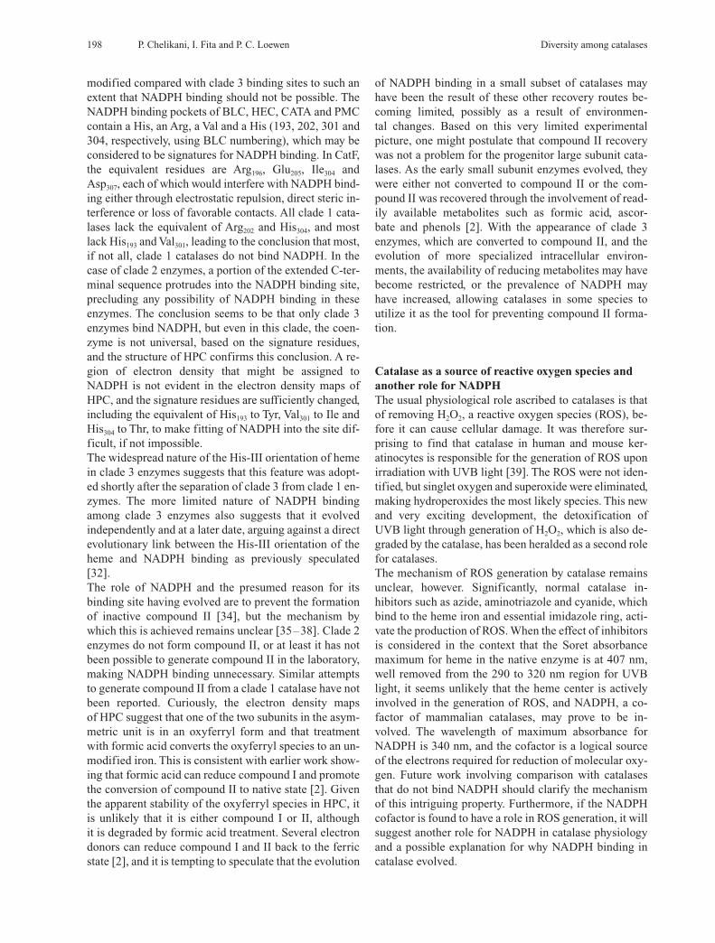
Channel architectureThe recently reported structures of CatF [32] and HPIIvariants [40] have provided significant insights into thechannel architecture in catalases. Three obvious channelsconnect the heme-containing active site with the surface.The main channel, so named because it is the most obvi-ous access route to the heme, approaches the heme per-pendicular to its plane and has long been considered theprimary access route for substrate H2O2, a concept sup-ported by molecular dynamics modeling [41, 42]. A com-parison of the main channels in CatF and HPII (fig. 3) re-veals the extended and bifurcated structure in HPII ascompared with the shorter funnel shape in CatF, which isrepresentative of other small subunit enzymes. A secondchannel approaches the heme laterally, almost in theplane of the heme, and has been referred to as the minoror lateral channel. Limited evidence pointing to a role forthe lateral channel in HPII includes a 3-fold increase inspecific activity resulting from an enlargement of thechannel through removal of Arg260, part of the Glu-Argionic pair situated in the channel. A third channel con-nects the heme with the central cavity, but no evidence ofit having a role has been presented. An extensive review of the waters occupying the mainchannels in each of the four subunits of CatF, HEC (+ CNand + peracetic acid), CATA + azide and HPII, and of thesingle subunits of MLC and PMC, revealed a number ofconsistently occupied positions as well as a number oflow occupancy sites [32]. All enzymes contain a water in-teracting with the catalytic residues His and Asn, andsome catalases, including HPII, MLC and PMC, containa second water in the active site interacting with the hemeiron and the active site His. Moving away from the hemein the channel, only one subunit of HPII and all subunitsin inactive HEC contain waters in the hydrophobic regionaround the conserved Val169 (HPII numbering). The lackof waters in the hydrophobic portion of the channel be-tween the conserved Asp (12 Å from the heme) and theactive site His was interpreted as a ‘molecular ruler’ ef-fect [26] attributed to the lack of interaction sites on theprotein and a distance that was inappropriate for the for-mation of a stable matrix of hydrogen-bonded waters, butappropriate for a matrix involving the slightly largerH2O2. While elements of the ruler model may be applica-ble, the new structures suggest that a more complex seriesof interactions control substrate access to the active site. The first point to consider is that a matrix of water canform in the hydrophobic portion of the channel of HECtreated with peracetic acid in the absence of any new con-stituents or structural changes. Secondly, there seems tobe an optimum three-dimensional shape and size for thisportion of the channel, because making the channel inCATA or HPII either smaller (V169A in HPII) or larger(V169I in HPII) reduced [25, 40] enzyme-specific activ-ity. Finally, changing the conserved Asp (181 in HPII) to
any uncharged residue, polar or nonpolar, including Asn,Gln, Ala, Ser and Ile caused a loss of enzyme activity anda reduction in solvent occupancy in the channel [40].Most notably, the sixth ligand water was absent in all sub-units of D181A, D181S and D181Q variants, and therewere generally fewer waters in the channel. By contrast,the D181E variant exhibited normal levels of activity andcontained not only the sixth ligand water in all subunits,but an unbroken water matrix extending the full length ofthe channels.
Figure 3. Structure of the channels providing access to the activesites in one subunit each of CatF (A), HPII (B), BpKatG (C) andLPC (D). The channel, as calculated by the program VOIDOO[121], is presented as a blue chicken-wire structure in a cross-sec-tion slab of the enzyme. In A and B, the key valine (V118 and V169)and aspartate residues (D130 and D181) discussed in the text are indi-cated. In C, the key serine (S324) discussed in the text is indicated,and its hydrogen bond with the propionate is indicated by a dashedline. Also in C, the region of unassigned electron density corre-sponding to an INH-like molecule is presented in a red chicken-wire structure. The active site heme is evident at the end of the chan-nels in A, B and C. In D, the dimanganese cluster is presented as twoblue balls (Mn) and four red balls (waters), and one of the coordi-nating histidines (His69) is indicated for reference. All four chan-nels are presented in the same scale for comparison. Note that theapproach to the heme in C is in the plane of the heme in compari-son to the approach perpendicular to the plane of the heme in A andB. The figure was prepared using SETOR [120].
This striking influence of the negatively charged sidechain at position 181 of HPII may lie in the generation ofan electrical potential field in the hydrophobic portion ofthe channel between the negative charge and the posi-tively charged heme iron. The potential field will influ-ence the orientation of any molecule with an electrical di-pole, including both water and hydrogen peroxide. Inboth cases, the preferred orientation will be with oxygenatoms pointed toward the positively charged heme ironand the hydrogens toward the negatively charged sidechain of aspartate or glutamate. Such a uniform orienta-tion in the population of solvent will facilitate the forma-tion of hydrogen bonds, providing an explanation for in-creased water occupancy when Asp or Glu are present atposition 181 in HPII. A second result of the induced ori-entation of hydrogen peroxide is that it will enter the ac-tive site with one oxygen oriented toward the heme iron,the hydrogen on this oxygen located within hydrogen-bonding distance of the essential His and the second oxy-gen situated within hydrogen-bonding distance of theNH2 of the active site Asn. This perfectly explains thesuggestion arising from molecular dynamics studies thatthe substrate hydrogen peroxide enters the active site in apreferred orientation. Further support for the involve-ment of a potential field is provided by the structures ofCatF, PMC and HEC [32], which reveal an increasinglyweaker interaction between the heme iron and the sixthligand water. The heme propionate in each case forms ahydrogen bond with either a Gln (CatF), a His-His com-plex (PMC) or a His-Asp-Tyr complex HEC. As the like-lihood for electron transfer into the heme ring increases,thereby decreasing the effective positive charge on theheme iron, the association of water with the heme iron de-creases. Despite this apparent correlation betweenweaker dissociation at the heme and inductive effects, thesituation is clearly more complex because HEC exhibitsone of the fastest turnover rates. Finally, it is conceivablethat the electrical potential field might serve to polarizeelectrons in the hydrogen peroxide, facilitating formationof the transition state. The folding of catalases has proven to be very sensitive tominor disruptions in structure within the active site. Forexample, the active site variants of HPII, H128A andH128N folded properly and protein accumulated, but thevariants H128E and H128Q did not accumulate any pro-tein despite the presence of sufficient room to accommo-date the longer side chain [43]. Given the central positionof heme in catalase structure, the isolation and character-ization of an iron-deficient PMC was a surprise. PMC ex-pressed from a gene under the control of a T7 promoterwas found to lack iron in about 70% of the molecules,presumably because the rapidly produced protein se-questered the protoporphyrin faster than iron could be in-serted [10]. Despite the lack of iron, there is very littledistortion in the heme or in the protein surrounding the
heme. Even the proximal tyrosine, which normally asso-ciates with the heme iron, is shifted very little, remainingassociated with the pyrrole nitrogens. Not surprisingly,the iron-deficient protein is less active in proportion tothe loss of iron.
Catalase-peroxidases
Despite a very different sequence and tertiary/quaternarystructure, the overall catalatic reaction of catalase-perox-idases takes place via the same two stages (reactions 2and 3) as were described for the monofunctional cata-lases. In large part this is because both types of enzymeare heme-containing, but it has the implication that theresidues in the active site will have similar roles. The per-oxidatic reaction presents another layer of complexity in-volving the use of organic electron donors for the reduc-tion of compound I to the resting state via two one-elec-tron transfers (reaction 4).
Cpd I(Por+.-FeIV= O) + 2AH2 ÆEnz(Por-FeIII) + 2AH• + H2O
(4)
In the presence of a suitable organic electron donor andlow levels of H2O2, the peroxidatic reaction becomes sig-nificant. Unfortunately, the in vivo peroxidatic substratefor the catalase-peroxidases has not been identified, leav-ing the actual role of the peroxidatic reaction undefined.
PhylogenyThe first review of KatG phylogeny appeared in 2000 andincluded 19 sequences [44]. The most recent report hasincluded 58 sequences that have become available, themajority from bacteria but with five each from archae-bacteria and fungi [12]. With the larger number of se-quences in the data set, the tree is not as robust as the ear-lier tree, and several interpretations of structure are pos-sible. When integrated into a conceptual tree of life, it isapparent that the catalase-peroxidases evolved much laterthan the heme-containing monofunctional catalases, anda significant frequency of lateral gene transfer is evident.Very significantly, the data are consistent with the inter-pretation that sometime after a lateral gene transfer eventfrom bacteria to the eukaryotic ancestor, the plant perox-idases evolved from the catalase-peroxidases.
Catalase-peroxidase structuresThe first catalase-peroxidase HPI of E. coli was purifiedand characterized in 1979 [45], and the sequence of its en-coding gene, katG, appeared in 1988 [46], providing thefirst catalase-peroxidase sequence and demonstrating theclose phylogenetic link to plant peroxidases. It remainedfor the demonstration that KatG from Mycobacterium tu-
200 P. Chelikani, I. Fita and P. C. Loewen Diversity among catalases

berculosis was responsible for the activation of the widelyused antitubercular drug isoniazid (INH) [47] to bring thecatalase-peroxidases into the spotlight. This led to exten-sive efforts around the world to crystallize the protein inorder to characterize at the molecular level the interactionof the protein with the drug. Attempts to crystallize HPIfrom E. coli had commenced unsuccessfully in 1987, andsuccess with the M. tuberculosis enzyme was no better.Persistence was finally rewarded in 2001 and 2002 withpreliminary reports of the crystallization of catalase-per-oxidases from the halophilic archaebacterium Haloarculamarismortui [48], from the cyanobacterium Synechococ-cus [49] and the Gram-negative bacterium Burkholderiapseudomallei [50], and of the C-terminal domain of HPIof E. coli [51]. The structure of the H. marismortui en-zyme (HmCPx) at 2.0 Å was reported first [52], followedby the structure of the B. pseudomallei enzyme (BpKatG)at 1.7 Å [53]. There are obvious and clear similarities be-tween the two enzymes, but the BpKatG structure presentsa number of unusual features that provided potentially sig-nificant insights into the function of the enzyme. In addi-tion, the structures have opened new avenues to studyingthe enzyme through the identification of specific struc-tural features to be investigated. One view of the BpKatGdimer is presented in figure 2.The asymmetric unit of both catalase-peroxidases con-tains two subunits related by noncrystallographic two-fold symmetry, consistent with the predominant form ofthe enzyme in solution being a dimer. Each subunit iscomposed of 20-a-helical sections joined by linker re-gions and just three or four b-strand segments, makingthe structure very different from monofunctional cata-lases. It had previously been proposed that the large genesize of katG had arisen through a gene duplication and fu-sion event [54], resulting in a gene with two distinct se-quence-related domains. Further support for this hypoth-esis is evident in the conservation of ten pseudo-symme-try-related a-helical segments in the N- and C-terminaldomains for which the r.m.s. deviation of 133 Ca car-bons following superimposition is just 2.19 Å in BpKatG.Superimposition of the N-terminal domain of BpKatG ontothe structures of cytochrome c peroxidase, ascorbate per-oxidase and horseradish peroxidase revealed r.m.s. devia-tions for the same 133 Ca atoms in the 10 a-helical seg-ments of 0.97 Å, 1.22 Å and 2.03 Å, respectively. Not sur-prisingly, the inactive C-terminal domain has sufferedgreater evolutionary drift, with the corresponding 113 Caatoms in 10 a-helical segments having r.m.s. deviations of3.62 Å, 3.75 Å and 4.06 Å, when compared with cy-tochrome c peroxidase, ascorbate peroxidase and horserad-ish peroxidase. The high similarity between the HMCPxand BpKatG is illustrated by the r.m.s. deviation of 0.43 Åfor the 133 Ca atoms and 1.05 Å for the 685 Ca atoms. The heme-containing active site is somewhat moredeeply buried compared with peroxidases but is accessed
CMLS, Cell. Mol. Life Sci. Vol. 61, 2004 Review Article 201
through a similar funnel-shaped channel that approachesthe heme laterally rather than perpendicularly as inmonofunctional catalases (fig. 3). Interpretation of possi-ble substrate binding sites is complicated by the presenceof a deep crevasse on the side of the protein that could po-tentially be the binding site of a substrate and the exis-tence of a second channel approaching a small centralcavity near the heme that also contains a single metal ionin BpKatG. Not knowing the natural peroxidatic substratemakes assigning roles to these features speculative at thispoint. HmCpx differs considerably in having 28 sulfate,chloride or potassium ions in its structure, consistent withthe halophilic origin of the organism and the high ionicstrength required to stabilize the enzyme.
Covalent linkage joining Trp-Tyr-MetThe most striking and unusual feature in both catalase-peroxidase structures is a covalent structure involving theindole ring of the active site Trp (residue 111 in BpKatG)and the sulfur of a Met (264 in BpKatG) joined to the or-tho positions of a Tyr ring (residue 238 in BpKatG) (fig.4). The structure is clearly evident in the electron densitymaps, although the refined bond lengths are a bit longerthan ideal for covalent bonds, and the bonds to the Tyr andTrp are not pure sp2 in character, deviating somewhatfrom planarity. Independent evidence for the existence ofthe covalent bonds has been obtained from a mass spec-trometry analysis of tryptic digests of BpKatG [55]. Sim-ilar results pointing to the presence of the Trp-Tyr-Metadduct in HPI of E. coli were also obtained, confirmingits presence in three catalase-peroxidases and supportingconjecture that the structure may be common to all cata-lase-peroxidases. The obvious question posed by such a covalent structureis, what is its role? It had previously been shown that theactive site Trp is essential for normal catalatic activity. Itsreplacement by Phe results in the loss of catalase activityand enhanced peroxidase activity in both HPI [56] andthe Synechocystis KatG [57, 58]. Subsequent work hasshown that replacement of either Met264 [55] or Tyr238 [59]has a similar effect in eliminating catalase activity withno effect or a positive effect on peroxidase activity. Inother words, the complete adduct is required for catalaticactivity but not for peroxidatic activity, providing a clearexplanation for why the apparently closely related plantperoxidases have no, or only a vestige, of catalase activ-ity. Given that the adduct is required for catalase activity,electronic or steric roles or a combination of both can beenvisioned. A very precise and immovable positioning ofthe indole ring may be necessary for interaction with thereducing H2O2 in order that correct bond lengths leadingto the transition state are realized. Alternatively or in ad-dition, the adduct may alter the electronic environment onthe indole, enhancing the interaction with the substrate

202 P. Chelikani, I. Fita and P. C. Loewen Diversity among catalases
and facilitating the formation of the transition state. De-termination of the structures of the variants individuallylacking each of the Trp, Tyr and Met residues involved inthe adduct will provide valuable evidence about the roleof the adduct. A very intriguing question that has not been addressed sofar is the reaction mechanism giving rise to the covalentstructure. A variety of mechanisms might be proposed,but a free radical mechanism involving hydrogen perox-ide as an oxidant for the formation of both covalent bondsis the simplest, and one version of such a mechanism ispresented in figure 5. An oxidized heme intermediatemay be involved in the formation of the nearby Trp-Tyrlinkage but seems less likely in the case of the Tyr-Metlinkage. The fact that the M264L variant loses significantactivity could be interpreted as being a result of the ab-sence of the Tyr-Trp portion because the Met-Tyr bondhas to form first or because the Tyr-Trp adduct, if it is pre-sent, is not sufficient to promote the catalatic reaction.Determination of the structure of the M264L variant willprovide insight into this question. Given that peroxidases probably evolved from catalase-peroxidases [12], the loss of catalatic activity was as sim-ple as disrupting the covalent adduct. Loss of the inactiveC-terminal domain and shrinkage of the remaining pro-tein through loss of looped regions also had to occur. Thisleads to the interesting question: was the progenitor ofcatalase-peroxidases a large subunit peroxidase that be-came catalase-proficient, or was it always a catalase thatevolved peroxidatic activity? At the moment, there is noanswer.
Heme modificationA second unusual modification in BpKatG [53], not evi-dent in the HmCPx structure [52], is an apparent perhy-droxy modification on the ring I vinyl group of the heme(fig. 4). The modification, first noticed in the electrondensity maps of BpKatG, has subsequently been con-firmed by mass spectrometry analysis [55]. The modifi-cation could arise from the simple hydration-like additionof hydrogen peroxide across the double bond of the vinylgroup. As such, its removal should be equally facile, andtreatment of the enzyme with INH, a peroxidatic sub-strate, does cause its removal. Another indication of thelabile nature of the modification is evident in the smallamount of oxidized heme in the mass spectrum. The pres-ence of an easily reversible modification involving thesubstrate in the active site presents a strong argument thatit has some mechanistic significance. The simplest inter-pretation is that the perhydroxy group serves as a reser-voir of H2O2. Normally, the heme would be oxidized tocompound I by H2O2, but compound I is very reactive and
Figure 4. Structure of the distal side residues that are critical forcatalatic activity in the catalase- peroxidases including the covalentadduct of Trp111-Tyr238-Met264. The structure and residue numberingin BpKatG is shown. The structure is viewed looking into the activesite from the access channel, resulting in the side chain of Asp141 be-ing located between the viewer and Trp111. The heme group, includ-ing the hydroperoxy group on the vinyl group of ring I, is shownalong with the three waters in the active site. Hydrogen-bonding in-teractions are indicated by dashed lines. Note that Asp141 does notinteract with any of the other residues in the diagram. The figurewas prepared using SETOR [120].
Figure 5. One possible mechanism for the creation of the covalentadduct among Trp111, Tyr238 and Met264. Hydrogen peroxide cat-alyzes the covalent bond formation at both sites, and a free radicalroute is presented. The methionine S is left with a positive chargebecause the presence of oxygen is not indicated in the electron den-sity maps, and this may be stabilized by the negative charge on thetyrosine, which can be associated with Arg426.

is rapidly degraded if substrate is not present. The perhy-droxy modification is less reactive than compound I andallows H2O2 to reside in the active site, available for im-mediate reaction when a peroxidatic substrate is con-tacted.
INH activationHow does KatG activate INH to be an antitubercular sub-stance? The initial KatG-catalyzed removal of the hy-drazine portion of INH, generating the isonicotinoyl rad-ical, presumably involves oxidation of the hydrazide rad-ical to diimide and reduction of compound I or II. Thesubsequent step of combining the acyl radical withNAD+ to generate the adduct that binds to InhA to inhibitmycolic acid synthesis is not as clearly defined. Initially,the adduct was characterized in the active site of InhA,leading to the conclusion that it was formed in InhA [60],although this would have necessitated the transfer of aradical species from KatG to InhA. Subsequently, it wassuggested that the isonicotinoyl radical and NAD+ re-acted independent of protein via the Minisci reaction[61]. The requirement of KatG for rapid radical genera-tion and InhA-inhibitor generation leads to the conclu-sion that KatG was responsible for both [62]. Unfortu-nately, with KatG in the reaction mixture, it is not possi-ble to define clearly the extent of its involvement. Is therole of KatG limited to radical production, or does it alsofacilitate the addition of the radical to NAD+? At the mo-ment, we can only state that KatG greatly enhances theproduction of the isonicotinoyl-NAD+ inhibitor of InhAthrough the generation of isonicotinoyl radical, but theinvolvement of KatG or any other protein in the additionof the acyl radical to NAD+ has not been convincinglydemonstrated. The structure of BpKatG contains a region of unassignedelectron density of a size and shape consistent with a mol-ecule similar to INH and in a location consistent with be-ing the INH binding site (fig. 3). Because the enzymeused for crystallization had never been treated with INHand INH is not a natural metabolite, the unknown ligandin BpKatG must be a metabolite with a structure similarto INH. A number of pyridine-based coenzymes, such aspyridoxal phosphate and nicotinamide, come immedi-ately to mind. The structure surrounding the ligand caneasily be rationalized as a site for radical generation andelectron transfer to the heme in MtKatG involving, in par-ticular, Ser324 (fig. 3), the equivalent of Ser315 the mostprevalent site for mutations causing INH resistance [63].The unknown ligand is near the narrow end of the funnel-shaped entrance channel, and NAD+ could potentially fitinto the funnel, placing it in close proximity to thenascent radical. Hence, it is physically possible for KatGto be involved in the generation of the InhA inhibitor, butNAD+ binding to KatG has never been demonstrated.
KatG variant characterizationBiochemical analyses of the catalase-peroxidases havebeen proceeding in parallel with the structural work. TheSynechocystis, E. coli and M. tuberculosis catalase-per-oxidases have been extensively dissected through a com-bination of site-directed mutagenesis and characteriza-tion of the variants. The M. tuberculosis katG has beenmodified to express a number of naturally occurringKatG variants that impart INH resistance [64, 65]. Acomparison of the variant properties with the location ofthe residue change in the protein structure reveals threecategories of mutations [53]. The first category, and pos-sibly the most interesting, includes only the S315T vari-ant, which interferes with INH activation without signif-icantly reducing peroxidatic activity. As noted above, anINH-like molecule is bound adjacent to and appears to in-teract with the main-chain atoms in the residue equivalentto Ser324 in BpKatG, suggesting a mechanism for how theMtKatG S315T change might affect INH binding. Withno change in main-chain conformation, the Thr side chainwould interact with the heme propionate, forcing a rota-tion of the Thr side chain. This would break the propi-onate-OH link, a likely electron transfer conduit betweenthe heme and the active site. In order to maintain the pro-pionate-SerOH association, a distortion of the main-chain atoms is required, and this would sterically interferewith INH binding. Electronic absorption and resonanceRaman spectroscopy suggest that while there are differ-ences in the heme between the native and S315T variant,the variant actually stabilizes the native conformation inthe long term [66]. Recent calorimetric and spectroscopiccharacterization has concluded that the change from Serto Thr reduces the affinity of the enzyme for INH [67],consistent with the concept that disturbance of the mainchain around Thr315 is the explanation for poor INH binding. The second group of MtKatG mutations includes a largernumber of variants that interfere generally with peroxi-datic activity by disrupting the matrix of active-siteresidues in the vicinity of the heme. The third category in-cludes a number of residues that are quite distant from theactive site, some resident in the C-terminal domain, sug-gesting that they interfere either with protein folding orwith subunit association. This interpretation is supportedby the lack of protein accumulation on expression. The initial mutageneses of E. coli HPI and SynechocystisKatG (SyKatG) were focused on the active-site residuesand generally confirmed the importance of the distal sidetriad of Arg (102 in HPI, 119 in SyKatG), Trp (105 inHPI, 122 in SyKatG) and His (106 in HPI, 123 inSyKatG), and His267 as the proximal side ligand of theheme iron in HPI [56–58]. Perhaps the most exciting ob-servation was that changing Trp105 (HPI) or Trp122
(SyKatG) for Phe, a common change in plant peroxi-dases, resulted in loss of catalase activity and an increase
CMLS, Cell. Mol. Life Sci. Vol. 61, 2004 Review Article 203

in peroxidatic activity. The ready accumulation of a com-pound I radical was demonstrated, confirming the impor-tance of the distal Trp in the second stage of the catalaticreaction and its lack of importance in the first-stage for-mation of compound I. More recently, the distal sideAsp152 [68] and Asn153 [69] of SyKatG were also found tobe critical for catalatic activity, but not for peroxidatic ac-tivity. These two residues are conserved in all knowncatalase-peroxidase sequences except that of Salmonellatyphimurium [70], where the Asn is replaced by a Thr.Given the similar codon sequences, ACPy for Thr andAAPy, this one exception may eventually prove to be theresult of a sequencing error. A continuation of this com-mon theme of an independent loss of catalatic activityalso occurs in the replacement of either Tyr249 of SyKatG[59] or Met268 of BpKatG [55]. Both these residues are in-volved in the covalent adduct, and in their absence thereis a significant reduction in catalase activity with little ef-fect on peroxidase activity. These catalase-defective vari-ants have provided a perfect opportunity to study the per-oxidatic activity of the enzyme in comparison with plantperoxidases, revealing that the mechanisms of compoundI formation and overall peroxidatic rates are similar be-tween the two classes of enzyme. Whether the compoundI radical proves to be a uniquely porphyrin cation radical[59] or a tyrosine radical [71] in all catalase-peroxidasesor whether different enzymes will have different radicallocations will be the subject of further study. At this time, the catalogue of residues required for cata-latic activity in the catalase-peroxidases includes theTrp111, Asp141, Asn142, Tyr238 and Met264 (BpKatG number-ing and residues 122, 152, 153, 249 and 275, respectively,in SyKatG). Each of these changes is essential, and it willbe interesting to see how many more residues will befound to be essential for catalatic activity. One featureunique to the catalase-peroxidases is the large subunit-2domain structure, and it is not yet clear whether both do-mains are required for catalatic activity.
Nonheme or manganese-containing catalases
Nonheme catalases were initially referred to as pseudo-catalases because they did not contain heme [72], butother names, including Mn-containing [73], nonheme [2]and dimanganese catalase [74] more accurately reflecttheir characteristics and have been applied. The nonhemecatalases are not as widespread as the heme-containingcatalases and so far have been identified only in bacteria.The number of available sequences has recently increasedto 29, allowing for a phylogenetic review, and the result-ing tree presents two main groups and suggests that theclass of enzymes appeared at a time intermediate betweenthe early appearance of monofunctional catalases and thelate appearance of catalase-peroxidases [12]. It is specu-
lated that the dimanganese catalases may not have be-come as widespread in nature because of their lower spe-cific activity in relation to other catalases that already ex-isted in multiple forms in many bacteria.The crystal structures of the two nonheme catalases, onefrom Thermus thermophilus (TTC) [74] and the secondfrom Lactobacillus plantarum (LPC) [75] reveal that thecatalytic center is a dimanganese group. The enzyme is ahomohexameric structure of approximately 30 kDamonomers (fig. 2). The four-helix bundle motif of the in-dividual monomers is highly conserved between the twoenzymes, with only the C-terminal tails differing. The di-manganese centers share very similar environments, withthe direct coordination of the Mn atoms involving a vir-tually identical matrix of glutamate and histidineresidues. The environments differ slightly in that one glu-tamate that normally interacts with Mn-associated watersin LPC is replaced by an arginine in TTC, and an argininein LPC is absent in TTC. Access to the dimanganese clus-ters is via a central channel that extends the full width ofthe hexamer, with branches into each subunit leading tothe active center. The branch leading from the centralchannel into one of the dimanganese clusters is shown infigure 3, and the similarity in length and narrowness ofthe channel to those in the monofunctional catalases isquite striking. In all three cases, CatF, HPII and LPC, thefinal 15 Å is uniformly narrow as compared with the fun-nel shape of the channel in the catalase-peroxidases, sug-gesting that restricted access to only substrate H2O2 isvery important to the catalatic reaction. The absence of aglutamate and an arginine in the TTC active site appar-ently creates a larger cavity and a second access channelthat allows in larger ions that cannot reach the LPC activesite, but viewing this expanded channel will have to awaitthe release of the TTC coordinates. The dimanganese catalase structures provide insight intothe mechanism of the catalatic reaction in a nonheme en-vironment. Like heme-containing catalases, the reactiontakes place in two stages, but here the similarity ends. Theoxidation state of the dimanganese cluster is equally sta-ble in either the 2,2 (MnII-MnII) or 3,3 (MnIII-MnIII) states,resulting in the enzyme being isolated primarily as a mix-ture of these two states. Consequently, there is no tempo-ral order to the oxidation and reduction stages, and eithercan occur first depending on the resting state of the en-zyme. If the 2,2 state is encountered, the H2O2 is an oxi-dant (reaction 5) and if the 3,3 state is encountered, theH2O2 is a reductant (reaction 6).
H2O2 + MnII-MnII(2H+) Æ MnIII-MnIII + 2 H2O (5)H2O2 + MnIII-MnIII Æ MnII-MnII(2H+) + O2 (6)
Reactions 5 and 6 are presented as being analogous to re-actions 2 and 3 in heme-containing catalases, but there isone overriding difference. Oxidation of the reaction cen-ter (reaction 5) involves removal of electrons from the ac-
204 P. Chelikani, I. Fita and P. C. Loewen Diversity among catalases

17 Chelikani P., Donald L. J., Duckworth H. W. and Loewen P. C.(2003) Hydroperoxidase II of Escheriochia coli exhibits en-hanced resistance to proteolytic cleavage compared to othercatalases. Biochemistry 42: 5729–5735
18 Sevinc M. S., Switala J., Bravo J., Fita I. and Loewen P. C.(1998) Truncation and heme pocket mutations reduce produc-tion of functional catalase HPII in Escherichia coli. ProteinEng. 11: 549–555
19 Ueda M., Kinoshita H., Maeda S. I., Zou W. and Tanaka A.(2003) Structure-function study of the amino-terminal stretchof the catalase subunit molecule in oligomerization, hemebinding and activity expression. Appl. Microbiol. Biotechnol.61: 488–494
20 Murthy M. R. N., Reid T. J., Sicignano A., Tanaka N. andRossmann M.G. (1981) Structure of beef liver catalase. J.Mol. Biol. 152: 465–499
21 Fita I., Silva A. M., Murthy M. R. N. and Rossmann M. G.(1986) The refined structure of beef liver catalase at 2.5 A res-olution Acta Crystallogr. B42: 497–515
22 Murshudov G. N., Melik-Adamyan W. R., Grebenko A. I.,Barynin V. V., Vagin A. A., Vainshtein B. K. et al. (1982)Three-dimensional structure of catalase from Micrococ-cus lysodeikticus at 1.5Å resolution. FEBS Lett. 312:127–131
23 Gouet P., Jouve H. M. and Dideberg O. (1995) Crystal struc-ture of Proteus mirabilis PR catalase with and without boundNADPH. J. Mol. Biol. 249: 933–954
24 Berthet S., Nykyri L., Bravo J., Maté M. J., Berthet-Colomi-nas C., Alzari P. M. et al. (1997) Crystallization and prelimi-nary structural analysis of catalase-A from Saccharomycescerevisiae. Protein Sci. 6: 481–483
25 Maté M. J., Zamocky M., Nykyri L. M., Herzog C., Alzari P.M., Betzel C. et al. (1999) Structure of catalase-A from Sac-charomyces cerevisiae. J. Mol. Biol. 286: 135–139
26 Putnam C. D., Arvai A. S., Bourne Y. and Tainer J.A. (1999)Active and inhibited human catalase sturctures: ligand andNADPH binding and catalytic mechanism. J. Mol. Biol. 296:295–309
27 Vainshtein B. K., Melik-Adamyan W. R., Barynin V. V., VaginA. A. and Grebenko A. I. (1981) Three-dimensional structureof the enzyme catalase. Nature 293: 411–412
28 Vainshtein B. K., Melik-Adamyan W. R., Barynin V. V., VaginA. A., Grebenko A. I., Borisov V. V. et al. (1986) Three-dimensional structure of catalase from Penicillium vitale, at2.0 Å resolution. J. Mol. Biol. 188: 49–61
29 Bravo J., Verdaguer N., Tormo J., Betzel C., Switala J., LoewenP. C. et al. (1995) Crystal structure of catalase HPII from Es-cherichia coli. Structure 3: 491–502
30 Bravo J., Maté M. J., Schneider T., Switala J., Wilson K.,Loewen P. C. et al. (1999) Structure of catalase HPII from Es-cherichia coli at 1.9 Å resolution. Proteins 34: 155–166
31 Carpena X., Perez R., Ochoa W. F., Verdaguer N., Klotz M. G.,Switala J. et al. (2001) Crystallization and preliminary X-rayanalysis of clade I catalases from Pseudomonas syringae andListeria seeligeri. Acta Crystallograph. D57: 1184–1186
32 Carpena X., Soriano M., Klotz M. G., Duckworth H. W., Don-ald L. J., Melik-Adamyan W. et al. (2003) Structure of theclade 1 catalase, CatF of Pseudomonas syringae, at 1.8 Å res-olution. Proteins 50: 423–436
33 Murshudov G. N., Grebenko A. I., Barynin V., Dauter Z., Wil-son K. S., Vainshtein B. K. et al. (1996) Structure of heme d ofPenicillium citale and Escherichia coli. J. Biol. Chem. 271:8863–8868
34 Kirkman H. N. and Gaetani G. F. (1984) Catalase: a tetramericenzyme with four tightly bound molecules of NADPH. Proc.Natl. Acad. Sci. USA 81: 4343–4347
35 Hillar A. and Nicholls P. (1992) A mechanism for NADPH in-hibition of catalase compound II formation. FEBS Lett. 314:179–182
CMLS, Cell. Mol. Life Sci. Vol. 61, 2004 Review Article 205
tive center, but a derivatized reactive intermediate is notproduced. As a result, the second stage does not involvereduction of a reactive intermediate, but a simple transferof electrons to the dimanganese center generating oxy-gen. Another nuance is that both product waters are gen-erated in reaction 5, unlike the heme catalases, where onewater is produced in each of reactions 2 and 3.
Acknowledgement. Preparation of this manuscript was supportedby Grant OGP9600 from the Natural Sciences and Engineering Re-search Council of Canada to P. C. Loewen.
1 Loew O. (1900) Physiological studies of Connecticut leaf to-bacco. U.S. Dept. of Agri. Repts. 56: 5–57
2 Nicholls P., Fita I. and Loewen P. C. (2001) Enzymology andstructure of catalases. Adv. Inorg. Chem. 51: 51–106
3 Maté M. J., Murshudov G., Bravo J., Melik-Adamyan W.,Loewen P. C. and Fita I. (2001) Heme catalases. In: Handbookof Metalloproteins, pp. 486–502, Messerschmidt A., HuberR., Poulos T. and Widghardt K. (eds), Wiley & Sons, Chich-ester, U.K.
4 Klotz M. G., Klassen G. R. and Loewen P. C. (1997) Phyloge-netic relationships among prokaryotic and eukaryotic cata-lases. Mol. Biol. Evol. 14: 951–958
5 Loewen P. C. (1997) Bacterial catalases. In: Oxidative Stressand the Molecular Biology of Antioxidant Defenses, pp.273–308. Scandalios J. G. (ed), Cold Spring Harbor
6 Jung I. L. and Kim I. G. (2003) Transcription of ahpC, katGand katE genes in Escherichia coli is regulated by polyamines:polyamine-deficient mutant sensitive to H2O2-induced oxida-tive damage. Biochem. Biophys. Res. Commun. 301: 915–922
7 Horsburgh M. J., Clements M. O., Crossley H., Ingham E. andFoster S. J. (2001) PerR controls oxidative stress resistanceand iron storage proteins and is required for virulence inStaphylococcus aureus. Infect. Immun. 69: 3744–5374
8 Ma J. F., Ochsner U. A., Klotz M. G., Nanayakkara V. K., How-ell M. L., Johnson Z. et.al. (1999) Bacterioferritin A modu-lates catalase A (KatA) activity and resistance to hydrogenperoxide in Pseudomonas aeruginosa. J. Bacteriol. 181:3730– 3742
9 Helmann J. D., Wu M. F., Gaballa A., Kobel P. A., MorshediM. M., Fawcett P. et al. (2003) The global transcriptional re-sponse of Bacillus subtilis to peroxide stress is coordinated bythree transcription factors. J. Bacteriol. 185: 243–253
10 Andreoletti P., Sainz G., Jaquinod M., Gagnon J. and Jouve, H.M. (2003) High resolution structure and biochemical proper-ties of a recombinant Proteus mirabilis catalase depleted iniron. Proteins 50: 261–271
11 Von Ossowski I., Hausner G. and Loewen P. C. (1993) Mole-cular evolutionary analysis based on the amino acid sequenceof catalase. J. Mol. Evol. 37: 71–76
12 Klotz M. G. and Loewen P. C. (2003) The molecular evolutionof catlatic hydroperoxidases: evidence for multiple lateraltransfer of genes between prokaryota and from bacteria intoeukaryota. Mol. Biol. Evol. 20: 1098–1112
13 Switala J. and Loewen P. C. (2002) Diversity of propertiesamong catalases. Arch. Biochem. Biophys. 401: 145–154
14 Meir E. and Yagil E. (1984) Catalase-negative mutants of Es-cherichia coli. Curr. Microbiol. 11: 13–18
15 Goldberg I. and Hochman A. (1989) Three different types ofcatalases in Klebsiella pneumoniae. Arch. Biochem. Biophys.268: 124–128
16 Switala J., O’Neil J. O. and Loewen P. C. (1999) Catalase HPIIfrom Escherichia coli exhibits enhanced resistance to denatu-ration. Biochemistry 38: 3895–3901

36 Olson L. P. and Bruice T. C. (1995) Electron tunneling and abinitio calculations related to the one-electron oxidation ofNAD(P)H bound to catalase. Biochemistry 34: 7335–7347
37 Almarsson O., Sinha A., Gopinath E. and Bruice T. C. (1993)Mechanism of one-electron oxidation of NAD(P)H and func-tion of NADPH bound to catalase. J. Am. Chem. Soc. 115:7093–7102
38 Kirkman H. N., Rolfo M., Ferraris A, M. and Gaetani G. F.(1999) Mechanisms of protection of catalase by NADPH. Ki-netics and stoichiometry. J. Biol. Chem. 274: 13908–13914
39 Heck D. E., Vetrano A. M., Marian T. M. and Laskin J. D.(2003) UVB light stimulates production of reactive oxygenspecies: unexpected role for catalase. J. Biol. Chem., in press
40 Chelikani P., Carpena X., Fita I, and Loewen P. C. (2003) Anelectrical potential in the access channel of catalases enhancescatalysis. J. Biol. Chem. 278: 31290–31296
41 Kalko S. G., Gelpi J. L., Fita I. and Orozco M. (2001) Theo-retical study of the mechanisms of substrate recognition bycatalase. J. Am. Chem. Soc. 123: 9665–9672
42 Amara P., Andreoletti P., Jouve H. M. and Field M. J. (2001)Ligand diffusion in the catalase from Proteus mirabilis: a mol-ecular dynamics study. Protein Sci. 10: 1927–1935
43 Loewen P. C., Switala J., von Ossowski I., Hillar A., ChristieA., Tattrie B. and Nicholls P. (1993) Catalase HPII of Esch-erichia coli catalyzes the conversion of protoheme to cis-hemed. Biochemistry 32: 10159–10164
44 Faguy D. M. and Doolittle W. F. (2000) Horizontal transfer ofcatalase-peroxidase genes between Archaea and pathogenicbacteria. Trends Genet. 16: 196–197
45 Claiborne A. and Fridovich I. (1979) Purification of the o-di-anisidine peroxidase from Escherichia coli B. J. Biol. Chem.254: 4245–4252
46 Triggs-Raine B. L., Doble B. W., Mulvery M. R., Sorby P. A.and Loewen P. C. (1988) Nucleotide sequence of katG, en-coding catalase HPI of Escherichia coli. J. Bacteriol. 170:4415–4419
47 Zhang Y., Heym B., Allen B., Young D. and Cole S. (1992) Thecatalase-peroxidase gene and isoniazid resistance of My-cobacterium tuberculosis. Nature 358: 591–593
48 Yamada Y., Saijo S., Sato T., Igarashi N., Usui H., Fujiwara T.et al. (2001) Crystallization and preliminary X-ray analysis ofcatalase-peroxidase from the halophilic archaeon Haloarculamarismortui. Acta Cryst. D57: 1157–1158
49 Wada K., Tada T., Nakamura Y., Kinoshita T., Tamoi M.,Sigeoka S. et al. (2002) Crystallization and preliminary X-raydiffraction studies of catalase-peroxidase from Synechococ-cus PCC7492. Acta Cryst. D58: 157–159
50 Carpena X., Switala J., Loprasert S., Mongkolsuk S., Fita I.and Loewen, P. C. (2002) Crystallization and preliminary X-ray analysis of the catalase-peroxidase KatG from Burkholde-ria pseudomallei. Acta Cryst. D58: 2184–2186
51 Carpena X., Guarne A., Ferrer J. C., Alzari P. M., Fita I. andLoewen, P. C. (2002) Crystallization and preliminary X-rayanalysis of the hydroperoxidase I C-terminal domain from Es-cherichia coli. Acta Cryst. D58: 853–855
52 Yamada Y., Fujiwara T., Sato T., Igarashi N. and Tanaka N.(2002) The 2.0 Å crystal structure of catalase-peroxidase fromHaloarcula marismortui. Nat. Struct. Biol. 9: 691–695
53 Carpena X., Loprasert S., Mongkolsuk S., Switala J., LoewenP. C. and Fita I. (2003) Catalase-peroxidase KatG of Burk-holderia pseudomallei at 1.7 Å resolution. J. Mol. Biol. 327:475–489
54 Welinder, K. G. (1991) Bacterial catalase-peroxidases aregene duplicated members of the plant peroxidase superfamily.Biochim. Biophys. Acta 1080: 215–220
55 Donald L. J., Krokhin O. V., Duckworth H. W., Wiseman B.,Deemagarn T., Singh R. et al. (2003) Characterization of thecatalase-peroxidase KatG from Burkholderia pseudomallei bymass spectrometry. J. Biol. Chem., in press
56 Hillar A., Peters B., Pauls R., Loboda A., Zhang H., Mauk A.G. et al. (2000) Modulation of the activities of catalase-perox-idase HPI of Escherichia coli by site directed mutagenesis.Biochemistry 39: 5868–5875
57 Regelsberger G., Jakopitsch C., Ruker F., Krois D., Peschek G.A. and Obinger C. (2000) Effect of distal cavity mutations onthe formation of compound I in catalase-peroxidases. J. Biol.Chem. 275: 22854–22861
58 Regelsberger G., Jakopitsch C., Furtmuller P. G., Rueker F.,Switala J., Loewen P. C. et al. (2001) The role of distal trypto-phan in the bifunctional activity of catlase-peroxidases.Biochem. Soc. Trans. 29: 99–105
59 Jakopitsch C., Auer M., Ivancich A., Rüker F., Furtmüller P. G.and Obinger C. (2003) Total conversion of bifunctional cata-lase-peroxidase (KatG) to monofunctional peroxidase by ex-change of a conserved distal side tyrosine. J. Biol. Chem. 278:20185–10191
60 Rozawarski D. A., Grant G. A., Barton D. H. R., Jacobs Jr W.R. and Sacchettini J. C. (1998) Modification of the NADH ofthe isoniazid target (INHA) from Mycobacterium tuberculo-sis. Science 279: 98–102
61 Wilming M. and Johnsson K. (1999) Spontaneous formationof the bioactive form of the tuberculosis drug isoniazid.Angew. Chem. Int. Ed. 38: 2588–2590
62 Lei B., Wei C. J. and Tu S. C. (2000) Activation mechanism ofantitubercular isoniazid: activation by Mycobacterium tuber-culosis KatG, isolation and characterization of InhA inhibitor.J. Biol. Chem. 275: 2520–2526
63 Ramaswamy S. and Musser J. M. (1998) Molecular geneticbasis of antimicrobial agent resistance in Mycobacterium tu-berculosis: 1998 update. Tuberc. Lung Dis. 79: 3–29
64 Saint-Joanis B., Souchon H., Wilming M., Johnsson K., AlzariP. and Cole S. T. (1999) Use of site-directed mutagenesis toprobe the structure, function and isonizid activation of the cat-lase/peroxidase, KatG, from Mycobacterium tuberculosis.Biochem. J. 338: 753–760
65 Rouse D. A., DeVito J. A., Li Z., Byer H. and Morris S. L.(1996) Site-directed mutagenesis of the katG gene of My-cobacterium tuberculosis: effects on catalase-peroxidase ac-tivities and isoniazid resistance. Mol. Microbiol. 22: 583–592
66 Kapetanaki S., Couchane S., Girotto S., Yu S., Magliozzo R.S. and Schelvis J. P. M. (2003) Conformational differences inMycobacterium tuberculosis catalase-peroxidase KatG and itsS315T mutant revealed by resonance Raman spectroscopy.Biochemistry 42: 3835–3845.
67 Yu S., Girotto S., Lee C. and Magliozzo R. S. (2003) Reducedaffinity for isoniazid in the S315T mutant of Mycobacteriumtuberculosis KatG is a key factor in antibiotic resistance. J.Biol. Chem. 278: 14769–14775
68 Jakopitsch C., Auer M., Regelsberger, G., Jantschko W., Furt-müller P. G., Rüker F. et al. (2003) Distal site aspartate is es-sential in the catalase activity of catalase-peroxidases. Bio-chemistry 42: 5292–5300
69 Jakopitsch C., Auer M., Regelsberger, G., Jantschko W., Furt-müller P. G., Rüker F. et al. (2003) The catalytic role of the dis-tal site asparagine-histidine couple in catalase-peroxidases.Eur. J. Biochem. 270: 1006–1013
70 Loewen P. C. and Stauffer G. V. (1990) Nucleotide sequenceof katG of Salmonella typhimurium and characterization of itsproduct, hydroperoxidase I. Mol. Gen. Genet. 224: 147–151
71 Couchane S., Girotto S., Yu S. and Magliozzo R. S. (2002)Identification and characterization of tyrosyl radical forma-tion in Mycobacterium tuberculosis catalase-peroxidase(KatG). J. Biol. Chem. 277: 42633–42638
72 Kono Y. and Fridovich I. (1983) Isolation and characterizationof the pseudocatalase of Lactobacillus plantarum. A newmanganese containing enzyme. J. Biol. Chem. 258: 6015–6019
206 P. Chelikani, I. Fita and P. C. Loewen Diversity among catalases

73 Allgood G. S. and Perry J. J. (1986) Characterization of a man-ganese-containing catalase from the obligate thermophileThermoleophilum album. J. Bacteriol. 168: 563–567
74 Antonyuk S. V., Melik-Adamyan V. R., Popov A. N., LamzinV. S., Hampstead P. D., Harrison P. M. et al. (2000) Three-di-mensional structure of the enzyme dimanganese catalase fromThermus Thermophilus at 1 Å resolution. Crystallogr. Reports45: 105–116
75 Barynin V. V., Whittaker M. M., Antonyuk S. V., Lamzin V. S.,Harrison P. M., Artymiuk P. J. et al. (2001) Crystal structure ofmanganese catalase from Lactobacillus plantarum. Structure9: 725–738
76 Nakjarung K., Mongkolsuk S. and Vattanaviboon P. (2003)The oxyR from Agrobacterium tumefaciens: evaluation of itsrole in the regulation of catalase and peroxide responses.Biochem. Biophys. Res. Commun. 304: 41–47
77 Navarro R. E. and Aguirre J. (1998) Posttranscriptional con-trol mediates cell type-specific localization of catalase A dur-ing Aspergillus nidulans development. J. Bacteriol. 180:5733–5738
78 Kawasaki L., Wysong D., Diamond R. and Aguirre J. (1997)Two divergent catalase genes are differentially regulated dur-ing Aspergillus nidulans development and oxidative stress. J.Bacteriol. 179: 3284–3292
79 Kawasaki L. and Aguirre J. (2001) Multiple catalase genes aredifferentially regulated Aspergillus nidulans. J. Bacteriol.183: 1434–1440
80 Scherer M., Wei H., Liese R. and Fischer R. (2002) As-pergillus nidulans catalase-peroxidase gene (cpeA) is tran-scriptionally induced during sexual development through thetranscription factor StuA. Eukaryot. Cell 1: 725–735
81 Witteveen F. B., van de Vondervoort P. J., van den Broeck H.C., van Engelenburg A. C., de Graaff L. H., Hillebrand M. H.,et al. (1993) Induction of glucose oxidase, catalase and lac-tonase in Aspergillus niger. Curr. Genet. 24: 408–416
82 Hicks D. B. (1995) Purification of three catalase isozymesfrom facultatively alkaliphilic Bacillus firmus OF4. Biochim.Biophys. Acta 1229: 347–355
83 Bsat N., Herbig A., Casillas-Martinez L., Setlow P. and Hel-mann J. D. (1998) Bacillus subtilis contains multiple Fur ho-mologues: identification of the iron uptake (Fur) and peroxideregulon (PerR) repressors. Mol. Microbiol. 29: 189–198
84 Bol D. K. and Yasbin R. E. (1994) Analysis of the dual regula-tory mechanisms controlling expression of the vegetative cata-lase gene of Bacillus subtilis. J. Bacteriol. 176: 6744– 6748
85 Engelmann S., Lindner C. and Hecker M. (1995) Cloning, nu-cleotide sequence and regulation of katE encoding a sigma B-dependent catalase in Bacillus subtilis J. Bacteriol. 177:5598–5605
86 Bagyan I., Casillas-Martinez L. and Setlow P. (1998) The katXgene, which codes for the catalase in spores of Bacillus sub-tilis, is a forespore-specific gene controlled by sigmaF, andKatX is essential for hydrogen peroxide resistance of the ger-minating spore. J. Bacteriol. 180: 2057–2062
87 Rocha E. R., Owens G. Jr and Smith C. J. (2000) The redox-sensitive transcriptional activator OxyR regulates the peroxideresponse regulon in the obligate anaerobe Bacteroides fragilis.J. Bacteriol. 182: 5059–5069
88 Kim J. A. and Mayfield J. (2000) Identification of Brucellaabortus OxyR and its role in control of catalase expression. J.Bacteriol. 182: 5631–5633
89 Wang P. and Schellhorn H. E. (1995) Induction of resistanceto hydrogen peroxide and radiation in Deinococcus radiodu-rans. Can. J. Microbiol. 41: 170–176
90 Christman M. F., Storz G. and Ames B. N. (1989) OxyR, apositive regulator of hydrogen peroxide-inducible genes in Es-cherichia coli and Salmonella typhimurium, is homologous toa family of bacterial regulatory proteins. Proc. Natl. Acad. Sci.USA 86: 3484–3488
91 Loewen P. C., Switala J. and Triggs-Raine B. L. (1985) Cata-lases HPI and HPII in Escherichia coli are induced indepen-dently. Arch. Biochem. Biophys. 243: 144–149
92 Bishai W. R., Smith H. O. and Barcak G. J. (1994) A perox-ide/ascorbate-inducible catalase from Haemophilus influen-zae is homologous to the Escherichia coli katE gene product.J. Bacteriol. 176: 2914–2921
93 Odenbreit S., Wieland B. and Haas R. (1996) Cloning and ge-netic characterization of Helicobacter pylori catalase and con-struction of a catalase-deficient mutant strain. J. Bacteriol.178: 6960–6967
94 Harris A. G, Hinds F. E., Beckhouse A. G., Kolesnikow T. andHazell S. L. (2002) Resistance to hydrogen peroxide in Heli-cobacter pylori: role of catalase (KatA) and Fur, and func-tional analysis of a novel gene product designated ‘KatA-as-sociated protein’, KapA (HP0874). Microbiology 148: 3813–3825
95 Zahrt T. C., Song J., Siple J. and Deretic V. (2001) Mycobac-terial FurA is a negative regulator of catalase-peroxidase genekatG. Mol. Microbiol. 39: 1174–1185
96 Pym A. S., Domenech P., Honore N., Song J., Deretic V. andCole S. T. (2001) Regulation of catalase-peroxidase (KatG)expression, isoniazid sensitivity and virulence by furA of My-cobacterium tuberculosis. Mol. Microbiol. 40: 879–889
97 Tseng H. J., McEwan A. G., Apicella M. A. and Jennings M.P. (2003) OxyR acts as a repressor of catalase expression inNeisseria gonorrhoeae. Infect. Immun. 71: 550–556
98 Calcutt M. J., Becker-Hapak M., Gaut M., Hoerter J. andEisenstark A. (1998) The rpoS gene of Erwinia carotovora:gene organization and functional expression in E. coli. FEMSMicrobiol. Lett. 159: 275–281
99 Hassett D. J., Sokol P. A., Howell M. L., Ma J. F., Schweizer H.T., Ochsner U. et al. (1996) Ferric uptake regulator (Fur) mu-tants of Pseudomonas aeruginosa demonstrate defectivesiderophore-mediated iron uptake, altered aerobic growth anddecreased superoxide dismutase and catalase activities. J.Bacteriol. 178: 3996–4003
100 Ochsner U.A., Vasil M.L., Alsabbagh E., Parvatiyar K. andHassett D.J. (2000) Role of the Pseudomonas aeruginosaoxyR-recG operon in oxidative stress defense and DNA re-pair: OxyR-dependent regulation of katB-ankB, ahpB andahpC-ahpF. J. Bacteriol. 182: 4533–4544
101 Katsuwon J. and Anderson A. J. (1989) Response of plant-col-onizing pseudomonads to hydrogen peroxide. Appl. Environ.Microbiol. 55: 986–989
102 Klotz M. G. and Hutcheson S. W. (1992) Multiple periplasmiccatalases in phytopathogenic strains of Pseudomonas sy-ringae. Appl. Environ. Microbiol. 58: 2468–2473
103 Amo T., Atomi H. and Imanaka T. (2002) Unique pre-sence of a manganese catalase in a hyperthermophilic ar-chaeon, Pyrobaculum calidifontis VA1. J. Bacteriol. 184:3305–3312
104 Del Carmen Vargas M., Encarnacion S., Davalos A., Reyes-Perez A., Mora Y., Garcia-De Los Santos A. et al. (2003) Onlyone catalase, katG, is detectable in Rhizobium etli and is en-coded along with the regulator OxyR on a plasmid replicon.Microbiology 149: 1165–1176
105 Robbe-Saule V., Coynault C., Ibanez-Ruiz M., Hermant D.and Norel F. (2001) Identification of a non-haem catalase inSalmonella and its regulation by RpoS (sigmaS). Mol. Micro-biol. 39: 1533–1545
106 Christman M. F., Morgan R. W., Jacobson F. S. and Ames B.N. (1985) Positive control of a regulon for defenses againstoxidative stress and some heat-shock proteins in Salmonellatyphimurium. Cell 41: 753–762
107 Fang F. C., Libby S. J., Buchmeier N. A., Loewen P. C., SwitalaJ., Harwood J. et al. (1992) The alternative sigma factor katF(rpoS) regulates Salmonella virulence. Proc. Natl. Acad. Sci.USA 89: 11978–11982
CMLS, Cell. Mol. Life Sci. Vol. 61, 2004 Review Article 207

115 Hahn J. S., Oh S. Y. and Roe J. H. (2000) Regulation of thefurA and catC operon, encoding a ferric uptake regulator ho-mologue and catalase-peroxidase, respectively, in Strepto-myces coelicolor A3(2). J. Bacteriol. 182: 3767–3774
116 Zou P., Borovok I., Ortiz de Orue Lucana D., Muller D. andSchrempf H. (1999) The mycelium-associated Streptomycesreticuli catalase-peroxidase, its gene and regulation by FurS.Microbiology 145: 549–559
117 Visick K. L. and Ruby E. G. (1998) The periplasmic, group IIIcatalase of Vibrio fischeri is required for normal symbioticcompetence and is induced both by oxidative stress and by ap-proach to stationary phase. J. Bacteriol. 180: 2087–2092
118 Mongkolsuk S., Loprasert S., Vattanaviboon P., Chan-vanichayachai C., Chamnongpol S. and Supsamran N. (1996)Heterologous growth phase- and temperature-dependent ex-pression and H2O2 toxicity protection of a superoxide-in-ducible monofunctional catalase gene from Xanthomonasoryzae pv. oryzae. J. Bacteriol. 178: 3578–3584.
119 Vattanaviboon P. and Mongkolsuk S. (2000) Expressionanalysis and characterization of the mutant of a growth-phase- and starvation-regulated monofunctional catalasegene from Xanthomonas campestris pv. phaseoli. Gene 241:259–265
120 Evans S. (1993). SETOR: hardware lighted three-dimensionalsolid model representations of macromolecules. J. Mol.Graphics. 11: 134–138
121 Kleywegt G. J. and Jones T. A. (1994). Detection, delineation,measurement and display of cavities in macromolecule struc-tures. Acta Crystallog. D50: 178–185
208 P. Chelikani, I. Fita and P. C. Loewen Diversity among catalases
108 Ohwada T., Shirakawa Y., Kusumoto M., Masuda H. and SatoT. (1999) Susceptibility to hydrogen peroxide and catalase ac-tivity of root nodule bacteria. Biosci. Biotechnol. Biochem.63: 457–462
109 Herouart D., Sigaud S., Moreau S., Frendo P., Touati D. andPuppo A. (1996) Cloning and characterization of the katAgene of Rhizobium meliloti encoding a hydrogen peroxide-in-ducible catalase. J. Bacteriol. 178: 6802–6809
110 Sigaud S., Becquet V., Frendo P., Puppo A. and Herouart D.(1999) Differential regulation of two divergent Sinorhizobiummeliloti genes for HPII-like catalases during free-livinggrowth and protective role of both catalases during symbiosis.J. Bacteriol. 181: 2634–2639
111 Fondren L. M., Sloan G. L., LeBlanc P. A. and Heath H. E.(1994) Plasmid-encoded catalase of Staphylococcus simulansbiovar staphylolyticus. FEMS Microbiol. Lett. 117: 231–235
112 Horsburgh M. J., Ingham E. and Foster S. J. (2001) In Staphy-lococcus aureus, fur is an interactive regulator with PerR, con-tributes to virulence and is necessary for oxidative stress re-sistance through positive regulation of catalase and ironhomeostasis. J. Bacteriol. 183: 468–475
113 Hahn J. S., Oh S. Y., Chater K. F., Cho Y. H. and Roe J. H.(2000) H2O2-sensitive fur-like repressor CatR regulating themajor catalase gene in Streptomyces coelicolor. J. Biol. Chem.275: 38254–38260.
114 Cho Y. H., Lee E. J. and Roe J. H. (2000) A developmentallyregulated catalase required for proper differentiation and os-moprotection of Streptomyces coelicolor. Mol. Microbiol. 35:150–160


![Structure of the monofunctional heme catalase DR1998 from ...cmromao/Articles-pdf/2014_FebsJ_KatDR19… · Penicillium vitale (PDB 4CAT) catalases [11–14]. The three catalases from](https://img.pdfslide.net/doc/110x75/5fd1b364c2d0642f3051de92/structure-of-the-monofunctional-heme-catalase-dr1998-from-cmromaoarticles-pdf2014febsjkatdr19.jpg)