Embed Size (px)
Citation preview
Review
Marjorie LeMay'
This article appears in the MaylJune 1984 issue of AJNR and the August 1984 issue of AJR.
Received November 19. 1982; accepted after revision November 1. 1983.
1 Department of Radiology, Massachusetts General Hospital and Harvard Medical School , Boston, MA 02114.
AJNR 5:269-275, MaylJune 1984 0195- 6108/84/0503-0269 $00 .00 © American Roentgen Ray Society
Radiologic Changes of the Aging Brain and Skull
269
Computed tomographic (CT) studies during life reveal the involutionary changes in the brain found in postmortem studies. Beginning about the fourth decade, gradual widening of the third ventricle, sylvian and interhemispheric fissures, superficial sulci , and basal cisterns occurs. Enlargement of the lateral ventricles is most striking after the sixth decade of life. Regression of the brain with aging is a normal process. There is marked individual variation in the degree of involutional changes; not all lives are identical, and the longer the life span the less predictable one would expect the involutionary changes to be.
Alterations in the brain with aging have been the focus of many investigations. Some have used morphometriC techniques in life and postmortem. Radiologic methods have been used, especially computed tomography (CT), which allows easy and safe noninvasive study of the functioning brain. The increasing abundance of CT data has prompted this review and correlation of radiologic findings with those neuropathologic observations of the maturing brain .
Neuropathologic Changes in the Brain with Normal Aging
The brain grows rapidly in early life and reaches its maximum weight by the third decade; thereafter, regression soon begins [1-4]. The regression tends to be slow at first, but it accelerates with advancing age, beginning usually by the seventh decade. Regression involves both the cerebrum and the cerebellum [5].
Ventricular Enlargement
The median and paramedian parts of the brain show regression early , and the third ventricle slowly begins to widen. In 1962, Yakovlev [6]. studying the "growth and maturation" of the nervous system, noted regression of the median nuclei of the thalami and widening of the third ventricle beginning by the fifth decade, and possibly before, and also a progressive diminution of the massa intermedia that joins the thalami.
Morel and Wildi [7] measured the volume of the ventricles of 423 fixed brains of individuals 55- 99 years old and noted a progressive increase in ventricular size up to the ninth decade. Knudson [8] studied changes in ventricular size with aging by making casts of the ventricles of 185 fixed brains considered to be normal. There was considerable variation in size up to the seventh decade; thereafter the size increased rapidly, but one-third of the over-70-years group still fell within the size range of 90% of the 20-40 year group. The ventricles of males were larger than those of females , and the left lateral ventricles were usually larger than the right. Knudson believed that with the use of linear measurements the septal-caudate measurement gave the highest correlation with the ventricular volume.
Involutionary changes with advancing age have also been described in the
270 LeMAY AJNR:5. Mayl June 1984
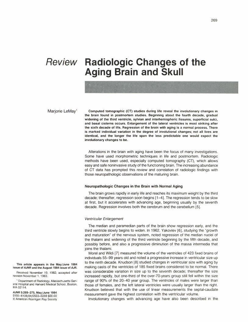
TABLE 1: CT Measurements of Third Ventricle, Anterior End of Sylvian Fissure, Anterior End of Interhemispheric Fissure, and Superficial Sulci with Aging
No. Patients per Age Group in Years Brain Part: Amount Seen on CT
20-29 30-39 40-49 50-59 60-69 70- 79 80+
Third ventricle: None or trace 16 8 2 3 1 0 0 < 2mm . 4 12 16 14 12 10 7 2-4 mm ................. . .... . . 0 0 2 3 7 10 13
Anterior end of sylvian fi ssure: Not seen . . . . . . . . . . . . 15 12 3 1 1 0 0 < 2mm .. 5 7 14 15 13 10 9 2: 2 mm . 0 1 3 4 6 10 11
Anterior end of interhemispheric fissure: Not seen ......................... .. . 13 8 0 2 0 0 0 < 2mm 7 11 18 18 16 14 12 2: 2 mm . . . . . . . . . . 0 1 2 0 4 6 8
Superficial sulci: Not seen 14 10 1 1 0 0 0 < 1 mm . 6 7 8 5 6 1 3 2: 1 mm . 0 3 11 14 14 19 17
Note.- Scans were from 140 patients without intracerebral lesions or obstructive hydrocephalus who were not demented or on medication at the time of the study. Measurements were made on Polaroid film. which showed the brain in transaxial sections reduced in size by a factor of 3.3.
temporal lobes, particularly in the hippocampus, uncus, parahippocampal and fusiform gyri, and about the insula [9, 10]. Von Braunmuhl [1] found these changes to be greater on the left.
Sulcal Enlargement
Widening of the superficial sulci has also been described in the aging brain , particularly in the frontal parasagittal region and in the parietal and temporal lobes [1 , 9, 11]. The reason for this is not clear. Neuronal cells are lost from the cortex, particularly in the frontal and temporal lobes [12-16] , but this causes little change in width of the cortex [15] compared with younger brains.
Meier-Ruge et al. [17] studied the diameter and length of the capillary network in various parts of the brain and found a decrease in the intercapillary distance in the putamen in the elderly, but no significant decrease in the intercapillary distance in the brain cortex. This would suggest that shrinking of the gyri with aging is not a change in width of the cortex but mainly a decrease in volume of subcortical structures.
Radiologic Changes in the Brain with Normal Aging
Ventricular Enlargement
Pneumoencephalographic studies , which allow more sharply defined measurements than do CT scans, have shown widening of the third ventricle beginning about the fourth decade [18-21] . Associated with widening of the third ventricle is regression of the median part of the thalami , manifested by progressive diminution of the massa intermedia, which , in most individuals , connects the thalami in early life. On pneumoencephalography the size and shape of the massa intermedia has shown regression first in its posterosuperior part [21] . In older individuals the anterosuperior part of the massa
intermedia is the last to disappear. The massa intermedia tends to be larger throughout life in women than in men and to persist more frequently in the aging brain of women than in men. The massa intermedia can often be seen on CT scans, but is not sharply defined and cannot be measured easily.
Most pneumoencephalographic studies of the aging brain have shown a slight increase in average size of the lateral ventricles up to the seventh decade and then a more rapid increase in size [19 , 22] . Bruijn [22] believed the measurement of the cella media (the smallest transverse diameter of the body of the lateral ventricles) gave the best correlation with the overall size of the lateral ventricles.
CT studies allow less accurate measurement of the ventricles than do pneumoencephalographic studies . The margins of the ventricles are less sharply defined on CT because of partial volume averaging and also because the shape and size of the ventricles and the skull may change due to variation in angulation of the head in the scanner [23] . The photographic printouts on which measurements are often made vary at times and distort the dimensions of the skull and its contents. The effect is identical to that sometimes observed on television screens when circular objects appear elliptical.
Both linear and planimetric CT measurements of the ventricles have usually been reported to show changes similar to those of pneumoencephalographic studies , that is, a statistical slight progression in size of the lateral ventricles beginning at about the fourth decade and a more striking increase after the sixth decade [24- 27]. The progression in size of the lateral ventricles with age is more variable than that of the third ventricle, and, as shown in neuropathologic studies, many persons in the over-70-years group still have relatively small ventricles .
Table 1 shows the CT findings in 140 patients examined at Massachusetts General Hospital who had no evidence of a local mass lesion or hydrocephalus and were not on medica-
AJNR:5, May/June 1984 AGING BRAIN AND SKULL 271
tion at the time of the study. There were 20 patients in each decade from 20 to 100 years . The third ventricle is invisible or minute on normal scans before the fourth decade, but it is commonly seen clearly by the fifth decade.
Linear Ventricular Measurements
Although measurement of the ventricular span is probably more accurate on coronal than on transaxial CT [28], linear measurements of the ventricles on transaxial CT and pneumoencephalography are comparable [29]. Ratios of the width of the ventricles to the width of the skull or brain are 'probably the most easily made and reproducible ventricular measurements on CT.
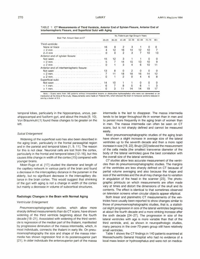
Evans ratio, the width of the greatest span of the frontal horns divided by the greatest width of the internal diameter of the skull, is a frequently used measurement (fig . 1). In normal individuals, even over the age of 60, Evans ratio is usually equal to or less than 0.29 [29, 30] .
The frontal horn ratio is the width of the frontal horns divided by the internal diameter of the skull at the same level [31, 32] (fig . 1). Hahn and Rim [31] , who first described this CT ratio, examined 200 normal brains in patients and volunteers 10-81 years old . The ratio varied from 0.19 to 0.39 (mean 0.31). Except in individuals who have suffered head trauma, the lateral ventricles usually do not enlarge as much with normal aging or degenerative diseases as with hydrostatic hydrocephalus. Although early in the course of obstructive hydrocephalus the bodies of the lateral ventricles may not be large, later they often enlarge more than in individuals with hydrocephalus ex vacuo. In a study of 100 patients with obstructive hydrocephalus and 50 patients with global cerebral atrophy [33] , the frontal horn ratio was found to be 0.5 or greater in 56% of the obstructive group and in only 6% of the atrophic group.
The bicaudate ratio is the width of the ventricles lying between the caudate nuclei divided by the internal diameter of the skull at the same level (fig. 1). This ratio is probably more sensitive in showing change than other linear ratios and it has been used particularly in studies of children . Pelicci et al. [32] considered ratios of 0.17 and lower to be normal and those over 0.2 to be definitely abnormal. Banna [34] considers the top normal bicaudate ratio to be 0.15.
The cella media ratio is the narrowest width of the bodies of the lateral ventricles divided by the greatest internal diameter of the vault. Haug (26) found a gradual increase in the mean cella media ratio with aging. In the 61-71 year range he found a mean ratio of 0.295. It was slightly larger in males than in females.
The third ventricle-sylvian fissure/skull ratio is the sum of the distances between each lateral margin of the third ventricle and the ipsilateral sylvian fissure divided by the internal diameter of the skull at the same level. Brinkman et al. (35) found this ratio to be 0.59 or less in 77% of their demented but in only 24% of their nondemented elderly patients.
Planimetric Ventricular Measurements
Volumetric ratios of the ventricles have been studied by measuring the size of the lateral ventricles with a planimeter
Fig. 1.-Linear ratios used in ventricular measurements. Frontal horn ratio = width of frontal horns (FH)/ internal diameter of vaul t at same level; bicaudate ratio = width of ventricles between caudate nuclei (BC)/ internal diameter of vault at same level; third ventricular ratio = greatest width of third ventricle (3V)/ internal diameter of vault at same level ; Evans ratio = width of frontal horns/greatest internal diameter of "ault (IS) .
[24] or using a grid method (36) and dividing it by the total area of the brain within the CT scan cut. The areas are presumed to be related to the volumes and therefore reflect the size of the ventricles. The measurements are usually made on one or two slices , and the anatomic areas measured may vary in different individuals depending partly on the shape of the head and its angulation in the scanner. Reproducible measurements are more difficult when the ventricles are small.
Volumetric Measurements of Cerebrospinal Fluid (CSF) Intracranial Space
On CT scans of 130 individuals without localized disease, Ito et al. (37) estimated the volume of the brain , CSF (ventricles), and cranial cavity by counting the number of pixels in each tissue on scans taken through the hemispheres. They found the mean CSF volume to increase progressively after the age of 40 years. Zatz et al. (38) studied the volume of intracranial fluid in 123 normal subjects 23-88 years old using semi automated computer analysis , and found a marked individual variation in the older patients but relatively little increase in the volumes until the seventh decade.
Fissural and Sulcal Enlargement
As has been found in neuropathologic studies , involutionary changes in the temporal lobes with widening of the anterior ends of the sylvian fissures is one of the earliest changes seen on CT in the aging brain. Table 1 shows that the anterior ends of the sylvian fissures are commonly seen on CT scans by the fifth decade. In CT studies the anterior end of the left sylvian fissure is more frequently wider than the right , which correlates with the findings of von Braunmuhl (1) , who found greater atrophic changes with aging in the anterior left temporal lobe than in the right on pathologic study.
Widening of superficial sulci also occurs with aging and has been described on pathologic studies, particularly in the parasagittal sulci of the frontal and parietal lobes [1 , 9, 11). On CT scans widening of the interhemispheric fissure is commonly seen anteriorly extending back to, and occasionally just beyond , the callosomarginal sulci , which commonly enlarge, but widening of the fissure more posteriorly is seen
272 LeMAY AJNR:5. May/June 1984
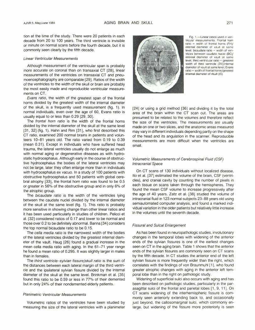
Fig . 2.-Whole brain and CT scan of 63-year-old man. Main sulci . which appear early in fetal life. are deeper and often more easily identified on CT scans than on inspection of the brain. pc = precentral; c = central ; poe = postcentral; cm = callosomarginal.
much less often . Cal a et al. [39] , studying sulcal widening on CT scans of 115 healthy volunteers 14-40 years old , found progressive sulcal widening , mainly in the frontal lobes and the cerebellar vermis, starting in the teens. Although there is statistically some progressive enlargement of the third and lateral ventricles and superficial sulci with age, there is often poor correlation between ventricular size and superficial sulcal widening [25] ; this suggests these atrophic changes may be independent of each other.
When the superficial sulci are widened over the hemispheres, the main sulci (central , pre- and postcentral, and superior frontal) , which are formed early in the developing brain, are deep, and when widened are seen on CT more clearly than on inspection of the brain surface [40] (fig. 2). Widening of superficial sulci seen with aging is often described in radiologic literature as "cortical atrophy, " but this is probably misleading, and "gyral atrophy" or "superficial atrophy" seems preferable. As noted before, neuropathologic studies have shown no statistically significant narrowing of the cerebral cortex with aging. Brody [12 , 13] has described a decrease in cells of the cortex that varies in different regions but is slight in the postcentral gyrus. Nevertheless, on CT the postcentral sulcus widens with aging, much like the central and precentral sulci (figs. 2 and 3). This suggests that the widening of the superficial sulci with aging must be associated ith considerable loss of adjacent white matter. Miller et al. [41] measured the volumes of the gray and white matter and found a relatively constant volume of cerebral tissue until age 50 and then a fairly constant loss per decade. The regression appeared to be mainly due to white-matter loss.
Changes in CT numbers , representing white matter, with normal aging have been reported on CT [42] , but such numbers are greatly influenced by the amount of bone in the overlying vault. A recent CT study of the aging brain that corrected for the thickness and density of overlying bone found no statistically significant change in white-matter density (Sandor T, Albert M, LeMay M, unpublished data).
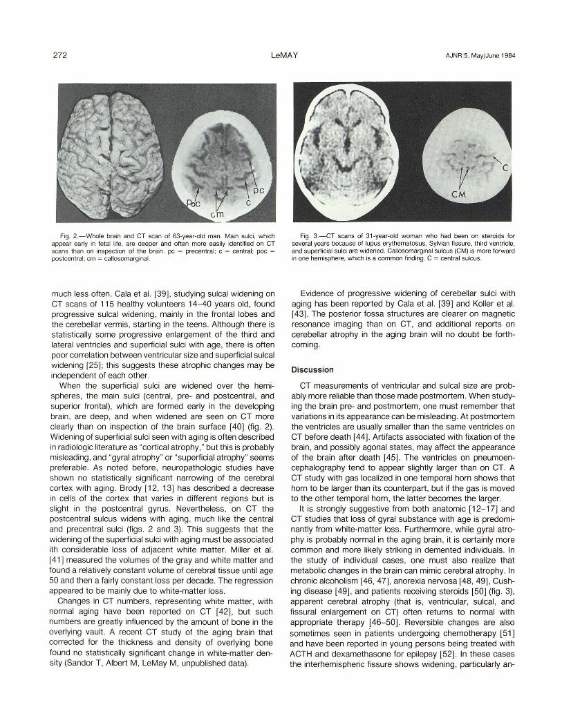
Fig. 3.-CT scans of 31-year-old woman who had been on steroids for several years because of lupus ery1hematosus. Sylvian fissure. third ventricle. and superficial sulci are widened . Callosomarginal sulcus (eM) is more forward in one hemisphere. which is a common finding . C = central sulcus.
Evidence of progressive widening of cerebellar sulci with aging has been reported by Cala et al. [39] and Koller et al. [43] . The posterior fossa structures are clearer on magnetic resonance imaging than on CT, and additional reports on cerebellar atrophy in the aging brain will no doubt be forthcoming.
Discussion
CT measurements of ventricular and sulcal size are probably more reliable than those made postmortem. When studying the brain pre- and postmortem, one must remember that variations in its appearance can be misleading. At postmortem the ventricles are usually smaller than the same ventricles on CT before death [44] . Artifacts associated with fixation of the brain, and possibly agonal states, may affect the appearance of the brain after death [45]. The ventricles on pneumoencephalography tend to appear slightly larger than on CT. A CT study with gas localized in one temporal horn shows that horn to be larger than its counterpart, but if the gas is moved to the other temporal horn, the latter becomes the larger.
It is strongly suggestive from both anatomic [12-17] and CT studies that loss of gyral substance with age is predominantly from white-matter loss. Furthermore, while gyral atrophy is probably normal in the aging brain, it is certainly more common and more likely striking in demented individuals. In the study of individual cases, one must also realize that metabolic changes in the brain can mimic cerebral atrophy. In chronic alcoholism [46, 47] , anorexia nervosa [48,49] , Cushing disease [49], and patients receiving steroids [50] (fig. 3), apparent cerebral atrophy (that is, ventricular, sulcal, and fissural enlargement on CT) often returns to normal with appropriate therapy [46-50]. Reversible changes are also sometimes seen in patients undergoing chemotherapy [51] and have been reported in young persons being treated with ACTH and dexamethasone for epilepsy [52]. In these cases the interhemispheric fissure shows widening, particularly an-
AJNR :5, May/June 1984 AGING BRAIN AND SKULL 273
teriorly , resembling the common involutionary pattern seen in the aging brain.
It is usually not possible on a routine CT study , when there is no evidence of a focal abnormality , to determine whether an individual will show diminished brain function clinically . Persons with diminished cognitive functioning tend to have larger ventricles than those of normally functioning individuals [53-60), but there is great variation in ventricular size in both groups [61-63].
Enlargement of the temporal horn tips is usually one of the earliest signs of pressure hydrocephalus, but the temporal horn tips are usually not seen at all , or are only slightly widened , on CT scans of normally functioning individuals showing generalized atrophic changes. They are more often widened, along with marked widening of the anterior ends of the sylvian fissures and temporal sulci in some individuals with dementia. These CT findings may correlate with the increased number of neurofibrillary tangles and granulovascular degeneration seen pathologically in the anterior temporal lobes of many individuals with a dementing illness [16, 64 , 65], but prominent temporal horn tips on CT studies should alert one to consider the diagnosis of pressure hydrocephalus.
Of the several linear measurements that have been used in recording CT images, the frontal horn and cella media ratios are the two most easily made. They do not show minor changes in ventricular size as accurately as volumetric measurements do [66], but they do help in recording gross differences in ventricular size. The bicaudate ratio is slightly more difficult to measure accurately in adults and is probably more helpful in recording ventricular measurements in children.
Skull Changes with Aging
When studying involutionary changes in the brain it should also be remembered that the skull also often changes in size with age. Studying repeated skull films of the same individuals over a long period of time, Finby and Kraft [67] and Israel [68] found in the majority an increase in size of the vault, facial bones, and paranasal sinuses, and in the thickness of the skull with aging. Most of the skulls also showed some increase in size of the sella turcica. An increase in width of some of the other bones of the body has been found with aging [69, 70]. A recent linear study measuring the breadth , length, and circumference of the heads of 2000 healthy veterans over a 5 year interval showed a statistical increase in these measurements with aging [71]. The phenomenon is presumably associated with the continuous resorption and regrowth of the skeleton and with the stresses to which the skeleton is subjected during life [72]. A few studies of the neurocranium [73, 74] have not found a significant increase in size with aging. However, it is important in such a study that skulls of the same individuals be measured over a long period and similar points of measurement be used.
Over 4000 years ago Ptahhotpe [75] , vizier to an early Egyptian king, described many of the changes in the human body occurring with aging; among them he noted, "the mind has come to an end and cannot remember yesterday. " Hip-
pocrates [76], in the fourth century B.C. , described shrinkage of the brain with aging. Now over 2000 years later we are still studying the anatomic changes with aging. Although we can see them with CT and other new visual modalities , further understanding of the involutionary changes in the brain and their regulation may require other, more sensitive methods.
REFERENCES
1. von Braunmuhl A. Alterserkrankungen des Zentralnervensystems. In: Lubarsch H, Henke F, Rossie R, eds. Handbuch der Speziel/en Pathologischen Anatomie and Histologie, vol. 13. Berlin: Springer-Verlag , 1957:337-539
2. Chernyshev SP. The weight of the human brain . In: Blinkov SM , Glezer II , eds. The human brain in figures and tables. New York: Plenum, 1968:337
3. De Kaban AS, Sadowsky BS. Changes in brain weights during the span of human life: relation of brain weights to body heights and body weights. Ann Neurol Psychiatr 1978;4 :345-356
4. Pakkenberg HL, Voigt J. Brain weight of the Danes. Acta Anat (Basel) 1964;56 :97-307
5. Ellis RJ. Norms for some structural changes in the human cerebellum from birth to old age. J Comp Neurol 1920;32: 1-35
6. Yakovlev PI. Morphological criteria of growth and maturation of the nervous system in man. Ment Retard 1961 ;39:3-46
7. Morel J, Wildi E. General and cellular pathochemistry of senile and presenile alterations of the brain . In: Proceedings of the First International Congress of Neuropathology , vol 2. Rome, 1952:347-374
8. Knudson PA. Veentriklernes Storrelsesforhold: Anatomisk normale Hjerner fra Voksne (thesis). Copenhagen. Odense, Denmark: Andeelsbogtykkeriet , 1958
9. Tomlinson BE, Blessed G, Roth M. Observations on the brains of non-demented old people. Neurol Sci 1968 ;7:331-356
10. Tomlinson BE. Morphological changes and dementia in old people . In: Smith WL, Kinsborne M, eds. Aging and dementia. New York: Spectrum, 1977 :25-26
11 . Hooper MW, Vogel FS. The limbic system in Alzheimer 's disease. Am J Pathol 1976;85: 1-13
12. Brody H. Organization of cerebral cortex. Study of aging in human cortex. J Comput Neuro/1955 ;1 02 :511 - 556
13. Brody H. Cell counts in cerebral cortex and brainstem in Alzheimer's disease. In: Katzman R, Terry RD, Bick KL, eds. Senile dementia and related disease, vol 7. New York : Raven , 1978 :345-351
14. Henderson G, Tomlinson BE, Gibson PH . Cell counts in human cerebral cortex in normal adults throughout life using an image analyzing computer. J Neurol Sci 1980;46 : 11 3-136
15. Shefer VF. Absolute number of neurons and thickness of the cerebral cortex during aging, senile and vascular dementia and Pick's and Alzheimer's disease. Zh Nevropatol Psikhiatr 1972;72: 1 024-1 029
16. Shefer VF. Development of senile plaques in the human brain . Arkh Anat Gistol Embrio/1977 ;73:97-1 03
17. Meier-Ruge W, Hunziker 0 , Schulg U, Tobler HJ , Schweizer A. Stereological changes in the capillary network and nerve cells of the aging human brain. Mech Ageing Dev 1980; 14: 233-234
18. Borgersen D. Width of the third ventricle. Acta Radiol (Stockh) 1966;4:645-661
19. Burhenne HJ, Davis W. The ventricular span in cerebral pneumography. AJR 1963;90 : 1176-1184
274 LeMAY AJNR:5, May/June 1984
20. Engeset A, Lonnum A. Third ventricles of 12 mm width or more. Acta Radiol (Stockh) 1958;50: 5-11
21. LeMay MJ. Neurological aspects of language disorders in the elderly . An anatomical overview. In: Obler LK, Alber ML, eds. Language and communication in the elderly. Lexington, MA: Heath, 1980 : 1 07-119
22. Bruijn JW. Pneumoencephalography in the diagnosis of cerebral atrophy. Utrecht, The Netherlands: Drukkeriz , Smits Oudergracht , 1959
23. Sabattini L. Evaluation and measurements of the normal ventricular system and subarachnoid spaces by CT. Neuroradiology 1982;23 :1-5
24 . Barron SA, Jacobs L, Kinkel WR . Change in size of normal lateral ventricles during aging determined by computerized tomography. Neurology (NY) 1976;26: 11 01-1113
25 . Gyldensted C. Measurements of the normal ventricular system and hemispheric sulci of 100 adults with computed tomography. Neuroradiology 1977;14: 183-192
26. Haug G. Age and sex dependence of the size of normal ventricles on computed tomography . Neuroradiology 1977;14:201-204
27. Meese W, Kluge W, Grumme TT, Hopfermuller W. CT evaluation of the CSF spaces of healthy persons. Neuroradiology 1980;19 :131-135
28 . Wolpert SM . The ventricular size on computed tomography. J Comput Assist Tomogr 1977 ;2:222-226
29. Gawler J, duBoulay GH, Bull JHD, Marshal.1 J. Computerized tomography: a comparison with pneumoencephalography and ventriculography. J Neurol Neurosurg Psychiatry 1976 ;39:203-211
30. Synek V, Tuben JR , duBoulay GH. Comparing Evan 's index and computerized axial tomography in assessing relationship of ventricular size to brain size. Neurology (NY) 1976;26:231-233
31. Hahn FJY, Rim K. Frontal ventricular dimensions or. normal computed tomography. AJR 1976;126:492-496
32. Pelicci LJ , Bedrick AD, Cruse RP, Vannucci RC. Frontal ventricular dimensions of the brain in infants and children . Arch Neurol 1979;35 :852-853
33. LeMay M, Hochberg FH. Ventricular differences between hydrostatic hydrocephalus and hydrocephalus ex vacuo by computed tomography . Neuroradiology 1979;17 : 191-195
34. Banna M. The ventriculo-cephalic ratio on computed tomography. J Can Assoc Radio/1977 ;28:208-210
35. Brinkman SD, Sarwar M, Levin H, Morris HH III. Quantitative indexes of computed tomography in dementia and normal aging. Radiology 1981 ; 138 : 89-92
36. Benes F, Sunderland P, Jones BD, LeMay M, Cohen BM, Lipinski JF. Normal ventricles in young schizophrenics . Br J Psychiatry 1982;141 :90-93
37. Ito M, Hatazawa J, Yamaura H, Matsuzawa T. Age-related brain atrophy and mental deterioration-a study with computed tomography. Br J Radio/1981 ;54:284-390
38. Zatz LM, Jernigan TL , Ahumada AJ Jr. Changes on computed cranial tomography with aging. Intracranial fluid volume. AJNR 1982;3 : 1- 11
39 . Cala LA, Thickbroom GW, Black JL, Collins DWK, Mastaglia FL. Brain density and cerebrospinal fluid space size: CT of normal volunteers. AJNR 1982;2:41 - 47
40 . Kido DK, LeMay M, Levinson AW, Benson W. Computed tomographic localization of the precentral gyrus. Radiology 1980;135 :373-377
41 . Miller AKH , Alston RL, Corsellis JAN. Variations with age in the volume of grey and white matter in the cerebral hemispheres of man: measurements with an image analyser. Neuropathol Appl
Neurobio/1980 ;6 : 119- 132
42 . Zatz LM , Jernigan TL, Ahumada AJ Jr. White matter changes in cerebral computed tomography related to aging. J Comput Assist Tomogr 1981 ;6 : 19-23
43. Koller WC, Glatt S, Fox HJ, et al. Cerebellar atrophy: relationship to aging and cerebral atrophy. Neurology (NY) 1981 ;31 :405-412
44. Jacobs L, Kinkel WR, Heffner RR. Autopsy correlations of computerized tomography: experience with 6000 CT scans. Neurology (NY) 1976;26: 1111-1118
45. Messert B, Wannamaker BB, Dudley AW. Reevaluation of the size of lateral ventricles of the brain . Neurology (NY) 1971 ;22:941-951
46. Artmann H, Gall MV, Hacker H, Herlieb J. Reversible enlargement of cerebral spinal fluid spaces in chronic alcoholics. AJNR 1981;2:23-27
47 . Carlen PL, Wortzmann G, Holgate RC, Wilkinson DA, Rankin JG. Reversible cerebral atrophy in recently abstinent chronic alcoholics measured by computed tomography scans. Science 1978;200: 1 076-1 078
48 . Enzmann DR , Lane B. Cranial computed tomography findings in anorexia nervosa. J Comput Assist Tomogr 1977;1 :410-414
49. Heinz ER , Martinez J, Haenggeli A. Reversibility of cerebral atrophy in anorexia nervosa and Cushing 's syndrome. J Comput Assist Tomogr 1977;1 :415-418
50. Bentson J, Resa M, Wilson G. Steroids and apparent cerebral atrophy on computed tomography scans. J Comput Assist Tomogr 1978 ;2:16-23
51. Enzmann DR , Lane B. Enlargement of subarachnoid spaces and lateral ventricles in pediatric patients undergoing chemotherapy. J Pediatr 1978 ;92:535-539
52. Langenstein L, Willig RP, Kuhne D. Cranial computed tomography (CCT) findings in children with ACTH and dexamethasone. First results. Neuropediatrics 1979;10:370-384
53. deLeon MJ, Ferris SH, George AE, et al. Computed tomography evaluation of brain behavior relationships in senile dementia of Alzheimer's type. Neurobiol Aging 1980;1 :69-79
54. Gonzalez CF, Lantieri RL, Nathan RJ. The CT scan appearance of the brain in the normally elderly population: a correlative study. Neuroradiology 1978;16: 120-122
55. Hubbard BM , Anderson JM. Age, senile dementia and ventricular enlargement. J Neurol Neurosurg Psychiatry 1981;44:631-635
56. Huckman MS, Fox J, Topel J. The validity of criteria for the evaluation of cerebral atrophy by computed tomography. Radiology 1975;116 :85-92
57. Jacoby R, Levy R. CT scanning and the investigation of dementia. A review. J R Soc Med 1980;13 :366-369
58. Kasniak AW, Garon DC, Fox JH , et al. Cerebral atrophy EEG slowing , age, education , and cognitive functioning in suspected dementia. Neurology (NY) 1979;29: 1273-1279
59. Roberts MA, Caird FI. Computerized tomography and intellectual impairment in the elderly. J Neurol Neurosurg Psychiatry 1976;39:986-989
60. Roberts MA, McGeorge AO, Caird FI. Electroncephalography and computerized tomography in vascular and nonvascular dementia in old age. J Neurol Neurosurg Psychiatry 1978;41 :903-906
61 . Earnest MP, Heaton RK, Wilkinson WE, Manke WF. Cortical atrophy. Ventricular enlargement and intellectual impairment in the aged. Neurology (NY) 1979;29: 138-143
62 . Hughes CO, Gado M. Computed tomography and aging of the brain . Radiology 1981 ;139:291-396
63. Jacoby RJ, Levy R, Dawson JM. Computed tomography in the elderly: the normal population . Br J Psychiatry 1980;136 :249-255
64. Dyan AD. Quantitative histological studies on the aged human
AJNR :5, May/June 1984 AGING BRAIN AND SKULL 275
brain . II. Senile plaques and neurofibrillary tangles in senile dementia. Acta Neuropathol (Berlin) 1970;16 :95-1 02
65. Kemper TL. Senile dementia: A focal disease in the temporal lobe. In : Nandy K, ed. Senile dementia: a biomedical approach. Developments in neuroscience , vol3 . Amsterdam: Elsevier/North Holland Biomedical , 1978 :105-113
66. Gado M, Hughes CP, Danziger W, Chi D, Jost G, Berg L. Volumetric measurements of the cerebrospinal fluid spaces in demented subjects and controls . Radiology 1982 ;144 :535-538
67. Finby N, Kraft E. The aging skull: comparative roentgen study, 25-34 year interval. Clin Radiol 1972;23:410-414
68. Israel H. The dichotomous pattern of craniofacial expansion. Am J Phys Anthropo/1977;47 :47-52
69 . Sedlin E, Frost HM, Villaneuva AR . Variations in cross-section area of rib cortex with age. J Geronto/1963;18:9-13
70. Smith RW, Walker RR. Femoral expansion in aging women.
Henry Ford Hosp Med J 1980 ;28 :168- 170 71. Friedlander JS, Costa PT, Bosse R, Ellis J, Rhoads JG, Stoudt
HW. Longitudinal physique changes among healthy white veterans at Boston. Hum Bioi 1977;49 :541-558
72. Johnson L. The kinetics of skeletal remodeling . Birth Defects 1966;2:66-142
73. Boersma H. Alteration of skull dimensions in aged persons. J Dent Res 1974;53 :678- 682
74. Tallgren A. Neurocranial morphology and aging- longitudinal roentgen cephalometric study of adult Finnish women. Am J Phys Anthropo/1974;41 :285-293
75. Simpson WK , ed . Ptahhotpe: the literature of ancient Egypt. New Haven: Yale University, 1973 :60
76. Farr S. The history of epidemics by Hippocrates. London: Cadell , 1780