Embed Size (px)
Citation preview

REVIEW RENAL FUNCTION
UNIVERSITY OF PNGSCHOOL OF MEDICINE AND HEALTH SCIENCES
DIVISION OF BASIC MEDICAL SCIENCES DISCIPLINE OF BIOCHEMISTRY & MOLECULAR BIOLOGY
PBL MBBS IV VJ. Temple
1

What are some of the functions of the kidneys?
• Regulation of water / fluid balance:
• Arginine Vasopressin (AVP) stimulates formation of Aqua-porins in Tubular cell, increasing reabsorption of Water from Glomerular filtrate;
• Regulation of Electrolyte:
• Aldosterone acts on Tubules causing reabsorption of Na+
ions in exchange for secretion of K+ ions and H+ ions,
• Regulation of Acid-Base balance:
• Maintenance of pH in blood and other body fluids,
• Excretion of metabolic waste products of Protein and Nucleic acid:
• Urea, Creatinine, Creatine, Uric acid, Sulphate, Phosphate2

• Parathyroid Hormone (PTH): Acts via the Kidneys:
• To promote Tubular Reabsorption of Calcium ions,
• For biosynthesis of 1,25-Dihydroxy-Cholecalciferol (Vit D3) that regulates Calcium absorption in GIT;
• Renin from Juxtaglomerular cells in kidneys regulate Aldosterone production:
• Renin converts Angiotensinogen to Angiotensin-1,
• Angiotensin Converting Enzyme (ACE) converts Angiotensin-1 to Angiotensin II,
• Angiotensin II stimulates biosynthesis of Aldosterone in Adrenal Cortex,
• Erythropoietin that promotes biosynthesis of Hb is partly regulated by kidneys,
3

What are the Renal Function Tests?
• Renal Function Tests: Procedures and Tests to evaluate Functional State of kidneys:
• Tests for Glomerular Function;
• Tests for Tubular Function;
• Specimens used are:
• Urine,
• Plasma or Serum,
4

• Renal Function Tests Include the following:
• Urinalysis: First line test for Renal Function,
• Creatinine Clearance (CC): to measure Glomerular Filtration Rate (GFR),
• Inulin Clearance: to measure GFR,
• Para-Amino-Hippuric Acid (PAH): to measure Renal Plasma Flow (RPF),
• Urine Osmolality,
• Plasma Creatinine,
• Plasma Urea,
• Plasma Electrolyte; 5

What test are carried out during urinalysis?
• Randomly collected urine sample is examined:
• Physically for:
Color, Odor, Appearance, Concentration (specific gravity) or Osmolarity;
• Chemically for:
• Protein, Glucose, Urine pH (acidity/ alkalinity);
• Microscopically for:
• Cellular elements (RBC, WBC, Epithelial cells),
• Bacteria, Crystals, Casts (deposit of protein, cells, and other substances in kidney tubules);
6

What is Glomerular Filtration Rate (GFR)?
• GFR: useful index of numbers of functioning Glomeruli,
• GFR: amount of filtrate kidneys made per minute,
• GFR: maximum rate that plasma can be ‘Cleared’ of a substance;
• GFR is related to body size and age, higher in males compared to females; reduced rate in elderly,
• Reduction in GFR can be caused by:
• Restriction of Renal blood supply,
• Low Cardiac Output,
• Destruction of Nephrons by Renal Diseases, etc
• Reduction in GFR results in Retention of Waste Products of Metabolism in blood;
7

How is GFR (Creatinine Clearance) calculated?
• GFR is directly related to Clearance,
• GFR can be calculated from Clearance of a compound in Plasma that is freely filtered at Glomerulus, and is not reabsorbed or Secreted by Tubules,
• Creatinine: normal product of muscle metabolism in blood is used to calculate GFR (Creatinine Clearance);
• GFR is calculated from Creatinine content of 24-hrs urine collection, and Plasma concentration of Creatinine within the 24-hrs period,
• Inulin can be used to measure GFR because it is filtered but not re-absorbed or secreted by Renal Tubules,
8

Take Note:
• GFR must be corrected for body surface area of patients;
• Correction factor is calculated from Age and Height of patient in relation to “Standard” Average Body Surface Area of an adult;
• ‘Standard’ average body surface area = 1.73m2 ;
• It is a common mistake to consider V as urine volume;
• V is Urine Flow Rate: Volume of Urine collected in 24hrs, expressed in ml/min
9

• Calculation of GFR or Creatinine Clearance (CC):
GFR = CC = (U x V)/P
• Where U = Urine concentration of Creatinine (mmol/L);
• P = Concentration of Creatinine in Plasma or Serum (mmol/L; mol/L)
• V = Urine Flow Rate (ml/minute);
10

How is GFR or CC calculated using Cockcroft and Gault equation?
(140 – Age in yrs) x Weight (Kg)
• CC (ml/min) =
0.814 x Serum Creatinine (umol/L)
• To correct for muscle mass:
• For Female multiply result by 0.85
• For Male multiply by 1.22
11

Another form of the Cockcroft and Gault equation
(140 – Age in yrs) x Weight (Kg)
• CC (ml/min) =
72 x Serum Creatinine (umol/L)
• NB: For Female multiply result by 0.85
• Limitations of Cockcroft and Gault equation:
• Patients should not be severely malnourished,
• Patients should not be very obese,
• Renal Function should not be severely impaired (GFR < 20 ml/min)
12

What is Proteinuria?
• Glomerular filtrate is an ultra-filtrate of plasma;
• Glomerular basement membrane does not allow passage of albumin and large molecular weight proteins,
• Small amount of protein, (<25mg/24h) may be in urine,
• Positive screening test for protein (routine urinalysis) on random urine sample should be followed-up with test on 24-h urine sample that precisely measures quantity of protein in urine,
• Protein, in excess of 250mg/24h urine sample indicates significant damage to Glomerular membrane,
• Persistent presence of significant amounts of protein in urine, is an indicator of kidney disease;
13

What are the different types and causes of Proteinuria?
• Glomerular Proteinuria:
• Abnormal leaking of large and small molecular weight proteins into filtrate resulting from damaged of Glomerular membrane,
• May be due to:
• Exercise,
• Fever (Febrile Proteinuria),
• Congestive Cardiac Failure,
• Glomerulonephritis,
• Renal Stenosis,
14

• Glomerulonephritis:
• Common cause of persistent Proteinuria
• Amount of protein in urine depends on:
• Extent of Glomerular damage,
• Molecular mass of protein,
• Capacity of Tubule to reabsorb or metabolize proteins
• May be mild, moderate or Severe Proteinuria
• Severe Proteinuria:
• Protein loss in urine exceeds synthetic capacity of liver to replace protein, resulting in Hypo-Proteinemia
• Severe persistent Proteinuria is one of the features of Nephrotic Syndrome,
15

• Nephrotic Syndrome:
• Large amount of protein loss in urine,
• Leads to Hypo-Proteinemia and Edema,
• Edema may be caused by low albumin or secondary Hyper-Aldosteronism,
• Patients may also develop Hyperlipidemia,
• Some causes of Nephrotic Syndrome:
• Glomerulonephritis,
• Systemic Lupus Erythematosus,
• Diabetes Nephropathy
16

• Tubular Proteinuria:
• Failure of Tubules to reabsorb filtered plasma proteins,
• Abnormal secretion of protein into urinary tract,
• May be due to Tubular or Interstitial damage,
• Proteins with low molecular wt are excreted by Tubules,
• Loss of protein is mild about 2.0g/24h urine sample,
• Sensitive test for assessment of Renal Tubular damage:
• Measure Urinary β2-Microglobulin: Values greater than 0.4mg/24h indicates tubular damage
17

• Overflow Proteinuria:
• Large amount of low molecular weight proteins in urine,
• Proteins are filtered at Glomerulus, but not reabsorbed or metabolized completely by Tubules,
• Some causes of Overflow Proteinuria:
• Acute Pancreatitis,
• Multiple Myeloma,
• Intravascular Hemolysis,
• Myelomonocytic Leukemia,
• Crush Injuries
• Orthostatic (Postural) Proteinuria:
• Proteinuria occurs after standing for a long time,
• Protein absent in early morning urine samples,18

Use of Plasma Creatinine in Renal Function Test
• Creatinine is a by-product of muscle energy metabolism,
• Creatinine is cleared from blood and excreted in urine,
• Level of creatinine in plasma depends on muscle mass, thus normally Creatinine in blood remains relatively constant,
• Plasma Creatinine level is inversely proportional to CC or GFR,
• Plasma Creatinine level is not affected by Liver function,
• Elevated Plasma Creatinine is sensitive indicator of impaired Renal function,
• Normal Plasma Creatinine conc. of a patient does not always indicate normal Renal function,
• Progressive rise in serial Plasma Creatinine levels may indicate impaired Renal function,
19

Use of Blood Urea Nitrogen (BUN) {H2N-CO-NH2 }
• Urea is a by-product of protein metabolism,
• Urea is formed in liver, released in blood then filtered by Glomerulus and excreted in urine,
• BUN is the amount of Nitrogen contained in Urea,
• High BUN indicates kidney dysfunction, but because BUN is also affected by Protein intake and Liver Function, the test is done in conjunction with Plasma Creatinine, a more specific indicator of kidney function
• Elevated BUN is suggestive, but not diagnostic of kidney dysfunction, because BUN is affected by other factors;
20

Other parameters in blood for assessing kidney function
• Measurement of blood levels of other compounds that are regulated in part by kidneys are useful in evaluating kidney function:
• These include:
• Electrolytes: Sodium, Potassium, Chloride;
• Bicarbonate, Calcium, Magnesium, Phosphorus,
• Protein,
• Uric Acid,
• Glucose
21

RENAL TUBULAR FUNCTION TESTS
• Glomeruli provide an efficient filtration mechanism for removal of waste products and toxic substances;
• Tubular reabsorption must be efficient to ensure that important constituents such as: Water, Sodium, Glucose, and Amino Acids are not lost in urine;
• About 180 liters of fluid is filtered by Glomerulus each day, and more than 99% is reabsorbed by Tubules;
• Of all tubular functions, the most frequently affected by disease is ability to concentrate the urine;
• Tubular function can be assessed by comparing Osmolality of Urine and Plasma;
22

• For “healthy” person under normal Physiological conditions Urine is more concentrated than Plasma
Urine Osmolality > Plasma Osmolality
• Urine-Plasma Osmolality Ratio is between 1.0 and 3.0,
• Urine / Plasma ratio < 1.0,
• Indicates poor reabsorption by Renal Tubules,
• Some disorders of Tubular function are inherited;
• Some patients cannot reduce their urine pH below 6.5, because of specific failure of Hydrogen ion secretion;
23
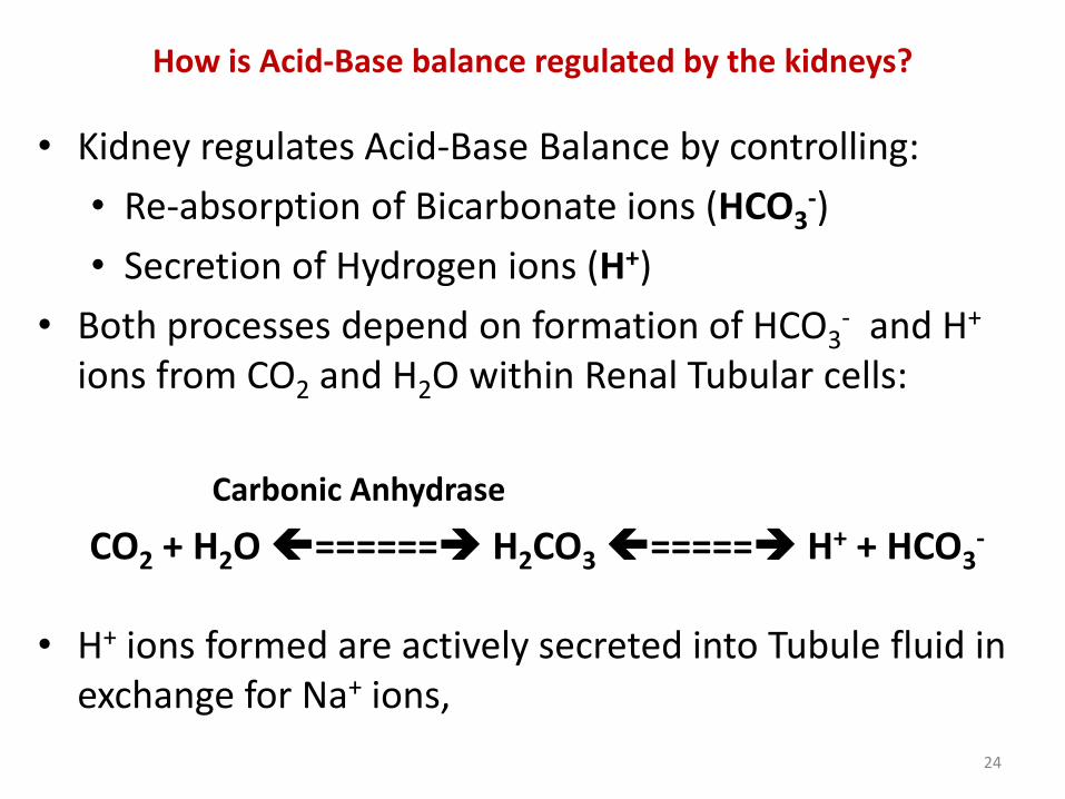
How is Acid-Base balance regulated by the kidneys?
• Kidney regulates Acid-Base Balance by controlling:
• Re-absorption of Bicarbonate ions (HCO3-)
• Secretion of Hydrogen ions (H+)
• Both processes depend on formation of HCO3- and H+
ions from CO2 and H2O within Renal Tubular cells:
Carbonic Anhydrase
CO2 + H2O ====== H2CO3===== H+ + HCO3-
• H+ ions formed are actively secreted into Tubule fluid in exchange for Na+ ions,
24

What mechanisms are used in the kidney for elimination of Acids?
• Mechanisms for elimination of Acids:
• Re-absorption of Sodium Bicarbonate (NaHCO3) by Proximal Renal Tubules, (Fig. 1);
• Regeneration of HCO-3 by Distal Renal Tubules (Fig. 2);
• Formation of Phosphate buffer in Distal Tubules (Fig. 3);
• Production of Ammonia (NH3) by Distal Renal Tubules for formation of Ammonium buffer (Fig. 4);
• Secretion of H+ ions by Tubular cells serves initially to reabsorb HCO3
- ions from the Glomerular filtrate;
• After all the HCO3- ions have been reabsorbed, any
deficit that occurs is regenerated;
25

Fig. 1: Reabsorption of Bicarbonate by Renal Tubules
26

Fig. 2: Regeneration of Bicarbonate ions by Renal Tubules
27

Fig. 3: Formation of Phosphate Buffer in Renal Tubules
28

Fig. 4: Formation of Ammonium Buffer in Renal Tubules
29

What is Anion Gap?
• Anion Gap (AG) calculation is the sum of routinely measured Cations minus routinely measured Anions:
Anion Gap = (Na+ + K+) – (Cl- + HCO3-)
• However, because K+ is a small value it is usually omitted from the AG equation; the most commonly use equation is:
Anion Gap = Na+ - (Cl- + HCO3-)
30

• Venous value of HCO3- should be used in calculation;
• Venous value of CO2 can be used in place of Bicarbonate
The equation will then be: AG = Na+ - (Cl- + CO2)
• Normal AG calculated without K+ is about 12.4mEq/L;
31

• Anion Gap exists because not all Electrolytes are routinely measured;
• Normally there is electrochemical balance in cells; thus the sum of all Anions equals the sum of all Cations;
• However, several Anions are not measured routinely, leading to the Anion Gap;
• Anion Gap is thus an artifact of measurement, and not a Physiologic reality;
32