Embed Size (px)
Citation preview

This article has been accepted for publication and undergone full peer review but has not been
through the copyediting, typesetting, pagination and proofreading process, which may lead to
differences between this version and the Version of Record. Please cite this article as doi:
10.1111/1749-4877.12318.
This article is protected by copyright. All rights reserved.
REVIEW
Running title: Oxytocin Pharmacology and Behavior
Oxytocin Structure and Function in New World Monkeys: From Pharmacology
to Behavior
Aaryn Mustoe, Jack H. Taylor, Jeffrey A. French
Program in Neuroscience and Behavior, University of Nebraska at Omaha, Omaha
NE 68182
Corresponding Author:
Aaryn Mustoe Allwine Hall 527 University of Nebraska at Omaha Omaha NE 68182 [email protected]
Abstract
Oxytocin (OT) is a hypothalamic nonapeptide that mediates a host of physiological and
behavioral processes including reproductive physiology and social attachments. While the
OT sequence structure is highly conserved among mammals, New World monkeys (NWMs)
represent an unusual „hot spot‟ in OT structure variability among mammals. At least six
distinct OT ligand variants among NWMs exist, yet it is currently unclear whether these

This article is protected by copyright. All rights reserved.
evolved structural changes result in meaningful functional consequences. NWMs offer a new
area to explore how these modifications to OT and its canonical G-protein coupled OT
receptor (OTR) may mediate specific cellular, physiological, and behavioral outcomes. In this
review, we highlight relationships between OT ligand and OTR structural variability
specifically examining coevolution between OT ligands, OTRs, and physiological and
behavioral phenotypes across NWMs. We consider whether these evolved modifications to
the OT structure alter pharmacological profiles at human and marmoset OTRs including
changes to receptor binding, intracellular signaling, and receptor internalization. Finally, we
evaluate whether exogenous manipulation using OT variants in marmoset monkeys
differentially enhance or impair behavioral processes involved in social relationships between
pairmates, opposite-sex strangers, and parents and their offspring. Overall, it appears that
changes to OT ligands in NWMs result in important changes ranging from cellular signaling
to broad measures of social behavior.
Keywords: oxytocin, oxytocin receptor, marmoset, social behavior, receptor
pharmacology, cell signaling
Introduction
Oxytocin (OT) can fairly be characterized as an essential element or the sine qua non of
eutherian, placental mammals. This nine amino acid peptide hormone, synthesized by
neurons in the supraoptic (SON) and paraventricular (PVN) nuclei of the neurohypophyseal
tract, regulates the two physiological characteristics that are ubiquitous for female placental
mammals. First, peripherally-secreted OT acts upon cells within the myometrium and
endometrium of the uterus to stimulate uterine smooth muscle contractions associated with
parturition and live birth (Arrowsmith & Wray 2014). Second, OT acting on the
myoepithelial cells in the mammary gland stimulates milk ejection during lactation (Crowley
2015). Clearly, successful offspring birth and postnatal growth are critically dependent on
these two OT-mediated facets of female reproductive physiology.

This article is protected by copyright. All rights reserved.
In addition to these important peripheral functions, OT release from oxytocinergic
neurons, particularly from the PVN, also project to critical mesolimbic and forebrain regions,
both through direct synaptic connections, and via volume transmission from neuronal
dendrites and soma (Ludwig & Leng 2006). These projections are known to mediate a host of
social processes. The canonical social relationship mediated by OT is the bond between
mothers and their offspring (Rilling & Young 2014). These social bonds ensure continued
spatial proximity of mother to offspring and offspring receipt of lactational sustenance, both
of which are necessary for growth and survival. It is no surprise, then, that the same neural
architecture that underlies maternal-offspring attachments also mediate other social
processes. This neural architecture appears to be critical for close affiliative social
relationships, and OT is an important element in the complex phenomenon associated with
the rewarding properties of affiliative social interactions (Lieberwirth & Wang 2014; Numan
& Young 2016). These social relationship traits include pair bonding among reproductive
adults, “friendships” among nonreproductive partners and social group members, and more
multifaceted sociocognitive traits such as trust, emotional intelligence, social memory, in-
group social preferences, and altruism (Feldman 2017).
The hypothalamus also contains neurons that synthesize an additional nonapeptide,
arginine vasopressin (AVP). AVP has considerable structural similarity to mammalian OT,
differing only at positions 3 and 8 in the mature peptides. AVP exerts significant effects on
peripheral function (particularly osmoregulation and vascular tone), but, like OT, AVP
projections in the CNS are involved in the regulation of multiple behavioral systems
including social attachments among adults, social communication and cognition, and
aggression (Caldwell & Young 2006; Caldwell et al. 2008; Albers 2012; Goodson 2013;
Kelly & Goodson 2014). In this review we focus primarily on OT, but given the structural

This article is protected by copyright. All rights reserved.
similarity of AVP and its important role in the modulation of social behavior, we also explore
details of AVP signaling at OT receptors (OTRs).
Structural Changes in Nonapeptides
A necessary condition for both the peripheral and central effects of OT and AVP is the
close coupling of the ligand with the cognate receptor(s). Nonapeptide-receptor binding
results in a host of changes in cell properties that underlie the modulation of both
physiological and behavioral states. Even subtle changes in ligand structure can alter binding
properties with receptors and can differentially activate signaling cascades (Koehbach et al.
2013). Successful docking of nonapeptides with the extracellular and/or transmembrane
elements of the receptors normally activate G protein-mediated intracellular processes
(Busnelli et al. 2012; Stoop 2012; Stoop 2014; Grinevich et al. 2016), and, from a behavioral
perspective, consequently alter neuronal function in cells that express receptors (Busnelli &
Chini 2017). As with any ligand-receptor signaling system, a variety of alterations or
modifications to the signaling molecule can enhance or inhibit cellular responses following
OTR binding (Gimpl & Fahrenholz 2001; Zingg & Laporte 2003; Wyttenbach et al. 2008;
Busnelli et al. 2016). An additional feature that makes the OT/AVP system intriguing is the
ability of both nonapeptides to bind with some affinity with both OT and AVP receptors
(Gruber et al. 2012; Manning et al. 2012). These „crosstalk‟ interactions between OT and
AVP can exert significant physiological changes (Chini et al. 1996; Meyer-Lindenberg et al.
2011) and alter behavioral outputs (Song et al. 2016; Caldwell 2017; Song & Albers 2017).
In the last few years, reports have emerged in the literature of nucleotide changes in the
coding regions for the OT gene (OXT) that leads to altered OT peptide structure among some
mammals, particularly in New World monkeys (NWMs) (Lee et al. 2011; Ren et al. 2015;
Vargas-Pinilla et al. 2015). These findings serve as a springboard to reevaluate the
This article is protected by copyright. All rights reserved.
assumption that the OT ligand is conserved across mammals, and to also explore the
consequences of this variation for cellular and behavioral outcomes. This review will first
highlight the broad phylogenetic and evolutionary lineage of nonapeptides, ultimately
focusing on the recent evidence for changes in the OXT gene in NWMs. As of this writing,
six structural OT variants (including the „consensus‟ mammalian sequence: Cys–Tyr–Ile–
Gln–Asn–Cys–Pro–Leu–Gly–NH2; Leu8-OT) have been identified in NWMs. We then
characterize correlated changes in the OTR associated with ligand variation in NWMs. We
also evaluate species differences among primates in AVP receptor structure, focusing on the
AVP V1a receptor gene (AVPR1a). Second, we review details of nonapeptide ligand-
receptor interactions in NWMs, focusing on receptor pharmacology and intracellular
signaling pathways. This section highlights the ways in which ligand variation in OT can
alter G-protein signaling cascades and points to the multiple ways in which ligand-receptor
interactions may ultimately alter neural function serving as potent modulators for behavioral
outputs. In our third section, we provide a description of the subtle and context-dependent
nature of ligand-specific modulation of social behavior via exogenous manipulation of
OT/AVP systems in marmoset monkeys (Callithrix spp.).
New World Monkey OT Evolution and Diversity
For those less familiar with primate taxonomy, we first provide a brief introduction to the
evolution and phylogeny of the primate lineage, with the most detailed treatment of the
NWMs. Diverging from other mammalian forms approximately 90 million years ago (Mya),
the Order Primates are currently classified into five major groupings, as derived from
extensive genomic phylogenies (Perelman et al. 2011). The most ancient primates include
Lemuriformes (lemurs and lorises) and Tarsiformes (tarsiers). The monkey and ape-like
primates (Simiiformes) include four major groupings including two groups of hominoid

This article is protected by copyright. All rights reserved.
primates: Hylobatidae (gibbons and siamangs) and hominid primates (orangutans, gorillas,
chimpanzees, bonobos, and humans). The African and Asian Old World primates
(superfamily Cercopithecoidea; e.g., macaques, baboons, guenons) constitute the third
grouping of Simiiformes, and the NWMs of South and Central America (parvorder
Platyrrhini) fill out the primate order. NWMs are estimated to have diverged from Old World
primates and hominoids ~43 Mya (Perelman, Johnson, Roos, et al. 2011). Within this
taxonomic group there are 17 genera that are classified into three families (see Fig. 1).
Atelidae are large-bodied (7 – 9 kg) and Pitheciidae are intermediate sized (1 – 3 kg), and
generally have diets of fruits and leaves. Cebidae are small-bodied primates (0.12 – 2.5 kg)
with more variable diets among species, including invertebrate and vertebrate prey (Hawes &
Peres 2014).
Nonapeptide evolution. Hypothalamic nonapeptides have a long evolutionary history –
these molecules are expressed in a number of invertebrate species, and appear to have
emerged approximately 600 million years ago (Gruber et al. 2012; Beets et al. 2013). During
the course of evolution, vasopressin-like molecules appear to be the „ancestral‟ peptide in
vertebrates (vasotocin), and the similarity of peptide structure and the close proximity of the
coding regions for OXT and AVP genes on the same chromosome in primitive jawed fishes
suggests a gene duplication event of very early evolutionary origin, first emerging in
ancestral jawed fishes ca. 500 million years ago (Gruber et al. 2012; Beets et al. 2013;
Koehbach et al. 2013). The vertebrate OT family (isotocin-mesotocin-oxytocin) is generally
associated with reproductive function, while the AVP family (vasotocin-vasopressin) is
presumed to have origins in fluid homeostasis (Acher et al. 1995). These two nonapeptide
families have significant similarities beyond both possessing nine amino acids, including an
N-terminal six-residue ring structure formed by a disulfide bridge between two Cys residues
This article is protected by copyright. All rights reserved.
at positions 1 and 6, a flexible C-terminal tail structure with a Pro and Gly at positions 7 and
9, and Asn at position 5.
Given the importance of the peripheral actions of OT for reproduction in placental
mammals, it is not surprising that an implicit assumption in the literature has been that the
mature OT peptide is highly conserved among the nearly 4,000 species of eutherians (Acher
et al. 1995; Caldwell et al. 2008; Donaldson & Young 2008; Insel 2010; Gruber 2014).
Comparative genomics analyses have supported this proposition. Wallis (2012) analyzed
variation in the 27 nucleotides that code for the mature OT peptide across placental
mammals, and calculated dN/dS ratios (the number of nonsynonymous nucleotide
substitutions relative to the number of synonymous substitutions), in which a ratio < 1.0
implies stabilizing selection for protein structure, while a ratio > 1.0 implies positive
selection for diversity in nucleotide sequences and hence protein variability (Wallis 2012).
The dN/dS ratio for eutherian OXT gene is remarkably low (0.009), reflecting a highly
conserved protein structure that is consistent with the centrality of OT for critical
reproductive and social processes in mammals. Wallis (2012) reported an even lower dN/dS
ratio for nucleotides coding for AVP gene among mammals (0.005), again implying an
extreme conservation in the AVP ligand structure.
In light of the evidence for conserved OT ligand structure in mammals, it comes as some
surprise that there is, in fact, considerable variation in OT structures among the NWMs. The
first data suggesting multiple ligand forms were published in 2011 (Lee et al. 2011), and
indicated that four species of NWMs sequenced for OXT (Callithrix, Aotus, Saimiri, and
Cebus) showed an in-frame nonsynonymous substitution (NS) in the OXT codon for the 8th
amino acid (CCGCTG). Wallis (2012) also reported a similar NS substitution in the tree
shrew (genus Tupaia). This NS leads to an alteration in the amino acid at the 8th position

This article is protected by copyright. All rights reserved.
from the consensus mammalian leucine (Leu) to a proline (Pro), yielding a unique
nonapeptide variant, Pro8-OT. Lee et al. (2011) further showed that this substitution was
expressed in mRNA and that neurohypophysial extracts contained Pro8-OT, indicating that
the altered OXT codon at position 8 in these species is translated into a mature Pro8-OT
structured peptide.
In 2015, two laboratories independently conducted a broader comparative analysis of
NWMs (Ren et al. 2015; Vargas-Pinilla et al. 2015), both confirming the presence of Pro8-
OT in the species sampled in Lee et al. (2011), and revealing additional variability in OXT in
this taxonomic group, ultimately characterizing six OT-like variants (Leu8-OT, Pro
8-OT,
Val3-Pro
8-OT, Phe
2-OT, Ala
8-OT, and Thr
8-OT). Accession of available sequence data from
NCBI for primates revealed that all species outside NWMs possess the consensus mammalian
Leu8-OT. Fig 1 characterizes the mapping of these OT variants on the consensus molecular
phylogeny of NWM, and full details on the amino acid substitutions in NWMs can be found
in French et al. (2016). All three Families of NWMs have at least two genera that have
divergent OT ligands from Leu8-OT, and there appears to have been multiple NS nucleotide
changes in Pitheciidae and Atelidae, since exemplar genera contain at least three variants of
OT. All genera of Cebidae, a Family that separated from the other NWM ~ 23 Mya, have
OXT coding regions for Pro8-OT, suggesting that the mutation that was associated with the
evolutionary divergence of this Family. Within the Cebidae, at least one species in the genera
Saguinus expresses an additional NS in the codon at position 3 (Valine for Isoleucine;
Vargas-Pinilla et al. 2015) as well as Proline at position eight. This finding, along with the
emergence of relatively recent (7.5 – 9 Mya) variants in the other Families, indicates that
mutations in the coding regions of OXT in NWM have continued after the separation of these
families, and that some mutations have occurred in relatively recent evolutionary time. There
has been a report of a seventh OT variant in NWMs, the howler monkey, Alouatta seniculus.

This article is protected by copyright. All rights reserved.
Campbell and Cortés-Ortiz (personal communication, Nov 2017) have sequenced a Val3-
Leu8-OT variant in this taxon. The potential existence of other, additional OT variants awaits
a more complete and systematic sampling of species within the NWMs.
The OT variants in NWMs represent both structural and physicochemical divergence
from consensus Leu8-OT. The Pro
8 substitution, found in all Cebids and one genera of
Atelidae, represents the most dramatic alteration from other variants, since the Pro-Pro
sequence in the tail portion of the OT peptide at positions 7 and 8 places constraints on the
rotational structure of the peptide, and can significantly alter ligand-cell membrane
interaction profiles (Geisler & Chmielewski 2009). In addition, Pro8-OT differs in its
hydrophilic profile from Leu8-OT, with greater hydrophilicity in Pro
8 (Ren et al. 2015;
Parreiras-e-Silva et al. 2017). For all OT variants in NWMs, each change in amino acid
residues represents at least one physicochemical change (polarity, charge, or hydrophobicity),
and hence each can be characterized as a „radical‟ residue substitution (Zhang 2000). Indeed,
Stoop (2012) argued that the structural changes associated with the Pro8 substitution
represents a more dramatic change in the molecular architecture of OT than the
corresponding changes from mesotocin to consensus Leu8-OT (Isoleucine to Leucine). In the
OTR, both the N-terminus, which shows considerable structural variability across NWMs
(Ren et al. 2015), and the first extracellular loop of the OTR are thought to be important for
ligand selectivity and binding (Zingg et al. 2003), with the latter thought to be primarily
interacting with the ligand C-terminus tail (where the majority of substitutions are found at
the 8th
position) of the OT peptide (Chini et al. 1996; Wesley et al. 2002). Thus, changes in
peptide structure of this magnitude have the potential to dramatically alter either receptor
binding properties or post-binding intracellular signaling.

This article is protected by copyright. All rights reserved.
In light of the widespread and potentially consequential variation in OT ligands among
NWMs, it is notable that NWMs have identical predicted protein structure for AVP. Ren et
al. (2014) characterized AVP coding sequences for 33 species of primates, representing all
four major clades. In all cases, the amino acid sequences for AVP were identical to the
majority of eutherian mammals (Cys-Tyr-Phe-Gln-Asn-Cys-Pro-Arg-Gly-NH2; interestingly
there are other variants (e.g., Lys8) present in a few nonprimate species). Among the available
hominoid, prosimian, and Old World monkey species, all had identical nucleotides in the
coding region for AVP (Ren et al. 2014).
Collectively, the findings on OT and AVP ligand variation in NWMs raise two questions
regarding genetic variability among species in the genes coding for these two peptide
hormones. First, in spite of the widespread structural stability of OT throughout mammalian
evolution, why do NWM represent a „hot-spot‟ for genetic variation among placental
mammals? As we have seen, multiple NS substitutions in OXT among these species lead to
structural and potentially functional differences in the OT molecule. One other taxon – the
cartilaginous sharks, skates, and rays – have also evolved multiple oxytocin-like peptides
(Acher et al. 1995). This group of marine fishes utilize urea rather than salts for fluid
homeostasis, and it has been argued that a conserved structure in OT-like peptides is not
necessary for these osmoregulatory functions, thus stabilizing selection has been relaxed
(Gimpl & Fahrenholz 2001). No comparable hypothesis has yet to be formulated for OT
ligand diversity in NWMs, and features like body size, geographic distribution, and feeding
ecology (Hawes & Peres 2014) fail to account for differences in OT structure. The second
outstanding question derives from the observation that despite considerable variation in the
OXT gene in NWM, there is no evidence for positive selection and hence variability in AVP.
Ligand-Receptor Coevolution: Macro- and Microscale

This article is protected by copyright. All rights reserved.
To exert their effects, OT and AVP must interact with the appropriate G protein-coupled
receptors to effect downstream changes in cell, and ultimately, organismal, function. Markov
et al. (2008) have argued that the analyses of ligand families and the corresponding structure
of their receptors are among the best tests of coevolutionary processes. This is certainly the
case with OT/AVP ligands and their receptors, given the centrality of their functions to
survivorship and reproductive success, the early appearance of these signaling systems in
vertebrates, and the diversity in nonapeptide ligand structures across different scales of
evolutionary time. A single receptor has been identified that binds OT with high affinity
(OTR), and it has wide distribution in the central nervous system (Vaidyanathan & Hammock
2017). Three receptor subtypes have been characterized for AVP (V1aR, V1bR, and V2R) in
mammals. V2R is primarily expressed in the kidney, but both V1aR and V1bR are expressed
widely throughout the brain (Song & Albers 2017).
When using a broad phylogenetic filter, nonapeptide receptor structure based on amino
acid sequences in invertebrates are statistically distinct from all vertebrate receptors, and
among vertebrates, AVP-like receptors are structurally distinct from all OT-like receptors.
Distinct clusters also form on the basis of OTR structure among taxa that have isotocin,
mesotocin, and oxytocin as the major OT ligand (Koehbach et al. 2013). It is clear that
genetic variation in nonapeptide receptors maps on to large-scale evolutionary changes that
are linked to differences in the signaling ligand structure.
We conducted a similar phylogenetic analysis of OTR variability within a more
restricted taxon, the Order Primates, with specific attention to nucleotide variation in the
OTR gene among the NWMs (French et al. 2016). Fig 2 portrays a phylogenetic analysis for
Simiiformes primates (monkeys and hominoids, with a prosimian as the reference out-group).
Taxa can be differentiated according to consensus primate phylogeny, with OTR structures

This article is protected by copyright. All rights reserved.
that are clearly distinct among prosimians, NWMs, Old World monkeys, and hominoids.
Among NWMs, the three major clades also tend to cluster together on the basis of OTR
structure. It is also apparent that OTR structure among species is significantly predicted by
OT ligand structure (Ren et al. 2015; Vargas-Panilla et al. 2015). OTR structure in one genus
of NWM (Ateles) stands out from the consensus primate phylogeny, in which the OTR
structure is more like those of the small-bodied Cebidae. Notably, Ateles is the only NWM
outside of the Cebids to express Pro8-OT (Fig 2). In this case, then, the OT ligand variant is a
better predictor of OTR structure than consensus phylogenies. Ren et al. (2015) confirmed
this trend by a comparing pairwise evolutionary distances among OT and OTR structure, and
provided evidence for significant coevolution for species-specific ligands and changes in
OTR structure among primates.
There are tantalizing results that link changes in OT and OTR structure with social
phenotypes among NWMs. Vargas-Pinilla et al. (2015) found a significant association
between the OT ligand and litter size, with species possessing Pro8-OT more commonly
producing more than one offspring at birth and extensive paternal care (a common trait
among Callitrichid primates). Ren et al. (2015) also demonstrated a significant association
between species classified as socially monogamous and OTR variability among primates.
This finding was also demonstrated between social monogamy and V1aR variability among
primates (Ren et al. 2014). Notably, V1aR receptor structure in the socially-monogamous and
biparental titi monkey (Bales et al. 2017) is highly similar to Cebid species that also share
these traits, despite considerable phylogenetic distance between titi monkeys and the Cebids
(Ren et al. 2014).
In the remainder of this review, we expand upon these statistical associations among
behavioral traits (social monogamy and biparental care) and nonapeptide and receptor
This article is protected by copyright. All rights reserved.
variability in two distinct ways. First, we examine ligand-receptor pharmacology and
downstream intracellular signaling via in vitro receptor binding and intracellular signaling
assays to ascertain whether there are consequences of nonapeptide ligand variation for these
processes. Second, we summarize our in vivo behavioral pharmacology experiments with
marmosets testing whether OT agonist and OTR antagonist manipulations alter social
behavior in marmosets in ways that are consistent with the phylogenetically-derived
statistical associations between nonapeptide systems and sociality.
Overview of OTR Signaling
OTRs are members of the G-protein coupled receptor (GPCR) Group-A family, which
are among the largest and most diverse mammalian protein families. GPCRs consist of seven
membrane-spanning helices, an extracellular N-terminus, and an intracellular C-terminus,
which interacts with G-protein messenger complexes. GPCRs ultimately function to
transduce extracellular stimuli (release of neurotransmitter or hormone) into intracellular
signaling events (changes in cell function and regulation). These signaling events are mostly
mediated by receptor intracellular coupling to a heterotrimeric G-protein complex (Gα, Gβ,
Gγ), which upon activation changes conformational states that lead to specific changes in
downstream cell activity. Much attention has focused on the Gα subunit due to the Gα subunit
consisting of a number of isoforms (Gq, Gi/o, Gs), any of which can be preferentially activated
by specific ligands. GPCRs possess multiple active conformation states that preferentially
couple to isoforms, and these responses lead to a variety of distinct intracellular responses
including stimulating adenynyl cyclase and production of cAMP (Gs), mobilizing
intracellular calcium ions (Ca2+
) from the endoplasmic reticulum and regulating potassium
ion (K+) channels (Gq/11), inhibiting production of cAMP and also regulating ion channels
(Gi/o), and all isoforms are involved in regulating a number of protein kinases which have

This article is protected by copyright. All rights reserved.
many diverse functions (Simon et al. 1991). Through preferential activation of specific G-
proteins, GPCR responses can exert a high degree of specificity in eliciting functional cellular
responses (Maudsley et al. 2005).
When activated by OT, OTRs preferentially couple to both Gq/11 and Gi/o-proteins
(Gimpl & Fahrenholz 2001; Busnelli et al. 2012). A number of factors can influence OTR
activation including circulating concentrations of OT ligand near OTRs and the density of
OTR expression. For example, at lower OT concentrations [1-10 nM], OTRs preferentially
couples to Gq, while at higher OT concentrations [11-100 nM] OTRs couple to both Gq and
Gi/o (Busnelli et al. 2012). Overall, OTRs primarily couple to Gq proteins, but this OT
concentration gradient governing preferential OTR coupling to G-proteins is also evident
within specific Gi isoform subtypes (Gi1, Gi2, and Gi3), and the Go subtypes (GoA and GoB).
Specifically, OTR activation via OT induces Gi3 coupling at lower concentrations and Gi1,
and GoB coupling only at higher concentrations (Busnelli et al. 2012). This functional
selectivity of coupling to Gq or Gi/o has differential cellular outcomes. It has been shown in
human embryonic kidney cells (HEK293) stably expressing OTRs that Gq coupling
stimulates cell growth while Gi/o coupling inhibits cell growth (Rimoldi et al. 2003).
Moreover, in olfactory neuron cell lines (GN11), OTR coupling to Gq was shown to decrease
K+ inward rectifying currents, while coupling to Gi/o increased these currents (Gravati et al.
2010). These data demonstrate that the coupling of OTR to specific G-proteins produce
differential cellular changes depending on a variety of cellular contexts, and that OTR
selectivity to specific G-proteins is physiologically relevant.
OTR coupling to Gq is most widespread because endogenous concentrations of OT
are usually in the 1-5 nM range, but it is possible that endogenous OT concentrations vary
across the brain. OT concentrations in the brain can be modulated by a number of neuronal

This article is protected by copyright. All rights reserved.
features including the number of OT fiber projections across various regions, local axonal
release from these fibers, and distance from passive diffusion of OT from dendrites of OT
fibers (Grinevich et al. 2016). Moreover, OT concentrations can change in response to
external stimuli. Some regions of the brain show up to 4-fold increases in basal OT
concentrations in response to social stimuli (Zoicas et al. 2014), particularly in areas where
levels of OTR expression are high (e.g., lateral septum, nucleus accumbens) (Ross et al.
2009), and this is an important mechanism by which OT modulates social behavior. Thus, the
relationship between OT release in response to external stimuli and the heterogeneous
distribution of OTRs across the brain are all key factors that can moderate specific cellular
processes in response of OTR G-protein coupling.
The heterogeneous distribution in the brain of OTRs and G-proteins are an important
mechanism by which OT system can finely tune cellular and behavioral responses. The
distribution of OTRs in the brain varies across species that display differential patterns of
social behavior (e.g., pair-living, biparental care, cooperation; (Freeman & Young 2016), and
OTR distribution also differs between males and females of the same species (Dumais &
Veenema 2016). Data on OTR expression in nonhuman primates is currently available for
marmosets (Schorscher-Petcu et al. 2009), titi monkeys (Freeman et al. 2014), and macaques
(Freeman et al. 2014). Marmosets show high OTR expression in the nucleus accumbens but
not in the hippocampus. Conversely, titi monkeys show no OTR expression in the nucleus
accumbens, but high OTR expression in the hippocampus (Freeman & Young 2016). Overall,
OTR expression levels across broader taxa (i.e., primates and rodents) demonstrate two key
phenomena. First, OTR expression is high for primates and rodents in areas involved in
visual and olfactory processing respectively such as nucleus basalis of Meynert and superior
colliculus in primates and olfactory bulb, lateral septum, and hippocampus in rodents
(Freeman & Young 2016). Second, OTR expression is more variable in limbic structures like

This article is protected by copyright. All rights reserved.
the nucleus accumbens and the bed nucleus of the stria terminalis (Freeman & Young 2016).
The overlap between OT fibers and OTRs within the brain is also region-specific. In rodents,
many regions that are innervated by OT neurons show prevalent OTR expression (Knobloch
& Grinevich 2014; Knobloch et al. 2012), but this isn‟t always the case. Regions including
the preoptic area, olfactory bulbs, ventral pallidum, and the ventromedial hypothalamic
nucleus have high OTR expression but do not receive direct OT projections (Grinevich et al.
2016). Overall, this variable OTR expression in limbic structures may underlie species- and
experience-dependent OTR regulation of social behavior, while high OTR expression in
sensory processing areas is likely a conserved phenomenon.
Our current knowledge regarding OTR signaling points to an important intersection
between variability in OTR induced cellular responses and variability in OTR expression
across the brain. This intersection is critical toward advancing our understanding of
variability in OT-mediated physiological and behavioral responses to social stimuli across
and within species. Like OTRs, many neurotransmitter receptor distributions in the brain are
also variable both across and within species. Importantly, unlike the OT system, in nearly all
neurotransmitter systems, the structure of the ligand is strictly conserved; yet we have shown
the OT peptide has undergone structural changes many times in the recent evolutionary
history of NWMs, despite its pivotal physiological functions. How changes in OT/OTR
structure-function relationships facilitate or respond to unique environmental conditions or
stimuli will provide new insights into OTR signaling at multiple levels of biological
organization.
Do Changes in OT Structure Correspond to Changes in OTR Signaling?
Here, we highlight how evolutionary changes in the structure of OT ligands can subtly
tune OTR function through a variety of pharmacological properties (Table 1).

This article is protected by copyright. All rights reserved.
Pharmacological data are just now emerging from work both in our lab and others
demonstrating that the specific OT variants Leu8-OT, Pro
8-OT, and Val
3-Pro
8-OT, and the
related nonapeptide AVP, show distinct functional fingerprints upon activating cells stably
expressing OTRs. We will focus on how these variants affect four functional cellular
outcomes including binding affinity, intracellular Ca2+
mobilization, G-protein functional
selectivity, and receptor internalization, and highlight how these outcomes elucidate
physiological mechanisms that underlie the OT regulation of social behavior (Table 1).
1) OT Binding Affinity at OTRs
The physiological responses induced by OT release in the brain generally require OT
interaction with OTRs. Importantly, the likelihood of OT induced OTR activation can vary
based on a variety of biochemical and biophysical properties associated with the ligand-
receptor interaction. Changes in the structure of the OT ligand can potentially enhance or
diminish the ligand‟s ability to bind to the receptor and induce a cellular response. We
currently know that both OT and AVP can bind to OTRs (Manning et al. 2012), but OT has a
much higher affinity than AVP at least partially due to the differences in AVP structure and
AVP‟s higher polarity. Conversely, AVP and OT both bind to vasopressin receptors with
AVP showing higher affinity (Manning et al. 2012).
Data are currently available for NWM OT ligand variants and binding affinity at
human (hOTR) and marmoset OTRs (mOTR). Pro8-OT has a stronger binding affinity than
Leu8-OT at the mOTR, where Pro
8-OT is the native ligand, and Pro
8-OT also has stronger
affinity than Leu8-OT at the hOTR, where Pro
8-OT is not the native ligand. [Taylor et al.
unpubl data]. Additionally Pro8-OT and Leu
8-OT both have stronger binding affinities than
AVP at each of these OTRs, which is consistent with previous findings of binding affinities
between Leu8-OT and AVP at the hOTR and rodent OTRs (Manning et al. 2012). Of the

This article is protected by copyright. All rights reserved.
OTRs tested thus far, Pro8-OT shows a higher OTR binding affinity than Leu
8-OT regardless
of whether it is the species‟ native ligand. Further work should explore whether this finding
translates more broadly to other NWM OTRs and whether these differences persist at V1a
and V1b receptors via OT crosstalk.
It is currently unknown whether Val3-Pro
8-OT has a stronger or weaker affinity at
hOTR and mOTRs. The structural changes of Val3-Pro
8-OT may offer interesting insights
into how conformational changes in the ligand can affect binding affinity at hOTRs and
mOTRs given that the Val3-Pro
8-OT variant has substitutions at both residue positions that
differentiate mammalian OT from AVP. Thus it seems plausible that Val3-Pro
8-OT may show
a distinct binding affinity profile compared to the other ligands and future work should
explore whether these changes result in enhanced or inhibited affinities to human and
marmoset OTRs and AVP receptors.
2) OT Induced Ca2+
Mobilization
Gq-mediated Ca2+
cellular responses are an important second messenger system that is
involved in numerous processes of signal transmission. Alterations in the efficacy or potency
of Ca2+
mobilization in the cell and corresponding disruption of ion channel regulation via
Ca2+
or K+ –dependent ion channels can have significant effects on physiological function.
While OT ligand changes can alter OTR binding affinities, OT ligand changes can also alter
corresponding OTR/G-protein coupling by enhancing or inhibiting the magnitude and
selectivity of OTR/Gq-coupling and downstream Ca2+
mobilization. While binding affinity
provides insight into the sensitivity of OTRs to respond to OT, the magnitude and selectivity
of the intracellular processes must be examined to evaluate meaningful physiological changes
associated with OT signaling.

This article is protected by copyright. All rights reserved.
Studies have begun to evaluate whether changes in the OT ligand produce changes in
the magnitude/efficacy (i.e., maximal calcium mobilization response) and potency (i.e., OT
concentration required to produce 50% of the maximal Ca2+
response) of the Ca2+
mobilization response upon activating OTRs. In HEK293T cell lines stably expressing
hOTRs, Parreira-e-Silva et al. (2017) tested the efficacy and potency of Pro8-, Val
3-Pro
8-, and
Leu8-OT at inducing Ca
2+ mobilization responses. They found that all three ligands produced
similar efficacy and potencies suggesting that all the OT ligands function as full agonists at
hOTRs via the classic OTR activated Gq pathway. These findings are similar to that found by
collaborative work in our lab exploring whether Leu8-OT and Pro
8-OT produced differences
in Ca2+
mobilization in CHO cell lines stably expressing hOTRs (Pierce et al. unpubl data).
However, at mOTRs, there are some notable differences in Ca2+
mobilization responses,
where Pro8-OT produced a larger maximal intracellular Ca
2+ mobilization response compared
to Leu8-OT, but the potency of this effect did not differ between OT ligands (Pierce et al.
unpubl data) and matched measures of potencies at hOTRs (Busnelli et al. 2012; Parreira-e-
Silva et al. 2017). These findings show that both Pro8-OT and Leu
8-OT activation elicits
similar patterns of Ca2+
mobilization triggered by OTR/Gq-coupling at hOTRs, while Pro8-
OT activation induces a stronger Ca2+
response compared to Leu8-OT at mOTRs. Overall,
Pro8-OT has a higher binding affinity and induces larger Gq-mediated Ca
2+ mobilization
responses compared to Leu8-OT in mOTRs. Given these changes are limited to mOTRs,
these pharmacological responses to Pro8-OT could have important physiological
consequences for underlying OT regulation of marmoset behavior.
3) Functional Selectivity of OT Ligands
OT and many synthetic OT analogues can activate specific G-proteins in a manner
that suggests OTRs display levels of functional selectivity. For example, carbetocin, atosiban,

This article is protected by copyright. All rights reserved.
D-Nal-OVT show distinct functional selectivity for specific forms OTR/G-protein coupling,
which have been used as important research tools and have important therapeutic
ramifications for targeted OT signaling (Busnelli et al. 2012; Busnelli et al. 2013). OT can
produce a variety of outcomes related to sociality, as well as changes in other processes
including pain modulation, appetite regulation, and reproduction. The variability in OT
induced functional selectivity can serve as a source of diversity in how OT signaling events at
OTRs enhance or inhibit many behavioral and physiological responses. Thus, demonstrating
that OT variants show functional selectivity of OTR/G-protein coupling can identify potential
mechanisms underlying coevolved behavioral processes in NWMs.
While OTRs predominately couple to Gq, OTRs also couple to Gi/o proteins, which
results in reduced cellular cAMP production and also regulates Ca2+
and K+ dependent ion
channels, potentially mediating cell growth, pro-inflammatory effects, and neuronal
excitability of OT (Rimoldi et al. 2003; Zhou et al. 2007; Gravati et al. 2010; Busnelli et al.
2012;). The regulation of these ion channels is important for signal transmission and altering
cellular membrane potentials that are critical components of neural transmission. This
functional selectivity of activating Gi/o or Gq more or less favorably is referred to as „biased
agonism‟ (Kenakin 2007; Busnelli et al. 2012), and can have important cellular consequences
based on the diversity of G-protein mediated responses.
OTR coupling to Gi/o can be measured in a number of ways. Coupling can be
measured directly via measuring bioluminescence resonance energy transfer (BRET) between
OTRs and G subunits (Busnelli et al. 2012; Parreira-e-Silva et al. 2017), or indirectly by
measuring changes in membrane potential (e.g., cell hyperpolarization) following
pharmacologic disruption of Gi/o coupling via pertussis toxin PTX (Pierce et al. unpubl data).
Parreira-e-Silva et al. (2017) showed that OT ligand induced OTR coupling varies by the

This article is protected by copyright. All rights reserved.
specific Gi subunit. Gi1 and Gi3 subunits show similar levels of hOTR coupling following OT
activation regardless of the OT ligand. On the other hand Gi2 showed stronger coupling to
hOTRs, but only when activated by Leu8-OT, suggesting that Leu
8-OT shows a lower degree
of functional selectivity at inducing G-coupling (Parreira-e-Silva et al. 2017). However, OT
ligand induced Gi coupling was significantly lower than Gq coupling for all ligands,
suggesting that OTRs mainly couple to Gq regardless of OTR activation by Leu8, Pro
8, or
Val3-Pro
8-OT. Whether these results are similar in mOTRs have yet to be evaluated using
BRET. However, recent work from our lab collaborators shows that when disrupting Gi/o
coupling via PTX, mOTRs showed a significant reduction in efficacy of Leu8-OT-mediated
hyperpolarization, while PTX treatment did not affect Pro8-OT-induced hyperpolarization.
Similarly at hOTRs, PTX treatment reduced the efficacy of Leu8-OT-mediated but did not
significantly affect Pro8-OT-mediated hyperpolarization (Pierce et al. unpubl data). Taken
together, these data suggest that hyperpolarization responses following Pro8-OT activation of
OTRs are almost completely Gq-mediated, and future work should explore potential
downstream functional consequences of via functional selectivity of Leu8-OT and Pro
8-OT at
OTRs.
Differences in the ways that Pro8-OT and Leu
8-OT induce Gi/o
coupling may have
important consequences on regulating Ca2+
and K+ dependent ion channels. Leu
8-OT and
Pro8-OT produced concentration-dependent decreases in hyperpolarization at both hOTRs
and mOTRs, and Leu8-OT displayed greater potency than Pro
8-OT with regard to changes in
membrane potential (Pierce et al. unpubl data). Given that Gq and Gi/o coupling are important
for regulating ion channels, future work should examine the degree to which these OT
ligands directly regulate Ca2+
and K+ -dependent inward rectifying and intermediate
conductance (IK) channels (Gravati et al. 2010) as a potential source for functional selectivity
of OT activation via OTR/G-protein coupling.

This article is protected by copyright. All rights reserved.
4) OTR Desensitization and Internalization
Receptor desensitization and internalization are important processes that can greatly
affect receptor sensitivity and responsiveness to long-term ligand stimulation. These
processes are regulated by a number of G-protein receptor kinases (GRK) and arrestins (e.g.,
β-arrestin1 and 2), which prevent G-protein coupling with the receptor and signal receptor
endocytosis. An important consideration for OTR function is the extent to which OT ligands
show stronger or weaker efficacy or biases at activating GRKs and β-arrestin recruitment
leading to receptor desensitization and internalization.
Thus far, one study has evaluated and demonstrated that OT ligands produce
differential effects on OTR desensitization and internalization. Parreira-e-Silva et al. (2017)
showed that Pro8-OT activation of OTRs resulted in a marked reduction in potency for both
β-arrestin1 and β-arrestin2 recruitment compared to Leu8-OT in hOTRs, and Val
3-Pro
8-OT
showed an even greater reduction in potency compared to Leu8- and Pro
8-OT. Pro
8-OT
showed a significant reduction in β-arrestin1 recruitment efficacy, producing only 72% of the
maximal response induced by Leu8-OT at β-arrestin1, but Pro
8-OT showed no significant
difference in β-arrestin2 recruitment efficacy compared to Leu8-OT at hOTRs. Val
3-Pro
8-OT
was associated with a significant reduction in recruitment efficacy at both β-arrestin1 and 2
producing only 62% and 70% of the Leu8-OT response, respectively. Overall these results
reveal that both Pro8-OT and Val
3-Pro
8-OT induce significantly diminished OTR
desensitization compared to Leu8-OT at hOTRs, suggesting potential biases in OTR
recruitment of β-arrestins. Notably, Val3-Pro
8-OT, shares a similar pharmacological profile
with AVP in regard to hOTR β-arrestin recruitment at OTRs.
Similarly to desensitization, Pro8-OT and Val
3-Pro
8-OT are both much weaker at
inducing hOTR internalization (Parreira-e-Silva et al. 2017). Leu8-OT induced a robust and

This article is protected by copyright. All rights reserved.
time-dependent loss of hOTRs from the plasma membrane (and thus a loss in OTR
signaling), and Pro8-OT only moderately induced hOTR loss from the membrane (about half
as potent). Val3-Pro
8-OT induced no internalization of hOTRs at all. These findings show that
Leu8-OT robustly desensitizes and internalizes hOTRs, Pro
8-OT does so to a lesser extent,
and Val3-Pro
8-OT induces very little desensitization and no internalization. When activated
by Pro8-OT or Val
3-Pro
8-OT, hOTRs are likely to remain more responsive to OT activation.
These OT differences are quite remarkable, and further experiments examining
desensitization, internalization, and the recruitment of other trafficking and recycling
signalers may be a fruitful area for future investigations.
Summary of OT Ligands and OTR Pharmacology
OT ligands clearly produce a number of different functional consequences in OTR
binding, signaling, and internalization. First, Pro8-OT appears to bind with higher affinity
and, at least in mOTRs, induces stronger Ca2+
mobilization responses. Second, Pro8-OT
appears to induce OTR coupling almost exclusively through Gq, while Leu8-OT shows less
selective G-protein coupling. Certainly more work is needed to evaluate whether Pro8-OT
shows true functional selectivity or „biased agonism‟. Finally, Pro8-OT and Val
3-Pro
8-OT
activation of hOTRs show lower affinities for β-arrestin recruitment, which likely results in
more responsive OTR functioning. Taken together, these pharmacological data suggest
mechanisms through which OT/OTR coevolution may lead to functional differences in
physiological and behavioral outcomes. Much more work is needed to chart out the
pharmacological cartography through which these ligands may directly modify signal
transduction. Such approaches should include directly measuring neural function to evaluate
whether the in vitro findings translate to native tissues including neuronal cultures and other
neuronal cell contexts. Moreover, exploring pharmacological profiles in other primate species

This article is protected by copyright. All rights reserved.
expressing Leu8-OT with similar behavioral repertoires (e.g., titi monkeys), or Pro
8-OT
expressing species with differing behavioral repertoires (e.g., capuchin or spider monkeys),
and the functional consequences of OT crosstalk at V1a and V1b receptors (Bales et al. 2007;
Gupta et al. 2008; Schorscher-Petcu et al. 2010; Song & Albers 2017) will advance our
understanding of the physiological importance of these signaling responses in OTRs.
Finally, recent attention has shifted toward potential signaling impacts related to OTR
homo- and heterodimerization with promising results. For instance, synthetic bivalent OT
ligands have been shown to display super-potent properties (~1000 times greater potency) to
activate OTR/Gq coupling compared to OT (Busnelli et al. 2016). This increased potency is
believed to be the result of bivalent OT ligands specifically targeting OTR dimers.
Additionally these bivalent OT ligands produce significant behavioral consequences
exhibiting 100 times greater potency at promoting sociability in mice compared to
endogenous OT and 40 times greater potency at promoting sociability in zebrafish compared
to endogenous isotocin (Busnelli et al. 2016). OTRs also form heterodimers with other Class
A GPCRs. One compelling example includes the formation of heterodimers between OTRs
and Dopamine (DA) 2 receptors (D2Rs), which, in turn, exhibit reciprocal interactions
(Romero-Fernandez et al. 2013; de la Mora et al. 2016). Specifically OT activation increases
D2R affinity for DA and DA agonists, while DA activation of D2Rs enhances OT-induced
signaling. With recently converging evidence for direct synergistic effects of OT neuronal
release modulating DA neural activity and social behavior (Hung et al. 2017; Xiao et al.
2017), OTR/D2R heterodimers may represent an important physiological mechanism through
which these behavioral outcomes can be modulated (De La Mora et al. 2016). Whether OT
ligand variants enhance or inhibit the conformational changes necessary to facilitate or impair
OTR homo- and heterodimerization is an important area for future investigation.

This article is protected by copyright. All rights reserved.
Do Changes in OT Structure Correspond to Changes in Social Behavior?
We have demonstrated there are considerable structural differences in OT ligands and
OTRs in NWM. We have also demonstrated these OT ligands induce differences in the
pharmacological properties of OTRs. The final arbiter of the importance of these structural
and functional changes from the perspective of social behavior is the degree to which Pro8-
OT ligand variants selectively modify behavioral and social processes in NWM. This
taxonomic group represents a „hot-spot‟ for OT ligand variation among placental mammals,
and this group corresponds with a „hot-spot‟ for many uncommon behavioral phenotypes
among mammals including biparental care and social monogamy (Lukas & Clutton-Brock
2013; French et al. 2017). Currently, data examining the impact of exogenous manipulation
with OT ligand variants on social behavior are available for marmoset monkeys (Callithrix
spp.). The social and mating systems of marmosets have been characterized variously as
socially monogamous, polyandrous, polygynous, and „flexible‟ (Díaz-Muñoz & Bales 2016).
Marmosets express many of the specific individual behavioral traits that are commonly
associated with social monogamy (French et al. 2017). For example, adult male and female
marmosets are successfully housed in long-term pair-living contexts, engage in high levels of
affiliative behavior, and maintain proximity with pairmates, all of which are hallmarks of
species that are traditionally viewed as socially monogamous (Agmo et al. 2012; Smith et al.
2010; Cavanaugh et al. 2014). Marmosets also live in extended family groups and exhibit
cooperative breeding with both parents and siblings providing caregiving to offspring (Digby
1995; Snowdon 1996). Finally, marmosets display prosocial preferences including „altruistic‟
food provisioning (Burkart et al. 2007; Mustoe et al. 2015). In this section, we evaluate
whether OT ligand variants (Pro8-OT and Leu
8-OT) influence behavioral outcomes across
several components of sociality (mate-directed behavior, stranger-directed behavior, and
parental behavior). To this end, we can determine whether specific behavioral components

This article is protected by copyright. All rights reserved.
show 1) OT-specific effects where treatment with Pro8-OT and/or Leu
8-OT produce different
behavioral outcomes, 2) OT-generic effects where both OT ligand treatments have similar
effects on behavioral outcomes, or 3) no OT effect where behavioral measures are not altered
by OT treatment.
In our work on nonapeptide modulation of social behavior in marmosets, we have
utilized intranasal administration of nonapeptides (Pro8-OT, Leu
8-OT, and/or AVP), and oral
administration of a non-peptide OTR antagonist (OTA: L368,899; Boccia et al. 2007).
Subsequent to these treatments, social behavior in marmosets was recorded in a number of
behavioral paradigms. First, we routinely record spontaneous social interactions that occur in
normal home environments. The most common experimental paradigm is the partner
preference test, in which an OT-treated marmoset can preferentially investigate and socially
interact with pairmates or strangers. In a novel environment stressor, OT-treated marmosets
were removed from their home enclosure and either separated with or isolated from their
pairmate. This task allows for the investigation of social buffering (Smith & French 1997;
Rukstalis & French 2005; Hennessy et al. 2009), a phenomenon in which stressors are
experienced as less severe (measured in animals physiologically or behaviorally) when a
social partner is present compared to when a social partner is absent. In a food-sharing task,
treated marmosets were given sole access to food and permitted to allocate to others (e.g.
mate, stranger, and offspring). This allows for the investigation of prosociality, altruism,
inequity aversion, and offspring provisioning. In cases when marmosets were removed from
their home environment and separated from their mates, we observed behavior during
reunion with their mate in their home enclosure. Finally, in an infant interest test, treated
marmosets were exposed to simulated infant stimuli and control stimuli. Much like in the
partner preference test, these stimuli are separated into different compartments, so marmosets
can investigate stimuli independently.

This article is protected by copyright. All rights reserved.
One can view how OT may influence social relationships through two distinct
behavioral processes. The first mechanism involves OT enhancing strong social attachment
relationships that are „endogenous‟ to bonded marmoset pairs. The second mechanism is
„exogenous‟ to the pairs, and derives from OT promoting disinterest toward unfamiliar
strangers of the opposite sex or by increasing aggressive exclusion of strangers by one or
both members of the marmoset pair. We review the evidence for each of these mechanisms
for maintaining social relationships among partners (Anzenberger 1985), first by discussing
the impact of OT manipulations on mate-directed behavior, followed by an evaluation of
these manipulations on stranger-directed behavior.
Mate-Directed Behavior
If OT is critical for regulating mate-directed behaviors, and Pro8-OT produces
enhanced mOTR signaling, then we would expect that Pro8-OT would show greater
enhancement of mate-directed affiliative behavior than Leu8-OT. It is clear that OT is
involved in mate-directed behavior in marmosets because evidence from OTA treated
marmosets show a reduction in mate-directed social approach in a variety of contexts,
including reduced proximity during partner preference tests (Smith et al. 2010; Cavanaugh et
al. 2014), reduced proximity during and after a novel environment stressor (Cavanaugh et al.
2016; Cavanaugh et al. unplubl data), and reduced food sharing behavior in the home cage
(Smith et al. 2010); though OTA doesn‟t always have this effect (Cavanaugh et al. 2014;
Mustoe et al. 2015; Mustoe et al. 2016).
Studies examining marmoset partner-preference tests only showed modest effects of
OT in the enhancement mate-directed behavior (Smith et al. 2010; Cavanaugh et al., 2014).
With regard to OT ligand specificity, Cavanaugh et al. (2014) found that Pro8-OT enhanced
the amount of time spent in close proximity with their mate during a partner-preference test

This article is protected by copyright. All rights reserved.
but in females only, while Leu8-OT had no effect for both males and females. However, the
vast majority of findings in marmosets suggest that both OT ligands produce little to no
effects on mate-directed behaviors including social approach, grooming, or huddling
behaviors (all affiliative behaviors associated with pair-bond maintenance) (Cavanaugh et al.
2014; Cavanaugh et al. 2015); cooperative food sharing in a experimental prosocial food-
sharing tasks (Mustoe et al. 2015; Mustoe et al. 2016); and affiliative behaviors (time in
proximity, grooming, huddling) upon reunion following a long-term social separation
(Cavanaugh et al. unpubl data). While it would seem most likely that Pro8-OT treatments
would produce larger effects on mate-directed behavior than Leu8-OT in marmosets, the data
suggest there are minimal OT-specific effects. Furthermore, OT in general (either ligand)
does not appear to strongly enhance mate-directed behaviors in marmosets, but OT does
affect marmoset social behavior in a variety of other important ways.
One way in which OT does produce behavioral changes between marmoset mates is
inducing changes in how untreated marmosets interact with their OT-treated mates.
Individuals who are treated with OT received increased affiliative behavior from their mates
(Cavanaugh et al. 2015). In some cases, the effects of OT are similar for both ligands. For
instance, females treated with either Pro8-OT or Leu
8-OT received more grooming from their
pairmates. Males treated with either Pro8-OT or Leu
8-OT are approached by their mates more
frequently (Cavanaugh et al. 2015), suggesting these are OT-general effects. In other cases,
the effect of OT appears to be specific to Pro8-OT, where Pro
8-OT treated individuals
received increased duration of gaze from their mate (Cavanaugh et al. unpubl data). Overall,
OT treatments may be subtly changing some characteristic(s) in marmosets that make them
more likely to receive increased affiliation from their mate, and these OT changes appear to
be both OT-specific and OT-general effects depending on the behavior.

This article is protected by copyright. All rights reserved.
Stranger-Directed Behavior
Our data reveal that OT manipulations produce more robust behavioral changes with
regard to interactions with opposite-sex strangers compared to interactions with their
pairmates. Marmosets gregariously interact with strangers when given the opportunity
(Cavanaugh et al. 2014; Mustoe et al. 2015; Smith et al. 2010). Activation of the OT system
significantly reduces these stranger-directed behaviors, and these effects are specific to Pro8-
OT. Pro8-OT treated marmosets spent less time with opposite sex strangers in the partner
preference test, and were slower to engage in sexual solicitation displays with strangers, but
these measures were unaffected by Leu8-OT treatments (Cavanaugh et al. 2014). Similarly,
untreated marmosets preferentially donated food rewards to strangers vs. pairmates in a
prosocial food-sharing task (Mustoe et al. 2015). Marmosets treated with Pro8-OT showed a
significant reduction in stranger-directed food sharing, but Leu8-OT treatment did not alter
stranger-directed food sharing. In two experimental contexts, then, Pro8-OT reduces stranger-
directed behavior while Leu8-OT had no effect, which supports the view that there is greater
OT-specificity for stranger-directed behavior than mate-directed behavior.
Parental Behavior
Experimental treatment with OT alters several components of parental behavior in
marmosets. Saito and Nakamura (2011) examined the rates fathers shared food with young
offspring under two conditions: intracerebroventricular administration of Leu8-OT or saline
treatment. Leu8-OT treatment lead to greater transfer of food from fathers to offspring, by
decreasing the rates of food sharing refusals (Saito & Nakamura 2011). In contrast, our lab
showed that Pro8-OT treated male marmosets were less likely to engage in alloparental care
via sharing food with their younger siblings (Taylor et al. 2017). Importantly, many
contextual and procedural features between these studies differed, making a direct

This article is protected by copyright. All rights reserved.
comparison between the opposing Leu8- and Pro
8-OT effects difficult. A recent study
addresses whether OT variants differentially affect parental behavior utilizing a rodent model
(a Leu8-OT species) (Parreiras-e-Silva et al. 2017). Administration of NWM OT ligand
variants (Pro8-OT and Val
3-Pro
8-OT) to adult male and female rats (intranasally) produced
differential effects on caregiving behavior toward pups. Female rats treated with Val3-Pro
8-
OT and Pro8-OT they were faster to retrieve separated pups compared to females treated with
Leu8-OT or saline. Male rats, not normally parental, retrieved pups more rapidly when treated
with any OT ligand (Pro8-OT, Leu
8-OT or Val
3Pro
8-OT) compared to saline (Parreiras-e-
Silva et al. 2017). This indicates that even in a Leu8-OT rodent species, Pro
8-OT is as
effective or more effective (in females at least) at enhancing parental behavior as Leu8-OT.
As is the case with mate- and stranger-directed behavior, Pro8-OT tends to produce stronger
behavioral effects.
Data comparing differences in parental behavior following either Pro8-OT or AVP
treatments are also of interest due to the potential for OT and AVP cross-activation at OTRs.
Taylor and French (2015) measured responses of adult male and female marmosets to
acoustic and visual stimuli associated with infants. Both Pro8-OT and AVP treatments
enhanced responsiveness to infant stimuli (reduced latency to approach and increased
investigation of infant stimuli), but the effects were sex-specific. Pro8-OT enhanced
responsiveness to infant stimuli in males, and AVP enhanced responsiveness to infant stimuli
in females. Pro8-OT and AVP do not produce the same effects on responses to infant and
juvenile stimuli (Taylor & French 2015).
Summary of Pro8-OT and Leu
8-OT Effects on Social Behavior
Treatment effects that are mixed and/or context dependent make summarizing the
collection of results from these experiments challenging but useful. As such we have taken a

This article is protected by copyright. All rights reserved.
“bird‟s eye view” of the published work comparing Pro8-OT, Leu
8-OT, OTA, and AVP, and
a summary is shown in Table 2. Here we highlight a few patterns that are apparent. First,
OTRs must be involved in augmenting social behavior, since blocking the OTR with an
antagonist reduces measures of sociality across a wide variety of experimental contexts.
Second, when Pro8-OT treatments produce an effect on behavior it is almost always in the
expected direction; Pro8-OT enhances mate-directed or mate-received behavior and reduces
stranger-directed behavior. Two notable exceptions are the findings that young male
marmosets treated with Pro8-OT reduce food sharing directed toward siblings (Taylor et al.
2017), and adult marmosets treated with Pro8-OT increased time spent alone and reduced
time spent with the mate (Cavanaugh et al. 2014). Third, Leu8-OT treatments produced
mixed effects on whether social behavior was enhanced or impaired, and Leu8-OT never
enhanced a social behavior that Pro8-OT did not also enhance. Differences between Pro
8-OT
and Leu8-OT across behavioral contexts may be explained by the observations that Leu
8-OT
functions as a partial agonist with lower efficacy than Pro8-OT for mOTRs, and that Leu
8-OT
leads to increased OTR internalization (Pierce et al. unpubl data; Parreiras-e-Silva et al.
2017), assuming full activation of OTRs are necessary to induce these behaviors. It is also
possible that the lack of effects or the mixed results may be due to inconsistent and poor
penetrance of OT into brain regions with receptors following intranasal delivery (Leng &
Ludwig 2016) or potential differences in Pro8-OT penetrance. Finally, the behaviors that
consistently appear most sensitive to OT treatments (stranger-directed behaviors) also show
the greatest degree of Pro8-OT specificity.
Concluding Remarks
It is clear that OT is among one of the most pervasive signaling systems modulating
physiology and social behavior in mammals, and changes to this signaling molecule are likely

This article is protected by copyright. All rights reserved.
to result in meaningful and targeted consequences. Pro8-OT as a signaling molecule at OTRs
shows increased binding affinity, produces increased intracellular Ca2+
mobilization, and
induces reduced receptor internalization compared to the ancestral mammalian Leu8-OT
molecule. Importantly, Pro8-OT also elicits specific behavioral changes in pair-bonded
marmosets in some contexts, especially with regard to reducing social interest directed
toward opposite-sex strangers. Together, these findings demonstrate that the unique OTR
pharmacological properties associated with activation by the Pro8-OT ligand result in
potentially important functional consequences (Fig 3). The extent to which these OT
structural changes modify neuronal processes, enhance or diminish OT/OTR interactions
with other neuronal systems, or possess targeted therapeutic potential has yet to be
thoroughly explored. However, our observations that naturally occurring modifications in the
OT/OTR system among NWMs lead to significant functional consequences provides an
intriguing „natural experiment‟ to explore OT under a new lens. Our approach should be
viewed as complementary to efforts that explore laboratory-induced changes in the OT
molecule as a means to discover super potent and efficacious synthetic ligands as potential
candidates for clinically relevant endpoints (e.g., Busnelli et al. 2016; Muttenthaler et al.
2017). Overall, these findings from OT pharmacology to OT-induced behavioral changes
clearly demonstrate that OT/OTR structural modifications in NWM that arise from natural
selection can orchestrate important changes in OT-mediated physiological and behavioral
outcomes.
Acknowledgements: The authors thank Dongren Ren, Thomas Murray, Marsha
Pierce, and Myron Toews for colleagueship and collaboration on the research
findings reviewed in this contribution, and for future directions in our work on
nonapeptide ligand/receptor variation. The work reported here, and the preparation
of this manuscript, are supported in part by funds from the NIH (HD042882,
HD089147).

This article is protected by copyright. All rights reserved.
References
Acher R, Chauvet J, Chauvet MT (1995). Man and the chimaera. Selective versus neutral oxytocin evolution. Advances in Experimental Medicine and Biology 395, 615–27.
Agmo A, Smith AS, Birnie AK, French JA (2012). Behavioral characteristics of pair bonding in the
black tufted-ear marmoset (Callithrix penicillata). Behaviour 149, 3–4. Albers HE (2012). The regulation of social recognition, social communication and aggression:
vasopressin in the social behavior neural network. Hormones and Behavior 6, 283–92. Anzenberger G (1985). How stranger encounters of common marmosets (Callithrix jacchus
jacchus) are influenced by family members: the quality of behavior. Folia Primatologica 45, 204–24.
Arnold C, Matthews LJ, Nunn CL (2010). The 10kTrees website: a new online resource for
primate phylogeny. Evolutionary Anthropology: Issues, News, and Reviews 19, 114-18. Arrowsmith S, Wray S (2014). Oxytocin: its mechanism of action and receptor signalling in the
myometrium. Journal of Neuroendocrinology 26, 356–69. Bales KL, del Razo RA, Conklin QA et al. (2017) Focus: Comparative medicine: Titi monkeys as a
novel non-human primate model for the neurobiology of pair bonding. The Yale journal of Biology and Medicine 90, 373.
Bales KL, Plotsky PM, Young LJ et al. (2007). Neonatal oxytocin manipulations have long-lasting,
sexually dimorphic effects on vasopressin receptors. Neuroscience 144, 38–45. Beets I, Temmerman L, Janssen T, Schoofs L (2013). Ancient neuromodulation by
vasopressin/oxytocin-related peptides. In: Worm. Taylor & Francis pp. e24246. Blume A, Bosch OJ, Miklos S et al. (2008). Oxytocin reduces anxiety via ERK1/2 activation: local
effect within the rat hypothalamic paraventricular nucleus. European Journal of Neuroscience 27, 1947-56.

This article is protected by copyright. All rights reserved.
Boccia ML, Goursaud APS, Bachevalier J et al. (2007). Peripherally administered non-peptide
oxytocin antagonist, L368,899, accumulates in limbic brain areas: A new pharmacological tool for the study of social motivation in non-human primates. Hormones and Behavior 52, 344–51.
Burkart JM, Fehr E, Efferson C, van Schaik CP (2007). Other-regarding preferences in a non-
human primate: Common marmosets provision food altruistically. Proceedings of the National Academy of Sciences 104, 19762–6.
Busnelli M, Chini B (2017). Molecular basis of oxytocin receptor signalling in the brain: What we
know and what we need to wnow. In: Berlin, Heidelberg, Springer Berlin Heidelberg. Busnelli M, Kleinau G, Muttenthaler M et al. (2016). Design and characterization of superpotent
bivalent ligands targeting oxytocin receptor dimers via a channel-like structure. Journal of Medicinal Chemistry 59, 7152–66.
Busnelli M, Sauliere A, Manning M et al. (2012) Functional selective oxytocin-derived agonists
discriminate between individual G protein family subtypes. Journal of Biological Chemistry 287, 3617–29.
Caldwell HK (2017). Oxytocin and vasopressin: powerful regulators of social behavior. The
Neuroscientist. 1073858417708284. Caldwell HK, Lee HJ, Macbeth AH, Young WS (2008). Vasopressin: Behavioral roles of an
“original” neuropeptide. Progress in Neurobiology 84, 1–24. Caldwell HK, Young WS (2006). Oxytocin and vasopressin: genetics and behavioral implications.
In: Handbook of neurochemistry and molecular neurobiology. Springer. pp. 573–607. Cavanaugh J, Carp SB, Rock CM, French JA (2016). Oxytocin modulates behavioral and
physiological responses to a stressor in marmoset monkeys. Psychoneuroendocrinology 66, 22–30.
Cavanaugh J, Huffman MC, Harnisch AM, French JA (2015). Marmosets treated with oxytocin are
more socially attractive to their long-term mate. Frontiers in Behavioral Neuroscience 9 Cavanaugh J, Mustoe AC, Taylor JH, French JA (2014). Oxytocin facilitates fidelity in well-
established marmoset pairs by reducing sociosexual behavior toward opposite-sex strangers. Psychoneuroendocrinology 49, 1–10.
Chatterjee O, Patil K, Sahu A, et al. (2016). An overview of the oxytocin-oxytocin receptor
signaling network. Journal of Cell Communication and Signaling 10, 355-360. Chini B, Mouillac B, Balestre MN et al. (1996) Two aromatic residues regulate the response of
the human oxytocin receptor to the partial agonist arginine vasopressin. FEBS Letters. 397, 201–6.
Crowley WR (2015). Neuroendocrine regulation of lactation and milk production.
Comprehensive Physiology 5, 255-91.

This article is protected by copyright. All rights reserved.
Díaz-Muñoz SL, Bales KL (2016). “Monogamy” in primates: Variability, trends, and synthesis: Introduction to special issue on primate monogamy. American Journal of Primatology 78, 283–7.
Digby LJ (1995). Social organization in a wild population ofCallithrix jacchus: II. Intragroup
social behavior. Primates 36, 361–75. Donaldson ZR, Young LJ (2008). Oxytocin, vasopressin, and the neurogenetics of sociality.
Science 322, 900–4. Dumais KM, Veenema AH (2016). Vasopressin and oxytocin receptor systems in the brain: sex
differences and sex-specific regulation of social behavior. Frontiers in Neuroendocrinology 40, 1–23.
Feldman R (2017). The neurobiology of human attachments. Trends in Cognitive Sciences 21,
80–99. Freeman SM, Inoue K, Smith AL et al. (2014). The neuroanatomical distribution of oxytocin
receptor binding and mRNA in the male rhesus macaque (Macaca mulatta). Psychoneuroendocrinology 45, 128–41.
Freeman SM, Walum H, Inoue K et al. (2014). Neuroanatomical distribution of oxytocin and
vasopressin 1a receptors in the socially monogamous coppery titi monkey (Callicebus cupreus). Neuroscience 273, 12–23.
Freeman SM, Young LJ (2016). Comparative perspectives on oxytocin and vasopressin receptor
research in rodents and primates: Translational implications. Journal of Neuroendocrinology 28
French JA, Cavanaugh J, Mustoe AC, Carp SB, Womack S (2017). Social monogamy in nonhuman
primates: Phylogeny, phenotype, and physiology. The Journal of Sex Research 13, 1–25. French JA, Taylor JH, Mustoe AC, Cavanaugh J (2016). Neuropeptide diversity and the regulation
of social behavior in New World primates. Frontiers in Neuroendocrinology 42, 18-39. Geisler I, Chmielewski J (2009). Cationic amphiphilic polyproline helices: Side-Chain variations
and cell-specific internalization. Chemical Biology & Drug Design 73, 39–45. Gimpl G, Fahrenholz F (2001). The oxytocin receptor system: structure, function, and regulation.
Physiological Reviews 81, 629–83. Gupta J, Russell RJ, Wayman CP, Hurley D, Jackson VM (2008). Oxytocin-induced contractions
within rat and rabbit ejaculatory tissues are mediated by vasopressin V1A receptors and not oxytocin receptors. British Journal of Pharmacology 155, 118–26.
Gong L, Gao F, Li J et al. (2015). Oxytocin-induced membrane hyperpolarization in pain-sensitive
dorsal root ganglia neurons mediated by Ca2+/nNOS/NO/K ATP pathway. Neuroscience 289, 417-28.
Goodson JL (2013). Deconstructing sociality, social evolution and relevant nonapeptide
functions. Psychoneuroendocrinology 38, 465–78.

This article is protected by copyright. All rights reserved.
Gravati M, Busnelli M, Bulgheroni E et al. (2010). Dual modulation of inward rectifier potassium currents in olfactory neuronal cells by promiscuous G protein coupling of the oxytocin receptor. Journal of Neurochemistry 114, 1424-35.
Grinevich V, Knobloch-Bollmann HS, Eliava M et al. (2016). Assembling the puzzle: pathways of
oxytocin signaling in the brain. Biological Psychiatry 79, 155–64. Gruber CW (2014). Physiology of invertebrate oxytocin and vasopressin neuropeptides.
Experimental Physiology 99, 55–61. Gruber CW, Koehbach J, Muttenthaler M (2012). Exploring bioactive peptides from natural
sources for oxytocin and vasopressin drug discovery. Future 4, 1791–98. Hawes JE, Peres CA (2014). Ecological correlates of trophic status and frugivory in neotropical
primates. Oikos 123, 365–77. Hennessy MB, Kaiser S, Sachser N (2009). Social buffering of the stress response: diversity,
mechanisms, and functions. Frontiers in Neuroendocrinology 30, 470–82. Hung LW, Neuner S, Polepalli JS et al. (2017). Gating of social reward by oxytocin in the ventral
tegmental area. Science 357, 1406–11 Insel TR (2010). The challenge of translation in social neuroscience: a review of oxytocin,
vasopressin, and affiliative behavior. Neuron 65, 768–79. Jurek B, Slattery DA, Maloumby R et al. (2012). Differential contribution of hypothalamic MAPK
activity to anxiety-like behaviour in virgin and lactating rats. Plos One 7, e37060. Kelly AM, Goodson JL (2014). Social functions of individual vasopressin–oxytocin cell groups in
vertebrates: What do we really know? Frontiers in Neuroendocrinology 35, 512–29. Knobloch HS, Charlet A, Hoffmann LC et al. (2012). Evoked axonal oxytocin release in the central
amygdala attenuates fear response. Neuron 73, 553–66. Knobloch HS, Grinevich V (2014). Evolution of oxytocin pathways in the brain of vertebrates.
Frontiers in Behavioral Neuroscience 8 Koehbach J, Stockner T, Bergmayr C et al. (2013) Insights into the molecular evolution of
oxytocin receptor ligand binding. Biochemical Society Transactions 41, 197–204. Lee AG, Cool DR, Grunwald WC et al. (2011). A novel form of oxytocin in New World monkeys.
Biology Letters 7, 584–7. Leng G, Ludwig M (2016). Intranasal oxytocin: myths and delusions. Biological Psychiatry 79,
243-50. Lieberwirth C, Wang Z (2014). Social bonding: regulation by neuropeptides. Frontiers in
Neuroscience 8 Lin YT, Huang CC, Hsu KS (2012). Oxytocin promotes long-term potentiation by enhancing
epidermal growth factor receptor-mediated local translation of protein kinase Mζ. Journal of Neuroscience, 32, 15476-488.

This article is protected by copyright. All rights reserved.
Ludwig M, Leng G (2006). Dendritic peptide release and peptide-dependent behaviours. Nature Reviews Neuroscience 7, 126–36.
Lukas D, Clutton-Brock TH (2013). The evolution of social monogamy in mammals. Science 341,
526–30. Manning M, Misicka A, Olma A et al. (2012). Oxytocin and vasopressin agonists and antagonists
as research tools and potential therapeutics. Journal of Neuroendocrinology 24, 609–28. Maudsley S, Martin B, Luttrell LM (2005). The origins of diversity and specificity in G protein-
coupled receptor signaling. Journal of Pharmacology and Experimental Therapeutics 314, 485–494.
Meyer-Lindenberg A, Domes G, Kirsch P, Heinrichs M (2011). Oxytocin and vasopressin in the
human brain: social neuropeptides for translational medicine. Nature Reviews Neuroscience 12, 524–38.
de la Mora MP, Pérez-Carrera D, Crespo-Ramírez M et al. (2016). Signaling in dopamine D2
receptor-oxytocin receptor heterocomplexes and its relevance for the anxiolytic effects of dopamine and oxytocin interactions in the amygdala of the rat. Biochimica et Biophysica Acta (BBA) - Molecular Basis of Disease. 1862, 2075-85
Mustoe AC, Cavanaugh J, Harnisch AM, Thompson BE, French JA (2015). Do marmosets care to
share? Oxytocin treatment reduces prosocial behavior toward strangers. Hormones and Behavior 71, 83–90.
Mustoe AC, Harnisch AM, Hochfelder B, Cavanaugh J, French JA (2016). Inequity aversion
strategies between marmosets are influenced by partner familiarity and sex but not by oxytocin. Animal Behaviour 115, 69-79.
Muttenthaler M, Andersson A, Vetter I et al. (2017). Subtle modifications to oxytocin produce
ligands that retain potency and improved selectivity across species. Science Signaling 10. Numan M, Young LJ (2016). Neural mechanisms of mother–infant bonding and pair bonding:
similarities, differences, and broader implications. Hormones and Behavior 77, 98–112. Parreiras-e-Silva LT, Vargas-Pinilla P, Duarte DA et al. (2017). Functional New World monkey
oxytocin forms elicit an altered signaling profile and promotes parental care in rats. Proceedings of the National Academy of Sciences 114, 9044–49.
Perelman P, Johnson WE, Roos C et al. (2011). A molecular phylogeny of living primates. Plos
Genetics 7, e1001342. Ren D, Chin KR, French JA (2014). Molecular variation in AVP and AVPR1a in New World
Monkeys (Primates, Platyrrhini): Evolution and implications for social monogamy. Plos One 9, e111638.
Ren D, Lu G, Moriyama H et al. (2015). Genetic diversity in oxytocin ligands and receptors in
New World Monkeys. Plos One 10, e0125775. Rilling JK, Young LJ (2014). The biology of mammalian parenting and its effect on offspring
social development. Science 345, 771–76.
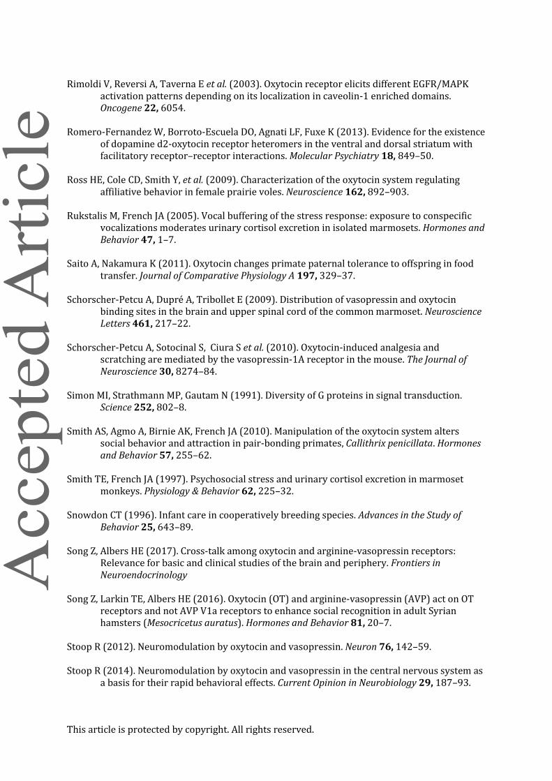
This article is protected by copyright. All rights reserved.
Rimoldi V, Reversi A, Taverna E et al. (2003). Oxytocin receptor elicits different EGFR/MAPK
activation patterns depending on its localization in caveolin-1 enriched domains. Oncogene 22, 6054.
Romero-Fernandez W, Borroto-Escuela DO, Agnati LF, Fuxe K (2013). Evidence for the existence
of dopamine d2-oxytocin receptor heteromers in the ventral and dorsal striatum with facilitatory receptor–receptor interactions. Molecular Psychiatry 18, 849–50.
Ross HE, Cole CD, Smith Y, et al. (2009). Characterization of the oxytocin system regulating
affiliative behavior in female prairie voles. Neuroscience 162, 892–903. Rukstalis M, French JA (2005). Vocal buffering of the stress response: exposure to conspecific
vocalizations moderates urinary cortisol excretion in isolated marmosets. Hormones and Behavior 47, 1–7.
Saito A, Nakamura K (2011). Oxytocin changes primate paternal tolerance to offspring in food
transfer. Journal of Comparative Physiology A 197, 329–37. Schorscher-Petcu A, Dupré A, Tribollet E (2009). Distribution of vasopressin and oxytocin
binding sites in the brain and upper spinal cord of the common marmoset. Neuroscience Letters 461, 217–22.
Schorscher-Petcu A, Sotocinal S, Ciura S et al. (2010). Oxytocin-induced analgesia and
scratching are mediated by the vasopressin-1A receptor in the mouse. The Journal of Neuroscience 30, 8274–84.
Simon MI, Strathmann MP, Gautam N (1991). Diversity of G proteins in signal transduction.
Science 252, 802–8. Smith AS, Agmo A, Birnie AK, French JA (2010). Manipulation of the oxytocin system alters
social behavior and attraction in pair-bonding primates, Callithrix penicillata. Hormones and Behavior 57, 255–62.
Smith TE, French JA (1997). Psychosocial stress and urinary cortisol excretion in marmoset
monkeys. Physiology & Behavior 62, 225–32. Snowdon CT (1996). Infant care in cooperatively breeding species. Advances in the Study of
Behavior 25, 643–89. Song Z, Albers HE (2017). Cross-talk among oxytocin and arginine-vasopressin receptors:
Relevance for basic and clinical studies of the brain and periphery. Frontiers in Neuroendocrinology
Song Z, Larkin TE, Albers HE (2016). Oxytocin (OT) and arginine-vasopressin (AVP) act on OT
receptors and not AVP V1a receptors to enhance social recognition in adult Syrian hamsters (Mesocricetus auratus). Hormones and Behavior 81, 20–7.
Stoop R (2012). Neuromodulation by oxytocin and vasopressin. Neuron 76, 142–59. Stoop R (2014). Neuromodulation by oxytocin and vasopressin in the central nervous system as
a basis for their rapid behavioral effects. Current Opinion in Neurobiology 29, 187–93.

This article is protected by copyright. All rights reserved.
Taylor JH, French JA (2015). Oxytocin and vasopressin enhance responsiveness to infant stimuli in adult marmosets. Hormones and Behavior 75, 154–59.
Taylor JH, Intorre AA, French JA (2017). Vasopressin and oxytocin reduce food sharing behavior
in male, but not female marmosets in family groups. Frontiers in Endocrinology 81 Vaidyanathan R, Hammock EA (2017). Oxytocin receptor dynamics in the brain across
development and species. Developmental Neurobiology 77, 143–57. Vargas-Pinilla P, Paixão-Côrtes VR, Paré P et al. (2015). Evolutionary pattern in the OXT-OXTR
system in primates: Coevolution and positive selection footprints. Proceedings of the National Academy of Sciences 112, 88–93.
Wallis M (2012). Molecular evolution of the neurohypophysial hormone precursors in
mammals: Comparative genomics reveals novel mammalian oxytocin and vasopressin analogues. General and Comparative Endocrinology 179, 313–18.
Wang Y, Zhao S, Wu Z, Feng Y, Zhao C, Zhang C (2015). Oxytocin in the regulation of social
behaviours in medial amygdala‐ lesioned mice via the inhibition of the extracellular signal‐ regulated kinase signalling pathway. Clinical and Experimental Pharmacology and Physiology 42, 465-74.
Wesley VJ, Hawtin SR, Howard HC, Wheatley M (2002). Agonist-specific, high-affinity binding
epitopes are contributed by an arginine in the N-terminus of the human oxytocin receptor. Biochemistry 41, 5086–92.
Wyttenbach T, Liu D, Bowers MT (2008). Interactions of the hormone oxytocin with divalent
metal ions. Journal of the American Chemical Society 130, 5993–6000. Xiao L, Priest MF, Nasenbeny J et al. (2017). Biased oxytocinergic modulation of midbrain
dopamine systems. Neuron 95, 368–84. Zhang, J. (2000) Rates of conservative and radical nonsynonymous nucleotide substitutions in
mammalian nuclear genes. Journal of molecular evolution. 50 (1), 56–68. Zingg HH, Laporte SA (2003). The oxytocin receptor. Trends in Endocrinology & Metabolism 14,
222–7. Zoicas I, Slattery DA, Neumann ID, (2014). Brain oxytocin in social fear conditioning and its
extinction: involvement of the lateral septum. Neuropsychopharmacology 39, 3027–35.

This article is protected by copyright. All rights reserved.
Table 1 Description of potential functional pharmacological outcomes associated with OTR signaling
1) OT Binding Affinity Definition: The concentration of OT ligand needed to occupy 50% of OTRs (IC50).
Question: Do different structural OT ligands enhance or reduce binding affinity with OTRs?
Outcome: OT ligand affinity to the receptor has important implications not only in how natural ligand variants interact
with OTRs, but these OT ligands may exhibit similar properties with regard to their crosstalk with AVP receptors.
2) OT Induced Ca2+
Mobilization
Definition: The maximal response/efficacy of Ca2+
mobilization and the OT ligand potency (EC50) of the concentration
needed to induce 50% maximal response.
Question: Are there differences in the magnitude of intracellular responses based on the structure of the OT ligand, and
does this response vary in potency between OT ligands?
Outcome: While certain ligands may share equal or better binding affinity to the receptor, differences in response
magnitude/potency may suggest that certain OT ligands are „better‟ at enhancing cellular function.

This article is protected by copyright. All rights reserved.
3) Functional Selectivity of OT
Ligands
Definition: Increased favorability/bias for OTRs to couple to one G-protein over another.
Question: Do specific OT ligands show different biases of activation of Gq over Gi/o or vise versa?
Outcome: OT ligands may preferentially activate certain physiological outcomes through activating specific cellular
pathways over others (e.g., OT ligands may only activate Gq and fail to activate Gi/o protein coupling).
4) OTR Desensitization and
Internalization
Definition: Decrease in receptor responsiveness over time via increased affinity for/recruitment of β-arrestins leading to
the transport and degradation of OTRs from the plasma membrane.
Question: Do specific OT ligands enhance β-arrestin recruitment and loss of OTR from the plasma membrane?
Outcome: Differences in how OT ligands facilitate faster or slower desensitization/internalization following acute or
chronic OTR stimulation can elucidate additional intracellular mechanisms for differential OT ligand function.

This article is protected by copyright. All rights reserved.
Table 2 Oxytocin ligand variants and social behavior in marmosets
Behavior Details Pro8-OT Leu8-OT AVP OTA
Social Approach
Partner preference test (3 weeks cohabitation)a -↑ ↓
Proximity (undisturbed)c - - -
Partner preference Test (8+ weeks cohabitation)b ↑F,↓M - -
Proximity during novel environment stressorf ↑ - ↓
Proximity during reunioni - - ↓
Responsiveness to infant Stimulie ↑M,-F -M,↑F -
Social Attention
Grooming(undisturbed)c - - -
Huddling(undisturbed)c - - -
Solicit grooming(undisturbed, reunion)c,i
- - -
Gazei - - -
Social Attractiveness
Received Grooming (undisturbed)c ↑F,-M ↑F,-M -

This article is protected by copyright. All rights reserved.
Received Grooming (reunion)i - ↓- ↓-
Approached by matei - - -
Received gazei ↑ ↓- ↓
Food Sharing
Mate-Directed prosocial food sharing taskd,g
- - -
Mate-Directed food sharing (undisturbed)a - ↓
Sibling directed food sharingh ↓M
Offspring directed food sharingh,j
↑M ↓M
Stranger-Directed Behavior
Stranger-Directed prosocial food sharing taskd,g
↓- - ↓-
Sexual solicitationb ↓ - -
Gazei - - -
Anxiety &Stress
Social Buffering of stress responsef - - -
Cortisol reactivityf - - ↑
Contact Callinge,f
- - - -
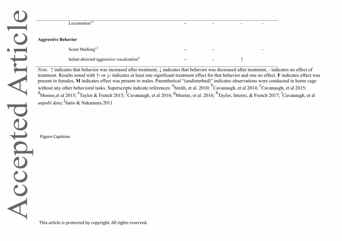
This article is protected by copyright. All rights reserved.
Locomotione,f
- - - -
Aggressive Behavior
Scent Markingc,f
- - -
Infant-directed aggressive vocalizationh - - ↑
Note. ↑ indicates that behavior was increased after treatment, ↓ indicates that behavior was decreased after treatment, - indicates no effect of
treatment. Results noted with ↑- or ↓- indicates at least one significant treatment effect for that behavior and one no effect. F indicates effect was
present in females, M indicates effect was present in males. Parenthetical “(undisturbed)” indicates observations were conducted in home cage
without any other behavioral tasks. Superscripts indicate references: aSmith, et al. 2010;
bCavanaugh, et al 2014;
cCavanaugh, et al 2015;
dMustoe,et al 2015;
eTaylor & French 2015;
fCavanaugh, et al 2016;
gMustoe, et al. 2016;
hTaylor, Intorre, & French 2017;
iCavanaugh, et al
unpubl data; jSaito & Nakamura 2011
Figure Captions

This article is protected by copyright. All rights reserved.
Figure 1. Consensus primate phylogeny constructed from data from 10kTrees (Arnold et al. 2010) for selected genera/species for which OT ligand is known.
Colored phylogenetic roots indicate separate OT ligands (2D structures shown for each OT ligand); red circles represent pair-living and beige variable pair-
living/non-pair-living, and stars indicate the presence of biparental care via provisioning and/or protecting behavior for the species. Behavioral phenotypes
derived from Lukas and Clutton-Brock 2013; French et al. 2017; and/or University of Michigan Museum of Zoology (animaldiversity.org).
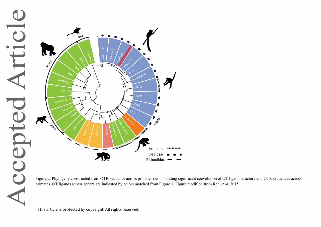
This article is protected by copyright. All rights reserved.
Figure 2. Phylogeny constructed from OTR sequence across primates demonstrating significant coevolution of OT ligand structure and OTR sequences across
primates. OT ligands across genera are indicated by colors matched from Figure 1. Figure modified from Ren et al. 2015.

This article is protected by copyright. All rights reserved.
Figure 3. A schematic summary of this review of the main findings based on OTR functional pharmacology outcomes such as receptor binding and G-protein
signaling, and social behavior outcomes (in marmosets) following activation of Pro8-OT. Arrows indicate direction of effect compared to Leu
8-OT induced
activation of OTRs at h- and mOTRs. “?” represent findings that have yet to be determined or evaluated. The pharmacological outcomes associated with OT
activation are known to be involved in neuronal membrane/cell excitability (Gong et al. 2015), long-term potentiation (Lin et al. 2012), and regulating many
facets of social behavior (Blume et al. 2008; Jurek et al. 2012; Wang et al. 2015). For a full representation of the OT signaling network see Chatterjee et al.
2016.