Embed Size (px)
Citation preview

REVIEW
Single-particle cryo-EM—How did itget here and where will it goYifan Cheng
Cryo–electron microscopy, or simply cryo-EM, refers mainly to three very different yetclosely related techniques: electron crystallography, single-particle cryo-EM, andelectron cryotomography. In the past few years, single-particle cryo-EM in particular hastriggered a revolution in structural biology and has become a newly dominant discipline.This Review examines the fascinating story of its start and evolution over the past40-plus years, delves into how and why the recent technological advances have been sogroundbreaking, and briefly considers where the technique may be headed in the future.
Physicist and Nobel laureate Richard Feyn-man once famously stated, “It is very easyto answer many fundamental biologicalquestions; you just look at the thing!” (1).Indeed, the central idea behind structural
biology is that onceweare able to “look” at “things”in great enough detail to discern their atomicstructures, we will naturally be able to answerhow and why the components and players ofcomplex biological processes work the way theydo. True to this aim, structural biology has con-tributed substantially to major biological discov-eries throughout history (2). It has also and willcontinue to facilitate developments of therapeu-tic agents to cure diseases or ameliorate patho-logical symptoms.The major techniques available to structural
biologists are x-ray crystallography, nuclear mag-netic resonance (NMR) spectroscopy, and electronmicroscopy (EM). Among them, x-ray crystallog-raphy contributes most of the atomic coordinatesof biological macromolecules deposited in theProtein Data Bank (PDB) (3). In this method,structures are determined fromdiffraction patternsgenerated from well-ordered three-dimensional(3D) crystals of biological macromolecules. The
resolutionsof structuresdetermineddepend largelyon the quality of the crystals; in short, obtainingwell-ordered 3D crystals of sufficient size is usuallya prerequisite for atomic structure determinationof any biological macromolecules by using thistechnique. It works well for many proteins orstable complexes; however, for certain categoriesof biological macromolecules, growing large andwell-ordered 3D crystals is a very difficult or im-possible task. For example, crystallizing integralmembrane proteins or large and dynamic com-plexes and machineries can be challenging. Anextension of the x-ray crystallography is the x-rayfree electron laser (XFEL), whose ultimate goal isto determine atomic structures without crystalsbut currently still requires a large amount of smallcrystals (4).Can atomic structures of biological macromole-
cules be determined without crystallization? Oris it possible, as Feynman once suggested, to de-termine their structures by “looking” at themby using a powerful electron microscope? Earlypioneers pursued this question in the 1970s anddeveloped a new EM-based method known todayas single-particle cryo–electron microscopy (cryo-EM) (5, 6). At the beginning, the method yielded
rather low-resolution results. Drawn to the prom-ise of being able to study biological macro-molecules without crystallizing them, however,the cryo-EM community dedicated itself to per-fecting the technique over more than four de-cades, yielding steady improvements in both thetechnique’s applicability and the resolution ofits results. Gradually, it has become a major toolin structural biology, complementary to x-raycrystallography and widely used to study largemacromolecular complexes that are difficult tobe crystallized. A few years ago, some amazingtechnological breakthroughs further enabled rou-tine atomic resolution structure determinationswith this method. Today, single-particle cryo-EMis no longer a complementary technique but adominant one, changing the field of structuralbiology in a profound and unprecedented wayand facilitating major new discoveries.
A brief history of single-particle cryo-EM
It is actually quite difficult to “look” at biologicalmacromolecules in an electron microscope. De-termining their atomic structures from electronmicrographs is evenmore complicated. First, EMimages are 2D projections of biological macro-molecules, but not their 3D structures. This wasresolved by De Rosier and Klug, who demon-strated that a 3D structure can be reconstructedby combining 2D projection images of the sameobject along different directions (7).Second, because of strong scattering, the elec-
tron beam has to be confined in a high vacuum,and all EM samples need to be placed inside saidvacuum. This is not a problem for inorganic ma-terials. But if one simply places a biological sam-ple inside an electronmicroscope, vacuum-causeddehydration would destroy the sample’s struc-tural integrity. The seemingly impossible taskof keeping protein samples hydrated in a high
TECHNOLOGIES TRANSFORMING BIOLOGY
Cheng, Science 361, 876–880 (2018) 31 August 2018 1 of 5
Howard Hughes Medical Institute, Department ofBiochemistry and Biophysics, University of California, SanFrancisco, San Francisco, CA, USA.
A B C D
Fig. 1. Establishing single-particle cryo-EM. (A) An electron diffractionpattern of frozen hydrated catalase crystal. [Reprinted from (9) withpermissions from Elsevier] The diffraction spots are visible at beyond 3-Åresolution. This experiment established the concept of cryo-EM. (B) Anelectron micrograph of frozen hydrated adenovirus particle recorded from afrozen hydrated grid prepared with plunge freezing.The micrograph is from the
same dataset described in (11). [Reprinted from (9) with permissions fromElsevier] (C) The first 3D reconstruction of bacteriorhodopsin determined withelectron crystallography. [Reprinted from (15) with permission from SpringerNature] (D) A 3D model of the 50S ribosome subunit determined withsingle-particle reconstruction of a negatively stained large ribosomal subunitfrom Escherichia coli. [Reprinted from (22) with permission from Wiley]
on June 5, 2020
http://science.sciencemag.org/
Dow
nloaded from

vacuumwas accomplished by Taylor and Glaeser.They recorded better than 3-Å resolution electrondiffraction patterns from frozen hydrated catalasecrystals, demonstrating that the structural integ-rity of biological macromolecules in a high vac-uum can bemaintained through frozen hydration(Fig. 1A) (8, 9). The practical implication of thisapproach, however, was not easy until a plungefreezing technique was developed by Dubochetand colleagues in the 1980s (10, 11). They appliedpurified protein samples in solution to anEMgridcovered with a thin layer of carbon holey filmand blotted the grid with a filter paper, whichremovedmost of the solution, and surface tensiondrove the remaining solution into a thin liquidfilm across holes in the carbon film. Plunging thegrid rapidly into liquid ethane cooled by liquidnitrogen froze it into a thin layer of amorphousice with the protein sample embedded within itin random orientations. After that, the frozen gridwas transferred into an electron microscope andkept at near–liquid nitrogen temperature for imag-ing (Fig. 1B). This method is still used routinelywithout major changes, except that we now usea machine to blot and plunge grids with tunableparameters.Third, radiation damage by the high-energy
electron beam limits the total electron dose thatcan be used to image biological samples. The con-sequence is that images recordedwith such a lowelectron dose have very poor signal-to-noise ratios(SNRs). Cooling the sample to liquid nitrogen oreven liquid helium temperature can reduce theradiation damage and allow the sample to toler-ate a higher electron dose (12, 13) but still far frombeing able to directly visualize high-resolutiondetails from rawmicrographs. Henderson andUnwin overcame this problem by use of a crystal-lographic approach, averaging images of manyidentical proteinspackedas 2Dcrystals (14). Imagesof glucose-embedded 2D crystals recorded withvery low electron doses show no visible features,but their Fourier transforms show clear reflec-tions. Similarly, electron diffraction from such
2D crystals at very low electron doses producedgood-quality diffraction patterns. Combining thephases calculated from the Fourier transforma-tions of images and the amplitudes obtained fromdiffractions produced a high-resolution projectionmap of the specimen (14). Combining data col-lected from specimens tilted at different anglesproduced a 3Dreconstruction similar to the densitymap of an x-ray crystal structure. This approachproduced the first structure of an integral mem-brane protein, initially at ~7-Å resolution (Fig.1C) (15) and finally at atomic resolution (16). Themethod became known as electron crystallogra-phy, which relies onwell-ordered 2D crystals. Thismethod has produced atomic structures of severalintegral membrane proteins (17, 18) and one solu-ble protein (19). The highest resolution achieved
was 1.9 Å, resolving a lipid bilayer surroundingan aquaporin-0 (AQP0) water channel (20). Butthe difficulty of growing well-ordered 2D crystalshinders the broad application of the method.In parallel, Frank proposed an idea to deter-
mine protein structures without crystallization:computationally combining images of many indi-vidual protein particles of the same type (5). Thisconceptually novel idea was first tested by usingprotein samples that were negatively stained forEM observation (Fig. 1D) (21, 22). The later com-bination of this approach with the plunge freez-ing sample preparation became what we now call“single-particle cryo-EM.” It does not require grow-ing proteins into crystals of any form. Instead, itdetermines structure by computationally align-ing and combining cryo-EM images of manybiological molecules randomly oriented within a
thin layer of vitreous ice. A large number of imagesis needed to both enhance SNR and to provide alldifferent views needed for 3D reconstruction.More detailed technical descriptions of thismethod can be found in many recent reviews,such as (23, 24).Furthermore, what electronmicrographs record
are projections of the specimen convoluted bya contrast transfer function (CTF). CTF is a sinefunction oscillating in a frequency-dependentmanner thatmodulates both phase and amplitudeof an image in frequency space (25). In additionto a number of microscope-dependent param-eters, CTF is determined by how much off thefocus an image is recorded—the so-called “defocus.”To retain the highest resolution, imagesmust berecorded very close to focus. Such images, how-ever, have very limited contrast. This is not a prob-lem for radiation-insensitive inorganicmaterials,which can be imaged with very high electrondoses so as to generate sufficient contrast whileretaining a high-resolution signal. However, imagesof radiation-sensitive frozen hydrated biologicalsamples have to be recorded with a large defocusin order to generate sufficient contrast, which inturn dampens the high-resolution signal.The resolution of a single-particle cryo-EM
structure depends onmany factors, including theresolution and contrast of individual particleimages, accuracy of aligning these images witheachother, obtaining a sufficient number of imagesfrom all necessary views of the macromoleculewithin a reasonable time frame, the conforma-tional and compositional homogeneity of theseparticles, and access to powerful enough computerswithwhich to process images efficiently. Less thanoptimal conditions for any of these factors—forexample, instability of the electronmicroscope,relatively poor performance of the image-recordingdevice and beam-induced sample motions, accu-racy of classifying and aligning particle imageslimited by image quality and computational al-gorithms, and limited computer power to processvery large numbers of particle images—can limit
Cheng, Science 361, 876–880 (2018) 31 August 2018 2 of 5
K2 Summit super-resolutionat 2.36 e-/pixel/sec
Gatan Ultrascan CCD cameraK2 Base
A
0.2 0.4 0.6 0.8 1.0 1.2 1.4 1.6 1.8 2.0
0.10.20.30.40.50.60.70.80.91.0
Fraction of physical Nyquist frequency
Dete
ctio
n qu
antu
m e
ffici
ency
(DQ
E)
PhysicalNyquist
DC
1/4.0Å-1
1/3.2Å-1
B
40 nm
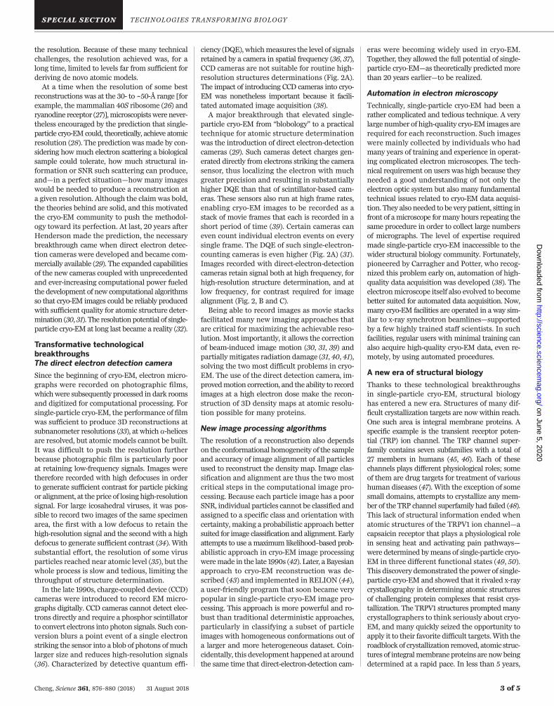
Fig. 2. Direct-electron-detection camera–enabled atomic structuredetermination. (A) Comparison of DQE curves of a scintillator-based CCDcamera (black), and direct electron detection camera K2 operating inbase mode (blue) and super-resolution counting mode (red) (31). (B) Atypical electron micrograph of archaeal 20S proteasome (~700 kDa in
molecular weight) recorded by use of a direct-electron-detection camera(60). (C) Fourier power spectrum calculated from the image in (B).[(B) and (C) reprinted from (60) with permission from Elsevier] High-resolution signal at ~3 Å resolution is clearly visible (60). (D) A portion ofdensity map from the 3D reconstruction of archaeal 20S proteasome (31).
“The resolution of a singleparticle cryo-EM structuredepends on many factors…”
on June 5, 2020
http://science.sciencemag.org/
Dow
nloaded from
the resolution. Because of these many technicalchallenges, the resolution achieved was, for along time, limited to levels far from sufficient forderiving de novo atomic models.At a time when the resolution of some best
reconstructions was at the 30- to ~50-Å range [forexample, the mammalian 40S ribosome (26) andryanodine receptor (27)],microscopistswere never-theless encouraged by the prediction that single-particle cryo-EMcould, theoretically, achieve atomicresolution (28). The prediction wasmade by con-sidering howmuch electron scattering a biologicalsample could tolerate, how much structural in-formation or SNR such scattering can produce,and—in a perfect situation—how many imageswould be needed to produce a reconstruction ata given resolution. Although the claimwas bold,the theories behind are solid, and this motivatedthe cryo-EM community to push the methodol-ogy toward its perfection. At last, 20 years afterHenderson made the prediction, the necessarybreakthrough came when direct electron detec-tion cameras were developed and became com-mercially available (29). The expanded capabilitiesof the new cameras coupled with unprecedentedand ever-increasing computational power fueledthe development of new computational algorithmsso that cryo-EM images could be reliably producedwith sufficient quality for atomic structure deter-mination (30, 31). The resolutionpotential of single-particle cryo-EM at long last became a reality (32).
Transformative technologicalbreakthroughsThe direct electron detection camera
Since the beginning of cryo-EM, electron micro-graphs were recorded on photographic films,whichwere subsequently processed in dark roomsand digitized for computational processing. Forsingle-particle cryo-EM, the performance of filmwas sufficient to produce 3D reconstructions atsubnanometer resolutions (33), at which a-helicesare resolved, but atomic models cannot be built.It was difficult to push the resolution furtherbecause photographic film is particularly poorat retaining low-frequency signals. Images weretherefore recorded with high defocuses in orderto generate sufficient contrast for particle pickingor alignment, at the price of losing high-resolutionsignal. For large icosahedral viruses, it was pos-sible to record two images of the same specimenarea, the first with a low defocus to retain thehigh-resolution signal and the second with a highdefocus to generate sufficient contrast (34). Withsubstantial effort, the resolution of some virusparticles reached near atomic level (35), but thewhole process is slow and tedious, limiting thethroughput of structure determination.In the late 1990s, charge-coupled device (CCD)
cameras were introduced to record EM micro-graphs digitally. CCD cameras cannot detect elec-trons directly and require a phosphor scintillatorto convert electrons into photon signals. Such con-version blurs a point event of a single electronstriking the sensor into a blob of photons ofmuchlarger size and reduces high-resolution signals(36). Characterized by detective quantum effi-
ciency (DQE), whichmeasures the level of signalsretained by a camera in spatial frequency (36, 37),CCD cameras are not suitable for routine high-resolution structures determinations (Fig. 2A).The impact of introducing CCD cameras into cryo-EM was nonetheless important because it facili-tated automated image acquisition (38).A major breakthrough that elevated single-
particle cryo-EM from “blobology” to a practicaltechnique for atomic structure determinationwas the introduction of direct electron-detectioncameras (29). Such cameras detect charges gen-erated directly from electrons striking the camerasensor, thus localizing the electron with muchgreater precision and resulting in substantiallyhigher DQE than that of scintillator-based cam-eras. These sensors also run at high frame rates,enabling cryo-EM images to be recorded as astack of movie frames that each is recorded in ashort period of time (39). Certain cameras caneven count individual electron events on everysingle frame. The DQE of such single-electron-counting cameras is even higher (Fig. 2A) (31).Images recorded with direct-electron-detectioncameras retain signal both at high frequency, forhigh-resolution structure determination, and atlow frequency, for contrast required for imagealignment (Fig. 2, B and C).Being able to record images as movie stacks
facilitated many new imaging approaches thatare critical for maximizing the achievable reso-lution. Most importantly, it allows the correctionof beam-induced image motion (30, 31, 39) andpartially mitigates radiation damage (31, 40, 41),solving the two most difficult problems in cryo-EM. The use of the direct detection camera, im-provedmotion correction, and the ability to recordimages at a high electron dose make the recon-struction of 3D density maps at atomic resolu-tion possible for many proteins.
New image processing algorithms
The resolution of a reconstruction also dependson the conformational homogeneity of the sampleand accuracy of image alignment of all particlesused to reconstruct the density map. Image clas-sification and alignment are thus the two mostcritical steps in the computational image pro-cessing. Because each particle image has a poorSNR, individual particles cannot be classified andassigned to a specific class and orientation withcertainty, making a probabilistic approach bettersuited for image classification and alignment. Earlyattempts to use amaximum likelihood–based prob-abilistic approach in cryo-EM image processingweremade in the late 1990s (42). Later, a Bayesianapproach to cryo-EM reconstruction was de-scribed (43) and implemented in RELION (44),a user-friendly program that soon became verypopular in single-particle cryo-EM image pro-cessing. This approach is more powerful and ro-bust than traditional deterministic approaches,particularly in classifying a subset of particleimages with homogeneous conformations out ofa larger and more heterogeneous dataset. Coin-cidentally, this development happened at aroundthe same time that direct-electron-detection cam-
eras were becoming widely used in cryo-EM.Together, they allowed the full potential of single-particle cryo-EM—as theoretically predictedmorethan 20 years earlier—to be realized.
Automation in electron microscopy
Technically, single-particle cryo-EM had been arather complicated and tedious technique. A verylarge number of high-quality cryo-EM images arerequired for each reconstruction. Such imageswere mainly collected by individuals who hadmany years of training and experience in operat-ing complicated electron microscopes. The tech-nical requirement on users was high because theyneeded a good understanding of not only theelectron optic system but also many fundamentaltechnical issues related to cryo-EM data acquisi-tion. They also needed to be very patient, sitting infront of amicroscope formany hours repeating thesame procedure in order to collect large numbersof micrographs. The level of expertise requiredmade single-particle cryo-EM inaccessible to thewider structural biology community. Fortunately,pioneered by Carragher and Potter, who recog-nized this problem early on, automation of high-quality data acquisition was developed (38). Theelectronmicroscope itself also evolved to becomebetter suited for automated data acquisition. Now,many cryo-EM facilities are operated in a way sim-ilar to x-ray synchrotron beamlines—supportedby a few highly trained staff scientists. In suchfacilities, regular users with minimal training canalso acquire high-quality cryo-EM data, even re-motely, by using automated procedures.
A new era of structural biology
Thanks to these technological breakthroughsin single-particle cryo-EM, structural biologyhas entered a new era. Structures of many dif-ficult crystallization targets are now within reach.One such area is integral membrane proteins. Aspecific example is the transient receptor poten-tial (TRP) ion channel. The TRP channel super-family contains seven subfamilies with a total of27 members in humans (45, 46). Each of thesechannels plays different physiological roles; someof them are drug targets for treatment of varioushuman diseases (47). With the exception of somesmall domains, attempts to crystallize any mem-ber of the TRP channel superfamily had failed (48).This lack of structural information ended whenatomic structures of the TRPV1 ion channel—acapsaicin receptor that plays a physiological rolein sensing heat and activating pain pathways—were determined bymeans of single-particle cryo-EM in three different functional states (49, 50).This discovery demonstrated the power of single-particle cryo-EM and showed that it rivaled x-raycrystallography in determining atomic structuresof challenging protein complexes that resist crys-tallization. The TRPV1 structures promptedmanycrystallographers to think seriously about cryo-EM, and many quickly seized the opportunity toapply it to their favorite difficult targets.With theroadblock of crystallization removed, atomic struc-tures of integralmembrane proteins are nowbeingdetermined at a rapid pace. In less than 5 years,
Cheng, Science 361, 876–880 (2018) 31 August 2018 3 of 5
TECHNOLOGIES TRANSFORMING BIOLOGY on June 5, 2020
http://science.sciencemag.org/
Dow
nloaded from

atomic structures of at least one member of eachof the seven TRP channel subfamilies have nowbeen determined (Fig. 3).Single-particle cryo-EM is also game-changing
for structural studies of many large and dynamiccomplexes andmachineries that were impossiblefor crystallization. A traditional approach was tofit crystal structures of individual componentsor domains into lower-resolution cryo-EM densi-ty maps of the whole complex (51). Now, atomicstructures of many large complexes are deter-mined directly by using single-particle cryo-EM.An excellent example is the spliceosome complex.Past efforts produced atomic structures of somesmall components (52), as well as low-resolutioncryo-EM structures of the whole complex (53).Now, in only a few short years, atomic structuresof the spliceosome in different functional stateshave been determined (54, 55).One can easily call forth many more examples
to illustrate how the recent technological break-throughs in single-particle cryo-EM have changedhow we tackle complex biological problems. Therapid pace of advancement in structural biol-ogy is unprecedented. It has also attractedmajorpharmaceutical companies, withmany hoping toimplement cryo-EM into structure-based drugdiscovery and optimization.
The future
Single-particle cryo-EM continues to move for-ward fast, with many new technologies beingdeveloped. One such example is the Volta phaseplate, which is a thin carbon film placed in theback focal plane of themicroscope’s objective lens.It adds a phase shift to the CTF so that an imagerecorded at near focus has good contrast (56),
facilitating the study of very small proteins (57).Another example is the development of a newsample preparation technology that is fundamen-tally different from the current blotting method(58). The new method promises many benefits,including reducing the total amount of sampleneeded from microliters to nanoliters.As impressive as these innovations have been,
there are still many technical details that can beimproved. Regularly achieving resolutions closeto 2 Å and beyond is one goal; improving robust-ness and throughput is another. Once very highresolutions can be achieved reliably, pharmaceu-tical companies will be able to routinely use EMto speed up structure-based drug discovery. Therange of what can be studied bymeans of single-particle cryo-EM can also be expanded to includesmaller or bigger targets with higher resolution,as well as more dynamic complexes or assemblieswith irregular shapes.There is still more that single-particle cryo-EM
can offer to further biological discoveries beyondstructure determination if we continue to pushthe boundary of this technology. For example,in theory, image classification can sort a cryo-EMdataset that contains particles of heterogeneousconformation or composition intomultiple classes,each corresponding to a different functional state.By freezing cryo-EM grids at specific time points,it could be possible to derive structural infor-mation in a time-dependent manner (59). It maytherefore be possible to understand and breakdown the complex cycles,movements, and processesof biologicalmacromolecular complexes andma-chineries step by step, in complete atomic detail.In the future, it may also be possible to studyboth physiological and pathological protein states
through affinity-purifying specific proteins directlyfrom cells onto EM grids for single-particle cryo-EM studies.The past 40 years have seen the evolution of
single-particle cryo-EM fromblobology to a routineand powerful method for atomic structure deter-mination. The technique’s new popularity is at-tracting not only more users but also talents fromdifferent fields, ranging from physics, materialscience, and mathematics, to machine learning.This infusion of new ideas and a larger commu-nity heralds an even brighter future for single-particle cryo-EM full of method development,collaboration, and most importantly, wonderfulnew biological discoveries.
REFERENCES AND NOTES
1. R. P. Feynman, in Feynman and Computation, A. J. G. Hey, Ed.(Perseus Books Cambridge, 1999), pp. 63–76.
2. Y. Shi, Cell 159, 995–1014 (2014).3. N. Jones, Nature 505, 602–603 (2014).4. Y. Kang et al., Nature 523, 561–567 (2015).5. J. Frank, Ultramicroscopy 1, 159–162 (1975).6. J. Dubochet et al., Q. Rev. Biophys. 21, 129–228 (1988).7. D. J. De Rosier, A. Klug, Nature 217, 130–134 (1968).8. K. A. Taylor, R. M. Glaeser, Science 186, 1036–1037 (1974).9. K. A. Taylor, R. M. Glaeser, J. Struct. Biol. 163, 214–223 (2008).10. J. Dubochet, J. J. Chang, R. Freeman, J. Lepault,
A. W. McDowall, Ultramicroscopy 10, 55–61 (1982).11. M. Adrian, J. Dubochet, J. Lepault, A. W. McDowall, Nature
308, 32–36 (1984).12. H. Stark, F. Zemlin, C. Boettcher, Ultramicroscopy 63, 75–79
(1996).13. Y. Fujiyoshi, Adv. Biophys. 35, 25–80 (1998).14. P. N. Unwin, R. Henderson, J. Mol. Biol. 94, 425–440
(1975).15. R. Henderson, P. N. Unwin, Nature 257, 28–32 (1975).16. R. Henderson et al., J. Mol. Biol. 213, 899–929 (1990).17. W. Kühlbrandt, D. N. Wang, Y. Fujiyoshi, Nature 367, 614–621
(1994).18. K. Murata et al., Nature 407, 599–605 (2000).
Cheng, Science 361, 876–880 (2018) 31 August 2018 4 of 5
TRPV1, 3J5P(2013)
TRPA1, 3J9P(2015)
TRPM4, 6BQR(2017)
TRPC4, 6G1K(2018)
NOMPC/TRPN1, 5VKQ(2017)
PKD2/TRPP2, 5T4D(2016)
TRPML1, 5WPQ(2017)
Fig. 3. Single-particle cryo-EM enables atomic structure determinationof TRPchannels.Ribbon diagrams of atomic structures from each subfamilyof TRP channel superfamily. They are TRPV1 (49), TRPA1 (61), TRPM4
(62),TRPC4 (63), NOMPC (also named TRPN1) (64), PKD2 (or TRPP2) (65),and TRPML (66). The rapid pace of integral membrane protein structuredetermination is enabled with single-particle cryo-EM and is unprecedented.
on June 5, 2020
http://science.sciencemag.org/
Dow
nloaded from

19. E. Nogales, S. G. Wolf, K. H. Downing, Nature 391, 199–203(1998).
20. T. Gonen et al., Nature 438, 633–638 (2005).21. J. Frank, W. Goldfarb, D. Eisenberg, T. S. Baker,
Ultramicroscopy 3, 283–290 (1978).22. M. Radermacher, T. Wagenknecht, A. Verschoor, J. Frank,
EMBO J. 6, 1107–1114 (1987).23. Y. Cheng, N. Grigorieff, P. A. Penczek, T. Walz, Cell 161,
438–449 (2015).24. R. Fernandez-Leiro, S. H. Scheres, Nature 537, 339–346
(2016).25. R. H. Wade, Ultramicroscopy 46, 145–156 (1992).26. S. Srivastava, A. Verschoor, M. Radermacher, R. Grassucci,
J. Frank, J. Mol. Biol. 245, 461–466 (1995).27. M. Radermacher et al., J. Cell Biol. 127, 411–423 (1994).28. R. Henderson, Q. Rev. Biophys. 28, 171–193 (1995).29. G. McMullan, A. R. Faruqi, R. Henderson, Methods Enzymol.
579, 1–17 (2016).30. X. C. Bai, I. S. Fernandez, G. McMullan, S. H. Scheres, eLife 2,
e00461 (2013).31. X. Li et al., Nat. Methods 10, 584–590 (2013).32. Y. Cheng, Cell 161, 450–457 (2015).33. B. Böttcher, S. A. Wynne, R. A. Crowther, Nature 386, 88–91
(1997).34. S. J. Ludtke, W. Chiu, J. Struct. Biol. 144, 73–78 (2003).35. X. Zhang, L. Jin, Q. Fang, W. H. Hui, Z. H. Zhou, Cell 141,
472–482 (2010).36. P. Mooney, Methods Cell Biol. 79, 661–719 (2007).
37. E. Samei, M. J. Flynn, D. A. Reimann, Med. Phys. 25, 102–113(1998).
38. C. Suloway et al., J. Struct. Biol. 151, 41–60 (2005).39. A. F. Brilot et al., J. Struct. Biol. 177, 630–637 (2012).40. S. H. Scheres, eLife 3, e03665 (2014).41. T. Grant, N. Grigorieff, eLife 4, e06980 (2015).42. F. J. Sigworth, J. Struct. Biol. 122, 328–339 (1998).43. S. H. Scheres, J. Mol. Biol. 415, 406–418 (2012).44. S. H. Scheres, J. Struct. Biol. 180, 519–530 (2012).45. D. E. Clapham, Nature 426, 517–524 (2003).46. D. Julius, Annu. Rev. Cell Dev. Biol. 29, 355–384 (2013).47. M. M. Moran, Annu. Rev. Pharmacol. Toxicol. 58, 309–330
(2018).48. M. Li, Y. Yu, J. Yang, Adv. Exp. Med. Biol. 704, 1–23 (2011).49. M. Liao, E. Cao, D. Julius, Y. Cheng, Nature 504, 107–112
(2013).50. E. Cao, M. Liao, Y. Cheng, D. Julius, Nature 504, 113–118
(2013).51. G. C. Lander et al., Nature 482, 186–191 (2012).52. W. P. Galej, T. H. Nguyen, A. J. Newman, K. Nagai, Curr. Opin.
Struct. Biol. 25, 57–66 (2014).53. M. D. Ohi, L. Ren, J. S. Wall, K. L. Gould, T. Walz, Proc. Natl.
Acad. Sci. U.S.A. 104, 3195–3200 (2007).54. S. M. Fica, K. Nagai, Nat. Struct. Mol. Biol. 24, 791–799 (2017).55. Y. Shi, Nat. Rev. Mol. Cell Biol. 18, 655–670 (2017).56. R. Danev, B. Buijsse, M. Khoshouei, J. M. Plitzko,
W. Baumeister, Proc. Natl. Acad. Sci. U.S.A. 111, 15635–15640(2014).
57. M. Khoshouei, R. Danev, J. M. Plitzko, W. Baumeister, J. Mol.Biol. 429, 2611–2618 (2017).
58. V. P. Dandey et al., J. Struct. Biol. 202, 161–169 (2018).59. S. Kaledhonkar, Z. Fu, H. White, J. Frank, Methods Mol. Biol.
1764, 59–71 (2018).60. X. Li, S. Q. Zheng, K. Egami, D. A. Agard, Y. Cheng, J. Struct.
Biol. 184, 251–260 (2013).61. C. E. Paulsen, J. P. Armache, Y. Gao, Y. Cheng, D. Julius,
Nature 520, 511–517 (2015).62. H. E. Autzen et al., Science 359, 228–232 (2018).63. D. Vinayagam et al., eLife 7, e36615 (2018).64. P. Jin et al., Nature 547, 118–122 (2017).65. P. S. Shen et al., Cell 167, 763–773.e11 (2016).66. Q. Chen et al., Nature 550, 415–418 (2017).
ACKNOWLEDGMENTS
This is a brief story of single-particle cryo-EM but not acomprehensive historical review. I apologize to many colleagueswhose important works are not cited here. I thank many of mycolleagues and all past and present members of my laboratory forsharing their thoughts about single-particle cryo-EM with me, andL. Wang for editing. The cryo-EM work in my laboratory is fundedby various NIH grants: R01GM082893, R01GM098672,R01HL134183, P50GM082250 (Nevan Krogan), P01GM111126(Robert Stroud), and S10OD020054 and S10OD021741. I am aHoward Hughes Medical Institute investigator.
10.1126/science.aat4346
Cheng, Science 361, 876–880 (2018) 31 August 2018 5 of 5
TECHNOLOGIES TRANSFORMING BIOLOGY on June 5, 2020
http://science.sciencemag.org/
Dow
nloaded from

How did it get here and where will it go−−Single-particle cryo-EMYifan Cheng
DOI: 10.1126/science.aat4346 (6405), 876-880.361Science
ARTICLE TOOLS http://science.sciencemag.org/content/361/6405/876
CONTENTRELATED
http://science.sciencemag.org/content/sci/361/6405/880.fullhttp://science.sciencemag.org/content/sci/361/6405/870.fullhttp://science.sciencemag.org/content/sci/361/6405/866.fullhttp://science.sciencemag.org/content/sci/361/6405/864.full
REFERENCES
http://science.sciencemag.org/content/361/6405/876#BIBLThis article cites 65 articles, 5 of which you can access for free
PERMISSIONS http://www.sciencemag.org/help/reprints-and-permissions
Terms of ServiceUse of this article is subject to the
is a registered trademark of AAAS.ScienceScience, 1200 New York Avenue NW, Washington, DC 20005. The title (print ISSN 0036-8075; online ISSN 1095-9203) is published by the American Association for the Advancement ofScience
Copyright © 2018, American Association for the Advancement of Science
on June 5, 2020
http://science.sciencemag.org/
Dow
nloaded from