Embed Size (px)
Citation preview

Behavioural Brain Research 192 (2008) 149–165
Contents lists available at ScienceDirect
Behavioural Brain Research
journa l homepage: www.e lsev ier .com/ locate /bbr
Review
Techniques and devices to restore cognition
Mijail Demian Serruya a,!, Michael J. Kahanab
a Department of Neurology, Hospital of the University of Pennsylvania, 3400 Spruce Street, Gates 3 HUP, Philadelphia, PA 19104-4283, United Statesb Department of Psychology, University of Pennsylvania, Suite 302C, 3401 Walnut Street, Philadelphia, PA 19104, United States
a r t i c l e i n f o
Article history:Received 26 May 2007Received in revised form 3 March 2008Accepted 9 April 2008Available online 20 April 2008
Keywords:Brain–computerBrain–machineNeuroprostheticProstheticNeurocognitiveMemoryCognitionImplantElectrodeHuman
a b s t r a c t
Executive planning, the ability to direct and sustain attention, language and several types of memory maybe compromised by conditions such as stroke, traumatic brain injury, cancer, autism, cerebral palsy andAlzheimer’s disease. No medical devices are currently available to help restore these cognitive functions.Recent findings about the neurophysiology of these conditions in humans coupled with progress in engi-neering devices to treat refractory neurological conditions imply that the time has arrived to considerthe design and evaluation of a new class of devices. Like their neuromotor counterparts, neurocognitiveprostheses might sense or modulate neural function in a non-invasive manner or by means of implantedelectrodes. In order to paint a vision for future device development, it is essential to first review what canbe achieved using behavioral and external modulatory techniques. While non-invasive approaches mightstrengthen a patient’s remaining intact cognitive abilities, neurocognitive prosthetics comprised of directbrain–computer interfaces could in theory physically reconstitute and augment the substrate of cognitionitself.
© 2008 Elsevier B.V. All rights reserved.
Contents
1. Introduction . . . . . . . . . . . . . . . . . . . . . . . . . . . . . . . . . . . . . . . . . . . . . . . . . . . . . . . . . . . . . . . . . . . . . . . . . . . . . . . . . . . . . . . . . . . . . . . . . . . . . . . . . . . . . . . . . . . . . . . . . . . . . . . . . . . . . . . . . 1502. Behavioral techniques . . . . . . . . . . . . . . . . . . . . . . . . . . . . . . . . . . . . . . . . . . . . . . . . . . . . . . . . . . . . . . . . . . . . . . . . . . . . . . . . . . . . . . . . . . . . . . . . . . . . . . . . . . . . . . . . . . . . . . . . . . . . . . . 150
2.1. Assistive devices . . . . . . . . . . . . . . . . . . . . . . . . . . . . . . . . . . . . . . . . . . . . . . . . . . . . . . . . . . . . . . . . . . . . . . . . . . . . . . . . . . . . . . . . . . . . . . . . . . . . . . . . . . . . . . . . . . . . . . . . . . . . . 1502.2. Virtual reality (VR). . . . . . . . . . . . . . . . . . . . . . . . . . . . . . . . . . . . . . . . . . . . . . . . . . . . . . . . . . . . . . . . . . . . . . . . . . . . . . . . . . . . . . . . . . . . . . . . . . . . . . . . . . . . . . . . . . . . . . . . . . . 1502.3. Cognitive training . . . . . . . . . . . . . . . . . . . . . . . . . . . . . . . . . . . . . . . . . . . . . . . . . . . . . . . . . . . . . . . . . . . . . . . . . . . . . . . . . . . . . . . . . . . . . . . . . . . . . . . . . . . . . . . . . . . . . . . . . . . . 151
3. Non-invasive modulation . . . . . . . . . . . . . . . . . . . . . . . . . . . . . . . . . . . . . . . . . . . . . . . . . . . . . . . . . . . . . . . . . . . . . . . . . . . . . . . . . . . . . . . . . . . . . . . . . . . . . . . . . . . . . . . . . . . . . . . . . . . 1523.1. Visual entrainment . . . . . . . . . . . . . . . . . . . . . . . . . . . . . . . . . . . . . . . . . . . . . . . . . . . . . . . . . . . . . . . . . . . . . . . . . . . . . . . . . . . . . . . . . . . . . . . . . . . . . . . . . . . . . . . . . . . . . . . . . . 1523.2. Transcranial magnetic stimulation . . . . . . . . . . . . . . . . . . . . . . . . . . . . . . . . . . . . . . . . . . . . . . . . . . . . . . . . . . . . . . . . . . . . . . . . . . . . . . . . . . . . . . . . . . . . . . . . . . . . . . . . . . 1523.3. Transcranial direct current stimulation . . . . . . . . . . . . . . . . . . . . . . . . . . . . . . . . . . . . . . . . . . . . . . . . . . . . . . . . . . . . . . . . . . . . . . . . . . . . . . . . . . . . . . . . . . . . . . . . . . . . . 1523.4. Neurofeedback. . . . . . . . . . . . . . . . . . . . . . . . . . . . . . . . . . . . . . . . . . . . . . . . . . . . . . . . . . . . . . . . . . . . . . . . . . . . . . . . . . . . . . . . . . . . . . . . . . . . . . . . . . . . . . . . . . . . . . . . . . . . . . . 153
4. Invasive techniques . . . . . . . . . . . . . . . . . . . . . . . . . . . . . . . . . . . . . . . . . . . . . . . . . . . . . . . . . . . . . . . . . . . . . . . . . . . . . . . . . . . . . . . . . . . . . . . . . . . . . . . . . . . . . . . . . . . . . . . . . . . . . . . . . 1544.1. Frequency-contingent learning . . . . . . . . . . . . . . . . . . . . . . . . . . . . . . . . . . . . . . . . . . . . . . . . . . . . . . . . . . . . . . . . . . . . . . . . . . . . . . . . . . . . . . . . . . . . . . . . . . . . . . . . . . . . . . 1544.2. Cell-triggered recall . . . . . . . . . . . . . . . . . . . . . . . . . . . . . . . . . . . . . . . . . . . . . . . . . . . . . . . . . . . . . . . . . . . . . . . . . . . . . . . . . . . . . . . . . . . . . . . . . . . . . . . . . . . . . . . . . . . . . . . . . 1554.3. Cortical microstimulation . . . . . . . . . . . . . . . . . . . . . . . . . . . . . . . . . . . . . . . . . . . . . . . . . . . . . . . . . . . . . . . . . . . . . . . . . . . . . . . . . . . . . . . . . . . . . . . . . . . . . . . . . . . . . . . . . . . 1554.4. Non-electrical neural stimulation . . . . . . . . . . . . . . . . . . . . . . . . . . . . . . . . . . . . . . . . . . . . . . . . . . . . . . . . . . . . . . . . . . . . . . . . . . . . . . . . . . . . . . . . . . . . . . . . . . . . . . . . . . . 1564.5. Subcortical and peripheral stimulation . . . . . . . . . . . . . . . . . . . . . . . . . . . . . . . . . . . . . . . . . . . . . . . . . . . . . . . . . . . . . . . . . . . . . . . . . . . . . . . . . . . . . . . . . . . . . . . . . . . . . 1564.6. Connectivity as cognition . . . . . . . . . . . . . . . . . . . . . . . . . . . . . . . . . . . . . . . . . . . . . . . . . . . . . . . . . . . . . . . . . . . . . . . . . . . . . . . . . . . . . . . . . . . . . . . . . . . . . . . . . . . . . . . . . . . . 1574.7. Expanding the neural substrate . . . . . . . . . . . . . . . . . . . . . . . . . . . . . . . . . . . . . . . . . . . . . . . . . . . . . . . . . . . . . . . . . . . . . . . . . . . . . . . . . . . . . . . . . . . . . . . . . . . . . . . . . . . . . 1584.8. Ectopic neural modules. . . . . . . . . . . . . . . . . . . . . . . . . . . . . . . . . . . . . . . . . . . . . . . . . . . . . . . . . . . . . . . . . . . . . . . . . . . . . . . . . . . . . . . . . . . . . . . . . . . . . . . . . . . . . . . . . . . . . . 1594.9. Internal neural assistant . . . . . . . . . . . . . . . . . . . . . . . . . . . . . . . . . . . . . . . . . . . . . . . . . . . . . . . . . . . . . . . . . . . . . . . . . . . . . . . . . . . . . . . . . . . . . . . . . . . . . . . . . . . . . . . . . . . . . 160
! Corresponding author. Tel.: +1 215 662 2700; fax: +1 215 746 6848.E-mail address: [email protected] (M.D. Serruya).
0166-4328/$ – see front matter © 2008 Elsevier B.V. All rights reserved.doi:10.1016/j.bbr.2008.04.007
150 M.D. Serruya, M.J. Kahana / Behavioural Brain Research 192 (2008) 149–165
5. Unique features of neurocognitive prosthetics . . . . . . . . . . . . . . . . . . . . . . . . . . . . . . . . . . . . . . . . . . . . . . . . . . . . . . . . . . . . . . . . . . . . . . . . . . . . . . . . . . . . . . . . . . . . . . . . . . . . . 1616. Conclusion. . . . . . . . . . . . . . . . . . . . . . . . . . . . . . . . . . . . . . . . . . . . . . . . . . . . . . . . . . . . . . . . . . . . . . . . . . . . . . . . . . . . . . . . . . . . . . . . . . . . . . . . . . . . . . . . . . . . . . . . . . . . . . . . . . . . . . . . . . . 162
Acknowledgments . . . . . . . . . . . . . . . . . . . . . . . . . . . . . . . . . . . . . . . . . . . . . . . . . . . . . . . . . . . . . . . . . . . . . . . . . . . . . . . . . . . . . . . . . . . . . . . . . . . . . . . . . . . . . . . . . . . . . . . . . . . . . . . . . . 162References . . . . . . . . . . . . . . . . . . . . . . . . . . . . . . . . . . . . . . . . . . . . . . . . . . . . . . . . . . . . . . . . . . . . . . . . . . . . . . . . . . . . . . . . . . . . . . . . . . . . . . . . . . . . . . . . . . . . . . . . . . . . . . . . . . . . . . . . . . . 162
1. Introduction
A range of medical devices to restore or augment humanfunctions are becoming available with the swift engineering andbiomedical advances in the new field of neurotechnology. Cochlearimplants have restored hearing to thousands of people, whiledevices to restore sight and movement are progressing rapidly. Sen-sory devices inject signals into the nervous system, while motorprosthetics extract signals from the nervous system and send themto control devices such as robotic arms or stimulators to re-activateparalyzed muscles [136]. Other types of neurotechnology devicesimprove health by modulating pre-existing systems: deep brainstimulators, for example, deliver targeted electrical stimulation tothe basal ganglia to relieve the symptoms of Parkinson’s disease.Non-invasive techniques under development include transcranialmagnetic or direct current stimulation, and biofeedback deliv-ered from quantitative electroencephalography (EEG) or functionalmagnetic resonance imaging (fMRI).
Medical devices seek to restore lost function and leverageremaining, intact aspects of the patient’s physiology. Deep brainstimulators appear to unfetter an otherwise intact voluntary motorcontrol system from the aberrant activity of one or more nuclei ofthe basal ganglia in patients with Parkinson’s disease or dystonia,whereas cochlear implants bypass a dysfunctional organ of Corti toactivate an intact vestibulocochlear nerve to re-instate audition. Amajor area not addressed by these devices is that of higher cogni-tive functions, such as memory, language and executive planning,all of which can be compromised in a wide range of neurologicaldiseases.
Cognitive function may be improved by using both non-invasivetechniques and invasive medical devices. Non-invasive techniquesrange from behavioral techniques or assistive software that providenew strategies to restoring memory and planning to electromag-netic stimulation and biofeedback that modulate activity in apatient’s brain as part of a rehabilitation program. Whereas thenon-invasive approaches may improve cognition by strengthen-ing retained skills or reorganizing the activity of intact brain,implantable medical devices that are able to record and stimulateensembles of neurons could, in theory, physically restore the sub-strate of cognition. Though pharmacotherapy will not be a focus ofthis review, it will be touched upon in the context of implantablemicrofluidic systems.
The central premise of direct brain–computer interfaces is thatlost function can be recovered by artificially recreating or bypass-ing the neural substrate lost by disease or injury. A successfulimplementation of such a strategy would be marked by a returnto baseline function as measured both by standardized neuropsy-chological testing, activities of daily living, and a person’s subjectiveappreciation of independent function. Such systems would ideallybe transparent and effortless to use. Yet to proceed with clinicaltrials developing such devices, it is essential to lay out what can beachieved with a variety of non-invasive means. To accept the risk ofany invasive intervention, patients and physicians must understandwhat, if any, alternatives exist. Thus, this paper will begin by review-ing behavioral and assistive device techniques aimed at helpingpatients with cognitive impairment, and proceed to approachesthat increasingly attempt to physically modulate or interface witha patient’s brain to improve function.
2. Behavioral techniques
2.1. Assistive devices
Simple, low-cost interventions such as a memory book or wallet,containing pictures of familiar places or people, can help patientswith impaired episodic memory better navigate conversations anddaily activities [16,119]. Mobile phones and pagers can be set up toactively remind patients of tasks [165,173]. Off-the-shelf systems(e.g., PDAs) can be modified to improve access and usability forpeople with cognitive impairment by creating a linear flow of use,making the system more resilient to user errors, and consistentlyrepeating items that users need help with [150].
Cognitive orthotic software can facilitate skill acquisition andself-sufficient management of daily tasks [13,36]. These systemscan be highly customized to take into account the deficits andrehabilitation goals of a particular patient. Expert systems com-prising mobile phones and palm-top computers linked by radio toweb-based central workstations have been found to help remindpatients with brain injury, stroke and dementia that certain tasksmust be performed such as taking a medication or calling a rela-tive [128]. A palmtop computer can literally step a patient througha task: it might alert them that it is time to call a sister, andthen either to provide the phone number and await confirmationthe task was completed, or give the patient the option to deferthe task and follow-up later [88,128]. Patients with brain injuriesmay benefit from both wearable computers that facilitate interac-tion with the environment [46], and computer-based diaries, withauditory alarms and linked entries [174]. Wearable cameras andmicrophones that continually record and monitor a person’s envi-ronment, coupled to sensors placed throughout the home whichall then feed into pattern recognition algorithms to alert a personto perform certain tasks, are in development [42,43,51]. Prelimi-nary field tests of these systems find that considerable challengesremain: as with able-bodied people, patients are averse to havingtheir privacy invaded by cameras and are wary of conspicuouslyplaced sensors; they do not want to be awoken from a nap to take amedicine a specific time [51]. To compensate for a patient’s deficit,computer-based memory-aids must be adaptive to the individualand incorporate input of multiple family members and health caregivers. A fundamental challenge for automated memory-aids is todevise a method by which computers could recognize the correctsituation when a particular reminder should be triggered.
Computerized memory-aids have been found to be useful forsimple reminding tasks but fall short of the kind of ongoingguidance that patients with severe cognitive impairments wouldrequire to become independent. Another approach is to train thepatient herself in order to improve cognition and memory. In the-ory such cognitive training approaches could help a patient in allaspects of daily life.
2.2. Virtual reality (VR)
VR has been shown to promote learning in people with mem-ory impairments; furthermore, this learning appears to transfer toimproved real-world performance [19,96,178]. Virtual worlds basedon a patient’s own home enable safe practice of daily activities andmemorization of the location of items [34,78]. Just as VR tools can

M.D. Serruya, M.J. Kahana / Behavioural Brain Research 192 (2008) 149–165 151
be used for data management by scientific teams to capture andmanage complex data [100], so too the annotated display mightbe adapted to help individuals with memory impairment. Giventhat enriched environments increase new neuron production inrodents, elaboration of gray matter and remyelination in white mat-ter [86], the mere act of having patients navigate through complexVR worlds may yield clinical benefits.
2.3. Cognitive training
A range of behavioral training techniques have been devel-oped to help impaired patients by leveraging remaining, intactcognitive structures. Animal studies have shown that experiencecan yield long-lasting effects on brain plasticity and morphology,hence training techniques might likewise induce beneficial changesin the human brain [18]. The approaches vary in sophisticationfrom the use of simple mnemonic aids to complex training regi-mens designed to reorganize cortical representations. In humans,epidemiological studies have explored a wide number of factorsthat have been suggested to reduce age-related cognitive declineincluding both formal and informal education, leisure pursuits,intellectual engagement, physical fitness training, and expertisein different skill domains such as music, computer-game playing,language translation or meditation [75]. Playing computer gamesappears to improve certain aspects of visual attention such as spa-tial resolution and number of objects being tracked, which in turncould improve visual memory [48]. Perhaps the best establishedtechniques for cognitive enhancement are healthy diet and physicalexercise; several epidemiological studies have revealed significantcorrelations between physical exercise and cognition [27,124]. Sim-ple procedural changes in the ways people are trained to learn canhave dramatic effects: testing retrieval, rather than merely study-ing items, can significantly improve retention [70]. Here we shalldefine and review the application of mnemonic techniques, error-less learning, spaced retrieval, vanishing cues, and a techniquenamed plasticity-based learning.
Mnemonic strategies range from verbal techniques such as ver-bal mediations, acronyms or poems, to spatial techniques suchas imagery and the “method of loci.” In method of loci, a fact ormemory is associated with a particular place in a real or imag-ined environment such that by mentally walking through this spaceand visiting particular locations, a person can sequentially recallthe memories ‘stored’ there. Method of loci has been shown to behelpful in adults and children with memory and other cognitiveimpairment [11,85]. People who are exceptionally good memoriz-ers often use mnemonics techniques to organize, remember andrecall large amounts of visual and verbal memories that do nota priori have spatial organization by using brain areas, as shownby fMRI, related to spatial navigation and memory (right cere-bellum, left medial superior parietal gyrus, bilateral retrosplenialcortex, right posterior hippocampus) [85]. In addition to spatial-location mnemonics, preliminary evidence suggests that musicalmnemonics (simply incorporating melody or rhythm in words tobe remembered) can improve memory [159].
Errorless learning comprises a technique in which patients areshown both the question and its answer during training, thus pro-viding participants ongoing positive feedback as they are giventhe right answer. While a meta-review of different techniques inamnesic patients found that errorless learning yielded the great-est effect size [73], a small cohort study of elderly with early-stagedementia suggested high-effort semantic learning was more effec-tive [30]. Incorporating errorless learning and spaced retrieval, inwhich newly acquired information is recalled at increasingly longerintervals or with more intervening items, was most effective forpatients with Alzheimer’s [47]. At best, the evidence suggests that
these approaches have a mild benefit on attention and memory,in a manner similar to cognition-enhancing drug trials, such asacetylcholinesterase inhibitors (e.g., donepezil) [47].
Patients who have severely impaired episodic memory due toencephalitis and other conditions may be able to learn new skillsusing remaining, intact implicit memory [45]. In teaching thesepatients word pairs to memorize, implicit learning can be lever-aged by systematically showing more and more letters of a wordto be remembered (e.g., for the word ‘cursor’ showing ‘c’ then ‘cu’then ‘cur’, etc.) and then subsequently removing the added letters.This technique is termed ‘vanishing cues,’ and while it appears tobenefit certain patients, a meta-review found it lacking [45,47]. Inpeople with brain injuries explicit strategies such as mnemonicsand practicing attention, were found to be the most effective inimproving performance on the Stroop-Color task and digit spanmemory [156,162].
Cognitive training approaches suffer from numerous drawbacksincluding a lack of statistical power due to small groups of par-ticipants, absence of long-term follow-up, and failure to measurethe impact of the intervention on measures of quality of dailylife [23,47]. Another common critique of behavioral techniques toenhance cognition is that the training effects are specific to the tasksused and generalize poorly to the kind of situations an impaired per-son needs to navigate in daily life [75]. Given that real-world skillsinvolve a variety of cognitive processes, it has been proposed thattraining interventions ought to a priori incorporate multiple pro-cesses (e.g., reasoning, verbal episodic memory, processing speed)rather than focus on a single one. Two large-scale, randomized trialsaddressed several of these concerns and provided some evidencethat cognitive training in well-functioning older adults improvedcognition in a manner that generalized to daily tasks and that wassustained for up to 5 years after the intervention [7,172]. Memorytraining comprised teaching mnemonic strategies (organization,visualization, association) for remembering verbal material [172].Reasoning training involved teaching techniques to find a patternin a series of words or letters, and then identifying the next itemin the series. Participants were also tested on applying the strate-gies to everyday problems (e.g. a mnemonic strategy to recall agrocery list; a reasoning strategy to comprehend a bus schedule[172]). Improvements appeared to generalize to a positive effecton daily function on the accuracy of verbal memory, self-ratingsof how independent they were on daily tasks such as preparing ameal, and the speed and accuracy of tasks such as finding a numberin a phonebook and reading a road sign [172].
An approach termed “plasticity-based learning” comprises exer-cises in which participants must: (1) identify whether a frequency-modulated sweep is upward or downward, (2) identify a synthet-ically generated syllable from a confusable pair (e.g., /ba/vs./da/),(3) match spoken confusable consonant–vowel–consonant words(e.g., bat, vat) from a spatial grid, (4) reconstruct a sequence of shortspoken words, (5) reconstruct spoken instructions by draggingicons on a computer screen, and (6) answer questions about shortnarratives. Exercises increase in difficulty, dynamically match-ing the participant’s performance. In a small study of childrenwith language-learning impairment, 1 month of daily training wasassociated with significant improvement in the ability to followcomplex auditory stimuli [154]. In a randomized, controlled studyof elderly adults, 8–10 weeks of training improved performance onimmediate recall of list of words, copying a figure, naming a picture,serial recall of digits, and recalling a figure or a story [87,120]. How-ever, as with the other cognitive training paradigms, it is not clearhow much plasticity-based learning generalizes to the real-worldenvironment.
Training techniques such as mnemonic aids, motivationaltechniques to enhance attention, customization to individual differ-

152 M.D. Serruya, M.J. Kahana / Behavioural Brain Research 192 (2008) 149–165
ences in personality and ability level, and integration of the trainingto ongoing medical care have been shown to help patients withmemory impairment [157]. New techniques should be developedwith an understanding of both what has been shown to be effec-tive in holistic approaches to neuropsychological rehabilitation andupon an understanding of the neurophysiology of synaptic plastic-ity [25,87]. Both assistive devices that a person can carry with them,or which are integrated into sensors in the home, and training tech-niques ranging from physical exercise to mnemonics, all promise tobring some benefit to patients with cognitive impairment. Yet, evenas these assistive devices and techniques are improved it is likelythere will be an upper limit of the degree to which they can helppatients overcome their impairments. Hypothetically, attempts tomodulate or restore the substrate of cognition itself could yieldmore fundamental benefit. The next two parts of this review covernon-invasive and invasive approaches to modify cortical functionin order to restore cognitive abilities.
3. Non-invasive modulation
3.1. Visual entrainment
Steady-state visual stimuli may be used to probe EEG responses:recent work comparing EEG and fMRI of human participantsexposed to flickering light found that medial frontal cortexmetabolic activity depended on the temporal frequency of visualinput implying that during visual stimulation, input frequency canbe used to activate distinct functional networks [146]. Indeed theuse of visual flicker to purposefully modify cortical activity in orderto improve memory has been explicitly tested [169,170].
The alpha rhythm is an oscillation in range of 8–12 Hz, withan average peak of 10–11 Hz in healthy adults. The peak alphafrequency (PAF) corresponds to the discrete frequency with thehighest power estimate in the alpha range. The specific discretefrequency of this oscillation appears correlated with mental perfor-mance at all ages, although it tends to be slowest in children and theelderly [2]. Alpha oscillations are associated with memory function:they decline in old age and Alzheimer’s disease and can be restoredby anti-dementia drugs. In a study of 550 normal participants (aged11–70 years) in the Brain Resource International Database, bothperformance on reverse digit span tests and PAF were found to sig-nificantly decrease with age [123]. For each increasing additional1 Hz of alpha peak frequency, a corresponding 0.21 digit increase inreverse digit span occurred, when age was held constant [123]. Inas-much as alpha activity reflects memory performance, one mighthypothesize that enhancing alpha might likewise enhance mem-ory.
Recent studies suggest that by exposing human participantsto light flickering at particular frequencies, episodic memory canbe improved. Visual flicker at 9.5–11.0 Hz (in 0.5 Hz intervals)displayed briefly before presenting a three-letter word to beremembered after a distracter, was found to improve word recallin both young adults and the elderly [169,170]. In the study of 51young adults, participants recognized more three-letter words thathad followed 0.5–1.5 s of 10.0 Hz flicker during learning, comparedto those that had followed 0, 8.7 and 11.7 Hz flicker [170]. In thestudy of 30 elderly, cognitively normal adults, flicker at frequen-cies close to 10.2 Hz, but not below 9.0 or above 11.0 Hz, improvedlater recognition of words from the learning phase [170]. Whileoscillating visual and auditory stimuli have been shown to increasefrequency-matched power spectra in the EEG, the mechanism bywhich flicker improves episodic memory is not known. RhythmicEEG activity induced by flicker may serve as a gain signal in recur-rent cortico-cortical or thalamo-cortical loops, or may predisposeLTP at hippocampal synapses.
3.2. Transcranial magnetic stimulation (TMS)
TMS comprises a non-invasive method of induction of focalcurrents in the brain and transient modulation of the function oftargeted cortex [80]. The direction of current flow in an overlyingcoil determines which neural elements are activated. A review ofmagnetically induced stimulation of non-motor areas revealed thatadverse effects were infrequent and usually mild [84]. rTMS hasbeen shown to ameliorate symptoms in conditions ranging frommigraine (the disappearance of abnormal visual evoked potentialsby 1 Hz over visual cortex [39]) to depression (5 Hz over left pre-frontal dorsolateral cortex [80]). The energy generated by TMS isestimated to be about 0.05% of that applied in a burst of electro-convulsive therapy.
In a study of four aphasics who were 5–11 years post-stroke,1 Hz rTMS applied daily for 10 days to an anterior portion of Broca’sarea induced significant and lasting improvement in picture nam-ing [103,104]. In addition to picture naming, rTMS may also be ableto inhibit cortical areas responsible for forming concepts such thatareas concerned with spatial detail are made directly accessible toconscious perception. In a small single-blind, randomized, sham-controlled study of the ability to estimate the number of discreteelements visually presented immediately after their presentation,15 min of 1 Hz rTMS applied to the left anterior temporal lobewas found to significantly improve number estimation accuracy[144]. Seven seconds of rTMS at 5 Hz over the parietal precuneusarea, applied immediately before a response was required in adelayed-match-to-sample working memory task, was found to sig-nificantly decrease reaction time in a sham-controlled study ofhealthy young adults [83]. rTMS has also been found to ameliorateage-related impairments in memory. In a randomized, double-blinded, sham-controlled study of 39 non-demented elders withmild memory problems, 5 min of periodic 5 Hz stimulation over theleft prefrontal cortex was found to significantly improve learning ofnovel face–name associations [145]. Furthermore, this behavioralimprovement was mirrored by changes in fMRI activation, namelyincreased activity in the left anterior cingulate, right middle andfrontal gyri. Magnetic stimulation hence provides a promising, non-invasive approach to improving memory and cognition in patientswith a wide range of disorders, both by activating networks thatsubserve these memory functions or by de-activating networks thatmay interfere with performance.
3.3. Transcranial direct current stimulation (tDCS)
Whereas rTMS induces currents in the brain using electromag-netic induction, tDCS involves placing metal electrodes on thescalp to directly apply a small (and harmless) current across thecranium. The effects are highly dependent on the polarity andgeometry of the electrodes on the head. The current can hyperpo-larize or depolarize neurons in the path of the current dependingon the electrode polarity [37]. A small trial of Parkinson’s diseasepatients with cognitive impairment suggests that tDCS appliedto dorsolateral prefrontal cortex might increase its excitability inturn improving performance on short-term verbal memory tasks[15].
Transcranial direct current stimulation over the motor cortex,premotor cortex, visual cortex, and left dorsolateral prefrontalcortex may improve working memory, implicit learning and ver-bal fluency in healthy adults [37,60,106]. In a study of 15 youngwomen where each subject served as her own control, 10 min ofanodal tDCS over left dorsolateral prefrontal cortex was found toimprove performance in a three-back letter working memory task,as compared to control conditions of sham stimulation or tDCSover primary motor cortex [37]. Transcranial application of poten-

M.D. Serruya, M.J. Kahana / Behavioural Brain Research 192 (2008) 149–165 153
tials oscillating at 0.75 Hz, a similar range as seen in endogenousslow-wave sleep, has been shown to induce the appearance ofoscillations at that specific frequency on the EEG. The inductionof these oscillations during sleep appeared to significantly improvethe retention of hippocampus-dependent declarative memories inhealthy humans [93]. Specifically, bilateral tDCS over frontal cortexduring slow-wave sleep (five 5-min trains at 0.75 Hz, applied 4 minafter entering stage 2 non-REM sleep for the first time) enhancedretention of word pairs learned during the preceding day [93].
Though both TMS and DCS ultimately modulate neural activityby inducing the passage of current, the way they achieve this andthe nature of the stimulation effect are distinct. TMS is thought toinduce neuronal depolarization and induction of action potentials,while DCS, when applied at low currents (e.g., 1 mA) is thoughtto only cause a slight change in the resting potential of stimu-lated cells [37]. By causing this change in resting potential, DCSmay improve information processing by bringing neurons closer todepolarization thresholds in response to appropriate inputs or bystrengthening effects on glutamatergic synapses. In addition, DCSmay induce extracellular potential oscillations comparable to thosethat would occur naturally, and that reach the extracellular spacebefore transmission by chemical signals would [93]. The two tech-niques or rTMS and tDCS may have distinct and complementaryroles in clinical applications that have yet to be fully characterized.The efficacy of either technique appears highly dependent on suchparameters as polarity, cortical region stimulated, frequency, dura-tion, and precise timing relative to an ongoing task and precedingbrain activity. While rTMS and tDCS appear to focally activate par-ticular brain areas, biofeedback in which participants view theirown brain-activity in real-time, represents another method to‘stimulate’ the brain and potentially modify cognition.
3.4. Neurofeedback
Given that gross patterns in scalp recorded potentials correlateto states of arousal, it is intuitive to hypothesize that gaining vol-untary control over EEG signals might provide better regulation ofarousal. Indeed, the most widely tested application of EEG biofeed-back, also known in the literature as neurofeedback, is to treatattention-deficit disorder, which can be considered a disorder ofabnormal arousal. The use of neurofeedback as a therapeutic inter-vention has been tainted by controversial claims and a dearth ofclinical trials to assess its efficacy. Fortunately, controlled studiesare being conducted and the procedure appears to pose no physicalrisks to patients.
Neurofeedback comprises self-modulation of the EEG by pos-itively reinforcing the production or suppression of specific EEGfrequencies. Neurofeedback originated out of studies conductedin the 1960s in which operant conditioning was used to traincats to increase the power of the 12–15 Hz EEG rhythm oversomatosensory cortex (the so called ‘sensorimotor’ or mu rhythm)while remaining alert and motionless [149]. The conditioning tookplace within a NASA study to investigate the toxic effects of arocket fuel component (hydrazine) on cats and it was fortuitouslynoticed that the cats that had undergone the sensorimotor train-ing had significantly elevated thresholds to developing seizuresafter being administered the hydrazine. These findings spurredinvestigations on neurofeedback in humans: in the 1970s it wasfound that occipital theta (4–7 Hz) amplitude was negatively cor-related with vigilance and that by conditioning human participantsto increase or decrease occipital theta amplitude, performanceon a visuospatial task worsened or improved, respectively [10].In terms of seizure control, several labs have gathered evidencethat reinforcing production of 12–15 Hz, while simultaneously sup-pressing theta 4–7 Hz, activity appears effective at reducing seizure
frequency in humans with epilepsy. Most of these studies wereunderpowered, however, and this approach is not part of standardclinical care [98,149].
Small, randomized controlled trials of neurofeedback in chil-dren with attention-deficit hyperactivity disorder (ADHD) havedemonstrated that the technique improves attention and appearsto induce changes in fMRI. Unlike those in control groups, childrenoperantly conditioned to enhance 12–15 and 15–18 Hz rhythms,and to concurrently suppress the 4–8 Hz theta rhythm, displayactivation of areas in the right (anterior cingulate and ventrolat-eral prefrontal cortices) and left (caudate nucleus, substantia nigra,thalamus) hemispheres. Neurofeedback might modulate the activ-ity of brain regions that mediate selective attention and responseinhibition [9,79].
As the neurophysiological underpinnings to the complex wave-forms of the EEG are just beginning to be elucidated [148], themechanism behind how neurofeedback exerts its effect on thesesystems remains unclear. The sensorimotor rhythm develops whena subject is motionless yet remains attentive. By remaining alertyet stationary, motor output to the thalamus and brainstem mightbe reduced, in turn leading to decreased red nucleus activity andreduced stretch reflex and muscle tone. The net effect of suchdecreased tone would be reduction of somatic afferent activityand a state shift of the ventroposterior lateral and reticular nucleiof the thalamus into oscillatory bursting, with the consequentdevelopment of the sensorimotor rhythm on the EEG. The sen-sorimotor rhythm appears similar in location and quality to sleepspindles; in fact, sensorimotor rhythm training appears to enhancespindle activity during quiet sleep [98]. Another class of neurofeed-back focuses on modifying slow cortical potentials: these shifts arethought to reflect widespread depolarization of apical dendrites ofcortical pyramidal cells. Negative slow cortical potential shifts arethought to reflect cortical arousal, attention and active processing,whereas positive shifts might represent relaxation. In contrast to12–15 Hz entrainment, self-regulation of slow cortical potentialsis correlated with significant changes in metabolic activity in thebasal ganglia and thalamus, implying that people can learn to reg-ulate a cortico-striatal-thalamic loop modulating local excitationthresholds of cortical assemblies [55].
As discussed earlier, PAF appears to be related to memory andcognition. A small pilot study of elderly people evaluated thedistinct effects of either reinforcing the production of a higherfrequency for peak alpha or reinforcing increased amplitude ofalpha-range activity regardless of specific frequency. Increasing PAFappeared to improve processing speed (e.g., Stroop Task speed), butnot memory, whereas increasing amplitude improved memory butactually worsened processing speed [3]. Though the study had toofew participants to assess whether alpha feedback training mightbe a clinically useful tool for improving cognitive skills, the findingsreveal that improving one set of skills might compromise others.Another small study found a correlation between augmenting thesensorimotor rhythm (12–15 Hz) while suppressing neighboringfrequencies improved performance on word list recall, a measureof semantic working memory [163].
While the various pilot neurofeedback studies imply that thetechnique can induce lasting cognitive processing changes inhumans, and while certain mechanisms based on thalamocorticalcircuitry have been proposed, most investigation remains largelyexploratory. In order to assess potential clinical benefits, therefore,studies must not only be better powered, with appropriate random-ization and controls, but they also must attempt to build upon ourgrowing knowledge of the relationships between oscillations andcognition.
Inasmuch as cognitive abilities depend on a person’s ability toinduce a particular neurophysiological state, neurofeedback may

154 M.D. Serruya, M.J. Kahana / Behavioural Brain Research 192 (2008) 149–165
improve cognitive function. Research on neuromotor EEG prosthet-ics has shown that people can learn to modulate oscillatory activityat a wide range of cortical locations and frequencies [14]. Possibili-ties for future research include the investigation of whether trainingpeople to increase oscillatory activity within the neural networkssupporting memory function can significantly enhance learningor recall. In particular, recent studies have shown that oscillatoryactivity in the gamma frequency band, especially in hippocam-pus and temporal cortex, increases during successful encodingof study items (as measured by whether those items are subse-quently recalled) [131]. The same pattern of gamma activity overhippocampus and cortex is recapitulated just prior to success-ful retrieval of previously learned items [132]. Training people toincrease gamma activity (recorded from the scalp or subdurally)in these regions, while decreasing alpha and beta activity overwidespread sites [131], could potentially result in improved learn-ing and recall. In addition to operant conditioning to consciouslyincrease the magnitude or precise frequency of gamma activity,participants could be engaged in closed-loop systems in whichstimuli to be recalled were repeatedly presented until the systemdetected gamma activity signatures that predict later successfulrecall.
In addition to increasing (or decreasing) the power of a givenfrequency band, neurofeedback could be used to increase thecoherence and synchronization of a particular frequency bandacross electrode sites. Coherence, a frequency domain metric whichdescribes the linear association between two variables, may reflectthe number and strength of corticocortical connections betweenneural ensembles when applied to EEG signals [155]. Synchro-nization refers to the temporal alignment of signals and has beenthought to play a role in binding disparate ensembles togetherto form conscious percepts. Like oscillatory power, coherence andsynchronization have been associated with successful memory pro-cessing [38]. It currently is unknown to what degree humans canachieve voluntary control of the coherence and synchronizationof oscillating local fields simultaneously recorded from spatiallyseparate electrodes.
Other physiological signals may play a role in neurocognitiverehabilitation. The P300, a marker related to unexpected stimuli[152], has been used in brain–computer interfaces to allow para-lyzed people to control software [133]. The P3b component, derivedfrom temporo-parietal activity, appears to be related to memoryprocessing and may serve as a useful neurofeedback target [114].In addition to the EEG, functional magnetic resonance imaging canalso be used as a form of biofeedback. A recent study reported thatadult participants, who visually monitored the metabolic activ-ity of their own anterior cingulate cortex, were able to controlthis brain activation and reduce chronic, medically refractory pain[28]. Recent evidence suggests that humans can achieve voluntarycontrol of the metabolic activity of other specific brain regions,such as insular cortex or the amygdala, via fMRI neurofeedback[22,32]. Adults and children with memory and other cognitiveimpairment could use variations of this closed-loop real-time fMRIbiofeedback to either boost the baseline activity in medial tempo-ral lobe structures known to serve as a substrate of memory, or toimprove spatial mnemonic techniques by increasing the activity ofareas such superior parietal cortex, right posterior hippocampus[85] while engaging memory tasks. Although considerable workremains before the full potential clinical benefit of non-invasivemodulatory techniques is characterized, approaches that attemptto directly interface with substrate of cognition might promise themost comprehensive therapy. The next section reviews prelimi-nary animal and human studies of implantable systems to enhancecognition and outlines new approaches that might re-instate lostfunction.
4. Invasive techniques
4.1. Frequency-contingent learning
Implanted electrodes can capture high-frequency electricalactivity that would otherwise be spatially filtered by the skull,and can provide high signal-to-noise recordings of oscillations atprecisely defined anatomical locations within the brain. Work inanimal models has established that fairly simple closed-loop sys-tems can augment memory. The theta rhythm (usually defined as4–8 Hz in humans, and as 3–10 Hz in animals) is known to enhancehippocampal plasticity and accelerate learning in rats, cats, rabbits,and other animal species [67], and thus manipulations based ontheta activity could be used to counteract impairments in learningdue to abnormal development, aging or pathology of the hippocam-pus and other memory structures. Rabbits that receive trainingtrials in the presence of theta were found to learn twice as fast asthose receiving trials in the absence of theta [130]. By comparingthe performance of two groups of animals to yoked controls trainedwith matched intertrial intervals, it was found that instead of thetabenefiting learning, rather nontheta was especially detrimental intasks that do not require the hippocampus such as delay condi-tioning. Pre-task theta activity was strongly associated with thesubsequent learning rate in operant conditioning [130]. Whereascerebellar and oculomotor pathways are clearly essential substratesof eyeblink conditioning, hippocampal influences appear to be par-allel and modulatory in nature. Theta-triggering accelerated boththe learning rate and the firing rate of individual hippocampal neu-rons as rabbits engaged in a difficult, hippocampus-dependent task[49].
The presence of high levels of theta may be optimal for learningbecause this oscillatory activity facilitates hippocampal plastic-ity at a cellular level. The approximately 200 ms period of thetheta rhythm matches the optimal window for coincident pre- andpost-synaptic activity for long-term potentiation induction at CA3synapses in the hippocampus [139]. The processes that increasetheta power may be due to increased attention or awareness ofenvironmental stimuli; awareness itself of stimulus contingencieshas been shown to enhance the learning rate in human eyeblinkconditioning [90]. The growing body of evidence that theta activityis present in human participants during performance of both spatialand verbal memory tasks [68] raises the possibility that learn-ing could be optimized by presenting information to be learnedcontingent on a person’s ongoing brain activity. In order to bepractically useful for people with cognitive impairment, scalp elec-trodes would have to be used during training sessions to monitortheta, or intracranial electrodes implanted for chronic recording.The ability of oscillatory-contingency to affect learning could itselfbe used as a diagnostic test to assess the nature of memory impair-ment in children with different cognitive disorders.
Contingency could be based on coherence across sites, suchas theta synchronization between hippocampus and entorhinal orfrontal cortex. In addition to enabling frequency-contingent learn-ing, invasive recordings with subdural grids and multi-electrodearrays could pave the way for neurofeedback based on high-gammafrequencies not recordable by scalp electrodes, and on the moreprecise anatomical localization [77]. Intracranial neurofeedbackcould be used to operantly condition increased phase synchroniza-tion between perirhinal cortex and the hippocampus, or to trainpatients to selectively increase oscillatory power within, or coher-ence between, any number of medial temporal lobe and frontalcortex structures. As more is learned about the specific oscilla-tory features associated with human cognition, these features inturn could be used for a principled exploration of intracranialcontingency-based training.

M.D. Serruya, M.J. Kahana / Behavioural Brain Research 192 (2008) 149–165 155
4.2. Cell-triggered recall
Individual neurons recorded in the human medial temporallobe fire selectively to images of faces, animals, objects or scenes[118]. A subset of these neurons have an invariant representationof individuals, namely they fire when patients are presented withstrikingly distinct pictures of a given individual, landmark or object(or even the name written as text on the screen) [118]. The ideathat individual neurons could carry an invariant representation hasexciting implications for a neurocognitive prosthetics. For peoplewith memory impairments, this raises the possibility that withintheir temporal lobes there may exist neurons whose individualability to classify external stimuli may outperform that of the indi-vidual. A chronically implanted microelectrode array could pick upthe activity of such a neuron, and based on a previous calibrationsession, use a look-up table to identify what the person perceived.This in turn could drive an explicit cue (such as the name of the iden-tified person or object whispered into the child’s ear via a speakerin the auditory canal or text appearing on an external computerscreen) or a perception induced by stimulation of auditory or visualcortex.
One might reasonably counter that a camera and microphone,coupled to already-available pattern recognition algorithms andrelational databases, could help patients remember certain items orperform certain tasks. Indeed, a prototype system uses inconspicu-ous motion sensors and an automated phone system to monitor anelder’s activity pattern at home, and appropriately call them withreminders to take medications or perform other tasks [51]. How-ever, no external sensor system will ever be able to detect memoryassociations in the absence of external stimuli: a patient wanderingthrough a particular landscape or talking to a person may strug-gle to remember an important association. Talking to a brothermight remind a person to call a son, or walking by a supermarketmight trigger thoughts of a certain meal they want to cook. Whiledatabases could conceivably be programmed to capture some ofthese spontaneous associational triggers, it is difficult to fathomhow it might fully emulate the combinatoric complexity of actualnetworks in the brain. Pattern detection based on temporal lobefiring could decode items that are imagined and have no simul-taneous referent in the external word, and identify items that areembedded in networks of associations.
Research in neuromotor prosthetics has revealed that humanscan bring the activity of both individual neurons and ensembles ofneurons under direct voluntary control [33,56]. Just as the behav-ioral correlate of motor cortical neuron discharge is real or imaginedmovement, patients with cognitive impairment could learn to vol-untarily control neurons in temporal cortex as part of memoryrehabilitation training. Implanted devices might also extract mul-timodality information about the identified invariant object withother cues not so easily measured by external microphones andcameras, such as emotional salience and the degree of covariancein firing rate with other neurons recorded elsewhere in the brain.
4.3. Cortical microstimulation
The neurosurgeon Wilder Penfield and his colleagues recog-nized that the experiential phenomena induced by microstim-ulation of the temporal cortex were similar to the symptomsexperienced by patients during spontaneous seizures [110]. Micros-timulation could thus be used to localize the seizure focus inorder to guide therapeutic resection for refractory epilepsy. Penfieldfound that while gross microstimulation usually induced a vague,generalized memory, in certain cases it could cause a vivid re-livingof a recollection by ‘replaying’ a memory in a way analogous to thatof a tape player. In one case, so long as the stimulation continued,
a patient heard an orchestra playing a song proceeding at the sametempo as it was originally experienced. The moment the electrodewas withdrawn, however, the song stopped, and when stimulatedagain, the song would restart at the beginning [111]. A more recentinvestigation found that a patient experienced, depending on whichsubdural electrode was stimulated, a female voice singing, particu-lar childhood songs, and the voice of a sports announcer whom hehad listened to [99]. That microstimulation can reliably induce real-istic perceptions to guide behavior has been shown in numerousanimal studies [107,125,153]. When the brain ‘induces’ a memory,it is not the same as hearing a semantic fact recited “an apple is afruit,” but a complex, brain-wide re-living of the item in which thesweetness of the apple and other associated memories lights up aparticular activation pattern of a collection of neurons and all oftheir related connected ensembles [115]. By activating these otherensembles, such a microstimulation-induced process would inducenot just a recollection but also a panoply of associations that couldnot be activated any other way. The notion that an experience canbe replayed may not be merely a metaphor: the temporally struc-tured ensemble spiking patterns that occur in the hippocampusand cortex of rats during awake exploration were recently foundto be repeated during REM and slow-wave sleep [65]. Not onlymight a transiently synchronized activation pattern carry percep-tual, mnemonic, and affective information, it might also, by virtueof emerging from the activity of an entire ensemble, resist noiseand tolerate the loss of individual neurons [65]. All medical devicesmust be able to restore lost function without compromising thepatient’s remaining function: microstimulation meets this criterioninasmuch as it can induce these phenomena without compromis-ing a person’s consciousness and ongoing experience of the world[64,110,177]. Given that the target beneficiaries of neuroprostheticdevices may have severe cortical and subcortical abnormalities, itis reassuring that microstimulation can induce distinct experientialmemories even in patients who have already undergone resectionof the anterior temporal lobe in previous attempts to treat seizures[99].
Neuromotor prosthetics research has demonstrated that com-plex information can be reliably be decoded from neural ensembleson subsequent days even if particular neurons may have beenremoved or added to the ensemble [135]. Furthermore, theinformation encoded in the ensemble activity of multiple, simulta-neously recorded individual neurons, can be decoded in real-timeand be used in behaviorally useful manner [137]. A central goalof neurocognitive prosthetic development will be the determina-tion of how to apply the real-time decoding techniques developedin neuromotor prosthetics to the growing body of data on howensembles of neurons throughout cortex and subcortical struc-tures represent cognitive features, such as spatial position in theenvironment [41,82,136].
In addition to anatomically organized spatial and temporaltopographies, the ongoing oscillatory state of underlying brainregions could be exploited. In one case study, the induction ofmemory experiences was found to occur only when the particu-lar brain structure being stimulated was synchronized in the thetarange [8], perhaps as part of a synchronized hippocampal-limbic-visual-cortex network. The effect of microstimulation on the brainis thus defined by ongoing behavioral context [52], the underlyingoscillatory state of the target brain region and the recent history ofprevious microstimulation episodes [20].
Whereas the cortical microstimulation described in the neuro-surgical mapping cases occurred in the context of mapping prior toresection, the approach could be systematized to create a kind ofpercept-stimulation database. A memory neuroprosthetic based onstimulation of the cortex could induce memories and perceptionswhen cued by an external computer in an attempt to help a person

156 M.D. Serruya, M.J. Kahana / Behavioural Brain Research 192 (2008) 149–165
remember a fact or an association. A lookup table could be builtassociating stimulus parameters with induced memories. If memo-ries comprise combinations of simpler ‘eigenmemory’ components,then perhaps one could retrieve nearly any memory using a combi-natorial microstimulation approach. For example, a patient couldperceive or imagine a certain person and associations with thatperson. Upon next meeting this person in real-life, the memory-impaired patient could use the look-up table to engage the electricalstimulation to induce the brain-state associated with that person.In a sense, a patient could be trained to perform a kind of proceduraltask of linking items to contexts such that he effectively becomeshis own hippocampus [31].
4.4. Non-electrical neural stimulation
Although electrical microstimulation may occasionally inducespecific percepts, the inadvertent activation of nearby neuronsand passing white fibers might render precise activation of cor-tical ensembles representing a particular memory impossible [65].Recent advances in transgenic engineering have made it possibleto achieve just such a type of selective stimulation of individualneurons with light. Photoresponsive proteins isolated from algae(channelrhodopsin) have successfully been transfected via virusesinto neurons, in both in vitro and in vivo experiments, making indi-vidual neurons fire action potentials when exposed to focused light[17]. In one recent proof-of-concept study, optical stimulation oflayer 2/3 neurons transgenically encoded with light-gated chan-nelrhodopsin in the mouse primary sensory cortex was shown tomodify behavior [59]. Hence arrays of optical fibers chronicallyimplanted into genetically modified cortical areas in a patient,could in theory be used to activate specific ensembles or individualneurons.
Direct infusion of chemicals might serve as another methodto stimulate neurons to induce specific local changes or diffusemodulatory effects. The ability to deliver neurotransmitters, med-ications and other compounds to precise anatomical locations atprecise times may be possible with new devices such as chroni-cally implantable hydrogel or microfluidic probes [105,122]. Arraysthat release chemicals could be used to stimulate, inhibit or mod-ulate cortical activity or effect more nonspecific antinflammatoryinterventions.
4.5. Subcortical and peripheral stimulation
Stimulation of several subcortical structures, such as the thala-mus and the septal nuclei, has been shown to improve memory andother aspects of cognition. These nuclei play a role in modulatingand facilitating memory, both as part of diffuse neuromodulatorysystems ascending from the brainstem and as sites of memory stor-age or addressing. In one case study, activation of a deep brainstimulator implanted in the thalamus (area Vim) in a man withParkinson’s disease was found to improve performance on a seman-tic memory task, yet impaired verbal fluency and recall from a wordlist [158]. Deep brain stimulation (DBS) has recently been used toameliorate symptoms in patients with severe psychiatric disorders;a recent study suggests that DBS improves memory in patientswith severe psychiatric illness in a manner that does not dependon the particular illness. In a recent study of 18 patients (8 withtreatment-resistant depression and 10 with obsessive compulsivedisorder), activation of bilateral DBS electrodes in the anterior limbof the internal capsule was associated with significantly improvedimmediate and delayed recall of prose passages [76].
The thalamus may also be a target of stimulation. Left thalamusstimulation appears to improve verbal memory when audition isdelivered to the contralateral ear [168]. Continuous high-frequency
(100 Hz) electrical stimulation of the central thalamus gener-ates widespread cortical activation and facilitates global arousal,goal-directed seeking behavior, and object recognition memory inrodents [141]. DBS targeting the central thalamus could restore sus-tained attention, working memory and awareness in patients withtraumatic brain injury [126] and appears to have achieved some ofthese goals in at least one patient [127].
The septal nucleus is part of an ascending pacemaker systemthat induces hippocampal theta activity in rodents. Direct electri-cal stimulation of the septal nucleus in rodents has been shownto induce the release of acetylcholine in the hippocampus andimprove memory in rodents in a frequency-dependent manner[66,86]. Stimulation of the septal nuclei in aged rats at 5 or 50 Hzwere found to be effective whereas 0.5 Hz or less had no effect. Like-wise stimulation at 7.7 Hz improved active avoidance memory andbrightness discrimination in young rats, whereas stimulation at 77or 100 Hz had no effect [66]. Pathological changes in Alzheimer’sdisease affect both the hippocampus and the deep septal nuclei.Direct electrical stimulation of the deep septal nuclei may be anopportunity to improve memory in patients with Alzheimer’s dis-ease and other memory disorders. Such stimulation might recruitremaining septo-hippocampal fibers and promote theta activityand hence improved memory encoding throughout the hippocam-pus. Humans have been implanted with electrodes in their septalnuclei for durations greater than 10 years to treat neurogenic pain[129], thus establishing a preliminary safety record.
Aside from the thalamus and the septal nuclei, other subcorticaltargets for the restoration of memory and cognition might includeother neuromodulatory nuclei. Other cholinergic nuclei such as thediagonal band of Broca or the nucleus basalis of Meynert couldbe stimulated; electrodes implanted in the noradrenergic locuscoeruleus or GABAergic basal forebrain could improve mental alert-ness and attentional focus; serotonergic raphe nucleus stimulationmight improve regulation of the time scale of reward prediction[86,89]. Other sites include the substantia nigra, caudate nucleus,nucleus accumbens, and the ventral tegmental area. A case studyof bilateral stimulation of the ventromedial nuclei of the hypotha-lamus in a human induce sensations of deja vu, vague flashes ofmemory, and appeared to show improved hippocampal-dependentmemory [53]. High-frequency (200 Hz) electrical stimulation of thecaudate, striatum or anterior cingulate during reinforcement mightaccelerate learning acquisition [171]. The hippocampus itself can bea target of open-loop stimulation: in-vivo stimulation given at 5 Hz(i.e., in the theta range) has been found to induce stable LTP in rats[147].
In evaluating the effects vagal nerve stimulators have on patientswho receive it to reduce seizure frequency, numerous investigatorshave coincidentally noticed a benefit on cognition [175]. In a studyof ten epileptic participants implanted with vagus nerve stimula-tors, 2 min of stimulation at 0.50 mA 30 s after reading text wasfound to enhance subsequent word recognition [26]. Animal stud-ies have shown that vagus nerve stimulation may enhance the rateof recovery from brain injury, as measured by time to remembera hidden platform in the Morris water maze [143]. Several smallstudies have explicitly investigated the potential benefit of VNS oncognition in patients with Alzheimer’s disease and preliminary evi-dence implies a mild improvement might be achieved [44,97,142].Although the studies are reassuring in the sense that the device didnot appear to pose any safety risk to the patients, the small num-ber of subjects makes it difficult to assess reliability and how anybenefits might extrapolate to daily life. Just as the precise mecha-nism by which VNS reduces seizure frequency is not understood,the method by which it might enhance cognition likewise remainsa matter of speculation. VNS may induce widespread release ofnorepinephrine by directly (via the vagus nerve up to the brain-

M.D. Serruya, M.J. Kahana / Behavioural Brain Research 192 (2008) 149–165 157
stem) or indirectly (via peripheral arousal changes) stimulating thelocus coeruleus, in turn increasing activity in the hippocampus andamygdala in a manner that enhances retention [44,143,179].
4.6. Connectivity as cognition
The number and strength of local intra-cortical and long-distance white matter interconnections between neurons isdirectly correlated with cognitive function [86,147], hence devicesthat restore or augment such connections may restore cognition.Neural prosthetics could conceivably address two aspects of suchdamage induced by ischemia or injury: first, they could recapitu-late the functionality due to loss of gray matter modules in cortexor subcortical areas, and second, they could restore connectivitybetween areas lost by interruption of white matter tracts. Fig. 1shows graphically the principle of how a pair of stimulating andrecording electrode arrays could bridge cortical areas whose con-nectivity was lost due to stroke or injury. The activity of a neuronin one area could induce the stimulation of one or more neuronsin another area. Even if the exact nature of recording and stimu-lation were modified (for example, optical stimulation of neuronsinstead of electrical) the principle of reconnection would be thesame. The prefrontal cortex, non-relay nuclei of the thalamus, basal
ganglia and the hippocampal formation are all thought to playcrucial roles in binding together of distributed cortical represen-tations. Hence, pairs of stimulating-recording arrays can be used tonot only re-instate lost connectivity, but to reinforce existing con-nectivity or induce completely new connectivity. In this manner adirect brain–computer interface could take on some of the organiz-ing memory functions that might be compromised due to damageto the medial temporal lobe or other structures.
Pairs of electrodes chronically implanted in the cortex of anon-human primate can artificially link two cortical areas thatwere not initially tied together such that the linkage induces anobservable behavioral effect [62]. Such linking can be achieved byspike-triggered stimulation, namely the activation of a stimulatingelectrode contingent upon a sufficiently high firing rate of neuronrecorded by a second, nearby electrode. Although these preliminarystudies [63,95] used only two electrodes, the number of channelscould in principle be increased. One might imagine the neurologistof the future, given a patient implanted with hundreds of record-ing and stimulating electrodes throughout the brain, could haveat her disposal a workstation in which she could view the neu-ral activity (individual units or focal high-gamma oscillations) andset stimulation parameters for selected electrodes or optical fibers.This future clinician could thus artificially reconnect any number
Fig. 1. Restoration or augmentation of a connection between two brain areas by means of paired electrode arrays. (a) Two brain areas are reciprocally linked by uncinatefibers in the white matter. (b) A subcortical stroke destroys the white matter tract that linked the areas. (c) Multi-electrode arrays implanted in each area are connected to amedical device or computer system that functionally restores the interconnection by means of recording and stimulation. (d) Two cortical areas are already physically linkedby uncinate fibers. (e) Correlated spike-triggered stimulation or firing induces plasticity in connecting synapses. (f) The two areas are now more strongly coupled.

158 M.D. Serruya, M.J. Kahana / Behavioural Brain Research 192 (2008) 149–165
Fig. 2. Connection rules. (a) Spike-triggered stimulation. (b) Spike-triggered stimulation gained by a weighting rule. (c) Artificial Hebbian or BCM learning rule, comparingthe relative spike timing of two units to decide if and at what frequency to stimulate one of the units. (d) Multiple neurons coupled to each other via artificial synapses in aneural network. (e) Connections routed through a neural circuit model or ectopic neural tissue.
of neurons to each other in a customized manner fit to promoteneurorehabilitation. One could envision several connection rulesbeyond spike-triggered stimulation, such as weighting the assign-ment or by interpolating a time sensitive Hebbian plasticity rule(Fig. 2b). This plasticity could be scaled up with multiple input andoutput neurons coupled to artificial, modifiable synapses (Fig. 2c).
Given the sheer magnitude of connectivity disruption found inconditions such as Alzheimer’s disease or cerebral palsy, it mightnot be anatomically feasible to implant a sufficient number of elec-trode or optical array pairs even if it were technologically possible.Hence purely biological approaches, in which engineered bioartifi-cial nerves, crafted into a kind of ectopic white matter, might benecessary to link diverse cortical and subcortical areas [69,112].Microsurgical techniques are already practiced to promote nerveregeneration along sheaths in the periphery [151], the outstandingquestion would be whether these could be adapted to create whitematter bundles placed atop the cortical mantle or running withinthe ventricular system to link multiple brain regions.
4.7. Expanding the neural substrate
Whether the purpose of paired arrays is to re-instate lost con-nectivity or induce new connectivity, the procedure generallyfollows the principle of recording in one area being used as a triggerto stimulate another area. The paradigm entails a fascinating oppor-tunity: instead of reconnecting a cortical area to another corticalarea within the patient’s brain directly, what about routing the sig-nals through an artificial model of cortex? If this were possible thenin addition to reconstituting pathways, one could provide patientswith additional neural substrate. Just as areas of the brain may bedamaged and become unusable due to stroke, injury or degener-ative conditions, so too there might be the possibility to add newvirtual or ectopic cortex to compensate for lost tissue. Recordingsof units throughout the brain could be fed into a software model, orinto actual ectopic neural tissue, and then activity from this modelor neural tissue would be used to drive stimulation back into thepatient’s brain (Fig. 2e).
Initially, such additional cortex may exist as computationalmodels in software programs in an external computer interlinkedto the patient through wireless connections. Eventually however,software could be rendered as neuromorphic very-large-scale-integrated (VLSI) microchips. Bidirectional recording-stimulatingdevices in the brain could be coupled – through wireless telemetry,high-bandwidth fiber optics [138], or bioengineered vitronerves[69,112] – to chips or ectopic neural tissues implanted in the chestor peritoneum to restore lost function.
The fundamental hypothesis of such a project is that if thebrain were given access to new cortical and subcortical “realestate” in time it would take over this real estate and use itin a behaviorally useful and measurable manner. This hypoth-esis is based on phenomena well documented in the basicneuroscience literature. Animals in which retinal projectionsare redirected neonatally to auditory thalamus develop visuallyresponsive cells in auditory thalamus and cortex, even devel-oping retinotopic maps in auditory cortex [164]. In addition toanimal models, early work in direct brain–computer interfacesin paralyzed humans suggests that human neocortex can takeover representation of novel outputs such as computer cursors, ineffect adding ‘cursor cortex’ to the traditional motor homunculus[72].
If the perceptual modality of a given neocortical region is guidedto a significant extent by extrinsic inputs, could artificial cortexbe made to follow the same rules? Could real cortex be rewiredby extrinsic inputs which derived from artificial neural circuitry?The columnar organization of neocortex, the ubiquitous featuresof the synaptic connectivity of circuits within these columns, andbasic thalamocortical architecture, are features that have beenwidely studied in vitro and modeled using computer simulation[29,58,91,101]. One can imagine a child with a degenerative con-dition implanted with multiple microelectrode arrays throughoutthe cortical mantle. These arrays could be reciprocally coupled toan artificial counterpart cortex such that even as the child’s real cor-tex degenerated, the artificial cortex, which over time had becometightly interlinked and entrained to the child’s normal neural pro-

M.D. Serruya, M.J. Kahana / Behavioural Brain Research 192 (2008) 149–165 159
cessing, would provide additional processing as a compensatorymechanism [134].
Subcortical structures such as the basal ganglia and hippocam-pus have been rendered in computational models or have beenfabricated as VLSI microchips [4,12,117]. Approaches have also beendeveloped to characterize closed-loop neural-computer systemssuch that the behavior of living neural tissue can be reduced tosets of equations; the equations in turn can be rendered in VLSIhardware [121]. Given a patient implanted with pairs of electrodearrays, one could track the interactions between neurons in thesystem and build a dynamical model such that if a target area inbetween the areas were removed, a VLSI module would take itsplace. Multiple areas could be duplicated such that a person withneurological abnormalities could have several auxiliary artificialneural modules, each in turn coupled to each other to form whatone might consider a second brain.
By directly coupling the surviving brain tissue to computer mod-els of lost or damaged nuclei, certain behaviors might be restored. Ina patient with working memory and executive planning difficultiesdue to frontal cortex damage from traumatic brain injury or tumorresection, arrays in parietal cortex, the amygdala and remainingbasal ganglia could be coupled to a firmware implementation ofa prefrontal cortex-basal ganglia working memory model [54]. Ifthe model can learn on its own to control itself in a strategic, task-appropriate manner, it might be possible to re-instate some of thesebehaviors to the impaired patient. Likewise hardware implementa-tions of minimal spiking networks that self-organize into a numberof groups that exceeds the number of neurons in the system mightserve as an ideal extracerebral buffer for patients with memoryimpairment [12,40,61,74,81].
In patients with disease processes that have a somewhat pre-dictable time course, such as degenerative conditions like ALS orMS, arrays could be implanted before cortex was compromised giv-ing clinicians the opportunity to map out a patient’s connectivitybefore it is lost. Anatomical MRI, including diffusion-tensor imag-ing of white fiber tracts, could be co-registered to maps based onknown human white fiber anatomical connections. This could becoupled to functional connectivity maps derived by coherence anal-yses of cortico-corticopotentials recorded by subdural grids and byfMRI time series data run through wavelet algorithms [1,94,109].
Given the remarkable degree of plasticity intrinsic to mam-malian brains, it may be possible to create entirely newsomatosensory modalities and motor output effectors to restoreand augment cognition. Comparative neuroanatomy reveals howdifferent species develop multiple somatotopic maps in cortex,while studies show not only how premature auditory cortex in a ratcan take on visual functions [164], but how additional barrel fieldscan be added to the somatosensory cortex of animals bred to havesupernumerary whiskers [24,167]. These empirical studies revealprinciples by which artificial extensions might be constructed andinterfaced to a patient’s brain. In addition to restoring lost func-tion, such extensions might provide humans with sensory organsthat already exist in other animals (e.g., electroreception of theOrnithorhynchidae) in order to grant patients novel compensatorytechniques.
Audition, somatic sensation and vision all involve transductionorgans (organ of Corti, skin receptors, retina), relay nuclei in the tha-lamus (MGN, VM, LGN), and primary cortices (A1, S1, V1). Could onemap abstract data on to its own relay nucleus and primary cortexto create artificial thalamocortical modules that facilitate databasecreation or internet searches in patients with memory or executiveimpairment? Just as vibrissae movements are tightly coupled togestalt sensory processing in rodents, novel somatomotor systemscould enable a patient to perceive, navigate through and ‘whisk’abstract data. Whether implemented in software or actual neural
Fig. 3. A hypothetical computer thalamocortical ‘organ’ to represent and processweb browser, relational database or other computer-based data. The artificial relayand primary cortices send their outputs to the real pulvinar nucleus and associationcortex of the patient.
tissue, novel organs could be connected to frontal or parietal inte-grative areas of a patient’s brain. Multimodal neurons in the parietalcortex and the thalamic pulvinar nuclei are known to coordinatecortical ensembles across multiple areas [140]. Hence the layer Voutput of an artificial primary cortex could be used to stimulateelectrodes chronically implanted in layer IV of patient’s associa-tion cortex, and layer VI of the artificial primary cortex to stimulatecells in the patient’s pulvinar nucleus of the thalamus (Fig. 3). Bidi-rectional interfaces could facilitate the integration of an artificialthalamocortical module into global brain processing states such aswakefulness, REM and slow-wave sleep (i.e., the artificial modulewould ‘dream’ along with the patient’s brain).
4.8. Ectopic neural modules
In addition to artificial neural circuits rendered in silicon, itmay also be possible to grow living ectopic cortex, basal ganglianuclei, hippocampus and other structures. Neural models ren-dered in VLSI or nanotechnologies are limited because these mediaoften suffer considerable cross-talk and cannot meet the wiringrequirements intrinsic to real neural tissue [21]. Ectopic neuralmodules could exist as extracorporeal, in vitro systems bidirection-ally linked, by means of telemetry or percutaneous connectors,to arrays implanted in a patient’s brain. Alternately these mod-ules could be encapsulated versions of the in vitro systems orbioengineered into self-contained organ-like structures such thatthey could be implanted within a patient’s body and connected toarrays in the brain by means of electrical leads, optical fibers, orvitronerves [112,138].
Neuron-chip interfaces could be built to perform process-ing as an in vitro computational module. In vitro neuron-siliconcircuit hybrid devices have been studied extensively [176,121].Complex networks of neurons can be cultured in vitro and keptalive and physiologically active for years at a time. Multiple dis-tinct spatiotemporal oscillatory patterns emerge spontaneously inthese chronic neural cultures [160,161,166]. Populations of neurons
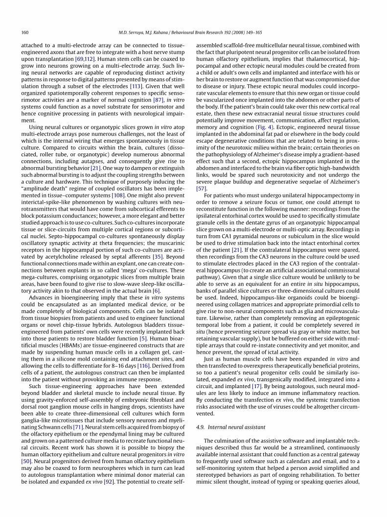
160 M.D. Serruya, M.J. Kahana / Behavioural Brain Research 192 (2008) 149–165
attached to a multi-electrode array can be connected to tissue-engineered axons that are free to integrate with a host nerve stumpupon transplantation [69,112]. Human stem cells can be coaxed togrow into neurons growing on a multi-electrode array. Such liv-ing neural networks are capable of reproducing distinct activitypatterns in response to digital patterns presented by means of stim-ulation through a subset of the electrodes [113]. Given that wellorganized spatiotemporally coherent responses to specific senso-rimotor activities are a marker of normal cognition [87], in vitrosystems could function as a novel substrate for sensorimotor andhence cognitive processing in patients with neurological impair-ment.
Using neural cultures or organotypic slices grown in vitro atopmulti-electrode arrays pose numerous challenges, not the least ofwhich is the internal wiring that emerges spontaneously in tissueculture. Compared to circuits within the brain, cultures (disso-ciated, roller tube, or organotypic) develop numerous abnormalconnections, including autapses, and consequently give rise toabnormal bursting behavior [21]. One way to dampen or extinguishsuch abnormal bursting is to adjust the coupling strengths betweena culture and hardware. This technique of purposely inducing the“amplitude death” regime of coupled oscillators has been imple-mented in tissue–computer systems [108]. One might also preventinterictal-spike-like phenomenon by washing cultures with neu-rotransmitters that would have come from subcortical efferents toblock potassium conductances; however, a more elegant and betterstudied approach is to use co-cultures. Such co-cultures incorporatetissue or slice-circuits from multiple cortical regions or subcorti-cal nuclei. Septo-hippocampal co-cultures spontaneously displayoscillatory synaptic activity at theta frequencies; the muscarinicreceptors in the hippocampal portion of such co-cultures are acti-vated by acetylcholine released by septal afferents [35]. Beyondfunctional connections made within an explant, one can create con-nections between explants in so called ‘mega’ co-cultures. Thesemega-cultures, comprising organotypic slices from multiple brainareas, have been found to give rise to slow-wave sleep-like oscilla-tory activity akin to that observed in the actual brain [6].
Advances in bioengineering imply that these in vitro systemscould be encapsulated as an implanted medical device, or bemade completely of biological components. Cells can be isolatedfrom tissue biopsies from patients and used to engineer functionalorgans or novel chip-tissue hybrids. Autologous bladders tissue-engineered from patients’ own cells were recently implanted backinto those patients to restore bladder function [5]. Human bioar-tificial muscles (HBAMs) are tissue-engineered constructs that aremade by suspending human muscle cells in a collagen gel, cast-ing them in a silicone mold containing end attachment sites, andallowing the cells to differentiate for 8–16 days [116]. Derived fromcells of a patient, the autologous construct can then be implantedinto the patient without provoking an immune response.
Such tissue-engineering approaches have been extendedbeyond bladder and skeletal muscle to include neural tissue. Byusing gravity-enforced self-assembly of embryonic fibroblast anddorsal root ganglion mouse cells in hanging drops, scientists havebeen able to create three-dimensional cell cultures which formganglia-like microtissues that include sensory neurons and myeli-nating Schwann cells [71]. Neural stem cells acquired from biopsy ofthe olfactory epithelium or the ependymal lining may be culturedand grown on a patterned culture media to recreate functional neu-ral circuits. Recent work has shown it is possible to biopsy thehuman olfactory epithelium and culture neural progenitors in vitro[50]. Neural progenitors derived from human olfactory epitheliummay also be coaxed to form neurospheres which in turn can leadto autologous transplantation where minimal donor material canbe isolated and expanded ex vivo [92]. The potential to create self-
assembled scaffold-free multicellular neural tissue, combined withthe fact that pluripotent neural progenitor cells can be isolated fromhuman olfactory epithelium, implies that thalamocortical, hip-pocampal and other ectopic neural modules could be created froma child or adult’s own cells and implanted and interface with his orher brain to restore or augment function that was compromised dueto disease or injury. These ectopic neural modules could incorpo-rate vascular elements to ensure that this new organ or tissue couldbe vascularized once implanted into the abdomen or other parts ofthe body. If the patient’s brain could take over this new cortical realestate, then these new extracranial neural tissue structures couldpotentially improve movement, communication, affect regulation,memory and cognition (Fig. 4). Ectopic, engineered neural tissueimplanted in the abdominal fat pad or elsewhere in the body couldescape degenerative conditions that are related to being in prox-imity of the neurotoxic milieu within the brain; certain theories onthe pathophysiology of Alzheimer’s disease imply a gradient-basedeffect such that a second, ectopic hippocampus implanted in theabdomen and interfaced to the brain via fiber optic high-bandwidthlinks, would be spared such neurotoxicity and not undergo thesevere plaque buildup and degenerative sequelae of Alzheimer’s[57].
For patients who must undergo unilateral hippocampectomy inorder to remove a seizure focus or tumor, one could attempt toreconstitute function in the following manner: recordings from theipsilateral entorhinal cortex would be used to specifically stimulategranule cells in the dentate gyrus of an organotypic hippocampalslice grown on a multi-electrode or multi-optic array. Recordings inturn from CA1 pyramidal neurons or subiculum in the slice wouldbe used to drive stimulation back into the intact entorhinal cortexof the patient [21]. If the contralateral hippocampus were spared,then recordings from the CA3 neurons in the culture could be usedto stimulate electrodes placed in the CA3 region of the contralat-eral hippocampus (to create an artificial associational commissuralpathway). Given that a single slice culture would be unlikely to beable to serve as an equivalent for an entire in situ hippocampus,banks of parallel slice cultures or three-dimensional cultures couldbe used. Indeed, hippocampus-like organoids could be bioengi-neered using collagen matrices and appropriate primordial cells togive rise to non-neural components such as glia and microvascula-ture. Likewise, rather than completely removing an epileptogenictemporal lobe from a patient, it could be completely severed insitu (hence preventing seizure spread via gray or white matter, butretaining vascular supply), but be buffered on either side with mul-tiple arrays that could re-instate connectivity and yet monitor, andhence prevent, the spread of ictal activity.
Just as human muscle cells have been expanded in vitro andthen transfected to overexpress therapeutically beneficial proteins,so too a patient’s neural progenitor cells could be similarly iso-lated, expanded ex vivo, transgenically modified, integrated into acircuit, and implanted [17]. By being autologous, such neural mod-ules are less likely to induce an immune inflammatory reaction.By conducting the transfection ex vivo, the systemic transfectionrisks associated with the use of viruses could be altogether circum-vented.
4.9. Internal neural assistant
The culmination of the assistive software and implantable tech-niques described thus far would be a streamlined, continuouslyavailable internal assistant that could function as a central gatewayto frequently used software such as calendars and email, and to aself-monitoring system that helped a person avoid simplified andstereotyped behaviors as part of ongoing rehabilitation. To bettermimic silent thought, instead of typing or speaking queries aloud,
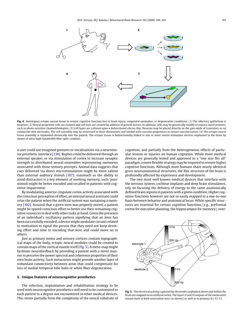
M.D. Serruya, M.J. Kahana / Behavioural Brain Research 192 (2008) 149–165 161
Fig. 4. Autologous ectopic neural tissue to restore cognitive function lost to brain injury, congenital anomalies, or degenerative conditions. (1) The olfactory epithelium isbiopsied. (2) Neural progenitor cells are isolated and cell lines are created by addition of growth factors. In addition, cells may be genetically modify to express novel proteins,such as photo-sensitive channelrhodopsin. (3) Cell types are cultured upon a bidirectional silicon chip. Neurons may be placed directly on the gate oxide of transistors or onconductive disk electrodes. The cell assembly may be structured in three dimensions and seeded with vascular progenitors to ensure vascularization. (4) The ectopic neuraltissue assembly is implanted chronically into the patient. The ectopic tissue is bidirectionally linked to one or more sensor-stimulator devices implanted in the brain bymeans of ultra-high-bandwidth fiber optic conduits.
a user could use imagined gestures or vocalizations via a neuromo-tor prosthetic interface [136]. Replies could be delivered through anexternal speaker, or via stimulation of cortex to increase synapticstrength to distributed neural ensembles representing memoriesassociated with those sensory precepts. Animal data suggests thatcues delivered via direct microstimulation might be more salientthan external auditory stimuli [107]; inasmuch as the ability toavoid distractors is a key element of working memory, such ‘pure’stimuli might be better encoded and recalled in patients with cog-nitive impairment.
By modulating anterior cingulate cortex activity associated withthe conscious perception of effort, an internal neural assistant couldrelax the patient when the artificial system was sustaining a mem-ory [102]. Assured that a given item was properly stored, a patientmight be spared conscious effort to better use their remaining cog-nitive resources to deal with other tasks at hand. Given the presenceof an individual’s oscillatory pattern signifying that an item hasbeen successfully encoded, a device might modulate circuits relatedto motivation to signal the person that they need not keep devot-ing effort and time to encoding that item and could move on toothers.
Just as primary motor and sensory cortices contain topograph-ical maps of the body, ectopic neural modules could be created tocontain maps of the cortical mantle itself (Fig. 5). A meta-map mightfacilitate neurofeedback by providing a patient with a novel man-ner to perceive the power spectral and coherence properties of theirown brain activity. Such metacortex might provide another layer ofredundant connectivity between areas that could compensate forloss of medial temporal lobe hubs or white fiber degeneration.
5. Unique features of neurocognitive prosthetics
The selection, implantation and rehabilitation strategy to beused with neurocognitive prosthetics will need to be customized toeach patient to a degree not encountered in other medical devices.This stems partially from the complexity of the neural substrate of
cognition, and partially from the heterogeneous effects of partic-ular lesions or injuries on human cognition. While most medicaldevices are generally tested and approved in a “one size fits all”paradigm, a more flexible strategy may be required to restore highercognitive functions. Although most humans share nearly identicalgross neuroanatomical structures, the fine structure of the brain isprofoundly affected by experience and development.
The two most well known medical devices that interface withthe nervous system, cochlear implants and deep brain stimulators,rely on focusing the delivery of energy to the same anatomicallydefined brain regions in patients with a given condition. Higher cog-nitive functions however are not so easily mapped in a one-to-onebasis between behavior and anatomical locus. While specific struc-tures are essential for certain cognitive functions (e.g., prefrontalcortex for executive planning, the hippocampus for memory), over-
Fig. 5. The electrical activity captured by electrodes implanted above and within thebrain are mapped on to artificial cortex. The layers V and VI outputs of the metacortexstream back to both association area (as shown) as well as to primary A1, V1, S1.
162 M.D. Serruya, M.J. Kahana / Behavioural Brain Research 192 (2008) 149–165
all cognition relies on the coordinated activity of widely distributedneural networks.
The development of the techniques and devices discussed in thisreview will continue to occur under the rubric of traditional clini-cal investigational trials. Withal, rather than having techniques ordevices that are designed to narrowly treat one defined behavior,it appears more likely that the clinicians of the future will insteadhave a toolbox of technique and device components. Though themanner in which FDA monitors safety of neurocognitive prostheticswill be the same as for other devices, the determination of efficacyconstitutes an extraordinary challenge. A new medical disciplinemay need to evolve in which neurologists, neurosurgeons, and neu-ropsychologists develop specialized expertise in evaluating whatbehavioral, non-invasive and implantable device based strategiesshould be used.
Though the various techniques in this review were discussedseparately, many techniques may bring fruit only when combined.It may be that the cognitive enhancement abilities of neurofeed-back and rTMS are synergistic. Likewise virtual reality could beintegrated with tDCS to help children with stroke re-acclimate to aclassroom or study new material. The choice of techniques, param-eters and the details of their combinations could be tailored toa person’s particular neuroanatomy, pathology, age, educationallevel, social context and rehabilitation goals.
Though the primary goal of neurocognitive prosthetics is torestore cognition and make quantifiable improvements in dailylife, this clinical mission goes hand in hand with a basic sciencemission of understanding the basis of cognition itself. As withany newly developing therapy, the line between basic science andclinical application will be blurred: there will be a continuousexchange of ideas and discovery between basic scientists and clin-icians. The possibility of tracking the unit activity of hundredsof individual neurons simultaneously, and to likewise have theability to inject signals back in to specific brain regions, wouldprovide an unprecedented scientific opportunity. Patients partic-ipating in clinical trials designed to assess the safety and clinicalutility of such devices might also elect to participate in additional,concurrent trials conducted in collaboration with basic science col-leagues.
6. Conclusion
Ongoing developments in non-invasive techniques and behav-ioral training promise to provide future clinicians with a greaterselection of options to help restore cognition in adults and childrenwith neurological disease and injury. Recent rapid developments indevices that can be chronically implanted in to the brain, coupledwith burgeoning knowledge of the neurophysiology of higher cog-nitive function, imply that the time has come to develop and testa new generation of implantable neurocognitive brain–computerinterfaces. For tens of thousands of children and adults disabledwith a range of diseases and injuries to their brain, treatmentoptions to ameliorate or cure their conditions are limited ornonexistent. The development of neurocognitive prosthetics mightrepresent a new way forward to address this urgent need and givethese individuals the possibility of life with greater enrichment andindependence.
Acknowledgments
Mijail Serruya was supported by the Brown University Depart-ment of Internal Medicine and the University of PennsylvaniaDepartment of Neurology. Mijail Serruya would like to thankDominick Tammaro, Fred Schiffman, Barry Connors, Eberhard Fetz,David Holtzman, Vernon Caviness, Gyorgy Buzsaki, William Hein-
del, Elena Festa, Kareem Zaghloul, Ammar Shaikhouni, AdamDingle, Geoffrey Aguirre and Mindy Aisen.
References
[1] Achard S, Salvador R, Whitcher B, Suckling J, Bullmore E. A resilient,low-frequency, small-world human brain functional network with highlyconnected association cortical hubs. J Neurosci 2006;26(January 4 (1)):63–72.
[2] Angelakis E, Lubar JF, Stathopoulou S. Electroencephalographic peak alphafrequency correlates of cognitive traits. Neurosci Lett 2004;371(November 16(1)):60–3.
[3] Angelakis E, Stathopoulou S, Frymiare JL, Green DL, Lubar JF, Kounios J.EEG neurofeedback: a brief overview and an example of peak alpha fre-quency training for cognitive enhancement in the elderly. Clin Neuropsychol2007;21(January (1)):110–29.
[4] Arthur JV, Boahen K. Learning in silicon: timing is everything. Advances inneural information processing systems, 18. 2006. p. 75–82.
[5] Atala A, Bauer SB, Soker S, Yoo JJ, Retik AB. Tissue-engineered autolo-gous bladders for patients needing cystoplasty. Lancet 2006;367(April 15(9518)):1241–6.
[6] Baker RE, Corner MA, van Pelt J. Spontaneous neuronal discharge patternsin developing organotypic mega-co-cultures of neonatal rat cerebral cortex.Brain Res 2006;1101(July 26 (1)):29–35.
[7] Ball K, Berch DB, Helmers KF, Jobe JB, Leveck MD, Marsiske M, et al. Effects ofcognitive training interventions with older adults—a randomized controlledtrial. JAMA 2002;288:2271–81.
[8] Barbeau E, Wendling F, Regis J, Duncan R, Poncet M, Chauvel P, et al. Recollec-tion of vivid memories after perirhinal region stimulations: synchronizationin the theta range of spatially distributed brain areas. Neuropsychologia2005;43(9):1329–37.
[9] Beauregard M, Levesque J. Functional magnetic resonance imaging inves-tigation of the effects of neurofeedback training on the neural bases ofselective attention and response inhibition in children with attention-deficit/hyperactivity disorder. Appl Psychophysiol Biofeedback 2006;31(March (1)):3–20.
[10] Beatty J, Greenberg A, Deibler WP, O’Hanlon JF. Operant control of occip-ital theta rhythm affects performance in a radar monitoring task. Science1974;183(March 1 (127)):871–3.
[11] Bender BG, Levin JR. Pictures, imagery, and retarded children’s prose learning.J Educ Psychol 1978;70(August (4)):583–8.
[12] Berger TW, Ahuja A, Courellis SH, Deadwyler SA, Erinjippurath G, Ger-hardt GA, et al. Restoring lost cognitive function. IEEE Eng Med Biol Mag2005;24(September–October (5)):30–44.
[13] Bergman MM. The essential steps cognitive orthotic. NeuroRehabilitation2003;18(1):31–46.
[14] Blankertz B, Muller KR, Curio G, Vaughan TM, Schalk G, Wolpaw JR, et al. TheBCI Competition 2003: progress and perspectives in detection and discrimi-nation of EEG single trials. IEEE Trans Biomed Eng 2004;51(June (6)):1044–51.
[15] Boggio PS, Ferrucci R, Rigonatti SP, Covre P, Nitsche M, Pascual-Leone A,et al. Effects of transcranial direct current stimulation on working mem-ory in patients with Parkinson’s disease. J Neurol Sci 2006;249(November1 (1)):31–8.
[16] Bourgeois MS. Evaluating memory wallets in conversations with persons withdementia. J Speech Hear Res 1992;35(December (6)):1344–57.
[17] Boyden ES, Zhang F, Bamberg E, Nagel G, Deisseroth K. Millisecondtimescale, genetically targeted optical control of neural activity. Nat Neurosci2005;8(September (9)):1263–8.
[18] Briones TL, Klintsova AY, Greenough WT. Stability of synaptic plasticity in theadult rat visual cortex induced by complex environment exposure. Brain Res2004;1018(August 20 (1)):130–5.
[19] Brooks BM, Rose FD. The use of virtual reality in memory rehabilitation:current findings and future directions. NeuroRehabilitation 2003;18(2):147–57.
[20] Brown TG, Sherrington CS. On the Instability of a Cortical Point. Proc R SocLond Ser B 1912;85(July 25 (579)):250–77 (Containing Papers of a BiologicalCharacter).
[21] Buzsaki, personal communication, 2007.[22] Caria A, Veit R, Sitaram R, Lotze M, Weiskopf N, Grodd W, et al. Regula-
tion of anterior insular cortex activity using real-time fMRI. Neuroimage2007;35(April 15 (3)):1238–46.
[23] Carney N, Chesnut RM, Maynard H, Mann NC, Patterson P, Helfand M. Effect ofcognitive rehabilitation on outcomes for persons with traumatic brain injury:a systematic review. J Head Trauma Rehabil 1999;14:277–307.
[24] Catania KC, Henry EC. Touching on somatosensory specializations in mam-mals. Curr Opin Neurobiol 2006;16(August (4)):467–73.
[25] Cicerone KD, Mott T, Azulay J, Friel JC. Community integration and satisfactionwith functioning after intensive cognitive rehabilitation for traumatic braininjury. Arch Phys Med Rehabil 2004;85(June (6)):943–50.
[26] Clark KB, Naritoku DK, Smith DC, Browning RA, Jensen RA. Enhanced recog-nition memory following vagus nerve stimulation in human participants. NatNeurosci 1999;2(January (1)):94–8.

M.D. Serruya, M.J. Kahana / Behavioural Brain Research 192 (2008) 149–165 163
[27] Deary IJ, Whalley LJ, Batty GD, Starr JM. Physical fitness and lifetime cognitivechange. Neurology 2006;67(October 10 (7)):1195–200.
[28] deCharms RC, Maeda F, Glover GH, Ludlow D, Pauly JM, Soneji D, et al. Controlover brain activation and pain learned by using real-time functional MRI. ProcNatl Acad Sci USA 2005;102(December 20 (51)):18626–31.
[29] Douglas RJ, Martin KA. Neuronal circuits of the neocortex. Annu Rev Neurosci2004;27:419–51.
[30] Dunn J, Clare L. Learning face–name associations in early-stage dementia:comparing the effects of errorless learning and effortful processing. Neuropsy-chol Rehabil 2007;(July 7):1.
[31] Eichenbaum H, Yonelinas AR, Ranganath C. The medial temporal lobe andrecognition memory. Annu Rev Neurosci 2007;(April 6).
[32] Eippert F, Veit R, Weiskopf N, Erb M, Birbaumer N, Anders S. Regulationof emotional responses elicited by threat-related stimuli. Hum Brain Mapp2007;28(May (5)):409–23.
[33] Fetz EE. Volitional control of neural activity: implications for brain–computerinterfaces. J Physiol 2007;579(March 15 (Pt 3)):571–9.
[34] Fidopiastis CM, Stapleton CB, Whiteside JD, Hughes CE, Fiore SM, Martin GA, etal. Human Experience Modeler: context-driven cognitive retraining to facili-tate transfer of learning. Cyberpsychol Behav 2006;9(April (2)):183–7.
[35] Fischer Y, Gahwiler BH, Thompson SM. Activation of intrinsic hippocam-pal theta oscillations by acetylcholine in rat septo-hippocampal cocultures.J Physiol 1999;519(September 1 (Pt 2)):405–13.
[36] Flannery MA, Butterbaugh GJ, Rice DA, Rice JC. Reminding technologyfor prospective memory disability: a case study. Pediatr Rehabil 1997;1(October–December (4)):239–44.
[37] Fregni F, Boggio PS, Nitsche M, Bermpohl F, Antal A, Feredoes E, et al. Anodaltranscranial direct current stimulation of prefrontal cortex enhances workingmemory. Exp Brain Res 2005;166(September (1)):23–30.
[38] Fries P, Fernandez G, Jensen O. When neurons form memories. Trends Neurosci2003;26(March (3)):123–4.
[39] Fumal A, Coppola G, Bohotin V, Gerardy PY, Seidel L, Donneau AF, et al. Induc-tion of long-lasting changes of visual cortex excitability by five daily sessionsof repetitive transcranial magnetic stimulation (rTMS) in healthy volunteersand migraine patients. Cephalalgia 2006;26(February (2)):143–9.
[40] Fusi S, Mattia M. Collective behavior of networks with linear (VLSI) integrate-and-fire neurons. Neural Comput 1999;11(April 1 (3)):633–52.
[41] Fyhn M, Hafting T, Treves A, Moser MB, Moser EI. Hippocampal remap-ping and grid realignment in entorhinal cortex. Nature 2007;446(March 8(7132)):190–4.
[42] Gemmell J, Bell G, Lueder R, Drucker S, Wong C. MyLifeBits: Fulfilling theMemex Vision, ACM Multimedia ’02. France: Juan-les-Pins, December 1–6,2002, p. 235–8.
[43] Gemmell J, Williams L, Wood K, Lueder R, Bell G. Passive capture and ensuingissues for a personal lifetime store. In: Proceedings of the 1st ACM workshopon continuous archival and retrieval of personal experiences. 2004.
[44] Ghacibeh GA, Shenker JI, Shenal B, Uthman BM, Heilman KM. Effect ofvagus nerve stimulation on creativity and cognitive flexibility. Epilepsy Behav2006;8(June (4)):720–5.
[45] Glisky EL, Schacter DL. Extending the limits of complex learning in organicamnesia: computer training in a vocational domain. Neuropsychologia1989;27:107–20.
[46] Gorman P, Dayle R, Hood CA, Rumrell L. Effectiveness of the ISAAC cognitiveprosthetic system for improving rehabilitation outcomes with neurofunc-tional impairment. NeuroRehabilitation 2003;18(1):57–67.
[47] Grandmaison E, Simard M. A critical review of memory stimulation pro-grams in Alzheimer’s disease. J Neuropsychiatry Clin Neurosci 2003;15(Spring(2)):130–44.
[48] Green CS, Bavelier D. Action-video-game experience alters the spatial resolu-tion of vision. Psychol Sci 2007;18(January (1)):88–94.
[49] Griffin AL, Asaka Y, Darling RD, Berry SD. Theta-contingent trial presentationaccelerates learning rate and enhances hippocampal plasticity during traceeyeblink conditioning. Behav Neurosci 2004;118(April (2)):403–11.
[50] Hahn CG, Han LY, Rawson NE, Mirza N, Borgmann-Winter K, Lenox RH, et al. Invivo and in vitro neurogenesis in human olfactory epithelium. J Comp Neurol2005;483(March 7 (2)):154–63.
[51] Haigh KZ, Kiff LM, Ho G. The Independent LifeStyle Assistant: lessons learned.Assist Technol 2006;18(Spring (1)):87–106.
[52] Halgren E. Mental phenomena induced by stimulation in the limbic system.Hum Neurobiol 1982;1(4):251–60.
[53] Hamani C, McAndrews MP, Cohn M, Oh M, Zumsteg D, Shapiro CM, et al. Mem-ory enhancement induced by hypothalamic/fornix deep brain stimulation.Ann Neurol 2008;63(January (1)):119–23.
[54] Hazy TE, Frank MJ, O’Reilly RC. Banishing the homunculus: making workingmemory work. Neuroscience 2006;139(April 28 (1)):105–18.
[55] Hinterberger T, Veit R, Wilhelm B, Weiskopf N, Vatine JJ, Birbaumer N. Neu-ronal mechanisms underlying control of a brain–computer interface. Eur JNeurosci 2005;21(June (11)):3169–81.
[56] Hochberg LR, Serruya MD, Friehs GM, Mukand JA, Saleh M, Caplan AH, et al.Neuronal ensemble control of prosthetic devices by a human with tetraplegia.Nature 2006;442(July 13 (7099)):164–71.
[57] Holtzman, Serruya, personal communication, 2005.[58] Hoppensteadt FC, Izhikevich EM. Thalamo-cortical interactions modeled by
weakly connected oscillators: could the brain use FM radio principles? Biosys-tems 1998;48(September–December (1–3)):85–94.
[59] Huber D, Petreanu L, Ghitani N, Ranade S, Hromadka T, Mainen Z, et al. Sparseoptical microstimulation in barrel cortex drives learned behaviour in freelymoving mice. Nature 2008;451(January 3 (7174)):61–4.
[60] Iyer MB, Mattu U, Grafman J, Lomarev M, Sato S, Wassermann EM. Safetyand cognitive effect of frontal DC brain polarization in healthy individuals.Neurology 2005;64(March 8 (5)):872–5.
[61] Izhikevich EM. Polychronization: computation with spikes. Neural Comput2006;18(February (2)):245–82.
[62] Jackson A, Mavoori J, Fetz EE. Long-term motor cortex plasticity inducedby an electronic neural implant. Nature 2006;444(November 2 (7115)):56–60.
[63] Jackson A, Moritz CT, Mavoori J, Lucas TH, Fetz EE. The Neurochip BCI: towardsa neural prosthesis for upper limb function. IEEE Trans Neural Syst Rehabil Eng2006;14(June (2)):187–90.
[64] Jackson HJ. On right- or left-sided spasm at the onset of epileptic parox-ysms, and on crude sensation warnings and elaborate mental states. Brain1880;3:192–206.
[65] Ji D, Wilson MA. Coordinated memory replay in the visual cortex and hip-pocampus during sleep. Nat Neurosci 2007;10(January (1)):100–7;Gloor P. Experiential phenomena of temporal lobe epilepsy: facts andhypotheses. Brain 1990;113:1673–94.
[66] Jiang F, Racine R, Turnbull J. Electrical stimulation of the septal regionof aged rats improves performance in an open-field maze. Physiol Behav1997;62(December (6)):1279–82.
[67] Kahana MJ, Seelig D, Madsen JR. Theta returns. Curr Opin Neurobiol2001;11(December (6)):739–44.
[68] Kahana MJ. The cognitive correlates of human brain oscillations. J Neurosci2006;26(February 8 (6)):1669–72.
[69] Kameswaran N, Pfister BJ, Cohen AS, Smith DH. A new approach for a cyber-netic interface with the nervous system. 148.22/L11. Atlanta: Society forNeuroscience; 2006.
[70] Karpicke JD, Roediger III HL. The critical importance of retrieval for learning.Science 2008;319(February 15 (5865)):966–8.
[71] Kelm JM, Ittner LM, Born W, Djonov V, Fussenegger M. Self-assembly of sen-sory neurons into ganglia-like microtissues. J Biotechnol 2006;121(January 2(1)):86–101.
[72] Kennedy PR, Bakay RA, Moore MM, Adams K, Goldwaithe J. Direct control ofa computer from the human central nervous system. IEEE Trans Rehabil Eng2000;8(June (2)):198–202.
[73] Kessels RP, de Haan EH. Implicit learning in memory rehabilitation: ameta-analysis on errorless learning and vanishing cues methods. J Clin ExpNeuropsychol 2003;25(September (6)):805–14.
[74] Kier RJ, Ames JC, Beer RD, Harrison RR. Design and implementation of mul-tipattern generators in analog VLSI. IEEE Trans Neural Netw 2006;17(July(4)):1025–38.
[75] Kramer AF, Bherer L, Colcombe SJ, Dong W, Greenough WT. Environmentalinfluences on cognitive and brain plasticity during aging. J Gerontol A Biol SciMed Sci 2004;59(September (9)):M940–57.
[76] Kubu CS, Azmi H, Greenberg B, Malone D, Machado A, Friehs G, RasmussenS, Malloy P, Rezai A. DBS in the Internal Capsule/Ventral Striatum ImprovesMemory in Patients with Severe Psychiatric Disorders. Am Acad Neurol Surg,Annual Meeting, Abstract, April 16, 2007. Article ID: 40788.
[77] Lachaux JP, Jerbi K, Bertrand O, Minotti L, Hoffmann D, Schoendorff B, et al.Blueprint for real-time functional mapping via human intracranial recordings.PLoS ONE 2007;2(October 31 (10)):e1094.
[78] Lee JH, Ku J, Cho W, Hahn WY, Kim IY, Lee SM, et al. A virtual reality system forthe assessment and rehabilitation of the activities of daily living. CyberpsycholBehav 2003;6(August (4)):383–8.
[79] Levesque J, Beauregard M, Mensour B. Effect of neurofeedback trainingon the neural substrates of selective attention in children with attention-deficit/hyperactivity disorder: a functional magnetic resonance imagingstudy. Neurosci Lett 2006;394(February 20 (3)):216–21.
[80] Lin KL, Pascual-Leone A. Transcranial magnetic stimulation and its applica-tions in children. Chang Gung Med J 2002;25(July (7)):424–36.
[81] Liu SC, Douglas R. Temporal coding in a silicon network of integrate-and-fireneurons. IEEE Trans Neural Netw 2004;15(September (5)):1305–14.
[82] Louie K, Wilson MA. Temporally structured replay of awake hippocampalensemble activity during rapid eye movement sleep. Neuron 2001;29(January(1)):145–56.
[83] Luber B, Kinnunen LH, Rakitin BC, Ellsasser R, Stern Y, Lisanby SH. Facilitationof performance in a working memory task with rTMS stimulation of the pre-cuneus: frequency- and time-dependent effects. Brain Res 2007;1128(January12 (1)):120–9.
[84] Machii K, Cohen D, Ramos-Estebanez C, Pascual-Leone A. Safety of rTMS tonon-motor cortical areas in healthy participants and patients. Clin Neuro-physiol 2006;117(February (2)):455–71.
[85] Maguire EA, Valentine ER, Wilding JM, Kapur N. Routes to remembering: thebrains behind superior memory. Nat Neurosci 2003;6(January (1)):90–5.
[86] Mahncke HW, Bronstone A, Merzenich MM. Brain plasticity and functionallosses in the aged: scientific bases for a novel intervention. Prog Brain Res2006;157:81–109 [Review].
[87] Mahncke HW, Connor BB, Appelman J, Ahsanuddin ON, Hardy JL, Wood RA,et al. Memory enhancement in healthy older adults using a brain plasticity-based training program: a randomized, controlled study. Proc Natl Acad SciUSA 2006;103(August 15 (33)):12523–8.
164 M.D. Serruya, M.J. Kahana / Behavioural Brain Research 192 (2008) 149–165
[88] Man DW, Tam SF, Hui-Chan CW. Learning to live independently withexpert systems in memory rehabilitation. NeuroRehabilitation 2003;18(1):21–9.
[89] Manns ID, Alonso A, Jones BE. Discharge profiles of juxtacellularly labeledand immunohistochemically identified GABAergic basal forebrain neuronsrecorded in association with the electroencephalogram in anesthetized rats.J Neurosci 2000;20(December 15 (24)):9252–63.
[90] Manns JR, Clark RE, Squire LR. Awareness predicts the magnitude of single-cuetrace eyeblink conditioning. Hippocampus 2000;10(2):181–6.
[91] Markram H. The blue brain project. Nat Rev Neurosci 2006;7(February(2)):153–60.
[92] Marshall CT, Lu C, Winstead W, Zhang X, Xiao M, Harding G, et al. The ther-apeutic potential of human olfactory-derived stem cells. Histol Histopathol2006;21(June (6)):633–43.
[93] Marshall L, Helgadottir H, Molle M, Born J. Boosting slow oscillations duringsleep potentiates memory. Nature 2006;444(November 30 (7119)):610–3.
[94] Matsumoto R, Nair DR, LaPresto E, Najm I, Bingaman W, Shibasaki H, et al.Functional connectivity in the human language system: a cortico-corticalevoked potential study. Brain 2004;127(October (Pt 10)):2316–30.
[95] Mavoori J, Jackson A, Diorio C, Fetz E. An autonomous implantable computerfor neural recording and stimulation in unrestrained primates. J NeurosciMethods 2005;148(October 15 (1)):71–7.
[96] McComas J, Pivik J, Laflamme M. Current uses of virtual reality for childrenwith disabilities. Stud Health Technol Inform 1998;58:161–9.
[97] Merrill CA, Jonsson MA, Minthon L, Ejnell H, C-son Silander H, Blennow K,et al. Vagus nerve stimulation in patients with Alzheimer’s disease: Addi-tional follow-up results of a pilot study through 1 year. J Clin Psychiatry2006;67(August (8)):1171–8.
[98] Monderer RS, Harrison DM, Haut SR. Neurofeedback and epilepsy. EpilepsyBehav 2002;3(June (3)):214–8.
[99] Moriarity JL, Boatman D, Krauss GL, Storm PB, Lenz FA. Human “memories”can be evoked by stimulation of the lateral temporal cortex after ipsilateralmedial temporal lobe resection. J Neurol Neurosurg Psychiatry 2001;71(Octo-ber (4)):549–51.
[100] Morie JF, Iyer K, Luigi DP, Williams J, Dozois A, Rizzo AS. Development of a datamanagement tool for investigating multivariate space and free will experi-ences in virtual reality. Appl Psychophysiol Biofeedback 2005;30(September(3)):319–31.
[101] Mountcastle VB. The columnar organization of the neocortex. Brain 1997;120(April (Pt 4)):701–22.
[102] Mulert C, Menzinger E, Leicht G, Pogarell O, Hegerl U. Evidence for a closerelationship between conscious effort and anterior cingulate cortex activity.Int J Psychophysiol 2005;56(April (1)):65–80.
[103] Naeser MA, Martin PI, Nicholas M, Baker EH, Seekins H, Helm-Estabrooks N,et al. Improved naming after TMS treatments in a chronic, global aphasiapatient—case report. Neurocase 2005;11(June (3)):182–93.
[104] Naeser MA, Martin PI, Nicholas M, Baker EH, Seekins H, Kobayashi M, etal. Improved picture naming in chronic aphasia after TMS to part of rightBroca’s area: an open-protocol study. Brain Lang 2005;93(April (1)):95–105.
[105] Neeves KB, Lo CT, Foley CP, Saltzman WM, Olbricht WL. Fabrication and char-acterization of microfluidic probes for convection enhanced drug delivery. JControl Release 2006;111(April 10 (3)):252–62.
[106] Nitsche MA, Fricke K, Henschke U, Schlitterlau A, Liebetanz D, Lang N, et al.Pharmacological modulation of cortical excitability shifts induced by tran-scranial direct current stimulation in humans. J Physiol 2003;553(November15 (Pt 1)):293–301.
[107] Otto KJ, Rousche PJ, Kipke DR. Cortical microstimulation in auditory cortexof rat elicits best-frequency dependent behaviors. J Neural Eng 2005;2(June(2)):42–51.
[108] Ozden I, Venkataramani S, Long MA, Connors BW, Nurmikko AV. Strongcoupling of nonlinear electronic and biological oscillators: reaching the“amplitude death” regime. Phys Rev Lett 2004;93(October 8 (15)):158102.
[109] Pascual-Marqui RD, Michel CM, Lehmann D. Low resolution electromagnetictomography: a new method for localizing electrical activity in the brain. Int JPsychophysiol 1994;18(October (1)):49–65.
[110] Penfield W, Perot P. The brain’s record of auditory and visual experience: afinal summary and discussion. Brain 1963;86:595–696.
[111] Penfield W. The excitable cortex in conscious man. Liverpool: Liverpool Uni-versity Press; 1958.
[112] Pfister BJ, Huang JH, Kameswaran N, Zager EL, Smith DH. Neural engineer-ing to produce in vitro nerve constructs and neurointerface. Neurosurgery2007;60(January (1)):137–41.
[113] Pizzi R, Cino G, Gelain F, Rossetti D, Vescovi A. Learning in human neuralnetworks on microelectrode arrays. Biosystems 2007;88(March (1–2)):1–15.
[114] Polich J. Updating P300: an integrative theory of P3a and P3b. Clin Neurophys-iol 2007;118(October (10)):2128–48.
[115] Polyn SM, Kahana MJ. Memory search and the neural representation of con-text. Trends Cogn Sci 2008;12(January (1)):24–30.
[116] Powell CA, Smiley BL, Mills J, Vandenburgh HH. Mechanical stimulationimproves tissue-engineered human skeletal muscle. Am J Physiol Cell Physiol2002;283(November (5)):C1557–65.
[117] Prescott TJ, Montes Gonzalez FM, Gurney K, Humphries MD, Redgrave P. Arobot model of the basal ganglia: behavior and intrinsic processing. NeuralNetw 2006;19(January (1)):31–61.
[118] Quiroga RQ, Reddy L, Kreiman G, Koch C, Fried I. Invariant visual repre-sentation by single neurons in the human brain. Nature 2005;435(June 23(7045)):1102–7.
[119] Quittre A, Olivier C, Salmon E. Compensating strategies for impaired episodicmemory and time orientation in a patient with Alzheimer’s disease. ActaNeurol Belg 2005;105(March (1)):30–8.
[120] Randolph C, Tierney MC, Mohr E, Chase TN. The repeatable battery for theassessment of neuropsychological status (RBANS): preliminary clinical valid-ity. J Clin Exp Neuropsychol (Neuropsychol, Dev) 1998;20(June (3)):310–9(10).
[121] Reger BD, Fleming KM, Sanguineti V, Alford S, Mussa-Ivaldi FA. Connectingbrains to robots: an artificial body for studying the computational propertiesof neural tissues. Artif Life 2000;6(Fall (4)):307–24.
[122] Retterer ST, Smith KL, Bjornsson CS, Neeves KB, Spence AJ, Turner JN, et al.Model neural prostheses with integrated microfluidics: a potential interven-tion strategy for controlling reactive cell and tissue responses. IEEE TransBiomed Eng 2004;51(November (11)):2063–73.
[123] Richard Clark C, Veltmeyer MD, Hamilton RJ, Simms E, Paul R, Hermens D, etal. Spontaneous alpha peak frequency predicts working memory performanceacross the age span. Int J Psychophysiol 2004;53(June (1)):1–9.
[124] Richards M, Hardy R, Wadsworth ME. Does active leisure protect cogni-tion? Evidence from a national birth cohort. Soc Sci Med 2003;56(February(4)):785–92.
[125] Romo R, Hernandez A, Zainos A, Salinas E. Somatosensory discriminationbased on cortical microstimulation. Nature 1998;392(March 26 (6674)):387–90.
[126] Schiff ND, Plum F, Rezai AR. Developing prosthetics to treat cognitive dis-abilities resulting from acquired brain injuries. Neurol Res 2002;24(March(2)):116–24.
[127] Schiff ND, Giacino JT, Kalmar K, Victor JD, Baker K, Gerber M, et al. Behaviouralimprovements with thalamic stimulation after severe traumatic brain injury.Nature 2007;448(August 2 (7153)):600–3.
[128] Schulze H. MEMOS: a mobile extensible memory aid system. Telemed J EHealth 2004;10(Summer (2)):233–42.
[129] Schvarcz JR. Long-term results of stimulation of the septal area for relief ofneurogenic pain. Acta Neurochir Suppl (Wien) 1993;58:154–5.
[130] Seager MA, Johnson LD, Chabot ES, Asaka Y, Berry SD. Oscillatory brain statesand learning: Impact of hippocampal theta-contingent training. Proc NatlAcad Sci USA 2002;99(February 5 (3)):1616–20.
[131] Sederberg PB, Schulze-Bonhage A, Madsen JR, Bromfield EB, McCarthyDC, Brandt A, et al. Hippocampal and neocortical gamma oscillationspredict memory formation in humans. Cereb Cortex 2007;17(May (5)):1190–6.
[132] Sederberg PB, Schulze-Bonhage A, Madsen JR, Bromfield EB, Litt B, Brandt A,et al. Gamma oscillations distinguish true from false memories. Psychol Sci2007;18(November (11)):927–32.
[133] Sellers EW, Donchin EA. P300-based brain–computer interface: initial testsby ALS patients. Clin Neurophysiol 2006;117(March (3)):538–48.
[134] Serruya, Caviness, personal communication, 2005.[135] Serruya M, Hatsopoulos N, Fellows M, Paninski L, Donoghue J. Robust-
ness of neuroprosthetic decoding algorithms. Biol Cybern 2003;88(March(3)):219–28.
[136] Serruya MD, Donoghue JP. Design principles for intracortical neuromotorprosthetics. In: Horch KW, Dhillon GS, editors. Neuroprosthetics: theory andpractice. Imperial College Press; 2004.
[137] Serruya MD, Hatsopoulos NG, Paninski L, Fellows MR, Donoghue JP. Instantneural control of a movement signal. Nature 2002;416(March 14 (6877)):141–2.
[138] Serruya MD, Song Y, Patterson WR, Bull CW, Nurmikko AV, Fellows MR, SimeralJD, Donoghue JP. Development of a brain implantable microsystem withinfrared optical telemetry for advanced neuromotor prosthesis. 148.15/L4.Atlanta: Society for Neuroscience; 2006.
[139] Shaikhouni A, Mehta MR. Enhancement of sequence learning by oscillations.68.17/BB25. 2006 Neuroscience Meeting Planner. Atlanta, GA: Society for Neu-roscience; 2006 [Online].
[140] Shipp S. The functional logic of cortico-pulvinar connections. Philos Trans RSoc Lond B Biol Sci 2003;358(October 29 (1438)):1605–24.
[141] Shirvalkar P, Seth M, Schiff ND, Herrera DG. Cognitive enhancement withcentral thalamic electrical stimulation. Proc Natl Acad Sci USA 2006;103(November 7 (45)):17007–12.
[142] Sjogren MJ, Hellstrom PT, Jonsson MA, Runnerstam M, Silander HC, Ben-Menachem E. Cognition-enhancing effect of vagus nerve stimulation inpatients with Alzheimer’s disease: a pilot study. J Clin Psychiatry 2002;63(November (11)):972–80.
[143] Smith DC, Modglin AA, Roosevelt RW, Neese SL, Jensen RA, Browning RA, etal. Electrical stimulation of the vagus nerve enhances cognitive and motorrecovery following moderate fluid percussion injury in the rat. J Neurotrauma2005;22(December (12)):1485–502.
[144] Snyder A, Bahramali H, Hawker T, Mitchell DJ. Savant-like numerosityskills revealed in normal people by magnetic pulses. Perception 2006;35(6):837–45.
[145] Sole-Padulles C, Bartres-Faz D, Junque C, Clemente IC, Molinuevo JL, BargalloN, et al. Repetitive transcranial magnetic stimulation effects on brain functionand cognition among elders with memory dysfunction. A randomized shamcontrolled study. Cereb Cortex 2006;16(October (10)):1487–93.

M.D. Serruya, M.J. Kahana / Behavioural Brain Research 192 (2008) 149–165 165
[146] Srinivasan R, Bibi FA, Nunez PL. Steady-state visual evoked potentials:distributed local sources and wave-like dynamics are sensitive to flicker fre-quency. Brain Topogr 2006;18(Spring (3)):167–87.
[147] Staubli U, Lynch G. Stable hippocampal long-term potentiation elicited by‘theta’ pattern stimulation. Brain Res 1987;435(December 1 (1–2)):227–34.
[148] Steriade M. Grouping of brain rhythms in corticothalamic systems. Neuro-science 2006;137(4):1087–106.
[149] Sterman MB, Egner T. Foundation and practice of neurofeedback for the treat-ment of epilepsy. Appl Psychophysiol Biofeedback 2006;31(March (1)):21–35.
[150] Stock SE, Davies DK, Davies KR, Wehmeyer ML. Evaluation of an applica-tion for making palmtop computers accessible to individuals with intellectualdisabilities. J Intellect Dev Disabil 2006;31(March (1)):39–46.
[151] Sulaiman WA, Kline DG. Nerve surgery: a review and insights about its future.Clin Neurosurg 2006;53:38–47.
[152] Sutton S, Braren M, Zubin J, John ER. Evoked-potential correlates of stimulusuncertainty. Science 1965;150(November 26 (700)):1187–8.
[153] Talwar SK, Xu S, Hawley ES, Weiss SA, Moxon KA, Chapin JK. Rat navigationguided by remote control. Nature 2002;417(May 2 (6884)):37–8.
[154] Temple E, Deutsch GK, Poldrack RA, Miller SL, Tallal P, Merzenich MM, et al.Neural deficits in children with dyslexia ameliorated by behavioral remedia-tion: evidence from functional MRI. Proc Natl Acad Sci USA 2003;100(March4 (5)):2860–5.
[155] Thatcher RW. EEG database-guided neurotherapy. In: Evans JE, Abarbanel A,editors. Introduction to quantitative EEG and neurofeedback. San Diego, CA:Academic Press; 1999. p. 48–9.
[156] Thoene AI, Glisky EL. Learning of name-face associations in memory impairedpatients: a comparison of different training procedures. J Int Neuropsychol Soc1995;1(January (1)):29–38.
[157] Treat NJ, Poon LW, Fozard JL, Popkin SJ. Toward applying cognitive skill trainingto memory problems. Exp Aging Res 1978;4(August (4)):305–19.
[158] Troster AI, Wilkinson SB, Fields JA, Miyawaki K, Koller WC. Chronic elec-trical stimulation of the left ventrointermediate (Vim) thalamic nucleusfor the treatment of pharmacotherapy-resistant Parkinson’s disease: a dif-ferential impact on access to semantic and episodic memory? Brain Cogn1998;38(November (2)):125–49.
[159] Thaut MH, Peterson DA, McIntosh GC. Temporal entrainment of cogni-tive functions: musical mnemonics induce brain plasticity and oscillatorysynchrony in neural networks underlying memory. Ann NY Acad Sci2005;1060(December):243–5.
[160] Van Pelt J, Corner MA, Wolters PS, Rutten WL, Ramakers GJ. Longterm stabilityand developmental changes in spontaneous network burst firing patterns indissociated rat cerebral cortex cell cultures on multielectrode arrays. NeurosciLett 2004;361(May 6 (1–3)):86–9.
[161] van Pelt J, Wolters PS, Corner MA, Rutten WL, Ramakers GJ. Long-term char-acterization of firing dynamics of spontaneous bursts in cultured neuralnetworks. IEEE Trans Biomed Eng 2004;51(November (11)):2051–62.
[162] van’t Hooft I, Andersson K, Sejersen T, Bartfai A, von Wendt L. Attentionand memory training in children with acquired brain injuries. Acta Paediatr2003;92(August (8)):935–40.
[163] Vernon D, Egner T, Cooper N, Compton T, Neilands C, Sheri A, et al. The effectof training distinct neurofeedback protocols on aspects of cognitive perfor-mance. Int J Psychophysiol 2003;47(January (1)):75–85.
[164] von Melchner L, Pallas SL, Sur M. Visual behaviour mediated by retinalprojections directed to the auditory pathway. Nature 2000;404(April 20(6780)):871–6.
[165] Wade TK, Troy JC. Mobile phones as a new memory aid: a preliminary inves-tigation using case studies. Brain Inj 2001;15(April (4)):305–20.
[166] Wagenaar DA, Madhavan R, Pine J, Potter SM. Controlling bursting incortical cultures with closed-loop multi-electrode stimulation. J Neurosci2005;25(January 19 (3)):680–8.
[167] Welker E, Van der Loos H. Quantitative correlation between barrel-field sizeand the sensory innervation of the whiskerpad: a comparative study in sixstrains of mice bred for different patterns of mystacial vibrissae. J Neurosci1986;6(November (11)):3355–73.
[168] Wester K, Hugdahl K. Thalamotomy and thalamic stimulation: effects on cog-nition. Stereotact Funct Neurosurg 1997;69(1–4 Pt 2):80–5.
[169] Williams J, Ramaswamy D, Oulhaj A. 10 Hz flicker improves recognition mem-ory in older people. BMC Neurosci 2006;7(March 5):21.
[170] Williams JH. Frequency-specific effects of flicker on recognition memory. Neu-roscience 2001;104:283–6.
[171] Williams ZM, Eskandar EN. Selective enhancement of associative learn-ing by microstimulation of the anterior caudate. Nat Neurosci 2006;9(April(4)):562–8.
[172] Willis SL, Tennstedt SL, Marsiske M, Ball K, Elias J, Koepke KM, et al., ACTIVEStudy Group. Long-term effects of cognitive training on everyday func-tional outcomes in older adults. JAMA 2006;296(December 20 (23)):2805–14.
[173] Wilson BA, Emslie HC, Quirk K, Evans JJ. Reducing everyday memory and plan-ning problems by means of a paging system: a randomised control crossoverstudy. J Neurol Neurosurg Psychiatry 2001;70:477–82.
[174] Wright P, Rogers N, Hall C, Wilson B, Evans J, Emslie H, et al. Compari-son of pocket-computer memory aids for people with brain injury. Brain Inj2001;15(September (9)):787–800.
[175] You SJ, Kang HC, Kim HD, Ko TS, Kim DS, Hwang YS, et al. Vagus nerve stim-ulation in intractable childhood epilepsy: a Korean multicenter experience. JKorean Med Sci 2007;22(June (3)):442–5.
[176] Zeck G, Fromherz P. Noninvasive neuroelectronic interfacing with synapticallyconnected snail neurons immobilized on a semiconductor chip. Proc Natl AcadSci USA 2001;98(August 28 (18)):10457–62.
[177] Zeman A. Tales from the temporal lobes. N Engl J Med 2005;352(January 13(2)):119–21.
[178] Zhang L, Abreu BC, Seale GS, Masel B, Christiansen CH, Ottenbacher KJ. A vir-tual reality environment for evaluation of a daily living skill in brain injuryrehabilitation: reliability and validity. Arch Phys Med Rehabil 2003;84(August(8)):1118–24.
[179] Zuo Y, Smith DC, Jensen RA. Vagus nerve stimulation potentiates hip-pocampal LTP in freely-moving rats. Physiol Behav 2007;90(March 16 (4)):583–9.