Embed Size (px)
Citation preview
Review
Temporomandibular Joint Disorders: Current Concepts and
Controversies in Diagnosis and Management
Dion Tik Shun LI 1 and Yiu Yan LEUNG 1 *
1 Oral and Maxillofacial Surgery, Faculty of Dentistry, The University of Hong Kong; [email protected],
* Correspondence: [email protected]; Tel.: +852 28890511
Abstract:
Temporomandibular disorders (TMD) is a group of orofacial pain conditions which is the most
common non-dental pain complaint in the maxillofacial region. Due to the complexity of the etiol-
ogy and often cyclical nature of the disease, the diagnosis and management of TMD remain a chal-
lenge where consensus is still lacking in many aspects. While clinical examination is considered the
most important process in the diagnosis of TMD, imaging may serve as a valuable adjunct in se-
lected cases. Depending on the type of TMD, many treatment modalities have been proposed,
ranging from conservative options to open surgical procedures. In this review, the authors discuss
the present thinking in the etiology and classification of TMD, followed by the diagnostic approach
and the current trend and controversies in management.
Keywords: Temporomandibular joint disorders; Temporomandibular Joint; Facial Pain; Cranio-
mandibular Disorders
1. Introduction
The diagnosis and management of the most common cause of non-dental pain in the
maxillofacial region, namely temporomandibular disorders (TMD), remains a challenge
for clinicians to this day, despite extensive clinical research into the topic. This is because
TMD is a broad term comprising of different conditions with complex etiologies, with
symptoms that are often sporadic and recurrent. Intriguingly, some signs and symptoms
resolve spontaneously even without treatment, whereas others persist for years despite
all treatment options have been exhausted. More perplexing is that while some may
have a recognizable physical basis, many cases of TMD also involve a significant biopsy-
chosocial component [1-3] with various associated psychological symptoms, such as de-
pression and anxiety [4-6]. Numerous treatment modalities have been proposed over the
years, with some becoming obsolete while others are gaining in popularity. Neverthe-
less, it seems that there is no single solution for every case. Controversies exist in the
literature regarding the diagnosis and the management protocol for TMD, hence the se-
lection of treatment modality may often be largely influenced by the expertise of the treat-
ing healthcare provider.
In general, TMD is believed to affect anywhere between 5%-15% of adults in the pop-
ulation [7-10], yet TMD related symptoms have been reported to be present in up to 50%
of adults [11]. Interestingly, there is evidence that the prevalence of TMD appears to be
on the rise in recent years [12-16]. A recent systematic review and meta-analysis in 2021
concluded that the prevalence of TMD was 31% for adults and 11% for children and ado-
lescence [17]. The fact that TMD encompasses a broad assortment of clinical diseases is
partially responsible for the wide range of prevalence rate estimates among studies, as the
Preprints (www.preprints.org) | NOT PEER-REVIEWED | Posted: 17 February 2021 doi:10.20944/preprints202102.0400.v1
© 2021 by the author(s). Distributed under a Creative Commons CC BY license.
classification of different types of TMD, the distinction between disease and non-disease,
as well as whether to include those with inactive disease as having TMD, may all be sub-
ject to the partialities of the assessing clinical researchers. In addition, studies that are
questionnaire-based might over-estimate the prevalence of TMD, as the symptoms of
many other conditions, such as headache not caused by TMD, dental pain, neuropathic
conditions, and otological diseases, can mimic the presentation of TMD.
TMD represents a significant and complex health problem, with opinions regarding
the appropriate course of management often equivocal. In this review, we discuss the
current concepts in the etiology and diagnosis of TMD, followed by an up-to-date man-
agement approach from a surgeons’ perspective.
2. Etiologies and Classifications
The etiologies of TMD are multi-factorial and can be attributed to both physical and
psychosocial factors [18-20]. Many believe that TMD symptoms of arthrogenous origin
may be related to internal derangement of the TMJ, which can be defined as a disruption
of the internal aspect of the joint, and usually pertains to an articular disc that has been
displaced. Although internal derangement does not necessarily lead to pain, it is gener-
ally believed that internal derangement precedes degenerative joint diseases, namely os-
teoarthritis [21]. Osteoarthritis is associated with pain and functional impairment of the
TMJ, and is characterized by subchondral bony changes such as cortical erosion and mar-
ginal lipping, secondary to pathological changes of the cartilaginous articular disc [22].
Note that the term “osteoarthrosis” has been used as a synonym of osteoarthritis, but also
has been used to describe degenerative joint changes of non-inflammatory cause [22].
The severity of internal derangement has been classified by Wilkes into five stages with
relations to pain, mouth opening, disc location and anatomy [21]. The classification
ranges from painless clicking of the joint (Stage I) to severe pain of the joint with severe
degenerative bony changes (Stage V), which has served as an aid to guide treatment op-
tions in the management of arthrogenous TMD.
While structural anomalies of the TMJ may predispose the patients to symptoms of
TMD [23], it should be noted that not all those with structural abnormalities suffer from
the same level of clinical symptoms. Apart from physical causes, the association between
biopsychosocial factors and TMD has been described by many [1-4,19,24]. Similar to
other chronic pain conditions, such as back pain and headache, it appears that there are
those in the population who are at risk for developing symptomatic TMD, who also share
a certain psychological profile and dysfunction [25,26]. Higher levels of depression and
somatization are associated with TMD of arthrogenous and myogenous origins [27].
Moreover, in those with pre-existing TMD, symptoms may be exacerbated during times
of stressful events. For example, recent studies have suggested that the during periods
of lockdown and social isolation due to the ongoing COVID-19 pandemic, an impact was
found on the prevalence of depressive symptoms, stress, as well as pain related to TMD
[28,29]. The finding that psychological variables are closely tied to the development of
TMD has been confirmed by the Orofacial Pain: Prospective Evaluation and Risk Assess-
ment (OPPERA) study, which found that TMD onset was strongly associated with somatic
symptoms, while previous life events, perceived stress and negative affect were also as-
sociated with the incidence of TMD [30].
As an umbrella term for pain and dysfunction of the temporomandibular regions,
TMD encompasses a wide variety of clinical conditions. What makes it more compli-
cated is that in many cases, patients present with multiple diagnoses of TMD simultane-
ously, and it is impossible to isolate the condition to a single particular cause. When dis-
cussing about TMD, most clinical researchers refer to those pain conditions that are most
commonly seen. However, one must not forget that disorders related to the TMJ include
Preprints (www.preprints.org) | NOT PEER-REVIEWED | Posted: 17 February 2021 doi:10.20944/preprints202102.0400.v1
those that are less routinely encountered. Importantly, the presentation of these uncom-
mon conditions of the TMJ may initially mimic those of the more common TMD, yet the
management approach may be completely different. For example, a patient who pre-
sents with ankylosis of the TMJ may initially present with signs and symptoms similar to
closed-lock due to disc displacement, but the standard treatment for ankylosis is surgical
release of ankylosis, while conservative or minimally invasive options, such as arthrocen-
tesis, are usually indicated for closed-lock of the TMJ due to disc displacement.
The crude classification of the most common diagnoses of TMD into arthrogenous,
myogenous, or of mixed origin is helpful in steering the clinician into the appropriate path
in the initial phases of management. However, more specific diagnoses are usually re-
quired, especially if the management progresses beyond conservative options. In the
past, classification was often confusing, with many different terminologies referring to
similar entities. Today, the Diagnostic Criteria for Temporomandibular Disorders
(DC/TMD) is the most widely accepted and standardized tool for assessment and classi-
fication of TMD, with sensitivity and specificity established for the most common diagno-
ses of TMD [31]. Recognizing that TMD contains a structural as well as a biopsychosocial
component, the DC/TMD consists of two Axes in its assessment. Axis-I contains a pro-
tocol for a prescribed physical examination to arrive at specific physical diagnoses of TMD
with regards to the joint and musculature, while Axis-II contains several instruments to
assess the psychological state of the patient.
There are 12 most common diagnoses of TMD described in Axis-I of the DC/TMD,
which are divided into painful conditions (myalgia, local myalgia, myofascial pain, myo-
fascial pain with referral, arthralgia, headache attributed to TMD) and non-painful condi-
tions (disc displacement with reduction, disc displacement with reduction with intermit-
tent locking, disc displacement without reduction with limited opening, disc displace-
ment without reduction without limited opening, degenerative joint disease, subluxation)
[31] (Table 1). Note that in many cases, multiple diagnoses are present at any timepoint
in a single patient, and that diagnoses may change as the disease progresses or resolves.
For example, a patient with complaint of joint clicking with pain in the TMJ and masseter
muscle, and headache during mouth opening may be diagnosed with having local myal-
gia, arthralgia, disc displacement with reduction, and headache attributed to TMD. The
classification of TMD also include those that are less common, but clinically important
diseases [32]. These include fractures of the TMJ, manifestations of systemic diseases, as
well as rare conditions such as neoplasms and developmental disorders (Table 2) [32].
Preprints (www.preprints.org) | NOT PEER-REVIEWED | Posted: 17 February 2021 doi:10.20944/preprints202102.0400.v1
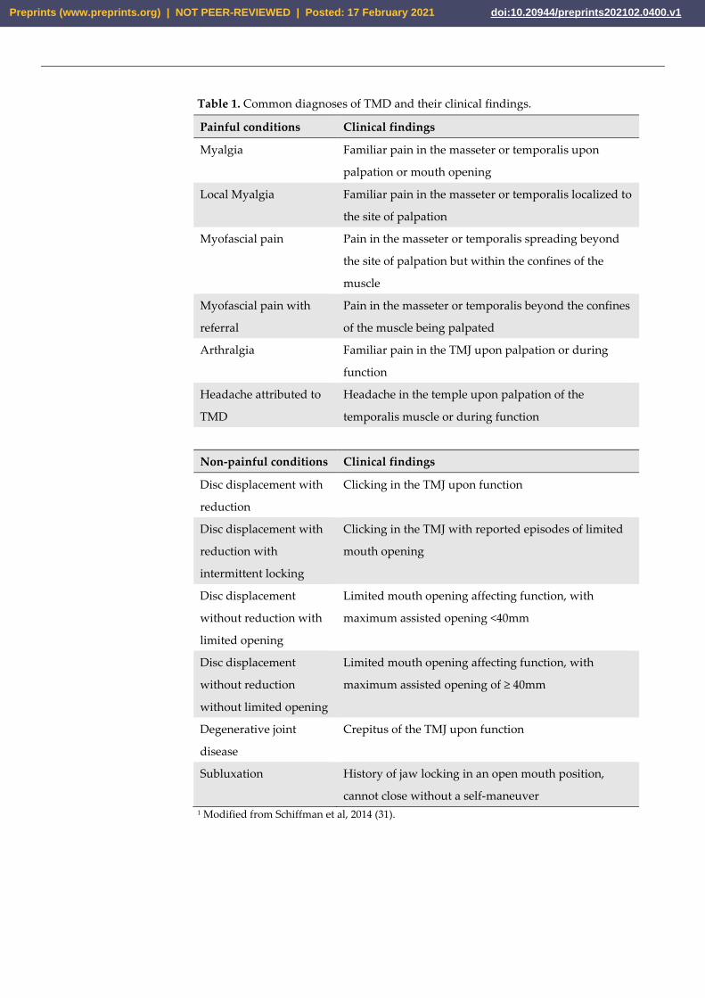
Table 1. Common diagnoses of TMD and their clinical findings.
Painful conditions Clinical findings
Myalgia Familiar pain in the masseter or temporalis upon
palpation or mouth opening
Local Myalgia Familiar pain in the masseter or temporalis localized to
the site of palpation
Myofascial pain Pain in the masseter or temporalis spreading beyond
the site of palpation but within the confines of the
muscle
Myofascial pain with
referral
Pain in the masseter or temporalis beyond the confines
of the muscle being palpated
Arthralgia Familiar pain in the TMJ upon palpation or during
function
Headache attributed to
TMD
Headache in the temple upon palpation of the
temporalis muscle or during function
Non-painful conditions Clinical findings
Disc displacement with
reduction
Clicking in the TMJ upon function
Disc displacement with
reduction with
intermittent locking
Clicking in the TMJ with reported episodes of limited
mouth opening
Disc displacement
without reduction with
limited opening
Limited mouth opening affecting function, with
maximum assisted opening <40mm
Disc displacement
without reduction
without limited opening
Limited mouth opening affecting function, with
maximum assisted opening of ≥ 40mm
Degenerative joint
disease
Crepitus of the TMJ upon function
Subluxation History of jaw locking in an open mouth position,
cannot close without a self-maneuver
1 Modified from Schiffman et al, 2014 (31).
Preprints (www.preprints.org) | NOT PEER-REVIEWED | Posted: 17 February 2021 doi:10.20944/preprints202102.0400.v1
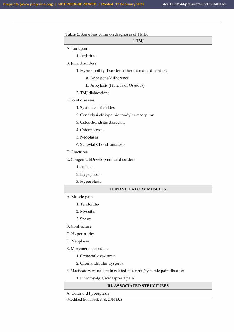
Table 2. Some less common diagnoses of TMD.
I. TMJ
A. Joint pain
1. Arthritis
B. Joint disorders
1. Hypomobility disorders other than disc disorders
a. Adhesions/Adherence
b. Ankylosis (Fibrous or Osseous)
2. TMJ dislocations
C. Joint diseases
1. Systemic arthritides
2. Condylysis/Idiopathic condylar resorption
3. Osteochondritis dissecans
4. Osteonecrosis
5. Neoplasm
6. Synovial Chondromatosis
D. Fractures
E. Congenital/Developmental disorders
1. Aplasia
2. Hypoplasia
3. Hyperplasia
II. MASTICATORY MUSCLES
A. Muscle pain
1. Tendonitis
2. Myositis
3. Spasm
B. Contracture
C. Hypertrophy
D. Neoplasm
E. Movement Disorders
1. Orofacial dyskinesia
2. Oromandibular dystonia
F. Masticatory muscle pain related to central/systemic pain disorder
1. Fibromyalgia/widespread pain
III. ASSOCIATED STRUCTURES
A. Coronoid hyperplasia 1 Modified from Peck et al, 2014 (32).
Preprints (www.preprints.org) | NOT PEER-REVIEWED | Posted: 17 February 2021 doi:10.20944/preprints202102.0400.v1
3. Diagnostic Approach
The signs and symptoms of TMD may mimic other orofacial pain conditions. Alt-
hough precise physical diagnosis into the type of TMD is helpful in developing an appro-
priate treatment plan, it might not be straight forward in every case. History taking forms
an important part of the diagnosis of a TMJ condition. The acquisition of history follows
the usual format. Apart from chief complaint, inquiries should be made regarding any
history of trauma or previous episodes, aggravating factors, such as eating, talking, yawn-
ing or spontaneous background pain, and any previous investigations or treatment. The
severity of pain should also be graded using a visual analogue scale (VAS), so treatment
progress can be quantitatively monitored. A past and current medical history, including
a full medications list, may reveal any comorbidities that may be related to TMD. The
clinician should note any habits such as smoking, drinking and recreational drug use, and
any history of clenching or bruxism as complained by the patients’ bed partner. Also,
the clinician should ask questions regarding stress and level of life satisfaction, and
whether there are any recent life events, such as change of job or loss of a loved one. Alt-
hough most clinicians treating TMD may be experienced with acquiring a clinical history,
some may not be comfortable with taking a psychological history. If desired, the clini-
cian may employ the numerous psychosocial instruments available to aid in their diagno-
sis, such as those in Axis-II of DC/TMD [31]. When necessary, the patient may be referred
for a psychological assessment.
Most clinicians who treat orofacial pain believe clinical examination is the most cru-
cial process of diagnosing TMD. Location of pain, and whether the pain is localized, re-
mains within or spreads beyond the confines of the muscle, should be confirmed with
palpation, which is done at rest and during mandibular function. Clicking or crepitus
upon mandibular function might be quite obvious in some cases, and the detection might
be aided by the use of a stethoscope. Intriguingly, the presence or location of clicking
detected by the clinician might be different from that reported by the patient, and this
should be documented. The range of mouth opening measured should include pain-free
maximum mouth opening, maximum unassisted mouth opening, and maximum assisted
mouth opening. Any deviation of the mandible may indicate differential obstruction of
the movement of the mandibular condyle in rotation and/or translation. An intra-oral
examination is performed to rule out any mucosal pathologies of the oral cavity and oro-
pharyngeal region, as well as to assess the state of the dentition.
Imaging and other investigations
Imaging is considered to be an useful adjunct in the diagnosis of TMD. Although the
diagnostic information provided by plain radiographs like orthopantomogram is limited,
they are convenient, simple and serve to rule out some of the differential diagnoses of the
bony TMJ, such as fractures, ankylosis, growth disturbances, as well as neoplasms. For
the most common types of TMD which clinical presentation is typical, many units might
not routinely employ additional imaging. This is due to availability and cost, and that
additional imaging might not alter the initial management plan. However, when further
information is desired, magnetic resonance imaging (MRI) is the gold standard for TMJ
imaging, and is useful in assessing the status of the osseous, as well as the non-osseous
structures of the TMJ, such as the masticatory muscles, ligaments and the cartilaginous
disc [33] (Figure 1). Classification systems, such as Wilkes [21], combine clinical and MRI
findings to stage the extent of internal derangement in order to guide treatment protocol.
MRI is therefore considered mandatory prior to any surgical intervention.
Preprints (www.preprints.org) | NOT PEER-REVIEWED | Posted: 17 February 2021 doi:10.20944/preprints202102.0400.v1
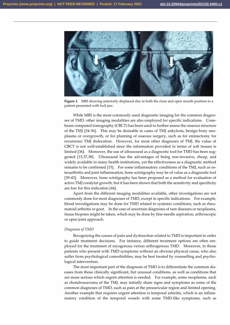
Figure 1. MRI showing anteriorly displaced disc in both the close and open mouth position in a
patient presented with lock jaw.
While MRI is the most commonly used diagnostic imaging for the common diagno-
ses of TMD, other imaging modalities are also employed for specific indications. Cone-
beam computed tomography (CBCT) has been used to further assess the osseous structure
of the TMJ [34-36]. This may be desirable in cases of TMJ ankylosis, benign bony neo-
plasms or overgrowth, or for planning of osseous surgery, such as for eminectomy for
recurrence TMJ dislocation. However, for most other diagnoses of TMJ, the value of
CBCT is not well-established since the information provided in terms of soft tissues is
limited [36]. Moreover, the use of ultrasound as a diagnostic tool for TMD has been sug-
gested [15,37,38]. Ultrasound has the advantages of being non-invasive, cheap, and
widely available in many health institutions, yet the effectiveness as a diagnostic method
remains to be confirmed [15]. For some inflammatory conditions of the TMJ, such as os-
teoarthritis and joint inflammation, bone scintigraphy may be of value as a diagnostic tool
[39-43]. Moreover, bone scintigraphy has been proposed as a method for evaluation of
active TMJ condylar growth, but it has been shown that both the sensitivity and specificity
are low for this indication [44].
Apart from the different imaging modalities available, other investigations are not
commonly done for most diagnoses of TMD, except in specific indications. For example,
blood investigations may be done for TMD related to systemic conditions, such as rheu-
matoid arthritis or gout. In the case of uncertain diagnoses of rare diseases or neoplasms,
tissue biopsies might be taken, which may be done by fine-needle aspiration, arthroscopic
or open joint approach.
Diagnosis of TMD
Recognizing the causes of pain and dysfunction related to TMD is important in order
to guide treatment decisions. For instance, different treatment options are often em-
ployed for the treatment of myogenous versus arthrogenous TMD. Moreover, in those
patients who present with TMD symptoms without an obvious physical cause, who also
suffer from psychological comorbidities, may be best treated by counselling and psycho-
logical intervention.
The most important part of the diagnosis of TMD is to differentiate the common dis-
eases from those clinically significant, but unusual conditions, as well as conditions that
are more serious which urgent attention is needed. For example, some neoplasms, such
as chondrosarcoma of the TMJ, may initially share signs and symptoms as some of the
common diagnoses of TMD, such as pain at the preauricular region and limited opening.
Another example that requires urgent attention is temporal arteritis, which is an inflam-
matory condition of the temporal vessels with some TMD-like symptoms, such as
Preprints (www.preprints.org) | NOT PEER-REVIEWED | Posted: 17 February 2021 doi:10.20944/preprints202102.0400.v1
headache, pain in the temporal region, and limited mouth opening. However, temporal
arteritis is a medical emergency which may cause permanent blindness if not treated
promptly. Some of the differential diagnoses of orofacial pain that may mimic TMD are
listed in Table 3 [45].
Table 3. Differential diagnosis of TMD
Neuropathic pain
Trigeminal neuralgia
Glossopharyngeal neuralgia
Postherpetic neuralgia
Traumatic neuralgia
Burning mouth syndrome
Atypical odontalgia
Atypical facial pain
Odontogenic pain
Dental caries
Periodontal disease
Dental abscess
Dental sensitivity
Cracked tooth syndrome
Periocoronitis
Intracranial pain
Tumours
Aneurysms
Bleeding
Infection
Pain from other adjacent structures
Ear
Nose
Throat
Eyes
Sinus
Salivary glands
Lymph nodes
Vasculature
Cervical region
Headaches not attributed to TMD
Migraine
Cluster headache
Tension-type headache
Temporal arteritis
Referred pain
Psychogenic pain 1 Modified from Kumar et al (2013) (45)
4. Treatment Modalities – A Change in Paradigm?
Preprints (www.preprints.org) | NOT PEER-REVIEWED | Posted: 17 February 2021 doi:10.20944/preprints202102.0400.v1
The goals of treatment for TMD include reduction of pain and improvement of jaw
function. Currently, physically restoring the disc position in the case of internal derange-
ment is not the primary treatment objective as it may not be relevant to clinical improve-
ment [46,47]. Symptoms of TMD should be addressed promptly, as chronic pain be-
comes more difficult to manage due to psychological deterioration and somatization
[2,19]. Since conservative options are less likely to cause any harm, they are usually in-
dicated in the early stages of treatment. This is especially true when definitive diagnosis
is difficult to ascertain and treatment is performed empirically. However, there is no
agreement on how long conservative treatment should be attempted before progressing
to other options when clear benefits are not observed. Although the treatment of TMD
has shifted away from open procedures which were once popular, the demonstrated suc-
cess of minimally invasive options may indicate that they may be considered as an early
option for those cases refractory to conservatory approaches.
Conservative options
The initial management of TMD may include various medications, such as analge-
sics, non-steroidal anti-inflammatory drugs (NSAIDs), muscle relaxants, anxiolytics, and
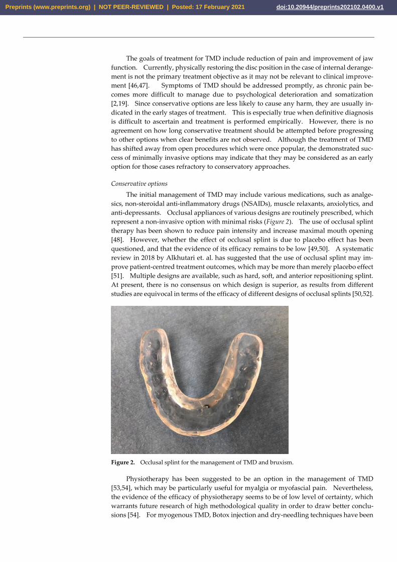
anti-depressants. Occlusal appliances of various designs are routinely prescribed, which
represent a non-invasive option with minimal risks (Figure 2). The use of occlusal splint
therapy has been shown to reduce pain intensity and increase maximal mouth opening
[48]. However, whether the effect of occlusal splint is due to placebo effect has been
questioned, and that the evidence of its efficacy remains to be low [49,50]. A systematic
review in 2018 by Alkhutari et. al. has suggested that the use of occlusal splint may im-
prove patient-centred treatment outcomes, which may be more than merely placebo effect
[51]. Multiple designs are available, such as hard, soft, and anterior repositioning splint.
At present, there is no consensus on which design is superior, as results from different
studies are equivocal in terms of the efficacy of different designs of occlusal splints [50,52].
Figure 2. Occlusal splint for the management of TMD and bruxism.
Physiotherapy has been suggested to be an option in the management of TMD
[53,54], which may be particularly useful for myalgia or myofascial pain. Nevertheless,
the evidence of the efficacy of physiotherapy seems to be of low level of certainty, which
warrants future research of high methodological quality in order to draw better conclu-
sions [54]. For myogenous TMD, Botox injection and dry-needling techniques have been
Preprints (www.preprints.org) | NOT PEER-REVIEWED | Posted: 17 February 2021 doi:10.20944/preprints202102.0400.v1
suggested [55,56]. Also, initial results regarding extracorporeal shock wave therapy for
myogenous TMD appear to show positive results [57,58].
There has been increasing evidence demonstrating that psychosocial assessment
serves as a powerful tool in terms of predicting treatment outcome [59,60]. For those
patients with a significant psychosocial component, counselling seems to be a promising
treatment adjunct [50,61-63], which might be most beneficial when included in a multi-
modal approach [50]. Other conservative treatment options for TMD include stress re-
duction techniques and diet modification. In the past, a causative relationship between
occlusion and TMD had been suggested, but it is now considered an outdated theory not
supported by robust evidence, and occlusal adjustment is an irreversible treatment which
is no longer supported by the recent literature [64-67].
Minimally invasive options – arthroscopy and arthrocentesis
In the 1980s, the availability of MRI has led clinicians to acknowledge the structural
anomalies related to TMD. This has resulted in a boom of open joint surgeries, which
were unfortunately ineffective in the most part. For those cases of TMD that are ar-
throgenous and not responsive to conservative treatment, more focus has since been
shifted to minimally invasive procedures which have shown promising clinical results.
Arthroscopy of the TMJ was initially pioneered by the Japanese in the 1970s [68,69],
and later popularized by the Americans [70-72]. TMJ arthroscopy may involve lysis and
lavage of the superior joint space, as well as operative procedures, such as repositioning
of a displaced disc, arthroplasty, and removal of inflamed tissues and adhesions. The
efficacy of arthroscopy has since been well-recognized [73-79], and has been found that
the therapeutic effect was mainly due to lysis and lavage but not disc position [80]. It
was due to this finding that a modification was made, where lysis and lavage was per-
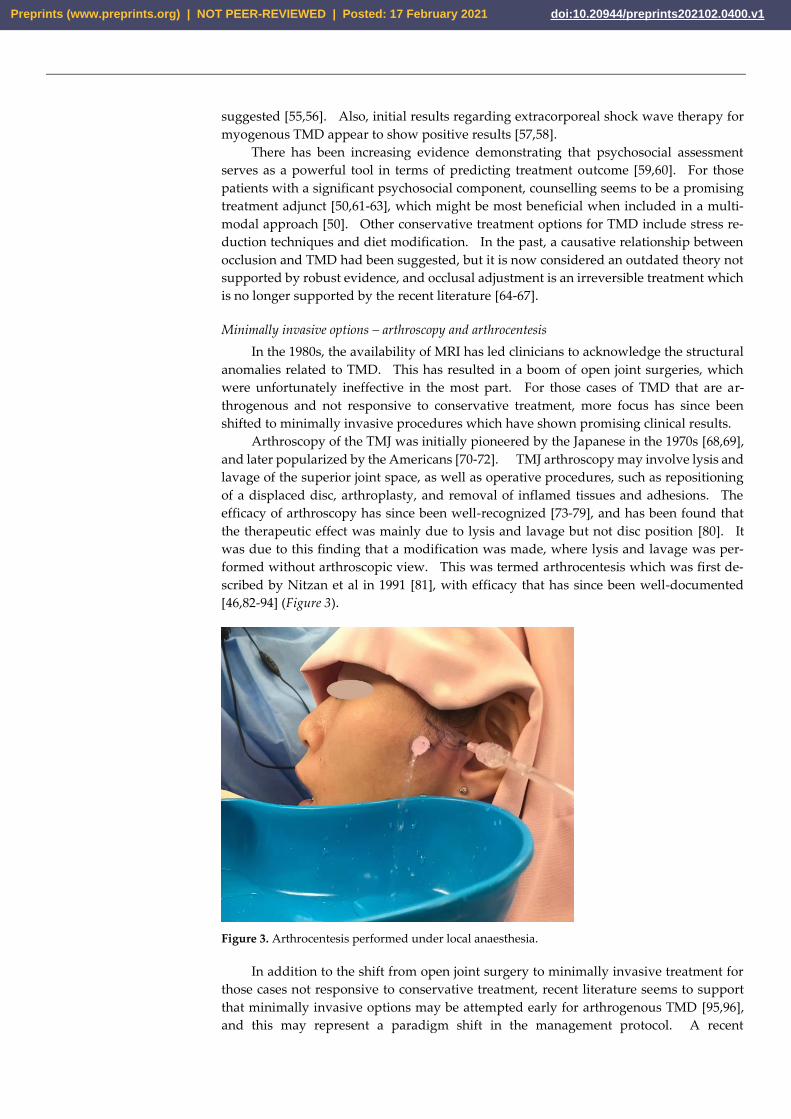
formed without arthroscopic view. This was termed arthrocentesis which was first de-
scribed by Nitzan et al in 1991 [81], with efficacy that has since been well-documented
[46,82-94] (Figure 3).
Figure 3. Arthrocentesis performed under local anaesthesia.
In addition to the shift from open joint surgery to minimally invasive treatment for
those cases not responsive to conservative treatment, recent literature seems to support
that minimally invasive options may be attempted early for arthrogenous TMD [95,96],
and this may represent a paradigm shift in the management protocol. A recent
Preprints (www.preprints.org) | NOT PEER-REVIEWED | Posted: 17 February 2021 doi:10.20944/preprints202102.0400.v1
integrated review and meta-analysis performed by the authors of this article showed that
arthrocentesis was beneficial, whether it was performed as an initial treatment, as an early
or late treatment with regards to conservative treatment [97]. However, the best timing
to perform arthrocentesis is still unclear due to the paucity of research on the topic, which
warrants more future well-designed clinical trials [97].
Although both arthroscopy and arthrocentesis have been shown to be beneficial in
the treatment of TMD, it is unclear which method produces better clinical results. In a
systematic review and meta-analysis by Al-Moraissi, it was revealed that arthroscopy
was superior to arthrocentesis in pain reduction and jaw function improvement, with sim-
ilar complication rates for both methods [78]. However, other studies have shown com-
parable results with the two procedures [98,99]. Nevertheless, arthrocentesis has been
suggested to be attempted first due to simplicity and cost-effectiveness, with a similar or
potentially lower complication rate [99].
Several modifications have been suggested for the conventional arthrocentesis,
which involves two puncture needles into the superior joint space guided by landmarks
in relations to adjacent structures, followed by lavage with an irrigation solution. For
example, single-puncture techniques employ specially designed devices, and may have
both the inflow and outflow fluid going through a single cannula but with different ports.
Although single-puncture techniques may appear more simple than double-puncture ar-
throcentesis, most studies to date have shown a similar clinical outcome between the two
techniques [83,100-102]. In addition, ultrasound-guided arthrocentesis has been pro-
posed to increase the accuracy of puncture into the superior joint space [103-106]. How-
ever, a recent systematic review by Leung et. al. has shown that no additional benefit is
seen with ultrasound-guided arthrocentesis compared to conventional arthrocentesis
[107]. Furthermore, different pharmacological agents for intra-articular injection have
been proposed, with the common ones including hyaluronic acid, corticosteroid, analge-
sics, and platelet-rich plasma [93,96,108,109]. Although promising results are seen in
some studies, there is currently no consensus regarding which intra-articular injection
agent is superior over the others.
Today, arthrocentesis is still considered to be a controversial procedure [87], despite
the documented efficacy and low complication rates. This is partly because some cases
of TMD improve with mere conservative options, or even without treatment. Also, many
cases of TMD are due to multiple etiologies, which may require a multimodal approach
before any clear clinical improvement can be appreciated. However, in those patients
with true arthrogenous TMD not responsive to conservative treatment options, whose
condition also lack a significant biopsychosocial component, minimally invasive proce-
dures, such as arthrocentesis or arthroscopy, may well be the answer.
Open joint surgery
Open surgical treatment for TMD is now uncommon, and is reserved for specific
indications as well as end-stage diseases. Though, surgery may be the only viable option
in some conditions, such as ankylosis and neoplasms, which require release of ankylosis
and removal of tumour, respectively. Pending on the availability of equipment and skills,
there is now an option of arthroscopic surgery for procedures that were only performed
with an open-joint approach in the past. These procedures include disc repositioning
procedures, removal of osteophyte, removal of pathologic tissue, and biopsy of the TMJ.
In recent years, much work has been done regarding replacement of the TMJ with allo-
plastic prosthesis [110-116] with an observed improvement in prognosis and longevity.
Due to this success, it is likely that we will see a continuous increase in popularity of allo-
plastic replacement of the TMJ for conditions such as end stage arthritic conditions, anky-
losis, post-tumour resection, and developmental anomalies of the TMJ.
Preprints (www.preprints.org) | NOT PEER-REVIEWED | Posted: 17 February 2021 doi:10.20944/preprints202102.0400.v1
5. Conclusions
TMD represents a complex group of orofacial pain conditions which shares similar-
ities with other chronic pain conditions. The etiology of TMD is often multi-factorial, and
precise causes for the symptoms may be difficult to pinpoint. In the past, focus has been
placed on the physical origins of TMD, but an at least equally significant psychosocial
factor is now well-recognized. Consequently, a multimodal approach, which might in-
clude counselling and psychological therapy, is being increasingly advocated. Most in-
stances of TMD are managed conservatively and empirically during the early phases of
treatment, yet lingering in the conservative phase for an extended period when clinical
improvement is unclear is not recommended. Though open joint surgery is rare nowa-
days and is reserved for specific situations, we may be in the midst of a changing para-
digm which favours early minimally invasive procedures.
Supplementary Materials: Not applicable.
Author Contributions: Both authors are responsible for all parts of the work. Both authors have
read and agreed to the published version of the manuscript.
Funding: This research received no external funding.
Institutional Review Board Statement: Not applicable.
Informed Consent Statement: Not applicable.
Conflicts of Interest: The authors declare no conflict of interest.
References
1. Von Korff, M.; Ormel, J.; Keefe, F.J.; Dworkin, S.F. Grading the severity of chronic pain. Pain 1992, 50, 133-149.
2. Ismail, F.; Eisenburger, M.; Lange, K.; Schneller, T.; Schwabe, L.; Strempel, J.; Stiesch, M. Identification of psychological
comorbidity in TMD-patients. Cranio 2016, 34, 182-187, doi:10.1179/2151090315Y.0000000008.
3. List, T.; Jensen, R.H. Temporomandibular disorders: Old ideas and new concepts. Cephalalgia 2017, 37, 692-704,
doi:10.1177/0333102416686302.
4. Bitiniene, D.; Zamaliauskiene, R.; Kubilius, R.; Leketas, M.; Gailius, T.; Smirnovaite, K. Quality of life in patients with
temporomandibular disorders. A systematic review. Stomatologija 2018, 20, 3-9.
5. Resende, C.; Rocha, L.; Paiva, R.P.; Cavalcanti, C.D.S.; Almeida, E.O.; Roncalli, A.G.; Barbosa, G.A.S. Relationship between
anxiety, quality of life, and sociodemographic characteristics and temporomandibular disorder. Oral Surg Oral Med Oral
Pathol Oral Radiol 2020, 129, 125-132, doi:10.1016/j.oooo.2019.10.007.
6. Dahlstrom, L.; Carlsson, G.E. Temporomandibular disorders and oral health-related quality of life. A systematic review.
Acta Odontol Scand 2010, 68, 80-85, doi:10.3109/00016350903431118.
7. Goncalves, D.A.; Camparis, C.M.; Speciali, J.G.; Franco, A.L.; Castanharo, S.M.; Bigal, M.E. Temporomandibular disorders
are differentially associated with headache diagnoses: a controlled study. Clin J Pain 2011, 27, 611-615,
doi:10.1097/AJP.0b013e31820e12f5.
8. Lim, P.F.; Smith, S.; Bhalang, K.; Slade, G.D.; Maixner, W. Development of temporomandibular disorders is associated with
greater bodily pain experience. Clin J Pain 2010, 26, 116-120, doi:10.1097/AJP.0b013e3181c507ef.
Preprints (www.preprints.org) | NOT PEER-REVIEWED | Posted: 17 February 2021 doi:10.20944/preprints202102.0400.v1
9. Facial Pain. Availabe online: http://www.nidcr.nih.gov/DataStatistics/FindDataByTopic/FacialPain/ (accessed on
09/06/2019).
10. Lipton, J.A.; Ship, J.A.; Larach-Robinson, D. Estimated prevalence and distribution of reported orofacial pain in the United
States. Journal of the American Dental Association (1939) 1993, 124, 115-121, doi:10.14219/jada.archive.1993.0200.
11. Locker, D.; Slade, G. Prevalence of symptoms associated with temporomandibular disorders in a Canadian population.
Community Dent Oral Epidemiol 1988, 16, 310-313, doi:10.1111/j.1600-0528.1988.tb01783.x.
12. Magnusson, T.; Egermark, I.; Carlsson, G.E. A longitudinal epidemiologic study of signs and symptoms of
temporomandibular disorders from 15 to 35 years of age. Journal of orofacial pain 2000, 14, 310-319.
13. Ebrahimi, M.; Dashti, H.; Mehrabkhani, M.; Arghavani, M.; Daneshvar-Mozafari, A. Temporomandibular Disorders and
Related Factors in a Group of Iranian Adolescents: A Cross-sectional Survey. J Dent Res Dent Clin Dent Prospects 2011, 5, 123-
127, doi:10.5681/joddd.2011.028.
14. Manfredini, D.; Piccotti, F.; Ferronato, G.; Guarda-Nardini, L. Age peaks of different RDC/TMD diagnoses in a patient
population. J Dent 2010, 38, 392-399, doi:10.1016/j.jdent.2010.01.006.
15. Klatkiewicz, T.; Gawriolek, K.; Pobudek Radzikowska, M.; Czajka-Jakubowska, A. Ultrasonography in the Diagnosis of
Temporomandibular Disorders: A Meta-Analysis. Med Sci Monit 2018, 24, 812-817, doi:10.12659/msm.908810.
16. Sena, M.F.; Mesquita, K.S.; Santos, F.R.; Silva, F.W.; Serrano, K.V. Prevalence of temporomandibular dysfunction in children
and adolescents. Rev Paul Pediatr 2013, 31, 538-545, doi:10.1590/S0103-05822013000400018.
17. Valesan, L.F.; Da-Cas, C.D.; Reus, J.C.; Denardin, A.C.S.; Garanhani, R.R.; Bonotto, D.; Januzzi, E.; de Souza, B.D.M.
Prevalence of temporomandibular joint disorders: a systematic review and meta-analysis. Clin Oral Investig 2021,
10.1007/s00784-020-03710-w, doi:10.1007/s00784-020-03710-w.
18. Rollman, G.B.; Gillespie, J.M. The role of psychosocial factors in temporomandibular disorders. Curr Rev Pain 2000, 4, 71-81,
doi:10.1007/s11916-000-0012-8.
19. Auerbach, S.M.; Laskin, D.M.; Frantsve, L.M.; Orr, T. Depression, pain, exposure to stressful life events, and long-term
outcomes in temporomandibular disorder patients. J Oral Maxillofac Surg 2001, 59, 628-633; discussion 634,
doi:10.1053/joms.2001.23371.
20. Toh, A.Q.J.; Chan, J.L.H.; Leung, Y.Y. Mandibular asymmetry as a possible etiopathologic factor in temporomandibular
disorder: a prospective cohort of 134 patients. Clin Oral Investig 2021, 10.1007/s00784-020-03756-w, doi:10.1007/s00784-020-
03756-w.
21. Wilkes, C.H. Internal Derangements of the Temporomandibular Joint: Pathological Variations. Archives of Otolaryngology–
Head & Neck Surgery 1989, 115, 469-477, doi:10.1001/archotol.1989.01860280067019.
22. Mercuri, L.G. Osteoarthritis, osteoarthrosis, and idiopathic condylar resorption. Oral Maxillofac Surg Clin North Am 2008, 20,
169-183, v-vi, doi:10.1016/j.coms.2007.12.007.
23. Bertram, S.; Rudisch, A.; Innerhofer, K.; Pümpel, E.; Grubwieser, G.; Emshoff, R. Diagnosing TMJ internal derangement and
osteoarthritis with magnetic resonance imaging. Journal of the American Dental Association (1939) 2001, 132, 753-761,
doi:10.14219/jada.archive.2001.0272.
24. Turk DC, G.R. Psychological approaches to pain management: A practitioner's hand book; The Gilford Press: New York, NY, 2002.
25. Dworkin, S.F.; Massoth, D.L. Temporomandibular disorders and chronic pain: disease or illness? The Journal of prosthetic
dentistry 1994, 72, 29-38, doi:10.1016/0022-3913(94)90213-5.
26. Suvinen, T.I.; Reade, P.C. Temporomandibular disorders: a critical review of the nature of pain and its assessment. Journal
of orofacial pain 1995, 9, 317-339.
27. Yap, A.U.; Tan, K.B.; Chua, E.K.; Tan, H.H. Depression and somatization in patients with temporomandibular disorders.
The Journal of prosthetic dentistry 2002, 88, 479-484, doi:10.1067/mpr.2002.129375.
Preprints (www.preprints.org) | NOT PEER-REVIEWED | Posted: 17 February 2021 doi:10.20944/preprints202102.0400.v1
28. Saccomanno, S.; Bernabei, M.; Scoppa, F.; Pirino, A.; Mastrapasqua, R.; Visco, M.A. Coronavirus Lockdown as a Major Life
Stressor: Does It Affect TMD Symptoms? Int J Environ Res Public Health 2020, 17, 8907, doi:10.3390/ijerph17238907.
29. Medeiros, R.A.; Vieira, D.L.; Silva, E.; Rezende, L.; Santos, R.W.D.; Tabata, L.F. Prevalence of symptoms of
temporomandibular disorders, oral behaviors, anxiety, and depression in Dentistry students during the period of social
isolation due to COVID-19. J Appl Oral Sci 2020, 28, e20200445, doi:10.1590/1678-7757-2020-0445.
30. Fillingim, R.B.; Ohrbach, R.; Greenspan, J.D.; Knott, C.; Diatchenko, L.; Dubner, R.; Bair, E.; Baraian, C.; Mack, N.; Slade,
G.D., et al. Psychological factors associated with development of TMD: the OPPERA prospective cohort study. The journal
of pain : official journal of the American Pain Society 2013, 14, T75-90, doi:10.1016/j.jpain.2013.06.009.
31. Schiffman, E.; Ohrbach, R.; Truelove, E.; Look, J.; Anderson, G.; Goulet, J.P.; List, T.; Svensson, P.; Gonzalez, Y.; Lobbezoo,
F., et al. Diagnostic Criteria for Temporomandibular Disorders (DC/TMD) for Clinical and Research Applications:
recommendations of the International RDC/TMD Consortium Network* and Orofacial Pain Special Interest Groupdagger.
J Oral Facial Pain Headache 2014, 28, 6-27, doi:10.11607/jop.1151.
32. Peck, C.C.; Goulet, J.P.; Lobbezoo, F.; Schiffman, E.L.; Alstergren, P.; Anderson, G.C.; de Leeuw, R.; Jensen, R.; Michelotti,
A.; Ohrbach, R., et al. Expanding the taxonomy of the diagnostic criteria for temporomandibular disorders. Journal of oral
rehabilitation 2014, 41, 2-23, doi:10.1111/joor.12132.
33. Al-Saleh, M.A.; Alsufyani, N.A.; Saltaji, H.; Jaremko, J.L.; Major, P.W. MRI and CBCT image registration of
temporomandibular joint: a systematic review. J Otolaryngol Head Neck Surg 2016, 45, 30, doi:10.1186/s40463-016-0144-4.
34. Al-Saleh, M.A.; Jaremko, J.L.; Alsufyani, N.; Jibri, Z.; Lai, H.; Major, P.W. Assessing the reliability of MRI-CBCT image
registration to visualize temporomandibular joints. Dentomaxillofac Radiol 2015, 44, 20140244, doi:10.1259/dmfr.20140244.
35. Ladeira, D.B.; da Cruz, A.D.; de Almeida, S.M. Digital panoramic radiography for diagnosis of the temporomandibular joint:
CBCT as the gold standard. Braz Oral Res 2015, 29, S1806-83242015000100303, doi:10.1590/1807-3107BOR-2015.vol29.0120.
36. Larheim, T.A.; Abrahamsson, A.K.; Kristensen, M.; Arvidsson, L.Z. Temporomandibular joint diagnostics using CBCT.
Dentomaxillofac Radiol 2015, 44, 20140235, doi:10.1259/dmfr.20140235.
37. Su, N.; van Wijk, A.J.; Visscher, C.M.; Lobbezoo, F.; van der Heijden, G. Diagnostic value of ultrasonography for the
detection of disc displacements in the temporomandibular joint: a systematic review and meta-analysis. Clin Oral Investig
2018, 22, 2599-2614, doi:10.1007/s00784-018-2359-4.
38. Talmaceanu, D.; Lenghel, L.M.; Bolog, N.; Popa Stanila, R.; Buduru, S.; Leucuta, D.C.; Rotar, H.; Baciut, M.; Baciut, G. High-
resolution ultrasonography in assessing temporomandibular joint disc position. Med Ultrason 2018, 1, 64-70,
doi:10.11152/mu-1025.
39. Choi, B.H.; Yoon, S.H.; Song, S.I.; Yoon, J.K.; Lee, S.J.; An, Y.S. Comparison of Diagnostic Performance Between Visual and
Quantitative Assessment of Bone Scintigraphy Results in Patients With Painful Temporomandibular Disorder. Medicine
(Baltimore) 2016, 95, e2485, doi:10.1097/MD.0000000000002485.
40. Epstein, J.B.; Rea, A.; Chahal, O. The use of bone scintigraphy in temporomandibular joint disorders. Oral Dis 2002, 8, 47-53,
doi:10.1034/j.1601-0825.2002.10753.x.
41. Kang, J.H.; An, Y.S.; Park, S.H.; Song, S.I. Influences of age and sex on the validity of bone scintigraphy for the diagnosis of
temporomandibular joint osteoarthritis. Int J Oral Maxillofac Surg 2018, 47, 1445-1452, doi:10.1016/j.ijom.2018.05.011.
42. Lee, Y.H.; Hong, I.K.; Chun, Y.H. Prediction of painful temporomandibular joint osteoarthritis in juvenile patients using
bone scintigraphy. Clin Exp Dent Res 2019, 5, 225-235, doi:10.1002/cre2.175.
43. Park, K.S.; Song, H.C.; Cho, S.G.; Kang, S.R.; Kim, J.; Jun, H.M.; Song, M.; Jeong, G.C.; Park, H.J.; Kwon, S.Y., et al. Open-
Mouth Bone Scintigraphy Is Better than Closed-Mouth Bone Scintigraphy in the Diagnosis of Temporomandibular
Osteoarthritis. Nucl Med Mol Imaging 2016, 50, 213-218, doi:10.1007/s13139-016-0407-z.
44. Chan, B.H.; Leung, Y.Y. SPECT bone scintigraphy for the assessment of condylar growth activity in mandibular asymmetry:
is it accurate? Int. J. Oral Maxillofac. Surg. 2018, 47, 470-479, doi:10.1016/j.ijom.2017.09.008.
Preprints (www.preprints.org) | NOT PEER-REVIEWED | Posted: 17 February 2021 doi:10.20944/preprints202102.0400.v1
45. Kumar, A.; Brennan, M.T. Differential diagnosis of orofacial pain and temporomandibular disorder. Dent Clin North Am
2013, 57, 419-428, doi:10.1016/j.cden.2013.04.003.
46. Alpaslan, G.H.; Alpaslan, C. Efficacy of temporomandibular joint arthrocentesis with and without injection of sodium
hyaluronate in treatment of internal derangements. J Oral Maxillofac Surg 2001, 59, 613-618; discussion 618-619,
doi:10.1053/joms.2001.23368.
47. Nitzan, D.W.; Dolwick, M.F.; Heft, M.W. Arthroscopic lavage and lysis of the temporomandibular joint: a change in
perspective. J Oral Maxillofac Surg 1990, 48, 798-801; discussion 802.
48. Zhang, C.; Wu, J.Y.; Deng, D.L.; He, B.Y.; Tao, Y.; Niu, Y.M.; Deng, M.H. Efficacy of splint therapy for the management of
temporomandibular disorders: a meta-analysis. Oncotarget 2016, 7, 84043-84053, doi:10.18632/oncotarget.13059.
49. Riley, P.; Glenny, A.M.; Worthington, H.V.; Jacobsen, E.; Robertson, C.; Durham, J.; Davies, S.; Petersen, H.; Boyers, D. Oral
splints for temporomandibular disorder or bruxism: a systematic review. Br Dent J 2020, 228, 191-197, doi:10.1038/s41415-
020-1250-2.
50. Al-Moraissi, E.A.; Farea, R.; Qasem, K.A.; Al-Wadeai, M.S.; Al-Sabahi, M.E.; Al-Iryani, G.M. Effectiveness of occlusal splint
therapy in the management of temporomandibular disorders: network meta-analysis of randomized controlled trials. Int J
Oral Maxillofac Surg 2020, 49, 1042-1056, doi:10.1016/j.ijom.2020.01.004.
51. Alkhutari, A.S.; Alyahya, A.; Rodrigues Conti, P.C.; Christidis, N.; Al-Moraissi, E.A. Is the therapeutic effect of occlusal
stabilization appliances more than just placebo effect in the management of painful temporomandibular disorders? A
network meta-analysis of randomized clinical trials. The Journal of prosthetic dentistry 2020, 10.1016/j.prosdent.2020.08.015,
doi:10.1016/j.prosdent.2020.08.015.
52. Seifeldin, S.A.; Elhayes, K.A. Soft versus hard occlusal splint therapy in the management of temporomandibular disorders
(TMDs). Saudi Dent J 2015, 27, 208-214, doi:10.1016/j.sdentj.2014.12.004.
53. Incorvati, C.; Romeo, A.; Fabrizi, A.; Defila, L.; Vanti, C.; Gatto, M.R.A.; Marchetti, C.; Pillastrini, P. Effectiveness of physical
therapy in addition to occlusal splint in myogenic temporomandibular disorders: protocol of a randomised controlled trial.
BMJ Open 2020, 10, e038438, doi:10.1136/bmjopen-2020-038438.
54. van der Meer, H.A.; Calixtre, L.B.; Engelbert, R.H.H.; Visscher, C.M.; Nijhuis-van der Sanden, M.W.; Speksnijder, C.M.
Effects of physical therapy for temporomandibular disorders on headache pain intensity: A systematic review. Musculoskelet
Sci Pract 2020, 50, 102277, doi:10.1016/j.msksp.2020.102277.
55. Kutuk, S.G.; Ozkan, Y.; Kutuk, M.; Ozdas, T. Comparison of the Efficacies of Dry Needling and Botox Methods in the
Treatment of Myofascial Pain Syndrome Affecting the Temporomandibular Joint. The Journal of craniofacial surgery 2019, 30,
1556-1559, doi:10.1097/SCS.0000000000005473.
56. Connelly, S.T.; Myung, J.; Gupta, R.; Tartaglia, G.M.; Gizdulich, A.; Yang, J.; Silva, R. Clinical outcomes of Botox injections
for chronic temporomandibular disorders: do we understand how Botox works on muscle, pain, and the brain? Int J Oral
Maxillofac Surg 2017, 46, 322-327, doi:10.1016/j.ijom.2016.11.004.
57. Kim, Y.H.; Bang, J.I.; Son, H.J.; Kim, Y.; Kim, J.H.; Bae, H.; Han, S.J.; Yoon, H.J.; Kim, B.S. Protective effects of extracorporeal
shockwave on rat chondrocytes and temporomandibular joint osteoarthritis; preclinical evaluation with in vivo(99m)Tc-
HDP SPECT and ex vivo micro-CT. Osteoarthritis Cartilage 2019, 27, 1692-1701, doi:10.1016/j.joca.2019.07.008.
58. Schenk, I.; Vesper, M.; Nam, V.C. [Initial results using extracorporeal low energy shockwave therapy ESWT in muscle reflex-
induced lock jaw]. Mund Kiefer Gesichtschir 2002, 6, 351-355, doi:10.1007/s10006-002-0365-8.
59. Dworkin, S.F.; Turner, J.A.; Mancl, L.; Wilson, L.; Massoth, D.; Huggins, K.H.; LeResche, L.; Truelove, E. A randomized
clinical trial of a tailored comprehensive care treatment program for temporomandibular disorders. Journal of orofacial pain
2002, 16, 259-276.
Preprints (www.preprints.org) | NOT PEER-REVIEWED | Posted: 17 February 2021 doi:10.20944/preprints202102.0400.v1
60. Türp, J.C.; Jokstad, A.; Motschall, E.; Schindler, H.J.; Windecker-Gétaz, I.; Ettlin, D.A. Is there a superiority of multimodal
as opposed to simple therapy in patients with temporomandibular disorders? A qualitative systematic review of the
literature. Clin Oral Implants Res 2007, 18 Suppl 3, 138-150, doi:10.1111/j.1600-0501.2007.01480.x.
61. Conti, P.C.; Correa, A.S.; Lauris, J.R.; Stuginski-Barbosa, J. Management of painful temporomandibular joint clicking with
different intraoral devices and counseling: a controlled study. J Appl Oral Sci 2015, 23, 529-535, doi:10.1590/1678-
775720140438.
62. de Resende, C.; de Oliveira Medeiros, F.G.L.; de Figueiredo Rego, C.R.; Bispo, A.S.L.; Barbosa, G.A.S.; de Almeida, E.O.
Short-term effectiveness of conservative therapies in pain, quality of life, and sleep in patients with temporomandibular
disorders: A randomized clinical trial. Cranio 2019, 10.1080/08869634.2019.1627068, 1-9, doi:10.1080/08869634.2019.1627068.
63. de Barros Pascoal, A.L.; de Freitas, R.; da Silva, L.F.G.; Oliveira, A.; Dos Santos Calderon, P. Effectiveness of Counseling on
Chronic Pain Management in Patients with Temporomandibular Disorders. J Oral Facial Pain Headache 2020, 34, 77-82,
doi:10.11607/ofph.2163.
64. Delgado-Delgado, R.; Iriarte-Álvarez, N.; Valera-Calero, J.A.; Centenera-Centenera, M.B.; Garnacho-Garnacho, V.E.;
Gallego-Sendarrubias, G.M. Association between temporomandibular disorders with clinical and sociodemographic
features: An observational study. Int J Clin Pract 2021, 10.1111/ijcp.13961, e13961, doi:10.1111/ijcp.13961.
65. Al-Ani, Z. Occlusion and Temporomandibular Disorders: A Long-Standing Controversy in Dentistry. Prim Dent J 2020, 9,
43-48, doi:10.1177/2050168420911029.
66. Manfredini, D.; Lombardo, L.; Siciliani, G. Temporomandibular disorders and dental occlusion. A systematic review of
association studies: end of an era? Journal of oral rehabilitation 2017, 44, 908-923, doi:10.1111/joor.12531.
67. Kakudate, N.; Yokoyama, Y.; Sumida, F.; Matsumoto, Y.; Gordan, V.V.; Gilbert, G.H.; Velly, A.M.; Schiffman, E.L. Dentist
Practice Patterns and Therapeutic Confidence in the Treatment of Pain Related to Temporomandibular Disorders in a Dental
Practice-Based Research Network. J Oral Facial Pain Headache 2017, 31, 152-158, doi:10.11607/ofph.1730.
68. Onishi, M. [Arthroscopy of the temporomandibular joint (author's transl)]. Kokubyo Gakkai Zasshi 1975, 42, 207-213.
69. Murakami, K.; Ono, T. Temporomandibular joint arthroscopy by inferolateral approach. Int J Oral Maxillofac Surg 1986, 15,
410-417, doi:10.1016/s0300-9785(86)80029-1.
70. Sanders, B. Arthroscopic surgery of the temporomandibular joint: treatment of internal derangement with persistent closed
lock. Oral surgery, oral medicine, and oral pathology 1986, 62, 361-372.
71. Sanders, B.; Buoncristiani, R. Diagnostic and surgical arthroscopy of the temporomandibular joint: clinical experience with
137 procedures over a 2-year period. Journal of craniomandibular disorders : facial & oral pain 1987, 1, 202-213.
72. McCain, J.P. Arthroscopy of the human temporomandibular joint. J Oral Maxillofac Surg 1988, 46, 648-655.
73. McCain, J.P.; Sanders, B.; Koslin, M.G.; Quinn, J.H.; Peters, P.B.; Indresano, A.T. Temporomandibular joint arthroscopy: a
6-year multicenter retrospective study of 4,831 joints. J Oral Maxillofac Surg 1992, 50, 926-930, doi:10.1016/0278-
2391(92)90047-4.
74. Reston, J.T.; Turkelson, C.M. Meta-analysis of surgical treatments for temporomandibular articular disorders. J Oral
Maxillofac Surg 2003, 61, 3-10; discussion 10-12, doi:10.1053/joms.2003.50000.
75. Schiffman, E.L.; Velly, A.M.; Look, J.O.; Hodges, J.S.; Swift, J.Q.; Decker, K.L.; Anderson, Q.N.; Templeton, R.B.; Lenton,
P.A.; Kang, W., et al. Effects of four treatment strategies for temporomandibular joint closed lock. Int J Oral Maxillofac Surg
2014, 43, 217-226, doi:10.1016/j.ijom.2013.07.744.
76. Dimitroulis, G. Outcomes of temporomandibular joint arthroscopy in patients with painful but otherwise normal joints. J
Craniomaxillofac Surg 2015, 43, 940-943, doi:10.1016/j.jcms.2015.03.035.
77. McCain, J.P.; Hossameldin, R.H.; Srouji, S.; Maher, A. Arthroscopic discopexy is effective in managing temporomandibular
joint internal derangement in patients with Wilkes stage II and III. J Oral Maxillofac Surg 2015, 73, 391-401,
doi:10.1016/j.joms.2014.09.004.
Preprints (www.preprints.org) | NOT PEER-REVIEWED | Posted: 17 February 2021 doi:10.20944/preprints202102.0400.v1
78. Al-Moraissi, E.A. Arthroscopy versus arthrocentesis in the management of internal derangement of the temporomandibular
joint: a systematic review and meta-analysis. Int J Oral Maxillofac Surg 2015, 44, 104-112, doi:10.1016/j.ijom.2014.07.008.
79. Liu, X.; Zheng, J.; Cai, X.; Abdelrehem, A.; Yang, C. Techniques of Yang's arthroscopic discopexy for temporomandibular
joint rotational anterior disc displacement. Int J Oral Maxillofac Surg 2019, 48, 769-778, doi:10.1016/j.ijom.2018.12.003.
80. Machoň, V.; Levorová, J.; Hirjak, D.; Beňo, M.; Drahoš, M.; Foltán, R. Does arthroscopic lysis and lavage in subjects with
Wilkes III internal derangement reduce pain? Oral Maxillofac Surg 2021, 10.1007/s10006-020-00935-7, doi:10.1007/s10006-020-
00935-7.
81. Nitzan, D.W.; Dolwick, M.F.; Martinez, G.A. Temporomandibular joint arthrocentesis: a simplified treatment for severe,
limited mouth opening. J Oral Maxillofac Surg 1991, 49, 1163-1167; discussion 1168-1170.
82. Alpaslan, C.; Kahraman, S.; Guner, B.; Cula, S. Does the use of soft or hard splints affect the short-term outcome of
temporomandibular joint arthrocentesis? Int J Oral Maxillofac Surg 2008, 37, 424-427, doi:10.1016/j.ijom.2008.01.022.
83. Bayramoglu, Z.; Tozoglu, S. Comparison of single- and double-puncture arthrocentesis for the treatment of
temporomandibular joint disorders: A six-month, prospective study. Cranio 2019, 10.1080/08869634.2019.1603796, 1-6,
doi:10.1080/08869634.2019.1603796.
84. Carvajal, W.A.; Laskin, D.M. Long-term evaluation of arthrocentesis for the treatment of internal derangements of the
temporomandibular joint. J Oral Maxillofac Surg 2000, 58, 852-855; discussion 856-857, doi:10.1053/joms.2000.8201.
85. Diracoglu, D.; Saral, I.B.; Keklik, B.; Kurt, H.; Emekli, U.; Ozcakar, L.; Karan, A.; Aksoy, C. Arthrocentesis versus nonsurgical
methods in the treatment of temporomandibular disc displacement without reduction. Oral Surg Oral Med Oral Pathol Oral
Radiol Endod 2009, 108, 3-8, doi:10.1016/j.tripleo.2009.01.005.
86. Emshoff, R.; Rudisch, A. Determining predictor variables for treatment outcomes of arthrocentesis and hydraulic distention
of the temporomandibular joint. J Oral Maxillofac Surg 2004, 62, 816-823.
87. Monje-Gil, F.; Nitzan, D.; Gonzalez-Garcia, R. Temporomandibular joint arthrocentesis. Review of the literature. Med Oral
Patol Oral Cir Bucal 2012, 17, e575-581.
88. Neeli, A.S.; Umarani, M.; Kotrashetti, S.M.; Baliga, S. Arthrocentesis for the treatment of internal derangement of the
temporomandibular joint. J Maxillofac Oral Surg 2010, 9, 350-354, doi:10.1007/s12663-010-0155-z.
89. Nitzan, D.W.; Price, A. The use of arthrocentesis for the treatment of osteoarthritic temporomandibular joints. J Oral
Maxillofac Surg 2001, 59, 1154-1159; discussion 1160, doi:10.1053/joms.2001.26716.
90. Nitzan, D.W.; Samson, B.; Better, H. Long-term outcome of arthrocentesis for sudden-onset, persistent, severe closed lock
of the temporomandibular joint. J Oral Maxillofac Surg 1997, 55, 151-157; discussion 157-158.
91. Nitzan, D.W.; Svidovsky, J.; Zini, A.; Zadik, Y. Effect of Arthrocentesis on Symptomatic Osteoarthritis of the
Temporomandibular Joint and Analysis of the Effect of Preoperative Clinical and Radiologic Features. J Oral Maxillofac Surg
2017, 75, 260-267, doi:10.1016/j.joms.2016.08.017.
92. Polat, M.E.; Yanik, S. Efficiency of arthrocentesis treatment for different temporomandibular joint disorders. Int J Oral
Maxillofac Surg 2020, 49, 621-627, doi:10.1016/j.ijom.2019.08.017.
93. Toameh, M.H.; Alkhouri, I.; Karman, M.A. Management of patients with disk displacement without reduction of the
temporomandibular joint by arthrocentesis alone, plus hyaluronic acid or plus platelet-rich plasma. Dent Med Probl 2019, 56,
265-272, doi:10.17219/dmp/109329.
94. Yilmaz, O.; Korkmaz, Y.T.; Tuzuner, T. Comparison of treatment efficacy between hyaluronic acid and arthrocentesis plus
hyaluronic acid in internal derangements of temporomandibular joint. J Craniomaxillofac Surg 2019, 47, 1720-1727,
doi:10.1016/j.jcms.2019.07.030.
95. Vos, L.M.; Huddleston Slater, J.J.; Stegenga, B. Arthrocentesis as initial treatment for temporomandibular joint arthropathy:
a randomized controlled trial. J Craniomaxillofac Surg 2014, 42, e134-139, doi:10.1016/j.jcms.2013.07.010.
Preprints (www.preprints.org) | NOT PEER-REVIEWED | Posted: 17 February 2021 doi:10.20944/preprints202102.0400.v1
96. Al-Moraissi, E.A.; Wolford, L.M.; Ellis, E., 3rd; Neff, A. The hierarchy of different treatments for arthrogenous
temporomandibular disorders: A network meta-analysis of randomized clinical trials. J Craniomaxillofac Surg 2020, 48, 9-23,
doi:10.1016/j.jcms.2019.10.004.
97. Li DTS, Wong NSM, Li SKY, McGrath CP, Leung YY. Timing of Arthrocentesis in the Management of Temporomandibular
Disorders: An Integrative Review and Meta-analysis. International journal of oral and maxillofacial surgery 2021, In press.
https://doi.org/10.1016/j.ijom.2021.01.011
98. Hobeich, J.B.; Salameh, Z.A.; Ismail, E.; Sadig, W.M.; Hokayem, N.E.; Almas, K. Arthroscopy versus arthrocentesis. A
retrospective study of disc displacement management without reduction. Saudi Med J 2007, 28, 1541-1544.
99. Laskin, D.M. Arthroscopy Versus Arthrocentesis for Treating Internal Derangements of the Temporomandibular Joint. Oral
and Maxillofacial Surgery Clinics of North America 2018, 30, 325-328, doi:https://doi.org/10.1016/j.coms.2018.04.008.
100. Monteiro, J.; de Arruda, J.A.A.; Silva, E.; Vasconcelos, B. Is Single-Puncture TMJ Arthrocentesis Superior to the Double-
Puncture Technique for the Improvement of Outcomes in Patients With TMDs? J Oral Maxillofac Surg 2020, 78, 1319.e1311-
1319.e1315, doi:10.1016/j.joms.2020.03.020.
101. Nagori, S.A.; Roy Chowdhury, S.K.; Thukral, H.; Jose, A.; Roychoudhury, A. Single puncture versus standard double needle
arthrocentesis for the management of temporomandibular joint disorders: A systematic review. Journal of oral rehabilitation
2018, 45, 810-818, doi:10.1111/joor.12665.
102. Folle, F.S.; Poluha, R.L.; Setogutti, E.T.; Grossmann, E. Double puncture versus single puncture arthrocentesis for the
management of unilateral temporomandibular joint disc displacement without reduction: A randomized controlled trial. J
Craniomaxillofac Surg 2018, 46, 2003-2007, doi:10.1016/j.jcms.2018.10.015.
103. Bhargava, D.; Thomas, S.; Pawar, P.; Jain, M.; Pathak, P. Ultrasound-guided arthrocentesis using single-puncture, double-
lumen, single-barrel needle for patients with temporomandibular joint acute closed lock internal derangement. Oral
Maxillofac Surg 2019, 23, 159-165, doi:10.1007/s10006-019-00753-6.
104. Antony, P.G.; Sebastian, A.; D, A.; Varghese, K.G.; S, M.; N, J.; Dominic, S.; John, B. Comparison of clinical outcomes of
treatment of dysfunction of the temporomandibular joint between conventional and ultrasound-guided arthrocentesis. Br J
Oral Maxillofac Surg 2019, 57, 62-66, doi:10.1016/j.bjoms.2018.11.007.
105. Bilgir, E.; Yildirim, D.; Senturk, M.F.; Orhan, H. Clinical and ultrasonographic evaluation of ultrasound-guided single
puncture temporomandibular joint arthrocentesis. Cranio 2020, 10.1080/08869634.2020.1853889, 1-10,
doi:10.1080/08869634.2020.1853889.
106. Hu, Y.; Zhang, X.; Liu, S.; Xu, F. Ultrasound-guided vs conventional arthrocentesis for management of temporomandibular
joint disorders: A systematic review and meta-analysis. Cranio 2020, 10.1080/08869634.2020.1829870, 1-10,
doi:10.1080/08869634.2020.1829870.
107. Leung, Y.Y.; Wu, F.H.W.; Chan, H.H. Ultrasonography-guided arthrocentesis versus conventional arthrocentesis in treating
internal derangement of temporomandibular joint: a systematic review. Clin Oral Investig 2020, 24, 3771-3780,
doi:10.1007/s00784-020-03408-z.
108. Haigler, M.C.; Abdulrehman, E.; Siddappa, S.; Kishore, R.; Padilla, M.; Enciso, R. Use of platelet-rich plasma, platelet-rich
growth factor with arthrocentesis or arthroscopy to treat temporomandibular joint osteoarthritis: Systematic review with
meta-analyses. Journal of the American Dental Association (1939) 2018, 149, 940-952.e942, doi:10.1016/j.adaj.2018.07.025.
109. Liu, Y.; Wu, J.S.; Tang, Y.L.; Tang, Y.J.; Fei, W.; Liang, X.H. Multiple Treatment Meta-Analysis of Intra-Articular Injection
for Temporomandibular Osteoarthritis. J Oral Maxillofac Surg 2020, 78, 373.e371-373.e318, doi:10.1016/j.joms.2019.10.016.
110. Chowdhury, S.K.R.; Saxena, V.; Rajkumar, K.; Shadamarshan, R.A. Evaluation of Total Alloplastic Temporomandibular
Joint Replacement in TMJ Ankylosis. J Maxillofac Oral Surg 2019, 18, 293-298, doi:10.1007/s12663-018-1136-x.
Preprints (www.preprints.org) | NOT PEER-REVIEWED | Posted: 17 February 2021 doi:10.20944/preprints202102.0400.v1
111. Bhargava, D.; Neelakandan, R.S.; Dalsingh, V.; Sharma, Y.; Pandey, A.; Pandey, A.; Beena, S.; Koneru, G. A three
dimensional (3D) musculoskeletal finite element analysis of DARSN temporomandibular joint (TMJ) prosthesis for total
unilateral alloplastic joint replacement. J Stomatol Oral Maxillofac Surg 2019, 120, 517-522, doi:10.1016/j.jormas.2019.04.001.
112. Mercuri, L.G. Costochondral Graft Versus Total Alloplastic Joint for Temporomandibular Joint Reconstruction. Oral
Maxillofac Surg Clin North Am 2018, 30, 335-342, doi:10.1016/j.coms.2018.05.003.
113. Lotesto, A.; Miloro, M.; Mercuri, L.G.; Sukotjo, C. Status of alloplastic total temporomandibular joint replacement
procedures performed by members of the American Society of Temporomandibular Joint Surgeons. Int J Oral Maxillofac
Surg 2017, 46, 93-96, doi:10.1016/j.ijom.2016.08.002.
114. Ramos, A.; Mesnard, M. Christensen vs Biomet Microfixation alloplastic TMJ implant: Are there improvements? A
numerical study. J Craniomaxillofac Surg 2015, 43, 1398-1403, doi:10.1016/j.jcms.2015.07.009.
115. Neelakandan, R.S.; Raja, A.V.; Krishnan, A.M. Total Alloplastic Temporomandibular Joint Reconstruction for Management
of TMJ Ankylosis. J Maxillofac Oral Surg 2014, 13, 575-582, doi:10.1007/s12663-013-0565-9.
116. Burgess, M.; Bowler, M.; Jones, R.; Hase, M.; Murdoch, B. Improved outcomes after alloplastic TMJ replacement: analysis of
a multicenter study from Australia and New Zealand. J Oral Maxillofac Surg 2014, 72, 1251-1257,
doi:10.1016/j.joms.2014.02.019.
Preprints (www.preprints.org) | NOT PEER-REVIEWED | Posted: 17 February 2021 doi:10.20944/preprints202102.0400.v1


![Malignant tumours of temporomandibular joint · 2020. 10. 6. · Temporomandibular joint (TMJ) disorders are very common and can be easily diagnosed [1, 2]. However, malignant tumours](https://img.pdfslide.net/doc/110x75/609cf658aa942f17d538f23e/malignant-tumours-of-temporomandibular-joint-2020-10-6-temporomandibular-joint.jpg)