Embed Size (px)
Citation preview
Review
The role of complement in the success of vaccination with conjugated vs
unconjugated polysaccharide antigen
Nur’ain Salehen and Cordula Stover, Department of Infection, Immunity, and
Inflammation, University of Leicester, Leicester LE1 9HN, England
Corresponding author:
Dr. Dr. Cordula Stover, Department of Infection, Immunity, and Inflammation, University
of Leicester, University Road, Leicester LE1 9HN, England; email: [email protected], tel
0044-116-252-5032, fax 0044-116-252-5030
1
Abstract
The complement system, a well-characterised arm of the innate immune system,
significantly influences the adaptive immune response via direct cell-cell interaction and
maintenance of lymphoid organ architecture. Development of vaccines is a major
advance in modern health care. In this review, we highlight the importance of the
marginal zone in response to both, polysaccharide and conjugated vaccines, and
discuss the relevance of complement herein, based on findings obtained from animal
models with specific deletions of certain complement components and from vaccination
reports of complement-deficient individuals. We conclude that both, intactness of the
complement system and maturity of expression of its components, are relatively more
important to aid in the immune response to polysaccharide vaccine than to conjugated
vaccines.
keywords (3): complement vaccine polysaccharide
running title
Complement essential for polysaccharide vaccines
2
The importance of complement activation in adaptive immunity
The complement system is part of the innate immune defence, because it
partakes in first-line, pattern-mediated recognition via its germ-line encoded serum
proteins and receptors. It has evolved from a primordial fluid-phase system that “simply”
tags and mediates uptake of micro-organisms by cells of early chordates. Large-scale
gene duplications, exon shuffling and transposition events are the main evolutionary
mechanisms that have generated a wide range of diversified molecules, which we now
ascribe to innate and adaptive immune defences [1]. Innate immune defence molecules
are of importance in the initial phase of an infection prior to the phase in which an
adaptive immune response is mounted in our secondary lymphoid organs, and may lead
to a resolution of local inflammation. However, pattern recognition systems in general,
such as TLRs, NODs, and scavenger receptors, do have an impact on the generation of
the adaptive immune response as well [2-6]. The complement system encompasses
both, fluid-phase molecules, and cell surface receptors. Components are widely
expressed, including in primary and secondary lymphoid organs, which are a more
recent acquisition in vertebrate evolution. The formative role of complement activation
for immune complex tagging, immune complex localisation, and B-cell co-receptor
triggering has now been clearly defined [7]. At the interface of innate and adaptive
immunity, complement activation effects an enhanced antibody response by directing
the binding of split products of one of its most abundant proteins, C3, to relevant
surfaces and molecules. Most studies have been conducted using T-dependent
antigens, therefore, the focus in the bigger body of literature is on follicular B cells;
antigen, when coated with C3d, binds to membrane-bound immunoglobulin of follicular
B-cells, the B-cell receptor (BCR), and, in addition, to one of the co-receptors, CD21.
This results in co-ligation of a receptor complex, composed of BCR, CD21, CD19, and
CD81 [8], which decreases a signalling threshold and, in the presence of T-cell help,
3
leads to expansion of B-cells and increase of secreted immunoglobulin production. C3d-
tagged antigen localises to follicular dendritic cells in lymphoid follicles by binding to the
receptors CD21 and CD35. This retention of antigen leads to stimulation of follicular B-
cells, affinity-driven selection and maintenance of memory B-cells. C3-coated immune
complexes are trapped by splenic marginal zone B-cells expressing high levels of CD21
and CD35 and are transported by these cells to the follicles.
The influence of complement on adaptive immunity involves, in particular,
survival, stimulation, and maintenance of B-cells. Here, we review the evidence that
complement influences differentially the response to unconjugated vs conjugated
polysaccharide antigen.
Microanatomic site of splenic immune reaction towards polysaccharide antigens
Total splenectomy or congenital asplenia in man carries a significant risk of
infections caused by encapsulated organisms. Complex polysaccharides, as they are
found in cell walls of encapsulated bacteria, such as Streptococcus pneumoniae,
Haemophilus influenzae, and Neisseria meningitidis, are poorly immunogenic in
comparison with peptide antigens owing to some degree to their chemical character
(negative charge), which can make phagocytic uptake difficult [9]. However, a group of
specialised cells in the marginal zone of the spleen appear to be of predominant
importance in dealing with this particular challenge. In the mouse, the marginal zone
surrounds the marginal sinus, which are vascular endbuds of the central artery with a
leaky endothelium, and harbors marginal zone macrophages, B-cells, dendritic cells and
metallophilic macrophages (fig. 1). In human, the splenic morphology differs in that a
marginal sinus as described for mice and marginal metallophilic macrophages are
absent [13]. Positivity for sialoadhesin, associated with marginal metallophilic
macrophages in rodents, is found in macrophages of the human perifollicular zone
4
(capillary sheaths), the proposed entry of antigen into the human spleen [14]. About 90%
of splenic blood throughput is directed into this area of discontinuous arterial terminals
and open-ended venous sinuses (slow flow), whereas the remainder passes via direct
arteriovenous connections through the red pulp (fast flow) [15].
Unconjugated polysaccharide antigens, due to the multivalent nature of the
repetitive epitopes [16] crosslink polyreactive B-cell receptors and lead to their
activation. Marginal zone B-cells are distinct from follicular B-cells (of the germinal
centers) and function within the innate immunity [17]. Polysaccharide antigens are avidly
bound by CD21hi marginal zone B-cells and are, in the mouse, additionally captured by
marginal zone macrophages expressing complement receptors CR3 and CR4 [18].
Marginal zone B-cells are a non-activated B-cell subset [19], but on activation, they
migrate to the follicular zone where CD21 (CR2) is proteolytically cleaved and antigen
transferred to follicular dendritic cells. There is, notably, no T-cell help, because i) there
is no efficient processing and presentation of polysaccharides, and ii) solely CR2
mediated B-cell triggering does not upregulate co-stimulatory molecules [20].
Importantly, upon binding of C1q-tagged complexes to the BCR of follicular B-cells, the
B-cell provides a surface on which complement activation can occur and C3 ligands that
are engendered can bind in strict vicinity of the BCR, to CR1 or CR2 [21].
Antibodies detected after immunisation with T-independent antigens, usually
generated simultaneously as IgM and IgG within 3 days, exhibit lesser avidity,
opsonophagocytic and bacteriolytic activities, compared to antibodies elicited by
immunisation with T-dependent antigens [22-24], reflecting absent affinity maturation of
these antibodies, but the polyreactive and persistent stimulation of B-cells by
polysaccharide structures can provide long lasting antibody levels. This is consistent
with the idea that antigen persistence and continuous stimulation are necessary to
maintain memory B-cells [25]. However, to date, polysaccharides were not thought to be
5
able to elicit a memory B cell response [26]. It seemed difficult to achieve, given the fact
that polysaccharide antigens are poorly presented by MHCII, thereby do not recruit T cell
help, may induce tolerance or generation of plasma cells by crosslinking BCR and might
be poorly retained in the germinal center [27]. Yet, importantly, recent analyses
demonstrate that IgM memory B-cells are indeed generated, independent of T-cell help
[28].
Coupling of vaccines with attenuated toxins, such as diphtheria or tetanus toxin,
enhances the delivery of low immunogenic vaccines, such as polysaccharides, to
antigen presenting cells, and recognition by T-cells. In the mouse, this T-dependent
immune response involves metallophilic macrophages of the marginal zone, marginal
zone B-cells and follicular B-cells of the germinal center in the spleen. Antibodies in
circulation, initially of the IgM-, subsequently of the IgG-type, are specific for the protein
carrier as well as the polysaccharides [29].
During T-dependent immune response to hen egg lysozyme, using an adoptive
transfer model in mice transgenic for anti hen egg lysozyme immunoglobulin, purified
marginal zone B-cells were less efficient in migrating to the outer periarteriolar lymphatic
sheath and were therefore less available for T-dependent responses than follicular B-
cells [30]. In principle, however, follicular B-cells and marginal zone B-cells are able to
respond to challenge with both, T-dependent and T-independent antigen [31].
Importantly, after subcutaneous administration of T-independent or T-dependent
antigens in mice, it is the spleen that reacts with formation of antibody forming cells
before the draining lymphnodes, especially in response to T-independent antigen [32].
Germinal centers are formed during an immune response elicited with a so-called T-
independent antigen. This reaction is, however, of short duration. It is possible that the
absence of somatic hypermutation of the immunoglobulin V genes is due to both, the
curtailed germinal center reaction – as somatic hypermutation is a relatively late event -
6
and the lack of T-cell help [33]. Of note, marginal zone B-cells are incapable of
participating in the process of somatic hypermutation due to the lack of expression of
activation-induced cytidine deaminase, an enzyme essential for this process [34].
The involvement of T-cells is not strictly excluded in the antibody response against
polysaccharides, as B7 ligand dependent costimulation of B-cells (by T-cells) is needed,
however, the interaction in this early response is short and T-cell receptor unspecific [35].
The absence of somatic hypermutation does, however, explain the low affinity binding of
anti-polysaccharide antibodies.
The contribution of complement activation towards vaccination success
In the marginal zone, innate and adaptive immunity interact on a cellular level in
the first encounter of hematogenously spread antigen with lymphatic tissue. Table 1 lists
the source of expression for scavenger receptors and complement receptors in this
anatomic area. Complement components assist in the marginal zone in focussing
antigenic material to specialised splenic cells, but they clearly shape the germinal center
reaction in the follicle of both, spleen (and lymphnodes). Table 2 lists the expression of
complement components in the germinal center.
Germinal centers of secondary lymphoid follicles are positive for the deposition of
C1, C4b, C4d, C3b, C3d, C5b-9, and C4Bp [45], which is consistent with a model of
complement activation on immobilised immune complexes. Relevant to this concept is
detection of C1r mRNA in the spleen [46], and of properdin mRNA, precursor of a
relevant amplifier of ongoing complement activation [47], which is possibly produced by
cells of the dendritic cell lineage [48].
Pneumococcal polysaccharides bind C1q [49] and can trigger alternative
pathway-mediated C3 deposition [50]. Pneumococcal polysaccharides and C1q both,
7
bind to SIGN-R1, a lectin expressed by marginal zone macrophages [51]. They are
highly phagocytic, but their antigen presentation potential is low.
In rats and non-immune humans, pneumococcal polysaccharide antigens
localise to splenic marginal zone B-cells and follicular dendritic cells of the germinal
center, in strict dependence of complement C3d and CR2 [52-54]. In immune humans,
pneumococcal polysaccarides co-localise with splenic CR1 [54], which captures immune
complexes and participates in enzymatic degradation of C3 and C4. Both, CR1 and CR2
are expressed by marginal zone B-cells. CR2 binding of marginal zone B-cells triggers
their migration to follicular dendritic cells, aided by myeloid dendritic cells (secretion of
activating signals). CR2 is able to promote alternative pathway-mediated C3 deposition
[55], so, on involvement, may provide the necessary ligands for CR3 expressing
dendritic cells.
The importance of complement in the generation of an adaptive immune
response towards T-dependent antigen is well studied using complement-deficient mice:
Expression of CR1 and CR2 on follicular dendritic cells and B-cells are necessary for a
specific antibody (IgM and IgG) response and increase of titre after secondary i.v.
immunisation with T-dependent antigen [44, 56], as deduced from CR1/2-deficient mice.
CR1/2-deficient mice may be impaired in germinal center formation and B1-cell
numbers, depending on the type of T-dependent antigen applied [57]. C1q-deficient mice
do not localise immune complexes to follicular dendritic cells [58]. C3- and C4-deficient
mice show a deficiency in isotype switching and formation of germinal centers in a model
of i.v. administration of T-dependent antigen [59]. After i.p. and epicutaneous
administration of T-dependent antigen, C3-deficient mice are impaired in both, Th1 and
Th2 responses [60]. As the humoral response to T-dependent antigen is normal in
Factor B-deficient mice [61], it seems that it is the classical pathway of complement
activation that is predominately more relevant than the lectin or alternative pathways for
8
generation of specific antibody response and maintenance of germinal center
architecture.
Complement activity can be depleted in vivo, for a limited period, by
administration of cobra venom factor (which forms a stable C3 convertase). In this
situation, there is absent splenic localisation of pneumococcal polysaccharide
(administered i.v.) and impairment of specific antibody response. This is not observed for
i.v. administered conjugate vaccine [53]. In splenectomised rats, polysaccharide
vaccines are far less efficient than conjugated vaccines [62].
Is complement relatively more important to raise an appropriate antibody
response to polysaccharide antigens, compared to proteinaceous, T-dependent
antigens? The fact that primary complement deficiency predisposes to infection with
micro-organisms with a complex polysaccharide capsule seems to suggest this and
casts doubt on the use of polysaccharide-type vaccines in these patients [63].
Complement deficiencies are rare [64], and few studies investigate the impact of
these deficiencies on vaccine success. In the largest of these studies, 53 patients with a
variety of genetic complement deficiencies (properdin, Factor H, late components) were
immunised with the tetravalent meningococcal polysaccharide vaccine ACYW135 [65].
There was wide variation in the specific antibody response between individuals but,
importantly, there were no vaccine failures. Of note, these patients on the whole showed
not only vaccination-induced, but also infection-induced, protective immunity.
Vaccination of three properdin-deficient patients with meningococcal vaccine was
protective against meningococcal disease [66, 67]. However, not every properdin-
deficient male succumbs to fatal meningococcal disease and there may indeed be
compensation of this defect by certain immunoglobulin allotypes that are especially
efficient towards polysaccharide antigens [68]. The naturally acquired anti-
polysaccharide antibody response of two C1q-deficient patients consisted of elevated
9
IgM levels compared to controls but lesser IgG and IgA levels [69], showing low class
switching ability in these patients. By contrast, three out of four C3-deficient patients
showed negligable levels of anti-pneumococcal capsular polysaccharide antibodies and
suffered from recurrent pneumococcal sepsis [70]. Hereditary C3-deficiency predisposes
to infections with not only encapsulated, but a variety of microorganisms [48], reflecting
the central role of C3 and its activation/degradation products in the complement
activation cascades.
Maturation of the immune response to polysaccharide vaccines
Immune response, splenic microarchitecture, and complement repertoire mature
with age. Neonatal mice have low C3 serum levels, low C3 production of peritoneal
macrophages, low follicular dendritic cell numbers, and immature B cell function [71,72].
The splenic marginal zone is the important substructure because it efficiently and quickly
captures, presents and relocates to the germinal centers T-dependent and T-
independent immunogens and other blood-borne antigens [73-75]. In term-newborns,
the spleen shows no evidence of a marginal zone [54]. In infant spleen, the marginal
zone shows low abundance of CD21 (CR2) expression, but is positive for CD35 (CR1).
Pneumococcal polysaccharides pre-incubated with normal human serum localise to
follicular dendritic cells in infants, but to follicular dendritic cells and marginal zone B-
cells in adults [54]. Development of the marginal zone is delayed in mouse (up to 3- 4
weeks) and human (up to two years, but it becomes visible as of 4 months) and
encompasses maturation and organisation of marginal zone B-cells (expressing CR1,
CR2, CD1d) [76] as well as population with memory B-cells [77, 78]. This is the basis for
the impairment in the immune response of the young in both, human and mouse, but it
can be overcome by conjugation of polysaccharide vaccines [79, 80]. In the elderly, by
10
contrast, unconjugated polysaccharides and protein-conjugated oligosaccharides prove
equally efficacious [81, 82], at least in the absence of functional hyposplenism [83].
Conclusions
Polysaccharides are poorly immunogenic. Marginal zone B-cells are instrumental
in generating an immediate anti-polysaccharide immunoglobulin response through their
ability to polyvalently bind polysaccharides on the one hand (via BCR) and to bind to
C3d-decoration on the other hand (via CR1/CR2). Marginal zone B-cells are found
concentrated in the spleen and in mesenteric lymphnodes [84].
Though the pieces of work using experimental mice aimed to elucidate relevant
immune mechanisms in human, it is important to remember that there are differences
that caution against over-extrapolation. Not only does the mouse spleen harbour
hematopoietic activity (signalling the presence of niches distinct form human), it exhibits
a different structure of the white pulp as explained above [85]. B1 cells, not or hardly
found in humans, can contribute significantly to the murine antibody response to
polysaccharide antigen [86]. However, as in humans, splenectomised mice are
significantly more susceptible to sepsis with encapsulated organisms [87]. The
significance of marginal zone B-cells as crucial effector cells in the response to T-
independent and T-dependent antigen lies in the rapidity of their involvement ad the
efficiency with which a specific antibody response is initiated [31]. In particular,
complement tags and captures polysaccharide antigens on marginal zone B-cells [88].
Cells expressing complement components or complement receptors are involved in the
transportation of both, polysaccharide and proteinaceous antigens, to the germinal
center. Where antigen processing and presentation occurs, namely in the case of
proteinaceous antigens, complement, in addition, aids in maintaining T-cell sustained
germinal center development and in establishing an adaptive immune response.
11
Receptors for the central complement component C3 and its cleavage /degradation
products are found on all immune effector cells, macrophages, follicular dendritic cells,
B-, T-cells. In the immune response to polysaccharide antigens, germinal centers do
develop, but, in the absence of T-cell help, they are transient. Through the ability to
retain and focus antigen, complement plays a role in the generation of memory B-cells.
These are selected in the spleen during both, T-dependent and T-independent antigen
challenges [78]. Mice genetically engineered to be deficient in complement components,
which, to date, have only been investigated in their responses to T-dependent antigens
[89], are suitable tools to investigate the relative contribution of complement activation
and complement-mediated effects on splenic antigen localisation, its maturation, and
ensuing antibody responses.
An intact complement system influences differentially the response to
unconjugated vs conjugated antigen because of the inability of non-zwitterionic
polysaccharide antigens to be presented in the context of MHC class II, causing
ineffectiveness of a cognate T-cell response. A defect in the complement system
therefore compromises the host especially on exposure to polysaccharide antigens. C3
deficiency is the most serious complement defect because C3 split and degradation
products link the adaptive and innate immune response on the surface of the B-cell [90].
In other complement-deficient individuals, adequate immunoglobulin responses to
polysaccharide antigens may be found, therefore, in persons with a complement
deficiency other than C3 and an inability to mount a protective titre of antibodies to
polysaccharide antigens, other functional polymorphisms, immaturity or old age may
play a more dominant role. After splenectomy, it is, on the one hand, the annihilation of
the anatomic substructure of the marginal zone of the white pulp that leads to a poor
immune response to encapsulated microorganisms. On the other hand, the significant
reduction of the MPS/RES represented by the red pulp with its repertoire of plasma cells
12
and C1q-producing macrophages leads to impairment in clearance of these organisms.
Hence, autotransplantation or maintenance of residual splenic tissue is now standard
practice [91]. Vaccinations against encapsulated organisms are administered to manage
these two cohorts showing physical and/or functional impairment of the splenic marginal
zone, however, the use of conjugate vaccines (where available) in these cases appears
to be more justified than that of polysaccharide antigens.
Acknowledgments
We thank Prof. Peter Andrew, Steven Hanson, Prof. Richard Camp, and Dr. Michael Browning
(University of Leicester) for useful discussions.
13
References
[ 1] Du Pasquier L. The immune system of invertebrates and vertebrates. Comp Biochem Physiol
2001; 129: 1-15.
[ 2] Figdor CG, van Kooyk Y, Adema G. C-type lectin receptors on dendritic cells and Langerhans
cells. Nature Rev Immunol 2002; 2: 77-84.
[ 3] Inohara N, Nuñez G. Cell death and immunity NODs: intracellular proteins involved in
inflammation and apoptosis Nature Rev Immunol 2003; 3: 371-82.
[4] Schjetne KW, Thompson KM, Nilsen N, Flo TH, Fleckenstein B, Iversen JG et al. Cutting
edge: link between innate and adaptive immunity: Toll-like receptor 2 internalizes antigen for
presentation to CD4+ T cells and could be an efficient vaccine target. Immunology 2003; 171: 32-
6.
[5 ] Shakushiro K, Yamasaki Y, Nishikawa M, Takakura Y. Efficient scavenger receptor-mediated
uptake and cross-presentation of negatively charged soluble antigens by dendritic cells.
Immunology. 2004; 112:211-8.
[ 6] Ruprecht CR, Lanzavecchia A.Toll-like receptor stimulation as a third signal required for
activation of human naive B cells. Eur J Immunol. 2006; 36: 810-6.
[7] Carroll MC. The complement system in regulation of adaptive immunity. Nat Immunol. 2004;
5:981-6.
[8] Cherukuri A, Shoham T, Sohn HW, Levy S, Brooks S, Carter R, Pierce SK. The tetraspanin
CD81 is necessary for partitioning of coligated CD19/CD21-B cell antigen receptor complexes
into signaling-active lipid rafts. J Immunol. 2004; 172:370-80.
[9] Lee CJ, Lee LH, Lu CS, Wu A. Bacterial polysaccharides as vaccines--immunity and chemical
characterization. Adv Exp Med Biol. 2001; 491: 453-71.
[10] Arnaiz-Villena A, Jones B, Roitt IM. Allograft cytotoxicity. Role of T lymphocytes bearing a
receptor for complement. Immunology 1975; 29: 903-8.
[11] Hammann KP, Scheiner O, Schulz T, Erdei A, Dierich MP. Binding of the third component of
complement (C3) to Thyl-1.2 positive Con-A-induced blasts of the murine spleen: modulation of
14
the C3 binding and cell aggregation by protease inhibitors. Immunology 1983; 48: 415–22.
[12] Fernandez-Centeno E, de Ojeda G, Rojo JM, Portoles P. Crry/p65, a membrane complement
regulatory protein, has costimulatory properties on mouse T cells. J Immunol 2000; 164:4533-42
[13] Steiniger B, Timphus EM, Barth P. The splenic marginal zone in humans and rodents: an
enigmatic compartment and its inhabitants. Histochem Cell Biol 2006; 126:641-8.
[14] Steiniger B, Barth P, Herbst B, Hartnell A, Crocker PR. The species-specific structure of
microanatomical compartments in the human spleen: strongly sialoadhesin-positive macrophages
occur in the perifollicular zone, but not in the marginal zone. Immunology 1997; 92: 307-16.
[15] Schmidt EE, MacDonald IC, Groom AC. Comparative aspects of splenic microcirculatory
pathways in mammals: the region bordering the white pulp. Scanning Microsc 1993; 7: 613-28.
[16] Mond JJ, Lees A, Snapper CM. T cell-independent antigens type 2. Ann Rev Immunol 1995;
13: 655-92.
[17] Viau M , Zouali M. B-lymphocytes, innate immunity, and autoimmunity. Clin Immunol 2005;
114: 17-26.
[18] Ochsenbein AF, Zinkernagel RM. Natural antibodies and complement link innate and
acquired immunity. Immunol Today 2000; 21:624-30.
[19] Timens W, Boes A, Poppema S. Human marginal zone B cells are not an activated B cell
subset: strong expression of CD21 as a putative mediator for rapid B cell activation. Eur J
Immunol 1989; 19: 2163-6.
[20] Brown SL, Barrault DV, Phythian-Adams A, Knight AM. Lack of induced co-stimulation as a
result of complement receptor 2 (CR2) ligation on mouse splenic B cells. Int Immunol.
2006;18(1):69-78.
[21] Rossbacher J, Shlomchik MJ. The B cell receptor itself can activate complement to provide
the complement receptor 1/2 ligand required to enhance B cell immune responses in vivo. J Exp
Med.2003; 198: 591-602.
[22] Harris S, Finn A, Granoff D. Disparity in functional activity between serum anticapsular
antibodies induced in adults by immunization with an investigational group A and C Neisseria
meningitidis-diphtheria toxoid conjugate vaccine and by a polysaccharide vaccine. Infect Immun.
15
2003; 71:3402-8.
[23] Hu Y, Test ST Functional differences in IgG anti-polysaccharide antibodies elicited by
immunization of mice with C3d versus ovalbumin conjugates of pneumococcal serotype 14
capsular polysaccharide. Vaccine 2004; 23:21-8.
[24] Lee CJ, Lee LH, Frasch CE. Protective immunity of pneumococcal glycoconjugates. Crit Rev
Microbiol. 2003;29(4):333-49.
[25] Macallan DC, Wallace DL, Zhang Y, Ghattas H, Asquith B, de Lara C et al. B-cell kinetics in
humans: rapid turnover of peripheral blood memory cells. Blood 2005; 105: 3633-40.
[26] Kelly DF, Snape MD, Cutterbuck EA, Green S, Snowden C, Diggle L et al. CRM197-
conjugated serogroup C meningococcal capsular polysaccharide, but not the native
polysaccharide, induces persistent antigen-specific memory B cells. Blood 2006; 108, 2642-7.
[27] Khan A, Lees A, Snapper C. Differential regulation of IgG anti-capsular polysaccharide and
antiprotein responses to intact Streptococcus pneumoniae in the presence of cognate CD4+ T
cell help. J Immunol 2004; 172: 532-9.
[28] Obukhanych T, Nussenzweig M. T-independent type II immune responses generate memory
B cells. J Exp Med 2006; 203: 305-10.
[29] McCool T, Harding C, Greenspan N, Schreiber J. B- and T-cell immune responses to
pneumococcal conjugate vaccines: divergence between carrier- and polysaccharide-specific
immunogenicity. Infect Imm 1999; 67: 4862-9.
[30] Phan T, Gardam S, Basten A, Brink R. Altered migration, recruitment, and somatic
hypermutation in the early response of marginal zone B cells to T cell-dependent antigen. J
Immunol 2005; 174: 4567-78.
[31] Martin F, Kearney JF. Marginal-zone B cells. Nature Rev Immunol 2002; 2: 323-35
[32] Delemarre FG, Claassen E, Van Rooijen N. Primary in situ immune response in popliteal
lymph nodes and spleen of mice after subcutaneous immunization with thymus-dependent or
thymus-independent (type 1 and 2) antigens. Anat Rec. 1989; 223:152-7.
[33] Lentz V, Manser T. Germinal centers can be induced in the absence of T cells. J Immunol
2001; 167: 15-20.
16
[34] Willenbrock K, Jungnickel B, Hansmann ML, Küppers R. Human splenic marginal zone B
cells lack expression of activation-induced cytidine deaminase. Eur J Immunol 2005; 35: 3002-7.
[35] Wu ZQ, Khan A, Shen Y, Schartman J, Peach R, Lees A et al. B7 requirements for primary
and secondary protein- and polysaccharide-specific Ig isotype responses to Streptococcus
pneumoniae. J Immunol 2000; 165: 6840-8.
[36] Gray D, McConnell I, Kumararatne DS, MacLennan IC, Humphrey JH, Bazin H. Marginal
zone B cells express CR1 and CR2 receptors. Eur J Immunol 1984; 14: 47-52.
[37] Karlsson M, Guinamard R, Bolland S, Sankala M, Steinman R, Ravetch J. Macrophages
control the retention and trafficking of B lymphocytes in the splenic marginal zone. J Exp Med
2003; 198: 333-40.
[38] Fischer MB, Ma M, Hsu NC, Carroll MC. Local synthesis of C3 within the splenic lymphoid
compartment can reconstitute the impaired immune response in C3-deficient mice. J Immunol.
1998; 160: 2619-25.
[39] Geijtembeck T, Groot P, Nolte M, van Vliet S, Gangaram-Panday S, van Duijnhoven G et al.
Marginal zone macrophages exress a murine homologue of DC-SIGN that captures blood-borne
antigens in vivo. Blood 2002; 100: 2908-16.
[40] Chen Y, Pikkarainen T, Elomaa O, Soininen R, Kodama T, Kraal G, Tryggvason K.
Defective microarchitecture of the spleen marginal zone and impaired response to a thymus-
independent type 2 antigen in mice lacking scavenger receptors MARCO and SR-A. J Immunol
2005; 175: 8173-80.
[41] Reynes M, Aubert JP, Cohen J, Audouin J, Tricottet V, Diebold J, Kazatchkine M. Human
follicular dendritic cels express CR1, CR2, and CR3 complement receptor antigens. J Immunol
1985; 135: 2687-94.
[42] Schwaeble W, Schafer MK, Petry F, Fink T, Knebel D, Weihe E, Loos M. Follicular dendritic
cells, interdigitating cells, and cells of the monocyte-macrophage lineage are the C1q-producing
sources in the spleen. Identification of specific cell types by in situ hybridization and
immunohistochemical analysis. J Immunol. 1995; 155: 4971-78.
[43] Vinci G, Lynch NJ, Duponchel C, Lebastard TM, Milon G, Stover C, Schwaeble W, Tosi M. In
17
vivo biosynthesis of endogenous and of human C1 inhibitor in transgenic mice: tissue distribution
and colocalization of their expression. J Immunol. 2002; 169:5948-54.
[44] Fang Y, Xu C, Fu Y-X, Holers VM, Molina H. Expression of complement receptors 1 and 2
on follicular dendritic cells is necessary for the generation of a strong antigen-specific IgG
response. J Immunol 1998; 160: 5273-9.
[45] Zwirner J, Felber E, Schmidt P, Rietmuller G, Feucht HE. Complement activation in human
lymphoid germinal centres. Immunology. 1998; 66: 270-7.
[46] Stover, C. PhD thesis University of Leicester, Structural and functional biology of MASP-2,
the third component of the novel antibody independent lectin route of complement activation.
1999.
[47] Maves KK, Weiler JM Detection of properdin mRNA in human peripheral blood monocytes
and spleen. J Lab Clin Med. 1992; 120: 762-6.
[48] Reis E, Falcao DA, Isaac L. Clinical aspects and molecular basis of primary deficiencies of
complement component C3 and its regulatory proteins factor I and factor H. Scand J Immunol.
2006; 63: 155-68.
[49] Volanakis JE. Complement-induced solubilization of C-reactive protein-pneumococcal C-
polysaccharide precipitates: evidence for covalent binding of complement proteins to C-reactive
protein and to pneumococcal C-polysaccharide. J Immunol. 1982; 128:2745-50.
[50] Winkelstein JA, Bocchini JA Jr, Schiffman G. The role of the capsular polysaccharide in the
activation of the alternative pathway by the pneumococcus. J Immunol 1976; 116: 367-70.
[51] Kang Y-S, Do Y, Lee H-K, Park S, Cheong C, Lynch R, Loeffler J, Steinman R, Park C. A
dominant complement fixation pathway for pneumococcal polysaccharides initiated by SIGN-R1
interacting with C1q. Cell 2006; 125: 47-58.
[52] Harms G, Hardonk MJ, Timens W. In vitro complement-dependent binding and in vivo
kinetics of pneumococcal polysaccharide TI-2 antigens in the rat spleen marginal zone and
follicle. Infect Immun. 1996; 64:4220-5.
[53] Breukels MA, Zandvoort A, Rijkers GT, Lodewijk ME, Klok PA, Harms G, Timens W.
Complement dependency of splenic localization of pneumococcal polysaccharide and conjugate
18
vaccines. Scand J Immunol. 2005; 61: 322-8.
[54] Peset Llopis MJ, Harms G, Hardonk MJ, Timens W. Human immune response to
pneumococcal polysaccharides: complement-mediated localization preferentially on CD21-
positive splenic marginal zone B cells and follicular dendritic cells. J Allergy Clin Immunol. 1996;
97:1015-24.
[55] Schwendinger M, Spruth M, Schoch J, Dierich M, Prodinger W. A novel mechanism of
alternative pathway complement activation accounts for the deposition of C3 fragments on CR2-
expressing homologous cells. J Immunol 1997; 158: 5455-63.
[56] Molina H, Holers VM, Li B, Fung Y, Mariathasan S, Goellner J et al. Markedly impaired
humoral immune response in mice deficient in complement receptors 1 and 2. Proc Natl Acad Sci
U S A. 1996; 93:3357-61.
[57] Ahearn JM, Fischer MB, Croix D, Goerg S, Ma M, Xia J et al. Disruption of the Cr2 locus
results in a reduction in B-1a cells and in an impaired B cell response to T-dependent antigen.
Immunity 1996; 4:251-62.
[58] Cutler AJ, Botto M, van Essen D, Rivi R, Davies KA, Gray D, Walport MJ. T cell-dependent
immune response in C1q-deficient mice: defective interferon gamma production by antigen-
specific T cells. J Exp Med. 1998; 187:1789-97.
[59] Fischer M, Ma M, Goerg S, Zhou X, Xia J, Finco O et al. Regulation of the B cell response to
T-dependent antigens by classical pathway complement. J Immunol 1996; 157: 549-56.
[60] Yalcindag A, He R, Laouini D, Alenius H, Carroll M, Oettgen HC, Geha RS. The complement
component C3 plays a critical role in both Th1 and Th2 responses to antigen. J Allergy Clin
Immunol. 2006; 117: 1455-61.
[61] Matsumoto M, Fukuda W, Circolo A, Goellner J, Strauss-Schoenberger J, Wang X et al.
Abrogation of the alternative complement pathway by targeted deletion of murine factor B. Proc
Natl Acad Sci U S A 1997; 94: 8720-5.
[62] Breukels MA, Zandvoort A, van Den Dobbelsteen GP, van Den Muijsenberg A, Lodewijk ME,
Beurret M, Klok PA, Timens W, Rijkers GT. Pneumococcal conjugate vaccines overcome splenic
dependency of antibody response to pneumococcal polysaccharides. Infect Immun. 2001;
19
69:7583-7.
[63] Weller S, Reynaud CA, Weill JC. Vaccination against encapsulated bacteria in humans:
paradoxes. Trends Immunol. 2005; 26:85-9.
[64] Ross SC and Densen P. Complement deficiency states and infection: epidemiology,
pathogenesis and consequences of neisserial and other infections in an immune deficiency.
Medicine (Baltimore). 1984; 63: 243-73.
[65] Fijen CA, Kuijper EJ, Drogari-Apiranthitou M, Van Leeuwen Y, Daha MR, Dankert J.
Protection against meningococcal serogroup ACYW disease in complement-deficient individuals
vaccinated with the tetravalent meningococcal capsular polysaccharide vaccine. Clin Exp
Immunol. 1998; 114: 362-9.
[66] Soderstrom C, Braconier JH, Kayhty H, Sjoholm AG, Thuresson B. Immune response to
tetravalent meningococcal vaccine: opsonic and bactericidal functions of normal and properdin
deficient sera. Eur J Clin Microbiol Infect Dis. 1989; 8: 220-4.
[67] Densen P, Weiler J, Griffiss J, Hoffmann L. Familial properdin deficiency and fatal
meningococcemia. Correction of the bactericidal defect by vaccination. N Engl J Med. 1987;
316:922-6.
[68] Spath PJ, Sjoholm AG, Fredrikson GN, Misiano G, Scherz R, Schaad UB et al. Properdin
deficiency in a large Swiss family: identification of a stop codon in the properdin gene, and
association of meningococcal disease with lack of the IgG2 allotype marker G2m(n). Clin Exp
Immunol. 1999; 118: 278-84.
[69] Griffioen AW, Rijkers GT, Janssens-Korpela P, Zegers BJ. Pneumococcal polysaccharides
complexed with C3d bind to human B lymphocytes via complement receptor type 2. Infect Immun
1991; 59:1839-45.
[70] Hazlewood MA, Kumararatne DS, Webster AD, Goodall M, Bird P, Daha M. An association
between homozygous C3 deficiency and low levels of anti-pneumococcal capsular
polysaccharide antibodies. Clin Exp Immunol. 1992; 87:404-9.
[71] Pihlgren M, Fulurija A, Villiers M-B, Tougne C, Lambert PH, Villiers CL, Siegrist CA.
Influence of complement C3 amount on IgG responses in early life: immunization with C3b-
20
conjugated antigen increases murine neonatal antibody responses. Vaccine. 2004; 23:329-35.
[72] Pihlgren M, Tougne C, Bozzotti P, Fulurija A, Duchosal MA, Lambert PH, Siegrist CA.
Unresponsiveness to lymphoid-mediated signals at the neonatal follicular dendritic cell precursor
level contributes to delayed germinal center induction and limitations of neonatal antibody
responses to T-dependent antigens. J Immunol. 2003; 170:2824-32.
[73] Oliver AM, Martin F, Kearney JF. IgMhighCD21high lymphocytes enriched in the splenic
marginal zone generate effector cells more rapidly than the bulk of follicular B cells. J Immunol.
1999; 162: 7198-207.
[74] Zandvoort A, Timens W. The dual function of the splenic marginal zone: essential for
initiation of anti-TI-2 responses but also vital in the general first-line defense against blood-borne
antigens. Clin Exp Immunol 2002; 130: 4-11.
[75] Attanavanich K, Kearney J. Marginal zone, but not follicular B cells, are potent activators of
naïve CD4 T cells. J Immunol 2004; 172: 803-11.
[76] Pillai S, Cariappa A, Moran S. Marginal zone B cells. Annu Rev Immunol 2005; 23: 161-96.
[77] Zandvoort A, Lodewijk ME, de Boer NK, Dammers PM, Kroese FG, Timens W. CD27
expression in the human splenic marginal zone: the infant marginal zone is populated by naïve B
cells. Tissue Antigens 2001; 58: 234-2.
[78] Kruetzmann S , Rosado MM, Weber H, Germing U, Tournilhac O, Peter HH et al. Human
immunoglobulin M memory B cells controlling Streptococcus pneumoniae infections are
generated in the spleen J Exp Med 2003; 197: 939-45.
[79] Adderson E. Antibody repertoires in infants and adults: effects of T-independent and T-
dependent immunizations. Springer Semin Immunopathol 2001; 23: 387-403.
[80] Jakobsen H, Hannesdottir S, Bjarnarson SP, Schulz D, Trannoy E, Siegrist CA, Jonsdottir I.
Early life T cell responses to pneumococcal conjugates increase with age and determine the
polysaccharide-specific antibody response and protective efficacy. Eur J Immunol. 2006; 36:287-
95
[81] Powers DC, Anderson EL, Lottenbach K, Mink CM. Reactogenicity and immunogenicity of a
protein-conjugated pneumococcal oligosaccharide vaccine in older adults. J Infect Dis. 1996;
21
173:1014-8.
[82] Lottenbach K, Mink C, Barenkamp S, Anderson E, Homan S, Powers D. Age-associated
differences in immunoglobulin G1 (IgG1) and IgG2 subclass antibodies to pneumococcal
polysaccharides following vaccination. Infect Imm 1999; 67: 4935-8.
[83] Werre JM, de Haan LD. Pneumococcal polysaccharide vaccines. Neth J Med 2004; 62:33-
35.
[84] Vinuesa CG, de Lucas C, Cook MC. Clinical implications of the specialised B cell response
to polysaccharide encapsulated pathogens. Postgrad Med J. 2001; 77:562-69.
[85] Mebius RE, Kraal G. Structure and function of the spleen. Nature Rev Immunol 2005; 5:
606-16.
[86] Martin F, Oliver AM, Kearney JF. Marginal zone and B1 B cells unite in the early response
against T-independent blood-borne particulate antigens. Immunity 2001; 14: 617-29.
[87] Coil JA, Dickerman J, Horner SR, Chalmer BJ. Pulmonary infection in splenectomized mice:
protection by splenic remnant. J Surg Res 1980; 28: 18-22.
[88] Guinamard R, Okigaki M, Schlessinger J, Ravetch J. Absence of marginal zone B cells in
Pyk-2-deficient mice defines their role in the humoral response. Nature Immunol 2000; 1: 31-6.
[89] Ferguson AR, Youd ME, Corley RB. Marginal zone B cells transport and deposit IgM-
containing immune complexes onto follicular dendritic cells. Int Immunol. 2004;16(10):1411-22.
[90] Rickert RC. Regulation of B lymphocyte activation by complement C3 and the B cell
coreceptor complex. Curr Opin Immunol 2005; 17: 237-43.
[91] Leemans R, Harms G, Rijkers G, Timens W. Spleen autotransplantation provides restoration
of functional splenic lymphoid compartments and improves the humoral immune response to
pneumococcal polysaccharide vaccine. Clin Exp Immunol. 1999; 117:596-604
22
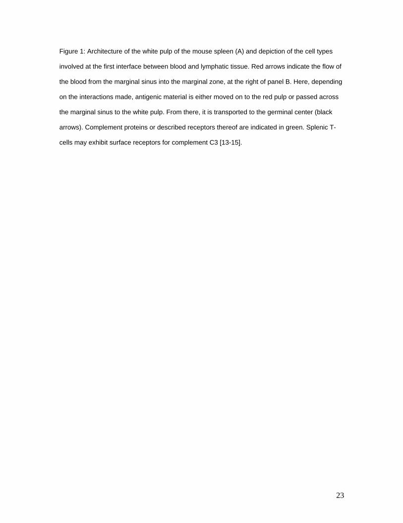
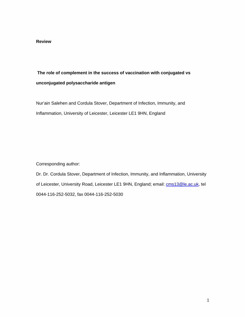
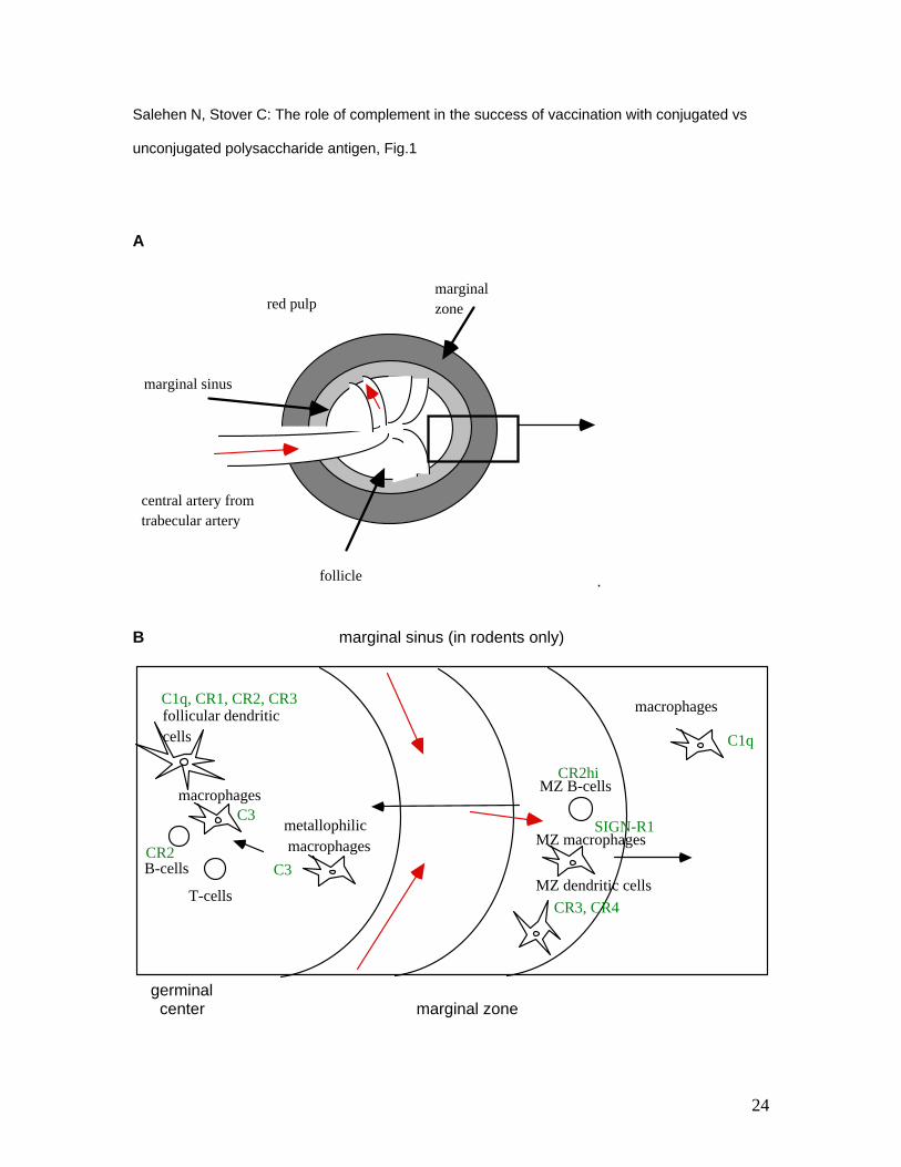
Figure 1: Architecture of the white pulp of the mouse spleen (A) and depiction of the cell types
involved at the first interface between blood and lymphatic tissue. Red arrows indicate the flow of
the blood from the marginal sinus into the marginal zone, at the right of panel B. Here, depending
on the interactions made, antigenic material is either moved on to the red pulp or passed across
the marginal sinus to the white pulp. From there, it is transported to the germinal center (black
arrows). Complement proteins or described receptors thereof are indicated in green. Splenic T-
cells may exhibit surface receptors for complement C3 [13-15].
23
Salehen N, Stover C: The role of complement in the success of vaccination with conjugated vs
unconjugated polysaccharide antigen, Fig.1
A
marginal sinus
marginal zonered pulp
follicle
central artery from trabecular artery
. B marginal sinus (in rodents only)
macrophages
MZ B-cells
MZ dendritic cells
MZ macrophagesmetallophilic macrophages
follicular dendritic cells
B-cells
T-cells
C1q
SIGN-R1
CR2hi
CR3, CR4
C3
C1q, CR1, CR2, CR3
CR2
C3macrophages
germinal center marginal zone
24
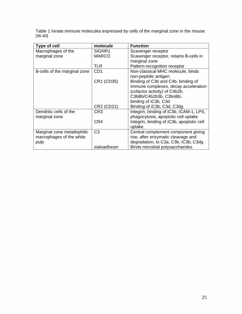
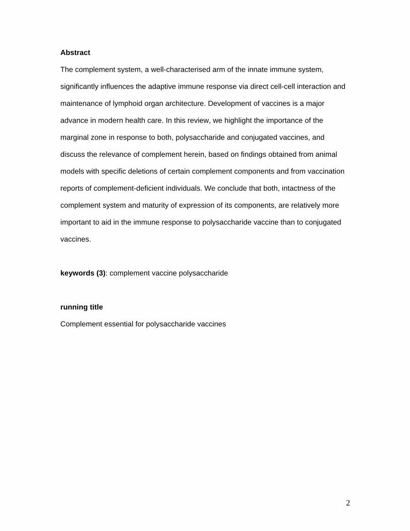
Table 1 Innate immune molecules expressed by cells of the marginal zone in the mouse [36-40] Type of cell molecule Function Macrophages of the marginal zone
SIGNR1 MARCO TLR
Scavenger receptor Scavenger receptor, retains B-cells in marginal zone Pattern-recognition receptor
B-cells of the marginal zone CD1 CR1 (CD35) CR2 (CD21)
Non-classical MHC molecule, binds non-peptidic antigen Binding of C3b and C4b, binding of immune complexes, decay acceleration (cofactor activity) of C4b2b, C3bBb/C4b2b3b, C3bnBb, binding of iC3b, C3d Binding of iC3b, C3d, C3dg
Dendritic cells of the marginal zone
CR3 CR4
Integrin, binding of iC3b, ICAM-1, LPS, phagocytosis, apoptotic cell uptake Integrin, binding of iC3b, apoptotic cell uptake
Marginal zone metallophilic macrophages of the white pulp
C3 sialoadhesin
Central complement component giving rise, after enzymatic cleavage and degradation, to C3a, C3b, iC3b, C3dg Binds microbial polysaccharides
25
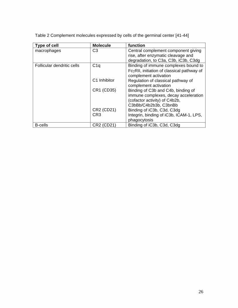
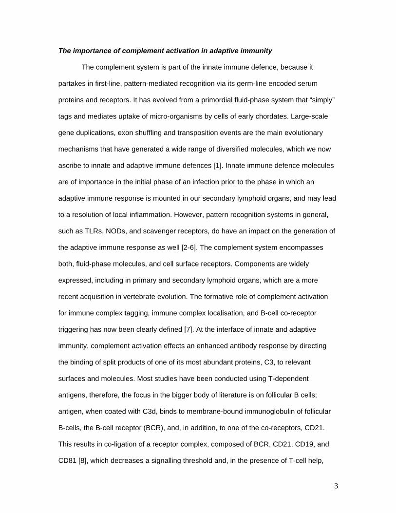
Table 2 Complement molecules expressed by cells of the germinal center [41-44] Type of cell Molecule function macrophages C3 Central complement component giving
rise, after enzymatic cleavage and degradation, to C3a, C3b, iC3b, C3dg
Follicular dendritic cells C1q C1 Inhibitor CR1 (CD35) CR2 (CD21) CR3
Binding of immune complexes bound to FcγRII, initiation of classical pathway of complement activation Regulation of classical pathway of complement activation Binding of C3b and C4b, binding of immune complexes, decay acceleration (cofactor activity) of C4b2b, C3bBb/C4b2b3b, C3bnBb Binding of iC3b, C3d, C3dg Integrin, binding of iC3b, ICAM-1, LPS, phagocytosis
B-cells CR2 (CD21) Binding of iC3b, C3d, C3dg
26