Embed Size (px)
Citation preview

117
INTRODUCTION
The immune system responds to injury or irritation through aninnate cascade known as inflammation. There are many cellularand biochemical events which occur as a result of tissue injurywhich are responsible for the outcome of inflammation. Centralto the formation of inflammation are the inflammatory mediators,which include proteins, peptides, glycoproteins, cytokines, arachi-donic acid metabolites (prostaglandins and leukotrienes), nitricoxide, and oxygen free radicals. These compounds are producedby epithelial cells, endothelial cells, and infiltrating inflammatorycells. Inflammatory mediators are a double-edged sword, havingthe potential to fight off infection, but also to damage the host.
Otitis media (OM) is an inflammatory response to either acute or
persistent stimuli, typified by the accumulation of both cellularand chemical mediators in the middle ear cleft (1). These media-tors have a role in the inflammatory processes of the middle earsuch as vascular permeability changes, chemotaxis, stimulation ofepithelial secretory activity, enhancement of mucous glycopro-tein secretion, and production of other mediators. This paperaims to review the important studies of inflammatory mediators inthe pathogenesis of OM and cholesteatoma in order to elucidatewhich mediators are the most significant. In addition, this paperreviews studies that have identified possible key factors respon-sible for the chronicity of OM.
INFLAMMATION AND OM
Initiation of inflammationOM is frequently caused by bacteria which enter the middle earfrom the nasopharynx via the eustachian tube. In cases of bac-terial otitis media, the source of bacteria is currently believed to bethe nasopharyngeal tonsils (adenoids). However, cases of sterileOM are not uncommon and the mechanism of this condition is
This review deals with the characteristics of various inflammatory mediators identified in the middle ear during otitis mediaand in cholesteatoma. The role of each inflammatory mediator in the pathogenesis of otitis media and cholesteatoma hasbeen discussed. Further, the relation of each inflammatory mediator to the pathophysiology of the middle and inner earalong with its mechanisms of pathological change has been described. The mechanisms of hearing loss including sensorineur-al hearing loss (SNHL) as a sequela of otitis media are also discussed. The passage of inflammatory mediators through theround window membrane into the scala tympani is indicated. In an experimental animal model, an application of cytokinesand lipopolysaccharide (LPS), a bacterial toxin, on the round window membrane induced sensorineural hearing loss asidentified through auditory brainstem response threshold shifts. An increase in permeability of the blood-labyrinth bar-rier (BLB) was observed following application of these inflammatory mediators and LPS. The leakage of the blood com-ponents into the lateral wall of the cochlea through an increase in BLB permeability appears to be related to the sensorineur-al hearing loss by hindering K+ recycling through the lateral wall disrupting the ion homeostasis of the endolymph. Furtherstudies on the roles of various inflammatory mediators and bacterial toxins in inducing the sensorineumral hearing lossin otitis media should be pursued.
Key Words. Otitis media, Inflammatory mediators, Cytokines, Chemokines, Cholesteatoma, Sensorineural hearing loss
The Role of Inflammatory Mediators in thePathogenesis of Otitis Media and Sequelae
Steven K. Juhn, MD Min-Kyo Jung, MD Mark D. Hoffman, MD Brian R. Drew, MD Diego A. Preciado, MDNicholas J. Sausen, BS Timothy T.K. Jung, MD1 Bo Hyung Kim, MD Sang-Yoo Park, MD Jizhen Lin, MD
Frank G. Ondrey, MD David R. Mains, BS Tina Huang, MD
Department of Otolaryngology, University of Minnesota Medical School, Minneapolis, MN; 1Department of Otolaryngology,
Loma Linda University, Loma Linda, CA, USA
�Received September 13, 2008 Accepted after revision September 20, 2008
�Corresponding author : Steven K. Juhn, MDDepartment of Otolaryngology, University of Minnesota Medical School,209 Lions Research Building, 2001 6th Street SE, Minneapolis, MN55455, USATel : +1-612-626-9879, Fax : +1-612-626-9871E-mail : [email protected]
DOI 10.3342/ceo.2008.1.3.117Clinical and Experimental Otorhinolaryngology Vol. 1, No. 3: 117-138, September 2008
Review

currently unclear. Endotoxin, a component of bacterial cell walls,is believed to be responsible for initiating inflammation in the mid-dle ear. Endotoxin is a potent inducer of various inflammatorymediators, as well as a modulator of the immune response, andstimulates local macrophages to produce tumor necrosis factor-α(TNF-α) and interleukin-1β(IL-1β). In addition, keratinocytes havethe ability to produce many soluble mediators independently fromimmune cells in response to injury, including TNF-α, IL-β, IL-1,IL-6, and IL-8 (2-5). Many of these cytokines contribute to the acutephase of the inflammatory response, prime the immune systemfor rapid activity, and promote the release of other cytokines(Fig. 1).
Sources of inflammatory mediatorsInflammatory mediators important in OM are produced by infil-trating immune cells such as neutrophils, monocytes, and lym-phocytes. In addition, local cells such as keratinocytes and mastcells have been shown to produce inflammatory mediators. It isalso coming to light that besides the middle ear, the inner ear tis-sues are able to produce inflammatory cytokines and use NF-κBactivation (6). The sources and functions of inflammatory medi-ators involved in OM are summarized in Table 1.
ComplementsComplement is one of the first mediators activated in the initia-tion of inflammation throughout the body and in OM. This path-way can act as the first cytotoxic response to invasion while alsoinitiating recruitment of leukocytes and vascular changes.Complement is part of the humoral immune system and consistsof multiple components that when activated, usually through cleav-age, interact to form enzymes or function as binding proteins. In-travascular activation of complement is initiated via a variety ofagents including antigen-antibody complexes, bacterial products,and toxins; injured endothelial cells can also activate the comple-ment system (7). The complement system is organized as two path-ways with C3 occupying the central position in both. Binding ofantibody to antigen or to foreign molecules results in activationof the antibody-dependent classical pathway, whereas the alter-
native pathway is activated by the lipopolysaccharide (LPS) por-tion of endotoxin and other nonprotein agents. C3 is biological-ly inactive, but cleavage by C3 convertase yields active fragmentsincluding C3a and C3b. Deposition of the active cleavage prod-uct C3b on the surface of foreign particles or target cells allowsrecognition by receptors on phagocytic cells. C3a is a potent activa-tor of mast cells and basophils and leads to release of histaminefrom secretory granules. Proteolytic cleavage of C5 occurs viaboth pathways and generates C5a, a potent mediator of inflam-mation. C5a is the major complement-derived chemotactic agentfor neutrophils, eosinophils, monocytes, and macrophages. C5aalso has the ability to activate platelets, leading to their aggrega-tion and surface expression of P-selectin.
Complement activation has been observed in human middleear effusion by some authors (8, 9). In animal models of acuteotitis media (AOM), animals were treated with cobra venom todeplete complement and then their middle ears were antigeni-cally challenged (10). Complement-depleted animals had signif-icantly less inflammation and decreased volume of effusion whencompared to controls. Middle ear mucosa from children with chron-ic otitis media (COM) with effusion has shown large amounts ofcomplement fragments C3 and C9 when analyzed by immuno-fluorescence microscopy (11). In humans, deficiencies of the com-plement system are known to cause recurrent or COM (12). Des-pite this fact, it appears that complement itself may be harmfulto the middle ear mucosa. Membrane cofactor protein (MCP) andprotectin (CD59) are two middle ear proteins which help preventunrestricted complement damage (11).
CytokinesCytokines are glycoproteins, produced by inflammatory cells andepithelial cells, which modulate the immune response. Cytokinesextensively conduct inter-cell communication. Inflammatory cellsincluding neutrophils, macrophages, and lymphocytes use cytokinesto coordinate all stages of the inflammatory response. Productionof cytokines is conducted by a wide variety of cell types. For exam-ple, IL-1 is produced and secreted by macrophages, lymphocytes,vascular endothelial cells, neutrophils, fibroblasts, and monocytes.There are many biological effects including: chemotaxis of cytotox-ic T-cells and B-lymphocytes, cytokine synthesis of IL-2 and IL-8 and TNF, neutrophil chemotaxis and degranulation, fibroblastand epithelial proliferation, and histamine release (13, 14). Thepresent theory is that cytokines are responsible for many of theinflammatory changes induced by pathogenic organisms duringOM (15).
Different cytokines are involved in the early and late stagesof inflammation and it has been observed in a bilateral study ofeffusion that cytokine production and concentration can varybetween the ears (16). IL-1 and TNF-αare early-response cytokineswith IL-1 being a more potent activator than TNF-α(17). IL-1 maybe more important than TNF-αin the elicitation stage of inflam-mation, while there is more of a need for TNF-αduring recruit-
118 Clinical and Experimental Otorhinolaryngology Vol. 1, No. 3: 117-138, September 2008
Fig. 1. Pathogenesis of otitis media.
Infection
SequelaeMediators
ofinflammation
Eustachiantube
dysfunction
MechanicalFunctional
BacterialViralOther Increased
vascularpermeability
Epithelialsecretoryactivity
Middleear
inflammationand
effusion
Resolution
Complications

Juhn SK et al.: Otitis Media and Inflammatory Mediators 119
Inflammatory mediator Source Principal activities
Table 1. Sources and functions of inflammatory mediators important in otitis media
(Continued to the next page)
CytokinesTumor necrosis factor-α Macrophages Prostaglandin release, activates neutrophils, eosinophils, macrophages, cytokine release
LymphocytesEpithelial cellsEndothelial cells
Interleukin-1 Macrophages Activates B- and T-cells, epithelial and fibroblast proliferation, cytokine synthesis, Neutrophils histamine release, induces fever, bone resorptionFibroblastsEndothelial cellsEpithelial cells
Interleukin-2 T-cells Activates T-cellsInterleukin-4 T-cells Stimulates TH2 differentiation and proliferation, anti-inflammatory action on T cells,
Mast cells B cells, monocytesBasophils
Interleukin-5 T-cells Stimulates and maintains IgA production by B-cells, eosinophil chemotaxisMast cells Stimulates eosinophil production in bone marrowBasophils
Interleukin-6 Macrophages Activates B- and T-cells, production of antibodies, induces fever, bone resorptionLymphocytesEpithelial cells
Interleukin-10 Macrophages Down regulates inflammatory properties of IL-1, IL-6, TNF-α
Interleukin-12 Macrophages Interferon release, activates macrophages, potentiates T-cell proliferation, cytokine production,Neutrophils and cytotoxicity of lymphocytes. Induces TH1 lympocyte developmentLangerhans cells
Interleukin-13 T-cells Similar to IL-4; molecular bridge linking allergic inflammation cells to the non-immune cells incontact with them
Transforming growth factor-β Neutrophils Initiation and maturation of inflammatory processes; recruitment, activation, and proliferation Macrophages of inflammatory tissues and cells
Granulocyte-macrophage Macrophages Stimulates production of granulocytes (neutrophils, eosinophils, and basophils)colony-stimulating factor
ChemokinesInterleukin-8 Macrophages Neutrophil chemotaxis and activation, angiogenesis
FibroblastsEpithelial cellsEndothelial cells
RANTES Epithelial cells Monocyte and T-cell chemotaxisMCP-1 Epithelial cells Monocyte and T-cell chemotaxisHistamine Mast cells Increases vascular permeability, vasodilation, neutrophil and eosinophil chemotaxis
BasophilsVascular endothelial Thrombocytes Increases vascular permeability, vasodilation, inflammatory cell infiltration
growth factorPlatelet activating factor Monocyte Chemotaxis and degranulation of neutrophils, increase vascular permeability
Neutrophils (1,000×more potent than histamine)Lymphocytes
Mast cells Bone marrow Release of preformed mediators: histamine and tryptaseexpressing CD34 Release of de novo synthesized mediators: leukotrienes and prostaglandinsmolecule
Arachidonic acid metabolitesProstaglandins
Prostaglandin E2 Mast cells Vasodilation, mucus production, induces cytoprotection to IL-1NeutrophilsMonocytes
ment and maintenance stages (18, 19). IL-1 and TNF-αfrom mac-rophages induce the expression of glycoprotein adhesion mole-cules on the surface of vascular endothelial cells which bind leuko-cytes so they can leave the circulation and enter the site of infec-tion.
IL-1IL-1 was originally identified as a lymphokine that was mitogenicfor murine thymocytes (20). Now IL-1 is known to be producedby many different cells to regulate immune responses (21). IL-1is one of the most active substances inducing bone resorptionthrough osteoclast activation (22). This IL-1-mediated bone destruc-tion is one of the clinical characteristics that signal the onset ofchronic OM. Neutrophils are a major producer of IL-1β. IL-1induces the production of IL-1 in neutrophils in a positive-feed-back mechanism (23). IL-1 has been shown to stimulate the syn-thesis of TNF, IL-2, IL-6, and IL-8 (24-27). IL-1 is comprised oftwo principal 17kDa polypeptides, IL-1αand IL-1β. Genes foundon chromosome 2 encode these two molecular species (28). Theyhave the same biological activities and bind to the same recep-tor on cell surfaces (28, 29). Both IL-1αand IL-1βare derivedby proteolytic cleavage of 33 kDa precursor molecules (28). IL-1αacts as a membrane-associated substance, whereas IL-1βisfound free in the circulation (30).
IL-1 promotes the release of other cytokines and stimulatesarachidonic acid metabolism in both cyclooxygenase and lipoxy-genase pathways (21, 31). IL-1 is primarily synthesized by acti-vated macrophages. In these, IL-1 production is stimulated byLPS and leukotrienes (28, 32, 33). TNF also stimulates synthe-sis of IL-1 (14). Platelet activating factor (PAF) can enhance IL-1 release by the production of leukotriene metabolites (34). IL-1βis mainly produced and released extracellularly by inflamma-tory cells such as macrophages/monocytes (35) and IL-1αis local-ized intracellularly or on the surface of such cells (36). IL-1 inducescell adhesion molecules (intercellular adhesion molecule-1 andvascular cell adhesion molecule-1) that in turn stimulate the migra-tion of leukocytes (18, 37).
IL-lβhas been shown to play an important role in the patho-genesis of otitis media with effusion (OME). When introduced
into the murine middle ear, endotoxin derived from Haemophilusinfluenzae produced middle ear effusion (MEE) with significant-ly higher levels of IL-1βthan controls, and inoculation of IL-1βcaused similar pathologic changes to that of endotoxin. It has beenobserved that IL-1βupregulates the activity of sodium-potassium-chloride cotransporters. Also, IL-1βsuppresses epithelial Na+
channel-dependent fluid transport. This upregulation of cotrans-porters may be one of the important factors in the excess fluidaccumulation that is observed in OM (38, 39).
Additionally, the administration of anti-IL-1 receptor antibod-ies with endotoxin reduced the incidence of MEE and the patho-logical changes of the middle ear mucosa (40). Viable pneumo-cocci inoculated into the middle ear of chinchillas caused an in-creased concentration of IL-1βafter 6 hr (41). In a guinea pigOM model induced with nonviable H. influenzae, Sato et al.(42) suggested that IL-1βand TNF-αwere produced by middleear mucosa in the early stage of OM, which caused subsequentinflammatory cell accumulation and further production ofcytokines such as IL-8 and TNF-αin the late stage.
Barzilai et al. (43) showed that IL-1 levels were significantlyhigher in culture-positive versus culture-negative AOM. Levelsof IL-1 significantly decreased on days 4-5 of antibiotic therapywhether the bacteria were eradicated or not. This is in contrastto measured levels of TNF-α, which are more closely correlatedwith the presence of bacteria (44). On the other hand, it hasbeen shown that age is inversely related to the concentration ordetection rate of IL-1βby enzyme-linked immunosorbent assay(ELISA) in the MEE of OME patients. Therefore, treatment thatreduces IL-1βmay be an effective adjuvant in the early stage ofOM in children (45, 46).
IL-2IL-2 is a 15.5 kDa glycoprotein synthesized principally by acti-vated T cells, although activated B cells may also have the abilityto produce small amounts of IL-2 (47). The gene of IL-2 is locat-ed on human chromosome 4q26. It induces the proliferation anddifferentiation of T-cells, B cells, NK cells, monocytes, and macro-phages. Upon release, IL-2 is bound by T-cells that have beenprimed by exposure to their distinctive antigens. Helper T-cells
120 Clinical and Experimental Otorhinolaryngology Vol. 1, No. 3: 117-138, September 2008
Inflammatory mediator Source Principal activities
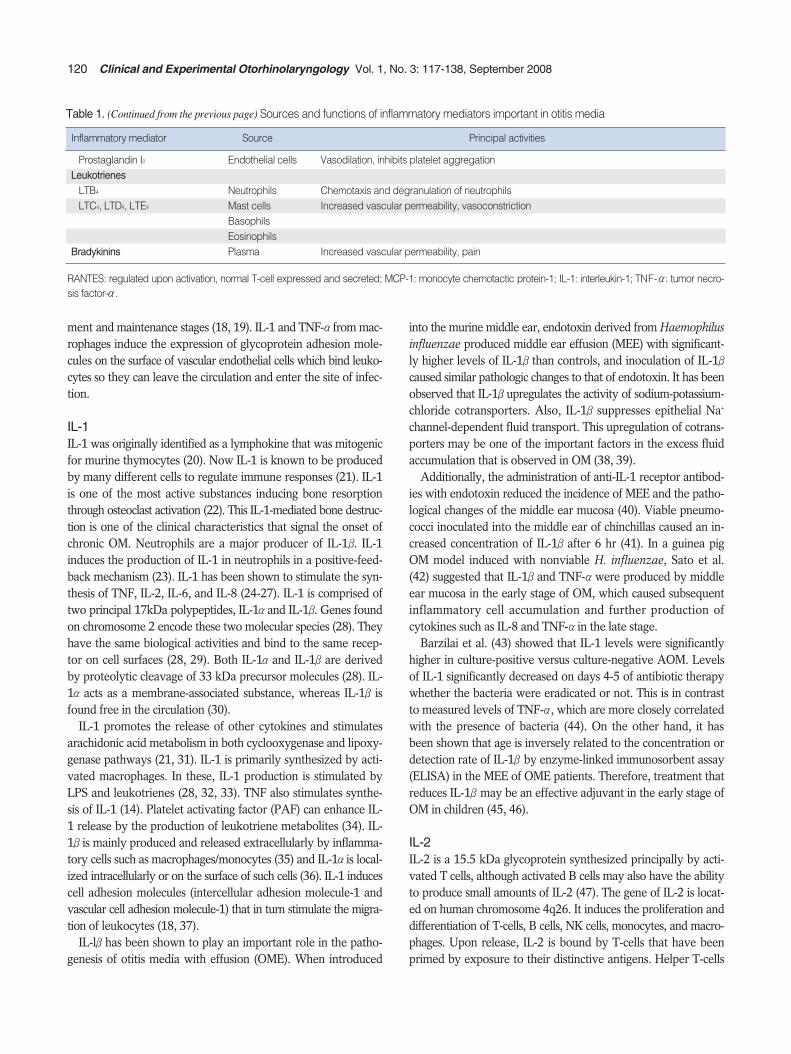
Table 1. (Continued from the previous page) Sources and functions of inflammatory mediators important in otitis media
Prostaglandin I2 Endothelial cells Vasodilation, inhibits platelet aggregationLeukotrienes
LTB4 Neutrophils Chemotaxis and degranulation of neutrophilsLTC4, LTD4, LTE4 Mast cells Increased vascular permeability, vasoconstriction
BasophilsEosinophils
Bradykinins Plasma Increased vascular permeability, pain
RANTES: regulated upon activation, normal T-cell expressed and secreted; MCP-1: monocyte chemotactic protein-1; IL-1: interleukin-1; TNF-α: tumor necro-sis factor-α.

stimulated by IL-2 secrete cytokines to stimulate other lympho-cytes. Cytotoxic T-cells and natural killer cells stimulated by IL-2 also proliferate and secrete cytokines, IFN-γ, granulocyte-macrophage colony-stimulating factor (GM-CSF), and TNF-αwhich preferentially activate monocytes and macrophages. IL-2reaches peak levels early in the inflammatory cascade, and lev-els quickly drop before there is any clinical evidence of inflamma-tion. IL-2 is a key factor in determining the TH1 response of T-cells to promote a strong cell-mediated response. TH1 cells areresponsible for orchestrating cellular immunity, while TH2 cells(stimulated by IL-4) provide signaling for humoral immunity.However, IL-2 was not detected in MEE of AOM patients byELISA nor was it found in an experimental rat model of AOMinduced by inoculation of Streptococcus pneumoniae type 3 andnontypable H. influenzae by reverse transcription-polymerasechain reaction (RT-PCR) (44, 48). On the contrary, IL-2 was detect-ed in the MEE of the chronic OME patients by ELISA (45, 49).It is believed that the inflammatory up-regulation of IL-2 is bal-anced by the down-regulatory effects of IL-10. Smirnova et al.(50) reported that an imbalance in the production of these cytokinesinduces switching to the chronic stage. Excessive IL-2 produc-tion can cause humoral inflammatory processes and/or chroniccell-mediated processes whereas IL-2 deficiency promotes per-sistence of OME, which can result in chronic OME. From this, itmay be deduced that the ongoing chronic inflammatory stateassociated with chronic OME may contribute different cell types,and thus different cytokines, than those with AOM.
Tumor necrosis factor (TNF)TNF is a pair of cytokines originally called cachectin (TNF-α) andlymphotoxin (TNF-β). TNF-αis a polypeptide produced mainlyby stimulated macrophages but made by wide variety of cellsincluding fibroblasts, T cells and B cells (51). TNF-βis producedpredominantly by lymphocytes and its active form is a 25 kDaglycosylated polypeptide (52). These two forms of TNF are 30%identical at the amino acid level (53).
Endothelial cells and epithelial cells all have the capacity to pro-duce TNF. TNF induces the acute phase of the inflammatory res-ponse and release of other cytokines. TNF activates polymor-phonuclear leukocytes, promotes fibroblast proliferation, inhibitsvascular endothelial and B-lymphocyte proliferation, and stim-ulates cartilage and bone resorption (54). Thus, TNF has manyof the same functions as IL-1. In fact, IL-1βand TNF-αinduce eachother’s production and have a synergistic effect in vivo (55-57).TNF stimulates arachidonic acid metabolism, leading to the pro-duction of prostaglandins and leukotrienes that are important inthe pathogenesis of OM (58, 59). TNF-αis a strong inducer ofIL-1, IL-2, IL-6, IL-8, and endothelial adhesion molecules includ-ing intercellular adhesion molecule-1 (ICAM-1) and vascular celladhesion molecule-1 (VCAM-1) (60-63). TNF-αis chemotacticto neutrophils, monocytes, macrophages, and lymphocytes (17).
TNF-αwas detected in human MEE and it is now apparent that
TNF-αis one of the most significant inflammatory mediators inOM (64). Animal models of OM induced by S. pneumoniae andLPS have all demonstrated elevated levels of TNF-αin the earlystage of OM. Ball et al. (65) indicated that TNF is a significantmediator of LPS-induced MEE in a rat model. Higher expressionof TNF-αin human OM confers higher risks of subsequentepisodes of OM, longer courses of OM, and chronicity of OM (66-68). The results of these studies suggest a potential therapeuticrole with the clinical application of TNF inhibitors, such as TNFbinding protein or inhibitor TNF soluble receptor, to control thesymptoms of AOM or to prevent chronicity and recurrence ofOM (65, 66).
IL-4, IL-5, IL-13, and GM-CSFIL-4, a 20 kDa glycosylated polypeptide, is produced mainly byCD4+ T cells, but also by mast cells and basophils. It functionsprimarily as a growth factor for B cells and some types of T cells(14). IL-4 supports the proliferation of activated T cells and dri-ves their differentiation to TH2 cells. These T cells support theproduction of antibodies in a humoral immune response. IL-4stimulates B-cells to increase IgG and IgE isotypes (21). Thereare also anti-inflammatory effects that are stimulated by IL-4. Itcontrols numerous molecular processes which cause deactivationof inflammatory macrophages, suppression of inflammation, down-regulation of the production and secretion of pro-inflammatorycytokines TNF-α, IL-1, and IL-8 (50). Although Melhus et al. (48)reported that they could not detect mRNA of IL-4 in the animalAOM model induced by S. pneumoniae and H. influenzae, Jangand Kim (49) could detect IL-4 in the MEE of chronic OME patientsand suggested that elevation of TH2 cytokines, such as IL-4 andIL-6, might be a contributing factor in the persistence of OMEwith allergy. In addition, IgG production shows increases in rela-tion to IL-4 prevalence in AOM in systemically immunized micedue to IL-4 having the effect of inducing B-cell differentiation intoIgG-secreting plasma cells (69). It is thought that this is the path-way leading to inflammation in patients with allergic disorderscausing OM. Smirnova et al. (50) reported that switching fromacute to chronic inflammation can be induced by IL-4 due to itsability to up-regulate mannose receptor expression on activatedmacrophages. Activated macrophage fusion and giant multinu-cleate cell formation are promoted by the mannose receptor andcreate the cellular background for the onset of chronic inflam-mation.
IL-5 is produced by T lymphocytes as a glycoprotein with aMr of 40-45 and is also involved in the TH2 pathway of inflam-mation (70). IL-5 functions to induce differentiation of eosinophilsand augments proliferation of activated B-cells (14). IL-5 specif-ically acts on B-cells to stimulate and maintain IgA production(71). In chronic otitis media with effusion (COME) in systemi-cally immunized mice, IL-5 positive cells were more numerousthan IL-4 cells (69). This finding is consistent with the finding ofincreased local IgA production seen during chronic otitis media
Juhn SK et al.: Otitis Media and Inflammatory Mediators 121

caused by allergy.Recent findings by Iino et al. suggest that IL-5 may play a key
role in the pathogenesis of eosinophilic otitis media (EOM), a formof infection often associated with bronchial asthma and/or aller-gic rhinitis. EOM is commonly seen in adults in contrast to AOMand OME which are more often diagnosed in children. The roleof IL-5 in EOM can be linked to its chemotactic effect on eosino-phils. EOM is characterized by having a markedly viscous or gelati-nous effusion with a high eosinophil count and middle ear mucousmembranes that are thickened with numerous eosinophils. Theeosinophilic infiltration is thought to be due to locally producedIL-5. Iino et al. (72) showed that higher concentrations of IL-5appeared in EOM middle ear effusions than in those of OMEpatients. This finding is further evidence of the role IL-5 has inEOM and OME patients.
IL-13 has also been implicated in the pathogenesis of OME. Thisinflammatory cytokine was found in 25.9% (7/27) of effusionsin a study by Smirnova et al. (73) It was also found that both IL-4and IL-13 had a significant positive correlation with the concen-tration of mucin in MEEs. It has been shown in an allergic air-way inflammation model that suppress both IL-4 and IL-13may be necessary to reduce mucus production and allergicinflammation (74). More research is needed to understand thefunctions of how IL-13 works in conjunction with IL-4 as a pro-inflammatory cytokine during OM.
GM-CSF has also been found to play a role in OM. GM-CSFpromotes the activation and prolonged existence of eosinophils.Together with IL-5, it is possible that up-regulation of GM-CSFcan cause eosinophil-mediated inflammation. In contrast, GM-CSFmay also prove to be a down-regulator of allergic inflammationdue to its ability to induce apoptosis in eosinophils (75). Furtherresearch is necessary to gain a better understanding of GM-CSFin the middle ear.
IL-6IL-6 is a glycoprotein with a molecular weight of 25 kDa. IL-6 isproduced by many cells, including helper T-cells, macrophages,mast cells, neutrophils, epithelial cells, and fibroblasts. It stimu-lates B cells to differentiate and is often upregulated followinginfection (76). Its main function is regulation of immune response,acute phase reaction, and hematopoiesis (77). IL-6 has been shownto play a key role in the acute phase reaction, including the induc-tion of C-reactive protein (78). C-reactive protein is commonlyused as a marker of bacterial infection. However, its use as a screen-ing test for bacterial AOM is limited by its limited sensitivity andnegative predictive value (79).
IL-6 seems to play an important role in the cytokine networkof OM. The concentrations of IL-1, IL-6, and TNF-αin MEE fromchildren were highly correlated with each other (80). Kerschneret al. (81) also discovered a significant correlation between MEEIL-6 concentrations and degree of hearing loss. In cultured mid-dle ear epithelial cells, mucin secretion is upregulated by IL-6 in
a dose and time-dependent manner. Interestingly, in a study ofchildren with AOM, serum samples from the AOM patients infect-ed with S. pneumoniae contained significantly higher levels ofIL-6 than those infected with other bacteria or no bacteria (82).It appears that S. pneumoniae stimulates IL-6 production morethan other types of bacteria. On the contrary, IL-6 was not detect-ed in the guinea pig OM model induced by nonviable H. influen-zae (41). Fogle-Ansson et al. (83) found that animals inoculatedwith gram-negative bacteria mediated a faster IL-6 response thananimals inoculated with gram-positive bacteria. They also foundthat it was possible to determine whether an infection was gram-positive or negative by analyzing the concentrations of IL-6. Ina rat model, IL-6 mRNA increased in AOM induced by both S.pneumoniae and H. influenzae (48). Additionally, it was foundthat the shorter the course of OM, the higher the concentrationof IL-6 in MEE (67).
In conclusion, expression of IL-6 is thought to be a potential can-didate for the detection of the severity or chronicity of AOM. Fu-rther proof of this can be seen in the recent findings linking IL-6to mucin upregulation. However, more reports demonstratingdirect correlation between IL-6 and upregulation of mucin secre-tion are necessary to show transition to chronicity of OM.
IL-10IL-10, known as cytokine synthesis inhibitory factor, is consid-ered an immunosuppressive regulator of acute inflammation. Itis produced by monocytes, CD4+ T cells, activated CD8+ T cells,and activated B cells following induction by bacterial productssuch as LPS. It is produced relatively late when compared to othercytokines, but there is evidence that IL-10 expression is upreg-ulated shortly after or even simultaneously with pro-inflammatorycytokines. IL-10 is the principal TH2-type cytokine that upregu-lates humoral immune responses and attenuates cell-mediatedimmune reactions. If IL-10 is present for an extended period inthe inflammation zone it can induce the amplification of chron-ic humoral inflammatory processes. Through this humoral inflam-matory amplication IL-10 is thought to contribute to the switch-ing of the infection into the chronic stage. A deficiency of IL-10has also been seen to result in the chronic condition because theinflammatory actions of IL-1β, TNF-α, and IL-6 are not downreg-ulated (50). IL-10, being the key anti-inflammatory cytokine,inhibits monocytes/macrophages and the production of TNF,IL-1, IL-6 and IL-8 (84). In addition, the production of oxygenradicals and proteases from activated neutrophils is directly sup-pressed. IL-10 was detected in the MEE of an experimental OMmodel by ELISA (85), and middle ear mucosal upregulation ofits mRNA expression was also reported in the early and in thelate stage of experimentally induced OM (48, 85). It is thoughtthat IL-10 contributes to the eradication of middle ear inflam-mation and provides a negative feedback mechanism related toTNF-αin OM.
It is also believed that production of IL-10 must be balanced
122 Clinical and Experimental Otorhinolaryngology Vol. 1, No. 3: 117-138, September 2008
′

relative to production of IL-2. These cytokines need to be pro-portional in the middle ear in order to cancel the effects of theother. IL-10 is responsible for the down-regulation of acute inflam-matory events while IL-2 leads to the up-regulation of cellularand molecular inflammation events (50).
Transforming growth factor (TGF)TGF is divided into two classes of polypeptide growth factors.Transforming growth factor alpha (TGF-α) and beta (TGF-β)are not structurally or genetically related to one another andthey act through different receptor mechanisms. TGF-βis knownto exist in at least five subtypes: TGF-β1 through TGF-β5. TGF-βis part of a supergene family of low molecular weight cytokinesthat play a crucial role in the initiation and maturation of inflam-matory processes including recruitment, activation, and prolifer-ation of inflammatory cells (86). TGF-βhas been found to stimu-late extracellular matrix formation (fibronectin, collagen, andproteoglycans), protease inhibitor, and fibroblast chemotaxis.TGF-βis also believed to have an effect on fibroblast prolifera-tion (87). The overproduction of TGF-βin the middle ear canelicit responses that may lead to chronic OME. Fibroblast accu-mulation and activation, IgA+ B cell secretion, and monocyte accu-mulation and activation are the result of TGF-βoverproduc-tion. In the presence of cytokines such as IL-10, IL-4, and GM-CSF, these effects can cause cell-mediated inflammation, humoralinflammation, and progressive fibrosis which leads to the chron-ic condition of OME (50). In a study by Cooter et al. (86), it wasfound that detectable amounts of TGF-β1 and TGF-β2 occurred in100% and 98% of middle ear effusions, respectively. The majorrole of TGF-βoccurs in the later stages of inflammation and itshigher levels indicate that it participates in the repair of dam-aged tissue in the middle ear (50).
IL-12IL-12, a heterodimeric pro-inflammatory cytokine, is producedby phagocytic cells such as macrophages, neutrophils, and Langer-hans cells. When stimulated by bacteria or bacterial toxins suchas LPS, these cells release large amounts of IL-12. This effect isfurther enhanced in the presence of interferon-γ(IFN-γ). IL-12 isa potent inducer of IFN-γproduction by T and NK cells. IL-12-induced IFN-γexerts positive feedback in inflammation by enhanc-ing IL-12 production. Wang et al. (88) found that IL-2 upregu-lates the expression of IL-12 and increases the response of NKto IL-12. IL-12 is the major cytokine responsible for the differ-entiation of TH1 cells, which are producers of IFN-γ. The enhanc-ing effect of IFN-γon IL-12 production may represent a mecha-nism by which TH1 responses are maintained. IFN-γactivatesmacrophages and NK cells, and potentiates the proliferation ofactivated T cells. IL-12 is important in early immunity becauseit stimulates innate resistance and generates a TH1-type immuneresponse. It is believed to form a link between innate and adap-tive immunity. There have been no reports about the expres-
sion of IL-12 in OM until now. It is thought that the function ofIL-12 in the middle ear is limited.
ChemokinesChemokines are a family of structurally similar small proteins,whose major role is to stimulate leukocyte chemotaxis (89). Inthis way, they are thought to regulate the immune response. Thereare 2 families of chemokines, α-chemokine (CXC) and β-chemo-kine (CC) chemokines. CXC chemokines include IL-8 and arechemotactic to neutrophils. CC chemokines are chemotactic tomonocytes and lymphocytes.
IL-8IL-8 comes from monocytes, macrophages, fibroblasts, endothe-lial cells, and lymphocytes (90). It is produced on stimulation withIL-1β, TNF-α, IL-13, LPS, Concanavalin A, or viruses from thepreviously listed cells (5, 17, 91, 92). Since IL-8 expression canbe controlled by the primary cytokines, IL-1βand TNF-α, it isconsidered to be a secondary cytokine in middle ear inflammation(73). IL-8 is a selective chemotaxic agent for neutrophils (93, 94).IL-8 induces neutrophil chemotaxis, activation and degranulation,surface expression of adhesion molecules, respiratory burst, andcytosolic calcium increase (93). IL-8 increases adhesion moleculesfor attachment and migration of neutrophils during acute inflam-matory response (95). Since IL-8 is capable of inducing lysoso-mal enzyme release, IL-8 may play a key role in causing tissuedamage, which leads to the prolongation of middle ear inflam-mation (96, 97). When directly injected into the middle ears ofhealthy mice, IL-8 causes more inflammatory changes than whenS. pneumoniae is injected into mouse middle ears (98). IL-8 secre-tion is enhanced in both S. pneumoniae and H. influenzae pro-voked cases of OM (73).
Several authors detected IL-8 in human MEE. Transcripts ofIL-8 were detected in 75% of both pediatric and adults MEEs (97).The mean level of IL-8 in MEE from children was higher thanthat of adults (99). The peak level of TNF-αand IL-1βin the MEEin a guinea pig OM model preceded the peak in IL-8 (41). Thisdelay of the IL-8 peak level suggests that IL-8 is produced by inf-lammatory cells which accumulated via the influence of TNF-αand IL-1β.
Levels of IL-8 were closely correlated with the presence of bac-teria and neutrophils in the MEE of OM. With initiation of antibi-otic therapy, levels of IL-8 were reported to fall as bacteria wereeradicated from the middle ear (100). IL-8 has also been shownto be predictive of the number of neutrophils in MEE (100-103).
Smirnova et al. (104) assessed the effects of IL-8 and other proin-flammatory cyokines on goblet cells using human goblet cell lineHT29- MTX which secretes two OME-related mucins, MUC5ACand MUC5B. They proposed that goblet cells are target cells forIL-8, TNF-α, and IL-1βand that IL-8 stimulates prolonged mucinsecretion from goblet cells and may be involved in the mainte-nance of OM in the the chronic stage. Additionally, recurrence
Juhn SK et al.: Otitis Media and Inflammatory Mediators 123

of AOM within 1 month was associated with high IL-8 levels inthe initial middle ear fluid (101). The results of these studies sug-gest the possibility of using the IL-8 level as a prognostic factor orparameter of clinical outcomes.
Monocyte chemotactic protein-1 (MCP-1)MCP-1 is expressed by endothelial cells, fibroblasts, and epithelialcells stimulated by either IL-1 or TNF (18). MCP-1 is chemotac-tic for both monocytes and lymphocytes in vitro (105, 106).Recently, MCP-1 has been shown to be chemotactic for mono-cytes and lymphocytes in vivo (107). MCP-1 expression alonedoes not activate these cells, but leads to enhanced inflammato-ry response upon treatment with other stimuli such as LPS(107). MCP-1 increases the adhesiveness of human monocytestoward endothelial cells by stimulating an increase in the surfaceexpression and activation of adhesion molecules (108, 109).MCP-1 has a central position in monocyte trafficking and activa-tion and has been implicated in diseases characterized bymonocyte-rich infiltrates, including atherosclerosis, rheumatoidarthritis, and multiple sclerosis (110). MCP-1 is important inrecruiting monocytes in chronic inflammation (17). MCP-1 hasbeen detected in higher levels in MEEs with combined bacterialand viral infections (111). MCP-1 can be seen to upregulate theactivation of inflammatory cells in the presence of LPS.
Macrophage inflammatory protein-1 (MIP-1 )MIP-lαis released by monocytes, mast cells, endothelial cells,fibroblasts, epithelial cells, B cells, and T cells. It has potent mono-cyte and lymphocyte chemoattractant properties and inducesrelease of histamine from basophils and mast cells (112, 113).MIP-1αwas detected 9 of 11 cases of MEE (111). It is likely thatMIP-1 plays an important role in the microenvironment of themiddle ear, especially with respect to basophil function (111). Thismediator has the potential to be important in acute and chronicOM as it is released by monocytes and induces histamine releaseand monocyte chemotaxis.
RANTESRegulated upon Activation, Normal T-cell Expressed and Secreted(RANTES) is a CC chemokine that is chemotactic to monocytesand lymphocytes. RANTES was reported to be detectable in 94(82%) of 114 effusions. The concentration of RANTES was pos-itively correlated with the endotoxin content (114). In vitro,RANTES was expressed in middle ear epithelium in response toproinflammatory stimuli (TNF-α) in a dose dependent manner.Schousboe et al. (114) speculated that the expression of RANTESmight explain the recruitment of monocytes in OME, possibly as aresult of TNF-α-mediated endotoxin stimulation. On the otherhand, Jang and Kim (115) demonstrated that RANTES andeosinophilic cationic protein (ECP) in MEE of OME with allergywere significantly correlated and significantly higher than con-trols. Their results suggest the allergic role of chemokines in the
pathogenesis of OME.
Mast cellsMast cells are the most numerous resident leukocytes in the mid-dle ear. When activated, mast cells release preformed mediators,histamine and tryptase, and de novo synthesized mediators, leu-kotrienes and prostaglandins. Mast cells also release various cyto-kines including tumor necrosis factor and IL-1. During OM themast cell population increases dramatically and mast cells will useToll-like receptors and CD48 to directly interact with bacterialproducts. Ebmeyer et al. (116) showed that mast cells may be amajor contributor to the early inflammatory response and mayprovide a link between infection and allergy in the generationof OM.
HistamineHistamine causes vasodilation, increased vascular permeability,and edema of middle ear mucosa (117-119). Histamine is releasedby mast cells present in the middle ear upon activation by eithercomplement or binding of antigen to IgE antibodies which studthe exterior surface of mast cells (120-122). Through its vasodila-tory effects, histamine can cause mucociliary and eustachian tubedysfunction (123-126). Histamine levels and mast cell numberswere higher in adenoids of children with serous OM than in chil-dren with normal ears, suggesting a possible mechanism of eustachi-an tube obstruction secondary to histamine released from the ade-noids (127, 128). In addition, middle ear mast cells were increasedtwofold in patients with chronic middle ear disease (129). Histaminelevels were found to be higher in mucoid effusions than serous-type effusions (121, 129). Thus, histamine appears to be an impor-tant mediator influencing both acute and chronic OME.
Vascular endothelial growth factor (VEGF)Another factor thought to be responsible for increased vascularpermeability is VEGF. It is extremely potent, estimated to be50,000 times as potent as histamine (130). Platelet-activating fac-tor is 1,000-fold more potent than histamine, which may meanthat the permeability effect of VEGF is approximately 50 timesgreater than PAF. Kim et al. (131) found that severe inflamma-tory cellular infiltration, subepithelial edema, and vascular dilata-tion occurred in Sprague Dawley rats that were exposed to 0.1μg and 1.0 μg of recombinant vascular endothelial growth fac-tor. It is thought that perhaps VEGF is primarily responsible forincreased vascular permeability and that it is expressed in thepresence of histamine (132).
Bradykinin/kallikrein-kinin systemThe plasma kinin-forming system consists of three plasma proteinsthat interact in a complex manner when bound to negatively ch-arged surfaces. These factors are factor XII (Hageman factor), pre-kallikrein and high molecular weight kininogen (HMWK). FactorXII, once converted to factor XIIa, converts plasma prekallikrein
124 Clinical and Experimental Otorhinolaryngology Vol. 1, No. 3: 117-138, September 2008

to kallikrein. Kallikrein enzymatically cleaves HMWK to liberatebradykinin. In serous OME, bradykinin enhances the vascular per-meability in the middle ear mucosa, causing plasma to leak intothe middle ear cleft (133). This plasma leakage characterizes serouseffusions (134). Active plasma leakage into the middle ear occursduring the early phase of inflammation (133). The degree of plas-ma leakage is low in mucoid effusions and varied in serous effu-sions (133). Decreased plasma leakage into the middle ear in mu-coid effusions may be explained by the consumption of the kalli-krein system (134). Thus, bradykinin is an acute phase mediator,induced by the kallikrein-kinin system, which enhances vascularpermeability in the middle ear leading to MEEs.
Platelet Activating Factor (PAF)PAF is a membrane phospholipid metabolite released through theremodeling pathway upon stimulation (135). PAF, IL-1, and TNFcan induce each other’s release and also their own generation inself-generating positive feedback cycles (35). PAF is released fromhuman neutrophils, platelets, eosinophils, macrophages, mast cells,and vascular endothelial cells. PAF synthesis is stimulated by PAF,LTB4, LTC4, and LTD4 (136, 137). Its activities include stimula-tion of arachidonic acid release and metabolism, increasing vas-cular permeability, and activation of neutrophils, monocytes, andmacrophages (138, 139). PAF induces neutrophil adhesion toendothelial cells (137). In addition, PAF appears to play a majorrole in the production of MEE by increasing vascular permeabili-ty, stimulating epithelial secretory activity, and reducing the mu-cociliary activity of the middle ear causing eustachian tube dys-function (140). By stimulating mucous glycoprotein release, PAFappears to be a mediator in the hypersecretion of mucus in themiddle ear cavity and eustachian tube (141). PAF has been shownto impair mucociliary clearance function in a dose dependent man-ner (142). The disruption of the mucociliary clearance functionmay be partly responsible for chronic MEE. Levels of PAF in sam-ples of human MEE have been reported to be in the range of5.5 μg/mL in purulent otitis media, 7.6 μg/mL in mucoid otitismedia, and 31.5 μg/mL in serous otitis media (143).
Arachidonic acid metabolites: prostaglandins andleukotrienesProducts derived from the metabolism of arachidonic acid (AA),also called eicosanoids, affect a variety of biologic processes, includ-ing inflammation and platelet function (Table 2). AA is derivedfrom cell membrane phospholipids of certain cells in response tosome stimulus which activates phospholipase A2 (PLA2). Possiblestimuli are many and include antigen-antibody complexes, oxy-gen free radicals, bradykinin, and thrombin. Leukotrienes (LTs)and prostaglandins (PGs) are enzymatically formed from AA bylipoxygenase and cyclooxygenase respectively. Metabolites ofAA have been identified in human MEE as well as in experimental-ly induced MEE in animals (58, 143, 144). Indomethacin or ibupro-fen, an inhibitor of prostaglandin-forming cyclooxygenase, has
been reported to be effective in reducing the accumulation ofMEE or mucosal thickness in the animal OM model (145, 146).However, LTs seem to be more pro-inflammatory than PGs inotitis media, according to previous animal studies (58, 144).
AA metabolites can affect the production of cytokines, and cyto-kines can regulate the production of AA metabolites. PLA2 acti-vating protein stimulates PLA2 enzymes to break down arachidon-ic acid. The synthesis of PLA2 activating protein is induced in endo-thelial cells by IL-1, TNF, LTD4, and bradykinin (147, 148). Ch-allenging the middle ear-eustachian tube (ME-ET) system witheicosanoids PGE1 and PGE2 resulted in increases in permeabili-ty and a vasodilation-edema. LTC4, LTD4, and PGE2 resulted indecreased ET patency and LTC4 slowed ME-ET clearance, sug-gesting that LT induces OME partly by impairing mucociliarytransport of eustachian tube mucosa (117, 124, 149).
LTB4 is primarily synthesized by granulocytes and is chemo-tactic for neutrophils, monocytes, and lymphocytes (150). Astudy of experimental pneumococcal OM in chinchillas has sug-gested that arachidonic acid products, including LTs, in the mid-dle ear are derived from inflammatory cells, since these productsappeared after influx of inflammatory cells into the area (59). Inanimal models of OM, concentrations of LTB4, as well as otherLTs, have been shown to correlate well with the degree of inflam-mation in the middle ear (146). LTB4 is higher in acute OM thanchronic OM, suggesting it is related to the acute stage of middleear disease (101). LTC4 was reported to be the most persistenteicosanoid in the infected chinchilla middle ear (151). Tada et al.(152) demonstrated that LTD4 can induce OME by a single inoc-ulation and OME could last over 14 days, as LTD4 stimulated therelease of proinflammatory cytokines. They also reported thatoral administration of a specific LT antagonist, pranlukast, alle-viated the experimental OME (152). Other LT-inhibitors, such asmontelukast sodium and SCH/37224, have been shown to havesome efficacy in prevention or treatment of OME experimentallyor clinically (141, 153).
Free radical production in OMNitric oxide (NO) is a free radical generated by NO synthase inmany cells and tissues including neurons, macrophages, neutrophils,endothelial cells, smooth muscle cells, lungs, and respiratory tractepithelial cells, including middle ear epithelial cells (154). InOM, it is largely responsible for vasodilation, increased vascular
Juhn SK et al.: Otitis Media and Inflammatory Mediators 125
Action Metabolite
Table 2. Inflammatory actions of eicosanoids
Vasoconstriction TXA2, Leukotrienes C4, D4, E4
Vasodilation PGI2, PGE1, PGE2, PGD2
Increased vascular permeability Leukotrienes C4, D4, E4
Chemotaxis Leukotriene B4, HETE
HETE: 12-hydroxy 5,8,10,14-eicosatetraenoic acid.

permeability, and production of mucoid effusions (155). NO syn-thesized by activated inflammatory cells regulates the functionsof the other cells involved in the inflammatory process. Thus, NOmay be considered a secondary mediator of inflammation producedby the middle ear epithelium in response to primary proinflam-matory cytokines, such as IL-1βand TNF-α(156). Watanabe etal. (157) demonstrated that both in vivo and in vitro applicationof these cytokines resulted in increased iNOS in the middle earepithelial cells of rats. Studies have demonstrated that inhibitionof intracellular NO production blocks the hypersecretion of mucinin guinea pig tracheal epithelial cells stimulated by reactive oxygenspecies, TNF-α, PAF, and histamine (158-160). In rat middle earsinjected with lipopolysaccharide, inhibition of intracellular NOrelease blocked mucin release (155). NO is also a likely media-tor of osteoclastic resorption in COM (161). Recent work by Jeonet al. (162) suggests that NO may be a crucial signaling moleculethat plays a role in nearly every step of OM pathophysiology. Itis also suggested that the damaging effects of NO might be a resultof its metabolite reactive nitrogen species which are products ofthe NO and reactive oxygen species reaction. In addition to NO,various other oxygen free radicals can directly damage middleear epithelium. Both S. pneumoniae and neutrophils are knownto produce highly reactive oxygen free radicals which have beenshown to directly damage middle ear mucosa in OM (163).
Acute and chronic inflammatory mediatorsIL-1 and TNF-αare early-response cytokines, although IL-1 appearsto be a more potent activator than TNF (17). IL-1βconcentrationswere higher in purulent middle ear fluids than in serous or mucoidMEE (1, 164), and significantly higher concentrations were alsofound in patients with culture-positive than in those with culture-negative AOM (1, 43, 164). On the other hand, high concentra-tions of TNF-αin MEE were reported to be associated with a his-tory of multiple placements of tympanostomy tubes (80). Culturedmonocytes, which had differentiated to macrophages, had a mar-kedly reduced LPS inducible IL-1, but not TNF-αsecretion, sug-gesting that TNF-αmay be involved in chronic otitis with differ-entiated macrophages and bacterial endotoxin retained in themiddle ear cleft (165). MEE from ears with chronic OME havebeen assayed for endotoxin and TNF-αwith 40% of ears hav-ing retained endotoxin. There was also a statistically significantcorrelation between both TNF-αand IL-1βwith endotoxin lev-els (166). More recently, Hebda et al. (85) demonstrated thatmucosal mRNAs for IL-6, IL-10, IFN-γ, TNF-α, MCP-1, and TGF-βwere upregulated through the 16 weeks of follow-up in an exper-imentally induced OM. Taken together, endotoxin and upregu-lated TNF-αproduction are considered to be important factorsfor the persistence or chronicity of OME.
LTB4 and IL-8 are produced during acute infection of the middleear and these PMN-related inflammatory substances may playan important role in delaying recovery or in recurrence of AOM(101). LTB4, LTC4, PGE2, and PAF appear to be important in the
acute inflammatory process of OM (58, 144, 167). Further researchis needed to gain a better understanding of the roles leukotrienes,prostaglandins, and platelet-activating factor play on each otherand on the middle ear.
Inflammatory cells in acute and chronic OMNeutrophils, monocytes, and macrophages are the major cell typesobserved in middle ear effusions (168, 169). Neutrophils are thepredominant cells involved in early host response against OMduring invasion of bacterial pathogens. Neutrophils have beenshown to be associated with the pathogenesis of not only acuteOM but also chronic OME in children (170). Lysozyme accumula-tion in MEE preceded significant inflammatory cell influx withlysozyme present at 6 hr and inflammatory cells present after 12hr in MEE in H. influenzae-induced experimental OM (171). Thepredominant cell populations in the mucosa and MEEs in COMEare mononuclear cells, macrophages, and lymphocytes, with plas-ma and mast cells also present (172-176). Lysozyme is a productof both PMNs and mononuclear inflammatory cells, with levelshigher in purulent otitis media (POM) than in serous OM (177).Inflammatory cells recruited to the ME were able to set up adelayed inflammatory reaction in chronic OM (138).
In chronic otitis media with repeated episodes of acute inflam-mation, neutrophils and macrophages infiltrate with each episode(178). The number of macrophages continued to increase for upto four weeks after eustachian tube obstruction in chinchillas (177).As the inflammatory process enters the chronic phase of OM,there is a continuing shift in the population of infiltrating leuko-cytes toward increasing numbers of mononuclear cells.
Pediatric and adult otitis mediaMost of the human studies discussed focus on children becauseadult OM occurs relatively infrequently. There are differencesin the inflammatory mediators involved in child and adult OME.Higher levels of lysozyme, a nonspecific defense lysosomal enzymeagainst bacterial infection, were found in the effusions of childrencompared to adults (179). The early response cytokines IL-1βandTNF-αwere present in higher percentages of effusions from chil-dren than adults (85% of children vs. 12% of adults for IL-1βand85% of children vs. 8% of adults for TNF-α), suggesting that adultOM is associated with lower expression of the early response cy-tokines (46). These observed differences in OM between childrenand adults may be due to adults having more developed immu-nity to different pathogens.
Histopathology of acute and chronic OMAcute inflammatory changes include vascular dilatation, sub-mucosal edema, and infiltration of phagocytic cells into the mid-dle ear. After the tissue damage has subsided, submucosal fibro-sis, calcification, ossification, and hypertrophy of squamous epithe-lia of the tympanic membrane develops (178). Serous otitis mediainduced by eustachian tube obstruction in an animal model
126 Clinical and Experimental Otorhinolaryngology Vol. 1, No. 3: 117-138, September 2008

revealed histopathological findings characterized by hyperpla-sia of epithelium and edema of the subepithelial space resultingfrom increased vascular permeabiltiy. POM induced by inocu-lation of pneumococci into the middle ear showed histopatho-logical changes characterized by metaplasia of the epithelial lin-ing and goblet cells, widening of the subepithelial space with cap-illary dilatation and neutrophilic infiltration, and the appearanceof lymphocytes and plasma cells (180).
Middle ear mucosal changes in patients with persistent OMEshow thickening of the pseudostratified epithelium with an increasein goblet cell density and pronounced basal cell hyperplasia (181).Goblet cell proliferation is commonly seen after toxic influenceon respiratory epithelium (182). Under abnormal conditions, theflat nonciliated epithelium of the middle ear was transformed intopseudostratified ciliary epithelium with an increase in goblet cells(183). A study of 123 temporal bones with chronic OM showedfibrous thickening of the mucoperiosteum and granulation tissueprincipally, but subepithelial glandular formation was also a com-mon finding (184). Influenza virus inoculated into the nose caus-es an increase in the frequency of goblet cells in the nasopharyn-geal portion of the eustachian tube (185). Increased frequencyof goblet cells was also induced by pneumococcal antigens in thebulla of guinea pigs (186). It has also been observed that the ratioof albumin to immunoglobulin can shed light on the stage of infec-tion and further deduce the pathomechanics of infection (187).Many of these changes are caused, in part, by inflammatory medi-ators in the middle ear.
Bacterial endotoxin and LPSA percentage of chronic OME may be related to retained bacte-rial antigens which produce chronic local stimulation of the immunesystem contributing to persistence of ME inflammation and effu-sion (188). Endotoxin is an integral component of the outer mem-brane of all gram-negative bacteria and consists of a complex ofLPS and protein. The LPS is composed of a lipid portion (lipid A)linked by 3-deoxy-D-manno-octulosonic acid (KDO) to a heteroge-nous sugar polymer referred to as O-antigens (189). The gram-negative OM pathogens, nontypable H. influenzae (NTHi) andMoraxella catarrhalis, however, do not have O-specific polysac-charide antigens. Endotoxin from these bacteria is therefore re-ferred to as being composed of lipooligosaccharide (LOS) andprotein (190).
Middle ear effusions containing 100 ng/mL to 1 μg/mL ofLPS and 25 to 100 ng/mL of LPS stimulated TNF-αproductionby cultured human monocytes (191). Therefore, monocytesand macrophages might be expected to be activated to produceinflammatory cytokines such as TNF-α, resulting in a chronicinflammatory response (192). LPS has been shown to induceproduction of IL-lβ, IL-6, and TNF-αand significant levels ofIL-1β, IL-6, and TNF-αhave been found in MEEs (45, 193, 194).LPS is a potent stimulus for the production of macrophage-derivedTNF and IL-1 (195). Non-typeable H. influenzae LOS induced
increases in cultured human middle ear epithelial (HMEE) cellexpression of IL-1β, TNF-α, MIP-1β, IL-8, IL-6, and MCP-1mRNA expression (196). Barrett et al. (197) demonstrated thatLPS activated ICAM-1 receptors, IL-8, and NF-κB, a ubiquitoustranscription factor complexed to its inhibitor within the cyto-plasm in in vitro studies using rabbit middle ear epithelial cells.Endotoxin is reported to be present in a high percentage ofmiddle ear effusions and even in nonpurulent MEEs (198-200).In an experiment examining the endotoxin levels in mucoid,serous, adult, and acute MEEs, endotoxin was found in 76.8%of the mucoid effusions, 55.6% of the serous effusions, 47.8% ofeffusions from adult patients, and 100% of acute effusions (201).
H. influenzae type b endotoxin and the outer cell wall of non-typable H. influenzae induced OM in guinea pigs (202, 203).Killed S. pneumoniae produced an inflammatory reaction con-sisting of epithelial metaplasia, increasing edema, and a PMNinfiltrate in MEEs in a chinchilla model (204). H. influenzae andS. pneumoniae antigen was present in 26% and 21% of humaneffusion samples, respectively (205). Inoculating a higher con-centration of LPS into the middle ear caused the developmentof a longer-term OME in the guinea pig (206). In a mouse modelit was shown that the subepithelial space of middle ear mucosawas severely thickened with the infiltration of a large number ofmononuclear cells induced by endotoxin (207). Bacterial endo-toxin delayed mucociliary transport in the chinchilla ME, suggest-ing that bacterial components trapped in ME may sustain the in-flammation and contribute to the chronicity of the MEE (208).MEE itself induces inflammatory reactions in the ME cavity inanimals (120). Intratympanic inoculation of human MEEs inducedOME in guinea pigs and reduced ciliary activity (209). Maeda etal. (207) found that endotoxin-induced OM resulted in mucoidMEEs and that eustachian tube dysfunction is essential to a serousMEE. Addition of endotoxin in vivo caused an increase in mucos-al cell proliferation and the formation of epithelial effusion prod-ucts (210, 211).
In view of the high incidence of endotoxin in MEE, a numberof studies have focused on the cochlear effects of endotoxin todetermine whether endotoxin can induce pathological changes,including hearing loss, after direct injection of the inner ear orsubsequent to the injection of the ME with endotoxin from var-ious sources (190, 212-214). When 1 mg/mL of endotoxin wasapplied onto the round window membrane in normal guineapigs, elevation of auditory brain stem response (ABR) thresh-olds were reported (213). Demaria (215) showed that endotox-in shed by NTHi during experimental OM penetrates the innerear and binds to both tissue components and inflammatory cellsin both the middle and inner ear. Therefore, it is probable thatendotoxin is a significant factor that can induce sensorineural hear-ing loss as a sequela of OM.
Transcription factor, toll-like receptor and cytokinesAmong a variety of transcription regulators, NF-κB has been shown
Juhn SK et al.: Otitis Media and Inflammatory Mediators 127

to play a critical role in regulating the expression of large num-bers of genes including cytokines, chemokines, and other medi-ators involved in inflammatory responses (216). NF-κB is a dimer-ic transcription factor that is composed of p50 (NF-κB1) and p65(RelA) subunits (216). NF-κB is rapidly activated by a large spec-trum of chemically diverse agents and cellular stress conditionsincluding bacterial LPS, microbial and viral pathogens, cytokinesand growth factors (197, 217, 218). In resting cells, NF-κB is re-tained in the cytoplasm but enters the nucleus in response to vari-ous stimuli including bacterial infections. Activation of NF-κB iscontrolled by an inhibitory subunit, IκB, which retains NF-κB inthe cytoplasm. The activation of NF-κB by a NF-κB translocation-dependent pathway requires sequential phosphorylation, ubiq-uitination, and degradation of IκB as well as consequent exposureof a nuclear localization signal on the NF-κB molecule. Multiplekinases have been shown to phosphorylate IκB at specific amino-terminal serine residues (219, 220). Phosphorylation of IκB byIκB kinase (IKK) pathways leads to the nuclear translocation ofNF-κB which, in turn, activates expression of target genes in thenucleus. NTHi, a well-known pathogen of otitis media, stronglyactivates NF-κB in a cultured human middle ear epithlelial cellline (HMEEC-1) via NF-κB translocation-dependent and indepen-dent pathways. The NF-κB translocation-independent pathwayinvolves activation of the MKK3/6-p38 mitogen-activated pro-tein kinase (MAPK) pathway (221). It has also been shown thatNF-κB is synergistically induced by the combination of TNF-αand NTHi (222).
In the host innate immune system, the surface epithelial cellsrecognize the invading bacteria by directly interacting with pa-thogen-associated molecular patterns on a variety of bacteria viaToll-like receptors (TLRs) expressed on the host (223). Of these,TLR2 has been well studied by researchers. TLR2 can respondto a variety of Gram-positive products, including peptidoglycan,lipoprotein, lipoteichoic acid, and lipoarabinomannan (224). TLR2also plays an important role in the NTHi-induced NF-κB activa-tion (221). Collectively, when NTHi binds TLR2, several key in-flammatory mediators including IL-1β, IL-8, and TNF-αare up-regulated by the activation of NF-κB (221).
On the other hand, the promoter regions of many of theTNF-α-regulated genes contain DNA binding sites for NF-κB.TNF-αalso contributes to the activation of NF-κB. The interac-tion of TNF-αwith its receptor has been shown to activate sev-eral signaling pathways, including a MAPK/extracellular signal-regulated kinase kinase 1 (MEKK-1)-dependent MAPK signal-ing pathway and a NF-κB-inducing kinase (NIK)-IKK-IκBαpath-way (225). Watanabe et al. (222) showed that NTHi and TNF-α,when present together, synergistically induce NF-κB activation viatwo distinct pathways: a NF-κB translocation-dependent and anindependent pathway in epithelial cell lines including HMEEC-1.
Mucous secretion and mucoid otitis media (MOM)MOM is characterized by viscous fluid, high in mucin concentra-
tion, which accumulates in the ME cavity. Hypersecretion ofmucous glycoprotein can be harmful to the mucociliary transportsystem (227). Mucocilary dysfunction in the tubotympanum playsan important role in the pathophysiology of OME and the rheo-logical properties of MEE also play an important role in the processof recovery from OME (228, 229). Mucin, or mucus glycoprotein,is the major viscous component of MEE (230). There was a pos-itive correlation between the viscoelasticity of MEE and gobletcell densities of middle ear mucosa (231). Mucociliary clearanceis dependent on the rheological properties or viscoelasticity ofMEE (232, 233). Mucociliary transport rates reduce when theviscosity of effusion is excessively high or low (233). Mucociliarydysfunction was suggested in the middle ears of children withOME caused by a decrease in the number of ciliated cells, hyper-plasia of goblet cells, and an abnormal interaction of cilia and mucusthat would fail to transport mucus, possibly causing mucus accu-mulation in the ME cavity (234).
Proinflammatory cytokines, such as TNF-αand IL-8, may playan important role in the pathogenesis of MOM. Mucoid effusionsfrom human MEE samples contained higher levels of IL-8 thanserous effusions (235, 236). TNF-αenhances PLA2 expression(237). PLA2, exogenously administrated, increased mucous gly-coprotein secretion in cultured chinchilla middle ear epithelialcells (238). This suggests that PLA2 increases the release of arachi-donic acid, since arachidonic acid metabolites are known to beinvolved in mucous glycoprotein production and secretion (159,239, 240). TNF-αmarkedly increased the steady-state level ofMUC2 mucin mRNA in ME epithelial cells of rats in a dose- andtime-dependent manner (237). Inoculation of TNF-αinto the MEcavity followed by ET obstruction stimulated mucous cell meta-plasia hyperplasia in the ME cleft, accompanied by abundant mucinor mucin-like glycoprotein in the MEE (241). Concentrations ofnitric oxide metabolites were higher in MOM than in serous OM(242). TNF-αwas reported to stimulate secretion of mucin byguinea pig tracheal epithelial cells via mechanisms involving acti-vation of NO synthase (NOS) and generation of NO (243). The-refore, it is conceivable that NO may also play an important rolein the pathogenesis of MOM.
PAF also stimulates secretion of mucus glycoprotein mediat-ed by arachidonic acid metabolites, especially metabolites of thelipoxygenase pathway via a PAF-receptor dependent mechanism(244), and inhibits mucociliary clearance of the eustachian tube(245). The expression of soluble adhesion molecules, such as inter-cellular adhesion molecule-1 (ICAM-1), is known to be upregu-lated at the cell surface in inflammatory disease. The levels ofsoluble ICAM-1 in mucoid effusions were reported to be signif-icantly higher than those in serous effusions (246). More recent-ly, Maeda et al. (207) demonstrated that additional administra-tion of endotoxin was needed to produce mucoid MEE after theinduction of serous MEE by ET blockage. Collectively, mucoidotitis media appears to be a more inflammatory and chronic formof OM than serous OM.
128 Clinical and Experimental Otorhinolaryngology Vol. 1, No. 3: 117-138, September 2008

NTHi utilize the TGF-β-Smad signaling pathway together withthe TLR2-MyD88-TAK1-NIK-IKKβ/γ-IκBαpathway to mediateNF-κB-dependent MUC2 mucin transcription in the epithelialcell lines including in the human middle ear epithelial cell lineHMEEC-1 (247). Jono et al. (248) also showed that NTHi strong-ly induced up-regulation of MUC5AC mucin via activation ofthe Toll-like receptor 2-MyD88-dependent p38 pathway in theepithelial cell lines including HMEEC-1. Activation of TGF-β-Smadsignaling, however, led to down- regulation of p38 by includingMAPK phosphatase-1 (MKP-1), thereby acting as a negative reg-ulator for MUC5AC induction (248).
The role of cytokines in cholesteatoma pathogenesisCholesteatomas are characterized by migration of keratinizedsquamous epithelium positioned in a fibrous stroma into the mid-dle ear and mastoid cavity. This review has focused mainly onthe associated inflammatory process seen in chronic otitis mediaand acquired cholesteatoma. Although the pathophysiology re-mains to be clearly elucidated, it is presumed to be multifactori-al, as many theories have been proposed and investigated (249).Generally, it is thought that cholesteatomas form from a retrac-tion pocket in the pars flaccida of the tympanic membrane (TM).Predisposing conditions causing TM invagination appear to bechronic eustachian tube dysfunction with negative middle earpressure, repeated bacterial infections, or chronic otitis media.In many of these cases, this induction of cholesteatoma forma-tion seems to be related to both internal molecular dysregulationand external stimuli in the form of pro-inflammatory cytokines,growth factors and/or bacterial toxins. Acquired cholesteatomasalmost universally arise in the setting of inflammation and infec-tion. Inflammatory granulation tissue always appears with invad-ing epithelium in active human cholesteatomas (250) and exper-imental animal cholesteatomas (251). The inflammatory milieuthat occurs in the setting of chronic otitis and cholesteatoma hasbeen well delineated. As previously described, a large numberof pro-inflammatory cytokines, prostaglandins, growth factors,and inflammatory cells have been identified in both chronic oti-tis media and cholesteatoma (249, 252-255).
The pathobiologic reason for the observed increase in prolif-eration in cholesteatoma tissue is not completely understood.Recent studies indicate a transcription factor, termed inhibitor ofdifferentiation (Id1), is involved in the proliferation of choles-teatoma epithelium via an NF-κB/cyclin D1 dependent mecha-nism (6, 187). First, Id1 is upregulated in the rat middle earmucosa following the infection with penumococcus (39). Second,upregulation of Id1 results in thickening of the middle ear mucosawith epithelial cell metaplasia. This is via the upregulation of cellcycle activity: the NF-κB/cyclin D1/cyclin-dependent kinases(Cdk2/4)/E2F/proliferating cell nuclear antigen (PCNA) signal-ing (Fig. 2). Phospholipase C-γ1 is the central molecule that acti-vates the phospho-inositide second messenger signal transductionpathway. It is responsible for transducing the signals from the
tyrosine kinase and epidermal growth factor receptors (EGFR).Increases in signal transduction from EGFRs result in a varietyof cellular events including the induction of hyperplasia, growth,proliferative cytokeratin expression, and tumor invasiveness.Immunohistochemical and protein immunoblot studies showedthat phospholipase C-γ1 is over-expressed across the matrix ofcholesteatoma (256). In cholesteatoma, EGFR expression is notlimited to the basal layer, as it is in normal squamous epithelia.Its expression is increased in the suprabasal strata (257, 258).75% of cholesteatoma keratinocytes express EGFR as opposedto only 10% of normal and canal keratinocytes (259). Transforminggrowth factor-α, the ligand for EGFR is also expressed across alllayers of the cholesteatoma matrix (258).
Although the molecular characteristics that contribute to ch-olesteatoma growth dysregulation have been qualitatively des-cribed, the exact pathobiologic mechanism leading to the observedhyper-proliferative state remains hypothetical. Evidence seemsto point to the inability of cholesteatoma cells to abrogate pro-liferative and migratory signals from local inflammatory cells.Cytokines secreted by these local immune cells may create alter-ations in internal keratinocyte biochemical pathways that lead tothe above-described hyper-proliferative phenotype. Moreover,noxious stimuli in the form of bacterial products and toxins couldmagnify this response. Ottaviani et al. (260) showed that the peri-matrix of cholesteatoma showed a sharp reactivity to the ICAM-1 and to the endothelial leukocyte adhesion molecule (ELAM-1), both responsible for the homing of inflammatory cells at theepithelial-stromal junction of cholesteatoma. This over-expressionof ICAM-1 and ELAM-1 seems to show that keratinocytes in
Juhn SK et al.: Otitis Media and Inflammatory Mediators 129
Fig. 2. Schematic representation of the Id1-induced cellular hyperpro-liferation and abundant keratin 10 production pathways in keratinocytes.CD1: cyclin D1; Cdks: cyclin-dependent kinases (cdks 4/6); Rb: reti-noblastoma; Rb-P: phosphorylated Rb; E2F: a transcription factor thatdrives the Go/G1-to-S phase transition of cells.

cholesteatoma are in an activated state and that their hyperpro-liferation is mediated through inflammatory and/or autocrinecytokines. To this point, Huang et al. (261) has shown that GM-CSF, a protein that is highly expressed by monocytes andfibroblasts within the subepithelial stroma of cholesteatoma, ispresent at high levels throughout all layers of cholesteatoma.GM-CSF is known to induce in vitro keratinocyte proliferationand protein production.
It is theorized that intense infiltration of cholesteatoma byimmune cells leads to chronic inflammation via continuous over-production of certain cytokines. The reasons for failure to con-trol this inflammatory response are not completely understood.It is possible that the microenvironment of the cholesteatomastate, namely granulation tissue, bacterial pathogens, and inva-sive hyper-proliferating keratinocytes, leads to a perpetual inflam-matory milieu. Studies have shown the overall number of mastcells to be highly increased in acquired cholesteatoma (262). Mastcells are responsible for the secretion of a variety of potent cyto-kines many of which have been implicated in the pathogenesisof cholesteatoma. These include IL-1, IL-6, GM-CSF, interferon-gamma (IFN-γ) and TNF-α. Much like increased mast cell infil-tration, acquired cholesteatomas contain increases in the num-ber activated T-cells and macrophages (263).
Among the above-mentioned cytokines, TNF-αplays a majorrole in acquired cholesteatoma biology (264, 265). It appears tobe preferentially localized to the connective tissue and epitheli-um of human cholesteatoma samples when compared to normalear canal skin. In vitro, it has been shown to induce the prolifer-ation, protein synthesis and terminal differentiation of basal ker-atinocytes. Although mainly secreted by activated macrophages,mast cells and cholesteatoma keratinocytes may also produce it.It induces osteoclast-mediated bone resorption (266), stimulatesfibroblasts to secrete collagenase and prostaglandin E2 that in turncause local soft tissue destruction (267), and may cause the resorp-tion and inhibition of proteoglycans in cartilage (266-268). Fur-thermore, the levels of TNF-αin acquired cholesteatoma were cor-related to the amount of inflammatory cell infiltration, bony de-struction, and severity of infection (269). These facts all suggestthat TNF-αplays a pivotal role in cholesteatoma pathogenesis.
Similarly, IL-1 was identified in the epidermis of cholesteatoma(270). IL-1 is produced both by cholesteatoma epithelial cells andby the inflammatory cells of the granulation tissue environment(252). Levels of both IL-1αand IL-1βappear to be much moreelevated in cholesteatoma than in normal squamous epithelium(271). IL-1 has been implicated in the bony resorption process(272), and has been shown to stimulate the proliferation of ker-atinocytes (273).
Matrix metalloproteinases (MMPs) have been implicated as cru-cial agents in the development of bony erosion seen with choles-teatoma. MMPs are important in the physiologic turnover of thecholesteatoma epithelium extracellular matrix. Normally, theiractivity is tightly controlled, as an increase in their activation would
cause denudement of the extracellular matrix and increased inva-siveness by the epithelium. In cholesteatoma, studies have indi-cated a clear imbalance in the regulation of MMPs, with an overallupregulation of MMP expression and a decrease in MMP inhibitorsresulting in degradation of the extracellular matrix. This causesthe aggressiveness seen in cholesteatoma with regards to its inva-siveness and bony destruction (274, 275). According to Wilmothet al. (276), MMP2 levels were increased with exposure to LPS orTNF-αin cultured gerbil TMs. This supports a link betweeninflammatory mediators and the secretion of potentially destruc-tive MMPs in the TM, which may contribute to the pathogenesisof cholesteatoma.
Interaction between middle and inner earMost of the middle ear inflammatory mediators and toxins fromthe middle ear enter into the inner ear through the round windowmembrane (277). It has been reported that the passage throughthe round window membrane (RWM) for small molecules is bypassive diffusion and for large molecules by endocytosis (278).The blood-labyrinth barrier (BLB) also plays a primary role in thehomeostasis of ion concentrations in the endolymph and periymph.Like other barrier systems, tight juctions within the capillaries ofthe spiral ligament form the morphological site of the BLB andprevent the passage of substances from blood into the fluid in theinner ear. Unlike other barrier systems, however, the BLB seemsto be less permeable to several ions (sodium, chloride and calci-um) and differentially modulates the passage of larger substancesin proportion to molecular weight (Fig. 3) (279).
Ion channels, including the delayed rectifier K+ channel, Ca2+-dependent K+ channel, Ca2+ voltage-sensitive channel, and recep-tors such as N-metyl-D-aspartic acid (NMDA) and nicotinic acetyl-choline receptors (nAchRs) have been localized to the stria vas-cularis, inner hair cells, and outer hair cells and are the primaryfactors responsible for maintaining the electrical potential of thehair cells and the process of auditory transduction (280). Morethan 75 different ion channels have been identified. Proper reg-ulation of ion channel conductance is mediated by protein phos-phorylation. Ion channel disorders such as tinnitus or metabolic
130 Clinical and Experimental Otorhinolaryngology Vol. 1, No. 3: 117-138, September 2008
Fig. 3. Pathological effects of inflammatory mediators in otitis media.
Lateral wallBlood-Labyrinth barrierHair cell damageSensorineural hearing loss
Infection
Tissue damage
Bacterial colonization
Inflammatorymediators
Inflammation/cell infiltration
Hypersecretionof mucins
Mucociliary dysfunction
Stagnation of mucus
Inner ear damage

disorders are characterized by a leakage in the apical or lateralpotassium channel, affecting intracellular calcium levels.
CONCLUSIONS
Inflammatory mediators play a central role in the pathogenesis ofOM and cholesteatoma by initiating and maintaining the inflam-matory response to infection and injury. Inflammatory mediatorsmay be one of the reasons that some patients progress from acuteto COM and cholesteatoma. It is possible that inflammatory medi-ators such as histamine and PAF can lead to eustachian tube dys-function resulting in decreased mucociliary clearance. This couldlead to retained bacterial products such as lipopolysaccharide, cellwalls, and bacterial antigens in the middle ear cleft causing a chron-ic inflammatory reaction with continual release of cytokines andarachidonic acid metabolites resulting in further inflammationand host cell damage. Another possible mechanism is throughinflammatory mediators such as TNF-α, interleukin-l, and PAFinduction of mucin secretion in the middle ear epithelium. Thiswould cause increased viscosity of the middle ear effusion andmay lead to decreased mucociliary clearance and retention ofbacterial products causing chronic inflammatory reactions andmediator release.
Future studies are necessary to elucidate the roles of the newlydiscovered chemokines in the pathogenesis of OM and cholestea-toma. More studies are needed on the use of specific blockingagents to inflammatory mediators with the aim of discoveringnew treatment options for chronic otitis media and cholesteatoma.The future holds much promise in the field of inflammatory medi-ator research and through new discoveries the pain and suffer-ing associated with otitis media can be reduced.
CONFLICT OF INTEREST STATEMENT
The authors declare that we have no conflicts in interests.
REFERENCES
1. Juhn SK, Garvis WJ, Lees CJ, Le CT, Kim CS. Determining otitis mediaseverity from middle ear fluid analysis. Ann Otol Rhinol LaryngolSuppl. 1994 May;163:43-5.
2. Kock A, Schwarz T, Kirnbauer R, Urbanski A, Perry P, Ansel JC, etal. Human keratinocytes are a source for tumor necrosis factor alpha:evidence for synthesis and release upon stimulation with endotoxinor ultraviolet light. J Exp Med. 1990 Dec 1;172(6):1609-14.
3. Kupper TS. The activated keratinocyte: a model for inducible cytokineproduction by non-bone marrow-derived cells in cutaneous inflamma-tory and immune responses. J Invest Dermatol. 1990 Jun;94(6 Suppl):146S-150S.
4. Hansen ER, Vejlsgaard GL, Lisby S, Heidenheim M, Baadsgaard O.Epidermal interleukin 1 alpha functional activity and interleukin 8immunoreactivity are increased in patients with cutaneous T-cell lym-
phoma. J Invest Dermatol. 1991 Nov;97(5):818-23.5. Luger TA, Schwarz T, Krutmann J, Kock A, Urbanski A, Kirnbauer
R. Cytokines and the skin. Curr Probl Dermatol. 1990;19:35-49.6. Ghaheri BA, Kempton JB, Pillers DA, Trune DR. Cochlear cytokine
gene expression in murine chronic otitis media. Otolaryngol Head NeckSurg. 2007 Aug;137(2):332-7.
7. Seifert PS, Catalfamo JL, Dodds J. Complement activation after aor-tic endothelial injury. Semin Thromb Hemost. 1986 Oct;12(4):280-3.
8. Bernstein JM, Schenkein HA, Genco RJ, Bartholomew WR. Comp-lement activity in middle ear effusions. Clin Exp Immunol. 1978 Aug;33(2):340-6.
9. Meri S, Lehtinen T, Palva T. Complement in chronic secretory otitismedia. C3 breakdown and C3 splitting activity. Arch Otolaryngol.1984 Dec;110(12):774-8.
10. Ryan AF, Catanzaro A, Wasserman SI, Harris JP, Vogel CW. Theeffect of complement depletion on immunologically mediated mid-dle ear effusion and inflammation. Clin Immunol Immunopathol. 1986Sep;40(3):410-21.
11. Narkio-Makela M, Jero J, Meri S. Complement activation and expres-sion of membrane regulators in the middle ear mucosa in otitis mediawith effusion. Clin Exp Immunol. 1999 Jun;116(3):401-9.
12. Leitao MF, Vilela MM, Rutz R, Grumach AS, Condino-Neto A,Kirschfink M. Complement factor I deficiency in a family with recur-rent infections. Immunopharmacology. 1997 Dec;38(1-2):207-13.
13. Treves AJ, Barak V, Tal T, Fuks Z. Constitutive secretion of interleukin1 by human monocytes. Eur J Immunol. 1983 Aug;13(8):647-51.
14. Strober W, James SP. The interleukins. Pediatr Res. 1988 Nov;24(5):549-57.
15. Juhn SK, Tolan CT, Garvis WJ, Cross DS, Giebink GS. The levelsof IL-1 beta in human middle ear effusions. Acta Otolaryngol Suppl.1992;493:37-42.
16. Matkovic S, Vojvodic D, Baljosevic I. Comparison of cytokine levelsin bilateral ear effusions in patients with otitis media secretoria. Otol-aryngol Head Neck Surg. 2007 Sep;137(3):450-3.
17. Kunkel SL. Inflammatory cytokines. In: Ward PA, editor. Manual ofvascular mediators. New York: HP Publishing Company; 1993. 2-14 p.
18. Kunkel SL, Chensue SW, Strieter RM, Lynch JP, Remick DG. Cellularand molecular aspects of granulomatous inflammation. Am J RespirCell Mol Biol. 1989 Dec;1(6):439-47.
19. Chensue SW, Shmyr-Forsch C, Weng A, Otterness IG, Kunkel SL.Biologic and immunohistochemical analysis of macrophage inter-leukin- 1 alpha, - 1 beta, and tumor necrosis factor production duringthe peritoneal exudative response. J Leukoc Biol. 1989 Dec;46(6):529-37.
20. Gery I, Waksman BH. Potentiation of the T-lymphocyte response tomitogens. II. The cellular source of potentiating mediator(s). J ExpMed. 1972 Jul 1;136(1):143-55.
21. Staren ED, Essner R, Economou JS. Overview of biological responsemodifiers. Semin Surg Oncol. 1989;5(6):379-84.
22. Gowen M, Mundy GR. Actions of recombinant interleukin 1, inter-leukin 2, and interferon-gamma on bone resorption in vitro. J Immunol.1986 Apr 1;136(7):2478-82.
23. Ghezzi P, Dinarello CA. IL-1 induces IL-1. III. Specific inhibition ofIL-1 production by IFN-gamma. J Immunol. 1988 Jun 15;140(12):4238-44.
24. Nawroth PP, Bank I, Handley D, Cassimeris J, Chess L, Stern D. Tumornecrosis factor/cachectin interacts with endothelial cell receptors toinduce release of interleukin 1. J Exp Med. 1986 Jun 1;163(6):1363-75.
25. Kilian PL, Kaffka KL, Stern AS, Woehle D, Benjamin WR, DechiaraTM, et al. Interleukin 1 alpha and interleukin 1 beta bind to the samereceptor on T cells. J Immunol. 1986 Jun 15;136(12):4509-14.
26. Kohase M, May LT, Tamm I, Vilcek J, Sehgal PB. A cytokine net-work in human diploid fibroblasts: interactions of beta-interferons,tumor necrosis factor, platelet-derived growth factor, and interleukin-
Juhn SK et al.: Otitis Media and Inflammatory Mediators 131
. .
. . . . . . . .
~
. .

1. Mol Cell Biol. 1987 Jan;7(1):273-80.27. Matsushima K, Morishita K, Yoshimura T, Lavu S, Kobayashi Y,
Lew W, et al. Molecular cloning of a human monocyte-derived neu-trophil chemotactic factor (MDNCF) and the induction of MDNCFmRNA by interleukin 1 and tumor necrosis factor. J Exp Med. 1988Jun 1;167(6):1883-93.
28. Cruse JM, Lewis RE. Cytokines and chemokines. In: Cruse JM,Lewis RE, editors. Atlas of immunology. 2nd ed. Boca Raton (FL):CRC Press LLC; 2004. p. 285-319.
29. Dinarello CA. Biology of interleukin 1. FASEB J. 1988 Feb;2(2):108-15.
30. Conlon PJ, Grabstein KH, Alpert A, Prickett KS, Hopp TP, Gillis S.Localization of human mononuclear cell interleukin 1. J Immunol.1987 Jul 1;139(1):98-102.
31. Dinarello CA. Interleukin-1. Dig Dis Sci. 1988 Mar;33(3 Suppl):25S-35S.
32. Kunkel SL, Chensue SW. Arachidonic acid metabolites regulateinterleukin-1 production. Biochem Biophys Res Commun. 1985 Apr30;128(2):892-7.
33. Durum SK, Schmidt JA, Oppenheim JJ. Interleukin 1: an immuno-logical perspective. Annu Rev Immunol. 1985;3:263-87.
34. Koltai M, Hosford D, Guinot P, Esanu A, Braquet P. Platelet activat-ing factor (PAF). A review of its effects, antagonists and possiblefuture clinical implications (Part I). Drugs. 1991 Jul;42(1):9-29.
35. Arend WP, Gordon DF, Wood WM, Janson RW, Joslin FG, JameelS. IL-1 beta production in cultured human monocytes is regulated atmultiple levels. J Immunol. 1989 Jul 1;143(1):118-26.
36. Goeken NE, Staggs TS, Ballas ZK. Monocyte suppressor factor isplasminogen activator inhibitor inhibition of membrane bound butnot soluble IL-1. J Immunol. 1989 Jul 15;143(2):603-8.
37. Springer TA. Adhesion receptors of the immune system. Nature. 1990Aug 2;346(6283):425-34.
38. Kim SJ, Choi JY, Son EJ, Namkung W, Lee MG, Yoon JH. Interleukin-1beta upregulates Na+-K+-2Cl- cotransporter in human middle earepithelia. J Cell Biochem. 2007 Jun 1;101(3):576-86.
39. Choi JY, Choi YS, Kim SJ, Son EJ, Choi HS, Yoon JH. Interleukin-1beta suppresses epithelial sodium channel beta-subunit expressionand ENaC-dependent fluid absorption in human middle ear epithelialcells. Eur J Pharmacol. 2007 Jul 12;567(1-2):19-25.
40. Watanabe T, Hirano T, Suzuki M, Kurono Y, Mogi G. Role of inter-leukin-1beta in a murine model of otitis media with effusion. AnnOtol Rhinol Laryngol. 2001 Jun;110(6):574-80.
41. Sato K, Liebeler CL, Quartey MK, Le CT, Giebink GS. Middle earfluid cytokine and inflammatory cell kinetics in the chinchilla otitismedia model. Infect Immun. 1999 Apr;67(4):1943-6.
42. Sato K, Kawana M, Nonomura N, Nakano Y. Course of IL-1beta,IL-6, IL-8, and TNF-alpha in the middle ear fluid of the guinea pigotitis media model induced by nonviable Haemophilus influenzae.Ann Otol Rhinol Laryngol. 1999 Jun;108(6):559-63.
43. Barzilai A, Leibovitz E, Laver JH, Piglansky L, Raiz S, AbboudMR, et al. Dynamics of interleukin-1 production in middle ear fluidduring acute otitis media treated with antibiotics. Infection. 1999May-Jun;27(3):173-6.
44. Barzilai A, Dekel B, Dagan R, Passwell JH, Leibovitz E. Cytokineanalysis of middle ear effusions during acute otitis media: significantreduction in tumor necrosis factor alpha concentrations correlateswith bacterial eradication. Pediatr Infect Dis J. 1999 Mar;18(3):301-3.
45. Yellon RF, Leonard G, Marucha PT, Craven R, Carpenter RJ,Lehmann WB, et al. Characterization of cytokines present in middleear effusions. Laryngoscope. 1991 Feb;101(2):165-9.
46. Ondrey FG, Juhn SK, Adams GL. Early-response cytokine expres-sion in adult middle ear effusions. Otolaryngol Head Neck Surg.1998 Oct;119(4):342-5.
47. Gaffen SL, Wang S, Koshland ME. Expression of the immunoglob-
ulin J chain in a murine B lymphoma is driven by autocrine produc-tion of interleukin 2. Cytokine. 1996 Jul;8(7):513-24.
48. Melhus A, Ryan AF. Expression of cytokine genes during pneumo-coccal and nontypeable Haemophilus influenzae acute otitis mediain the rat. Infect Immun. 2000 Jul;68(7):4024-31.
49. Jang CH, Kim YH. Characterization of cytokines present in pediatricotitis media with effusion: comparison of allergy positive and nega-tive. Int J Pediatr Otorhinolaryngol. 2002 Oct 21;66(1):37-40.
50. Smirnova MG, Birchall JP, Pearson JP. The immunoregulatory andallergy-associated cytokines in the aetiology of the otitis media witheffusion. Mediators Inflamm. 2004 Apr;13(2):75-88.
51. Pennica D, Nedwin GE, Hayflick JS, Seeburg PH, Derynck R,Palladino MA, et al. Human tumour necrosis factor: precursor struc-ture, expression and homology to lymphotoxin. Nature. 1984 Dec20-1985 Jan 2;312(5996):724-9.
52. Gray PW, Aggarwal BB, Benton CV, Bringman TS, Henzel WJ,Jarrett JA, et al. Cloning and expression of cDNA for human lym-photoxin, a lymphokine with tumour necrosis activity. Nature. 1984Dec 20-1985 Jan 2;312(5996):721-4.
53. Nedwin GE, Naylor SL, Sakaguchi AY, Smith D, Jarrett-Nedwin J,Pennica D, et al. Human lymphotoxin and tumor necrosis factorgenes: structure, homology and chromosomal localization. NucleicAcids Res. 1985 Sep 11;13(17):6361-73.
54. DeMaria TF, Murwin DM. Tumor necrosis factor during experimen-tal lipopolysaccharide-induced otitis media. Laryngoscope. 1997 Mar;107(3):369-72.
55. Dinarello CA, Cannon JG, Wolff SM, Bernheim HA, Beutler B, CeramiA, et al. Tumor necrosis factor (cachectin) is an endogenous pyrogenand induces production of interleukin 1. J Exp Med. 1986 Jun 1;163(6):1433-50.
56. Ikejima T, Okusawa S, Ghezzi P, van der Meer JW, Dinarello CA.Interleukin-1 induces tumor necrosis factor (TNF) in human periph-eral blood mononuclear cells in vitro and a circulating TNF-likeactivity in rabbits. J Infect Dis. 1990 Jul;162(1):215-23.
57. Waage A, Espevik T. Interleukin 1 potentiates the lethal effect oftumor necrosis factor alpha/cachectin in mice. J Exp Med. 1988 Jun1;167(6):1987-92.
58. Jung TT. Prostaglandins, leukotrienes, and other arachidonic acidmetabolites in the pathogenesis of otitis media. Laryngoscope. 1988Sep;98(9):980-93.
59. Nonomura N, Giebink GS, Zelterman D, Harada T, Juhn SK. Earlybiochemical events in pneumococcal otitis media: arachidonic acidmetabolites in middle ear fluid. Ann Otol Rhinol Laryngol. 1991May;100(5 Pt 1):385-8.
60. Norris JG, Tang LP, Sparacio SM, Benveniste EN. Signal transduc-tion pathways mediating astrocyte IL-6 induction by IL-1 beta andtumor necrosis factor-alpha. J Immunol. 1994 Jan 15;152(2):841-50.
61. Maxwell KS, Fitzgerald JE, Burleson JA, Leonard G, Carpenter R,Kreutzer DL. Interleukin-8 expression in otitis media. Laryngoscope.1994 Aug;104(8 Pt 1):989-95.
62. Lane TA, Lamkin GE, Wancewicz EV. Protein kinase C inhibitorsblock the enhanced expression of intercellular adhesion molecule-1on endothelial cells activated by interleukin-1, lipopolysaccharideand tumor necrosis factor. Biochem Biophys Res Commun. 1990Nov 15;172(3):1273-81.
63. Mattila P, Majuri ML, Mattila PS, Renkonen R. TNF alpha-inducedexpression of endothelial adhesion molecules, ICAM-1 and VCAM-1, is linked to protein kinase C activation. Scand J Immunol. 1992Aug;36(2):159-65.
64. Ophir D, Hahn T, Schattner A, Wallach D, Aviel A. Tumor necrosisfactor in middle ear effusions. Arch Otolaryngol Head Neck Surg.1988 Nov;114(11):1256-8.
65. Ball SS, Prazma J, Dais CG, Triana RJ, Pillsbury HC. Role of tumornecrosis factor and interleukin-1 in endotoxin-induced middle ear
132 Clinical and Experimental Otorhinolaryngology Vol. 1, No. 3: 117-138, September 2008

effusions. Ann Otol Rhinol Laryngol. 1997 Aug;106(8):633-9.66. Maxwell K, Leonard G, Kreutzer DL. Cytokine expression in otitis
media with effusion. Tumor necrosis factor soluble receptor. ArchOtolaryngol Head Neck Surg. 1997 Sep;123(9):984-8.
67. Xie M, Zhou L, Jin X. (Interleukin-6 and tumor necrosis factor-alphain middle ear effusions) Zhonghua Er Bi Yan Hou Ke Za Zhi. 1997Oct;32(5):280-2.
68. Nell MJ, Grote JJ. Endotoxin and tumor necrosis factor-alpha inmiddle ear effusions in relation to upper airway infection. Laryngoscope.1999 Nov;109(11):1815-9.
69. Bikhazi P, Ryan AF. Expression of immunoregulatory cytokinesduring acute and chronic middle ear immune response. Laryngoscope.1995 Jun;105(6):629-34.
70. Sanderson CJ. Interleukin-5. In: Thomson AW, editor. The cytokinehandbook. 3rd ed. London: Academic Press Limited; 1998. p. 175-95.
71. Sonoda E, Hitoshi Y, Yamaguchi N, Ishii T, Tominaga A, Araki S,et al. Differential regulation of IgA production by TGF-beta and IL-5: TGF-beta induces surface IgA-positive cells bearing IL-5 recep-tor, whereas IL-5 promotes their survival and maturation into IgA-secreting cells. Cell Immunol. 1992 Mar;140(1):158-72.
72. Iino Y, Kakizaki K, Katano H, Saigusa H, Kanegasaki S. Eosinophilchemoattractants in the middle ear of patients with eosinophilic otitismedia. Clin Exp Allergy. 2005 Oct;35(10):1370-6.
73. Smirnova MG, Birchall JP, Pearson JP. Evidence of T-helper cell 2cytokine regulation of chronic otitis media with effusion. Acta Otola-ryngol. 2005 Oct;125(10):1043-50.
74. Perkins C, Wills-Karp M, Finkelman FD. IL-4 induces IL-13-inde-pendent allergic airway inflammation. J Allergy Clin Immunol. 2006Aug;118(2):410-9.
75. Nutku E, Aizawa H, Hudson SA, Bochner BS. Ligation of Siglec-8:a selective mechanism for induction of human eosinophil apoptosis.Blood. 2003 Jun 15;101(12):5014-20.
76. Heinrich PC, Castell JV, Andus T. Interleukin-6 and the acute phaseresponse. Biochem J. 1990 Feb 1;265(3):621-36.
77. Hirano T. Interleukin-6. In: Thomson AW, editor. The cytokinehandbook. 3rd ed. London: Academic Press Limited; 1998. p. 198-228.
78. Kishimoto T. The biology of interleukin-6. Blood. 1989 Jul;74(1):1-10.79. Principi N, Marchisio P, Bigalli L, Massironi E. C-reactive protein in
acute otitis media. Pediatr Infect Dis. 1986 Sep-Oct;5(5):525-7.80. Yellon RF, Doyle WJ, Whiteside TL, Diven WF, March AR, Fireman
P. Cytokines, immunoglobulins, and bacterial pathogens in middleear effusions. Arch Otolaryngol Head Neck Surg. 1995 Aug;121(8):865-9.
81. Kerschner JE, Meyer TK, Yang C, Burrows A. Middle ear epithelialmucin production in response to interleukin-6 exposure in vitro.Cytokine. 2004 Apr 7;26(1):30-6.
82. Heikkinen T, Ghaffar F, Okorodudu AO, Chonmaitree T. Seruminterleukin-6 in bacterial and nonbacterial acute otitis media. Pediatrics.1998 Aug;102(2 Pt 1):296-9.
83. Fogle-Ansson M, White P, Hermansson A, Melhus A. Otomicroscopicfindings and systemic interleukin-6 levels in relation to etiologicagent during experimental acute otitis media. APMIS. 2006 Apr;114(4):285-91.
84. Fiorentino DF, Zlotnik A, Mosmann TR, Howard M, O’Garra A. IL-10 inhibits cytokine production by activated macrophages. J Immunol.1991 Dec 1;147(11):3815-22.
85. Hebda PA, Piltcher OB, Swarts JD, Alper CM, Zeevi A, Doyle WJ.Cytokine profiles in a rat model of otitis media with effusion causedby eustachian tube obstruction with and without Streptococcus pneu-moniae infection. Laryngoscope. 2002 Sep;112(9):1657-62.
86. Cooter MS, Eisma RJ, Burleson JA, Leonard G, Lafreniere D, KreutzerDL. Transforming growth factor-beta expression in otitis media witheffusion. Laryngoscope. 1998 Jul;108(7):1066-70.
87. Somers T, Goovaerts G, Schelfhout L, Peeters S, Govaerts PJ, Offeciers
E. Growth factors in tympanic membrane perforations. Am J Otol. 1998Jul;19(4):428-34.
88. Wang KS, Frank DA, Ritz J. Interleukin-2 enhances the response ofnatural killer cells to interleukin-12 through up-regulation of the inter-leukin-12 receptor and STAT4. Blood. 2000 May 15;95(10): 3183-90.
89. Oppenheim JJ, Zachariae CO, Mukaida N, Matsushima K. Propertiesof the novel proinflammatory supergene “intercrine” cytokine family.Annu Rev Immunol. 1991;9:617-48.
90. Schroder JM. The neutrophil-activating peptide 1/interleukin 8, anovel neutrophil chemotactic cytokine. Arch Immunol Ther Exp(Warsz). 1992;40(1):23-31.
91. Baggiolini M, Dewald B, Walz A. Interleukin-8 and related chemo-tactic cytokines. In: Gallin J, Goldstein I, Snyderman R, editors. In-flammation: Basic principles and clinical correlates. 2nd ed. New York(NY): Raven Press; 1992. p. 247-63.
92. Strieter RM, Kunkel SL, Showell HJ, Remick DG, Phan SH, WardPA, et al. Endothelial cell gene expression of a neutrophil chemotac-tic factor by TNF-alpha, LPS, and IL-1 beta. Science. 1989 Mar 17;243(4897):1467-9.
93. Baggiolini M, Walz A, Kunkel SL. Neutrophil-activating peptide-1/interleukin 8, a novel cytokine that activates neutrophils. J ClinInvest. 1989 Oct;84(4):1045-9.
94. Leonard EJ, Yoshimura T. Neutrophil attractant/activation protein-1(NAP-1 [interleukin-8]). Am J Respir Cell Mol Biol. 1990 Jun;2(6):479-86.
95. Huber AR, Kunkel SL, Todd RF 3rd, Weiss SJ. Regulation of transen-dothelial neutrophil migration by endogenous interleukin-8. Science.1991 Oct 4;254(5028):99-102.
96. Zachariae CO. Matsushima K. Interleukin-8. In: Aggarwal BB, Butte-rman JU, editors. Human cytokines. Boston (MA): Blackwell ScientificPublications; 1992. p. 181-95.
97. Takeuchi K, Maesako K, Yuta A, Sakakura Y. Interleukin-8 geneexpression in middle ear effusions. Ann Otol Rhinol Laryngol. 1994May;103(5 Pt 1):404-7.
98. Johnson M, Leonard G, Kreutzer DL. Murine model of interleukin-8-induced otitis media. Laryngoscope. 1997 Oct;107(10):1405-8.
99. Hotomi M, Samukawa T, Yamanaka N. Interleukin-8 in otitis mediawith effusion. Acta Otolaryngol. 1994 Jul;114(4):406-9.
100. Leibovitz E, Dagan R, Laver JH, Piglansky L, Raiz S, Abboud MR,et al. Interleukin 8 in middle ear fluid during acute otitis media: cor-relation with aetiology and bacterial eradication. Arch Dis Child.2000 Feb;82(2):165-8.
101. Chonmaitree T, Patel JA, Sim T, Garofalo R, Uchida T, Sim T, et al.Role of leukotriene B4 and interleukin-8 in acute bacterial and viralotitis media. Ann Otol Rhinol Laryngol. 1996 Dec;105(12):968-74.
102. Storgaard M, Larsen K, Blegvad S, N dgaard H, Ovesen T, AndersenPL, et al. Interleukin-8 and chemotactic activity of middle ear effusions.J Infect Dis. 1997 Feb;175(2):474-7.
103. Nassif PS, Simpson SQ, Izzo AA, Nicklaus PJ. Interleukin-8 concen-tration predicts the neutrophil count in middle ear effusion. Laryngo-scope. 1997 Sep;107(9):1223-7.
104. Smirnova MG, Birchall JP, Pearson JP. In vitro study of IL-8 andgoblet cells: possible role of IL-8 in the aetiology of otitis media witheffusion. Acta Otolaryngol. 2002 Mar;122(2):146-52.
105. Valente AJ, Graves DT, Vialle-Valentin CE, Delgado R, SchwartzCJ. Purification of a monocyte chemotactic factor secreted by non-human primate vascular cells in culture. Biochemistry. 1988 May 31;27(11):4162-8.
106. Carr MW, Roth SJ, Luther E, Rose SS, Springer TA. Monocyte chemoat-tractant protein 1 acts as a T-lymphocyte chemoattractant. Proc Natl AcadSci U S A. 1994 Apr 26;91(9):3652-6.
107. Gunn MD, Nelken NA, Liao X, Williams LT. Monocyte chemoat-tractant protein-1 is sufficient for the chemotaxis of monocytes andlymphocytes in transgenic mice but requires an additional stimulus
Juhn SK et al.: Otitis Media and Inflammatory Mediators 133
. .

for inflammatory activation. J Immunol. 1997 Jan 1;158(1):376-83.108. Jiang Y, Beller DI, Frendl G, Graves DT. Monocyte chemoattractant
protein-1 regulates adhesion molecule expression and cytokine pro-duction in human monocytes. J Immunol. 1992 Apr 15;148(8):2423-8.
109. Vaddi K, Newton RC. Regulation of monocyte integrin expressionby beta-family chemokines. J Immunol. 1994 Nov 15;153(10):4721-32.
110. Rollins BJ. Monocyte chemoattractant protein 1: a potential regula-tor of monocyte recruitment in inflammatory disease. Mol MedToday. 1996 May;2(5):198-204.
111. Patel JA, Sim T, Owen MJ, Howie VM, Chonmaitree T. Influenceof viral infection on middle ear chemokine response in acute otitismedia. In: Lim DJ, editor. Abstracts of the sixth international sympo-sium on recent advances in otitis media. Ft Lauderdale (FL):1995. p.178-9.
112. Baggiolini M, Dahinden CA. CC chemokines in allergic inflamma-tion. Immunol Today. 1994 Mar;15(3):127-33.
113. Schall TJ, Bacon KB. Chemokines, leukocyte trafficking, andinflammation. Curr Opin Immunol. 1994 Dec;6(6):865-73.
114. Schousboe LP, Rasmussen LM, Ovesen T. RANTES in otitis mediawith effusion: presence, role and correlation with cytokines andmicrobiology. APMIS. 2001 Jun;109(6):441-6.
115. Jang CH, Kim YH. Demonstration of RANTES and eosinophiliccataionic protein in otitis media with effusion with allergy. Int JPediatr Otorhinolaryngol. 2003 May;67(5):531-3.
116. Ebmeyer J, Furukawa M, Pak K, Ebmeyer U, Sudhoff H, Broide D,et al. Role of mast cells in otitis media. J Allergy Clin Immunol.2005 Nov;116(5):1129-35.
117. Dennis RG, Whitmire RN, Jackson RT. Action of inflammatorymediators on middle ear mucosa. A method for measuring perme-ability and swelling. Arch Otolaryngol. 1976 Jul;102(7):420-4.
118. Boisvert P, Wasserman SI, Schiff M, Ryan AF. Histamine-inducedmiddle ear effusion and mucosal histopathology in the guinea pig.Ann Otol Rhinol Laryngol. 1985 Mar-Apr;94(2 Pt 1):212-6.
119. Goldie P, Hellstrom S, Idahl LA. Middle ear effusion induced byvarious inflammatory mediators and neuropeptides. An experimentalstudy in the rat. Acta Otolaryngol. 1989 Sep-Oct;108(3-4):246-52.
120. Nakata J, Suzuki M, Kawauchi H, Mogi G. Experimental otitismedia with effusion induced by middle ear effusion. Laryngoscope.1992 Sep;102(9):1037-42.
121. Skoner DP, Stillwagon PK, Casselbrandt ML, Tanner EP, DoyleWJ, Fireman P. Inflammatory mediators in chronic otitis media witheffusion. Arch Otolaryngol Head Neck Surg. 1988 Oct;114(10):1131-3.
122. Berger G, Hawke M, Ekem JK, Johnson A. Mast cells in humanmiddle ear mucosa in health and in disease. J Otolaryngol. 1984Dec;13(6):370-4.
123. Esaki Y, Ohashi Y, Furuya H, Sugiura Y, Ohno Y, Okamoto H, etal. Histamine-induced mucociliary dysfunction and otitis media witheffusion. Acta Otolaryngol Suppl. 1991;486:116-34.
124. Minami T, Kubo N, Tomoda K, Kumazawa T. Effects of various inflam-matory mediators on eustachian tube patency. Acta Otolaryngol. 1992;112(4):680-5.
125. Minami T, Kubo N, Tomoda K, Yamashita T, Kumazawa T. Regionalblood flow volume in the eustachian tube. Acta Otolaryngol Suppl.1993;500:80-3.
126. Downs BW, Butehorn HF 3rd, Prazma J, Rose AS, Stamat JC, PillsburyHC 3rd. Action of histamine on eustachian tube function. OtolaryngolHead Neck Surg. 2001 Apr;124(4):414-20.
127. Collins MP, Church MK, Bakhshi KN, Osborne J. Adenoid hista-mine and its possible relationship to secretory otitis media. J LaryngolOtol. 1985 Jul;99(7):685-91.
128. Berger G, Ophir D. Possible role of adenoid mast cells in the patho-genesis of secretory otitis media. Ann Otol Rhinol Laryngol. 1994Aug;103(8 Pt 1):632-5.
129. Berger G, Hawke M, Proops DW, Ranadive NS, Wong D. Histaminelevels in middle ear effusions. Acta Otolaryngol. 1984 Nov-Dec;98(5-6): 385-90.
130. Connolly DT, Heuvelman DM, Nelson R, Olander JV, Eppley BL,Delfino JJ, et al. Tumor vascular permeability factor stimulates endothe-lial cell growth and angiogenesis. J Clin Invest. 1989 Nov;84(5):1470-8.
131. Kim TH, Chae SW, Kim HJ, Jung HH. Effect of recombinant vascu-lar endothelial growth factor on experimental otitis media with effu-sion. Acta Otolaryngol. 2005 Mar;125(3):256-9.
132. Jung HH, Kim MW, Lee JH, Kim YT, Kim NH, Chang BA, et al.Expression of vascular endothelial growth factor in otitis media.Acta Otolaryngol. 1999;119(7):801-8.
133. Hamaguchi Y, Sakakura Y, Majima Y, Ukai K, Miyoshi Y. Plasmacomponent of middle ear effusion evaluated by prekallikrein level.ORL J Otorhinolaryngol Relat Spec. 1985;47(3):145-50.
134. Hamaguchi Y, Majima Y, Ukai K, Sakakura Y, Miyoshi Y. Significanceof kallikrein-kinin system in otitis media with effusion. Ann Otol RhinolLaryngol Suppl. 1986 May-Jun;124:5-8.
135. Prescott SM, Zimmerman GA, McIntyre TM. Platelet-activating fac-tor. J Biol Chem. 1990 Oct 15;265(29):17381-4.
136. Tessner TG, O’Flaherty JT, Wykle RL. Stimulation of platelet-acti-vating factor synthesis by a nonmetabolizable bioactive analog ofplatelet-activating factor and influence of arachidonic acid metabo-lites. J Biol Chem. 1989 Mar 25;264(9):4794-9.
137. McIntyre TM, Zimmerman GA, Prescott SM. Leukotrienes C4 andD4 stimulate human endothelial cells to synthesize platelet-activat-ing factor and bind neutrophils. Proc Natl Acad Sci USA. 1986Apr;83(7):2204-8.
138. Nakashima S, Suganuma A, Sato M, Tohmatsu T, Nozawa Y. Mech-anism of arachidonic acid liberation in platelet-activating factor-stim-ulated human polymorphonuclear neutrophils. J Immunol. 1989 Aug15;143(4):1295-302.
139. Hayashi H, Kudo I, Inoue K, Onozaki K, Tsushima S, Nomura H, etal. Activation of guinea pig peritoneal macrophages by platelet acti-vating factor (PAF) and its agonists. J Biochem. 1985 Jun;97(6):1737-45.
140. Tsuji H, Furukawa M, Ikeda H, Asako M, Yamashita T. The presenceof platelet-activating factor-acetylhydrolase in human middle ear effu-sions. ORL J Otorhinolaryngol Relat Spec. 1998 Jan-Feb;60(1):25-9.
141. Jung TT, Park SK, Rhee CK. Effect of inhibitors of leukotriene and/or platelet activating factor on killed H. influenzae induced experimen-tal otitis media with effusion. Int J Pediatr Otorhinolaryngol. 2004 Jan;68(1):57-63.
142. Rhee CK, Jeong PS, Kim YH, Chung PS, Jung TT. Effect of plateletactivating factor and its antagonist on the mucociliary clearance ofthe eustachian tube in guinea pigs. Ann Otol Rhinol Laryngol. 1999May;108(5):453-8.
143. Jung TT, Rhee CK, Heinrich JA, Hwang SJ, Tambunan DJ. Profile ofarachidonic acid metabolites and platelet activating factor in humanmiddle ear effusion. In: Lim DJ, Bluestone CD, Klein JO, Nelson JD,Ogra PL, editors. Recent advances in otitis media, Proceedings of thefifth international symposium. Ontario: Decker Periodicals; 1993. p.415-9.
144. Jung TT, Park YM, Schlund D, Weeks D, Miller S, Wong O, et al.Effect of prostaglandin, leukotriene, and arachidonic acid on experi-mental otitis media with effusion in chinchillas. Ann Otol RhinolLaryngol Suppl. 1990 Jun;148:28-32.
145. Goldie P, Jung TT, Hellstrom S. Arachidonic acid metabolites inexperimental otitis media and effects of anti-inflammatory drugs.Ann Otol Rhinol Laryngol. 1993 Dec;102(12):954-60.
146. Diven WF, Evans RW, Alper C, Burckart GJ, Jaffe R, Doyle WJ.Treatment of experimental acute otitis media with ibuprofen andampicillin. Int J Pediatr Otorhinolaryngol. 1995 Oct;33(2):127-39.
147. Clark MA, Chen MJ, Crooke ST, Bomalaski JS. Tumour necrosis
134 Clinical and Experimental Otorhinolaryngology Vol. 1, No. 3: 117-138, September 2008
. .
. .
factor (cachectin) induces phospholipase A2 activity and synthesis ofa phospholipase A2-activating protein in endothelial cells. BiochemJ. 1988 Feb 15;250(1):125-32.
148. Clark MA, Bomalaski JS, Conway TM, Cook M, Dispoto J, MongS, et al. The role of phospholipase A2 activating protein (PLAP) inregulating prostanoid production in smooth muscle and endothelialcells following leukotriene D4 treatment. Adv Exp Med Biol. 1990;275:125-44.
149. Ganbo T, Hisamatsu K, Shimomura S, Nakajima T, Inoue H, MurakamiY. Inhibition of mucociliary clearance of the eustachian tube by leuko-trienes C4 and D4. Ann Otol Rhinol Laryngol. 1995 Mar;104(3):231-6.
150. Piper PJ. Formation and actions of leukotrienes. Physiol Rev. 1984Apr;64(2):744-61.
151. Swarts JD, Westcott JY, Chan KH. Eicosanoid synthesis and inacti-vation in healthy and infected chinchilla middle ears. Acta Otolaryngol.1997 Nov;117(6):845-50.
152. Tada N, Furukawa M, Ogura M, Arai S, Adachi Y, Ikehara S, et al.Experimental otitis media with effusion induced by leukotriene D4.Auris Nasus Larynx. 2002 Apr;29(2):127-32.
153. Combs JT. The effect of montelukast sodium on the duration of effu-sion of otitis media. Clin Pediatr (Phila). 2004 Jul-Aug;43(6):529-33.
154. Moncada S. “Ottorino Rossi” Award 1997. The biology of nitricoxide. Funct Neurol. 1997 May-Aug;12(3-4):134-40.
155. Rose AS, Prazma J, Randell SH, Baggett HC, Lane AP, PillsburyHC. Nitric oxide mediates mucin secretion in endotoxin-induced oti-tis media with effusion. Otolaryngol Head Neck Surg. 1997 Mar;116(3):308-16.
156. Li W, Lin J, Adams GL, Juhn SK. Expression of inducible nitricoxide synthase (iNOS) in middle ear epithelial cells by IL-1beta andTNF-alpha. Int J Pediatr Otorhinolaryngol. 2000 Sep 29;55(2):91-8.
157. Watanabe K, Tomiyama S, Jinnouchi K, Pawankar R, Yagi T. Expres-sion of inducible nitric oxide synthase in the cochlea following immuneresponse in the endolymphatic sac of guinea pigs. ORL J Otorhinola-ryngol Relat Spec. 2001 May-Jun;63(3):155-9.
158. Wright DT, Fischer BM, Li C, Rochelle LG, Akley NJ, Adler KB.Oxidant stress stimulates mucin secretion and PLC in airway epithe-lium via a nitric oxide-dependent mechanism. Am J Physiol. 1996Nov;271(5 Pt 1):L854-61.
159. Fischer BM, Krunkosky TM, Wright DT, Dolan-O’Keefe M, AdlerKB. Tumor necrosis factor-alpha (TNF-alpha) stimulates mucinsecretion and gene expression in airway epithelium in vitro. Chest.1995 Mar;107(3 Suppl):133S-135S.
160. Adler KB, Fischer BM, Li H, Choe NH, Wright DT. Hypersecretionof mucin in response to inflammatory mediators by guinea pig tra-cheal epithelial cells in vitro is blocked by inhibition of nitric oxidesynthase. Am J Respir Cell Mol Biol. 1995 Nov;13(5):526-30.
161. Chole RA, Tinling SP, Leverentz E, McGinn MD. Inhibition ofnitric oxide synthase blocks osteoclastic bone resorption in adaptivebone modeling. Acta Otolaryngol. 1998 Sep;118(5):705-11.
162. Jeon EJ, Park YS, Lee SK, Yeo SW, Park SN, Chang KH. Effect ofnitric oxide and peroxynitrite on mucociliary transport function ofexperimental otitis media. Otolaryngol Head Neck Surg. 2006 Jan;134(1):126-31.
163. Takoudes TG, Haddad J Jr. Evidence of oxygen free radical damagein human otitis media. Otolaryngol Head Neck Surg. 1999 May;120(5):638-42.
164. Himi T, Suzuki T, Kodama H, Takezawa H, Kataura A. Immunologiccharacteristics of cytokines in otitis media with effusion. Ann Otol RhinolLaryngol Suppl. 1992 Oct;157:21-5.
165. Burchett SK, Weaver WM, Westall JA, Larsen A, Kronheim S,Wilson CB. Regulation of tumor necrosis factor/cachectin and IL-1secretion in human mononuclear phagocytes. J Immunol. 1988 May15;140(10):3473-81.
166. Willett DN, Rezaee RP, Billy JM, Tighe MB, DeMaria TF. Relationship
of endotoxin to tumor necrosis factor-alpha and interleukin-1 beta inchildren with otitis media with effusion. Ann Otol Rhinol Laryngol.1998 Jan;107(1):28-33.
167. Rhee CK, Jung TT, Miller S, Weeks D. Experimental otitis mediawith effusion induced by platelet activating factor. Ann Otol RhinolLaryngol. 1993 Aug;102(8 Pt 1):600-5.
168. Giebink GS, Juhn SK, Weber ML, Le CT. The bacteriology andcytology of chronic otitis media with effusion. Pediatr Infect Dis.1982 Mar-Apr;1(2):98-103.
169. Sipila P, Karma P. Inflammatory cells in mucoid effusion of secreto-ry otitis media. Acta Otolaryngol. 1982 Nov-Dec;94(5-6):467-72.
170. Kamimura M, Himi T, Yosioka I, Kataura A. Adhesion molecules inimmune-mediated otitis media with effusion. In: Lim DJ, editor.Abstracts of the sixth international symposium on recent advances inotitis media. Ft. Lauderdale (FL): 1995. p. 193-5.
171. Kawana M, Nonomura N, Fujioka O, Nakano Y. Early inflammato-ry changes of the middle ear in Haemophilus influenzae- inducedexperimental otitis media. In: Mogi G, editors. Recent advances inotitis media. Proceedings of the second extraordinary internationalsymposium on recent advances in otitis media. Amsterdam: KuglerPublications; 1994. p. 591-4.
172. Ichimiya I, Kawauchi H, Mogi G. Analysis of immunocompetentcells in the middle ear mucosa. Arch Otolaryngol Head Neck Surg.1990 Mar;116(3):324-30.
173. Sipila P. Aspects of biological potentials of mononuclear cells inmiddle ear effusions. Acta Otolaryngol Suppl. 1984;414:138-42.
174. Lim DJ. Functional morphology of the lining membrane of the middleear and Eustachian tube: an overview. Ann Otol Rhinol Laryngol.1974 Mar-Apr;83:Suppl 11:5-26.
175. Alm PE, Bloom GD, Hellstrom S, Salen B, Stenfors LE. The releaseof histamine from the pars flaccida mast cells one cause of otitismedia with effusion? Acta Otolaryngol. 1982 Nov-Dec;94(5-6):517-22.
176. Bernstein JM, Yamanaka T, Cumella J, Ogra PL. Characteristics oflymphocyte and macrophage reactivity in otitis media with effusion.Acta Otolaryngol Suppl. 1984;414:131-7.
177. Juhn SK, Sipila P, Jung TT, Edlin J. Biochemical pathology of otitismedia with effusion. Acta Otolaryngol Suppl. 1984;414:45-51.
178. Fujioka O, Nonomura M, Kawana S, Tagami S, Nakano Y. Otitismedia induced by a chemotactic factor in mongolian gerbils. In:Mogi G, editors. Recent advances in otitis media. Proceedings of thesecond extraordinary international symposium on recent advances inotitis media. Amsterdam: Kugler Publications; 1994. p. 187-91.
179. Harada T, Juhn SK, Adams GL. Lysozyme levels in middle ear effu-sion and serum in otitis media. Arch Otolaryngol Head Neck Surg.1990 Jan;116(1):54-6.
180. Juhn SK, Giebink GS, Huff JS, Mills EL. Biochemical and immuno-chemical characteristics of middle ear effusions in relation to bacteri-ological findings. Ann Otol Rhinol Laryngol Suppl. 1980 May-Jun;89(3 Pt 2):161-7.
181. Tos M. Middle ear epithelia in chronic secretory otitis. Arch Otolaryngol.1980 Oct;106(10):593-7.
182. Tos M. Goblet cells and glands in the nose. In: Proctor DF, AndersenI, editors. The nose. Amsterdam: Elsevier; 1982. p.99-144.
183. Tos M. Anatomy and histology of the middle ear. Clin Rev Allergy.1984 Nov;2(4):267-84.
184. Meyerhoff WL, Kim CS, Paparella MM. Pathology of chronic otitismedia. Ann Otol Rhinol Laryngol. 1978 Nov-Dec;87(6 Pt 1):749-60.
185. Giebink GS, Ripley ML, Wright PF. Eustachian tube histopathologyduring experimental influenza A virus infection in the chinchilla.Ann Otol Rhinol Laryngol. 1987 Mar-Apr;96(2 Pt 1):199-206.
186. Lowell SH, Juhn SK, Giebink GS. Experimental otitis media follow-ing middle ear inoculation of nonviable Streptococcus pneumoniae.
Juhn SK et al.: Otitis Media and Inflammatory Mediators 135
. .
. .
. .
. .
′

Ann Otol Rhinol Laryngol. 1980 Sep-Oct;89(5 Pt 1):479-82.187. Rezes S, Kesmerki K, Sipka S, Sziklai I. Characterization of otitis
media with effusion based on the ratio of albumin and immunoglob-ulin G concentrations in the effusion. Otol Neurotol. 2007 Aug;28(5):663-7.
188. Johnson MD, Fitzgerald JE, Leonard G, Burleson JA, Kreutzer DL.Cytokines in experimental otitis media with effusion. Laryngoscope.1994 Feb;104(2):191-6.
189. Gibson BW, Melaugh W, Phillips NJ, Apicella MA, Campagnari AA,Griffiss JM. Investigation of the structural heterogeneity of lipooligosac-charides from pathogenic Haemophilus and Neisseria species and ofR-type lipopolysaccharides from Salmonella typhimurium by elec-trospray mass spectrometry. J Bacteriol. 1993 May;175(9):2702-12.
190. Hitchcock PJ, Leive L, Makela PH, Rietschel ET, Strittmatter W,Morrison DC. Lipopolysaccharide nomenclature--past, present, andfuture. J Bacteriol. 1986 Jun;166(3):699-705.
191. Iino Y, Kaneko Y, Takasaka T. Endotoxin in middle ear effusionstested with Limulus assay. Acta Otolaryngol. 1985 Jul-Aug;100(1-2):42-50.
192. Iino Y, Toriyama M, Kudo K, Natori Y, Yuo A. Erythromycin inhi-bition of lipopolysaccharide-stimulated tumor necrosis factor alphaproduction by human monocytes in vitro. Ann Otol Rhinol LaryngolSuppl. 1992 Oct;157:16-20.
193. Waage A, Halstensen A, Shalaby R, Brandtzaeg P, Kierulf P, EspevikT. Local production of tumor necrosis factor alpha, interleukin 1, andinterleukin 6 in meningococcal meningitis. Relation to the inflamma-tory response. J Exp Med. 1989 Dec 1;170(6):1859-67.
194. Van Snick J. Interleukin-6: an overview. Annu Rev Immunol. 1990;8:253-78.
195. Le J, Vilcek J. Tumor necrosis factor and interleukin 1: cytokineswith multiple overlapping biological activities. Lab Invest. 1987 Mar;56(3):234-48.
196. Tong HH, Chen Y, James M, Van Deusen J, Welling DB, DeMariaTF. Expression of cytokine and chemokine genes by human middleear epithelial cells induced by formalin-killed Haemophilus influen-zae or its lipooligosaccharide htrB and rfaD mutants. Infect Immun.2001 Jun;69(6):3678-84.
197. Barrett TQ, Kristiansen LH, Ovesen T. NF-kappaB in cultivated mid-dle ear epithelium. Int J Pediatr Otorhinolaryngol. 2003 Aug;67(8):895-903.
198. Ovesen T, Ledet T. Bacteria and endotoxin in middle ear fluid andthe course of secretory otitis media. Clin Otolaryngol Allied Sci.1992 Dec;17(6):531-4.
199. DeMaria TF, Prior RB, Briggs BR, Lim DJ, Birck HG. Endotoxin inmiddle-ear effusions from patients with chronic otitis media witheffusion. J Clin Microbiol. 1984 Jul;20(1):15-7.
200. Bernstein JM, Praino MD, Neter E. Detection of endotoxin in earspecimens from patients with chronic otitis media by means of thelimulus amebocyte lysate test. Can J Microbiol. 1980 Apr;26(4):546-8.
201. Sakakura K, Hamaguchi Y, Harada T, Yamagiwa M, Sakakura Y.Endotoxin and lysosomal protease activity in acute and chronic otitismedia with effusion. Ann Otol Rhinol Laryngol. 1990 May;99(5 Pt1):379-85.
202. Nonomura N, Nakano Y, Satoh Y, Fujioka O, Niijima H, Fujita M.Otitis media with effusion following inoculation of Haemophilusinfluenzae type b endotoxin. Arch Otorhinolaryngol. 1986;243(1):31-5.
203. Nonomura N, Nakano Y, Fujioka O, Niijima H, Kawana M, FujitaM. Experimentally induced otitis media with effusion followinginoculation with the outer cell wall of nontypable Haemophilusinfluenzae. Arch Otorhinolaryngol. 1987;244(4):253-7.
204. Ripley-Petzoldt ML, Giebink GS, Juhn SK, Aeppli D, Tomasz A, Tu-omanen E. The contribution of pneumococcal cell wall to the patho-genesis of experimental otitis media. J Infect Dis. 1988 Feb;157(2):
245-55.205. Miller MB, Koltai PJ, Hetherington SV. Bacterial antigens and neu-
trophil granule proteins in middle ear effusions. Arch OtolaryngolHead Neck Surg. 1990 Mar;116(3):335-7.
206. Ohashi Y, Nakai Y, Esaki Y, Ohno Y, Sugiura Y, Okamoto H.Experimental otitis media with effusion induced by lipopolysaccha-ride from Klebsiella pneumoniae. Mucociliary pathology of theeustachian tube. Acta Otolaryngol Suppl. 1991;486:105-15.
207. Maeda K, Hirano T, Ichimiya I, Kurono Y, Suzuki M, Mogi G.Cytokine expression in experimental chronic otitis media with effu-sion in mice. Laryngoscope. 2004 Nov;114(11):1967-72.
208. Bakaletz LO, Griffith SR, Lim DJ. Effect of prostaglandin E2 andbacterial endotoxin on the rate of dye transport in the eustachian tubeof the chinchilla. Ann Otol Rhinol Laryngol. 1989 Apr;98(4 Pt 1):278-82.
209. Ohashi Y, Nakai Y, Ohno Y, Sugiura Y, Okamoto H. Effects of humanmiddle ear effusions on the mucociliary system of the tubotympanumin the guinea pig. Eur Arch Otorhinolaryngol. 1995;252 (1):35-41.
210. Chung MH, Enrique R, Lim DJ, De Maria TF. Moraxella (Branhamella)catarrhalis-induced experimental otitis media in the chinchilla. ActaOtolaryngol. 1994 Jul;114(4):415-22.
211. Sakakura Y, Majima Y, Hamaguchi Y, Takeuchi K, Jin CS, Juhn SK.Effects of endotoxin and neutrophil lysate on experimental otitismedia with effusion in cats. Acta Otolaryngol Suppl. 1991;483:30-6.
212. Kawauchi H, DeMaria TF, Lim DJ. Endotoxin permeability throughthe round window. Acta Otolaryngol Suppl. 1989;457:100-15.
213. Kim CS, Kim HJ. Auditory brain stem response changes after appli-cation of endotoxin to the round window membrane in experimentalotitis media. Otolaryngol Head Neck Surg. 1995 Apr;112(4):557-65.
214. Stenqvist M, Anniko M, Pettersson A. Electrophysiological effectsof multiple instillation of Haemophilus influenzae type b endotoxinon the inner ear. Acta Otolaryngol. 1997 May;117(3):352-7.
215. Demaria TF. Localization of nontypeable Haemophilus influenzaeendotoxin in the middle and inner ear during experimental otitis media.Acta Otolaryngol. 1999;119(5):583-7.
216. Hatada EN, Krappmann D, Scheidereit C. NF-kappaB and the innateimmune response. Curr Opin Immunol. 2000 Feb;12(1):52-8.
217. Baeuerle PA, Baltimore D. NF-kappa B: ten years after. Cell. 1996Oct 4;87(1):13-20.
218. Ovesen T, Barrett TQ. Nuclear factor-kappaB in middle ear epithe-lial cells: a methodological study using an ELISA. Acta Otolaryngol.2003 Jan;123(2):306-9.
219. Stancovski I, Baltimore D. NF-kappaB activation: the I kappaBkinase revealed? Cell. 1997 Oct 31;91(3):299-302.
220. Karin M. How NF-kappaB is activated: the role of the IkappaB kinase(IKK) complex. Oncogene. 1999 Nov 22;18(49):6867-74.
221. Shuto T, Xu H, Wang B, Han J, Kai H, Gu XX, et al. Activation ofNF-kappa B by nontypeable Hemophilus influenzae is mediated bytoll-like receptor 2-TAK1-dependent NIK-IKK alpha/beta-I kappa Balpha and MKK3/6-p38 MAP kinase signaling pathways in epithe-lial cells. Proc Natl Acad Sci USA. 2001 Jul 17;98(15):8774-9.
222. Watanabe T, Jono H, Han J, Lim DJ, Li JD. Synergistic activation ofNF-kappaB by nontypeable Haemophilus influenzae and tumornecrosis factor alpha. Proc Natl Acad Sci USA. 2004 Mar 9;101(10):3563-8.
223. Aderem A, Ulevitch RJ. Toll-like receptors in the induction of theinnate immune response. Nature. 2000 Aug 17;406(6797):782-7.
224. Shuto T, Imasato A, Jono H, Sakai A, Xu H, Watanabe T, et al.Glucocorticoids synergistically enhance nontypeable Haemophilusinfluenzae-induced Toll-like receptor 2 expression via a negativecross-talk with p38 MAP kinase. J Biol Chem. 2002 May 10;277(19):17263-70.
225. Chen G, Goeddel DV. TNF-R1 signaling: a beautiful pathway. Science.2002 May 31;296(5573):1634-5.
136 Clinical and Experimental Otorhinolaryngology Vol. 1, No. 3: 117-138, September 2008
. . . .

226. Shen H, Yoshida H, Yan F, Li W, Xu F, Huang H, et al. Synergisticinduction of MUC5AC mucin by nontypeable Haemophilus influen-zae and Streptococcus pneumoniae. Biochem Biophys Res Comm.2008 Jan 25; 365(4):795-800.
227. Ganbo T, Hisamatsu K. Mucosal dysfunction and damage inducedby platelet activating factor (PAF). Acta Otolaryngol. 1990 Nov-Dec;110(5-6):427-36.
228. Karja J, Nuutinen J, Karjalainen P. Mucociliary function in childrenwith secretory otitis media. Acta Otolaryngol. 1983 May-Jun;95(5-6):544-6.
229. Takeuchi K, Majima Y, Hirata K, Morishita A, Hattori M, SakakuraY. Prognosis of secretory otitis media in relation to viscoelasticity ofeffusions in children. Ann Otol Rhinol Laryngol. 1989 Jun;98(6):443-6.
230. Kubba H, Pearson JP, Birchall JP. The aetiology of otitis media witheffusion: a review. Clin Otol Allied Sci. 2001 Dec 25;25(3):181-194.
231. Carrie S, Hutton DA, Birchall JP, Green GG, Pearson JP. Otitismedia with effusion: components which contribute to the viscousproperties. Acta Otolaryngol. 1992;112(3):504-11.
232. Takeuchi K, Majima Y, Hirata K, Hattori M, Sakakura Y. Quantitationof tubotympanal mucociliary clearance in otitis media with effusion.Ann Otol Rhinol Laryngol. 1990 Mar;99(3 Pt 1):211-4.
233. Majima Y, Sakakura Y, Matsubara T, Hamaguchi Y, Hirata K, TakeuchiK, et al. Rheological properties of middle ear effusions from childrenwith otitis media with effusion. Ann Otol Rhinol Laryngol Suppl. 1986May-Jun;124:1-4.
234. Inagaki M, Sakakura Y, Shimizu T, Majima Y, Ukai K. Ultrastructureof mucous blanket in otitis media with effusion. Ann Otol RhinolLaryngol. 1988 May-Jun;97(3 Pt 1):313-7.
235. Pospiech L, Jaworska M, Kubacka M. Soluble L-selectin and inter-leukin-8 in otitis media with effusion. Auris Nasus Larynx. 2000Jul;27(3):213-7.
236. Chung MH, Choi JY, Lee WS, Kim HN, Yoon JH. Compositional dif-ference in middle ear effusion: mucous versus serous. Laryngoscope.2002 Jan;112(1):152-5.
237. Lin J, Haruta A, Kawano H, Ho SB, Adams GL, Juhn SK, et al.Induction of mucin gene expression in middle ear of rats by tumornecrosis factor-alpha: potential cause for mucoid otitis media. JInfect Dis. 2000 Sep;182(3):882-7.
238. Lin J, Juhn SK, Adams GL, Giebink GS, Kim Y. Dexamethasoneinhibits mucous glycoprotein secretion via a phospholipase A2-depen-dent mechanism in cultured chinchilla middle ear epithelial cells. ActaOtolaryngol. 1997 May;117(3):406-13.
239. Lin J, Kim Y, Lees C, Juhn SK. Effects of platelet-activating factor(PAF) receptor blockage on mucous glycoprotein secretion in cul-tured chinchilla middle ear epithelium. Acta Otolaryngol. 1996Jan;116(1):69-73.
240. Amesara R, Kim Y, Juhn SK. Effects of arachidonic acid metabo-lites on mucous glycoprotein production in middle ear epithelial cellculture. In: Lim DJ, Bluestone CD, Klein JO, Nelson JD, Ogra PL,editors. Recent advances in otitis media. Proceedings of the fifthinternational symposium. New York (NY): Decker Periodicals; 1993.p. 411-3.
241. Kawano H, Haruta A, Tsuboi Y, Kim Y, Schachern PA, PaparellaMM, et al. Induction of mucous cell metaplasia by tumor necrosis fac-tor alpha in rat middle ear: the pathological basis for mucin hyperpro-duction in mucoid otitis media. Ann Otol Rhinol Laryngol. 2002 May;111(5 Pt 1):415-22.
242. John EO, Russell PT, Nam BH, Jinn TH, Jung TT. Concentration ofnitric oxide metabolites in middle ear effusion. Int J Pediatr Otorhino-laryngol. 2001 Jul 30;60(1):55-8.
243. Fischer BM, Rochelle LG, Voynow JA, Akley NJ, Adler KB. Tumornecrosis factor-alpha stimulates mucin secretion and cyclic GMP pro-duction by guinea pig tracheal epithelial cells in vitro. Am J Respir
Cell Mol Biol. 1999 Mar;20(3):413-22.244. Ganbo T, Hisamatsu K, Kikushima K, Nakajima M, Inoue H, Mura-
kami Y. Effects of platelet activating factor on mucociliary clearanceof the eustachian tube. Ann Otol Rhinol Laryngol. 1996 Feb;105(2):140-5.
245. Ohashi Y, Nakai Y, Tanaka A, Kakinoki Y, Washio Y. Soluble adhe-sion molecules in middle ear effusions from patients with chronic oti-tis media with effusion. Clin Otolaryngol Allied Sci. 1998 Jun;23(3):231-4.
246. Juhn SK, Huff JS. Biochemical characteristics of middle ear effu-sions. Ann Otol Rhinol Laryngol. 1976 Mar-Apr;85(2 Suppl 25 Pt2):110-6.
247. Jono H, Shuto T, Xu H, Kai H, Lim DJ, Gum JR Jr, et al. Transforminggrowth factor-beta -Smad signaling pathway cooperates with NF-kappa B to mediate nontypeable Haemophilus influenzae-inducedMUC2 mucin transcription. J Biol Chem. 2002 Nov 22;277(47):45547-57.
248. Jono H, Xu H, Kai H, Lim DJ, Kim YS, Feng XH, et al. Transforminggrowth factor-beta-Smad signaling pathway negatively regulatesnontypeable Haemophilus influenzae-induced MUC5AC mucintranscription via mitogen-activated protein kinase (MAPK) phos-phatase-1-dependent inhibition of p38 MAPK. J Biol Chem. 2003Jul 25;278(30):27811-9.
249. Albino AP, Kimmelman CP, Parisier SC. Cholesteatoma: a molecu-lar and cellular puzzle. Am J Otol. 1998 Jan;19(1):7-19.
250. Ruedi L. Pathogenesis and surgical treatment of the middle earcholesteatoma. Acta Otolaryngol Suppl. 1979;361:1-45.
251. Friedmann L. The comparative pathology of otitis media, experimen-tal and human. II. The histopathology of experimental otitis of theguinea-pig with particular reference to experimental cholesteatoma. JLaryngol Otol. 1955 Sep;69(9):588-601.
252. Chung JW, Yoon TH. Different production of interleukin-1alpha,interleukin-1beta and interleukin-8 from cholesteatomatous and nor-mal epithelium. Acta Otolaryngol. 1998 Jun;118(3):386-91.
253. Kato A, Ohashi Y, Masamoto T, Sakamoto H, Uekawa M, Nakai Y.Interleukin-6 and tumour necrosis factor alpha synthesized by choles-teatoma cells affect mucociliary function in the eustachian tube. ActaOtolaryngol Suppl. 1998;538:90-7.
254. Sudhoff H, Dazert S, Gonzales AM, Borkowski G, Park SY, BairdA, et al. Angiogenesis and angiogenic growth factors in middle earcholesteatoma. Am J Otol. 2000 Nov;21(6):793-8.
255. Yetiser S, Satar B, Aydin N. Expression of epidermal growth factor,tumor necrosis factor-alpha, and interleukin-1alpha in chronic otitismedia with or without cholesteatoma. Otol Neurotol. 2002 Sep;23(5):647-52.
256. Myers EN, Park K, Chun Y, Lee D, Hwang S. Signal transduction path-way in human middle ear cholesteatoma. Otolaryngol Head Neck Surg.1999 Jun;120(6):899-904.
257. Bujia J, Holly A, Schilling V, Negri B, Pitzke P, Schulz P. Aberrantexpression of epidermal growth factor receptor in aural cholesteatoma.Laryngoscope. 1993 Mar;103(3):326-9.
258. Ergun S, Zheng X, Carlsoo B. Expression of transforming growthfactor-alpha and epidermal growth factor receptor in middle earcholesteatoma. Am J Otol. 1996 May;17(3):393-6.
259. Bujia J, Kim C, Holly A, Sudhoff H, Ostos P, Kastenbauer E. Epidermalgrowth factor receptor (EGF-R) in human middle ear cholesteatoma:an analysis of protein production and gene expression. Am J Otol. 1996Mar;17(2):203-6.
260. Ottaviani F, Neglia CB, Berti E. Cytokines and adhesion molecules inmiddle ear cholesteatoma. A role in epithelial growth? Acta Otolaryngol.1999;119(4):462-7.
261. Huang T, Yan SD, Huang CC. Colony-stimulating factor in middleear cholesteatoma. Am J Otolaryngol. 1989 Nov-Dec;10(6):393-8.
262. Albino AP, Reed JA, Bogdany JK, Sassoon J, Parisier SC. Increased
Juhn SK et al.: Otitis Media and Inflammatory Mediators 137
. . . .
. .
. . . . . .
′
′

numbers of mast cells in human middle ear cholesteatomas: implica-tions for treatment. Am J Otol. 1998 May;19(3):266-72.
263. Schilling V, Bujia J, Negri B, Schulz P, Kastenbauer E. Immunologicallyactivated cells in aural cholesteatoma. Am J Otolaryngol. 1991 Sep-Oct;12(5):249-53.
264. Yan SD, Huang CC. The role of tumor necrosis factor-alpha in boneresorption of cholesteatoma. Am J Otolaryngol. 1991 Mar-Apr;12(2):83-9.
265. Yan SD, Huang CC. Tumor necrosis factor alpha in middle earcholesteatoma and its effect on keratinocytes in vitro. Ann Otol RhinolLaryngol. 1991 Feb;100(2):157-61.
266. Bertolini DR, Nedwin GE, Bringman TS, Smith DD, Mundy GR.Stimulation of bone resorption and inhibition of bone formation invitro by human tumour necrosis factors. Nature. 1986 Feb 6-12;319(6053):516-8.
267. Dayer JM, Beutler B, Cerami A. Cachectin/tumor necrosis factor stim-ulates collagenase and prostaglandin E2 production by human synovialcells and dermal fibroblasts. J Exp Med. 1985 Dec 1;162(6):2163-8.
268. Saklatvala J. Tumour necrosis factor alpha stimulates resorption andinhibits synthesis of proteoglycan in cartilage. Nature. 1986 Aug 7-13;322(6079):547-9.
269. Akimoto R, Pawankar R, Yagi T, Baba S. Acquired and congenitalcholesteatoma: determination of tumor necrosis factor-alpha, intercel-lular adhesion molecule-1, interleukin-1-alpha and lymphocyte func-tional antigen-1 in the inflammatory process. ORL J OtorhinolaryngolRelat Spec. 2000 Sep-Oct;62(5):257-65.
270. Shiwa M, Kojima H, Kamide Y, Moriyama H. Involvement of inter-leukin-1 in middle ear cholesteatoma. Am J Otolaryngol. 1995 Sep-Oct;16(5):319-24.
271. Kim CS, Lee CH, Chung JW, Kim CD. Interleukin-1 alpha, inter-
leukin-1 beta and interleukin-8 gene expression in human auralcholesteatomas. Acta Otolaryngol. 1996 Mar;116(2):302-6.
272. Ahn JM, Huang CC, Abramson M. Third place--Resident Basic ScienceAward 1990. Interleukin 1 causing bone destruction in middle ear ch-olesteatoma. Otolaryngol Head Neck Surg. 1990 Oct;103(4):527-36.
273. Didierjean L, Salomon D, Merot Y, Siegenthaler G, Shaw A, DayerJM, et al. Localization and characterization of the interleukin 1immunoreactive pool (IL-1 alpha and beta forms) in normal humanepidermis. J Invest Dermatol. 1989 Jun;92(6):809-16.
274. Schonermark M, Mester B, Kempf HG, Blaser J, Tschesche H,Lenarz T. Expression of matrix-metalloproteinases and their inhibitorsin human cholesteatomas. Acta Otolaryngol. 1996 May;116(3):451-6.
275. Schonermark MP, Issing PR, Erbrich BK, Lenarz T. Expression pat-tern of the plasminogen activator-plasmin system in human cholestea-toma. Ann Otol Rhinol Laryngol. 1999 Mar;108(3):245-52.
276. Wilmoth JG, Schultz GS, Antonelli PJ. Tympanic membrane metal-loproteinase inflammatory response. Otolaryngol Head Neck Surg.2003 Dec;129(6):647-54.
277. Juhn SK, Jung TT, Lin J, Rhee CK. Effects of inflammatory media-tors on middle ear pathology and on inner ear function. Ann N YAcad Sci. 1997 Dec 29;830:130-42.
278. Goycoolea MV, Muchow D, Martinez GC, Aguila PB, GoycooleaHG, Goycoolea CV, et al. Permeability of the human round-windowmembrane to cationic ferritin. Arch Otolaryngol Head Neck Surg.1988 Nov;114(11):1247-51.
279. Juhn SK, Rybak LP, Prado S. Nature of blood-labyrinth barrier in exper-imental conditions. Ann Otol Rhinol Laryngol. 1981 Mar-Apr;90(2 Pt 1):135-41.
280. Juhn SK, Hunter BA, Odland RM. Blood-labyrinth barrier and fluiddynamics of the inner ear. Int Tinnitus J. 2001;7(2):72-83.
138 Clinical and Experimental Otorhinolaryngology Vol. 1, No. 3: 117-138, September 2008
′
. .
. .
. .