Embed Size (px)
Citation preview
Reviewer #1 (Remarks to the Author):
Two-side n→π* interactions in small molecules and proteins
This is an interesting paper on a potential new manifestation of the now largely accepted n→π*
interaction in small molecules and proteins. In short, the hypothesis is that in addition to the
established one-sided interaction, two-sided interactions are possible between nearby carbonyl
groups that are appropriately configured in space to allow what I would prefer to call “reciprocated
n→π* interactions”*. Impressively, the authors combine experimental studies in a small-molecule
model, analysis of two databases (namely the CSD and the PDB), and theoretical calculations to
make and support their hypothesis. I am left in no doubt that these two-sided n→π* interactions
do occur in small and protein molecules, and that they may well be important in contributing to
stabilising the final conformations of small and protein molecules, though I am more sceptical
about roles that they might play in protein folding. As a result, I would guardedly say that this
could be published in Nat Commun. However, I would suggest that if the editors do decide to
proceed that they ask for major revisions before accepting the paper. This is for several reasons.
First, whilst I am in no doubt that the detail, quality and depth of this analysis and paper far
outstrips any foregoing work, there are precedents for describing this type of arrangement of
carbonyl groups in the literature, regardless of the mechanism of interaction. See:
Allen et al., Acta Cryst B54: 320-29 (1998) (Figure 1).
MacCallum et al (Milner-White and co), JMB (1995), 248:374-384 (Figure 3 and text).
For the background and context of this new work, these foregoing papers must be cited and
discussed in a revised manuscript.
Page 4. I am confused by the energies of interactions 1 and 2. These are in effect closed systems,
and one might anticipate that the electrons become evenly distributed. So why the difference in
energy between the “1st” and “2nd” n→π* interaction? I’m not sure that I buy the explanation
given. Moreover, the argument made does not tally with the data given in Table 2, where all
possibilities are seen, interaction 1 > interaction 2, 2 > 1 and 1 ≈ 2.
Figures 2 and 3. The distances d1 and d2 are described and shown for the systems that test
positive for the two-sided n→π* only. But what is the distribution of these distances in
unconstrained systems. (I realise that this probably only has meaning in the context of i,i+1
residues in proteins.) This is important to see if the short distances are really outliers in the full
distributions or not.
None of the data plotted in Figs. 4a-c are normalized, ie raw counts or frequencies are given rather
than percentages, or, better, propensities. This is also important. It is best illustrated by example:
Clearly, in panel a two-sided n→π* are rare in alpha and beta structures, but we can’t compare
these numbers with coil and turn, because these states occur in different amounts across the
whole PDB. Therefore, the numbers for each state need to be normalized somehow for the % of
that state in the PDB as a whole. Numbers could then be compared. This will help the authors’
argument I’m sure, as coil is rarer than alpha etc.
The same is true for the amino acids in panel b. Met, Cys and Trp are the amino acids that occur
least frequently in proteins, so it is no surprise that the sit on the RHS of the plot. I suspect that
normalising for amino acid occurrence in the PDB would flatten all amino acids other than Pro and
Gly, which would add weight to the authors’ argument.
While on this, panel c seems redundant as it only highlights the Pro and Gly-containing pairs
really. Panel d is hard to fathom and explain, and should be removed in my view.
While on Pro, have the authors considered cis/trans forms and how these differ in propensity to
form two-sided n→π* interactions?
*I mentioned my preference for “reciprocated n→π* interactions” as I find “two-sided n→π*
interactions” confusing. The authors may wish to consider this alternative name.
Finally, I am not taken by the arguments that these could influence folding. Even at ¼ kcal/mol for
each n→π* interaction these would perturb equilibria between states only slightly. This is before
solvent is considered. Surely, water competes well for solvation of carbonyl groups in a flexible
unfolded state? I would recommend that this section is removed or trimmed at least. In other
words, as the focus of the analysis and results is on equilibrium structures, discussion of kinetic
processes might best be left out of this paper.
Even if the paper does not appear in Nat Commun, we would advise the authors to make these
changes anyway.
Reviewer #2 (Remarks to the Author):
This manuscripts introduces a new type of through-space interactions between oxygen lone pair
atoms of carbonyl bonds and non-bonding pi orbitals of the carbonyl groups. This interaction is
termed two sided n-> pi star interactions. The authors synthesized a set of hydrazine molecules
(see Fig. 1c), determined their X-ray structures and then performed electronic structure
calculations. They analyzed the orbital energies using the NBO-method of Weinhold. Figs. 1e and
1f illustrate the favorable spatial contacts/overlaps between the molecular orbitals of the twisted
hydrazine side-chains. The authors then searched the CSD and RCSB databases and discovered
sizeable numbers of related interactions in the X-ray structures of small molecules (CSD) and of
protein (RCSB). Fig. 4a shows that, in proteins, such contacts occur primarily in coils and turns
which lack regular through-space interactions up to date. The authors suggest that this new type
of interactions may provide a novel kind of stabilization to these elements, and may contribute to
narrowing down the search space upon protein folding and thus to overcome the Levinthal’s
paradox.
Overall, the manuscript is well written and well accessible. The authors performed a good amount
of experimental and theoretical work. The conclusions appear plausible and may have wide-
ranging implications.
I only noticed one critical point that must be addressed plus a few minor points.
(1 – critical) The authors used X-ray coordinates of the small molecules as input for the electronic
structure calculations. Although the resolution of the structures may be high (I could not find the
resolution in Tables 1 and 2 nor in the text), it is common practice to relax molecular
conformations during electronic structure calculations before providing final coordinates and
energies. In the present case, with the twisted side chain conformations, it should be okay to keep
the involved dihedral angles of the side chains frozen to their X-ray values.
However, the authors need to demonstrate – at least for their sequence of hydrazine derivatives
in Fig. 1c – that geometry optimization of the remaining degrees of freedom (bond lengths, angles,
and dihedrals) has a negligible effect on the overlap of the computed n/pi* orbitals and on the
NBO energies in comparison to calculations done on the unrelaxed X-ray coordinates.
(2 – minor) p.6 two lines from bottom: insert how many molecules were randomly chosen from
CSD
(3 – minor) p.7 seven lines from bottom and p.17 line 5: does „redundancy“ mean „pairwise
sequence identity“? If yes, I suggest to use the latter expression.
(4 – minor) p.8 four lines from bottom: „in most number“ -> „in the largest number“
(5 – minor) p.10 lines 7/8: „providing an answer to Levinthal’s paradox“ should be replaced by
something like „contribute to answering Levinthal’s paradox in addition to other concepts such as
the free energy funnel model for protein folding (cite Onuchic, Wolynes, Proteins 1995)“.
Reviewer #3 (Remarks to the Author):
The manuscript reports the observation of previously uncharacterized reciprocating n→π*
interactions, wherein a carbonyl group accepting an interaction donates back to the carbonyl group
from which it receives electron density. The existence of these reciprocal interactions was
hypothesized based on the prediction that donation of an interaction into a particular carbonyl
group should enhance the acceptor’s ability to donate to subsequent interactions. In support of
this hypothesis, the authors provide crystal structures of several diacyl hydrazines that have
structures consistent with reciprocal n→π* interactions, which is further supported by quantum
mechanical calculations. The authors then query the CSD and find numerous putative examples of
reciprocal n→π* interactions in diverse small molecules. Finally, the authors argue that similar
interactions are common in proteins, based on geometries observed in protein crystal structures.
The characterization of protein structural motifs and their underlying determinants remains an
important issue in protein science. As such, the identification of previously unrecognized
interactions in proteins is likely to be of broad interest. Indeed, I am unaware of any reports
explicitly addressing the possibility of such reciprocal n→π* interactions. Though this hypothesis is
very provocative, I believe that there are some important questions that need to be answered in
order to be confident in the conclusions.
Overall, I find eight important points to be addressed:
1) The energy of these interactions in proteins is unclear. There are relatively few calculations
performed on protein geometries, and those limited calculations sample a relatively small
conformational space. In particular, the reported examples have angles of approach between 70°
and 90°, whereas the data presented in Figure 3b suggest that many interactions have larger
angles of approach. It is unclear, therefore, if all of the interactions identified herein have
significant energy. Do they have energies similar to other n→π* interactions? Or does the distorted
geometry required for back donation affect the energy?
Because exhaustive calculations of n→π* energies have been performed previously (see Bartlett et
al. 2010), it should not be necessary to calculate energies for all of the observed geometries.
Rather, calculations on a more representative subset should allow readers compare the energy of
these newly identified interactions with the energy of previously reported n→π* interactions. It
would also be helpful if energy data for examples in proteins were incorporated into the main
article, as opposed to residing in the supplement.
2) The most critical signature of the n→π* interaction is the pyramidalization induced upon the
acceptor carbonyl group, as the authors note in their introduction. What is the degree of
pyramidalization in the synthetic diacyl hydrazines? Importantly, is pyramidalization observed in
both carbonyl groups? This would lend strong credence to the existence of these interactions, and
the data should already be available from the crystal structures. Pyramidalization data should also
be available for CSD structures, though a certain amount of noise is to be expected (see Kamer, et
al. 2013). Again, significant pyramidalization of both carbonyl groups participating in the
interaction would greatly strengthen the authors’ claims.
3) The authors claim that d2 distances in compounds 6-8 are shorter than d1 because electronic
donation from the halogen lone pairs to the carbonyl π* orbital make the carbonyl a stronger
donor. However, no evidence is presented that such donation occurs. In fact, in all of the
presented crystal structures, the halogen is proximal (cis) to the carbonyl oxygen, rather than the
carbonyl carbon. Should halogen electron density be transferred to the carbonyl group, it should
almost certainly occur through the carbonyl carbon, not oxygen. In addition, if such donation were
to occur, it should be more apparent in 8 than in 6, due to the higher polarizability of bromine. It
is therefore unlikely that the cause of d2 constriction in compounds 6-8 is donation of electron
density from the proximal halogen. The fact that d2 is shorter than d1 in these cases is likely due
to other effects. Perhaps it is the case that, even in the absence of any back donation,
enhancement of a single n→π* interaction would decrease both distances.
4) I do not believe that the manuscript contains sufficient data to conclude that n→π* interactions
are cooperative. Though it is certainly intriguing that the energies of the paired n→π* interactions
are correlated, this does not necessarily imply cooperativity. As the authors note, higher n→π*
energies are observed when the donor–acceptor distance shortens. This should also cause a
contraction of the second, reciprocal donor–acceptor distance, thereby increasing the strength of
the second interaction independent of any polarization. It is therefore unclear if the increase in
energy of the second interaction is due to polarization by the first interaction or if it merely results
from the particular geometries observed. Though the authors are clear that the results are only
suggestive of cooperativity, I feel that this point is too speculative given the data herein.
5) How much overlap is there between this dataset and previous examples? In particular, the
authors report that 7% of residues form reciprocal n→π* interactions; presumably, half of those
residues were previously reported to engage in one-sided n→π* interactions. However, it is
difficult to compare this study with previous ones, as previous reports include an angular criterion
for examining n→π* interactions. How many of the interactions reported herein were not reported
previously by Bartlett et al? That is, how many n→π* interactions have we been missing by not
considering reciprocal interactions?
6) The two rotatable bonds between each pair of carbonyl groups in the protein backbone both
belong to the same amino acid residue, the second residue of the pair. Therefore, to plot the
backbone dihedral angles of both residues in the Ramachandran plot in Figure 3d is very
misleading, because the dihedral angles of the first residue do not affect the formation of the
interaction. In fact, previous computations have shown no possibility for n→π* interactions in
many of the regions indicated in Figure 3d (see Bartlett, et al. 2010). It is unreasonable, therefore,
to conclude that “two-sided n→π* interactions have a widespread presence in the allowed regions
of the Ramachandran plot.” Plotting only the dihedral angles of the residue between the two
relevant carbonyl groups should bring the data in line with expectations from previous
computations.
7) With regard to the frequency of these interactions in different secondary structure types or
different amino acids, it is important that the data be normalized. For example, in Figure 4a, could
it be that the interactions are observed more frequently in “coil” than in “turn” because “coil” is
simply more frequent than “turn”? The data from the last column of Extended figure 5a would be
more appropriate. Similarly, the plots in Figures 4b and 4c should be normalized to the frequency
of the amino acids. In particular, the observation that Leu-Pro is most common might simply be
due to the fact that leucine is the single most common amino acid in proteins. When normalized to
account for amino acid frequency, the data might show that other residues have stronger
preferences for these reciprocal interactions. This is also relevant for Figure 4d, as hydrolases are
the most well-represented class of enzymes in the PDB, and therefore might not be more prone to
reciprocal n→π* interactions than other classes. Finally, it is unclear what “frequency” is
considered in Extended Figure 5b. Are there really proteins where 92% of residues engage in
reciprocal n→π* interactions? Perhaps “frequency” in this last case actually refers to the number of
examples in that protein?
8) It is unclear where the data from Extended Figure 6 come from. Which structures were
subjected to these calculations? Importantly, panel (a) appears to concern different molecules
than panels (b/c), which are still different from those of panels (d/e). The datasets for these
panels should be made clear. In addition, what is the horizontal axis for Extended Figures 6f/g?
What conclusions are to be drawn from these data?
In addition to the important issues above, I believe that the manuscript would benefit from the
following considerations.
1) How is the fit of Extended Figure 6a determined? What is the resulting model?
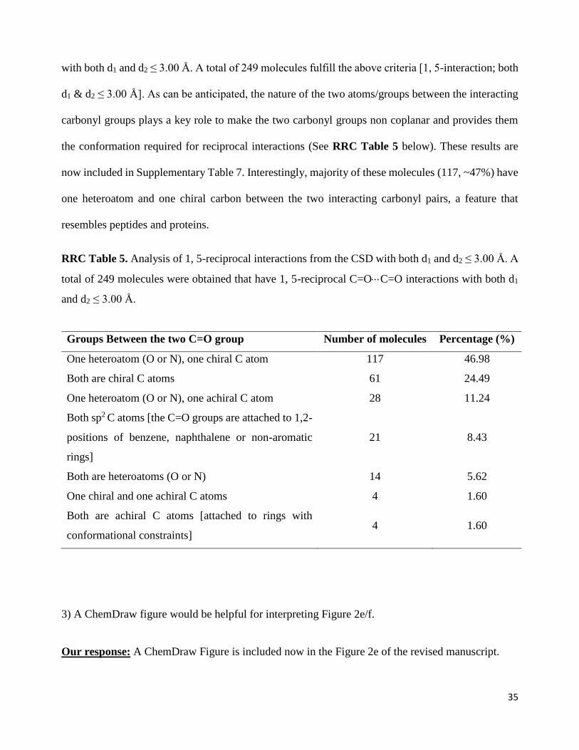
2) I think that it is important to note that many of the CSD hits, at least as described by Extended
Figure 3c, are very conformationally constrained, either by additional rings, highly substituted
centers, or stereoelectronic effects. This makes it all the more remarkable that reciprocal n→π*
interactions are observed in the much less-constrained protein backbone.
3) A ChemDraw figure would be helpful for interpreting Figure 2e/f.
4) The authors have not commented on the types of small molecules that form these reciprocal
n→π* interactions, which might reveal some interesting trends.
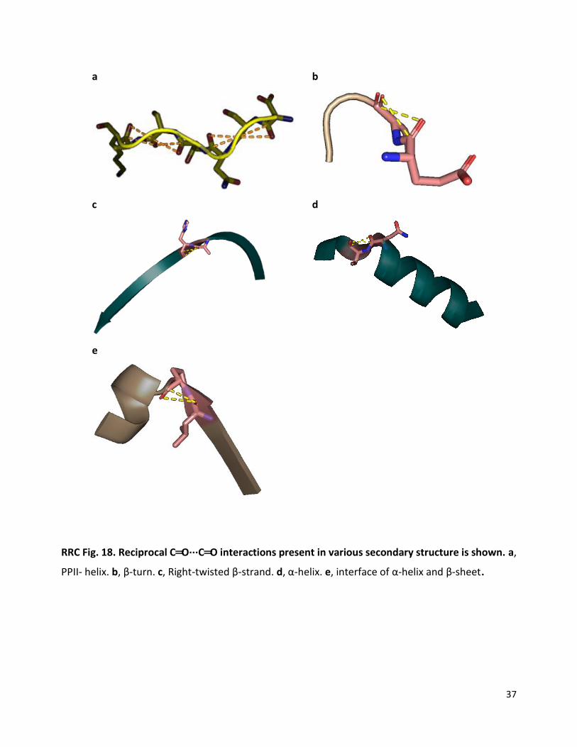
5) It would be helpful for the authors to show illustrative examples of the interactions they find in
proteins, particularly in different secondary structure contexts. For example, the authors note that
“α-helices that have two-sided n→π* interactions are distorted, while the β-sheets having two
sided n→π* interactions are twisted.” It would be very helpful for readers to be able to visualize
these distortions, as they could have important consequences for protein structure. Such examples
would, in my opinion, be more informative than the examples shown in Extended Figure 4.
1
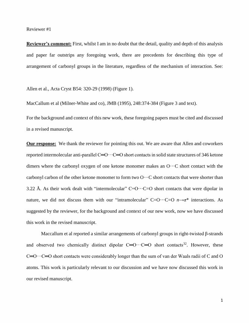
Reviewer #1
Reviewer’s comment: First, whilst I am in no doubt that the detail, quality and depth of this analysis
and paper far outstrips any foregoing work, there are precedents for describing this type of
arrangement of carbonyl groups in the literature, regardless of the mechanism of interaction. See:
Allen et al., Acta Cryst B54: 320-29 (1998) (Figure 1).
MacCallum et al (Milner-White and co), JMB (1995), 248:374-384 (Figure 3 and text).
For the background and context of this new work, these foregoing papers must be cited and discussed
in a revised manuscript.
Our response: We thank the reviewer for pointing this out. We are aware that Allen and coworkers
reported intermolecular anti-parallel C═O···C═O short contacts in solid state structures of 346 ketone
dimers where the carbonyl oxygen of one ketone monomer makes an O···C short contact with the
carbonyl carbon of the other ketone monomer to form two O···C short contacts that were shorter than
3.22 Å. As their work dealt with “intermolecular” C=O···C=O short contacts that were dipolar in
nature, we did not discuss them with our “intramolecular” C=O···C=O n→π* interactions. As
suggested by the reviewer, for the background and context of our new work, now we have discussed
this work in the revised manuscript.
Maccallum et al reported a similar arrangements of carbonyl groups in right-twisted β-strands
and observed two chemically distinct dipolar C═O···C═O short contacts32. However, these
C═O···C═O short contacts were considerably longer than the sum of van der Waals radii of C and O
atoms. This work is particularly relevant to our discussion and we have now discussed this work in
our revised manuscript.
2
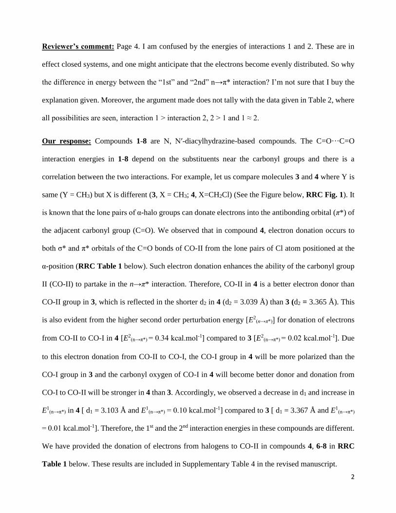
Reviewer’s comment: Page 4. I am confused by the energies of interactions 1 and 2. These are in
effect closed systems, and one might anticipate that the electrons become evenly distributed. So why
the difference in energy between the “1st” and “2nd” n→π* interaction? I’m not sure that I buy the
explanation given. Moreover, the argument made does not tally with the data given in Table 2, where
all possibilities are seen, interaction 1 > interaction 2, 2 > 1 and 1 ≈ 2.
Our response: Compounds 1-8 are N, Nʹ-diacylhydrazine-based compounds. The C=O···C=O
interaction energies in 1-8 depend on the substituents near the carbonyl groups and there is a
correlation between the two interactions. For example, let us compare molecules 3 and 4 where Y is
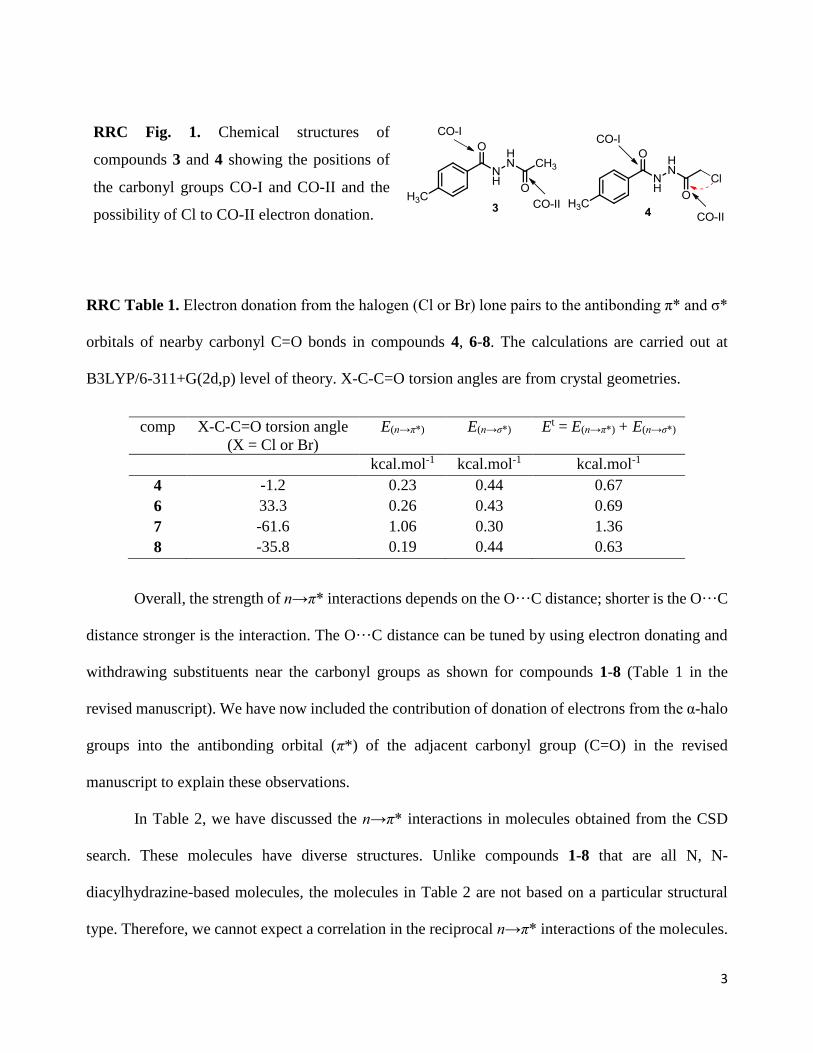
same (Y = CH3) but X is different (3, X = CH3; 4, X=CH2Cl) (See the Figure below, RRC Fig. 1). It
is known that the lone pairs of α-halo groups can donate electrons into the antibonding orbital (π*) of
the adjacent carbonyl group (C=O). We observed that in compound 4, electron donation occurs to
both σ* and π* orbitals of the C=O bonds of CO-II from the lone pairs of Cl atom positioned at the
α-position (RRC Table 1 below). Such electron donation enhances the ability of the carbonyl group
II (CO-II) to partake in the n→π* interaction. Therefore, CO-II in 4 is a better electron donor than
CO-II group in 3, which is reflected in the shorter d2 in 4 (d2 = 3.039 Å) than 3 (d2 = 3.365 Å). This
is also evident from the higher second order perturbation energy [E2(n→π*)] for donation of electrons
from CO-II to CO-I in 4 [E2(n→π*) = 0.34 kcal.mol-1] compared to 3 [E2
(n→π*) = 0.02 kcal.mol-1]. Due
to this electron donation from CO-II to CO-I, the CO-I group in 4 will be more polarized than the
CO-I group in 3 and the carbonyl oxygen of CO-I in 4 will become better donor and donation from
CO-I to CO-II will be stronger in 4 than 3. Accordingly, we observed a decrease in d1 and increase in
E1(n→π*) in 4 [ d1 = 3.103 Å and E1
(n→π*) = 0.10 kcal.mol-1] compared to 3 [ d1 = 3.367 Å and E1(n→π*)
= 0.01 kcal.mol-1]. Therefore, the 1st and the 2nd interaction energies in these compounds are different.
We have provided the donation of electrons from halogens to CO-II in compounds 4, 6-8 in RRC
Table 1 below. These results are included in Supplementary Table 4 in the revised manuscript.
3
RRC Fig. 1. Chemical structures of
compounds 3 and 4 showing the positions of
the carbonyl groups CO-I and CO-II and the
possibility of Cl to CO-II electron donation.
RRC Table 1. Electron donation from the halogen (Cl or Br) lone pairs to the antibonding π* and σ*
orbitals of nearby carbonyl C=O bonds in compounds 4, 6-8. The calculations are carried out at
B3LYP/6-311+G(2d,p) level of theory. X-C-C=O torsion angles are from crystal geometries.
comp X-C-C=O torsion angle
(X = Cl or Br)
E(n→π*)
E(n→σ*)
Et = E(n→π*) + E(n→σ*)
kcal.mol-1 kcal.mol-1 kcal.mol-1
4 -1.2 0.23 0.44 0.67
6 33.3 0.26 0.43 0.69
7 -61.6 1.06 0.30 1.36
8 -35.8 0.19 0.44 0.63
Overall, the strength of n→π* interactions depends on the O···C distance; shorter is the O···C
distance stronger is the interaction. The O···C distance can be tuned by using electron donating and
withdrawing substituents near the carbonyl groups as shown for compounds 1-8 (Table 1 in the
revised manuscript). We have now included the contribution of donation of electrons from the α-halo
groups into the antibonding orbital (π*) of the adjacent carbonyl group (C=O) in the revised
manuscript to explain these observations.
In Table 2, we have discussed the n→π* interactions in molecules obtained from the CSD
search. These molecules have diverse structures. Unlike compounds 1-8 that are all N, N-
diacylhydrazine-based molecules, the molecules in Table 2 are not based on a particular structural
type. Therefore, we cannot expect a correlation in the reciprocal n→π* interactions of the molecules.

4
Moreover, we have randomly chosen the two carbonyl groups as CO-I and CO-II in these molecules
in Table 2. Only correlation we can expect here is an increase in the interaction energy [E1(n→π*) or
E2(n→π*)] with a decrease in O···C distance (d1 or d2).
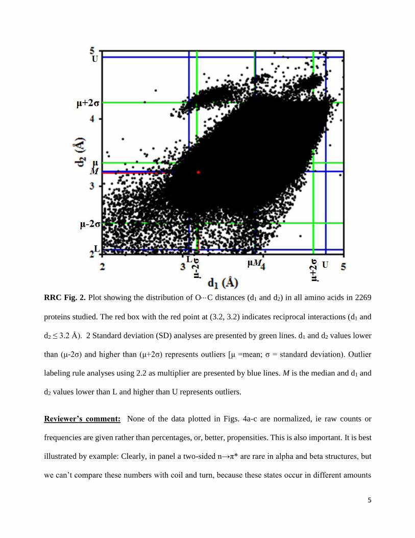
Reviewer’s comment: Figures 2 and 3. The distances d1 and d2 are described and shown for the
systems that test positive for the two-sided n→π* only. But what is the distribution of these distances
in unconstrained systems. (I realise that this probably only has meaning in the context of i,i+1 residues
in proteins.) This is important to see if the short distances are really outliers in the full distributions
or not.
Our Response: We agree with the reviewer that the distribution of unconstrained distances d1 and d2
can give us information regarding whether the short distances are outliers in the full distributions or
not. As pointed out by the reviewer, such a distribution would be meaningful for proteins as the
carbonyl groups of ith and (i+1)th residues in proteins are always separated by three covalent bonds.
For comparison, we have now plotted the distribution of distances d1 and d2 for all amino acid residues
in the 2184 proteins studied in this work (see RRC Fig. 2 below). To find out if the shorter distances
(d1 and d2 ≤ 3.2 Å) are outliers in the overall distribution, we used two different methods; two standard
deviation (SD) method and outlier labeling rule method [Hoaglin, D. C. & Iglewicz, B. Fine tuning
some resistant rules for outlier labeling, J. Am. Stat. Assoc. 82, 1147-1149 (1987)]. Both methods
suggest that short distances d1 & d2 ≤ 3.0 Å are not completely outliers in the overall distribution.
They may be considered as tail of short distances in the full distribution.
5
RRC Fig. 2. Plot showing the distribution of OC distances (d1 and d2) in all amino acids in 2269
proteins studied. The red box with the red point at (3.2, 3.2) indicates reciprocal interactions (d1 and
d2 ≤ 3.2 Å). 2 Standard deviation (SD) analyses are presented by green lines. d1 and d2 values lower
than (μ-2σ) and higher than (μ+2σ) represents outliers [μ =mean; σ = standard deviation). Outlier
labeling rule analyses using 2.2 as multiplier are presented by blue lines. M is the median and d1 and
d2 values lower than L and higher than U represents outliers.
Reviewer’s comment: None of the data plotted in Figs. 4a-c are normalized, ie raw counts or
frequencies are given rather than percentages, or, better, propensities. This is also important. It is best
illustrated by example: Clearly, in panel a two-sided n→π* are rare in alpha and beta structures, but
we can’t compare these numbers with coil and turn, because these states occur in different amounts
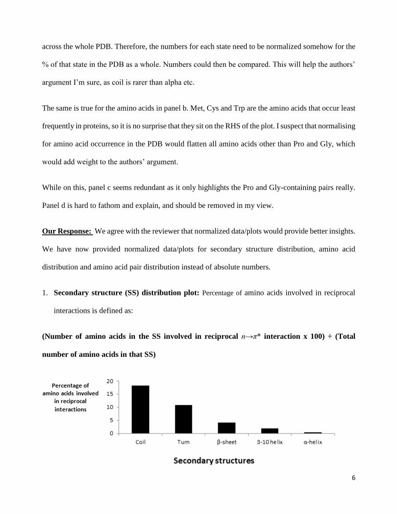
6
across the whole PDB. Therefore, the numbers for each state need to be normalized somehow for the
% of that state in the PDB as a whole. Numbers could then be compared. This will help the authors’
argument I’m sure, as coil is rarer than alpha etc.
The same is true for the amino acids in panel b. Met, Cys and Trp are the amino acids that occur least
frequently in proteins, so it is no surprise that they sit on the RHS of the plot. I suspect that normalising
for amino acid occurrence in the PDB would flatten all amino acids other than Pro and Gly, which
would add weight to the authors’ argument.
While on this, panel c seems redundant as it only highlights the Pro and Gly-containing pairs really.
Panel d is hard to fathom and explain, and should be removed in my view.
Our Response: We agree with the reviewer that normalized data/plots would provide better insights.
We have now provided normalized data/plots for secondary structure distribution, amino acid
distribution and amino acid pair distribution instead of absolute numbers.
1. Secondary structure (SS) distribution plot: Percentage of amino acids involved in reciprocal
interactions is defined as:
(Number of amino acids in the SS involved in reciprocal n→π* interaction x 100) ÷ (Total
number of amino acids in that SS)
7
RRC Fig. 3. Plot of percentage of amino acids involved in reciprocal interactions in various
secondary structures.
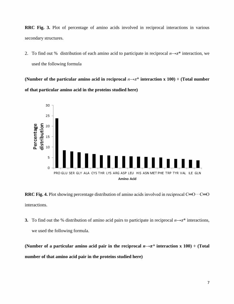
2. To find out % distribution of each amino acid to participate in reciprocal n→π* interaction, we
used the following formula
(Number of the particular amino acid in reciprocal n→π* interaction x 100) ÷ (Total number
of that particular amino acid in the proteins studied here)
RRC Fig. 4. Plot showing percentage distribution of amino acids involved in reciprocal C═O···C═O
interactions.
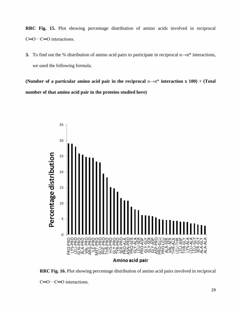
3. To find out the % distribution of amino acid pairs to participate in reciprocal n→π* interactions,
we used the following formula.
(Number of a particular amino acid pair in the reciprocal n→π* interaction x 100) ÷ (Total
number of that amino acid pair in the proteins studied here)
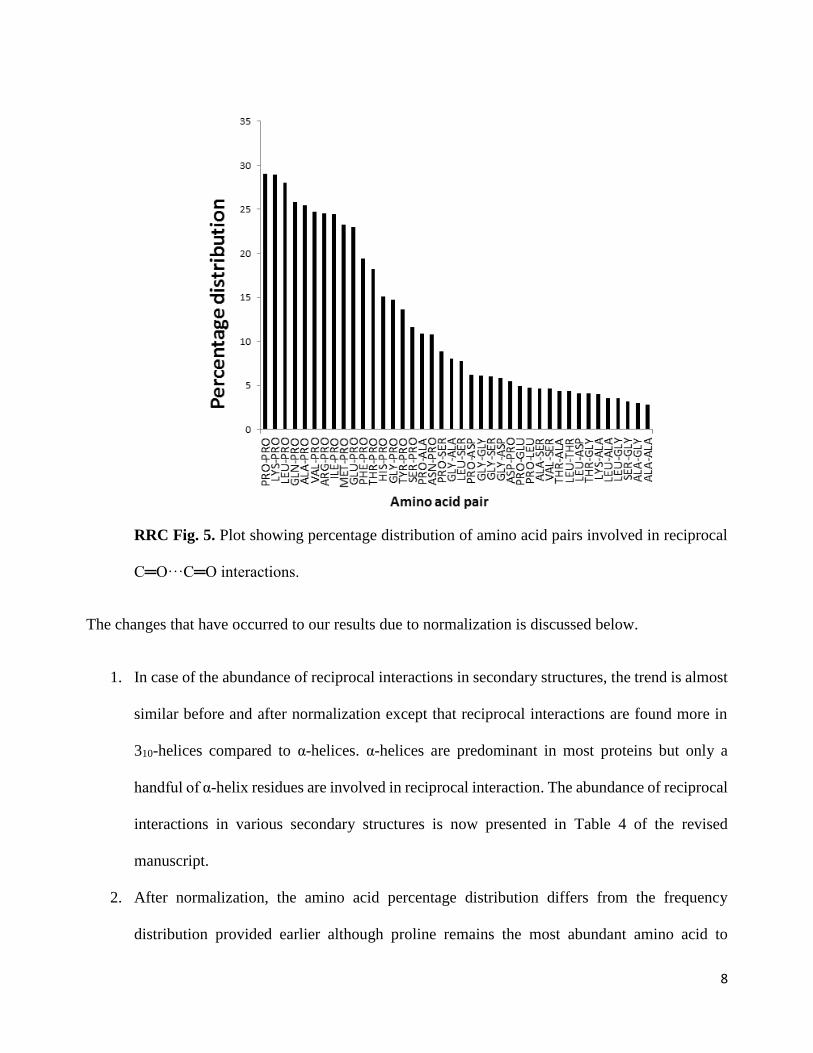
8
RRC Fig. 5. Plot showing percentage distribution of amino acid pairs involved in reciprocal
C═O···C═O interactions.
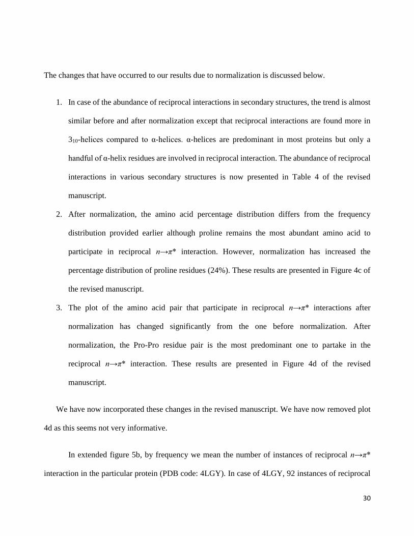
The changes that have occurred to our results due to normalization is discussed below.
1. In case of the abundance of reciprocal interactions in secondary structures, the trend is almost
similar before and after normalization except that reciprocal interactions are found more in
310-helices compared to α-helices. α-helices are predominant in most proteins but only a
handful of α-helix residues are involved in reciprocal interaction. The abundance of reciprocal
interactions in various secondary structures is now presented in Table 4 of the revised
manuscript.
2. After normalization, the amino acid percentage distribution differs from the frequency
distribution provided earlier although proline remains the most abundant amino acid to
9
participate in reciprocal n→π* interaction. However, normalization has increased the
percentage distribution of proline residues (24%). These results are presented in Figure 4c of
the revised manuscript.
3. The plot of the amino acid pair that participate in reciprocal n→π* interactions after
normalization has changed significantly from the one before normalization. After
normalization, the Pro-Pro residue pair is the most predominant one to partake in the
reciprocal n→π* interaction. These results are presented in Figure 4d of the revised
manuscript.
We have now incorporated these changes in the revised manuscript. As suggested by reviewer,
we have also removed plot 4d.
Reviewer’s comment: While on Pro, have the authors considered cis/trans forms and how these
differ in propensity to form two-sided n→π* interactions?
Our Response: It is known that n→π* interaction is possible only if the proline residue exists in
trans conformation. Based on this observation, the ratio of isomers (Ktrans/cis) is used to report the
energy of n→π* interaction using NMR spectroscopy [J. Am. Chem. Soc. 135, 7843-7846 (2013)].
Therefore, we assumed that the conformation of Pro amide bonds involved in reciprocal interactions
would also be trans. However, after this query from the reviewer, we looked into the conformation
of the amide bond (C-N-C=O dihedral angle) in Pro in Leu-Pro and Lys-Pro amino acid pairs (two
among the most abundant amino acid pairs to participate in reciprocal interactions) and observed that
the Pro conformations in the residues involved in reciprocal interactions are trans. Please see the files
ROM-1-Leu-Pro and ROM-2-Lys-Pro provided as review only material.
10
Reviewer’s comment: *I mentioned my preference for “reciprocated n→π* interactions” as I find
“two-sided n→π* interactions” confusing. The authors may wish to consider this alternative name.
Our response: We thank the reviewer for this suggestion. We have now modified the name of the
interaction to “reciprocal interaction” and title of the manuscript is now changed to “Reciprocal
carbonyl-carbonyl interactions in small molecules and proteins.”
Reviewer’s comment: Finally, I am not taken by the arguments that these could influence folding.
Even at ¼ kcal/mol for each n→π* interaction these would perturb equilibria between states only
slightly. This is before solvent is considered. Surely, water competes well for solvation of carbonyl
groups in a flexible unfolded state? I would recommend that this section is removed or trimmed at
least. In other words, as the focus of the analysis and results is on equilibrium structures, discussion
of kinetic processes might best be left out of this paper.
Our response: We agree with the reviewer that mixing kinetics and thermodynamic is not a good
idea. However, as these interactions are most abundant in polyproline II helices that are the most
abundant secondary structure in unfolded proteins, gives rise to the possibility of these interactions
playing some role in protein folding. Also, turn regions that are stabilized by reciprocal interactions
are known to act as nucleation sites for protein folding. Therefore, an open question is how important
such reciprocal interactions might be for protein folding. We have now modified the discussion
regarding protein folding both in the revised manuscript and the abstract.
11
Reviewer #2
Reviewer’s comment: The authors used X-ray coordinates of the small molecules as input for the
electronic structure calculations. Although the resolution of the structures may be high (I could not
find the resolution in Tables 1 and 2 nor in the text), it is common practice to relax molecular
conformations during electronic structure calculations before providing final coordinates and
energies. In the present case, with the twisted side chain conformations, it should be okay to keep the
involved dihedral angles of the side chains frozen to their X-ray values.
However, the authors need to demonstrate – at least for their sequence of hydrazine derivatives
in Fig. 1c – that geometry optimization of the remaining degrees of freedom (bond lengths, angles,
and dihedrals) has a negligible effect on the overlap of the computed n/pi* orbitals and on the NBO
energies in comparison to calculations done on the unrelaxed X-ray coordinates.
Our response: The resolution of the X-ray crystal structures of the compound 1-8 were high (See
RRC Table 1 below). We have now included this information in the Supplementary Table 2 of the
revised manuscript. We agree with the reviewer that providing optimized geometry and electronic
energy calculation on optimized geometry is the common practice. However, as we have discussed
in the manuscript, the donor and acceptor abilities of the carbonyl groups in compounds 1-8 are
dependent on the substituents attached to the carbonyl groups and their geometrical arrangements.
For example, the halogen atoms present in the substituent X in compounds 4, 6-8 highly influence
the donor ability of the nearby carbonyl group (CO-II) (See RRC Fig. 6 below). A change in the
orientation of the halogen with respect to the CO-II group would change the donor ability of the CO-
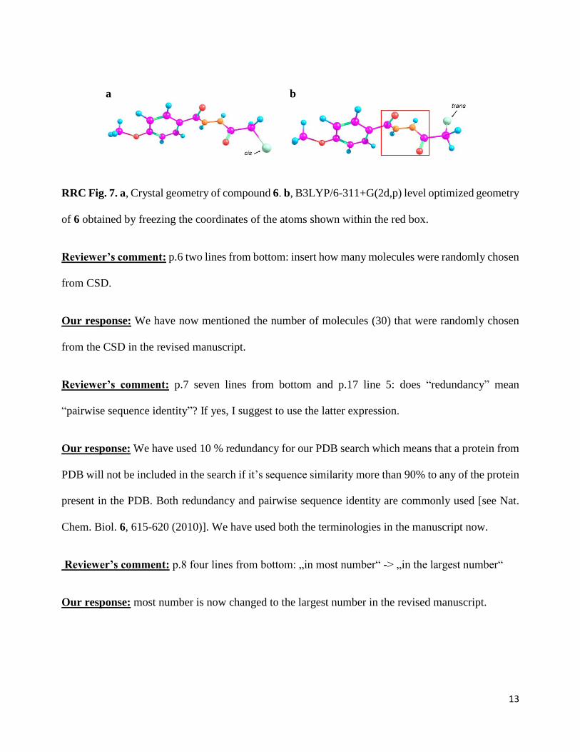
II group. We carried out optimization of compound 6 by freezing the coordinates of the atoms shown
within the box (see the RHS ball and stick RRC Fig. 7b below). However, during optimization the
chlorine (Cl) atom moved to an anti-periplanar geometry (trans) from an almost syn-periplanar
12
geometry (cis) with respect to the oxygen atom of the nearby carbonyl group (CO-II). In fact, when
a completely relaxed geometry optimization of 6 was done at B3LYP/6-311+G(2d,p) level of theory,
the Cl atom moved to an anti-periplanar position and the geometry of 6 became almost planar without
any reciprocal interaction. In such a scenario, one would have to come up with the best theoretical
method and basis set to study these interaction, which itself would be a separate project for
investigation. As this study deals with the possibility of reciprocal interactions in the crystal
geometries of small molecules and proteins, to avoid such deviation from crystal geometries, we
avoided free optimization the molecules.
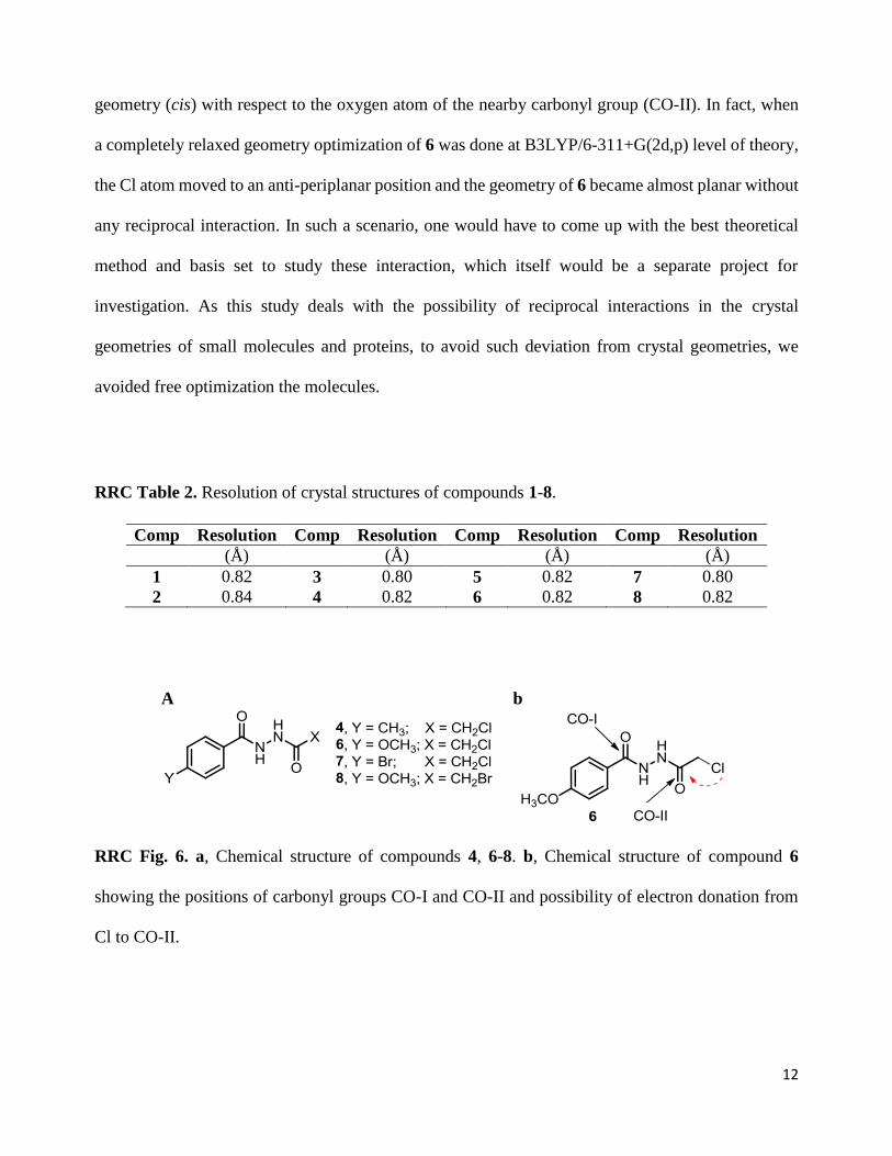
RRC Table 2. Resolution of crystal structures of compounds 1-8.
Comp Resolution Comp Resolution Comp Resolution Comp Resolution
(Å) (Å) (Å) (Å)
1 0.82 3 0.80 5 0.82 7 0.80
2 0.84 4 0.82 6 0.82 8 0.82
A b
RRC Fig. 6. a, Chemical structure of compounds 4, 6-8. b, Chemical structure of compound 6
showing the positions of carbonyl groups CO-I and CO-II and possibility of electron donation from
Cl to CO-II.
13
a b
RRC Fig. 7. a, Crystal geometry of compound 6. b, B3LYP/6-311+G(2d,p) level optimized geometry
of 6 obtained by freezing the coordinates of the atoms shown within the red box.
Reviewer’s comment: p.6 two lines from bottom: insert how many molecules were randomly chosen
from CSD.
Our response: We have now mentioned the number of molecules (30) that were randomly chosen
from the CSD in the revised manuscript.
Reviewer’s comment: p.7 seven lines from bottom and p.17 line 5: does “redundancy” mean
“pairwise sequence identity”? If yes, I suggest to use the latter expression.
Our response: We have used 10 % redundancy for our PDB search which means that a protein from
PDB will not be included in the search if it’s sequence similarity more than 90% to any of the protein
present in the PDB. Both redundancy and pairwise sequence identity are commonly used [see Nat.
Chem. Biol. 6, 615-620 (2010)]. We have used both the terminologies in the manuscript now.
Reviewer’s comment: p.8 four lines from bottom: „in most number“ -> „in the largest number“
Our response: most number is now changed to the largest number in the revised manuscript.

14
Reviewer’s comment: p.10 lines 7/8: “providing an answer to Levinthal’s paradox” should be
replaced by something like “contribute to answering Levinthal’s paradox in addition to other concepts
such as the free energy funnel model for protein folding (cite Onuchic, Wolynes, Proteins 1995)”.
Our response: This section is now modified. Please see the revised manuscript.
15
Reviewer #3
Reviewer’s comment:
The energy of these interactions in proteins is unclear. There are relatively few calculations performed
on protein geometries, and those limited calculations sample a relatively small conformational space.
In particular, the reported examples have angles of approach between 70° and 90°, whereas the data
presented in Figure 3b suggest that many interactions have larger angles of approach. It is unclear,
therefore, if all of the interactions identified herein have significant energy. Do they have energies
similar to other n→π* interactions? Or does the distorted geometry required for back donation affect
the energy?
Because exhaustive calculations of n→π* energies have been performed previously (see
Bartlett et al. 2010), it should not be necessary to calculate energies for all of the observed geometries.
Rather, calculations on a more representative subset should allow readers compare the energy of these
newly identified interactions with the energy of previously reported n→π* interactions. It would also
be helpful if energy data for examples in proteins were incorporated into the main article, as opposed
to residing in the supplement.
Our response: The strength of n→π* interactions depends on the O···C distance. As the O···C
distance increases, the stabilization due individual n→π* interactions decreases. This can be seen
from Figure 2c-2d of the revised manuscript for the molecules obtained from the CSD search where
the distances d1 and d2 increase as the angles θ1 and θ2 deviate from the Bürgi-Dunitz trajectory.
To confirm that reciprocal n→π* interaction is present when the angle of approach is greater
than 90°, we have now performed additional NBO calculations on representative subsets that cover
the complete range of observed O···C=O angles (70-110º) and observed that reciprocal n→π*
interaction is present even the angle of approach is greater than 90 °. The calculations were done for
16
amino acid pairs extracted from the PDB as well as small molecules obtained from the CSD that have
reciprocal interactions. We observed substantial C═O···C═O π→π* interactions in molecules having
θ1 and θ2 values > 90º (Supplementary Table 6 and Supplementary Table 8) for both CSD molecules
and amino acid pairs taken from PDB. In some cases, π→π* interactions are even stronger than n→π*
interactions. When θ1 and θ2 values were < 90º, π→π* interactions were observed for molecules
having relatively stronger n→π* interactions (both d1 and d2 < 2.90 Å). This indicates that although
the individual n→π*interaction in reciprocal interaction is weak, the sum of the two n→π*
interactions together with two π→π* interactions provide substantial stabilization to small molecule
and proteins. Based on NBO calculations at B3LYP/6-311+G(2d,p) level, we observed 0.11-3.37
kcal.mol-1 (with an average 0.98 kcal.mol-1) stabilization of small molecules from reciprocal
C═O···C═O interactions (See the last column in Supplementary Table 6). Similarly, we observed
that reciprocal C═O···C═O interactions contribute 0.27-4.41 kcal.mol-1 (with an average value 1.34
kcal.mol-1) stabilization to proteins per each amino acid pair (See the last column in Supplementary
Table 8). These results are now discussed in the revised manuscript (See Table 2 and Table 3) and
also incorporated in the Supplementary Table 6 and Supplementary Table 8.
Reviewer’s comment: The most critical signature of the n→π* interaction is the pyramidalization
induced upon the acceptor carbonyl group, as the authors note in their introduction. What is the degree
of pyramidalization in the synthetic diacyl hydrazines? Importantly, is pyramidalization observed in
both carbonyl groups? This would lend strong credence to the existence of these interactions, and the
data should already be available from the crystal structures. Pyramidalization data should also be
available for CSD structures, though a certain amount of noise is to be expected (see Kamer, et al.
2013). Again, significant pyramidalization of both carbonyl groups participating in the interaction
would greatly strengthen the authors’ claims.
17
Our response: We agree that the pyramidality of the acceptor carbonyl carbon atom measured by
parameters Δ and Θ is another important signature of n→π* interactions. Positive values of Δ and Θ
indicate pyramidalization of the acceptor carbonyl carbon towards the donor oxygen atom whereas
negative values of Δ and Θ indicate pyramidalization of the acceptor carbon away from the donor
oxygen atom. We have now tabulated the Δ and Θ values of compounds 1-8 in the Table 1 of the
revised manuscript. In compounds 1-8, however, we have not observed a correlation of pyramidality
(Θ) with O···C distance and the strength of n→π* interactions. One reason for this could be the
stronger donation from the α-halogen atoms to the nearby carbonyl, which would force the acceptor
carbonyl carbons towards the halogens atoms away from the donor oxygen atoms. Also, the crystal
packing forces may have some influence in the observed geometries and the pyramidalization of the
two nitrogen atoms between the carbonyl groups may influence the pyramidalization of the acceptor
carbonyl carbons. Moreover, the individual n→π* interactions in compounds 1-8 may not be strong
enough to exert a significant effect on pyramidalization of the carbonyl carbons.
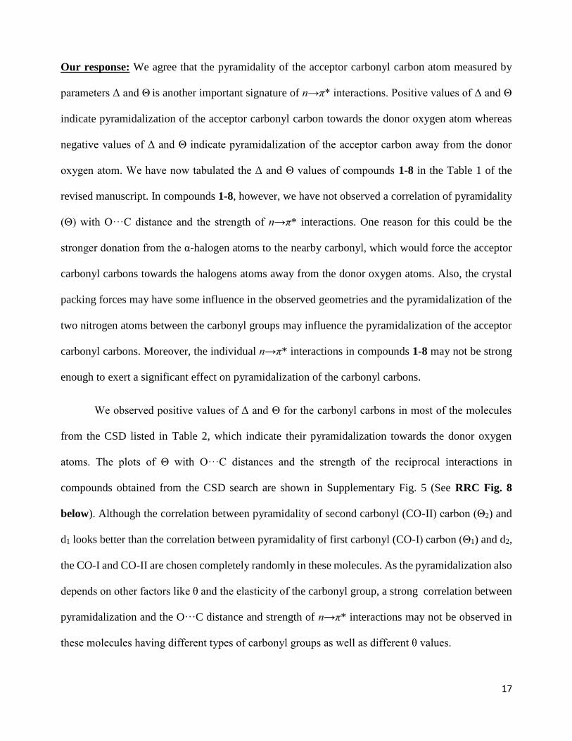
We observed positive values of Δ and Θ for the carbonyl carbons in most of the molecules
from the CSD listed in Table 2, which indicate their pyramidalization towards the donor oxygen
atoms. The plots of Θ with O···C distances and the strength of the reciprocal interactions in
compounds obtained from the CSD search are shown in Supplementary Fig. 5 (See RRC Fig. 8
below). Although the correlation between pyramidality of second carbonyl (CO-II) carbon (Θ2) and
d1 looks better than the correlation between pyramidality of first carbonyl (CO-I) carbon (Θ1) and d2,
the CO-I and CO-II are chosen completely randomly in these molecules. As the pyramidalization also
depends on other factors like θ and the elasticity of the carbonyl group, a strong correlation between
pyramidalization and the O···C distance and strength of n→π* interactions may not be observed in
these molecules having different types of carbonyl groups as well as different θ values.
18
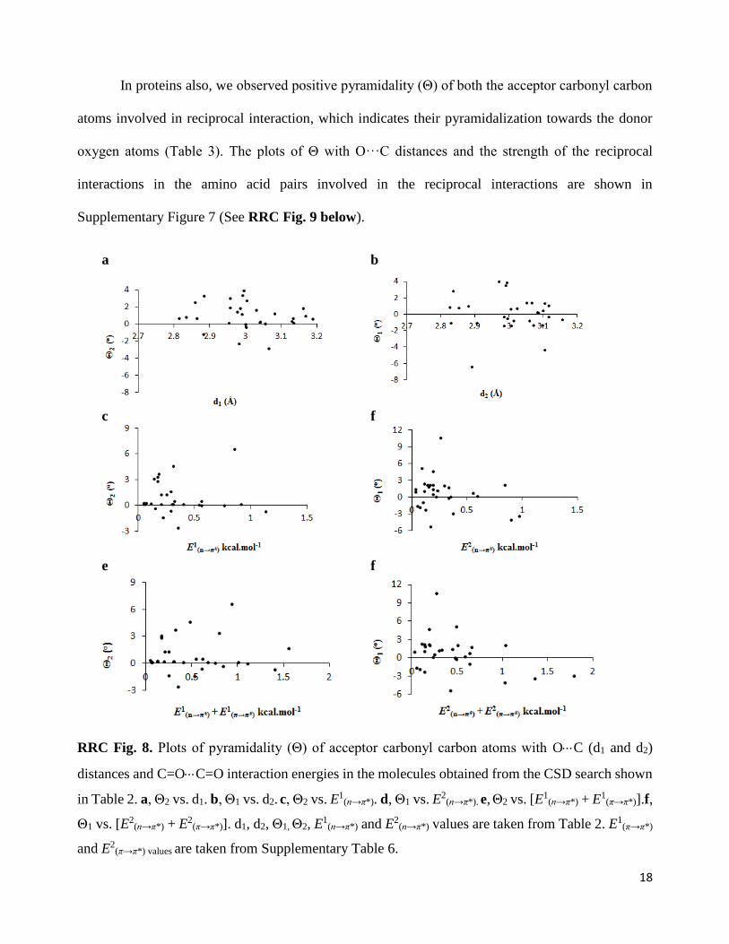
In proteins also, we observed positive pyramidality (Θ) of both the acceptor carbonyl carbon
atoms involved in reciprocal interaction, which indicates their pyramidalization towards the donor
oxygen atoms (Table 3). The plots of Θ with O···C distances and the strength of the reciprocal
interactions in the amino acid pairs involved in the reciprocal interactions are shown in
Supplementary Figure 7 (See RRC Fig. 9 below).
a b
c f
e f
RRC Fig. 8. Plots of pyramidality (Θ) of acceptor carbonyl carbon atoms with OC (d1 and d2)
distances and C=OC=O interaction energies in the molecules obtained from the CSD search shown
in Table 2. a, Θ2 vs. d1. b, Θ1 vs. d2. c, Θ2 vs. E1(n→π*). d, Θ1 vs. E2
(n→π*). e, Θ2 vs. [E1(n→π*) + E1
(π→π*)].f,
Θ1 vs. [E2(n→π*) + E2
(π→π*)]. d1, d2, Θ1, Θ2, E1
(n→π*) and E2(n→π*) values are taken from Table 2. E1
(π→π*)
and E2(π→π*) values are taken from Supplementary Table 6.
19
a
b
c d
e f
RRC Fig. 9. Plots of pyramidality (Θ) of acceptor carbonyl carbon atoms with OC (d1 and d2)
distances and C=OC=O interaction energies in the amino acid pairs shown in Table 3. a, Θ2 vs. d1.
b, Θ1 vs. d2. c, Θ2 vs. E1(n→π*). d, Θ1 vs. E2
(n→π*). e, Θ2 vs. [E1(n→π*) + E1
(π→π*)].f, Θ1 vs. [E2(n→π*) +
E2(π→π*)]. d1, d2, Θ1, Θ2, E
1(n→π*) and E2
(n→π*) values are taken from Table 3. E1(π→π*) and E2
(π→π*) values
are taken from Supplementary Table 8.
20
Reviewer’s comment: The authors claim that d2 distances in compounds 6-8 are shorter than d1
because electronic donation from the halogen lone pairs to the carbonyl π* orbital make the carbonyl
a stronger donor. However, no evidence is presented that such donation occurs. In fact, in all of the
presented crystal structures, the halogen is proximal (cis) to the carbonyl oxygen, rather than the
carbonyl carbon. Should halogen electron density be transferred to the carbonyl group, it should
almost certainly occur through the carbonyl carbon, not oxygen. In addition, if such donation were to
occur, it should be more apparent in 8 than in 6, due to the higher polarizability of bromine. It is
therefore unlikely that the cause of d2 constriction in compounds 6-8 is donation of electron density
from the proximal halogen. The fact that d2 is shorter than d1 in these cases is likely due to other
effects. Perhaps it is the case that, even in the absence of any back donation, enhancement of a single
n→π* interaction would decrease both distances.
Our response: We have now provided evidence for electron donation from the α-halogen lone pairs
to carbonyl group’s σ* and π* orbitals (see RRC Table 3 below). These results are included in
supplementary Table 4 in the revised manuscript. We agree with the reviewer that a halogen atom
anti-periplanar (trans) to the carbonyl oxygen would donate electrons much better to the carbonyl
group than when it is in syn-periplanar (cis). As can be seen from the X-C-C=O torsion angles in the
RRC Table 3 below, the halogen atoms in the crystal geometries are not exactly cis to the carbonyl
group except in compound 4. There is substantial electron donation from the halogen atoms to the σ*
and π* orbitals of the C=O group, which should increase the donor ability of the carbonyl group CO-
II.
As can be seen from the Table below, 6 and 8 have similar X-C-C=O torsion angles but
stabilization due to donation from Br lone pairs to CO-II in 8 is less than the stabilization due to
donation from Cl lone pairs to CO-II in 6 even though Br is more polarizable. This expected as the
21
size of Br orbitals will be much higher than C and O orbitals that will hamper efficient delocalization
between the Br lone pairs and the σ* and π* orbitals of C=O bond. Compared to Br, relatively smaller
Cl lone pair orbitals will have better overlap with σ* and π* orbitals of the C=O group. These
observations are now included in the revised paper and Supplementary Table 4. The chemical
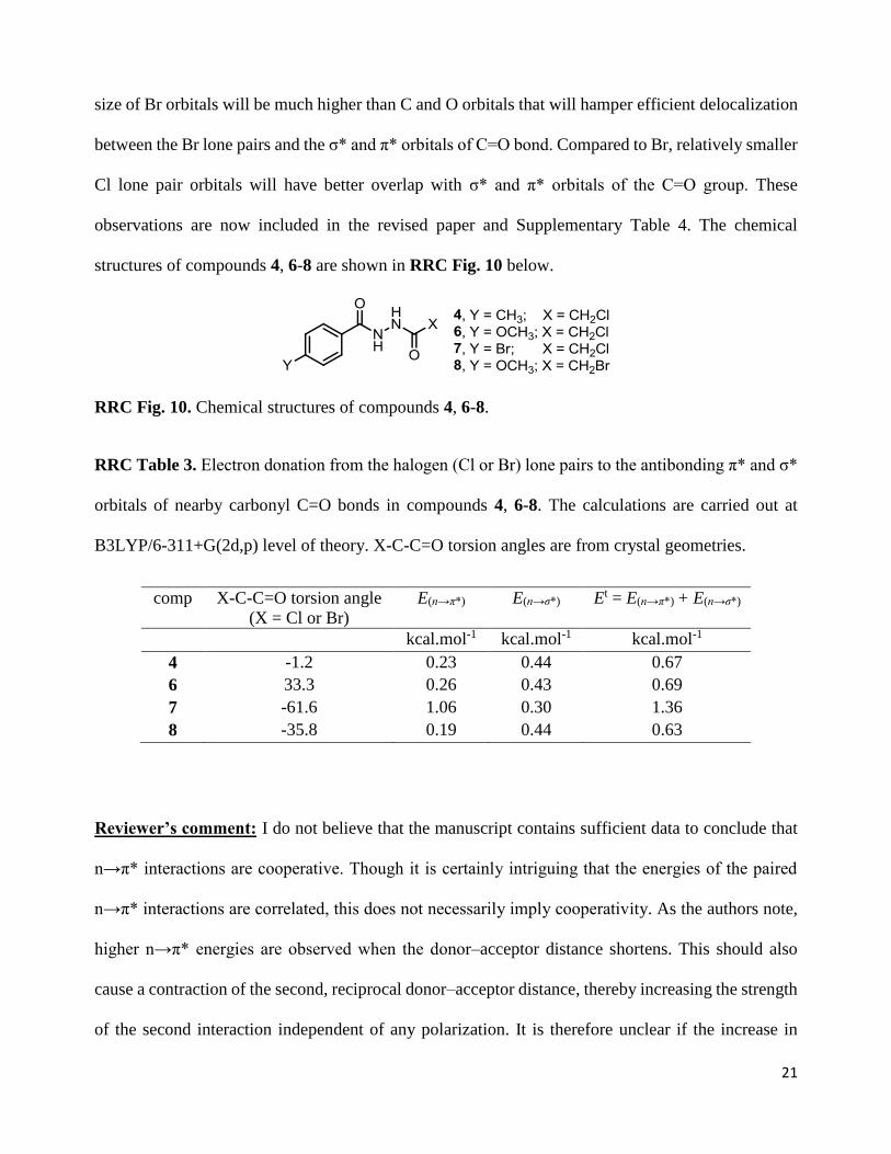
structures of compounds 4, 6-8 are shown in RRC Fig. 10 below.
RRC Fig. 10. Chemical structures of compounds 4, 6-8.
RRC Table 3. Electron donation from the halogen (Cl or Br) lone pairs to the antibonding π* and σ*
orbitals of nearby carbonyl C=O bonds in compounds 4, 6-8. The calculations are carried out at
B3LYP/6-311+G(2d,p) level of theory. X-C-C=O torsion angles are from crystal geometries.
comp X-C-C=O torsion angle
(X = Cl or Br)
E(n→π*)
E(n→σ*)
Et = E(n→π*) + E(n→σ*)
kcal.mol-1 kcal.mol-1 kcal.mol-1
4 -1.2 0.23 0.44 0.67
6 33.3 0.26 0.43 0.69
7 -61.6 1.06 0.30 1.36
8 -35.8 0.19 0.44 0.63
Reviewer’s comment: I do not believe that the manuscript contains sufficient data to conclude that
n→π* interactions are cooperative. Though it is certainly intriguing that the energies of the paired
n→π* interactions are correlated, this does not necessarily imply cooperativity. As the authors note,
higher n→π* energies are observed when the donor–acceptor distance shortens. This should also
cause a contraction of the second, reciprocal donor–acceptor distance, thereby increasing the strength
of the second interaction independent of any polarization. It is therefore unclear if the increase in
22
energy of the second interaction is due to polarization by the first interaction or if it merely results
from the particular geometries observed. Though the authors are clear that the results are only
suggestive of cooperativity, I feel that this point is too speculative given the data herein.
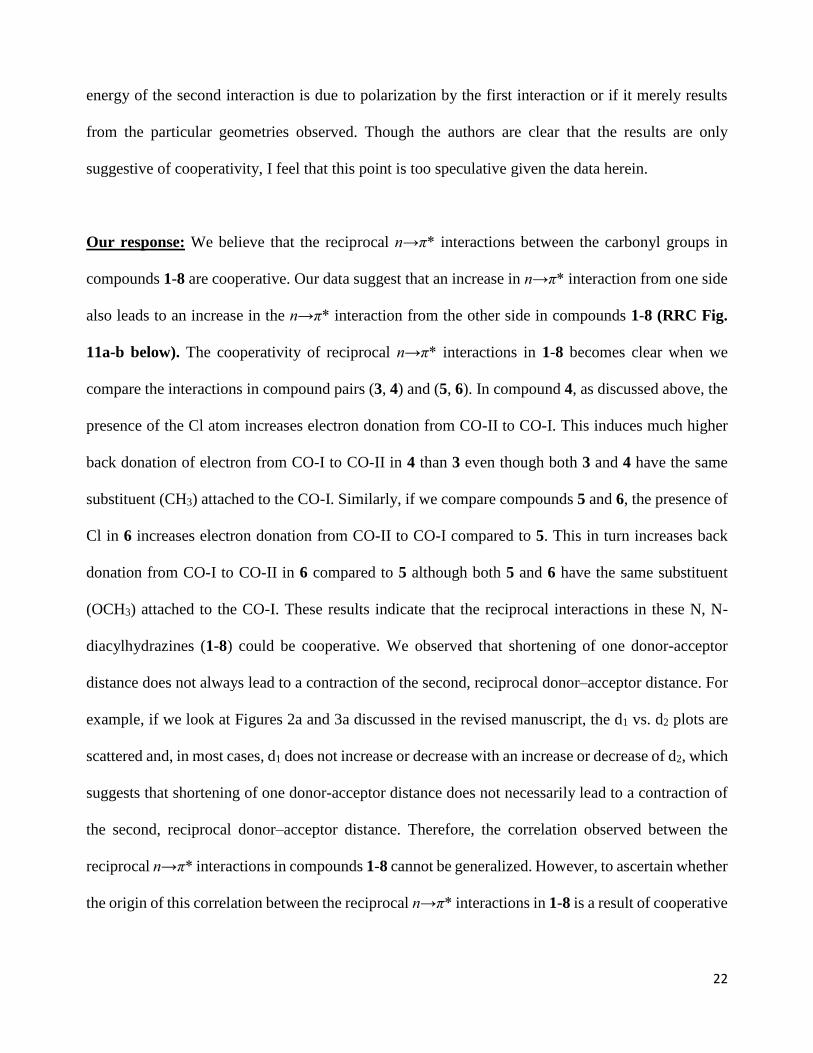
Our response: We believe that the reciprocal n→π* interactions between the carbonyl groups in
compounds 1-8 are cooperative. Our data suggest that an increase in n→π* interaction from one side
also leads to an increase in the n→π* interaction from the other side in compounds 1-8 (RRC Fig.
11a-b below). The cooperativity of reciprocal n→π* interactions in 1-8 becomes clear when we
compare the interactions in compound pairs (3, 4) and (5, 6). In compound 4, as discussed above, the
presence of the Cl atom increases electron donation from CO-II to CO-I. This induces much higher
back donation of electron from CO-I to CO-II in 4 than 3 even though both 3 and 4 have the same
substituent (CH3) attached to the CO-I. Similarly, if we compare compounds 5 and 6, the presence of
Cl in 6 increases electron donation from CO-II to CO-I compared to 5. This in turn increases back
donation from CO-I to CO-II in 6 compared to 5 although both 5 and 6 have the same substituent
(OCH3) attached to the CO-I. These results indicate that the reciprocal interactions in these N, N-
diacylhydrazines (1-8) could be cooperative. We observed that shortening of one donor-acceptor
distance does not always lead to a contraction of the second, reciprocal donor–acceptor distance. For
example, if we look at Figures 2a and 3a discussed in the revised manuscript, the d1 vs. d2 plots are
scattered and, in most cases, d1 does not increase or decrease with an increase or decrease of d2, which
suggests that shortening of one donor-acceptor distance does not necessarily lead to a contraction of
the second, reciprocal donor–acceptor distance. Therefore, the correlation observed between the
reciprocal n→π* interactions in compounds 1-8 cannot be generalized. However, to ascertain whether
the origin of this correlation between the reciprocal n→π* interactions in 1-8 is a result of cooperative
23
effects or merely a result of the particular geometries adopted by 1-8 needs further investigation. The
plots shown below (RRC Fig.11) are now included in Fig. 1g-h of the revised manuscript.
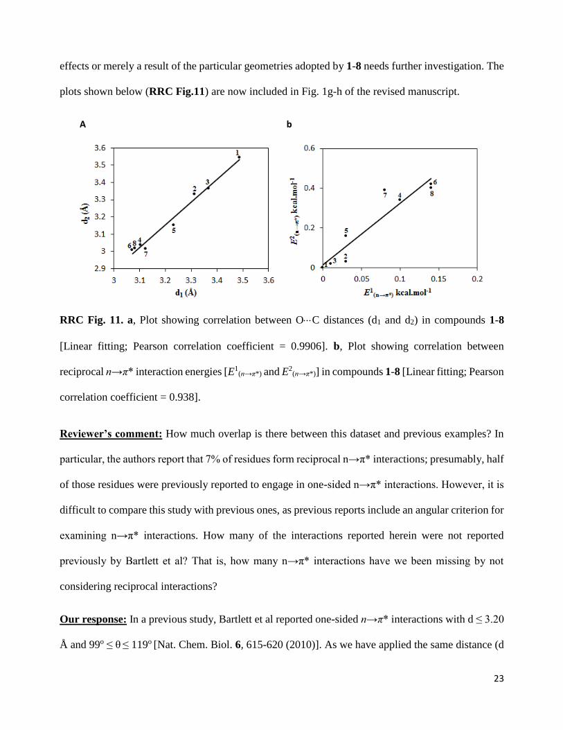
A b
RRC Fig. 11. a, Plot showing correlation between OC distances (d1 and d2) in compounds 1-8
[Linear fitting; Pearson correlation coefficient = 0.9906]. b, Plot showing correlation between
reciprocal n→π* interaction energies [E1(n→π*) and E2
(n→π*)] in compounds 1-8 [Linear fitting; Pearson
correlation coefficient = 0.938].
Reviewer’s comment: How much overlap is there between this dataset and previous examples? In
particular, the authors report that 7% of residues form reciprocal n→π* interactions; presumably, half
of those residues were previously reported to engage in one-sided n→π* interactions. However, it is
difficult to compare this study with previous ones, as previous reports include an angular criterion for
examining n→π* interactions. How many of the interactions reported herein were not reported
previously by Bartlett et al? That is, how many n→π* interactions have we been missing by not
considering reciprocal interactions?
Our response: In a previous study, Bartlett et al reported one-sided n→π* interactions with d ≤ 3.20
Å and 99o ≤ θ ≤ 119o [Nat. Chem. Biol. 6, 615-620 (2010)]. As we have applied the same distance (d
24
≤ 3.20 Å) and resolution (< 1.6 Å) criteria, the reciprocal interactions observed here for angles 99o ≤
θ1,θ2 ≤ 119o must have been reported by Bartlett et al as one-sided n→π* interactions. However, our
data (downloaded on 19th January 2016) contains many additional proteins apart from what Bartlett
et al studied (downloaded on 18th December 2007). Also, we have used a lower redundancy (pairwise
sequence identity) value (10%) than Bartlett et al (30%), which will also increase the number of
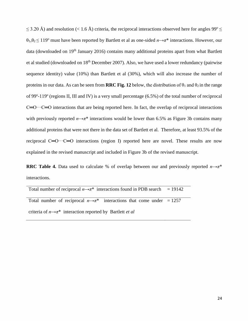
proteins in our data. As can be seen from RRC Fig. 12 below, the distribution of θ1 and θ2 in the range
of 99º-119º (regions II, III and IV) is a very small percentage (6.5%) of the total number of reciprocal
C═O···C═O interactions that are being reported here. In fact, the overlap of reciprocal interactions
with previously reported n→π* interactions would be lower than 6.5% as Figure 3b contains many
additional proteins that were not there in the data set of Bartlett et al. Therefore, at least 93.5% of the
reciprocal C═O···C═O interactions (region I) reported here are novel. These results are now
explained in the revised manuscript and included in Figure 3b of the revised manuscript.
RRC Table 4. Data used to calculate % of overlap between our and previously reported n→π*
interactions.
Total number of reciprocal n→π* interactions found in PDB search = 19142
Total number of reciprocal n→π* interactions that come under
criteria of n→π* interaction reported by Bartlett et al
= 1257
25
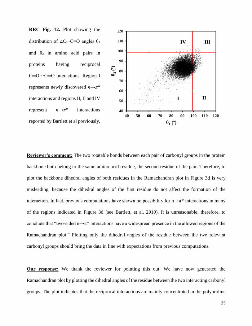
RRC Fig. 12. Plot showing the
distribution of ∠OC=O angles θ1
and θ2 in amino acid pairs in
proteins having reciprocal
C═O···C═O interactions. Region I
represents newly discovered n→π*
interactions and regions II, II and IV
represent n→π* interactions
reported by Bartlett et al previously.
Reviewer’s comment: The two rotatable bonds between each pair of carbonyl groups in the protein
backbone both belong to the same amino acid residue, the second residue of the pair. Therefore, to
plot the backbone dihedral angles of both residues in the Ramachandran plot in Figure 3d is very
misleading, because the dihedral angles of the first residue do not affect the formation of the
interaction. In fact, previous computations have shown no possibility for n→π* interactions in many
of the regions indicated in Figure 3d (see Bartlett, et al. 2010). It is unreasonable, therefore, to
conclude that “two-sided n→π* interactions have a widespread presence in the allowed regions of the
Ramachandran plot.” Plotting only the dihedral angles of the residue between the two relevant
carbonyl groups should bring the data in line with expectations from previous computations.
Our response: We thank the reviewer for pointing this out. We have now generated the
Ramachandran plot by plotting the dihedral angles of the residue between the two interacting carbonyl
groups. The plot indicates that the reciprocal interactions are mainly concentrated in the polyproline
40
50
60
70
80
90
100
110
120
40 50 60 70 80 90 100 110 120θ
2(º
)
θ1 (º)
I II
IIIIV
26
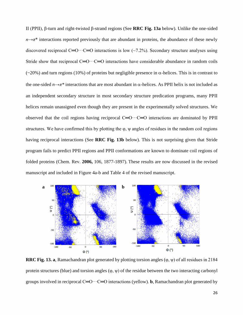
II (PPII), β-turn and right-twisted β-strand regions (See RRC Fig. 13a below). Unlike the one-sided
n→π* interactions reported previously that are abundant in proteins, the abundance of these newly
discovered reciprocal C═O···C═O interactions is low (~7.2%). Secondary structure analyses using
Stride show that reciprocal C═O···C═O interactions have considerable abundance in random coils
(~20%) and turn regions (10%) of proteins but negligible presence in -helices. This is in contrast to
the one-sided n→π* interactions that are most abundant in -helices. As PPII helix is not included as
an independent secondary structure in most secondary structure predication programs, many PPII
helices remain unassigned even though they are present in the experimentally solved structures. We
observed that the coil regions having reciprocal C═O···C═O interactions are dominated by PPII
structures. We have confirmed this by plotting the φ, ψ angles of residues in the random coil regions
having reciprocal interactions (See RRC Fig. 13b below). This is not surprising given that Stride
program fails to predict PPII regions and PPII conformations are known to dominate coil regions of
folded proteins (Chem. Rev. 2006, 106, 1877-1897). These results are now discussed in the revised
manuscript and included in Figure 4a-b and Table 4 of the revised manuscript.
a
b
RRC Fig. 13. a, Ramachandran plot generated by plotting torsion angles (φ, ψ) of all residues in 2184
protein structures (blue) and torsion angles (φ, ψ) of the residue between the two interacting carbonyl
groups involved in reciprocal C═O···C═O interactions (yellow). b, Ramachandran plot generated by
27
plotting torsion angles (φ, ψ) of all residues in 2184 protein structures (blue) and torsion angles (φ,
ψ) of the residue between the two interacting carbonyl groups involved in reciprocal C═O···C═O
interactions present only in the coil regions (yellow).
Reviewer’s comment: With regard to the frequency of these interactions in different secondary
structure types or different amino acids, it is important that the data be normalized. For example, in
Figure 4a, could it be that the interactions are observed more frequently in “coil” than in “turn”
because “coil” is simply more frequent than “turn”? The data from the last column of Extended figure
5a would be more appropriate. Similarly, the plots in Figures 4b and 4c should be normalized to the
frequency of the amino acids. In particular, the observation that Leu-Pro is most common might
simply be due to the fact that leucine is the single most common amino acid in proteins. When
normalized to account for amino acid frequency, the data might show that other residues have stronger
preferences for these reciprocal interactions. This is also relevant for Figure 4d, as hydrolases are the
most well-represented class of enzymes in the PDB, and therefore might not be more prone to
reciprocal n→π* interactions than other classes. Finally, it is unclear what “frequency” is considered
in Extended Figure 5b. Are there really proteins where 92% of residues engage in reciprocal n→π*
interactions? Perhaps “frequency” in this last case actually refers to the number of examples in that
protein?
Our Response: We agree with the reviewer that normalized data/plots would provide better insights.
We have now provided normalized data/plots for secondary structure distribution, amino acid
distribution and amino acid pair distribution instead of absolute numbers.
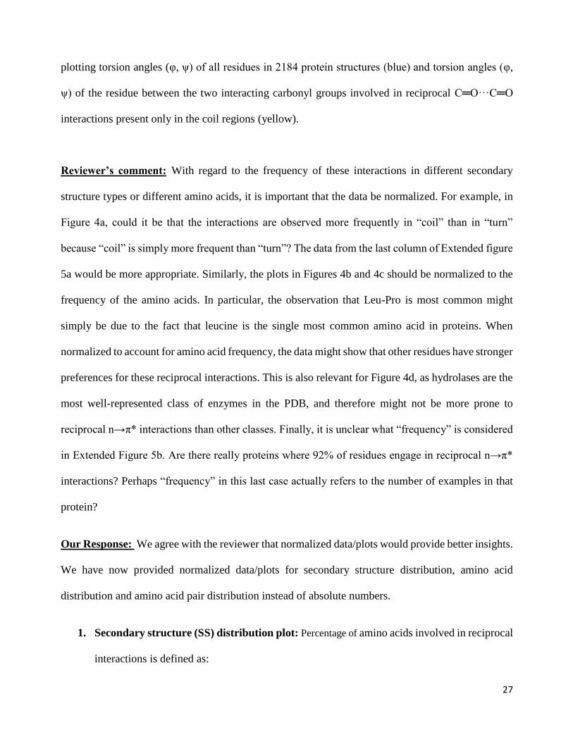
1. Secondary structure (SS) distribution plot: Percentage of amino acids involved in reciprocal
interactions is defined as:
28
(Number of amino acids in the SS involved in reciprocal n→π* interaction x 100) ÷ (Total
number of amino acids in that SS)
RRC Fig. 14. Plot of percentage of amino acids involved in reciprocal interactions in various
secondary structures.
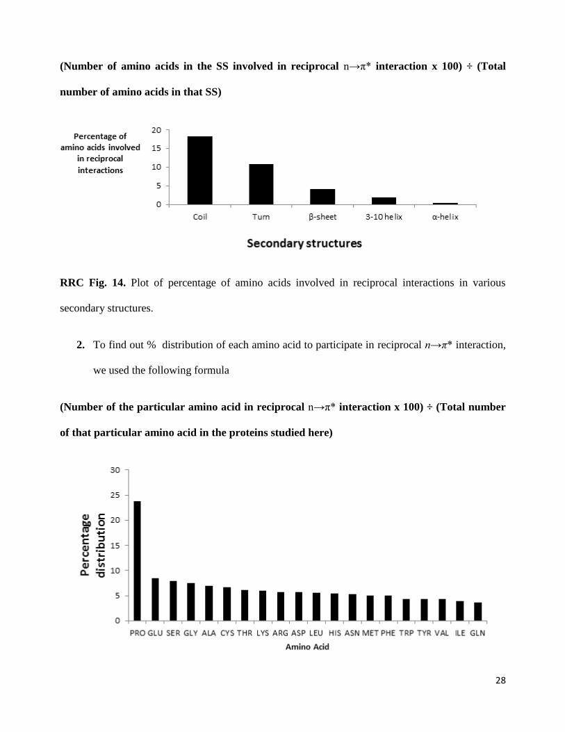
2. To find out % distribution of each amino acid to participate in reciprocal n→π* interaction,
we used the following formula
(Number of the particular amino acid in reciprocal n→π* interaction x 100) ÷ (Total number
of that particular amino acid in the proteins studied here)
29
RRC Fig. 15. Plot showing percentage distribution of amino acids involved in reciprocal
C═O···C═O interactions.
3. To find out the % distribution of amino acid pairs to participate in reciprocal n→π* interactions,
we used the following formula.
(Number of a particular amino acid pair in the reciprocal n→π* interaction x 100) ÷ (Total
number of that amino acid pair in the proteins studied here)
RRC Fig. 16. Plot showing percentage distribution of amino acid pairs involved in reciprocal
C═O···C═O interactions.
30
The changes that have occurred to our results due to normalization is discussed below.
1. In case of the abundance of reciprocal interactions in secondary structures, the trend is almost
similar before and after normalization except that reciprocal interactions are found more in
310-helices compared to α-helices. α-helices are predominant in most proteins but only a
handful of α-helix residues are involved in reciprocal interaction. The abundance of reciprocal
interactions in various secondary structures is now presented in Table 4 of the revised
manuscript.
2. After normalization, the amino acid percentage distribution differs from the frequency
distribution provided earlier although proline remains the most abundant amino acid to
participate in reciprocal n→π* interaction. However, normalization has increased the
percentage distribution of proline residues (24%). These results are presented in Figure 4c of
the revised manuscript.
3. The plot of the amino acid pair that participate in reciprocal n→π* interactions after
normalization has changed significantly from the one before normalization. After
normalization, the Pro-Pro residue pair is the most predominant one to partake in the
reciprocal n→π* interaction. These results are presented in Figure 4d of the revised
manuscript.
We have now incorporated these changes in the revised manuscript. We have now removed plot
4d as this seems not very informative.
In extended figure 5b, by frequency we mean the number of instances of reciprocal n→π*
interaction in the particular protein (PDB code: 4LGY). In case of 4LGY, 92 instances of reciprocal
31
n→π* interaction exist, whilst only 7 % of the amino acid residues present in the protein partake
reciprocal n→π* interaction. A separate column of the percentage of residues involved in reciprocal
interaction is now incorporated in Supplementary Table 9 to clear any confusion.
Reviewer’s comment: It is unclear where the data from Extended Figure 6 come from. Which
structures were subjected to these calculations? Importantly, panel (a) appears to concern different
molecules than panels (b/c), which are still different from those of panels (d/e). The datasets for these
panels should be made clear. In addition, what is the horizontal axis for Extended Figures 6f/g? What
conclusions are to be drawn from these data?
Our response: We have now brought this Figure to the main manuscript in the revised version
(Figure 6). This Figure is necessary to understand the nature of reciprocal C═O···C═O interactions.
The nature of C═O···C═O interactions is highly been debated in the literature. While some consider
them n→π* orbital interaction, others believe them to be dipolar in nature. We have discussed
reciprocal C═O···C═O interactions as orbital interactions (n→π* and π→π*) because of the
following reasons. Firstly, the plots of the n→π* and sum of n→π* and π→π* orbital interaction
energies against the O···C distances (d) show a strong correlation [See Figure below] (Figure 6 in the
revised manuscript). In RRC Fig. 17a, we have plotted the distances (d1 and d2 values) against the
n→π* interaction stabilization energies (NBO second order perturbation energies E1(n→π*) and
E2(n→π*)) reported in Table 1-3. The plot suggest that the stabilization energies E(n→π*) for n→π*
interactions decreases with an increase in the O···C (d) in synthetic molecules 1-8, molecules taken
from CSD and interacting amino acid pairs obtained from PDB (Table 1-3). The plot of overall orbital
interaction energies [sum of n→π* {E(n→π*)} and π →π* {E(π →π*)}] interaction energies reported in
Table 1-3 also shows a similar correlation with O···C (d) distances (RRC Fig. 17b) (Fig. 6b in the

32
revised manuscript). This correlation indicate that orbital interaction is the major mechanism for the
stabilization of these reciprocal C═O···C═O short contacts. Secondly, C=O···C=O torsion angles of
the carbonyl groups involved in reciprocal interactions indicate a net zero dipole-dipole interaction
eliminating the possibility of these interactions being dipolar in nature. To emphasize this point, in
RRC Fig. 17c-d (Figure 6c-d in the revised manuscript), we have plotted the values of C=O···C=O
torsion angles of the 1432 molecules obtained from the CSD search. The torsion angle (T) between
two dipoles could be used to understand the dipolar nature of interaction between them. As we know,
antiparallel (T ~ 180º) dipoles attract and parallel dipoles (T ~ 0º) repel each other whereas two
orthogonal dipoles (T ~ 90º) have net zero dipolar interaction. In case of reciprocal interaction, the
C=O···C=O torsion angles show an orientational preference [C═O···C═O torsion angle falls in 60º
to 90º or -60º to -90º range] as a consequence of the simultaneous restrictions on d1 and d2 (≤ 3.2 Å).
However, the values of the C═O···C═O torsion angles (~90º) suggest that there would be almost net
zero interaction between the dipoles, eliminating the possibility of strong dipolar interactions.
Therefore, we conclude that orbital delocalization is the major driving force for the stabilization of
reciprocal C═O···C═O interactions. However, an elaborate energy decomposition analysis may be
required for the accurate deconvolution of various forces contributing to the stabilization of reciprocal
C═O···C═O short contacts.
We have now omitted panel b-e as they have not provided much additional insight into
understanding the nature of reciprocal C=O…C=O interaction.
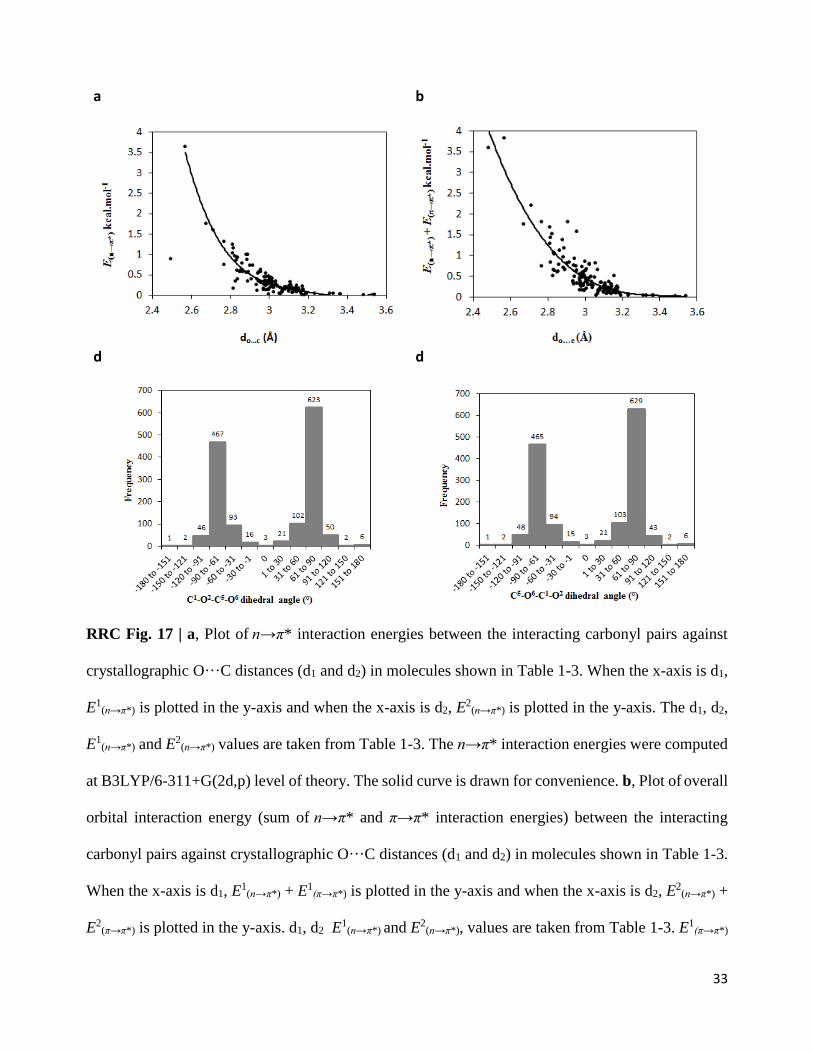
33
a b
d d
RRC Fig. 17 | a, Plot of n→π* interaction energies between the interacting carbonyl pairs against
crystallographic O···C distances (d1 and d2) in molecules shown in Table 1-3. When the x-axis is d1,
E1(n→π*) is plotted in the y-axis and when the x-axis is d2, E
2(n→π*) is plotted in the y-axis. The d1, d2,
E1(n→π*) and E2
(n→π*) values are taken from Table 1-3. The n→π* interaction energies were computed
at B3LYP/6-311+G(2d,p) level of theory. The solid curve is drawn for convenience. b, Plot of overall
orbital interaction energy (sum of n→π* and π→π* interaction energies) between the interacting
carbonyl pairs against crystallographic O···C distances (d1 and d2) in molecules shown in Table 1-3.
When the x-axis is d1, E1(n→π*) + E1
(π→π*) is plotted in the y-axis and when the x-axis is d2, E2
(n→π*) +
E2(π→π*) is plotted in the y-axis. d1, d2 E
1(n→π*) and E2
(n→π*), values are taken from Table 1-3. E1(π→π*)
34
and E2(π→π*) values are taken from Supplementary Table 3, Supplementary Table 6 and
Supplementary Table 8. The orbital interaction energies were computed at B3LYP/6-311+G(2d,p)
level of theory. The solid curve is drawn for convenience. c, Histogram plot showing the frequency
of the C1═O2···C5═O6 dihedral angles (see Supplementary Fig. 3 for atom numbers) for 1432
molecules obtained from the CSD search. d, Histogram plot showing the frequency of the
C5═O6···C1═O2 dihedral angles (see Supplementary Fig. 3 for atom numbers) for 1432 molecules
obtained from the CSD search.
Reviewer’s Comments
In addition to the important issues above, I believe that the manuscript would benefit from the
following considerations.
(1) How is the fit of Extended Figure 6a determined? What is the resulting model?
Our response: The data points in Figure 6a is not fitted to any equation. The solid curve is used only
for convenience to illustrate the trend.
2) I think that it is important to note that many of the CSD hits, at least as described by Extended
Figure 3c, are very conformationally constrained, either by additional rings, highly substituted
centers, or stereoelectronic effects. This makes it all the more remarkable that reciprocal n→π*
interactions are observed in the much less-constrained protein backbone.
Our response: The Figure pointed out by the reviewer only has 15 of 1432 molecules. In fact, there
are many molecules that are less constrained than what was presented in that Figure. To get some
insights into the structures of the small molecules having reciprocal C═O···C═O interactions, we
have now manually analyzed small molecules from the CSD having 1,5-type reciprocal interactions
35
with both d1 and d2 ≤ 3.00 Å. A total of 249 molecules fulfill the above criteria [1, 5-interaction; both
d1 & d2 ≤ 3.00 Å]. As can be anticipated, the nature of the two atoms/groups between the interacting
carbonyl groups plays a key role to make the two carbonyl groups non coplanar and provides them
the conformation required for reciprocal interactions (See RRC Table 5 below). These results are
now included in Supplementary Table 7. Interestingly, majority of these molecules (117, ~47%) have
one heteroatom and one chiral carbon between the two interacting carbonyl pairs, a feature that
resembles peptides and proteins.
RRC Table 5. Analysis of 1, 5-reciprocal interactions from the CSD with both d1 and d2 ≤ 3.00 Å. A
total of 249 molecules were obtained that have 1, 5-reciprocal C=OC=O interactions with both d1
and d2 ≤ 3.00 Å.
Groups Between the two C=O group Number of molecules Percentage (%)
One heteroatom (O or N), one chiral C atom 117 46.98
Both are chiral C atoms 61 24.49
One heteroatom (O or N), one achiral C atom 28 11.24
Both sp2 C atoms [the C=O groups are attached to 1,2-
positions of benzene, naphthalene or non-aromatic
rings]
21 8.43
Both are heteroatoms (O or N) 14 5.62
One chiral and one achiral C atoms 4 1.60
Both are achiral C atoms [attached to rings with
conformational constraints] 4 1.60
3) A ChemDraw figure would be helpful for interpreting Figure 2e/f.
Our response: A ChemDraw Figure is included now in the Figure 2e of the revised manuscript.
36
4) The authors have not commented on the types of small molecules that form these reciprocal n→π*
interactions, which might reveal some interesting trends.
Our response: To get some insights into the structures of the small molecules having reciprocal
C═O···C═O interactions, we manually analyzed small molecules from the CSD having 1,5-type
reciprocal interactions with both d1 and d2 ≤ 3.00 Å. A total of 249 molecules fulfill the above criteria
[1, 5-interaction; both d1 & d2 ≤ 3.00 Å]. As can be anticipated, the nature of the two atoms/groups
between the interacting carbonyl groups plays a key role to make the two carbonyl groups non
coplanar and provides them the conformation required for reciprocal interactions (See RRC Table 5
above). These results are now included in Supplementary Table 7. Interestingly, majority of these
molecules (117, ~47%) have one heteroatom and one chiral carbon between the two interacting
carbonyl pairs, a feature that resembles peptides and proteins. Analysis of the substituents attached to
the carbonyl groups show that of 249 molecules, only 24 (9.6%) have the presence of strong electron
withdrawing groups attached to at least one C=O group.
5) It would be helpful for the authors to show illustrative examples of the interactions they find in
proteins, particularly in different secondary structure contexts. For example, the authors note that “α-
helices that have two-sided n→π* interactions are distorted, while the β-sheets having two sided
n→π* interactions are twisted.” It would be very helpful for readers to be able to visualize these
distortions, as they could have important consequences for protein structure. Such examples would,
in my opinion, be more informative than the examples shown in Extended Figure 4.
Our response: We agree with the reviewer. We have now included a Figure in the revised manuscript
(Fig. 5 in the revised manuscript) to illustrate the reciprocal interactions found in different secondary
structures. Please see the RRC Fig. 18 below.
37
RRC Fig. 18. Reciprocal C═O···C═O interactions present in various secondary structure is shown. a,
PPII- helix. b, β-turn. c, Right-twisted β-strand. d, α-helix. e, interface of α-helix and β-sheet.
a b
c d
e
Reviewer #1 (Remarks to the Author):
The authors have responded to my comments very well, and I feel that the data presentation,
arguments, and the overall paper are much improved. In my view, the revised paper is now
suitable for publication in Nature Communications.
Reviewer #2 (Remarks to the Author):
The authors have appropriately responded to most of my points BUT my main initial concern has
not been clarified.
My main point of criticism was as follows "... the authors need to demonstrate – at least for their
sequence of hydrazine derivatives in Fig. 1c – that geometry optimization of the remaining degrees
of freedom (bond lengths, angles, and dihedrals) has a negligible effect on the overlap of the
computed n/pi* orbitals and on the NBO energies in comparison to calculations done on the
unrelaxed X-ray coordinates."
The authors replied "We agree with the reviewer that providing optimized geometry and electronic
energy calculation on optimized geometry is the common practice."
Then, they write that they have performed new unconstrained calculations of a single molecule
(compound 6). However, they observed that the molecular geometry of the chloride atom
distorted significantly from its starting (X-ray) geometry - which is of course UNWANTED.
They argue that "one would have to come up with the best theoretical method and basis set to
study these interaction, which itself would be a separate project for investigation."
IF this means that the level of theory used here is NOT ADEQUATE to properly describe the
electronic structure of the molecules considered here, in my view this also casts doubts on the
reported interactions between orbitals of these molecules.
Hartree-Fock and DFT calculations are pretty cheap methods these days.
The authors close their discussion of the calculations on compound 6 with "As this study deals with
the possibility of reciprocal interactions in the crystal geometries of small molecules and proteins,
to avoid such deviation from crystal geometries, we
avoided free optimization the molecules. "
Contrary to this statement, the title of the manuscript and everything else suggest that the
authors believe that reciprocal interactions are important for small molecules and proteins in
general, and not only when they adopt their crystal geometries and are in the packing
environment of a crystal.
Reviewer #3 (Remarks to the Author):
The revisions submitted for the manuscript regarding “Reciprocal carbonyl-carbonyl interactions in
small molecules and proteins” resolve most of the key issues raised during initial review. The
authors are to be commended for the thoroughness of their analysis. However, a few issues
remain to be addressed:
1) I do not believe that the set of protein structures examined in this manuscript is of sufficient
resolution to report data regarding carbonyl pyramidalization, which is a most subtle distortion.
Previous studies of this parameter in proteins have focused on ultrahigh-resolution structures (less
than 1.0 Å), which probably represent only about 10% of the structures considered herein.
Therefore, I am not sure that these data are reliable, and I would discourage their inclusion (Table
3) unless a more appropriate dataset is examined.
2) I still do not believe that the diacyl hydrazines can be used to demonstrate cooperativity of
n→π* interactions because the two interaction distances are not independent from one another.
When carbonyl 1 gets closer to carbonyl 2, it is necessary that carbonyl 2 gets closer to carbonyl
1. That is why there is such a strong correlation in Figure 1g. Cooperativity need not be present for
this trend to be observed. In order to truly demonstrate cooperativity, a change in the strength of
one interaction must affect the strength of an independent interaction. In other words, as the
interaction between carbonyls 1 and 2 gets stronger, the interaction between carbonyls 2 and 3
should also get stronger. Without that type of experimental design, I do not believe that
cooperativity can be invoked.
3) I apologize for the poor phrasing of my question regarding the overlap between the datasets of
this manuscript and that of Bartlett et al. The absolute number of interactions identified by the two
studies cannot be compared, as the authors note, due to differences in the size of the protein
dataset under examination. However, my interest is not in the absolute number of interactions
present in each dataset, but rather in the relative number that would be identified using the
criteria of Bartlett et al. compared to the number that would be identified using the criteria
employed in this manuscript. The important piece of information is that 6.5% of the interactions
identified by this study would have been identified using the criteria reported by Bartlett et al.,
demonstrating that reciprocal carbonyl interactions are largely distinction from sequential n→π*
interactions reported previously. The discussion of the differences in the protein datasets is
therefore unnecessary and can be removed.
Robert W. Newberry
Reviewer #4 (Remarks to the Author):
The authors report studies of reciprocal carbonyl-carbonyl interactions in small molecules and
proteins. This is a somewhat unexplored previously type of interactions and it can play a significant
role in determining 3-D protein structure. The manuscript is well written and generally clear. The
level of science is good. In my opinion, the article should be publishable once the following issues
have been addressed:
(1) The authors call these interactions "unprecedented" in the introduction part of the paper. I do
not believe the interactions themselves are without a precedent. Moreover, I would recommend to
tone down this and similar sentences in general.
(2) More thorough extensive analysis should be done for the computational results. This should
definitely include analyzing charge redistribution.
Reviewer #2
Reviewer’s comment: The authors have appropriately responded to most of my points BUT my main
initial concern has not been clarified.
My main point of criticism was as follows "... the authors need to demonstrate – at least for their
sequence of hydrazine derivatives in Fig. 1c – that geometry optimization of the remaining degrees
of freedom (bond lengths, angles, and dihedrals) has a negligible effect on the overlap of the computed
n/pi* orbitals and on the NBO energies in comparison to calculations done on the unrelaxed X-ray
coordinates."
The authors replied "We agree with the reviewer that providing optimized geometry and electronic
energy calculation on optimized geometry is the common practice."
Then, they write that they have performed new unconstrained calculations of a single molecule
(compound 6). However, they observed that the molecular geometry of the chloride atom distorted
significantly from its starting (X-ray) geometry - which is of course UNWANTED.
They argue that "one would have to come up with the best theoretical method and basis set to study
these interaction, which itself would be a separate project for investigation."
IF this means that the level of theory used here is NOT ADEQUATE to properly describe the
electronic structure of the molecules considered here, in my view this also casts doubts on the reported
interactions between orbitals of these molecules.
Hartree-Fock and DFT calculations are pretty cheap methods these days.
The authors close their discussion of the calculations on compound 6 with "As this study deals with
the possibility of reciprocal interactions in the crystal geometries of small molecules and proteins, to
avoid such deviation from crystal geometries, we avoided free optimization the molecules. "
Contrary to this statement, the title of the manuscript and everything else suggest that the authors
believe that reciprocal interactions are important for small molecules and proteins in general, and not
only when they adopt their crystal geometries and are in the packing environment of a crystal.
Our response: We thank the reviewer for the suggestions. Reporting n→π* interaction energies by
performing NBO analysis on high resolution crystal structures without geometry optimization is

known and B3LYP/6-311+G(2d,p) level of theory [the same method that we used in this manuscript]
worked well for that purpose (ref. 16 in the manuscript). Based on the reviewer’s suggestions, we
have now carried out geometry optimizations in compounds 1-8 by freezing the dihedral angles of
the side chains involved in reciprocal interactions to their X-ray values (see Figure below and
Supplementary Figure 3 in the revised manuscript) to find out if free optimization of the remaining
degrees of freedom (bond lengths, angles, and dihedrals) have any effect on the overlap of the
computed n→π* interactions in comparison to calculations done on the unrelaxed X-ray geometries.
We observed that reciprocal n→π* interactions were retained after geometry optimizations but they
became slightly weaker than what were observed from the NBO calculations on the crystal geometries
(Supplementary Table 5). We also observed that, during gas phase geometry optimization, in absence
of any packing and intermolecular forces that are present in the X-ray geometries, the Cl or Br atoms
attached to the methylene carbons in 4, 6-8 moved to an anti-periplanar geometry (trans) with respect
to the oxygen atom of the nearby carbonyl group (CO-II). This is probably due to higher
hyperconjugative delocalization between the halogen lone pairs and carbonyl π* orbital in the anti-
periplanar geometry that would provide more stability to the isolated gas phase molecule. Note that
such elongation of carbonyl-carbonyl (O···C) short contacts (weakening of n→π* interactions) in gas
phase optimized geometry relative to the X-ray geometries are well known (see ref. 9,13 and 14 in
the manuscript).
RRC Figure 1. Representative molecular geometry to show the frozen dihedral angles during
geometry optimization.
Reviewer #3
Reviewer’s comment: I do not believe that the set of protein structures examined in this manuscript
is of sufficient resolution to report data regarding carbonyl pyramidalization, which is a most subtle
distortion. Previous studies of this parameter in proteins have focused on ultrahigh-resolution
structures (less than 1.0 Å), which probably represent only about 10% of the structures considered
herein. Therefore, I am not sure that these data are reliable, and I would discourage their inclusion
(Table 3) unless a more appropriate dataset is examined.
Our response: We agree with the reviewer. Accordingly, we have now removed this section from
the manuscript. We have also removed columns 8-11 from Table 3 in the revised manuscript.
Reviewer’s comment: I still do not believe that the diacyl hydrazines can be used to demonstrate
cooperativity of n→π* interactions because the two interaction distances are not independent from
one another. When carbonyl 1 gets closer to carbonyl 2, it is necessary that carbonyl 2 gets closer to
carbonyl 1. That is why there is such a strong correlation in Figure 1g. Cooperativity need not be
present for this trend to be observed. In order to truly demonstrate cooperativity, a change in the
strength of one interaction must affect the strength of an independent interaction. In other words, as
the interaction between carbonyls 1 and 2 gets stronger, the interaction between carbonyls 2 and 3
should also get stronger. Without that type of experimental design, I do not believe that cooperativity
can be invoked.
Our response: “When carbonyl 1 gets closer to carbonyl 2, it is necessary that carbonyl 2 gets
closer to carbonyl 1. That is why there is such a strong correlation in Figure 1g.”
This is not true in general. If this were true we should have observed a similar correlations in Figure
2a and 3a, which we have not. The correlations were observed only for compounds 1-8 in Fig 1g. In
Figures 2a and 3a, the d1 vs. d2 plots are scattered and, in most cases, d1 does not increase or decrease
with an increase or decrease of d2, which suggests that shortening of one donor-acceptor distance
does not necessarily lead to a contraction of the second, reciprocal donor–acceptor distance.
“In other words, as the interaction between carbonyls 1 and 2 gets stronger, the interaction
between carbonyls 2 and 3 should also get stronger. Without that type of experimental design,
I do not believe that cooperativity can be invoked.”
I think, we have a disagreement in the use of terminology “cooperative” here. May be we should have
used “synergistic” instead of “cooperative”. A famous textbook example of synergistic bonding is
metal carbonyl bonding. As we know, in case of metal carbonyls, higher σ donation from CO to metal
increases backdonation from metal’s filled π orbital to empty π* orbital of CO, which are termed as
“synergistic”. In case of synergistic interaction, we don’t need two independent interactions (1→2
and 2→3) as evident from metal carbonyl’s synergistic σ-bonding and π-back bonding.
In view of the above discussion, we have now replaced “cooperative” with “synergistic” in
the manuscript. We believe that there is a strong possibility that the correlation between the two n→π*
interactions in compounds 1-8 is synergistic. However, to ascertain whether the origin of this
correlation between the reciprocal n→π* interactions in 1-8 is a result of synergistic effects or merely
a result of the particular geometries adopted by 1-8 needs further investigation. Therefore, we have
now modified this discussion in the revised manuscript and restricted ourselves to discuss the
possibility of having synergistic interaction between the carbonyl groups in 1-8.
Reviewer’s comment: I apologize for the poor phrasing of my question regarding the overlap
between the datasets of this manuscript and that of Bartlett et al. The absolute number of interactions
identified by the two studies cannot be compared, as the authors note, due to differences in the size
of the protein dataset under examination. However, my interest is not in the absolute number of
interactions present in each dataset, but rather in the relative number that would be identified using
the criteria of Bartlett et al. compared to the number that would be identified using the criteria
employed in this manuscript. The important piece of information is that 6.5% of the interactions
identified by this study would have been identified using the criteria reported by Bartlett et al.,
demonstrating that reciprocal carbonyl interactions are largely distinction from sequential n→π*
interactions reported previously. The discussion of the differences in the protein datasets is therefore
unnecessary and can be removed.
Our response: We have now modified this section and removed the discussion on the differences in
the protein datasets from the revised manuscript.
Reviewer #4
Reviewer’s comment: The authors call these interactions "unprecedented" in the introduction part of
the paper. I do not believe the interactions themselves are without a precedent. Moreover, I would
recommend to tone down this and similar sentences in general.
Our response: We have now modified this section and replaced “unprecedented” with “unexplored”.
Please see the revised manuscript.
Reviewer’s comment: More thorough extensive analysis should be done for the computational
results. This should definitely include analyzing charge redistribution.
Our response: To analyze charge redistribution, we first took help of NBO deletion analysis. Due
to n→π* electron delocalization between donor oxygen lone pair and acceptor carbonyl π* orbital of
C=O bond, there should be a reduction on the electronic charge on the donor oxygen atom and a
corresponding increase in the electronic charge on the π* orbital of the acceptor carbonyl group.
Conversely, deletion of n→π* interaction should increase charge on the donor oxygen atom’s lone
pair and decrease charge on the acceptor carbonyl’s π* orbital, which could be studied by using NBO
deletion analysis. Therefore, NBO deletion analysis should provide some understanding of the
increase or decrease of charge on donor oxygen lone pair and acceptor carbonyl’s π*C═O orbital.
Importantly, this change in charges should correlate with the strength of n→π* interactions and hence
O···C distances. We have now carried out NBO deletion analysis (Supplementary Table 13 of revised
manuscript) and observed that deletion of n→π* interactions increases charge on donor oxygen lone
pair (nO) and depletes it on the acceptor carbonyl π*C═O orbital. Although the values of charge increase
or decrease is small, these values correlate well with O···C distances suggesting electron
delocalization between the two carbonyl groups involved in C═O···C═O interactions
(Supplementary Table 13 of revised manuscript). Similarly, deletion of π→π* interactions between
the carbonyl groups leads to charge increase in the donor π orbital and decrease in the acceptor π*
orbital, which can be correlated to the strength of C═O···C═O interactions. The overall accumulation
of charges on the acceptor carbonyl π*C═O orbitals due to donation from the oxygen lone pairs and
πC═O orbital of donor carbonyl is shown in Figure 6c-d of revised manuscript, which correlate well
with the strength of C═O···C═O short contacts.
We have also carried out CHelpG charge calculations on compounds 1-8, the molecules
obtained from CSD search and amino acid dimers listed in Table 1-3. However, it is difficult to
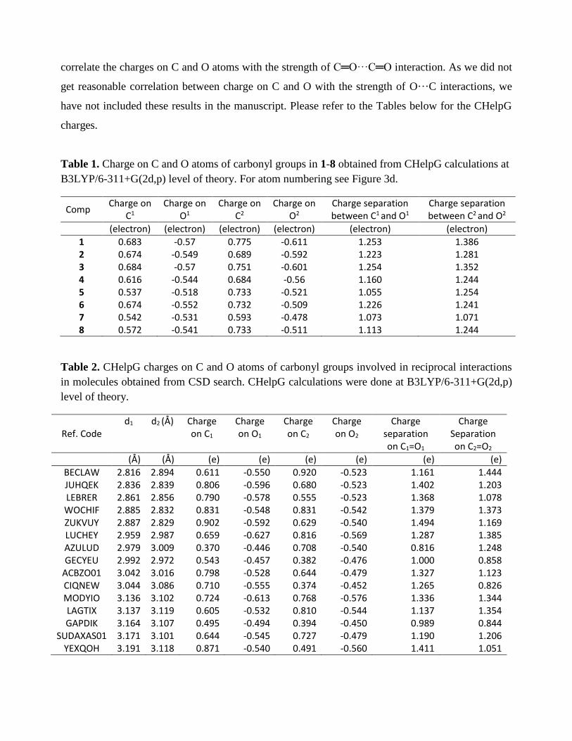
correlate the charges on C and O atoms with the strength of C═O···C═O interaction. As we did not
get reasonable correlation between charge on C and O with the strength of O···C interactions, we
have not included these results in the manuscript. Please refer to the Tables below for the CHelpG
charges.
Table 1. Charge on C and O atoms of carbonyl groups in 1-8 obtained from CHelpG calculations at
B3LYP/6-311+G(2d,p) level of theory. For atom numbering see Figure 3d.
Comp Charge on
C1 Charge on
O1 Charge on
C2 Charge on
O2 Charge separation between C1 and O1
Charge separation between C2 and O2
(electron) (electron) (electron) (electron) (electron) (electron)
1 0.683 -0.57 0.775 -0.611 1.253 1.386 2 0.674 -0.549 0.689 -0.592 1.223 1.281 3 0.684 -0.57 0.751 -0.601 1.254 1.352 4 0.616 -0.544 0.684 -0.56 1.160 1.244 5 0.537 -0.518 0.733 -0.521 1.055 1.254 6 0.674 -0.552 0.732 -0.509 1.226 1.241 7 0.542 -0.531 0.593 -0.478 1.073 1.071 8 0.572 -0.541 0.733 -0.511 1.113 1.244
Table 2. CHelpG charges on C and O atoms of carbonyl groups involved in reciprocal interactions
in molecules obtained from CSD search. CHelpG calculations were done at B3LYP/6-311+G(2d,p)
level of theory.
Ref. Code d1 d2 (Å) Charge
on C1
Charge on O1
Charge on C2
Charge on O2
Charge separation on C1=O1
Charge Separation on C2=O2
(Å) (Å) (e) (e) (e) (e) (e) (e)
BECLAW 2.816 2.894 0.611 -0.550 0.920 -0.523 1.161 1.444 JUHQEK 2.836 2.839 0.806 -0.596 0.680 -0.523 1.402 1.203 LEBRER 2.861 2.856 0.790 -0.578 0.555 -0.523 1.368 1.078 WOCHIF 2.885 2.832 0.831 -0.548 0.831 -0.542 1.379 1.373 ZUKVUY 2.887 2.829 0.902 -0.592 0.629 -0.540 1.494 1.169 LUCHEY 2.959 2.987 0.659 -0.627 0.816 -0.569 1.287 1.385 AZULUD 2.979 3.009 0.370 -0.446 0.708 -0.540 0.816 1.248 GECYEU 2.992 2.972 0.543 -0.457 0.382 -0.476 1.000 0.858
ACBZO01 3.042 3.016 0.798 -0.528 0.644 -0.479 1.327 1.123 CIQNEW 3.044 3.086 0.710 -0.555 0.374 -0.452 1.265 0.826 MODYIO 3.136 3.102 0.724 -0.613 0.768 -0.576 1.336 1.344 LAGTIX 3.137 3.119 0.605 -0.532 0.810 -0.544 1.137 1.354 GAPDIK 3.164 3.107 0.495 -0.494 0.394 -0.450 0.989 0.844
SUDAXAS01 3.171 3.101 0.644 -0.545 0.727 -0.479 1.190 1.206 YEXQOH 3.191 3.118 0.871 -0.540 0.491 -0.560 1.411 1.051

Table 3. CHelpG charges on C and O atoms of carbonyl groups involved in reciprocal interactions
in amino acid pairs taken from PDB. CHelpG calculations were done at B3LYP/6-311+G(2d,p) level
of theory.
AA pair pdb stretch d1 d2 Charge on C1
Charge on O1
Charge on C2
Charge on O2
Charge separation on C1=O1
Charge Separation on C2=O2
(Å) (Å) (e) (e) (e) (e) (e) (e)
Ala-Ala 3s5m 402-403 2.485 2.999 0.523 -0.574 0.434 -0.514 1.097 0.948 Ala-Pro 2xu9 264-265 2.568 2.852 0.710 -0.548 0.545 -0.535 1.262 1.080 Ile-Pro 2opc 135-136 2.675 2.768 0.566 -0.545 0.574 -0.472 1.111 1.046 Leu-Pro 2x5o 141-142 2.712 2.771 0.821 -0.628 0.585 -0.552 1.449 1.137 Cys-Pro 1gcy 251-252 2.815 2.834 0.259 -0.511 0.639 -0.466 0.770 1.105 Gln-Lys 3wcq 9-10 2.815 2.816 0.713 -0.591 0.635 -0.553 1.304 1.188 Leu-Tyr 3u26 101-102 2.826 2.893 0.991 -0.674 0.625 -0.573 1.665 1.198 Pro-Pro 3cx2 186-187 2.89 2.838 0.582 -0.611 0.62 -0.519 1.193 1.139 Leu-Pro 1a2p 20-21 2.904 3.007 0.604 -0.596 0.626 -0.577 1.200 1.203 Thr-Glu 4pdy 108-109 2.918 2.936 0.827 -0.643 0.588 -0.579 1.470 1.167 Phe-Pro 1n08 76-77 2.943 2.941 0.220 -0.564 0.723 -0.547 0.784 1.270 Ala-Phe 4y1w 139-140 2.947 2.973 0.878 -0.643 0.529 -0.566 1.521 1.095 Ser-Asp 3ry4 79-80 2.952 2.981 1.092 -0.689 0.364 -0.585 1.781 0.949 Leu-Pro 1g5a 379-380 2.956 2.978 0.581 -0.577 0.626 -0.488 1.158 1.114 Lys-Pro 1k3i 50-51 2.975 2.938 0.573 -0.515 0.579 -0.511 1.088 1.090 Ala-Asp 2bi8 95-96 2.985 2.973 0.629 -0.645 0.608 -0.567 1.274 1.175 Ile-Pro 1o7i 107-108 2.986 3.082 0.620 -0.626 0.616 -0.589 1.246 1.205 Thr-Thr 3uxf 349-350 2.987 2.975 0.902 -0.629 0.814 -0.565 1.531 1.379 Pro-Ser 4psc 32-33 2.998 2.946 1.010 -0.645 0.584 -0.493 1.655 1.077 Asp-Pro 2vzp 2-3 3.026 3.026 0.728 -0.647 0.577 -0.566 1.375 1.143 Thr-Pro 1fj2 3-4 3.035 3.015 0.712 -0.632 0.537 -0.520 1.344 1.057 Ala-Pro 1g12 103-104 3.108 3.152 0.395 -0.568 0.621 -0.474 0.963 1.095 Ile-Pro 1e2w 208-209 3.114 3.143 0.594 -0.582 0.671 -0.494 1.176 1.165 Val-Pro 1jnd 294-295 3.119 3.135 0.778 -0.653 0.680 -0.499 1.431 1.179 His-Ser 1b6a 331-332 3.128 3.108 0.593 -0.550 0.607 -0.553 1.143 1.160 Phe-Gly 1odv 28-29 3.129 3.169 0.817 -0.628 0.656 -0.576 1.445 1.232 Ala-Leu 1ikp 388-389 3.141 3.183 0.650 -0.625 0.632 -0.522 1.275 1.154 Glu-Pro 1eu1 623-624 3.144 3.194 0.443 -0.600 0.614 -0.466 1.043 1.080 Ala-Arg 1ejd 119-120 3.175 3.171 0.830 -0.647 0.632 -0.539 1.477 1.171 Leu-Pro 1eb6 110-111 3.176 3.175 0.457 -0.610 0.689 -0.545 1.067 1.234
Reviewer #3 (Remarks to the Author):
I am satisfied with the adjustments made in this round of revision and feel the paper can be
published as written. Though I remain unconvinced that cooperativity, or “synergy,” has been
adequately demonstrated by the diacyl hydrazines, the results are described in appropriately
speculative terms.
Robert W. Newberry
Reviewer #4 (Remarks to the Author):
In my opinion, the authors have addressed the reviewers' comments in a satisfactory fashion, and
the manuscripts should now be publishable.