Embed Size (px)
Citation preview
45710.2217/17460816.3.5.457 © 2008 Future Medicine ISSN 1746-0816Future Rheumatol. (2008) 3(5), 457–473
part of
Futu
re R
he
um
ato
log
y
The spondyloarthropathies (SpA) are a heterogeneous group of rheumatic inf lammatory interrelated diseases that share common etiopathogenesis, clinical, genetic and radiological features, with frequent association with overlapping extraarticular features [1]. At least five entities comprise this group, as follows:
Ankylosing spondylitis (AS) is the prototype n
of this family, characterized by predominant axial skeletal involvement and advanced radiographic sacroiliitis;
Reactive arthritis (ReA);n
A subset of psoriatic arthritis (PsA);n
Arthritis associated with inflammatory bowel n
disease (IBD);
Undifferentiated SpA (UndSpA).n
They have as a common denominator their strong, but variable among them, association to human leukocyte antigen (HLA)B27. At onset, SpA present four syndromes that are expressed to different degrees in each of them:
Axial syndrome (the most important hallmark n
is the involvement of the axial skeleton with
spondylitis and/or sacroiliitis). This is clinically characterized by inflammatory back pain and stiffness, the latter being due to both inflammation and progressive bony ankylosis of the spine;
Peripheral syndrome (generally asymmetrical n
oligoarthritis predominantly of the lower limbs);
Enthesitic syndrome;n
Extraskeletal syndrome (SpA is associated n
with several extraarticular manifestations, including inflammatory intestinal lesions, acute uveitis and skin lesions) [2].
It is especially relevant that we acknowledge that in the same patient we can find any of the four syndromes, or all of them in diverse combinations.
Ankylosing spondylitis is a chronic systemic inflammatory disorder, with an estimated prevalence of 0.2–1.2%, involving the sacroiliac (SI) joints, the spine, and often the hips or peripheral joints, which predominantly affects young men. In the latest years, special attention has been paid to the enthesis involvement in AS – some authors propose that enthesis is a key structure in the
Ankylosing spondylitis and other spondyloarthropathies: extra-articular manifestations and their management
Ruxandra Elena Schiotis, Alejandro Escudero-Contreras†, Janitzia Vazquez-Mellado & Eduardo Collantes-Estévez†Author for correspondence: Servicio de Reumatología. Hospital Universitario ‘Reina Sofía’, Avda. Menendez Pidal s/n, 14004 Córdoba, Spain n Tel.: +34 957 011 631 n Fax: +34 957 011 683 n [email protected]
The spondylarthropathies (SpA) are a group of rheumatic inflammatory inter-related diseases that share common etiopathogenesis, clinical, genetic and radiological features, in which ankylosing spondylitis is the prototype of the family. All SpA are, to some extent, associated with human leukocyte antigen-B27, but the frequency varies among the several diseases. Extra-articular involvement is a frequent feature of the SpA. By the way they relate with the SpA concept, two different categories of extra-articular manifestations may be identified. First, is the extra-articular manifestation concept-associated, which has eye, bowel and skin involvement. This category may be present in all groups of the SpA family; they share common etiopathogenesis with the disease itself and can be present at any moment during disease evolution. Second, is the extra-articular manifestations non-concept associated, which involve cardiac, pulmonary, renal and neurologic manifestations, as well as osteoporosis and amyloidosis. This category is most frequently described in relation to ankylosing spondylitis, and often occurs in long-standing uncontrolled disease. In this article, we review the clinical features and therapeutic approach to the main extra-articular involvement of the SpA, paying special attention to ankylosing spondylitis.
Keywords
ankylosing spondylitisn extra-articular manifestationsn spondyloarthropathies n treatment
Revie
w
Future Rheumatol. (2008) 3(5)458 future science group
Review Schiotis, Escudero-Contreras, Vazquez-Mellado & Collantes-Estévez
pathogenesis of the disease and so, in this context, synovitis is considered secondary to enthesitis [3,4]. Peripheral arthritis could be detected frequently in large joints such as shoulders, hips or knees, but probably the most important of them is hip impairment, as it can indicate a poor prognosis for the disease course [5,6].
Extraarticular involvement is a frequent feature of the SpA, gathering all the signs and symptoms that share the same etiopathogeny with the disease itself (SpA conceptrelated manifestation), and can be present at any moment in the disease evolution, even at the first manifestation, before the axial or articular manifestations. These extraarticular manifestations include eye, skin and bowel involvement, and classical SpA treatment and antitumor necrosis factor (TNF) therapy influences the course of these symptoms.
On the other hand, there are other extraarticular manifestations that are seldom related to joint or axial manifestations (nonconceptrelated manifestation of SpA), and classical SpA drugs have no effect on them. They include cardiac, pulmonary, renal and neurologic manifestations, as well as ostoporosis and amyloidosis (Table 1).
Concept-related extra-articular manifestations
Eye manifestationsThe most frequent extraarticular involvement in AS is inflammation of the uvea (uveitis), the prevalence reported for anterior acute uveitis (AAU) ranging from 16 to 40% [7,8].
In a large series of patients, the mean frequency of active episodes of uveitis was 0.8 ± 0.6 per year [9]. Uveitis usually occurs after the onset of rheumatologic symptoms, but AAU may be the first symptom of the systemic disease, preceding other clinical manifestations. Several studies have shown that the diagnosis of previously unknown systemic inflammatory disease was made as a result of the ophthalmological consultation after an episode of uveitis in more than 50% of the patients with SpA and uveitis, and up to 91% in UndSpA [10].
The association between the human major histocompatibility complex (MHC), HLA‑B27, and AAU was originally described in 1973 and remains one of the strongest HLA–disease associations [11].
AAU in AS HLA‑B27positive patients is characterized by a clear male preponderance, males being affected 1.5 to 2.5times more often than females [12], in contrast to HLA‑B27negative AAU, which does not show any gender difference [13].
The first episode of anterior uveitis in HLA‑B27positive patients most commonly occurs between the ages of 20 and 40 years [14], whereas the age at onset of uveitis in HLA‑B27negative patients tends to occur a decade later [15].
Intraocular inflammation is generally an acute unilateral episode, with sudden onset and limited duration accompanied by photophobia and hyper eye discharge. Bilateral involvement is infrequently seen during the course of the disease. In rare cases, prolonged, uncontrolled, anterior uveitis can cause extended inflammation of the posterior segment of the eye and subsequent cystoid macular edema, which is the main cause of irreversible visual impairment in patients with prolonged, uncontrolled uveitis.
The first line of treatment usually prescribed is topical corticosteroids and cycloplegic agents, in order to prevent iris–lens synechia. When additional treatment is needed, periocular injections of corticosteroids is used more often than systemic corticosteroids.
Tumor necrosis factor inhibitors have been successfully used in the treatment of refractory uveitis, particularly in patients with Behçet’s disease [16].
Moreover, high levels of TNF have been detected in ocular fluids of patients with SpA [17], and there is evidence that showed the participation of this cytokine in its pathogenesis [18]. The study of Sugita et al. first provided evidence for the presence of an increased level of the soluble TNFα receptors in the ocular fluids of patients with active uveitis, which stimulates T cells that infiltrate from ocular fluids to increase TNFα production during the inflammatory condition [19]. Thus, the authors conclude that the soluble forms of the TNF receptors are among the inflammatory cytokines that play a major role in ocular inflammation. Therefore, it has been suggested that TNF inhibitors can prevent recurrences of uveitis. The results of the study of Gritz et al. included a large series of patients with AS under treatment with TNF inhibitor, demonstrating the reduction in the incidence of anterior uveitis [20], as was also shown in the study of Braun et al. [7], which revealed that antiTNF therapy (infliximab and etancercept) prevented flares of anterior uveitis in patients with severe AS. Another study performed by Guignard et al. evaluated the efficacy of antiTNF agents (infliximab, etanercept and adalimumab) in reducing the occurrence of uveitis flares in patients with SpA, suggesting a difference in the efficacies of the soluble TNF receptor and antiTNF antibody treatments [21]. The study demonstrated that the

www.futuremedicine.com 459future science group
Ankylosing spondylitis & other spondyloarthropathies Review
overall incidence of uveitis flares in SpA patients, decreased with antiTNF treatment, with a relative risk of 2.4. However, when analyzing each agent, it was concluded that soluble TNF receptor treatment did not reduce flares, whereas antiTNF antibodies greatly reduced flares. The authors also observed that there were patients receiving etanercept treatment in whom uveitis flares appeared for the first time during the treatment (which did not occur with antiTNF antibody treatment), as was previously seen by Reddy et al. [22].
These episodes were often severe and occurred not only in patients with AS, but also in patients with rheumatoid arthritis, which is usually associated with scleritis but not with isolated uveitis. In addition, exacerbation of intraocular inflammation by TNF inhibitors has occasionally been observed in experimental models of autoimmune uveitis [23].
An important question that arises is: why is etanercept associated with cases of uveitis as compared with infliximab and adalimumab? One explanation could be that this is due to the additional inhibitory effect of etanercept on TNFβ, in contrast with infliximab and adalimumab that are only effective against TNFα. However, TNFβ has been associated with uveitis in animal models [23] and, therefore, it would be expected that etanercept could produce a greater inhibitory effect on uveitis than infliximab and adalimumab, rather than vice versa. Another theory suggests that etanercept and infliximab have different effects on cell apoptosis. As etanercept does not induce apoptosis, this may represent the cause of its ineffectiveness in Crohn’s disease (CD), where, in the case of uveitis, the explanantion could be that, as a soluble receptor, etanercept prolongs the halflife of TNF within the eye, and thus potentially leads to uveitis if the receptor ligand complex is not cleared. Thus, etanercept is different from the antiTNFα monoclonal antibodies in terms of treating the uveitis flares, and it has been suggested that antiTNFα antibody treatment is preferable to treatment with the soluble TNFα receptor agent in patients with SpA experiencing recurrent uveitis flares [24,25].
Other options in reducing uveitis flares, usually used as a steriodsparing medication are: sulfasalazine [26], methotrexate [27], and mycophenolate mofetil, which has been proven to be safe and effective as a second or thirdline adjunct/alternative immunosuppressant in the difficult or refractory cases of uveitis, and also when used in combination with ciclosporin A, tacrolimus and antiTNF agents [28]. In a
recent study comparing antimetabolite drugs in patients with noninfectious uveitis, Galor et al. suggested that the time to control ocular inflammation was faster with mycophenolate than with metho trexate, and for azathioprine therapy, a higher rate of treatmentrelated side effects compared with the former two agents has been noted [29].
Intestinal manifestations Spondylarthropathies are associated with inf lammatory gut lesions that can evolve to IBD. Between 5 and 10% of AS patients have IBD, particularly CD, but also ulcerative colitis (UC) [30]. Ileocolonoscopy and further histological ana lysis revealed subclinical gut
Table 1. Extra-articular manifestation of spondylarthropathies.
Clinical features Frequency
Concept related extra-articular manifestations
Eye
Anterior acute uveitis 30–40% symptomatic HLA‑B27 strongly associated
Intestinal
Intestinal inflammation 5–10% symptomatic60% asymptomaticHLA‑B27 associated
Skin HLA‑B27 without clear association
PsoriasisPyoderma gangrenosumErythema nodosumCircinate balanitisKeratoderma blennorrhagica
90% psoriasis vulgarisIn 5% of UC and 2% of CDUp to 15% of patients with IBD12–14% of patients with ReATypical for ReA
Non-concept related extra-articular manifestations
Pulmonary
Restrictive pulmonary pattern Subclinical
Apical fibrosis 1–2%, subclinical
Pleuro-pulmonary lesions 60–80%, subclinical
Cardiac
Valvular pathology 2–10% symptomatic
Conduction disturbances Frequently asymptomatic
Myocardiopathy Increased with disease duration of more than 15 years and patient age of more than 45 years
Renal
IgA nephropathy Rare
Amyloid nephropathy Symptomatic (>90% proteinuria)
NSAID nephropathy Rare, symptomatic
Osteoporosis & vertebral fractures: 10–20% vertebral fractures
Neurological
Atlanto-axial subluxation Frequently subclinical
Cauda equina syndrome Rare, symptomatic
Radiculophaties Variable symptomatology CD: Crohn’s disease; HLA: Human leukocyte antigen; IBD: Inflammatory bowel disease; ReA: Reactive arthritis; UC: Ulcerative colitis.

Future Rheumatol. (2008) 3(5)460 future science group
Review Schiotis, Escudero-Contreras, Vazquez-Mellado & Collantes-Estévez
inflammation in approximately 60% of patients with AS [31], frequently of the terminal ileum. The intestinal lesions are histologically divided into acute (neutrophil dominant) and chronic (lympho cyte dominant). This classification refers to the morpho logical characteristics and not to the onset or duration of the disease. The acute type resembles an acute and selflimiting bacterial enterocolitis, with a wellpreserved mucosal architecture (Figure 1). By contrast, intestinal architecture is profoundly altered in the chronic type of inflammation, with blunted and fused villi and distorted crypts (Figure 2). The infiltrate consists of a mixed cell population, with lymphoid infiltrates in the chronic subtype, and primarily neutrophilic predominance in acute inflammation. The chronic form closely resembles ileal CD, being characterized by the formation of granulomas. It has been suggested that ileitis in SpA represents subclinical CD that does not evolve to CD in most of the patients due to a lack of genetic or environmental factors. The acute type was more frequently observed in ReA, whereas chronic inflammation is more linked with UndSpA and AS, especially in AS patients with peripheral arthritis [32]. Furthermore, ileocolonoscopy studies confirmed the strong relationship between gut lesions and the synovial
lesions in the swollen joints [32], which revealed that gut and joint infammmation are related in SpA [33–35].
This theory is also sustained by the fact that chronic lesions on gut histology were associated with more advanced radiological signs of sacroiliitis and spondylitis, and with more erosive and destructive peripheral articular disease. In addition, on followup in patients with SpA in whom a second ileocolonoscopy was performed, remission of joint inflammation was associated with the disappearance of the gut inflammation, whereas persistence of arthritis was mainly associated with the persistence of gut inflammation [35]. Most patients with normal histology or acute intestinal lesions had exhibited transient arthritis, whereas the majority of those with chronic intestinal lesions had persistent inflammatory joint symptoms. It has also been shown that the presence of chronic ileal lesions predict an aggressive evolution of the arthritis in patients with AS [34], but systematic screening of AS patients by ileocolonoscopy is not routinely recommended in the absence of gut symptoms, as only a small group of AS patients with subclinical gut inflammation will develop IBD over time. Of 123 patients with SpA, eight (6.5%) patients developed CD 2–9 years after the appearance of rheumatic symptoms in the study by Mielants et al. [33].
These clinical findings demonstrate that gut inflammation is linked to peripheral joint disease in SpA, raising the question of a common immuno logical and inflammatory pathway in these two distinct organs. In this regard, there were initial hypotheses that had tried to demonstrate an intestinal–spondylitic association in HLA‑B27positive patients with AS in relation to a shared amino acid sequence between B27 and Klebsiella’s nitrogenase enzyme [36], or between certain subtypes of collagen and some other bacteria such as Yersinia enterocolitica and Saccharomyces cerevisiae [37]. MakiIkola et al. studied the relationship between levels of IgA antiKlebsiella pneumoniae and the inflammatory changes of the intestinal mucosa in AS patients with the axial and peripheral form of disease [38]. They observed a high level of IgA antiKlebsiella pneumoniae in the plasma of patients with the axial form of the disease and associated intestinal inflammation, but not in patients with the peripheral form of AS or the axial form without inflammatory intestinal involvement, which may support the idea of the involvement of K. pneumoniae in different forms of AS. However, these studies do not provide any explanation as to why the disease should develop,
Figure 1. Acute ileitis. The neutrophilic infiltrate resembles bacterial enterocolitis. Reproduced from [153].

www.futuremedicine.com 461future science group
Ankylosing spondylitis & other spondyloarthropathies Review
since the above findings relate to disease activity rather than to the diagnosis. More recent studies that evaluated both humoral and cellular response in AS patients compared with healthy relatives, and could not support the above hypothesis [39]. Despite sharing a common sequence of amino acids [36], the tertiary structure is rarely affected (the basis of antigenicity), a fact that cast doubt on this Klebsiella hypothesis.
The association with HLA‑B27 is less strong in IBDassociated AS, with only 25–33% [40–42] of the patients being HLA‑B27positive, in contrast with idiopathic AS in which 75–95% of the patients are HLA‑B27positive. The observation that HLA‑B27negative patients with SpA seemed more susceptible to develop ileitis (in particular, CD), suggested that other genes are also implicated [42,43].
There is evidence for an association between intestinal inf lammation in AS with the CDrelated CARD15 mutations [44]. Laukens et al. demonstrated that CARD15 polymorphisms in patients with SpA are associated with a higher risk of evolution to chronic gut inflammation. Of all patients with SpA carrying the CARD15 polymorphisms tested, 71% displayed chronic gut inflammation, none had acute inflammation and 29% had normal histology, suggesting that these polymorphisms are associated with a higher risk for development of chronic gut inflammation.
The association between CARD15 and radiographic sacroiliitis was independent of HLA‑B27 [45], which is consistent with the nonassociation or weak association of radiographic sacroiliitis with HLA‑B27 in IBD patients [43,45,46].
Although carrying CARD15 mutations puts AS patients at risk for subclinical intestinal inflammation [44], this remains asymptomatic in the majority of patients with SpA, and it can be considered a unique model for detection of early genetic markers for CD [47]. It is thus questionable if an ileocolonoscopy should be performed in SpA. Therefore, if the patient has HLA‑B27negative AS, an ileocolonoscopy may be indicated, since this subgroup may be at particular risk for developing symptomatic CD. It is also important that an ileoscopic distinction is made between ileitis of SpA and NSAID enteropathy (which affect the small intestine), as metronidazole, sulfasalazine and misoprostol all reduce the inflammation, bleeding and protein loss in NSAID enteropathy, while only sulfasalazine is known to affect the spondylarthropathic ileitis. Ileocolonoscopy can differentiate between the two conditions, since the ‘diaphragmatic’
NSAIDinduced strictures are very characteristic; however, in certain cases, surgery is necessary for the differential diagnostic [48].
The treatment options for patients with active AS associated with IBD include physiotherapy, local corticosteroid injections, NSAIDs and antiTNF agents. Unfortunately, conventional DMARDs, such as methotrexate, azathioprine or sulfasalazine, which are all effective for treating IBD, are considered to be ineffective or poorly effective in the axial form of AS [49,50]. Likewise, anakinra, an IL1 receptor antagonist, and leflunomide are not effective in treating axial symptoms in AS, and have not yet been tested in IBD [51,52]. The cooccurrence of IBD in patients with AS has important therapeutical implications in the light of the use of NSAIDs, because the underlying bowel disease can be reactivated. NSAIDs are the firstline therapy in all AS patients [49]. There are numerous reports on patients without AS, suggesting that NSAIDs exacerbate IBD or reactivate preexisting IBD [53,54]. Both NSAIDs and Cox2 inhibitors appear to be capable of triggering a flareup of IBD by inhibiting the intestinal production of prostaglandins involved in the tissue reparative process, and therefore they should be avoided, when possible, in patients with
Figure 2. Chronic ileitis: the villus architecture is disturbed. Villi are irregular and surrounded by oedematous lamina propria containing mainly mononuclear cells. Reproduced from [153].
Future Rheumatol. (2008) 3(5)462 future science group
Review Schiotis, Escudero-Contreras, Vazquez-Mellado & Collantes-Estévez
IBD [55]. On the other hand, a retrospective evaluation of the effects of NSAID therapy in patients with IBD failed to reveal any correlation between NSAID use and the likelihood of active IBD [56]. Given the aforementioned data, in daily practice AS patients should be assessed with regards to the symptoms suggestive of IBD. If IBD is suspected on clinical grounds, endoscopic examinations should be performed. If a diagnosis of IBD is then made, NSAIDs should be used intermittently in low to moderate doses [47].
TNFα antagonists are effective for both articular and intestinal inflammation, and are currently used for the induction of remission and for maintenance of IBD associated with active AS. Although all three antiTNFα compounds, infliximab, etanercept and adalimumab, had shown shortterm and longterm efficacy in controlling signs and symptoms in patients with active AS [57], for both spinal disease and peripheral arthritis, their action upon intestinal inflammation is not equally effective. Infliximab [58,59], and more recently adalimumab [60,61], were proven effective in CD and UC [62,63], whereas etanercept treatment failed to provide clinical efficacy [64,65]. Thus, it seems that although etanercept is clearly effective in treating signs and symptoms of sacroiliitis and spondylitis, with clear regression of active inflammatory lesions of the spine as detected by magnetic resonance imaging [66], gut inflammation does not respond to etanercept therapy [64,65,67]. The differences in incidence of new flares of IBD in patients with AS treated with antiTNF therapies were recently reviewed [57], with data from seven placebocontrolled trials and two open trials. The authors performed a combined ana lysis of all mentioned studies on IBD flares in patients with AS during antiTNF therapy, and concluded that, unlike etanercept, infliximab efficiently prevents flares and newly developed IBD. In this review, the incidence of IBD in AS patients treated with infliximab was 0.2/100 patientyears, while for etanercepttreated groups it was 2.2/100 patientyears, compared with 1.3/100 patientyears in the placebo groups. On average, the risk for a patient with AS developing a flare or newonset IBD appears to be tentimes higher for etanercept than for infliximab. The few data available on adalimumab also suggest a decreased incidence of IBD flares in AS patients [68,69], but cases of clinical activity of CD and UC in AS patients treated with adalimumab were reported [69]. One possible explanation for this difference in activity is provided by the signaling characteristics of these biologicals. Both antiTNF antibodies, but not etanercept, bind transmembranebound TNF
on lamina propria T lymphocytes and, by reverse signaling (dimerization of transmembrane TNF by infliximab) and activationinduced cell death, induce apoptosis of activated lymphocytes, thereby covering a fundamental defect in CD [70,71].
Thus, both infliximab and adalimumab are capable of improving IBD and reducing IBD flares, while etanercept is not, which is why a positive history of IBD should be considered as a contraindication for etanercept therapy in AS patients. However, there is still no evidence regarding the screening for silent gut inflammation in patients evaluated for antiTNF therapy.
Skin manifestationsA variety of skin manifestations may often be seen in patients with AS and other SpA, yet there is no clear data about the relationship between skin lesions and the HLA‑B27 antigen. Four types of skin lesions are usually found in spondylitic patients.
Circinate balanitis & keratoderma blennorrhagicaCircinate balanitis and keratoderma blennorrhagica are both very typical lesions seen in ReA and frequently precede the onset of arthritis. Circinate balanitis is defined by circinate or gyrate white ulcerative plaques that grow centrifugally and eventually cover the entire surface of the gland penis. The penile shaft and scrotum can be involved. The lesions become keratotic and painful in circumcised patients. Keratoderma blennorrhagica develops 1–2 months after the onset of arthritis – it begins as clear vesicles on erythematous bases and progresses to pustular keratotic lesions that coalesce to form plaques that are painful under pressure and affect 12–14% of the patients with ReA (palmoplantar pustulosis). The palms and soles are most commonly involved with these keratotic papules, plaques and pustules that resemble pustular psoriasis, from which it cannot be clinically or histologically differentiated [72]. Other possible locations of kertatodermic lesions are the finger tips, trunk and skull. These lesions are not associated with the disease evolution.
Erithema nodosumErithema nodosum is the cutaneous disorder most frequently seen in patients with associated IBD, occurring in up to 15% of patients [73]. It typically appears as raised, tender, red or violet subcutaneous nodules that often do not exceed 5 cm in diameter. The nodules are most often located on the extensor surfaces of the
www.futuremedicine.com 463future science group
Ankylosing spondylitis & other spondyloarthropathies Review
extremities, particularly over the anterior tibial area. Biopsy of these lesions shows focal panniculitis. Commonly, it appears at the same time as intestinal disease activity, and the treatment of the underlying bowel disease usually determines healing of the lesions.
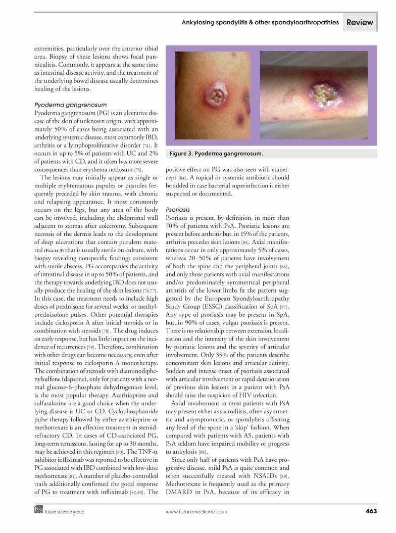
Pyoderma gangrenosumPyoderma gangrenosum (PG) is an ulcerative disease of the skin of unknown origin, with approximately 50% of cases being associated with an underlying systemic disease, most commonly IBD, arthritis or a lymphoproliferative disorder [74]. It occurs in up to 5% of patients with UC and 2% of patients with CD, and it often has more severe consequences than erythema nodosum [75].
The lesions may initially appear as single or multiple eryhtematous papules or pustules frequently preceded by skin trauma, with chronic and relapsing appearance. It most commonly occurs on the legs, but any area of the body can be involved, including the abdominal wall adjacent to stomas after colectomy. Subsequent necrosis of the dermis leads to the development of deep ulcerations that contain purulent material (Figure 3) that is usually sterile on culture, with biopsy revealing nonspecific findings consistent with sterile abscess. PG accompanies the activity of intestinal disease in up to 50% of patients, and the therapy towards underlying IBD does not usually produce the healing of the skin lesions [76,77]. In this case, the treatment needs to include high doses of prednisone for several weeks, or methylprednisolone pulses. Other potential therapies include ciclosporin A after initial steroids or in combination with steroids [78]. The drug induces an early response, but has little impact on the incidence of recurrences [79]. Therefore, combination with other drugs can become necessary, even after initial response to ciclosporin A monotherapy. The combination of steroids with diaminodiphenylsulfone (dapsone), only for patients with a normal glucose6phosphate dehydrogenase level, is the most popular therapy. Azathioprine and sulfa salazine are a good choice when the underlying disease is UC or CD. Cyclophosphamide pulse therapy followed by either azathioprine or methotrexate is an effective treatment in steroidrefractory CD. In cases of CDassociated PG, longterm remissions, lasting for up to 30 months, may be achieved in this regimen [80]. The TNFα inhibitor infliximab was reported to be effective in PG associated with IBD combined with lowdose metho trexate [81]. A number of placebocontrolled trails additionally confirmed the good response of PG to treatment with infliximab [82,83]. The
positive effect on PG was also seen with etanercept [84]. A topical or systemic antibiotic should be added in case bacterial superinfection is either suspected or documented.
PsoriasisPsoriasis is present, by definition, in more than 70% of patients with PsA. Psoriatic lesions are present before arthritis but, in 15% of the patients, arthritis precedes skin lesions [85]. Axial manifestations occur in only approximately 5% of cases, whereas 20–50% of patients have involvement of both the spine and the peripheral joints [86], and only those patients with axial manifestations and/or predominately symmetrical peripheral arthritis of the lower limbs fit the pattern suggested by the European Spondyloarthropathy Study Group (ESSG) classification of SpA [87]. Any type of psoriasis may be present in SpA, but, in 90% of cases, vulgar psoriasis is present. There is no relationship between extension, localization and the intensity of the skin involvement by psoriatic lesions and the severity of articular involvement. Only 35% of the patients describe concomitant skin lesions and articular activity. Sudden and intense onset of psoriasis associated with articular involvement or rapid deterioration of previous skin lesions in a patient with PsA should raise the suspicion of HIV infection.
Axial involvement in most patients with PsA may present either as sacroiliitis, often asymmetric and asymptomatic, or spondylitis affecting any level of the spine in a ‘skip’ fashion. When compared with patients with AS, patients with PsA seldom have impaired mobility or progress to ankylosis [88].
Since only half of patients with PsA have progressive disease, mild PsA is quite common and often successfully treated with NSAIDs [89]. Methotrexate is frequently used as the primary DMARD in PsA, because of its efficacy in
Figure 3. Pyoderma gangrenosum.

Future Rheumatol. (2008) 3(5)464 future science group
Review Schiotis, Escudero-Contreras, Vazquez-Mellado & Collantes-Estévez
treating both skin and joint involvement and its low cost [90,91]. Sulfasalazine showed modest benefit in patients with PsA [92], while leflunomide showed a 59% probability of achieving Psoriatic Arthritis Response Criteria (PsARC) in patiens in a randomized, doubleblind, placebocontrolled study [93]. Other DMARDs, including antimalarials, ciclosporin and gold, are less frequently used because evidence for their efficacy is less convincing than for methotrexate, sulfasalazine and leflunomide [ 89].
Most dermatologists avoid systemic corticosteroids in the treatment of patients with psoriasis because of the potential risk of pustular and erythrodermic flares when discontinued. However, rheumatologists often use systemic cortico steroids in the short and longterm treatment of PsA, in small dosages (5–10 mg/day). Shortterm data indicated that both etanercept [94] and infliximab can delay radiographic progression in patients with PsA [95]. For adalimimab it was also shown to reduce signs, symptoms and disability in patients with active PsA, as well as improving psoriasis lesions in patients [96]. In a recent study, the biologic treatment (infliximab, etanercept and adalimumab) for PsA lead to a sustained response in longterm therapy with a low rate of non response; the majority of nonresponder patients had responded to second and thirdline therapy [97]. There are published cases describing psoriasis or psoriasiform lesions after TNFα antagonist therapy in some patients. The development of psoriasis was seen in all types of inflammatory diseases treated with TNFα antagonists [98], and it is considered a class effect that has been reported with all the currently available TNFα antagonists [99]. The prevalence of this adverse effect has been estimated at 1.5–5% of patients taking TNFα antagonists [100]. The skin lesions develop within the first few months of therapy, palmoplantar pustulosis being the most common feature [99]. Out of the three antiTNF agents, a previous history of psoriasis seemed more common in patients who experience psoriasis onset or exacerbation during etanercept therapy (55%). Thus, previous psoriasis may be a risk factor for psoriasis exacerbation during etanercept therapy [100].
Non-concept extra-articular manifestations
Cardiac manifestationsIn daily clinical practice symptoms of heart involvement in AS patients are rarely seen. Based on published findings, clinically significant cardiac disease occurs only in 2–10% of patients [101].
Recent studies have shown that cardio vascular complications in SpA also include impaired endothelial function and coronary micro vascular function, suggesting that vascular pathologies such as atherosclerosis could also be involved in the increased cardiovascular mortality rates. Potential mechanisms for cardiovascular complications include a chronic inf lammatory condition with increased levels of circulating cytokines and acute phase reactants and a more atherogenic lipid profile. The beneficial effects of statin treatment on circulating inflammatory mediators and atherogenic lipid profiles could reveal new therapeutic options for patients with SpA [102].
Three types of cardiac lesions can be found in AS patients: valvular pathology, conduction disturbances of the atrioventricular node and myocardial involvement.
Valvular pathologyValvular pathology consists of aortitis and aortic insufficiency (isolated aortic regurgitation, aortic root and ascendant aorta dilatation), sometimes with the need for cardiac surgery. In addition, the affectation of the anterior mitral valve leaflet fibrosis and mitral regurgitation may be seen. These abnormalities are the result of the sclerosing inflammatory process, involving aortic root, aortic valve cusps that may extend into the ventricular septum and the atrioventricular node, leading to conduction disorders. The prevalence of subclinical aortic root and valve disease may be as high as 82% in AS patients when transesophageal echocardiography is performed [103].
Conduction disturbances of the atrioventricular nodeConduction disturbances of the atrioventricular node are in most cases silent incomplete blocks, revealed through routine electrocardiograms, and are only in a few cases complete blocks that require a pacemaker. Screening for a prolonged QT time has been recommended since it might be associated with arrhythmias in asymptomatic patients [104].
Myocardial involvementSeveral studies have reported cardiac abnormalities of left ventricular diastolic function [105–107]; nevertheless, cardiologic evaluation with echocardiography should not be recommended routinely in patients with longstanding AS [105].
Although the prevalence of cardiac abnormalities does not seem to be related to therapy or the severity of the skeletal disease, cardiac
www.futuremedicine.com 465future science group
Ankylosing spondylitis & other spondyloarthropathies Review
manifestations have been associated with age over 45 years and disease duration of longer than 15 years [108].
Pleuropulmonary manifestationsLung involvement in AS was initially described in 1941, but it has only been since 1965 that it was considered as an extraarticular manifestation of the disease. Respiratory abnormalities have been reported in from 0% to over 30% of AS patients [109].
Pleuropulmonary lesions are believed to be an uncommon and delayed manifestation of AS. Histological studies of transbronchial biopsies found nonspecific interstitial fibrosis with degeneration of collagen and elastic fibers and, in some cases, focal lymphocytic infiltrates were seen. In patients with advanced disease, cystic cavities were visible within the fibrous areas. No granulomas or signs of vasculitis were found [110–112].
The highresolution computed tomography (HRCT) changes included apical fibrosis, interstitial lung disease, emphysema, bronchiectasis and pleural thickening. In general, the HRCT changes were of a mild degree, and no correlation was observed between HRCT abnormalities, pulmonary function test parameters and the indices of AS. Spontaneous pneumothorax was reported to be a rare complication, but tended to occur in those patients with fibrobullous disease [113].
Respiratory function impairmentCostovertebral joint involvement, which leads to chest wall rigidity, is responsible for the restrictive ventilation pattern of the disease. Although expiratory muscle performance is maintained, the chest wall rigidity causes muscle exhaustion, so that complete expiration cannot be achieved. Vital capacity (VC) is proportionately decreased with disease severity, but patients with advanced disease rarely have a VC of less than 60% of that predicted. Residual volume (RV) and functional residual capacity (FRC) are increased or normal, in contrast to findings in other restrictive diseases. Thus, the decrease in total lung capacity (TLC = VC + RV) is less important than the decrease in VC [114].
Pulmonary fibrosisApical fibrosis is the most recognized pulmonary manifestation of AS, and it develops in the upper lung lobes and follows a progressive course. The frequency of upper lobe fibrosis is variable in different published series. A retrospective review of 1028 AS patients found 22 patients (2.1%) with apical lung fibrosis detected on chest
radiographs [115]. Pulmonary fibrosis associated with AS by its location in the upper part of the lungs is, in contrast with the basal involvement, frequently seen in pulmonary fibrosis associated to other systemic diseases [111,116,117]. The main complications associated with apical fibrosis are infections (Aspergillus and atypical mycobacteria) and pneumothorax. Smoking is widely believed to promote apical fibrosis and interstitial inflammation. Exposure to tobacco smoke is associated with increased macrophages and neutrophil counts in the deep lung parenchyma [118].
Pleural & parenchyma involvementThickening of the apical pleura is commonly concomitant with apical fibrosis, although it may precede the fibrosis, indicating that the two abnormalities are related to each other. Apical bronchiectasis is usually an asymptomatic lesion, due to the airways traction by the fibrotic lung parenchyma, which in its early stages is not visible on chest xrays [119].
Chest xray should be routinely obtained in AS patients, and HRCT should be considered in patients with normal radiographic findings, but abnormal physical findings [120]. In addition, with the increasing use of antiTNFα agents in patients with AS, both HRCT and bronchioloalveolar lavage are useful for ruling out TB before treatment initiation in patients with chest radiographic abnormalities. Physicians should be aware of the abnormalities associated with AS, so that they can distinguish them from changes caused by TB or other infections. Prophylactic measures, including smoking cessation and physiotherapy to improve overall and respiratory function, are useful. It is not known if early appropriate treatment of AS decreases the risk of pleuropulmonary involvement [116.118]. Lifethreatening pulmonary complications include massive hemoptysis secondary to Aspergillus [121], and cardiorespiratory failure due to bacterial infection.
Renal manifestationsThe most frequent etiology of renal impairment in patients with AS is secondary amyloidosis, affecting approximately 5–13% of the studied populations with AS [122,123], followed by IgA nephropathy, mesangioproliferative glomerulonephritis, membranous nephropathy, focal segmental glomerulosclerosis and focal proliferative glomerulonephritis [124].
Treatmentassociated nephrotoxicity may result in NSAID nephropathy or diseasemodifying agent toxicity, which are used in SpA on a longterm basis. The nephrotoxic potential of

Future Rheumatol. (2008) 3(5)466 future science group
Review Schiotis, Escudero-Contreras, Vazquez-Mellado & Collantes-Estévez
NSAIDs is well known – renal deterioration may result in tubulointerstitial nephritis or as decrease in medullary blood flow in patients with preexisting renal damage [125].
Amyloid nephropathy is the most frequent renal manifestation seen in AS. Clinically, it appears as a late complication of AS, associated with a longterm inflammatory status, and it is frequently seen in patients who also present peripheral arthritis. Histologically, it is characterized by important glomerular amyloid deposits. Proteinuria is present in 90% of the cases and may be accompanied by microhematuria. The prognosis is related to the extent of renal failure (creatininne >2 mg/dl) and not to the severity of proteinuria. The factors implicated in producing renal failure are not well known, but it has been suggested that vascular deposits of amyloid in the renal vessels could be involved in the progression of renal failure [126].
TNFα has a central role in the pathogenesis of amyloidosis. TNFα, like other proinflammatory cytokines including IL6 and IL1, stimulates hepatocytes to produce serum amyloid A (SAA), which is the precursor of type amyloid A amyloid fibrils [127]. TNFα also plays a role in the proteolysis of SAA to AA amyloid in macrophages. Furthermore, TNFα enables the expression of receptor for advanced glycation end products (RAGE) by macrophages in amyloid deposits. RAGE and amyloid fibrils interaction is responsible for cytotoxicity and tissue damage by signaling the cascade that causes sustained activation of NFκB [128].
Although renal disease is the most significant organ manifestation of secondary amyloidosis, digestive manifestations may also be present, and they together account for 90% of the symptoms associated with secondary amyloidosis. This is why colestasis, diarrheic or pseudoocclusive syndrome due to big amyloid masses in intestinal sub mucosa may occur. In rare cases, vascular amyloid deposits can lead to intestinal perforation.
The diagnosis of secondary amyloidosis needs to be demonstrated by either subcutaneous fat biopsy or biopsies taken from rectal submucosa that will confirm the diagnosis.
Diminishing the overproduction of SSA may be achieved by reducing the inflammatory status of the disease. It was shown that the adequate treatment of AS also diminishes proteinuria and slows down renal failure, but disease flares were associated with the reactivation of proteinuria [129]. There is literature data that have shown the efficacy of azathioprine or methotrexate [130,131] in patients. Chlorambucil
and cyclophosphamide have also shown some efficacy in renal amyloidosis, as compared with historical controls [132]. However, the risk of myelotoxicity, leukemia, and sterility [133] is to be considered. Colchicine has also been used for proteinuria; some studies even described histological regression of amyloid deposits [134].
Although there are few published reports about the efficacy of antiTNF agents in secondary AA, they are a promising treatment as they reduce amyloid load and disrupt RAGEinduced propagation of amyloidogenesis and tissue damage. It is known that patients with amyloidosis can have renal, hepatic and/or cardiac failure that might alter the pharmacokinetics of antiTNF and/or the tolerance to treatment. These patients can also be at high risk for infections and thrombosis. Etanercept had been used as a choice treatment for amyloidotic renal involvement complicating AS and it was well tolerated, rapid and highly effective in suppressing proteinuria and stabilizing renal function [127,135].
IgA nephropathyIgA nephropathy represents a rare cause of glomerulo nephritis associated with AS. Histologically, there are mesangial deposits indistinguishable from primary IgA glomerulonephritis (Berger’s disease). High levels of IgA in the serum of patients with AS have shown association with disease activity in some studies [136]. There is no specific treatment for the condition – in advanced cases dialysis and renal transplant are the only possible therapeutic choice.
Osteoporosis & vertebral fracturesThe association of AS and osteoporosis (OP) has been described by several studies since the 1990s. It was usually the AS that was diagnosed first, although occasionally OP may be the presenting feature [137].
An increased prevalence of axial OP occurs even in early, mild forms of AS, and the de mineralization process continues for many years until the advanced stages of the disease. In AS patients, OP is largely confined to the axial skeleton, in contrast to the pattern of OP seen in rheumatoid arthritis. OP is also more common in patients with syndesmophytes, cervical fusion and peripheral joint involvement. These variables are not all independent, as they may be indicators of disease duration.
Cytokines such as TNFα and IL6 may play an important part in the pathogenesis of OP in early AS, and IL6 levels have been correlated with markers of disease activity and severity. In
www.futuremedicine.com 467future science group
Ankylosing spondylitis & other spondyloarthropathies Review
late AS, mechanical factors such as decreased mobility and the support provided by extra spinal bone may play a role in vertebral OP [138]. The rigid spine of spondylitic patients is at risk of fracture, deformities and kyphosis. Increased hyperkyphosis can, therefore, be regarded not only as a clinical consequence of the disease, but also as an indicator of vertebral fractures [139].
Measurements of bone formation and bone degradation markers may provide insight into the pathogenesis of OP in AS. Currently, the data are scarce for determining what role they should play in routine clinical management. Both decreased bone formation and increased bone catabolism have been reported in patients with AS, although there is no clear consensus of findings between different groups. For this reason it is difficult to justify the use of such markers in clinical practice.
Measurements of 25OHD3, 1,25OHD
3 and
PTH were normal, while osetocalcin level was low in AS patients studied by Franck and Keck [139], but abnormal for Lange et al. [140], who observed decreased levels of 1,25OHD
3 and
PTH in patients with AS. These studies emphasize the necessity of simultaneous measurement of many indicators of bone formation and resorption [138]. Bone formation and resorption biochemical markers do not correlate with bone mineral density (BMD) of the lumbar spine or femoral neck [141,142]. No significant differences were seen in any marker of bone turnover in AS patients with vertebral fractures compared with AS patients without vertebral fractures [143].
Bone mineral density measurement early in the disease should include dual xray absorptiometry of both spine and hip, as BMD may be severely reduced at these sights. The development of syndesmophytes in late AS can lead to difficulties in lumbar dual energy xray absorptiometry (DEXA) interpretation, as they may hide osteoporotic vertebras. In this regard, more accurate assessment of lumbar BMD, and one that correlates better with femoral neck BMD, may be obtained by quantitative CT scanning or DEXA scanning of the lateral aspect of L3 vertebra [138].
The main clinical manifestations of OP are due to vertebral fracture. In daily practice, vertebral fractures can be overlooked, even when they are symptomatic, as acute and chronic back pain is common in AS patients. Vertebral pain is frequently attributed to disease activity, as long as the vertebral fracture is not considered in the differential diagnosis of back pain. The delay in diagnosis of the vertebral fractures in patients
with preexisting back pain is responsible of increased morbidity in AS patients [144]. The prevalence and incidence of vertebral fractures is therefore influenced by the definition of the degree of deformation of the vertebral body. In crosssectional studies, there are controversies regarding which deformities of vertebral bodies should be considered fractures. In addition, there are no available data about the prevalence of vertebral deformities/fractures in the young adult population, which make it difficult to interpret them in AS patients younger than 50 years of age. In 2006, seven new case reports were published in addition to the already long list on distinctive characteristics of some aspects of vertebral fractures in AS [145–147].
These fractures are characterized by their unusual location (e.g., at the cervical spine, involvement of the posterior arch of the vertebrae), their anatomic location (transvertebral with variable degrees of dislocation, transdiscal through the syndesmophytes) and their complications with variable degrees of neurological deficits [148].
In strong contrast with postmenopausal OP, there are reports that suggest the presence of neurological complications after fractures of the vertebral body or its dorsal components in patients with AS. These complications include spinal cord or nerve root lesions, and the occurrence of paravertebral hematomas resulting in variable degrees of sensory or motor deficits. Due to defective fracture healing, pseudoarthrosis with instability in the posterior arch structures of the vertebrae can occur [148]. The resulting neurological deficit ranges from mild sensory loss to complete paraplegia, with a higher incidence of spinal cord injury among patients with AS. Vertebral compression fractures are more frequent in AS males (13.7%) than females (8.3%), and increase with age, disease duration and spinal restriction, the presence of peripheral joint involvement and more extensive syndesmophyte formation [148].
If there is suspicion of vertebral fractures, radiographs of the spine should be performed. Additional imaging techniques are indicated in cases of persistent posttraumatic pain, in order to avoid delays in diagnosis and therapy.
The treatment of OP in AS is at present similar to that used for OP, although in this population, most being male and young, there is a limited role for hormonereplacement therapy, exercise regimens and bisphosphonates being widely used. Drug treatment for OP should be considered in patients with vertebral fractures or a BMD with a T score of less than 2.5. From
Future Rheumatol. (2008) 3(5)468 future science group
Review Schiotis, Escudero-Contreras, Vazquez-Mellado & Collantes-Estévez
a pathophysiological perspective, the target for avoiding vertebral fractures is not only to prevent bone loss within the vertebrae, but also to prevent excessive bone formation around the vertebrae. Recently, Demis et al. observed that the antiTNFα therapies are effective in this type of OP, probably due to their capacity to inhibit the inflammatory response, as well as balancing the bone remodeling [149].
Neurological manifestationsAtlantoaxial subluxation is a latephase complication, affecting 2% of patients, less than in rheumatoid arthritis and, in contrast with rheumatoid arthritis, it is not accompanied by neurological symptomatology [150]. Isolated noncompressive thoracic and lumbar lesions are associated with disease activity and are typically transitory. Cauda equina syndrome affects patients with advanced AS, and is due to increased pressure of the cerebrospinal fluid, which determines arachnoidal dilatation and cyst formation, finally affecting nerve roots down to D12. Other authors consider this is secondary to arachnoiditis, which would be responsible for initiating the symptomat ology. Clinical features consist of sensitive disturbances of the inferior limbs and of the perinea region. Motor and sphincter alterations (rectal and urinary) are less frequent at initial phases, and become evident with late disease. The diagnosis is clinical and it is sustained by CT and MRI findings. Electromyography is not necessary for diagnosis, although it is useful in evaluating the extent of neurological lesions and their progression in time.
There is no effective treatment, either medical or surgical, for this condition, but rehabilitation for the neurological deficit may be helpful [151].
Expert commentary & future perspectiveSince the introduction of TNF blockers, important changes have been obtained in the therapeutic response previously not known with molecules such as NSAIDs, methotrexate or sulfasalazine. Nevertheless, there is literature data that shows a lack of a suitable therapeutic response in at least 30% of the patients treated with these new antiTNF therapies.
New molecules have been developed for the control and treatment of patients with AS (golimumab, abatacept and rituximab). These agents were also initially used in patients with rheumatoid arthritis in which they have demonstrated good therapeutic effect in controlling disease activity.
Golimumab, a nextgeneration human antiTNFα monoclonal antibody (CNTO 148), has demonstrated good results in rheumatoid arthritis patients, and also in a Phase III study in AS patients. Golimumab is being studied as a monthly sub cutaneous injection, and as an every12week intravenous infusion (approximately 30min) therapy. According to the findings from the Golimumab – A Randomized Study in Ankylosing Spondylitis Subjects of a Novel AntiTNF mAB Injection (sc.) Given Every Four Weeks (GORAISE) trial, more than half of the patients receiving monthly subcutaneous injections of golimumab 50 mg and 100 mg experienced significant and sustained improvement in the signs
Executive summary
Uveitis is the most frequent extra-articular manifestation of ankylosing spondylitis (AS), with the strongest human leukocyte antigen n
(HLA)-B27 disease association.
Anti-tumor necrosis factor (TNF)-n α therapy has proved to be efficient in controlling uveitis attacks, although there is evidence of uveitis induced by anti-TNF-α therapy in patients.
Gut inflammation is linked to peripheral joint disease in spondylarthropathies (SpA), and there is a strong relationship between gut n
lesions and synovial modifications of the inflamed joints.
Both infliximab and adalimumab are capable of improving inflammatory bowel disease (IBD) associatated with AS and reduce IBD flares n
when compared with etanercept, which is why a positive history of IBD should be considered as contraindication for etanercept therapy in AS.
Psoriasis or psoriasiform exanthema may be induced by all anti-TNF agents – skin lesions develop within the first few months of therapy, n
with palmoplantar pustulosis being the most common feature.
Screening for prolonged QT time has been recommended, since it might be associated with asymptomatic arrhythmias in AS patients.n
Pulmonary fibrosis in AS is typically located in the upper lungs, in contrast with the pulmonary fibrosis due to other systemic diseases.n
Secondary amyloidosis is a late complication of AS, being associated with a long inflammatory status in patients in which renal and n
digestive symptoms are the most frequent, but peripheral arthritis is also seen in patients. There are studies that have demonstrated the efficiency of biological therapy in treating this complication, particularly etanercept.
Axial osteoporosis is an early manifestation of AS. Bone mineral density measurements should be made early in the disease course and n
should include dual x-ray absorptiometry measurement of both the spine and hip.
Vertebral fractures of osteoporotic spine occur in unusual locations and, in contrast with postmenopausal osteoporosis, present a n
higher degree of neurological complications.
www.futuremedicine.com 469future science group
Ankylosing spondylitis & other spondyloarthropathies Review
and symptoms of active AS. Clinical benefit was demonstrated as early as week 4 after one dose, and was maintained through week 24 [152].
Compounds such as abatacept (CTLA4 Ig recombinat protein that blocks the process of costimulation with the CD 80/86 and CD28) and rituximab (anti CD20) have shown excellent results in rheumatoid arthritis patients, which have motivated new clinical trials in AS. An openlabel pilot clinical trial with abatacept in AS (AbaAS01 [201]) investigates the efficacy and safety of abatacept in 30 AS patients from baseline up to week 30. Abatacept will be administered intravenously according to the prescription used in rheumatoid arthritis.
The rituximab trial is a Phase II openlabel clinical trial in AS [202] for evaluating the efficacy and safety of rituximab when added to NSAIDs and/or methotrexate both for TNFα inhibitornaive patients or patients who have experienced TNFα inhibitor failure, in patients with moderate to severe AS.
These are Phase II trials, in the recruitment stage, with good expectations regarding the results of management of the approach for AS patients.
Added to the development of ambulatory consults focused on an early diagnosis of AS, these new therapies could open an encouraging horizon for the patients with early diagnosis, by providing a more effective new therapeutic approach and also by avoiding the possible late complications of the disease.
Financial & competing interests disclosureThe authors have no relevant affiliations or financial involvement with any organization or entity with a financial interest in or financial conflict with the sub‑ject matter or materials discussed in the manuscript. This includes employment, consultancies, honoraria, stock ownership or options, expert testimony, grants or patents received or pending, or royalties.
No writing assistance was utilized in the production of this manuscript.
BibliographyPapers of special note have been highlighted as:n of interestnn of considerable interest
Wright V: Seronegative polyarthritis: 1.
a unified concept. Arthritis Rheum. 21, 619–633 (1978).
Rudwaleitt M, Khan MA, Sieper J: 2.
The challenge of diagnosis and classification in early ankylosing spondylitis. Arthritis Rheum. 52, 1000–1008 (2005).
Mc Gonagle D, Khan MA, MarzoOrtega H, 3.
O’Conor P, Gibbono W, Emery P: Enthesitis in spondylarthropaty. Curr. Opin. Rheum. 11, 244–250 (1999).
D’Agostino MA, Olivieri I: Enthesitis. 4.
Res. Clin. Rheumatol. 20, 473–487 (2006).
Braun J, Pincus T: Mortality, course of 5.
disease and prognosis of patients with ankylosing spondylitis. Clin. Exp. Rheumatol. 20, S16–S22 (2002).
Braun J: Epidemiology and prognostic aspects 6.
of ankylosing spondylitis. Radiologe 44, 209–10, 212–216 (2004).
Braun J, Baraliakos X, Listing J, Sieper J: 7.
Decreased incidence of anterior uveitis in patients with ankylosing spondylitis treated with the antitumor necrosis factor agents infliximab and etanercept. Arthritis Rheum. 52, 2447–2451 (2005).
Demonstrated the efficacy of the nn
anti-TNF-α agents in treating uveitis.
Collantes E, Zarco P, Muñoz E 8. et al.: Disease pattern of spondyloarthropathies in Spain. Rheumatology (Oxford) 46, 1309–1315 (2007).
Monnet D, Moachon L, Dougados M, 9.
Brézin AP: Severe uveitis in an HLAB27positive patient with ankylosing spondylitis. Nat. Clin. Pract. Rheum. 2, 393–397 (2006).
Chang JH, McCluskey PJ, Wakefield D: 10.
Acute anterior uveitis and HLAB27. Surv. Ophthalmol. 50, 364–388 (2005).
Brewerton DA, Caffrey M, Nicholls A 11. et al.: Acute anterior uveitis and HLA 27. Lancet 2, 994–996 (1973).
Beckingsale B, Davies J, Gibson JM, 12.
Rosenthal AR: Acute anterior uveitis, ankylosing spondylitis, back pain, and HLAB27. Br. J. Ophthalmol. 68, 741–745 (1984).
Linssen A, Meenken C: Outcomes of 13.
HLAB27positive and HLAB27negative acute anterior uveitis. Am. J. Ophthalmol. 120, 351–361 (1995).
Monnet D, Breban M, Hudry C 14. et al.: Ophthalmic findings and frequency of extraocular manifestations in patients with HLAB27 uveitis: a study of 175 cases. Ophthalmology 111, 802–809 (2004).
Rothova A, Buitenhuis HJ, Christiaans BJ 15.
et al.: Acute anterior uveitis (AAU) and HLAB27. Br. J. Rheumatol. 22, 144–145 (1983).
ElShabrawi Y, Hermann J: Antitumor 16.
necrosis factorα therapy with infliximab as an alternative to corticosteroids in the treatment of human leukocyte antigen B27associated acute anterior uveitis. Ophthalmology 109, 2342–2346 (2002).
PérezGuijo V, SantosLacomba M, 17.
SánchezHernandez M et al.: Tumour necrosis factorα levels in aqueous humour and serum from patients with uveitis: the involvement of HLAB27. Cur. Med. Res. Opin. 20, 155–157 (2004).
Santos Lacomba M, Marcos Martin C, 18.
Gallardo Galera JM et al.: Aqueous humor and serum necrosis factor α in clinical uveitis. Ophtalmic Res. 33, 251–255 (2001).
Sugita S, Takase H, Taguchi C, 19.
Mochizuki M: The role of soluble TNF receptors for TNFα in uveitis. Invest. Ophthalmol. Vis. Sci. 48, 3246–3252 (2007).
The first study to show the involvement of nn
the soluble receptor of TNF-α in uveitis.
Gritz DC, Wong IG: Incidence and 20.
prevalence of uveitis in Northern California: the Northern California Epidemiology of Uveitis Study. Ophthalmology 111, 491–500 (2004).
Guignard S, Gossec L, Salliot C 21. et al.: Efficacy of tumour necrosis factor blockers in reducing uveitis flares in patients with spondylarthropathy: a retrospective study. Ann. Rheum. Dis. 65, 1631–1634 (2006).
Future Rheumatol. (2008) 3(5)470 future science group
Review Schiotis, Escudero-Contreras, Vazquez-Mellado & Collantes-Estévez
Reddy AR, Backhouse OC: Does etanercept 22.
induce uveitis? Br. J. Ophthalmol. 87, 925 (2003).
Demonstrated that etanercept is associated nn
with more frequent uveitis flares.
Kasner L, Chan CC, Whitcup SM, Gery I: 23.
The paradoxical effect of tumor necrosis factor α (TNFα) in endotoxininduced uveitis. Invest. Ophthalmol. Vis. Sci. 34, 2911–2917 (1993).
Lim LL , Fraunfelder FW, Rosenbaum JT: 24.
Do tumor necrosis factor inhibitors cause uveitis? A registrybased study. Arthritis Rheum. 56, 3248–3252 (2007).
Scrivo R , Spadaro A, Spinelli FR, Valesini G: 25.
Uveitis following the use of tumor necrosis factorα inhibitors: Comment on the article by Lim et al. Arthritis Rheum. 58, 1555–1556 (2008).
MuñozFernández S, Hidalgo V, Fernández26.
Melón J et al.: Sulfasalazine reduces the number of flares of acute anterior uveitis over a oneyear period. J. Rheumatol. 30, 1277–1279 (2003).
MuñozFernández S, GarcíaAparicio AM, 27.
Hidalgo MV et al.: Methotrexate: an option for preventing the recurrence of acute anterior uveitis. Eye DOI 10.1038/eye.2008.198 (2008) (Epub ahead of print).
Choudhary A, Harding SP, Bucknall RC, 28.
Pearce IA: Mycophenolate mofetil as an immunosuppressive agent in refractory inflammatory eye disease. J. Ocul. Pharmacol. Ther. 22, 168–175 (2006).
Galor A, Jabs DA, Leder HA 29. et al.: Comparison of antimetabolite drugs as corticosteroidsparing therapy for noninfectious ocular inflammation. Opthalmology DOI 10.1016/j.ophtha.2008.04.026 (2008) (Epub ahead of print).
De Vos M, Mielants H, Cuvelier C 30. et al.: Longterm evolution of gut inflammation in patients with spondyloarthropathy. Gastroenterology 110, 1696–1703 (1996).
De Keyser F, Baeten D, van den Bosch F, 31.
de Vos M et al.: Gut inflamation and spondylarthropathies. Curr. Rheumatol Rep. 4, 252–253 (2002).
Mielants H, Veys EM, Cuvelier C, De Vos M: 32.
Course of gut inflammation in spondylarthropathies and therapeutic consequences. Baillieres Clin. Rheumatol. 10, 147–164 (1996).
Mielants H, Veys EM, De Vos M 33. et al.: The evolution of spondyloarthropathies in relation to gut histology. Part I: clinical aspects. J. Rheumatol. 22, 2266–2272 (1995).
Important review of the relation between n
the gut and the development of spondylarthropathies (SpA).
Mielants H, Veys EM, De Vos M 34. et al.: The evolution of spondyloarthropathies in relation to gut histology. Part II: histological aspects. J. Rheumatol. 22, 2273–2278 (1995).
Important review of the relation between n
the gut and the development of SpA.
Mielants H, Veys EM, Cuvelier C 35. et al.: The evolution of spondyloarthropathies in relation to gut histology. Part III: relation between gut and joint. J. Rheumatol. 22, 2279–2284 (1995).
Important review of the relation between n
the gut and the development of SpA.
Schwimmbeck PL, Oldstone MB: Molecular 36.
mimicry between human leukocyte antigen B27 and Klebsiella. Consequences for spondyloarthropathies. Am. J. Med. 85(6A), 51–53 (1988).
Perez Guijo VC, Collantes E: Flora intestinal 37.
y espondiloartropatías. In: Monografias SER: Artritis Infecciosas. Sociedad Española de Reumatología (Ed.), Panamericana, Madrid, 269–279 (2006).
MakiIkola O, Nissila M, Lhetinen K, 38.
Grandfors K: IgA class serum antibodies against three different Klebsiella serotypes in ankylosing spondylitis. Br. J. Rheumatol. 37, 1299–1302 (1998).
Stone MA, Payne U, Schentag C, Rahman P, 39.
PachecoTena C, Inman RD: Comparative immune responses to candidate arthritogenic bacteria do not confirm a dominant role for Klebsiella pneumonia in the pathogenesis of familial ankylosing spondylitis. Rheumatology (Oxford) 43, 148–155 (2004).
de Vlam K, Mielants H, Cuvelier C 40. et al.: Spondyloarthropathy is underestimated in inflammatory bowel disease: prevalence and HLA association. J. Rheum. 27, 2860–2865 (2000).
Podswiadek M, Punzi L, Stramare R 41. et al.: The prevalence of radiographic sacroiliitis in patients affected by inflammatory bowel disease with inflammatory low back pain. Reumatismo 56, 110–113 (2004).
Enlow RW, Bias WB, Arnett FC: 42.
The spondylitis of inflammatory bowel disease. Evidence for a non HLA linked axial arthropathy. Arthritis Rheum. 23, 1359–1365 (1980).
Steer S, Jones H, Hibbert J 43. et al.: Low back pain, sacroiliitis, and the relationship with HLAB27 in Crohn’s disease. J. Rheumatol. 30, 518–522 (2003).
Laukens D, Peeters H, Marichal D 44. et al.: CARD15 gene polymorphisms in patients with spondyloarthropathies identify a specific phenotype previously related to Crohn’s disease. Ann. Rheum. Dis. 64, 930–935 (2005).
Offers an explanation for the low incidence n
of HLA-B27 positivity in ankylosing spondylitis (AS) secondary to inflammatory bowel disease.
Peeters H, Van der Cruyssen B, Laukens D 45.
et al.: Radiological sacroiliitis, a hallmark of spondylitis, is linked with CARD15 gene polymorphisms in patients with Crohn’s disease. Ann. Rheum. Dis. 63, 1131–1134 (2004).
Queiro R, Maiz O, Intxausti J 46. et al.: Subclinical sacroiliitis in inflammatory bowel disease: a clinical and followup study. Clin. Rheumatol. 19, 445–449 (2000).
Rudwaleit M: Ankylosing spondylitis and 47.
bowel disease. Best Prac. Res. Clin. Rheumatol. 20, 451–471 (2006).
Review of the entire mechanisms of gut nn
involvement in AS, pointing out the management of the condition.
Smale S, Natt RS, Orchard TR, Russell AS, 48.
Bjarnason I: Inflammatory bowel disease and spondyloarthropathy. Arthritis Rheum. 44, 2728–2736 (2001).
Zochling J, van der Heijde D, 49.
BurgosVargas R et al.: ASAS/EULAR recommendations for the management of ankylosing spondylitis. Ann. Rheum. Dis. 65, 442–452 (2006).
Dougados M, Dijkmans B, Khan M 50. et al.: Conventional treatments for ankylosing spondylitis. Ann. Rheum. Dis. 61(Suppl. 3), iii40–iii50 (2002).
Haibel H, Rudwaleit M, Braun J, Sieper J: 51.
Six months open label trial of leflunomide in active ankylosing spondylitis. Ann. Rheum. Dis. 64, 124–126 (2005).
Haibel H, Rudwaleit M, Listing J, Sieper J: 52.
Open label trial of anakinra in active ankylosing spondylitis over 24 weeks. Ann. Rheum. Dis. 64, 296–298 (2005).
Felder JB, Korelitz BI, Rajapakse R 53. et al.: Effects of nonsteroidal antiinflammatory drugs on inflammatory bowel disease: a case–control study. Am. J. Gastroenterol. 95, 1949–1954 (2000).
Cipolla G, Crema F, Sacco S 54. et al.: Nonsteroidal antiinflammatory drugs and inflammatory bowel disease: current perspectives. Pharmacol. Res. 46, 1–6 (2002).
Guslandi M: Exacerbation of inflammatory 55.
bowel disease by nonseroidal antiinflammatory drugs and cyclooxigenase2 inhibitors: fact or fiction? World J. Gastroenterol. 12, 1509–1510 (2006).
Bonner GF, Walczak M, Kitchen L, 56.
Bayona M: Tolerance of nonsteroidal antiinflammatory drugs in patients with inflammatory bowel disease. Am. J. Gastroenterol. 95, 1946–1948 (2000).

www.futuremedicine.com 471future science group
Ankylosing spondylitis & other spondyloarthropathies Review
Braun J , Baraliakos X, Listing J 57. et al.: Differences in the incidence of flares or new onset of inflammatory bowel diseases in patients with ankylosing spondylitis exposed to therapy with antitumor necrosis factorα agents. Arthritis Care Res. 57, 639–647 (2007).
Ascertains the difference between the nn
available biological treatment upon their intestinal action.
Doubremelle M, Bourreille A, Zerbib F 58. et al.: Treatment of Crohn’s disease with antiTNFα antibodies (infliximab): results of a multicentric and retrospective study. Gastroenterol. Clin. Biol. 26, 973–979 (2002).
Hanauer SB, Feagan BG, Lichtenstein GR 59.
et al.: Maintenance infliximab for Crohn’s disease: the ACCENT I randomised trial. Lancet 359(9317), 1541–1549 (2002).
Papadakis KA, Shaye OA, Vasiliauskas EA 60.
et al.: Safety and efficacy of adalimumab (D2E7) in Crohn’s disease patients with an attenuated response to infliximab. Am. J. Gastroenterol. 100, 75–79 (2005).
Hanauer SB, Sandborn WJ, Rutgeerts P 61. et al.: Human antitumor necrosis factor monoclonal antibody (adalimumab) in Crohn’s disease: the CLASSICI trial. Gastroenterology 130, 323–333 (2006).
Rutgeerts P, Sandborn WJ, Feagan BG 62. et al.: Infliximab for induction and maintenance therapy for ulcerative colitis. N. Engl. J. Med. 353, 2462–2476 (2005).
Jarnerot G, Hertervig E, FriisLiby I 63. et al.: Infliximab as rescue therapy in severe to moderately severe ulcerative colitis: a randomized, placebocontrolled study. Gastroenterology 128, 1805–1811 (2005).
D’Haens G, Swijsen C, Noman M 64. et al.: Etanercept in the treatment of active refractory Crohn’s disease: a singlecenter pilot trial. Am. J. Gastroenterol. 96, 2564–2568 (2001).
Sandborn WJ, Hanauer SB, Katz S 65.
et al.:Etanercept for active Crohn’s disease: a randomized, doubleblind, placebocontrolled trial. Gastroenterology 121, 1088–1094 (2001).
Baraliakos X, Brandt J, Listing J 66. et al.: Outcome of patients with active ankylosing spondylitis after two years of therapy with etanercept: clinical and magnetic resonance imaging data. Arthritis Rheum. 53, 856–863 (2005).
MarzoOrtega H, McGonagle D, 67.
O’Connor P, Emery P: Efficacy of etanercept for treatment of Crohn’s related spondyloarthritis but not colitis. Ann. Rheum. Dis. 62, 74–76 (2003).
Haibel H, Rudwaleit M, Brandt HC 68. et al.: Adalimumab reduces spinal symptoms in active ankylosing spondylitis: clinical and
magnetic resonance imaging results of a fiftytwoweek openlabel trial. Arthritis Rheum. 54, 678–681 (2006).
van der Heijde D, Kivitz A, Schiff MH 69. et al.: Efficacy and safety of adalimumab in patients with ankylosing spondylitis: results of a multicenter, randomized, doubleblind, placebocontrolled trial. Arthritis Rheum. 54, 2136–2146 (2006).
Boirivant M, Marini M, Di Felice G 70. et al.: Lamina propria T cells in Crohn’s disease and other gastrointestinal inflammation show defective CD2 pathwayinduced apoptosis. Gastroenterology 116, 557–565 (1999).
Ina K, Itoh J, Fukushima K 71. et al.: Resistance of Crohn’s disease T cells to multiple apoptotic signals is associated with a Bcl2/Bax mucosal imbalance. J. Immunol. 163, 1081–1090 (1999).
Calin A: Keratodermia blennorrhagica and 72.
mucocutaneous manifestations of Reiter’s syndrome. Ann. Rheum. Dis. 38(Suppl.), 68–72 (1979).
Veloso FT, Carvalho J, Magro F: 73.
Immunerelated systemic manifestations of inflammatory bowel disease. A prospective study of 792 patients. J. Clin. Gastroenterol. 23, 29–34 (1996).
Hickman JG: Pyoderma gangrenosum. 74. Clin. Dermatol. 1, 102–13 (1983).
MirMadjlessi SH, Taylor JS, Farmer RG: 75.
Clinical course and evolution of erythema nodosum and pyoderma gangrenosum in chronic ulcerative colitis: a study of 42 patients. Am. J. Gastroenterol. 80, 615–20 (1985).
Troma A, Almus E, Voigt E, Greing I, 76.
Schwegle RU, Gria T: Cutaneous manifestations in inflammatory bowel disease. Z. Gastroenterol. 39, 137–144 (2001).
Menachem Y, Gotsman I: Clinical 77.
manifestations of pyoderma gangrenosum associated with inflammatory bowel disease. Istr. Med. Assoc. J. 6, 88–90 (2004).
Wollina U: Clinical management of 78.
pyoderma gangrenosum. Am J. Clin. Dermatol. 3, 149–158 (2002).
Vidal D, Puig L, Gilaberte M 79. et al.: Review of 26 cases of classical pyoderma gangrenosum: clinical and therapeutic features. J. Dermatol. Treat. 15, 146–152 (2004).
Schmidt C, Wittig BM, Moser C 80. et al.: Cyclophosphamide pulse therapy followed by azathioprine or methotrexate induces longterm remission in patients with steroidrefractory Crohn’s disease. Aliment Pharmacol. Ther. 24, 343–350 (2006).
Girolomoni G, Pastore S, Albanesi C 81. et al.: Targeting tumor necrosis factorα as a
potential therapy in inflammatory skin diseases. Curr. Opin. Investig. Drugs 3, 1590–159 (2002).
Brooklyn TN, Dunnill MG, Shetty A 82. et al.: infliximab for the treatment of pyoderma gangrenosum: a randomized, double blind placebo controlled trial. Gut 55, 505–509 (2006).
Regueiro M, Valentine J, Plevy S, 83.
Fleisher MR, Lichtenstein GR: Infliximab for treatment of pyoderma gangrenosum associated with inflammatory bowel disease. Am. J. Gastroenterol. 98, 1821–1826 (2003).
Roy DB, Conte ET, Cohen DJ: The treatment 84.
of pyoderma gangrenosum using etanercept. J. Am. Acad. Dermatol. 54, S128–S134 (2006).
Gladman DD: Psoriatic arthritis. 85. Rheum. Dis. Clin. North Am. 24, 829–844 (1998).
Helliwell PS, Taylor WR: Classification and 86.
diagnostic criteria for psoriatic arthritis. Ann. Rheum. Dis. 64(Suppl. 2), ii3–ii8 (2005).
Dougados M, Van der Linden S, Juhlin R 87.
et al.: The ESSG preliminary criteria for the classification of spondylarthropathies. Arhtritis Rheum. 34, 1218–1227 (1991).
Gottlieb A, Korman NJ, Gordon K 88. et al.: Guidelines of care for the management of psoriasis and psoriatic arthritis Section 2. Psoriatic arthritis: Overview and guidelines of care for treatment with an emphasis on the biologics. J. Am. Acad. Dermatol. 58, 851–864 (2008).
Nash P, Clegg DO: Psoriatic arthritis therapy: 89.
NSAIDs and traditional DMARDs. Ann. Rheum. Dis. 64(Suppl.), ii74–ii77 (2005).
Black RL, O’Brien WM, Vanscott EJ 90. et al.: Methotrexate therapy in psoriatic arthritis; doubleblind study on 21 patients. JAMA 189, 743–747 (1964).
Willkens RF, Williams HJ, Ward JR 91. et al.: Randomized, doubleblind, placebocontrolled trial of lowdose pulse methotrexate in psoriatic arthritis. Arthritis Rheum. 27, 376–381 (1984).
Clegg DO, Reda DJ, Mejias E 92. et al.: Comparison of sulfasalazine and placebo in the treatment of psoriatic arthritis: a Department of Veterans Affairs cooperative study. Arthritis Rheum. 39, 2013–2020 (1996).
Kaltwasser JP, Nash P, Gladman D 93. et al.: Efficacy and safety of leflunomide in the treatment of psoriatic arthritis and psoriasis: a multinational, doubleblind, randomized, placebocontrolled clinical trial. Arthritis Rheum. 50, 1939–1950 (2004).
Woolacott N, Bravo Vergel Y, Hawkins N 94. et al.: Etanercept and infliximab for the treatment of psoriatic arthritis: a systematic review and economic evaluation. Health Technol. Assess. 10, iii–iv, xiii–xvi, 1–239 (2006).
Future Rheumatol. (2008) 3(5)472 future science group
Review Schiotis, Escudero-Contreras, Vazquez-Mellado & Collantes-Estévez
van der Heijde D, Kavanaugh A, 95.
Gladman DD et al.: Infliximab inhibits progression of radiographic damage in patients with active psoriatic arthritis through one year of treatment: Results from the induction and maintenance psoriatic arthritis clinical trial 2. Arthritis Rheum. 56, 2698–2707 (2007).
Genovese MC, Mease PJ, Thomson GT 96. et al.: Safety and efficacy of adalimumab in treatment of patients with psoriatic arthritis who had failed disease modifying antirheumatic drug therapy. J. Rheumatol. 34, 1040–1050 (2007).
Coates LC, Cawkwell LS, Ng NW 97. et al.: Sustained response to longterm biologics and switching in psoriatic arthritis: results from real life experience. Ann. Rheum. Dis. 67, 717–719 (2008).
Cohen JD, Bournerias I, Buffard V 98. et al.: Psoriasis induced by tumor necrosis factorα antagonist therapy: a case series. J. Rheumatol. 34, 380–385 (2007).
A class side effect of anti-TNF-n α agents important in patients with psoriatic lesions.
Wollina U, Hansel G, Koch A, Schönlebe J, 99.
Köstler E, Haroske G: Tumor necrosis factorα inhibitorinduced psoriasis or psoriasiform exanthemata: first 120 cases from the literature including a series of six new patients. Am. J. Clin. Dermatol. 9, 1–14 (2008).
Wendling D, Balblanc JC, Briançon D 100. et al.: Onset or exacerbation of cutaneous psoriasis during TNFα antagonist therapy. Joint Bone Spine 75, 315–318 (2008).
A newly reviewed class side effect of nn
anti-TNF-α agents important in patients with psoriatic lesions.
Huffer LL, Furgerson JL: Aortic root 101.
dilatation with sinus of valsalva and coronary artery aneurysms associated with ankylosing spondylitis. Tex. Heart Inst. J. 33, 70–73 (2006).
Heeneman S, Daemen MJ: Cardiovascular 102.
risks in spondyloarthritides. Curr. Opin. Rheumatol. 19, 358–62 (2007).
Roldan CA, Chavez J, Wiest PW 103. et al.: Aortic root disease and valve disease associated with ankylosing spondylitis. J. Am. Coll. Cardiol. 32, 1397–1404 (1998).
Yildirir A, Aksoyek S, Calguneri M 104. et al.: QT dispersion as a predictor of arrhythmic events in patients with ankylosing spondylitis. Rheumatology 39, 875–879 (2000).
Yildirir A, Aksoyek S, Calguneri M 105. et al.: Echocardiographic evidence of cardiac involvement in ankylosing spondylitis. Clin. Rheumatol. 21, 129–134 (2002).
Peters MJ, van der HorstBruinsma IE, 106.
Dijkmans BA, Nurmohamed M: Cardiovascular risk profile of patients with spondylarthropathies, particularly ankylosing spondylitis and psoriatic arthritis. Semin. Arthritis Rheum. 34, 585–592 (2004).
Lautermann D, Braun J: Ankylosing 107.
spondylitis – cardiac manifestations. Clin. Exp. Rheumatol. 20, 11–5 (2002).
Brunner F, Kunz A, Weber U, Kissling R: 108.
Ankylosing spondylitis and heart abnormalities: do cardiac conduction disorders, valve regurgitation and diastolic dysfunction occur more often in male patients with diagnosed ankylosing spondylitis for over 15 years than in the normal population? Clin. Rheumatol. 25, 24–29 (2006).
SampaioBarros PD, Elza Maria FP, 109.
Cerqueira FP et al.: Pulmonary involvement in ankylosing spondylitis. Clin. Rheumatol. 26, 225–230 (2007).
Jeandel P, Bonnet D, Chouc PY 110. et al.: Demonstration of subclinical pulmonary alveolitis in spondylarthropathies. Rev. Rhum. Ed. Fr. 61, 301–309 (1994).
Ferdoutsis M, Bouros D, Meletis G, 111.
Patrsourakis G, Siafakas NM: Diffuse interstitial lung disease as an early manifestation of ankylosing spondylitis. Respiration 62, 286–289 (1995).
Kchir MM, Mtimet S, Kochbati S, Zouari R, 112.
Ayed M, Gharb T: Bronchoalveolar lavage and transbronchial biopsy in spondylarthropathies, J. Rheumatol. 19, 913–916 (1992).
Quismorio FP Jr: Pulmonary involvement in 113.
ankylosing spondylitis. Curr. Opin. Pulmonary Med. 12, 342–435 (2006).
Fournié B, Boutes A, Dromer C, Sixou L, 114.
Le Guennec P, Granel J: Prospective study of anterior chest wall involvement in ankylosing spondylitis and psoriatic arthritis. Rev. Rhum. Engl. Ed. 64, 22–25 (1997).
Lee CC, Lee SH, Chang IJ 115. et al.: Spontaneous pneumothorax associated with ankylosing spondylitis. Rheumatol. 44, 1538–1541 (2005).
Richards AJ: Ankylosing spondylitis and 116.
pulmonary apical fibrosis. Proc. R. Soc. Med. 67, 45–46 (1974).
Rumancik WM, Firooznia H, Davis MS Jr, 117.
Leitman BS, Golimbu C, Rafii M: Fibrobullous disease of the upper lobes: an extraskeletal manifestation of ankylosing spondylitis, J. Comput. Tomogr. 8, 225–229 (1984).
Abdellah El Maghraoui: Pleuropulmonary 118.
involvement in ankylosing spondylitis. Joint Bone Spine 72, 496–502 (2005).
Bouros D, Meletis G, Patsourakis G, 119.
Siafakas NM: Diffuse interstitial lung disease
as an early manifestation of ankylosing spondylitis. Respiration 62, 286–289 (1995).
Maghraoui AE, Chaouir S, Abid A 120. et al.: Lung findings on thoracic highresolution computed tomography in patients with ankylosing spondylitis. Correlations with disease duration, clinical findings and pulmonary function testing. Clin. Rheum. 23, 123–128 (2004).
Franchet T, Muller NL, Flint JD: A patient 121.
with ankylosing spondylitis and recurrent hemoptysis. Eur. Respir. J. 23, 488–491 (2004).
Lehtinen K: Mortality and causes of death in 122.
398 patients admitted to hospital with ankylosing spondylitis. Ann. Rheum. Dis. 52, 174–176 (1993).
Escalante A, Weaver WJ, Beardmore TD: 123.
An estimate of the prevalence of reactive systemic amyloidosis in ankylosing spondylitis [letter]. J. Rheumatol. 22, 2192–2193 (1995).
Strobel AS, Fristchka E: Renal diseases on 124.
ankylosing spondylitis. Review of the literature illustrated by cases report. Clin. Rheumatol. 17, 524–530 (1998).
Kirschenbaum MA, Anderson DA: 125.
Nephropathies of nonsteroidal antiinflammatory agents. In: Textbook of nephrology (2nd edition). Massry SG, Glassock RJ (Eds). Williams and Wilkins, Baltimore, USA, 823–828 (1989).
Sasatomi Y, Kiyoshi Y, Uesugi N, Hisano S, 126.
Takebayashi S: Prognosis of renal amyloidosis: a clinicopathological study using cluster analysis. Nephron 87, 42–9 (2001).
Kobak S, Oksel F, Kabasakal Y, 127.
Doganavsargil E: Ankylosing spondylitisrelated secondary amyloidosis responded well to etanercept: a report of three patients. Clin. Rheumatol. 26, 2191–2194 (2007).
Tanaka N, Yonekura H, Yamagishi S, 128.
Fujimori H, Yamamoto Y, Yamamoto H: The receptor for advanced glycation end products is induced by the glycation products themselves and tumor necrosis factorα through nuclear factorκB, and by 17βestradiol through Sp1 in human vascular endothelial cells. J. Biol. Chem. 275, 25781–25790 (2000).
Berglund K, Keller C, Thysell H: Alkylating 129.
cytostatic treatment in renal amyloidosis secondary to rheumatic disease. Ann. Rheum. Dis. 48, 435–437 (1989).
Shapiro DL, Spiera H: Regression of the 130.
nephrotic syndrome in rheumatoid arthritis and amyloidosis treated with azathioprine: a case report. Arthritis Rheum. 38, 1851–1854 (1995).
www.futuremedicine.com 473future science group
Ankylosing spondylitis & other spondyloarthropathies Review
Komatsuda A, Morita K, Ohtani H, 131.
Yamaguchi A, Miura AB: Remission of the nephrotic syndrome in a patient with renal amyloidosis due to rheumatoid arthritis treated with prednisolone and methotrexate. Am. J. Kidney Dis. 32, E7 (1998).
Savolainen HA: Chlorambucil in severe 132.
juvenile chronic arthritis: long term followup with special reference to amyloidosis. J. Rheumatol. 26, 898–903 (1999).
David J, Vouyiouka O, Ansell BM, Hall A, 133.
Woo P: Amyloidosis in juvenile chronic arthritis: a morbidity and mortality study. Clin. Exp. Rheumatol. 11, 85–90 (1993).
Escalante A, Ehresmann GR, Quismorio FP 134.
Jr.: Regression of reactive systemic amyloidosis following the administration of colchicines. Arthritis Rheum. 34, 920–922 (1991).
Gottenberg JE, MerleVincent FM, 135.
Bentaberry F et al.: Antitumor necrosis factorα therapy in fifteen patients with AA amyloidosis secondary to inflammatory arthritides: a followup report of tolerability and efficacy. Arthritis Rheum. 48, 2019–2024 (2003).
New therapeutic approach for treatment of n
secondary amyloidosis.
Collado A, Sanmarti R, Brancos MA 136. et al.: Immunglobulin A and C reactive protein levels in ankylosing spondylitis. Ann. Rheum. Dis. 46, 719–720 (1987).
La Roche M, Lassoued S, Billey T, Bernard J, 137.
Mazières B: Male osteoporosis with vertebral fractures? Look for ankylosing spondylitis! A report of 10 cases. J. Rheumatol. 34, 2271–2272 (2007).
Bessant R, Keat A: How should clinicians 138.
manage osteoporosis in ankylosing spondylitis? J. Rheuamtol. 29, 1511–1519 (2002).
Hand F, Keck E: Serum osteocalcin and 139.
vitamin D metabolites in patients with ankylosing spondylitis. Ann. Rheum. Dis. 52, 343–346 (1993).
Lange U, Jung O, Teichmann J, Neeck G: 140.
Relationship between disease activity and serum levels of vitamin D metabolites and parathyroid hormone in ankylosing spondylitis. Osteoporos. Int. 12, 1031–1035 (2001).
Mitra D, Elvins DM, Collins AJ: Biochemical 141.
markers of bone metabolism in mild ankylosing spondylitis and their relationship with bone mineral density and vertebral fractures. J. Rheumatol. 26, 2201–2204 (1999).
Karberg K, Zochling J, Sieper J, Felsenberg D, 142.
Braun J: Bone loss is detected more frequently in patients with ankylosing spondylitis with syndesmophytes. J. Rheumatol. 32, 1290–1298 (2005).
Mitra D, Elvins DM, Speden DJ, Collins AJ: 143.
The prevalence of vertebral fractures in mild ankylosing spondylitis and their relationship to bone mineral density. Rheumatology (Oxford). 39, 85–89 (2000).
Geusens P, Vosse D, van der Linden SJ: 144.
Osteoporosis and vertebral fractures in ankylosing spondylitis. Curr. Opin. Rheumatol. 19, 335–339 (2007).
Sinigaglia L, Varenna M, Girasole G, 145.
Bianchi G: Epidemiology of osteoporosis in rheumatic diseases. Rheum. Dis. Clin. North Am. 32, 631–658 (2006).
Vosse D, Feldtkeller E, Erlendsson J 146. et al.: Clinical vertebral fractures in patients with ankylosing spondylitis. J. Rheumatol. 31, 1981–1985 (2004).
Feldtkeller E, Vosse D, Geusens P, van der 147.
Linden S: Prevalence and annual incidence of vertebral fractures in patients with ankylosing spondylitis. Rheumatol. Int. 26, 234–239 (2006).
Wang YF, Teng MM, Chang CY 148. et al.: Imaging manifestations of spinal fractures in ankylosing spondylitis. AJNR Am. J. Neuroradiol. 26, 2067–2076 (2005).
Demis E, Roux C, Breban M, Dougados M: 149.
Infliximab in spondylarthropathy – influence on bone density. Clin. Exp. Rheumatol. 20(Suppl. I), 85–86 (2002).
RamosRemus C, GomezVargas A, 150.
GuzmanGuzman JL et al.: Frequency of atlantoaxial subluxation and neurologic involvement in patients with ankylosing spondylitis. J. Rheumatol. 22, 2120–2125 (1995).
Fox MW, Onofrio BM, Kilgon JE: 151.
Neurological complication of ankylosing spondylitis. J. Neurosurg. 871–878 (1993).
Braun J, Inman R, van der Heijde D 152. et al.: Golimumab significantly improves productivity in patients with ankylosing spodylitis: results from Phase 3 GORAISE STUDY. Ann. Rheum. Dis. 67(Suppl. II), 381 (2008).
Collantez Estévez E: El intestino en la 153.
patogenia de las Espondiloartritis. II Simposio de Espondiloartritis de la S.E.R. Reumatol. Clin. 3(Suppl. 2), S29–S32 (2007).
WebsitesUS NIH: Pilot Open Label Clinical Trial 201.
With Abatacept in Ankylosing Spondylitis (AbaAS01). ClinicalTrials.gov Identifier: NCT00558506 http://clinicaltrials.gov/ct2/show/NCT00558506
US NIH: Open Label Clinical Trial With 202.
Rituximab (MabThera®) in Ankylosing Spondylitis. ClinicalTrials.gov Identifier:NCT00432653 http://clinicaltrials.gov/ct2/show/NCT00432653
AffiliationsRuxandra Elena Schiotis, MD n
Clinica Reumatologica, UMF ‘Iuliu Haieganu’, ClujNapoca, Romania; and, Becaria del Programa European Scientific Training bursary of EULAR at Rheumatology Department of the Universitary Hospital ‘Reina Sofía’, Córdoba [email protected]
Alejandro EscuderoContreras, MD n
Associate Professor of the University of Córdoba, Spain; and, Rheumatologist, Rheumatology Departament of the University Hospital ‘Reina Sofía’, Córdoba, Spain [email protected]
Janitzia VazquezMellado, PhD n
Associate Professor of the National University of Mexico; Rheumatologist, Rheumatology Department of the General Hospital, Mexico DF, México
Eduardo CollantesEstévez, MD, PhD n
Professor University of Córdoba, Head of Rheumatology Department of the University Hospital ‘Reina Sofía’, Córdoba, Spain [email protected]
Alejandro EscuderoContreras n
Servicio de Reumatología, Hospital Universitario ‘Reina Sofía’, Avda. Menendez Pidal s/n, 14004 Córdoba, Spain Tel.: +34 957 011 631 Fax: +34 957 011 683 [email protected]