Embed Size (px)
Citation preview
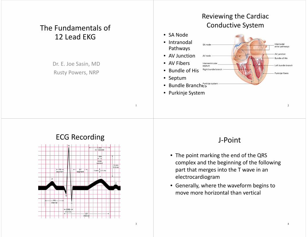
The Fundamentals of12 Lead EKG
Dr. E. Joe Sasin, MDRusty Powers, NRP
1
Reviewing the CardiacConductive System
• SA Node• Intranodal Pathways
• AV Junction• AV Fibers• Bundle of His• Septum• Bundle Branches• Purkinje System
2
ECG Recording
3
J‐Point
• The point marking the end of the QRS complex and the beginning of the following part that merges into the T wave in an electrocardiogram
• Generally, where the waveform begins to move more horizontal than vertical
4
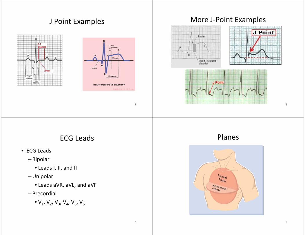
J Point Examples
5
More J‐Point Examples
6
ECG Leads• ECG Leads
–Bipolar• Leads I, II, and II
–Unipolar• Leads aVR, aVL, and aVF
–Precordial• V1, V2, V3, V4, V5, V6
7
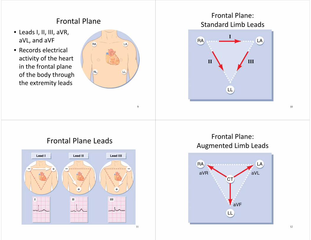
Planes
8
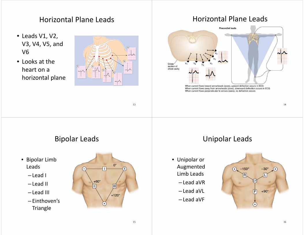
Frontal Plane• Leads I, II, III, aVR, aVL, and aVF
• Records electrical activity of the heart in the frontal plane of the body through the extremity leads
9
Frontal Plane:Standard Limb Leads
10
Frontal Plane Leads
11
Frontal Plane:Augmented Limb Leads
12
Horizontal Plane Leads
• Leads V1, V2, V3, V4, V5, and V6
• Looks at the heart on a horizontal plane
13
Horizontal Plane Leads
14
Bipolar Leads
• Bipolar Limb Leads– Lead I– Lead II– Lead III– Einthoven’s Triangle
15
Unipolar Leads
• Unipolar or Augmented Limb Leads– Lead aVR– Lead aVL– Lead aVF
16
Precordial Leads• Precordial Leads
– Lead V1
– Lead V2
– Lead V3
– Lead V4
– Lead V5
– Lead V6
17
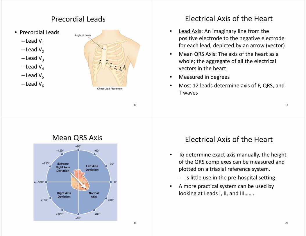
Electrical Axis of the Heart• Lead Axis: An imaginary line from the
positive electrode to the negative electrode for each lead, depicted by an arrow (vector)
• Mean QRS Axis: The axis of the heart as a whole; the aggregate of all the electrical vectors in the heart
• Measured in degrees• Most 12 leads determine axis of P, QRS, and
T waves
18
Mean QRS Axis
19
Electrical Axis of the Heart
• To determine exact axis manually, the height of the QRS complexes can be measured and plotted on a triaxial reference system.– Is little use in the pre‐hospital setting
• A more practical system can be used by looking at Leads I, II, and III…….
20
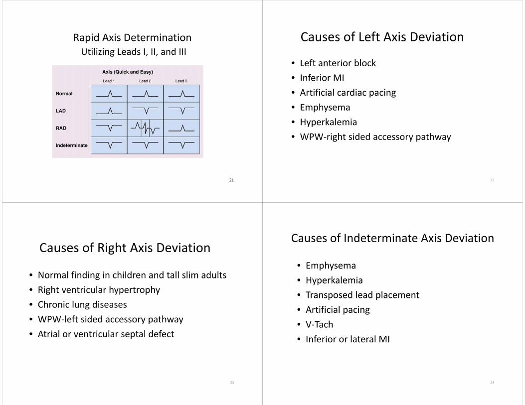
Rapid Axis DeterminationUtilizing Leads I, II, and III
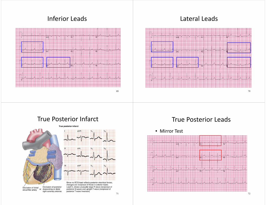
21
Causes of Left Axis Deviation
• Left anterior block• Inferior MI• Artificial cardiac pacing• Emphysema• Hyperkalemia• WPW‐right sided accessory pathway
22
Causes of Right Axis Deviation
• Normal finding in children and tall slim adults• Right ventricular hypertrophy• Chronic lung diseases• WPW‐left sided accessory pathway• Atrial or ventricular septal defect
23
Causes of Indeterminate Axis Deviation
• Emphysema• Hyperkalemia• Transposed lead placement• Artificial pacing• V‐Tach• Inferior or lateral MI
24
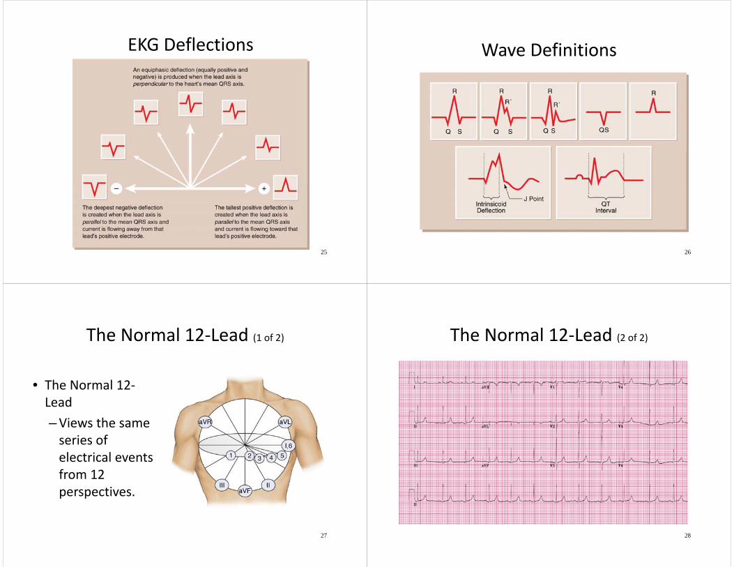
EKG Deflections
25
Wave Definitions
26
The Normal 12‐Lead (1 of 2)
• The Normal 12‐Lead–Views the same series of electrical events from 12 perspectives.
27
The Normal 12‐Lead (2 of 2)
28
Another Standard Layout
29
Interpretation of 12 Lead EKGs
• 5 + 3 Approach• Five basic steps
–Rate–Rhythm–P‐wave–PR Interval–QRS complex
30
Interpretation of 12 Lead EKGs• +3
– ST Depression• Present?• In which leads?• Reciprocal?
– ST Elevation• Present?• In which leads?• Is there reciprocal ST depression present?
–Q Waves• Present?• In which leads?• Are they pathologic or nonpathologic?
31
Multi‐Lead Heart Assessment
32
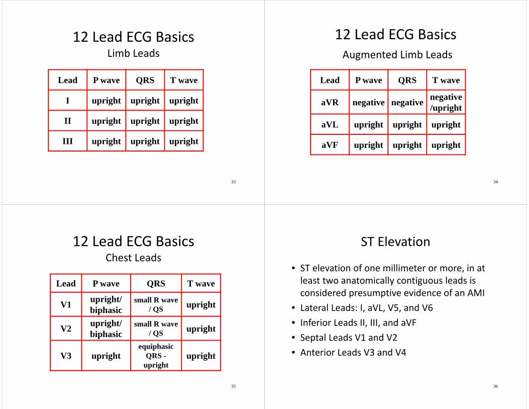
12 Lead ECG BasicsLimb Leads
33
Lead P wave QRS T wave
I upright upright upright
II upright upright upright
III upright upright upright
12 Lead ECG BasicsAugmented Limb Leads
34
Lead P wave QRS T wave
aVR negative negative negative/upright
aVL upright upright upright
aVF upright upright upright
12 Lead ECG BasicsChest Leads
35
Lead P wave QRS T wave
V1 upright/ biphasic
small R wave / QS upright
V2 upright/ biphasic
small R wave / QS upright
V3 uprightequiphasic
QRS -upright
upright
ST Elevation
• ST elevation of one millimeter or more, in at least two anatomically contiguous leads is considered presumptive evidence of an AMI
• Lateral Leads: I, aVL, V5, and V6• Inferior Leads II, III, and aVF• Septal Leads V1 and V2• Anterior Leads V3 and V4
36
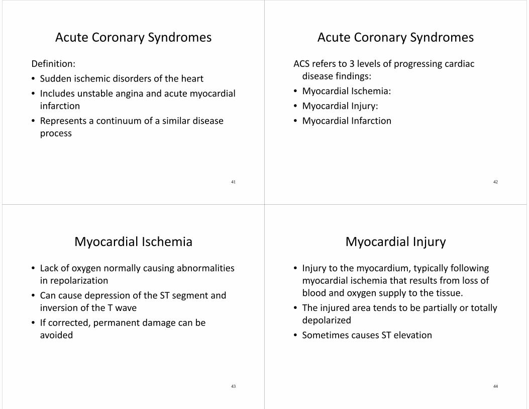
Leads and Artery Correlation• Leads I, aVL, V5, and V6
–Circumflex artery– Left anterior descending artery (LAD)
• Leads II, III, and aVF–Right coronary artery (RAC)
• Leads V1 and V2– Left anterior descending artery (LAD)
• Leads V5 and V6–Circumflex artery– Left anterior descending artery (LAD)
37
Coronary Perfusion
38
Pathologic Q Waves
• Also referred to as “significant” Q waves• Defined as a width greater than or equal to 1 small box (0.04msec) or a depth greater than 1/3 of the R wave in the same lead
• Indicates irreversible tissue damage
39
Physiologic Q Waves
• AKA: Non‐pathologic Q Waves• Less than 0.04msec (one small box)• Considered “Normal”
40
Acute Coronary Syndromes
Definition:• Sudden ischemic disorders of the heart• Includes unstable angina and acute myocardial infarction
• Represents a continuum of a similar disease process
41
Acute Coronary Syndromes
ACS refers to 3 levels of progressing cardiac disease findings:
• Myocardial Ischemia: • Myocardial Injury: • Myocardial Infarction
42
Myocardial Ischemia
• Lack of oxygen normally causing abnormalities in repolarization
• Can cause depression of the ST segment and inversion of the T wave
• If corrected, permanent damage can be avoided
43
Myocardial Injury
• Injury to the myocardium, typically following myocardial ischemia that results from loss of blood and oxygen supply to the tissue.
• The injured area tends to be partially or totally depolarized
• Sometimes causes ST elevation
44
Myocardial Infarction
• Death of the heart muscle• Due to lack of blood and oxygen• Location of the MI affects corresponding lead changes
• Can cause ST elevation
45
Acute Coronary Syndromes
• All have sudden ischemia• Cannot be differentiated in the first hours of episode
• All have the same initiating events–Plaque Rupture– Thrombus Formation–Vasoconstriction
46
Risk Factors for ACS
• Diabetes• Smoking• Hypertension• Age• Hyperlipidemia
• Family history of CAD
• Obesity• Stress• Sedentary• Non‐estrogenized females
47
Atypical Presentations of ACS
• Pain that is sharp or intermittent• Pain that is in the teeth, neck, shoulder, arm, or abdomen
• Mostly affects females, diabetics, and the elderly
48
Anginal Equivalents
• Dyspnea• Palpitations• Syncope or near syncope• Generalized weakness with no history of a GI bleed or recent fever
• DKA• May be the only signs/symptoms of ACS
49
Recognizing ACS
50
Story+
Risk Factors+
EKG=
Treatment
STEMI and Non‐STEMI
• STEMI: ST elevation MI• Non‐STEMI: non ST elevation MI
51
Disease Findings• Ischemia
– ST segment depression with or without T wave inversion
• Injury– ST elevation >1mm in 2 congruent leads
• With or without loss of R wave• >2mm in septal leads (V1, V2)
• Infarction– Pathological Q waves
• >.04 sec wide or 1/3 of R, with ST elevation– STEMI– Non‐STEMI
52
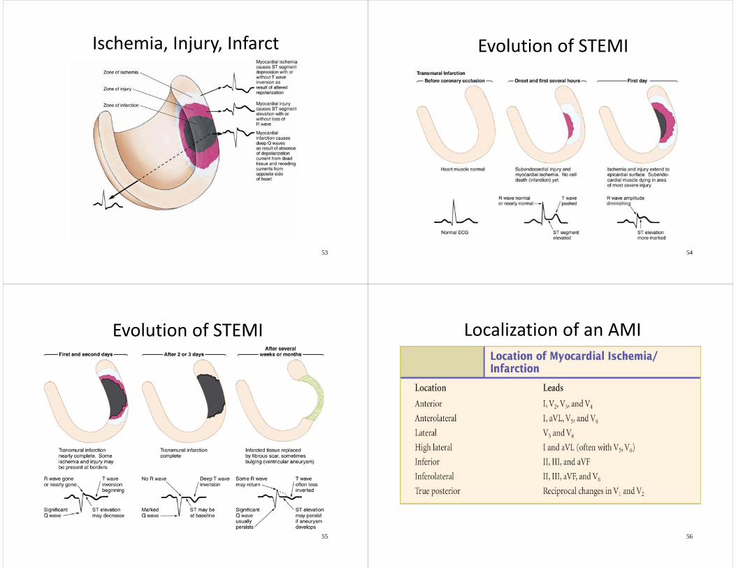
Ischemia, Injury, Infarct
53
Evolution of STEMI
54
Evolution of STEMI
55
Localization of an AMI
56
Reciprocal Changes
• ST elevation may show as ST Depression in reciprocal leads and vice‐versa
• Not necessary to presume infarction
• Strong confirming evidence when present
57
II, III, aVF I, aVL, V-Leads
Reciprocal Changes
Region of ST ElevationRegion of ST DepressionAnterior (V1‐V4) Inferior (true posterior)Inferior (II, III, aVF) Anterior (V1‐V3 or aVL)Lateral (I, aVL, V5, V6) Inferior (II, III, aVF)True Posterior Anterior (V1‐V‐3)
58
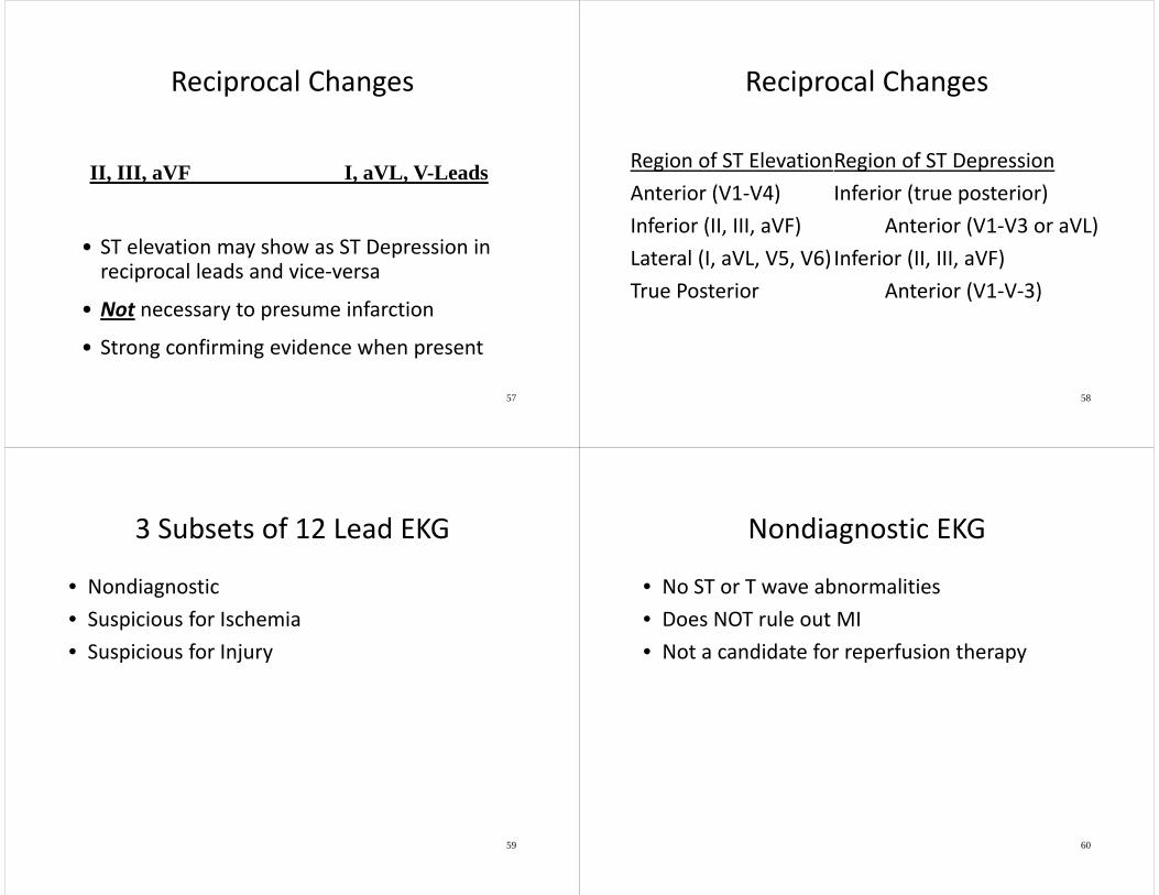
3 Subsets of 12 Lead EKG
• Nondiagnostic• Suspicious for Ischemia• Suspicious for Injury
59
Nondiagnostic EKG
• No ST or T wave abnormalities• Does NOT rule out MI• Not a candidate for reperfusion therapy
60
Suspicious for Ischemia EKG
• ST depression or T wave inversion• Does NOT rule out MI• Not a candidate for reperfusion therapy
61
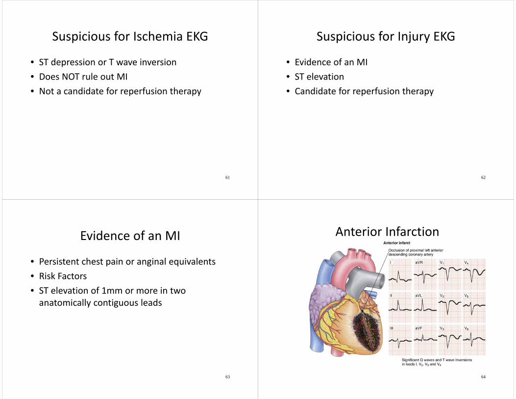
Suspicious for Injury EKG
• Evidence of an MI• ST elevation• Candidate for reperfusion therapy
62
Evidence of an MI
• Persistent chest pain or anginal equivalents• Risk Factors• ST elevation of 1mm or more in two anatomically contiguous leads
63
Anterior Infarction
64
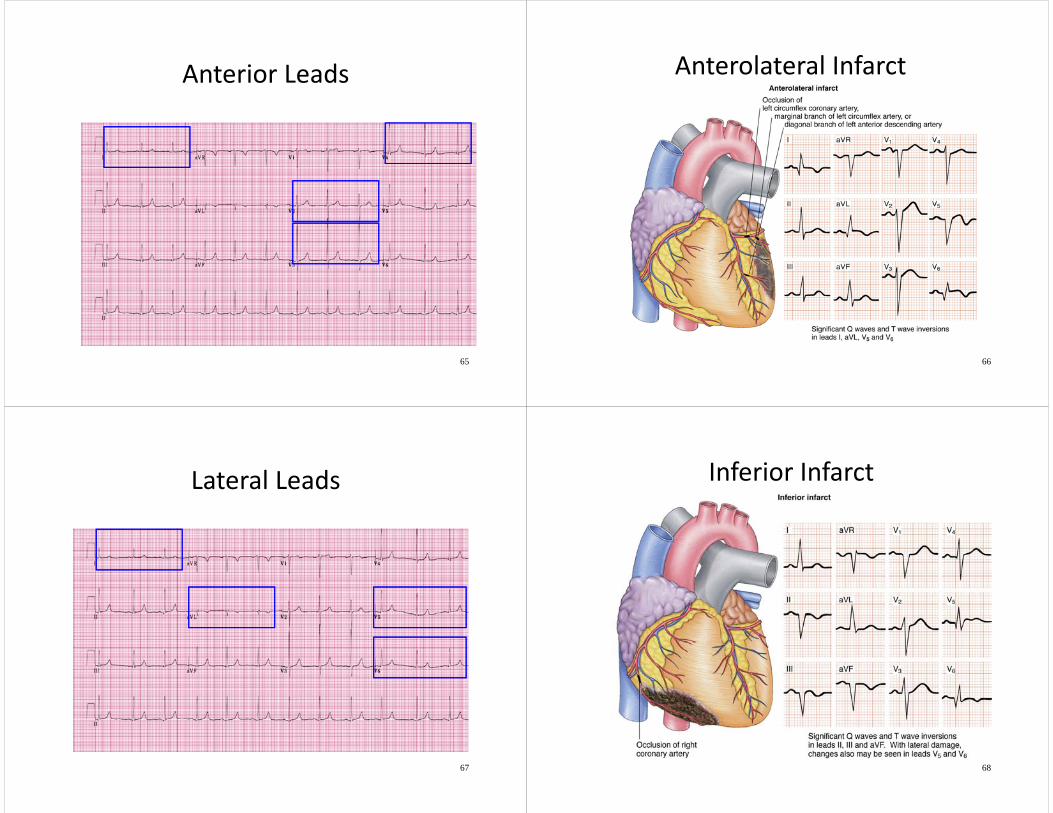
Anterior Leads
65
Anterolateral Infarct
66
Lateral Leads
67
Inferior Infarct
68
Inferior Leads
69
Lateral Leads
70
True Posterior Infarct
71
True Posterior Leads• Mirror Test
72
Acute Anterior MIS‐T elevation, and T wave inversion in leads I, V2, V3, and V4
Reciprical ST depression in leads III and aVF
73
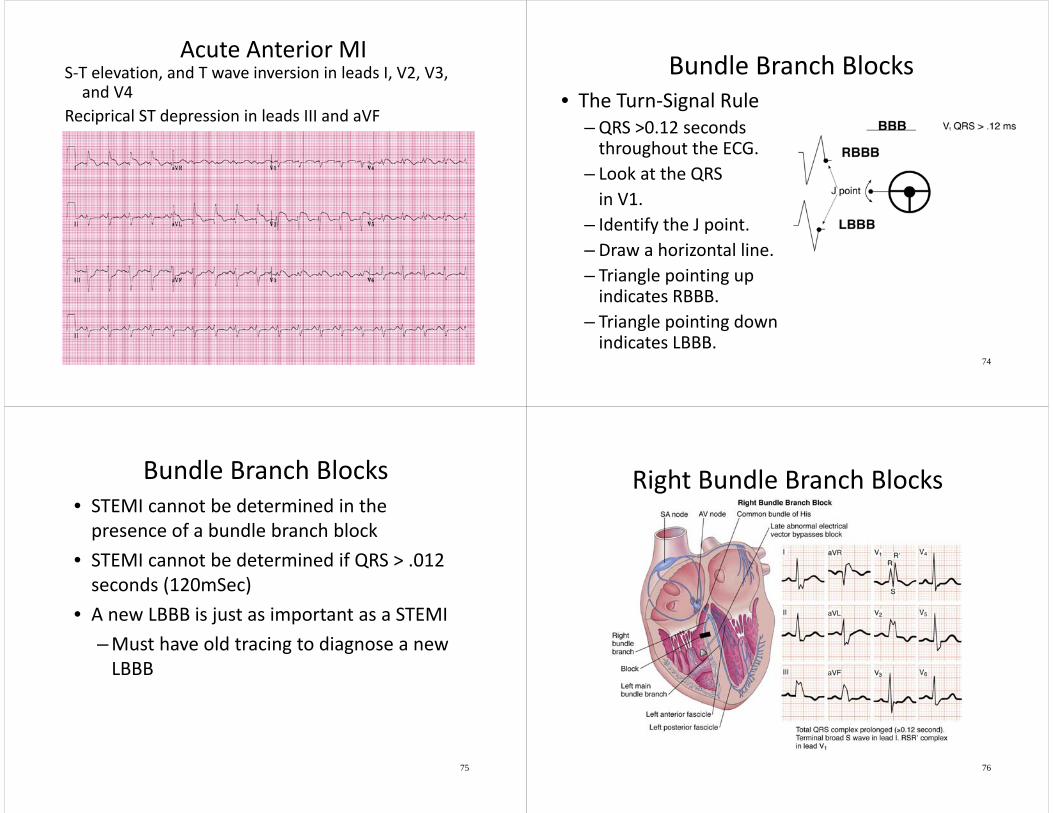
Bundle Branch Blocks• The Turn‐Signal Rule
–QRS >0.12 seconds throughout the ECG.
– Look at the QRS in V1.
– Identify the J point.– Draw a horizontal line.– Triangle pointing up indicates RBBB.
– Triangle pointing down indicates LBBB.
74
Bundle Branch Blocks• STEMI cannot be determined in the presence of a bundle branch block
• STEMI cannot be determined if QRS > .012 seconds (120mSec)
• A new LBBB is just as important as a STEMI–Must have old tracing to diagnose a new LBBB
75
Right Bundle Branch Blocks
76
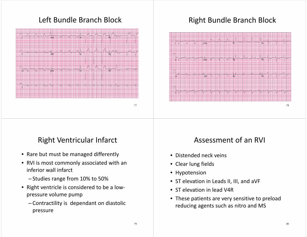
Left Bundle Branch Block
77
Right Bundle Branch Block
78
Right Ventricular Infarct
• Rare but must be managed differently• RVI is most commonly associated with an inferior wall infarct– Studies range from 10% to 50%
• Right ventricle is considered to be a low‐pressure volume pump–Contractility is dependant on diastolic pressure
79
Assessment of an RVI
• Distended neck veins• Clear lung fields• Hypotension• ST elevation in Leads II, III, and aVF• ST elevation in lead V4R• These patients are very sensitive to preload reducing agents such as nitro and MS
80

Right Ventricular Infarct
• ST elevation in leads II, III, and aVF• Note that the elevation is greater in lead III than lead II– Typical for inferior MI with RVI
81
Right Ventricular Infarct
• Right sided V4R showing ST elevation along with ST elevation in Leads II, III, and aVF
• When using R sided EKG or just V4R, label the EKG as “Right‐Sided” or “V4R” and disregard machine’s interpretation
82
Management of an RVI
• Extreme caution must be used with nitro and MS–Use small incremental doses of MS–NTG best given by drip
• Fluid therapy if hypotensive• Vasopressors if fluid is ineffective
–Dopamine–Dobutamine
83
Wolff‐Parkinson‐White Syndrome• WPW is a syndrome of pre‐excitation of the ventricles due to an accessory pathway called the Bundle of Kent which is an abnormal pathway from the atria to the ventricles.
• Effects 0.15 to 0.2% of the population• Normally asymptomatic
84
Wolff‐Parkinson‐White Syndrome
• Risk of sudden death due to tachydysrthymias(rare)
• Produces a delta wave– Slurred upstroke in the QRS complex with a short PRI
– Type I WPW produces positive delta waves– Type II WPW produces negative delta waves
• Commonly causes syncope and/or palpitations
85
Delta Wave in WPW
86
Wolff‐Parkinson‐White
87
Let’s Practice
88
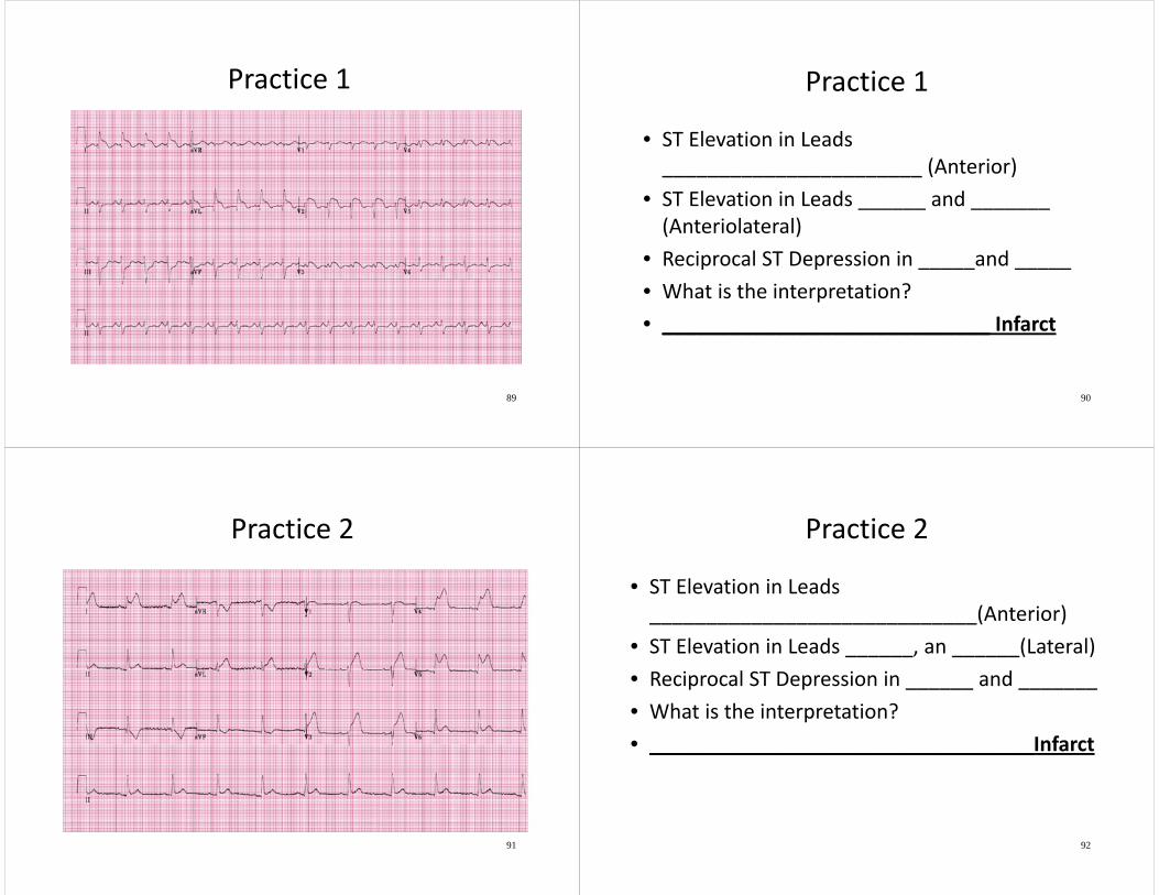
Practice 1
89
Practice 1
• ST Elevation in Leads _______________________ (Anterior)
• ST Elevation in Leads ______ and _______ (Anteriolateral)
• Reciprocal ST Depression in _____and _____• What is the interpretation?• _____________________________ Infarct
90
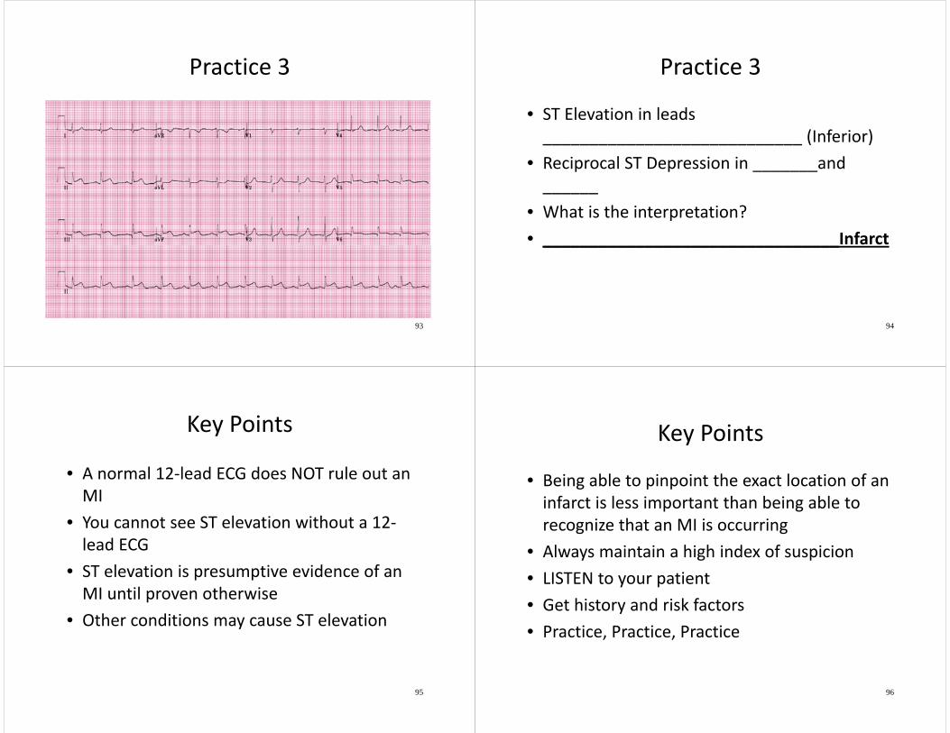
Practice 2
91
Practice 2
• ST Elevation in Leads _____________________________(Anterior)
• ST Elevation in Leads ______, an ______(Lateral)• Reciprocal ST Depression in ______ and _______• What is the interpretation?• __________________________________Infarct
92
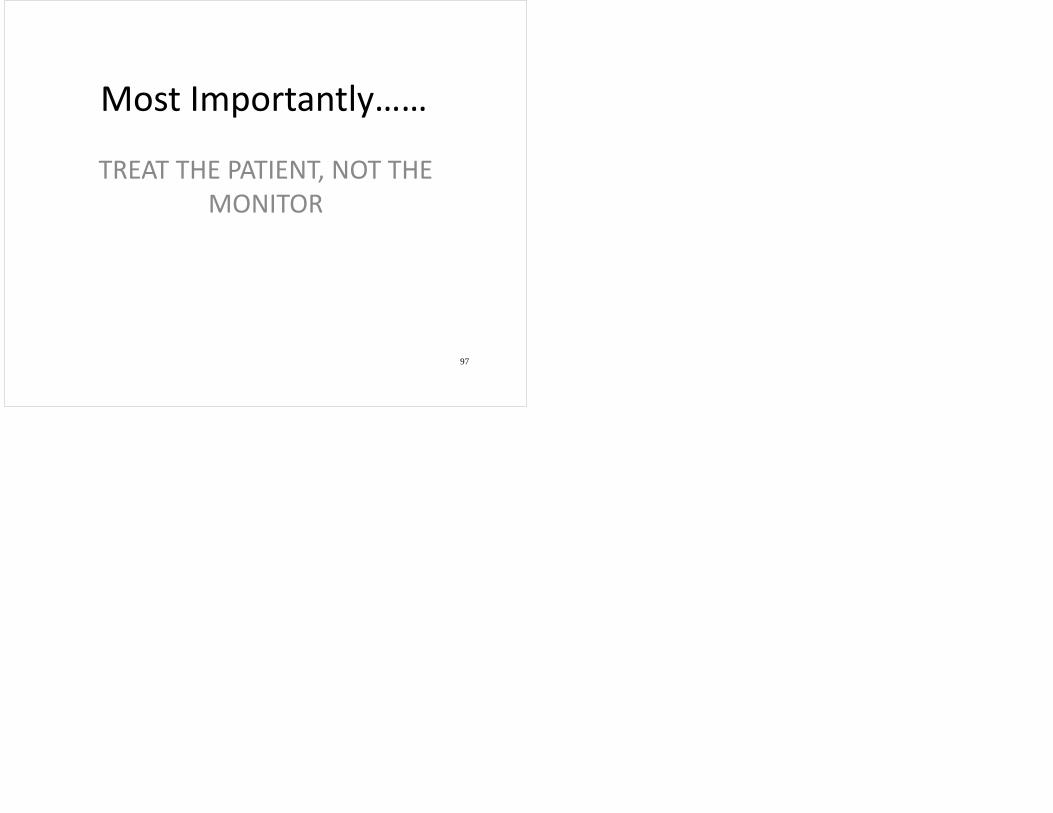
Practice 3
93
Practice 3
• ST Elevation in leads ____________________________ (Inferior)
• Reciprocal ST Depression in _______and ______
• What is the interpretation?• ________________________________Infarct
94
Key Points
• A normal 12‐lead ECG does NOT rule out an MI
• You cannot see ST elevation without a 12‐lead ECG
• ST elevation is presumptive evidence of an MI until proven otherwise
• Other conditions may cause ST elevation
95
Key Points
• Being able to pinpoint the exact location of an infarct is less important than being able to recognize that an MI is occurring
• Always maintain a high index of suspicion• LISTEN to your patient• Get history and risk factors• Practice, Practice, Practice
96
Most Importantly……
TREAT THE PATIENT, NOT THE MONITOR
97