Embed Size (px)
Citation preview
Leading Edge
Review
Revisiting the Central DogmaOne Molecule at a Time
Carlos Bustamante,1,2,3,4,5,* Wei Cheng,6,8 and Yara X. Mejia7,81Jason L. Choy Laboratory of Single-Molecule Biophysics2QB3 Institute3Physics Department4Howard Hughes Medical InstituteUniversity of California, Berkeley, CA 94720, USA5Physical Biosciences Division, Lawrence Berkeley National Laboratory, Berkeley, CA 94720, USA6Department of Pharmaceutical Sciences, College of Pharmacy, University of Michigan, 428 Church Street, Ann Arbor, MI 48109, USA7Biological Micro and Nanotechnology, Max Planck Institute for Biophysical Chemistry, Am Faßberg 11, D-37077 Gottingen, Germany8These authors contributed equally to this work
*Correspondence: [email protected] 10.1016/j.cell.2011.01.033
The faithful relay and timely expression of genetic information depend on specialized molecularmachines, many of which function as nucleic acid translocases. The emergence over the lastdecade of single-molecule fluorescence detection and manipulation techniques with nm and Aresolution and their application to the study of nucleic acid translocases are painting an increasinglysharp picture of the inner workings of these machines, the dynamics and coordination of theirmoving parts, their thermodynamic efficiency, and the nature of their transient intermediates.Here we present an overview of the main results arrived at by the application of single-moleculemethods to the study of the main machines of the central dogma.
Introduction‘‘The operative industry of Nature is so prolific that machines
will be eventually found not only unknown to us but also
unimaginable by our mind.’’ So wrote in De Viscerum Structura
Marcello Malpighi (Malpighi, 1666), the founder of microscopic
anatomy. Malpighi (1628–1694), a Professor at the University
of Bologna, was the leader of the revolution that swept through
the biological sciences in the 17th century and that mirrored
the parallel revolution that was occurring in physics. Coinciden-
tally, during the latter, Galileo and Newton refined the concepts
of inertia, force, and acceleration that establish the foundations
of kinematics and dynamics and that became the language
to describe the operation of machines. Coincidentally again, in
both revolutions, the invention of instruments that made it
possible to observe and measure what was not directly visible
to the human eye, the microscope and the telescope, became
the catalyst that unleashed, in both sciences, the modern scien-
tific imagination.
Since the era of Malpighi, the mechanical paradigm has been
a recurrent idea in biology. In recent decades, the molecular
biology revolution has revealed that much of the inner workings
of the cell are the result of specialized units or assembly lines that
function as molecular machines (Alberts, 1998). Many of these
entities operate as molecular motors, converting chemical
energy into mechanical work, and their description must be
done in the language of mechanics: ‘‘moving parts,’’ forces,
torques, displacements, thermodynamic efficiencies, and time.
And once again, the recent advent of single-molecule methods,
which permit to follow in real-time the individual molecular trajec-
480 Cell 144, February 18, 2011 ª2011 Elsevier Inc.
tories without having to synchronize a population of molecules,
and specifically the development of single-molecule manipula-
tion, whose direct observables are precisely displacements,
forces, and torques, is making it possible to formulate an
accurate description of molecular machines and to uncover
the physical principles and diverse biological designs that
underlie their operation.
Most of these machines are enzymes that couple a thermody-
namically spontaneous chemical reaction (typically nucleotide
hydrolysis) to a mechanical task. Because of their microscopic
dimensions, the many small parts that make up these
machine-like devices operate at energies only marginally higher
than that of the thermal bath and, hence, their operation is
subjected to large fluctuations. The fluctuations revealed by
single-molecule analyses are not just a nuance or an artifact of
studying them in singulo. In fact, many of them are present and
need only be present in very small numbers to carry their physi-
ological role in the cell, a role, therefore, subjected to large fluc-
tuations. Behaving as true thermodynamic open systems, these
devices can exchange energy and matter with the bath and take
advantage of fluctuations to operate, sometimes, as energy
rectifiers. Like ‘‘honest’’ Maxwell Demons that sit astride the
line that separates stochastic from deterministic phenomena,
the function of these molecular machines is to tame the random-
ness of molecular events and generate directional processes in
the cell.
How does this taming take place? How does this noise affect
the coordinated operation required to maintain cellular homeo-
stasis? How should we modify our concepts from macroscopic

Figure 1. f29 Packaging Motor(A) Cryo-electron microscopy of the packaging motor. Left: Packaging motorwith capsid and DNA modeled in for scale. Right: Close-up on packagingmotor. Modified from Morais et al. (2008).(B) Optical tweezers packaging assay. Left: An optical trap exerts a force, F, ona single packaging bacteriophage while monitoring the length, L, of theunpackaged DNA. Right: DNA length versus time. Different colors correspondto different concentrations of [ATP].(C) High-resolution packaging reveals a burst-dwell packaging mechanism.Left: Cartoon layout of high-resolution packaging assay. Right: Schematicdiagram of the kinetic events that occur during the dwell and burst phasesoverlaid on packaging data.
chemistry and biochemistry to obtain a more faithful description
of these stochastic devices? These and other questions are
becoming the common thread that ties the ever-increasing
number of single-molecule studies of cellular machines, some
of which are the subjects of this Review.
Here we will restrict our review to single-molecule studies
of the machinery involved in the metabolism and transactions
of nucleic acids, primary protagonists of the central dogma of
molecular biology, the operating system of the cell. Processes
such as replication, transcription, and translation require the
information encoded in the sequence of nucleic acids to be
read and copied in a directional manner. Therefore, these
machines are all, necessarily, translocases. We have accord-
ingly organized this article following the cell’s operational
logic. First we will review single-molecule studies of machines
involved in the packaging and storage of the genome. This
section will be followed by a review of helicases, followed
in turn by a review of single-molecule studies of genome
replication and DNA transcription, and will end with translation
studies.
Translocases in Chromosomal Partitioningand SegregationNewly replicated DNA molecules must be properly partitioned
and segregated into daughter cells, spores, or viral capsids. In
many cases, these processes utilize an active mechanism that
involves an ATP-dependent translocase. Generally the viral
packaging and prokaryotic segregation ATPases belong to the
P loop NTPase fold and appear to have an ancient common
origin (Catalano, 2005; Iyer et al., 2004b; Koonin et al., 1993).
Members of the P loop NTPase fold possess a conserved nucle-
otide-binding and Mg2+-binding motif (Walker A) and a water-
activating motif (Walker B) and belong to one of two major
divisions: the KG division, which includes P loop kinases and
GTPases, and the ASCE (additional strand conserved E [gluta-
mate]) division. Due to space limitations, we will only review
here the main single-molecule results obtained on viral pack-
aging systems.
Viral Packaging SystemsThe machinery involved in the packaging of viral DNA has two
components, the portal-connector and the ATPase (Catalano,
2005; C.L. Hetherington, J.R. Moffitt, P.J. Jardine, and C.B.,
unpublished data; Jardine and Anderson, 2006). The phyloge-
netic origin of these components and their spatial and functional
relationships define four different types of viral genome
packaging systems: (1) terminase-portal systems, (2) the pack-
aging systems of lipid inner membrane-containing viruses, (3)
the 429-like packaging system, and (4) the adenovirus pack-
aging apparatus (Burroughs et al., 2007). (See Supplemental
Information).
Viral DNA packaging has been divided into initiation, elonga-
tion, and termination. So far, single-molecule studies have
been restricted to bacteriophages T4, lambda, and 429. The
DNA packaging motor of bacteriophage 429, the best studied
so far, is made up of three concentric rings (Grimes et al.,
2002) (Figure 1A): (1) the head–tail connector, a dodecamer
that fits in the pentameric opening at one of the ends of the
capsid; (2) a ring of five molecules of RNA, each 174 nucleotides
(nt) long of unknown function; and (3) a pentameric ring (Morais
et al., 2008) of gp16, an ATPase that belongs to the FtsK/HerA
family of the ASCE superfamily of P loop NTPases.
Packaging Initiation
Initiation of viral DNA packaging requires recognition of the viral
genome by the packaging machinery. This process is done
either through binding of a specific DNA sequence (reviewed in
Catalano, 2005; Jardine and Anderson, 2006) or through
a terminal protein bound to the ends of the viral DNA. Only
the latter form of initiation has been studied by single-molecule
methods. In bacteriophage 429, a terminal protein, gp3, is
bound to both 50 ends of the viral genome, and at least one
of them is required for robust packaging in vitro. In EM studies,
the terminal protein is seen to induce a loop or lariat on the
Cell 144, February 18, 2011 ª2011 Elsevier Inc. 481
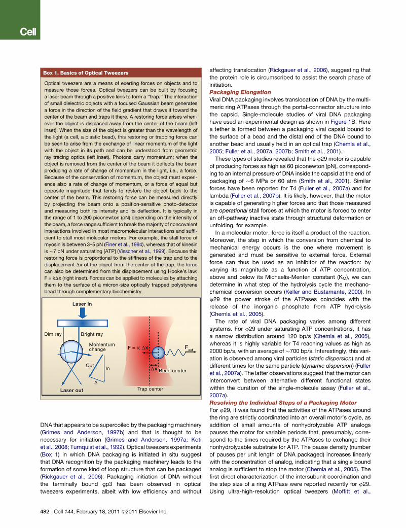
Box 1. Basics of Optical Tweezers
Optical tweezers are a means of exerting forces on objects and to
measure those forces. Optical tweezers can be built by focusing
a laser beam through a positive lens to form a ‘‘trap.’’ The interaction
of small dielectric objects with a focused Gaussian beam generates
a force in the direction of the field gradient that draws it toward the
center of the beam and traps it there. A restoring force arises when-
ever the object is displaced away from the center of the beam (left
inset). When the size of the object is greater than the wavelength of
the light (a cell, a plastic bead), this restoring or trapping force can
be seen to arise from the exchange of linear momentum of the light
with the object in its path and can be understood from geometric
ray tracing optics (left inset). Photons carry momentum; when the
object is removed from the center of the beam it deflects the beam
producing a rate of change of momentum in the light, i.e., a force.
Because of the conservation of momentum, the object must experi-
ence also a rate of change of momentum, or a force of equal but
opposite magnitude that tends to restore the object back to the
center of the beam. This restoring force can be measured directly
by projecting the beam onto a position-sensitive photo-detector
and measuring both its intensity and its deflection. It is typically in
the range of 1 to 200 piconewton (pN) depending on the intensity of
the beam, a force range sufficient to break themajority of noncovalent
interactions involved in most macromolecular interactions and suffi-
cient to stall most molecular motors. For example, the stall force of
myosin is between 3–5 pN (Finer et al., 1994), whereas that of kinesin
is �7 pN under saturating [ATP] (Visscher et al., 1999). Because this
restoring force is proportional to the stiffness of the trap and to the
displacement Dx of the object from the center of the trap, the force
can also be determined from this displacement using Hooke’s law:
F = kDx (right inset). Forces can be applied to molecules by attaching
them to the surface of a micron-size optically trapped polystyrene
bead through complementary biochemistry.
DNA that appears to be supercoiled by the packagingmachinery
(Grimes and Anderson, 1997b) and that is thought to be
necessary for initiation (Grimes and Anderson, 1997a; Koti
et al., 2008; Turnquist et al., 1992). Optical tweezers experiments
(Box 1) in which DNA packaging is initiated in situ suggest
that DNA recognition by the packaging machinery leads to the
formation of some kind of loop structure that can be packaged
(Rickgauer et al., 2006). Packaging initiation of DNA without
the terminally bound gp3 has been observed in optical
tweezers experiments, albeit with low efficiency and without
482 Cell 144, February 18, 2011 ª2011 Elsevier Inc.
affecting translocation (Rickgauer et al., 2006), suggesting that
the protein role is circumscribed to assist the search phase of
initiation.
Packaging Elongation
Viral DNA packaging involves translocation of DNA by the multi-
meric ring ATPases through the portal-connector structure into
the capsid. Single-molecule studies of viral DNA packaging
have used an experimental design as shown in Figure 1B. Here
a tether is formed between a packaging viral capsid bound to
the surface of a bead and the distal end of the DNA bound to
another bead and usually held in an optical trap (Chemla et al.,
2005; Fuller et al., 2007a, 2007b; Smith et al., 2001).
These types of studies revealed that the 429 motor is capable
of producing forces as high as 60 piconewton (pN), correspond-
ing to an internal pressure of DNA inside the capsid at the end of
packaging of �6 MPa or 60 atm (Smith et al., 2001). Similar
forces have been reported for T4 (Fuller et al., 2007a) and for
lambda (Fuller et al., 2007b). It is likely, however, that the motor
is capable of generating higher forces and that those measured
are operational stall forces at which the motor is forced to enter
an off-pathway inactive state through structural deformation or
unfolding, for example.
In a molecular motor, force is itself a product of the reaction.
Moreover, the step in which the conversion from chemical to
mechanical energy occurs is the one where movement is
generated and must be sensitive to external force. External
force can thus be used as an inhibitor of the reaction: by
varying its magnitude as a function of ATP concentration,
above and below its Michaelis-Menten constant (KM), we can
determine in what step of the hydrolysis cycle the mechano-
chemical conversion occurs (Keller and Bustamante, 2000). In
429 the power stroke of the ATPases coincides with the
release of the inorganic phosphate from ATP hydrolysis
(Chemla et al., 2005).
The rate of viral DNA packaging varies among different
systems. For 429 under saturating ATP concentrations, it has
a narrow distribution around 120 bp/s (Chemla et al., 2005),
whereas it is highly variable for T4 reaching values as high as
2000 bp/s, with an average of �700 bp/s. Interestingly, this vari-
ation is observed among viral particles (static dispersion) and at
different times for the same particle (dynamic dispersion) (Fuller
et al., 2007a). The latter observations suggest that the motor can
interconvert between alternative different functional states
within the duration of the single-molecule assay (Fuller et al.,
2007a).
Resolving the Individual Steps of a Packaging Motor
For 429, it was found that the activities of the ATPases around
the ring are strictly coordinated into an overall motor’s cycle, as
addition of small amounts of nonhydrolyzable ATP analogs
pauses the motor for variable periods that, presumably, corre-
spond to the times required by the ATPases to exchange their
nonhydrolyzable substrate for ATP. The pause density (number
of pauses per unit length of DNA packaged) increases linearly
with the concentration of analog, indicating that a single bound
analog is sufficient to stop the motor (Chemla et al., 2005). The
first direct characterization of the intersubunit coordination and
the step size of a ring ATPase were reported recently for 429.
Using ultra-high-resolution optical tweezers (Moffitt et al.,

2006), it was found that this motor packages the DNA in incre-
ments of 10 bp separated by stochastically varying dwell times
(Moffitt et al., 2009). Statistical analysis of the dwell times
revealed that multiple ATPs bind during each dwell; application
of high force showed that these 10 bp increments are
composed of four 2.5 bp steps. Further analysis demonstrated
that the hydrolysis cycles of the individual subunits are highly
coordinated: the ATP binding to all subunits occurs during
the ‘‘dwell’’ phase that is completely segregated from and
followed by the translocation or ‘‘burst’’ phase (Figure 1C).
Interestingly, the strong coordination among the ATPase activ-
ities in the ring is not consistent with the Hill coefficient of �1
measured experimentally. It turns out that if the binding of the
individual ATPs to the various subunits is separated by an
irreversible step, the Hill analysis will yield n = 1 despite the
strong coordination and cooperativity among these subunits
(Moffitt et al., 2009).
The Nature of the DNA-Motor Interaction
Little is known about the interactions responsible for the large
forces displayed by these motors and the noninteger base pair
steps observed for 429. The role played by the phosphate back-
bone charge in the motor-DNA interaction was investigated
recently in single-molecule packaging experiments by
challenging the motor with DNA constructs bearing inserted
regions of neutral DNA segments containing methylphospho-
nate (MeP) modifications (Aathavan et al., 2009). Remarkably,
the motor actively traverses these inserts, though with reduced
probability compared to regular DNA, indicating that phosphate
charges are important but not essential for translocation. By
changing the length of the MeP inserts and selectively restoring
the charge to one or the other DNA strand, it was found that
important contacts are made with phosphate charges every
10 bp on the 50/30 strand only. High-resolution measurements
of the dynamics through the insert reveal that, in addition to
providing a load-bearing contact, these phosphate contacts
also play a role regulating the timing of the mechanochemical
cycle (Aathavan et al., 2009).
A step size that is a noninteger number of base pairs requires
motor-DNA interactions that do not depend on any given peri-
odic structure in the DNA molecule, and that are of steric nature.
Thus, the motor was challenged with a series of additional
inserts: DNA lacking bases and sugars, single-stranded gaps,
unpaired bulges, and a nonbiological linker (Aathavan et al.,
2009). Surprisingly, none of themodifications abolish packaging,
indicating that the motor makes promiscuous, steric contacts
with a wide variety of chemical moieties over a range of geome-
tries, helping to rationalize the observed 2.5 A steps. These
results suggest that the 2.5 bp step is determined by the magni-
tude of the conformational change that the individual ATPases
undergo during their power stroke.
The Structural Basis of Force Generation
Several sequence motifs define the members of the ASCE family
of P loop NTPases (Erzberger and Berger, 2006; Iyer et al.,
2004a; Thomsen and Berger, 2008), including the Walker A
andWalker B motifs—known to coordinate binding of the nucle-
otides and to catalyze hydrolysis (Dhar and Feiss, 2005) —and
the arginine finger. In addition, the Q-motif and the C-motif are
present in some of the packaging ATPases (Mitchell et al.,
2002; Rao and Feiss, 2008). These conserved sequence
elements are likely to be involved in the mechanochemical
energy transduction of viral packaging machines and are, there-
fore, prime targets for combined mutational and single-molecule
studies. Tsay et al. (2009) used optical tweezers to investigate
the effect of mutations in the large terminase subunit of bacterio-
phage l on the dynamics of packaging. One of the mutations,
K84A, near the Walker A motif reduced packaging velocity by
�40% but did not affect the processivity of the motor nor its
force sensitivity (i.e., the distance to the transition state) (see
Supplemental Information). The other mutant, Y46F, was found
to reduce the rate of the motor by �40% but to decrease also
its processivity 10-fold. This same mutant greatly weakened
the motor mechanically (Tsay et al., 2009). These findings
indicate that viral motors contain an adenine-binding motif that
regulates ATP hydrolysis and substrate affinity analogous to
the Q-motif recently identified in DEAD-box RNA helicases.
Furthermore, the Q-motif appears to be involved in coupling
the conformational changes in the ATP-binding pocket to
substrate translocation (Worrall et al., 2008). In a separate study,
Tsay et al. (2010) found that mutation T194M downstream of the
Walker Bmotif slows themotor 8-fold withoutmodifying its proc-
essivity or force generation. In contrast, mutation G212S in the
C-motif causes a 3-fold reduction in velocity but also a 6-fold
reduction in processivity. Future studies using A-resolution
optical tweezers should help establish which phase of the
dynamic cycle of the motor, relative to nucleotide binding and
hydrolysis, is directly affected by these modifications.
Helicases: Keys to the Sequence VaultHelicases constitute a large class of motor proteins that play
indispensible roles in almost every aspect of nucleic acid metab-
olism (Matson et al., 1994; Rocak and Linder, 2004). Most organ-
isms encode multiple helicases, and genes encoding proteins
with helicase/translocase activities comprise close to 2% of
the eukaryotic genome (Shiratori et al., 1999). Conventionally,
helicases are defined as enzymes that utilize ATP to break the
complementary hydrogen bonds in double-stranded nucleic
acids (dsNA), a process essential for DNA or RNA replication
(Lohman and Bjornson, 1996). Biochemical functions of
helicases go beyond the mere catalytic opening of double-
stranded DNA (dsDNA) or RNA (dsRNA), however. Many heli-
cases not only perform canonical functions but also catalyze
disassembly of protein-nucleic acid complexes (PNAC), an
important activity required in many essential cellular processes
(Jankowsky and Bowers, 2006; Krejci et al., 2003). In addition,
some helicase proteins may not function to unwind dsNA but
rather serve other biological functions inside the cell, like
chromatin remodeling (Saha et al., 2006). This multifunctional
facet begs important questions about helicases: How do
helicases use ATP to catalyze the opening of dsDNA or the
disassembly of PNAC? How are these activities integrated in
a given molecule? How is ATP hydrolysis coordinated with the
mechanical tasks of the enzyme? Research over the last 10
years, often using single-molecule techniques, has yielded
a tremendous amount of information at a mechanistic level on
how these proteins catalyze the opening of dsNA and the disas-
sembly of PNAC. These advances will be reviewed here.
Cell 144, February 18, 2011 ª2011 Elsevier Inc. 483
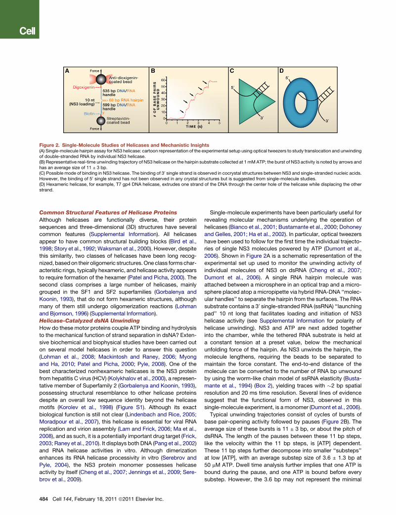
Figure 2. Single-Molecule Studies of Helicases and Mechanistic Insights(A) Single-molecule hairpin assay for NS3 helicase: cartoon representation of the experimental setup using optical tweezers to study translocation and unwindingof double-stranded RNA by individual NS3 helicase.(B) Representative real-time unwinding trajectory of NS3 helicase on the hairpin substrate collected at 1 mMATP; the burst of NS3 activity is noted by arrows andhas an average size of 11 ± 3 bp.(C) Possible mode of binding in NS3 helicase. The binding of 30 single strand is observed in cocrystal structures between NS3 and single-stranded nucleic acids.However, the binding of 50 single strand has not been observed in any crystal structures but is suggested from single-molecule studies.(D) Hexameric helicase, for example, T7 gp4 DNA helicase, extrudes one strand of the DNA through the center hole of the helicase while displacing the otherstrand.
Common Structural Features of Helicase Proteins
Although helicases are functionally diverse, their protein
sequences and three-dimensional (3D) structures have several
common features (Supplemental Information). All helicases
appear to have common structural building blocks (Bird et al.,
1998; Story et al., 1992;Waksman et al., 2000). However, despite
this similarity, two classes of helicases have been long recog-
nized, basedon their oligomeric structures.One class formschar-
acteristic rings, typically hexameric, and helicase activity appears
to require formation of the hexamer (Patel and Picha, 2000). The
second class comprises a large number of helicases, mainly
grouped in the SF1 and SF2 superfamilies (Gorbalenya and
Koonin, 1993), that do not form hexameric structures, although
many of them still undergo oligomerization reactions (Lohman
and Bjornson, 1996) (Supplemental Information).
Helicase-Catalyzed dsNA Unwinding
How do these motor proteins couple ATP binding and hydrolysis
to the mechanical function of strand separation in dsNA? Exten-
sive biochemical and biophysical studies have been carried out
on several model helicases in order to answer this question
(Lohman et al., 2008; Mackintosh and Raney, 2006; Myong
and Ha, 2010; Patel and Picha, 2000; Pyle, 2008). One of the
best characterized nonhexameric helicases is the NS3 protein
from hepatitis C virus (HCV) (Kolykhalov et al., 2000), a represen-
tative member of Superfamily 2 (Gorbalenya and Koonin, 1993),
possessing structural resemblance to other helicase proteins
despite an overall low sequence identity beyond the helicase
motifs (Korolev et al., 1998) (Figure S1). Although its exact
biological function is still not clear (Lindenbach and Rice, 2005;
Moradpour et al., 2007), this helicase is essential for viral RNA
replication and virion assembly (Lam and Frick, 2006; Ma et al.,
2008), and as such, it is a potentially important drug target (Frick,
2003; Raney et al., 2010). It displays both DNA (Pang et al., 2002)
and RNA helicase activities in vitro. Although dimerization
enhances its RNA helicase processivity in vitro (Serebrov and
Pyle, 2004), the NS3 protein monomer possesses helicase
activity by itself (Cheng et al., 2007; Jennings et al., 2009; Sere-
brov et al., 2009).
484 Cell 144, February 18, 2011 ª2011 Elsevier Inc.
Single-molecule experiments have been particularly useful for
revealing molecular mechanisms underlying the operation of
helicases (Bianco et al., 2001; Bustamante et al., 2000; Dohoney
and Gelles, 2001; Ha et al., 2002). In particular, optical tweezers
have been used to follow for the first time the individual trajecto-
ries of single NS3 molecules powered by ATP (Dumont et al.,
2006). Shown in Figure 2A is a schematic representation of the
experimental set up used to monitor the unwinding activity of
individual molecules of NS3 on dsRNA (Cheng et al., 2007;
Dumont et al., 2006). A single RNA hairpin molecule was
attached between a microsphere in an optical trap and a micro-
sphere placed atop a micropipette via hybrid RNA-DNA ‘‘molec-
ular handles’’ to separate the hairpin from the surfaces. The RNA
substrate contains a 30 single-stranded RNA (ssRNA) ‘‘launching
pad’’ 10 nt long that facilitates loading and initiation of NS3
helicase activity (see Supplemental Information for polarity of
helicase unwinding). NS3 and ATP are next added together
into the chamber, while the tethered RNA substrate is held at
a constant tension at a preset value, below the mechanical
unfolding force of the hairpin. As NS3 unwinds the hairpin, the
molecule lengthens, requiring the beads to be separated to
maintain the force constant. The end-to-end distance of the
molecule can be converted to the number of RNA bp unwound
by using the worm-like chain model of ssRNA elasticity (Busta-
mante et al., 1994) (Box 2), yielding traces with �2 bp spatial
resolution and 20 ms time resolution. Several lines of evidence
suggest that the functional form of NS3, observed in this
single-molecule experiment, is a monomer (Dumont et al., 2006).
Typical unwinding trajectories consist of cycles of bursts of
base pair-opening activity followed by pauses (Figure 2B). The
average size of these bursts is 11 ± 3 bp, or about the pitch of
dsRNA. The length of the pauses between these 11 bp steps,
like the velocity within the 11 bp steps, is [ATP] dependent.
These 11 bp steps further decompose into smaller ‘‘substeps’’
at low [ATP], with an average substep size of 3.6 ± 1.3 bp at
50 mM ATP. Dwell time analysis further implies that one ATP is
bound during the pause, and one ATP is bound before every
substep. However, the 3.6 bp may not represent the minimal
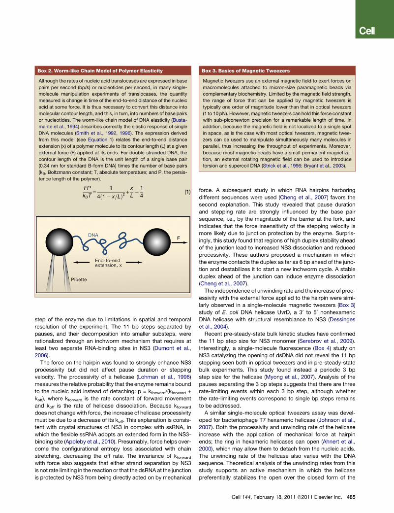
Box 2. Worm-like Chain Model of Polymer Elasticity
Although the rates of nucleic acid translocases are expressed in base
pairs per second (bp/s) or nucleotides per second, in many single-
molecule manipulation experiments of translocases, the quantity
measured is change in time of the end-to-end distance of the nucleic
acid at some force. It is thus necessary to convert this distance into
molecular contour length, and this, in turn, into numbers of base pairs
or nucleotides. The worm-like chain model of DNA elasticity (Busta-
mante et al., 1994) describes correctly the elastic response of single
DNA molecules (Smith et al., 1992, 1996). The expression derived
from this model (see Equation 1) relates the end-to-end distance
extension (x) of a polymer molecule to its contour length (L) at a given
external force (F) applied at its ends. For double-stranded DNA, the
contour length of the DNA is the unit length of a single base pair
(0.34 nm for standard B-form DNA) times the number of base pairs
(kB, Boltzmann constant; T, absolute temperature; and P, the persis-
tence length of the polymer).
FP
kBT=
1
4ð1� x=LÞ2 +x
L� 1
4(1)
Box 3. Basics of Magnetic Tweezers
Magnetic tweezers use an external magnetic field to exert forces on
macromolecules attached to micron-size paramagnetic beads via
complementary biochemistry. Limited by the magnetic field strength,
the range of force that can be applied by magnetic tweezers is
typically one order of magnitude lower than that in optical tweezers
(1 to 10 pN). However, magnetic tweezers can hold this force constant
with sub-piconewton precision for a remarkable length of time. In
addition, because the magnetic field is not localized to a single spot
in space, as is the case with most optical tweezers, magnetic twee-
zers can be used to manipulate simultaneously many molecules in
parallel, thus increasing the throughput of experiments. Moreover,
because most magnetic beads have a small permanent magnetiza-
tion, an external rotating magnetic field can be used to introduce
torsion and supercoil DNA (Strick et al., 1996; Bryant et al., 2003).
step of the enzyme due to limitations in spatial and temporal
resolution of the experiment. The 11 bp steps separated by
pauses, and their decomposition into smaller substeps, were
rationalized through an inchworm mechanism that requires at
least two separate RNA-binding sites in NS3 (Dumont et al.,
2006).
The force on the hairpin was found to strongly enhance NS3
processivity but did not affect pause duration or stepping
velocity. The processivity of a helicase (Lohman et al., 1998)
measures the relative probability that the enzyme remains bound
to the nucleic acid instead of detaching: p = kforward/(kforward +
koff), where kforward is the rate constant of forward movement
and koff is the rate of helicase dissociation. Because kforward
does not change with force, the increase of helicase processivity
must be due to a decrease of its koff. This explanation is consis-
tent with crystal structures of NS3 in complex with ssRNA, in
which the flexible ssRNA adopts an extended form in the NS3-
binding site (Appleby et al., 2010). Presumably, force helps over-
come the configurational entropy loss associated with chain
stretching, decreasing the off rate. The invariance of kforward
with force also suggests that either strand separation by NS3
is not rate limiting in the reaction or that the dsRNA at the junction
is protected by NS3 from being directly acted on by mechanical
force. A subsequent study in which RNA hairpins harboring
different sequences were used (Cheng et al., 2007) favors the
second explanation. This study revealed that pause duration
and stepping rate are strongly influenced by the base pair
sequence, i.e., by the magnitude of the barrier at the fork, and
indicates that the force insensitivity of the stepping velocity is
more likely due to junction protection by the enzyme. Surpris-
ingly, this study found that regions of high duplex stability ahead
of the junction lead to increased NS3 dissociation and reduced
processivity. These authors proposed a mechanism in which
the enzyme contacts the duplex as far as 6 bp ahead of the junc-
tion and destabilizes it to start a new inchworm cycle. A stable
duplex ahead of the junction can induce enzyme dissociation
(Cheng et al., 2007).
The independence of unwinding rate and the increase of proc-
essivity with the external force applied to the hairpin were simi-
larly observed in a single-molecule magnetic tweezers (Box 3)
study of E. coli DNA helicase UvrD, a 30 to 50 nonhexameric
DNA helicase with structural resemblance to NS3 (Dessinges
et al., 2004).
Recent pre-steady-state bulk kinetic studies have confirmed
the 11 bp step size for NS3 monomer (Serebrov et al., 2009).
Interestingly, a single-molecule fluorescence (Box 4) study on
NS3 catalyzing the opening of dsDNA did not reveal the 11 bp
stepping seen both in optical tweezers and in pre-steady-state
bulk experiments. This study found instead a periodic 3 bp
step size for the helicase (Myong et al., 2007). Analysis of the
pauses separating the 3 bp steps suggests that there are three
rate-limiting events within each 3 bp step, although whether
the rate-limiting events correspond to single bp steps remains
to be addressed.
A similar single-molecule optical tweezers assay was devel-
oped for bacteriophage T7 hexameric helicase (Johnson et al.,
2007). Both the processivity and unwinding rate of the helicase
increase with the application of mechanical force at hairpin
ends; the ring in hexameric helicases can open (Ahnert et al.,
2000), which may allow them to detach from the nucleic acids.
The unwinding rate of the helicase also varies with the DNA
sequence. Theoretical analysis of the unwinding rates from this
study supports an active mechanism in which the helicase
preferentially stabilizes the open over the closed form of the
Cell 144, February 18, 2011 ª2011 Elsevier Inc. 485

Box 4. Basics of Single-Molecule Fluorescence
The ability to detect the fluorescence emitted by certain dyes at the
single-molecule level has furnished another way to follow the
dynamics of complex biochemical processes in real-time. Single-
molecule fluorescence methods make it possible, for example, to
localize the emitter with nanometer precision (Yildiz et al., 2003). In
particular, single-molecule fluorescence resonance energy transfer,
FRET, takes advantage of the fact that the fluorescence emission of
a molecule (called a donor) is influenced by a neighboring molecule
(the acceptor) through dipolar coupling. The efficiency of this coupling
is determined by the spectral overlap between the emission of the
donor and the absorbance of the acceptor, the distance, and the
orientation between these two molecules. Because this efficiency
decreases with the sixth power of the distance between the donor
and the acceptor, this method can be used to monitor conformational
changes of macromolecules or changes in the relative orientation
between macromolecules. In practice, FRET is better used to monitor
relative changes in distance and/or orientation because the absolute
distance measurements require information about the orientation of
the fluorophores, which is not always available (Muschielok et al.,
2008). The application of single-molecule fluorescence techniques
to nucleic acid translocases has revealed many novel insights and
mechanistic details of these motors (Ha et al., 2002). These experi-
ments are mostly carried out using evanescent field excitation to
reduce fluorescence background and achieve single-molecule sensi-
tivity. This particular experimental design also permits to monitor
many individual molecules simultaneously.
junction (Betterton and Julicher, 2005) (see Supplemental Infor-
mation for passive versus active unwinding). Although not
directly observed in this study, the analysis of unwinding rates
indicates a step size of 2 bp. Using a magnetic tweezers assay,
Lionnet et al. studied the DNA-unwinding mechanism catalyzed
by bacteriophage T4 helicase gp41, a hexameric helicase
involved in phage DNA replication (Lionnet et al., 2007). The
difference between the rate of unwinding in these experiments
(30 bp/s) and the expected rate in vivo (400 bp/s) suggests
that gp41 must interact with other components of the replisome
to achieve rapid and processive unwinding of the T4 genome.
Interestingly, this study showed a clear dependence of DNA-
unwinding rate on the tension applied to the hairpin.
Hexameric versus Nonhexameric Helicases
A comparison of the behavior of hexameric and nonhexameric
helicases reveals that for both groups processivity increases
with applied force and the rate of dsNA unwinding depends on
the thermodynamic stability of the base pair at the junction.
However, the unwinding rate of nonhexameric helicases is insen-
sitive to mechanical force on the hairpin (Dessinges et al., 2004;
Dumont et al., 2006), whereas that of hexameric helicases
studied so far increases with force (Johnson et al., 2007; Lionnet
et al., 2007). The speeding up of hexameric helicases with force
indicates that strand separation constitutes the rate-limiting step
of their mechanochemical cycle. It also suggests that these two
classes of helicases may interact with their dsNA substrates in
different ways: whereas nonhexameric helicases may protect
the junction and hold onto the single-stranded nucleic acids
(ssNA) chains immediately after separation, preventing the force
to reach the junction (Figure 2C), hexameric helicases do not
486 Cell 144, February 18, 2011 ª2011 Elsevier Inc.
protect the junction (Figure 2D). Structural (Enemark and
Joshua-Tor, 2008) and biochemical studies (Patel and Picha,
2000) have shown that ring-shaped helicases pass one strand
of the dsDNA through the center channel of the ring while
excluding the other strand, consistent with a simple picture of
a ‘‘wire stripper’’ (Figure 2D). In contrast, single-molecule fluo-
rescence studies on NS3 suggest that the helicase maintains
contact with the 50 displaced single strand during unwinding
(Myong et al., 2007). This notion is supported by the observation
that domain II of the protein contains a positive patch that may
form part of the exit path for the displaced 50 single strand
(Serebrov et al., 2009).
Protein-Displacement Activity of Helicases
Although genetic studies have long implied the role of helicases
in DNA recombination and repair (Aboussekhra et al., 1992; Pal-
ladino and Klein, 1992), it was not until recently that biochemical
studies demonstrated unambiguously their requirement for
disassembly of the DNA-Rad51 complex, the recombination
intermediate in eukaryotes (Krejci et al., 2003; Veaute et al.,
2003). Helicase malfunction in this case leads to hyperrecombi-
nation and cell death (Gangloff et al., 2000). There are also
numerous examples of the involvement of RNA helicases in
disassembly of RNA-protein complexes (Jankowsky and
Bowers, 2006).
The mechanisms by which helicases catalyze protein
displacement are just beginning to be explored (Antony et al.,
2009). In particular, single-molecule fluorescence studies
in vitro showed that the repetitive movement of the E. coli Rep
translocase monomer on single-stranded DNA (ssDNA) can
delay the formation of recombination intermediates (Myong
et al., 2005), and in the case of PcrA helicase, this activity can
lead to catalytic disruption of the recA-DNA filament (Park
et al., 2010). Direct observation of repetitive helicase transloca-
tion on ssNA is a capability unique to single-molecule methods
and highlights their power in mechanistic studies of nucleic
acid translocases.
DNA ReplicationIn the decade that followed the now famous paper by Watson
and Crick on the structure of DNA, Arthur Kornberg and his
group, working with E. coli cell extracts, showed that the building
blocks of the reaction were deoxynucleoside tri-phosphates
(Bessman et al., 1958; Lehman et al., 1958), that these building
blocks could yield a copy of the DNA molecule in a thermody-
namically spontaneous reaction with a DNA template, and that
this reaction, however energetically possible, required a catalyst
to proceed at biologically compatible rates; they called the
enzyme that they isolated ‘‘DNA polymerase’’ (Lehman et al.,
1958) (now called DNA polymerase I). These enzymes are univer-
sally present across species (see Supplemental Information).
Many of these enzymes contain two active sites, a polymeriza-
tion (pol) site that catalyzes the synthesis of dsDNA from an
ssDNA template and an exonucleolysis (exo) site, capable of
hydrolyzing and excising bases incorporated erroneously,
greatly increasing the fidelity of the enzyme. DNA polymerases
are distributive enzymes that require processivity factors to
remain bound to the DNA template during replication. Thus,
instead of tethering the enzyme and one end of the template,

as in transcription assays (see below), in single-molecule manip-
ulation experiments one must tether both ends of the template.
In the first study of this type, a single molecule of ssDNA was
spanned between one bead held atop a micropipette by suction
and another kept in an optical trap (Wuite et al., 2000). To follow
the activity of T7DNApolymerase, these authors took advantage
of the difference in extension between ssDNA and dsDNA under
all tensions (Box 2). As the enzyme converted ssDNA into
dsDNA, the tweezers instrument, to keep the tension on the
DNA constant at a preset value, changed the separation
between the beads in an amount proportional to the progress
of the enzyme. The authors observed bursts of polymerization
activity, whose lengths were enzyme concentration and force
independent, followed by gaps of constant extension whose
lengths depended on enzyme concentration. These data indi-
cated that each burst of activity corresponded to a different
DNA polymerase binding, polymerizing, and falling off the
template. It was estimated that the processivity of this poly-
merase is only around 420 bases (at 15 pN of tension). The
rate of DNA polymerization decreased with increasing template
tension until a (reversible) stall was reached at tensions around
34 pN. Surprisingly, the application of tension around and above
this value induced exonucleolysis at rates 100 times faster than
in solution. Based on these observations and analysis of the
crystal structure of the ternary elongation complex (polymerase,
incoming nucleotide, and DNA) (Doublie et al., 1998), the authors
proposed a model in which two bases of ssDNA are organized
within the enzyme during polymerization. Application of high
forces deforms the DNA at the active site triggering the
transfer of the 30 end to the exonucleolysis site. Lowering the
force below this threshold value allows the enzyme to resume
polymerization.
Experiments with T7 DNA polymerase were complicated by
the enzyme’s low processivity: the observed kinetics of polymer-
ization and exonucleolysis were convolved with the enzyme’s on
and off rates. Ibarra et al. (2009) studied the effect of force on the
transfer dynamics between the pol and exo sites of 429 DNA
polymerase, an enzyme with a processivity greater than 70 kb.
Again, this assay monitored the single-molecule conversion of
ssDNA into dsDNA and vice versa by individual polymerases.
Two mutants were studied besides the wild-type enzyme, an
exo-deficient variant that lacks exo activity and a transfer-defi-
cient mutant that cannot transfer the DNA between the pol and
exo domains. Polymerization rate was found to be independent
of force for a wide range of forces. However, above this range,
polymerization speed decreased rapidly until all activity ceased
at a force of �37 pN for the wild-type enzyme. Upon lowering
the tension, activity resumed, indicating that the stalling was
reversible. Tensions above 46 pN or as low as 30 pN induced
exonucleolysis activity in the presence (saturating conditions)
or absence of dNTPs, respectively. Analysis of the enzyme’s
pausing and elongating behavior as a function of tension
suggests that the tension mimics the presence of a nucleotide
mismatch that distorts the DNA primer-template interactions
triggering the exo editing response. This study revealed two
intermediate states of the replication complex in the pol-exo
transfer reaction. One of them appears to be a fidelity checkpoint
before the pol-exo transfer.
Still, DNA replication in vivo is a more complex process
because it involves both leading- and lagging-strand synthesis,
as well as additional proteins that together form the replisome.
Furthermore, due to the antiparallel nature of DNA strands and
the 50-30 polarity of DNA polymerases, discontinuous pieces of
DNA, known as Okazaki fragments, must be synthesized on
the lagging strand (Ogawa and Okazaki, 1980). In order to coor-
dinate the synthesis of the Okazaki fragments with the leading-
strand polymerase, a DNA loop is thought to be formed between
the leading polymerase at the replication fork and the polymeri-
zation site on the lagging strand (Alberts et al., 1983). Hamdan
et al. (2009) have used a single-molecule technique to monitor
the formation and release of these loops for single bacteriophage
T7 replisomes. Four proteins form the T7 replisome, one of the
simplest known: the polymerase, the helicase-primase protein
gp4, the gp5-thioredoxin protein clamp, and the gp2.5 single-
stranded binding protein. In this single-molecule experiment,
the lagging strand of a DNA replication fork was attached to
a glass slide while the downstream DNA was attached to
a bead and kept under force (see Figure S2). In the presence
of all four proteins as well as a full set of deoxynucleotides and
the ribonucleotides required for primer synthesis, a shortening
followed by a lengthening of the DNAwas observed, presumably
corresponding to the formation and release of the loop.
Two models have been proposed for the triggering of loop
release: the signaling and collision models. In the signaling
model, primase activity is responsible for the timing of loop
release, independently of the completion of the Okazaki
fragment. In contrast, the collision model proposes that the
arrival of DNA polymerase to the end of the previous Okazaki
fragment causes loop release. For this model, however,
leading-strand polymerization must continue even after loop
release to allow the primase to find its next starting sequence.
This additional polymerization length would then increase the
size of the next loop formed, a directly testable prediction.
Indeed, analyses of the data show a positive correlation of the
lag time between the formation of two loops and the loop length,
consistent with the collision model. However, by changing the
concentration of the available ribonucleotides for primer
synthesis or by substituting them with dinucleotides, a change
in both the length of the loop and the lag time between loops
was observed. These data then indicated that the first step in
RNA primer synthesis—the formation of the first two RNA
bases—triggered loop release and argued instead for the
signaling model. The authors concluded that not being mutually
exclusive, both mechanisms operate during DNA replication.
Additionally, using single-molecule fluorescence resonance
energy transfer (FRET), researchers have begun to understand
other specialized types of DNA polymerases, such as the HIV
reverse transcriptase (Liu et al., 2008) and telomerase
(Wu et al., 2010) (see Supplemental Information). Even though
some progress has been made, there is still a long way before
the complex dynamics of these enzymes are fully revealed.
DNA TranscriptionRNA polymerase is the enzyme responsible for the first step of
gene expression: copying the information stored in DNA into
the messenger RNA (mRNA). The prokaryotic RNA polymerase,
Cell 144, February 18, 2011 ª2011 Elsevier Inc. 487
RNAP, is a 450 kDa protein with five core subunits and one initi-
ation factor. Of the various RNA polymerases that exist in
eukaryotes, RNA polymerase II (Pol II)—the one responsible for
the synthesis of mRNA, some small nuclear RNAs (sNRNA),
andmostmicroRNAs—is themost studied. Pol II has amolecular
weight close to that of its prokaryotic counterpart (550 kDa), it is
composed of 12 subunits, and it requires a rather large number
of factors to initiate transcription. For both enzymes, the tran-
scription cycle consists of three stages: initiation, elongation,
and termination. During initiation, the polymerase, with the help
of initiation factors, binds to the promoter sequence in the
template DNA and unwinds the duplex, forming a transcription
bubble in an open promoter complex (OPC). The polymerase
then undergoes a series of attempts known as abortive initiation
in which short pieces of RNA are formed but detach from the
complex. It is not until the growing RNA reaches a length of
around 9–11 bases that the complex makes the transition into
the elongation stage. As part of elongation and as it reads the
template DNA in the 30 to 50 direction, RNAP displaces the tran-
scription bubble base by base, opening the next base pair in
front and closing a base pair at its back. At each DNA base,
RNAP binds the next correct ribonucleoside tri-phosphate
(NTP), hydrolyzes it, incorporates it into the 30 end of the RNA
growing chain, and releases pyrophosphate (PPi). During termi-
nation, the enzyme reads the terminator sequence and detaches
from the DNA, releasing the transcript. Termination can occur
either in a Rho-independent or in a Rho-dependent manner. In
the former, a very stable RNA hairpin and a U-rich track are
required to destabilize the complex. In the latter, Rho, an RNA
helicase, moves along the transcript until it reaches the enzyme
and releases the transcript.
RNA polymerase has been studied by means of an ever-
growing array of techniques. Traditional biochemical bulk
methods, together with recent structural breakthroughs, have
set the stage for much of what is known about this molecular
motor. However, these approaches cannot provide a detailed
picture of the dynamics of transcription, as much of the details
of the individual molecular trajectories are lost in the asynchro-
nous average of the signals. In contrast, single-molecule
methods have made it possible to follow the individual transcrip-
tion traces, characterize their heterogeneity, and reveal their
stochastic alternation in periods of continuous translocation
and pauses.
Initiation Studies
Initiation is the process by which RNA polymerase binds to
a promoter sequence and locally unwinds the DNA template to
form the OPC. Atomic force microscopy (AFM) studies have
revealed that lPR promoter wraps around the polymerase over
270� in OPCs and that 2/3 of this wrapping involves extensive
contacts of the enzyme with the upstream DNA (Rivetti et al.,
1999). At this point, the catalytic center of the polymerase will
be located at the +1 site of the template from which RNA
synthesis will start. Most single-molecule studies of initiation
have been performed on prokaryotic RNA polymerase due to
the vast complexity of eukaryotic initiation. In bacteria, only
one transcription factor is required for initiation, the sigma factor.
In E. coli sigma-70 is the housekeeping factor, but other factors,
like sigma-32, the heat shock sigma factor, also exist.
488 Cell 144, February 18, 2011 ª2011 Elsevier Inc.
In recent years, single-molecule fluorescence has risen as
a powerful tool for analyzing the dynamics of initiation. Kapanidis
et al. (2005) used FRET to render the first quantitative study of the
extent of sigma-70 retention during the transition from initiation
to elongation. The authors placed a donor-acceptor pair on the
sigma subunit of the polymerase and on either the downstream
or upstream template DNA. By measuring changes in FRET
efficiency they were able to assess both the translocation state
of the polymerase and the presence or absence of the sigma
factor as a function of transcript length. Contrary to previous
biochemical results that argued sigma-70 detachment upon
the transition from initiation to elongation, this single-molecule
experiment proved that, for the lacUV5 promoter, the sigma
factor is retained for approximately half of the transcription elon-
gation complexes, even for mature elongation complexes with
50 bp transcripts. Margeat et al. (2006) performed a similar
experiment but with surface-immobilized complexes and not
only again confirmed sigma-70 retention by elongation
complexes but, more importantly, conclusively eliminated the
possibility of sigma factor rebinding, a plausible concern for
solution experiments. Together, these experiments convincingly
demonstrate that sigma release is not required for promoter
escape and challenge the conventional belief of sigma disen-
gagement as part of the transition between initiation and elonga-
tion. However, as Kapanidis et al. point out, sigma retention
in vivo could be different due to the presence of other transcrip-
tion factors that might facilitate sigma release.
Three different movement mechanisms involved in the early
dynamics of transcription initiation have been proposed: in-
chworming, scrunching, and transient excursions (Kapanidis
et al., 2006; Revyakin et al., 2006; and references therein). In
the inchworming model, a portion of the polymerase containing
its catalytic center and the complete transcription bubble moves
forward on the DNA, while its trailing edge remains static. This
mechanism requires that the polymerase be somewhat elastic,
extending and contracting as it moves along DNA. The scrunch-
inghypothesis postulates that thepolymerase remains staticwith
respect to the DNA, but that it reels in the template keeping the
extraDNA inside. Finally, the transient excursionmodel proposes
that the entire polymerase moves rapidly forward and backward
along the DNA as it creates abortive products. Two separate
studies, using two distinct single-molecule methods, have evalu-
ated the predictions of these three models. Revyakin et al. (2006)
usedamagnetic tweezers assay inwhich changes in extension of
supercoiled DNA are observed upon unwinding due to initiation.
Their results show an initial unwinding due to DNA bubble
opening as expected but, surprisingly, also an additional
unwinding whose extent depends on the length of the abortive
RNA product. Only the scrunching model predicts increased
unwinding during abortive initiation because the reeled-in DNA
bases are unwound and kept as single-stranded bulges inside
the polymerase. The two othermechanisms should only advance
the transcription bubble but not change the unwound state of the
DNA. Based on these results the authors conclude that all tran-
scription complexes undergo scrunching during initiation for
transcripts longer than 2 bp and propose that it is precisely the
creation of this stressed intermediate that facilitates promoter
escape. Along the same lines, Kapanidis et al. (2006) have used

a single-molecule FRET experiment to test these three models.
Donor-acceptor pairs in specific locations on the initiation
complex are used as reporters of changes in distance. With this
method the authors find that during abortive initiation, there is
a change in extension between the leading edge of the poly-
merase and the downstream end of the DNA, as expected, but
not a measurable distance change between the trailing edge of
the polymerase and the upstream DNA (eliminating the transient
excursion model) or between two positions on the enzyme (inva-
lidating the inchworming model). These results, again, indepen-
dently support the DNA scrunching mechanism.
The observation of partial loss of upstream contacts during
abortive transcription of a 6-mer and 8-mer (Straney and
Crothers, 1987) suggests that abortive initiation may result
from the failure of the enzyme to fully break these contacts.
The energy required to break the association of the enzyme to
the promoter has been estimated in roughly 10–15 kcal/mol
(Murakami et al., 2002). On the other hand, the maximum
work that the prokaryotic enzyme can generate is roughly
�0.8–1 kcal/mol using one-half of 0.34 nm per base pair for
the distance to the transition state and between 20–25 pN for
the stall force of the motor (see below). Therefore, the motor
cannot climb the required energetic hill in a single step. More
likely, the enzyme ‘‘peels’’ itself off from the promoter through
successive catalytic cycles during abortive initiation, breaking
partial interactions with the promoter one step at a time. It was
early suggested that some kind of stress intermediate could be
responsible for the escape and clearance of the promoter (Stra-
ney andCrothers, 1987). The finding of DNA scrunching provides
a candidate for that intermediate and a mechanism for the
storage and accumulation of at least part of the work done by
the enzyme during its separation from the promoter. This stored
energy should increase as the DNA is compressed inside the
enzyme until the scrunched DNA is released either at the front
of the polymerase (abortion followed by release of the short tran-
script) or at its back (formation of stable elongation complex).
Elongation and Pausing
The first study of RNAP’s ability to move against an external
opposing force and generate work was done by Yin et al.
(Yin et al., 1995) using optical tweezers. By immobilizing an
E. coli RNA polymerase molecule on a glass slide and attaching
a polystyrene bead to the downstream end of the DNA, they
observed individual transcription events under force. These
authors determined that E. coli’s enzyme generates average
forces as high as 14 pN before stalling. Later experiments
(Wang et al., 1998) yielded mean stall forces of 25 pN, a value
more than five times those of myosin and kinesin but small
compared to forces exerted by other DNA translocases (Chemla
et al., 2005), as described before.
Analysis of the RNAP’s force-velocity behavior (Wang et al.,
1998) revealed that the translocation velocity of the enzyme is
largely unaffected by the force until the maximum force is
reached, and that, at the single-molecule level, transcription
was made up of alternating continuous translocation and
stochastic pausing events. A more systematic study of the
kinetics of the enzyme’s pausing behavior (Davenport et al.,
2000) demonstrated that translocation and pausing compete
kinetically, suggesting that pauses states are off the main
elongation pathway. This study also revealed that the paused
state is an intermediary to irreversible motor arrest. Forde et al.
(2002) studied the effect of opposing and assisting force and
of nucleotide concentration on elongation velocity and pause
entry. Their data show that lower nucleotide concentrations
lead to decreased velocity and increased pausing, again con-
firming the kinetic competition between the main elongation
pathway and the off-pathway paused state.
As the resolution and precision of optical tweezers experi-
ments improved, more detailed studies of RNAP pausing
became possible. Shaevitz et al. (2003) observed backward
movements along the DNA and identified them with the back-
tracking events described by bulk studies when the polymerase
was shown to move backward displacing the 30 end of the tran-
script from its catalytic center (Nudler et al., 1997). In parallel,
Neuman et al. described short polymerase pauses that could
be well fit by a double exponential and were force independent,
arguing against a backtracking mechanism (Neuman et al.,
2003). Therefore, these two studies claimed the existence of
two distinct types of pauses: ‘‘ubiquitous’’ pausing in which
backtracking does not occur, and backtracked pauses. Another
study (Dalal et al., 2006) analyzed the effect of RNA secondary
structure on ubiquitous pauses by pulling on the 50 end of the
nascent RNA during transcription. They did not observe a signif-
icant effect on the enzyme’s processivity, elongation rate, pause
frequency, or pause lifetimes, thereby concluding that ubiqui-
tous pauses are not related to the formation of RNA hairpins.
In addition, Herbert et al. (2006) studied the sequence depen-
dence of pausing and proposed that ubiquitous pauses are
associated with DNA sequences similar to known regulatory
pause sequences.
This conclusion was challenged when Galburt et al. (2007)
demonstrated that pause durations for the yeast polymerase
(Pol II) follow a power-law distribution of t�3/2. These authors
proposed that such dependence arises naturally if, during back-
tracking, the transcription bubble moves backward and forward
executing an isoenergetic one-dimensional diffusion along the
DNA. A pause ends when the 30 end of the RNA realigns at the
active site so that elongation can resume. These distributions
suggested that most if not all pauses observed are backtracking
pauses. This same mechanism for pausing was later verified for
the E. coli polymerase as well (Mejia et al., 2008).
The earlier observation that some pauses do not appear to
involve backtracks was recently addressed by Depken et al.
(2009). In this work, backtracking was modeled as a discrete
one-dimensional random walk, with an absorbing boundary,
along the periodic potential of the DNA. The distribution derived
from their model predicts three regimes as a function of pause
duration. Short pauses have a probability density that falls off
exponentially, whereas intermediate pause durations follow
a t�3/2 decay that is then cut off by an exponential behavior for
even longer pause durations. Furthermore, they also showed
that the pauses within the short time limit would display apparent
force insensitivity, and very brief and shallow backward excur-
sions, both characteristics observed for the ubiquitous pauses.
Therefore, these authors conclude that a single mechanism,
backtracking, can account for the behavior of most if not all
pauses observed.
Cell 144, February 18, 2011 ª2011 Elsevier Inc. 489
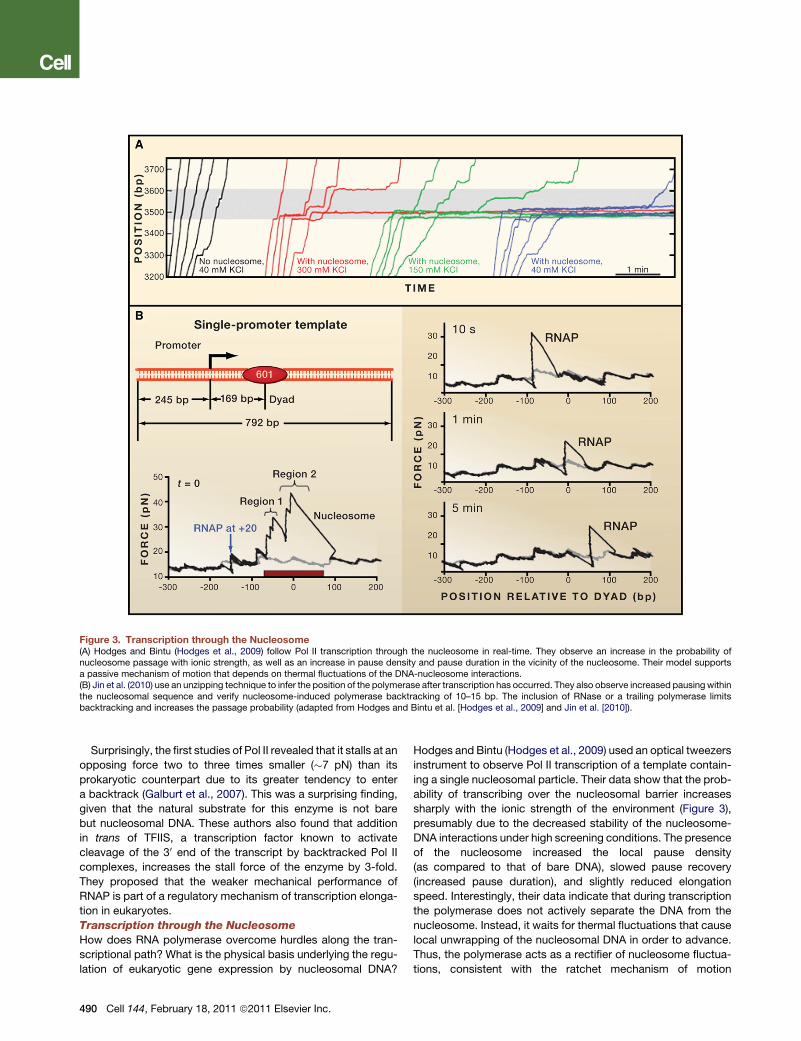
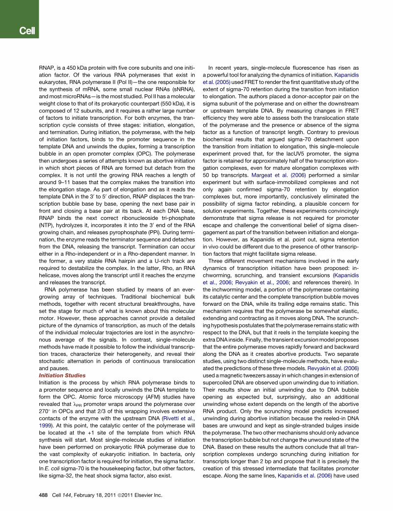
Figure 3. Transcription through the Nucleosome(A) Hodges and Bintu (Hodges et al., 2009) follow Pol II transcription through the nucleosome in real-time. They observe an increase in the probability ofnucleosome passage with ionic strength, as well as an increase in pause density and pause duration in the vicinity of the nucleosome. Their model supportsa passive mechanism of motion that depends on thermal fluctuations of the DNA-nucleosome interactions.(B) Jin et al. (2010) use an unzipping technique to infer the position of the polymerase after transcription has occurred. They also observe increased pausing withinthe nucleosomal sequence and verify nucleosome-induced polymerase backtracking of 10–15 bp. The inclusion of RNase or a trailing polymerase limitsbacktracking and increases the passage probability (adapted from Hodges and Bintu et al. [Hodges et al., 2009] and Jin et al. [2010]).
Surprisingly, the first studies of Pol II revealed that it stalls at an
opposing force two to three times smaller (�7 pN) than its
prokaryotic counterpart due to its greater tendency to enter
a backtrack (Galburt et al., 2007). This was a surprising finding,
given that the natural substrate for this enzyme is not bare
but nucleosomal DNA. These authors also found that addition
in trans of TFIIS, a transcription factor known to activate
cleavage of the 30 end of the transcript by backtracked Pol II
complexes, increases the stall force of the enzyme by 3-fold.
They proposed that the weaker mechanical performance of
RNAP is part of a regulatory mechanism of transcription elonga-
tion in eukaryotes.
Transcription through the Nucleosome
How does RNA polymerase overcome hurdles along the tran-
scriptional path? What is the physical basis underlying the regu-
lation of eukaryotic gene expression by nucleosomal DNA?
490 Cell 144, February 18, 2011 ª2011 Elsevier Inc.
Hodges and Bintu (Hodges et al., 2009) used an optical tweezers
instrument to observe Pol II transcription of a template contain-
ing a single nucleosomal particle. Their data show that the prob-
ability of transcribing over the nucleosomal barrier increases
sharply with the ionic strength of the environment (Figure 3),
presumably due to the decreased stability of the nucleosome-
DNA interactions under high screening conditions. The presence
of the nucleosome increased the local pause density
(as compared to that of bare DNA), slowed pause recovery
(increased pause duration), and slightly reduced elongation
speed. Interestingly, their data indicate that during transcription
the polymerase does not actively separate the DNA from the
nucleosome. Instead, it waits for thermal fluctuations that cause
local unwrapping of the nucleosomal DNA in order to advance.
Thus, the polymerase acts as a rectifier of nucleosome fluctua-
tions, consistent with the ratchet mechanism of motion

proposed for the operation of RNAP (Bar-Nahum et al., 2005).
Based on these results, they developed a quantitative model in
which the nucleosome behaves as a fluctuating mechanical
barrier that slows forward translocation and causes the poly-
merase to enter backtracked/paused states and, as a result,
increases the probability of enzyme arrest. Furthermore, during
backtracks the nucleosome can rewrap the newly exposed
DNA, a process that slows down the recovery from a pause.
As a way to better understand the interactions between the
DNA and the nucleosome, Jin et al. (2010) developed a DNA
unzipping technique that monitors the position of RNA poly-
merase from E. coli on the DNA template after transcription
(Figure 3B). In this experiment, a nucleosome is placed down-
stream of a polymerase and, after transcription is allowed to
take place for varying periods of time, the two strands of the tran-
scribed molecule are pulled apart. The bacterial polymerase
does not encounter nucleosomes in vivo, however it is used as
a model system warranted by the high level of functional
homology with Pol II (Walter et al., 2003). The resulting force
extension curves show characteristic transitions that indicate
the position of the polymerase on the DNA (to avoid additional
transitions due to nucleosome unwrapping, the nucleosome
was removed from the template using heparin). The authors
observed nucleosome-induced polymerase pausing with
a 10 bp periodicity that was sequence independent and corre-
lated with the periodicity of the interactions between the nucleo-
some and the DNA. Moreover, by comparing the size of the RNA
formed (using a transcription gel) with the position of the poly-
merase on the template (using the unzipping assay), they
estimated that the polymerase backtracks between 10–15 bases
when it encounters the nucleosome. They further reasoned that,
if backtracking and arrest occur upon transcription through the
nucleosome, conditions under which backtracking is limited
should facilitate passage. As predicted, the use of RNase, as
a way to reduce the number of RNA bases the polymerase could
backtrack on, decreased the number of backtracked bases and
increased the number of complexes that passed the nucleo-
some. Similarly, the addition of a second trailing polymerase
that physically limited the number of bases the leading poly-
merase could move back enhanced passage by a factor of 5,
an amount similar to experiments using RNase. From these
experiments the authors speculate that the presence of multiple
polymerases in vivowill further facilitate transcription through the
nucleosome by preventing or reducing backtracking. Also,
transcription factors like TFIIS could rescue backtracked poly-
merases, expediting nucleosome passage. It would be inter-
esting to repeat these experiments with the eukaryotic enzyme.
RNA polymerase pausing and backtracking are intrinsic and
complex properties of RNA polymerase important for transcrip-
tion regulation and control of transcription fidelity. Future use of
mutant polymerases with altered pausing behavior and the
reconstitution in vitro of ever more complex single-molecule
transcription reactions should provide a more complete picture
of the mechanisms that control gene expression during tran-
scription elongation.
Transcription Termination
To investigate the importance of mechanical force on termina-
tion, forces up to 30 pN were applied to the nascent RNA tran-
script (Dalal et al., 2006). No significant effect was found on
enzyme processivity, elongation rates, pause frequencies, and
lifetimes. It is unlikely that the termination hairpin or Rho could
exert larger forces; thus if force plays any role in termination, it
must be aided by an allosteric effect wherein the binding energy
of the hairpin and/or Rho to the complex pay in part the energetic
price of disrupting the DNA-RNA hybrid. Larson et al. (2008)
found that pulling between RNAP and upstream DNA does not
affect termination efficiency on two out of three terminators
studied, indicating that hypertranslocation (forward movement
of the bubble with respect to RNA’s 30 end) either cannot beeffected mechanically or is not the only mechanism of termina-
tion. In fact, the authors propose that depending on the identity
of the terminator, shearing of the RNA-DNA hybrid or hypertrans-
location, or both, can occur during transcript release.
Prokaryotic TranslationRibosomes are the cellular machines that hydrolyze GTP to
‘‘read’’ and translate the information encoded in mRNA into
protein (Moore and Steitz, 2003). Single-molecule studies of
translation are quite recent and restricted to prokaryotic
ribosomes. Translation is an extremely complex process also
conveniently divided in three phases: initiation, elongation, and
termination (Ramakrishnan, 2002).
In prokaryotes, initiation begins with the binding of the ribo-
some to themethionine-encoding mRNA translation start codon,
AUG, whose placing at the P site of the ribosome is directed by
an upstream Shine-Dalgarno (SD) sequence complementary to
a segment of the 16S ribosomal RNA. Initiation requires initiation
factors IF1, IF2,GTP, and IF3.
In elongation, ternary complexes of tRNAs charged with the
correct amino acids, elongation factor EF-Tu, and GTP bind to
ribosome. The correct amino acid-carrying tRNA is selected by
its complementarity to the codon on the mRNA and interactions
with the small and large subunits at the A site of the ribosome.
Upon GTP hydrolysis and release of EF-Tu, the tRNA is bound
in the ‘‘classical’’ position at the A site, adjacent to the
peptide-containing tRNA bound in the classical position at the
P site. Subsequently, a new peptide bond is formed as the poly-
peptide in the P site is transferred to the A site tRNA, a reaction
catalyzed by the peptidyl transferase active site in the 23S rRNA
of the 50S subunit. This event allows the tRNAs to access inter-
mediate binding conformations called ‘‘hybrid’’ states, in which
the anticodon ends of the tRNAs remain in their classical A and
P sites in the 30S subunit but their acceptor stems make
contacts in the P and E sites of the 50S subunit, respectively
(Moore and Steitz, 2003). The elongation cycle is completed
with the translocation of the ribosome relative to the mRNA
upon binding of another elongation factor, EF-G,GTP, and the
subsequent hydrolysis of GTP. In this process, the tRNA at the
A site moves to the P site and the tRNA at the P site moves to
the exit or E site.
Termination occurs when the ribosome encounters a stop
codon (either UAA, UAG, or UGA). Protein release factors are
bound that cleave the peptide from the tRNA at the P site;
release factor 1 (RF1) recognizes UAA and UAG; release factor
2 (RF2) recognizes UAA and UGA. The ribosome then remains
attached to the mRNA. Dissociation of the ribosome into its
Cell 144, February 18, 2011 ª2011 Elsevier Inc. 491
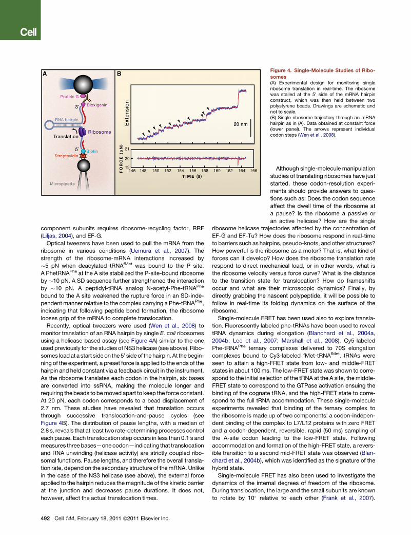
Figure 4. Single-Molecule Studies of Ribo-
somes(A) Experimental design for monitoring singleribosome translation in real-time. The ribosomewas stalled at the 50 side of the mRNA hairpinconstruct, which was then held between twopolystyrene beads. Drawings are schematic andnot to scale.(B) Single ribosome trajectory through an mRNAhairpin as in (A). Data obtained at constant force(lower panel). The arrows represent individualcodon steps (Wen et al., 2008).
component subunits requires ribosome-recycling factor, RRF
(Liljas, 2004), and EF-G.
Optical tweezers have been used to pull the mRNA from the
ribosome in various conditions (Uemura et al., 2007). The
strength of the ribosome-mRNA interactions increased by
�5 pN when deacylated tRNAfMet was bound to the P site.
A PhetRNAPhe at the A site stabilized the P-site-bound ribosome
by �10 pN. A SD sequence further strengthened the interaction
by �10 pN. A peptidyl-tRNA analog N-acetyl-Phe-tRNAPhe
bound to the A site weakened the rupture force in an SD-inde-
pendent manner relative to the complex carrying a Phe-tRNAPhe,
indicating that following peptide bond formation, the ribosome
looses grip of the mRNA to complete translocation.
Recently, optical tweezers were used (Wen et al., 2008) to
monitor translation of an RNA hairpin by single E. coli ribosomes
using a helicase-based assay (see Figure 4A) similar to the one
used previously for the studies of NS3 helicase (see above). Ribo-
somes loadata start sideon the50 sideof thehairpin.At thebegin-ning of the experiment, a preset force is applied to the ends of the
hairpin and held constant via a feedback circuit in the instrument.
As the ribosome translates each codon in the hairpin, six bases
are converted into ssRNA, making the molecule longer and
requiring the beads to bemoved apart to keep the force constant.
At 20 pN, each codon corresponds to a bead displacement of
2.7 nm. These studies have revealed that translation occurs
through successive translocation-and-pause cycles (see
Figure 4B). The distribution of pause lengths, with a median of
2.8 s, reveals that at least two rate-determining processes control
each pause. Each translocation step occurs in less than 0.1 s and
measures three bases—onecodon—indicating that translocation
and RNA unwinding (helicase activity) are strictly coupled ribo-
somal functions. Pause lengths, and therefore the overall transla-
tion rate, depend on the secondary structure of themRNA. Unlike
in the case of the NS3 helicase (see above), the external force
applied to the hairpin reduces themagnitude of the kinetic barrier
at the junction and decreases pause durations. It does not,
however, affect the actual translocation times.
492 Cell 144, February 18, 2011 ª2011 Elsevier Inc.
Although single-molecule manipulation
studies of translating ribosomes have just
started, these codon-resolution experi-
ments should provide answers to ques-
tions such as: Does the codon sequence
affect the dwell time of the ribosome at
a pause? Is the ribosome a passive or
an active helicase? How are the single
ribosome helicase trajectories affected by the concentration of
EF-G and EF-Tu? How does the ribosome respond in real-time
to barriers such as hairpins, pseudo-knots, and other structures?
How powerful is the ribosome as a motor? That is, what kind of
forces can it develop? How does the ribosome translation rate
respond to direct mechanical load, or in other words, what is
the ribosome velocity versus force curve? What is the distance
to the transition state for translocation? How do frameshifts
occur and what are their microscopic dynamics? Finally, by
directly grabbing the nascent polypeptide, it will be possible to
follow in real-time its folding dynamics on the surface of the
ribosome.
Single-molecule FRET has been used also to explore transla-
tion. Fluorescently labeled phe-tRNAs have been used to reveal
tRNA dynamics during elongation (Blanchard et al., 2004a,
2004b; Lee et al., 2007; Marshall et al., 2008). Cy5-labeled
Phe-tRNAPhe ternary complexes delivered to 70S elongation
complexes bound to Cy3-labeled fMet-tRNAfMet. tRNAs were
seen to attain a high-FRET state from low- and middle-FRET
states in about 100 ms. The low-FRET state was shown to corre-
spond to the initial selection of the tRNA at the A site, themiddle-
FRET state to correspond to the GTPase activation ensuing the
binding of the cognate tRNA, and the high-FRET state to corre-
spond to the full tRNA accommodation. These single-molecule
experiments revealed that binding of the ternary complex to
the ribosome is made up of two components: a codon-indepen-
dent binding of the complex to L7/L12 proteins with zero FRET
and a codon-dependent, reversible, rapid (50 ms) sampling of
the A-site codon leading to the low-FRET state. Following
accommodation and formation of the high-FRET state, a revers-
ible transition to a second mid-FRET state was observed (Blan-
chard et al., 2004b), which was identified as the signature of the
hybrid state.
Single-molecule FRET has also been used to investigate the
dynamics of the internal degrees of freedom of the ribosome.
During translocation, the large and the small subunits are known
to rotate by 10� relative to each other (Frank et al., 2007).

This ‘‘ratchet’’ motion is thought to accompany the formation of
the hybrid states (Ermolenko et al., 2007). Fluctuations in the
spatial orientations of the large and small subunits were followed
in real-time by FRET changes between Cy3-labeled protein L9
and Cy5-labeled protein S6 (Cornish et al., 2008). Ribosomes
fluorescently labeled at L1 and L33 were also used to monitor
the movement of the L1 stalk of the E site (Cornish et al.,
2009). A correlation between the L1 stalk position and the
binding, movement, and release of the deacylated tRNA at the
E site was found, suggesting that conformational changes of
the stalk are responsible for the tRNA transitions. Consistent
with these observations, fluctuating FRET signals between the
L1 stalk and the incoming tRNA were also detected (Fei et al.,
2008). These signals were thought to represent the stochastic
movements of the L1 stalk between open and closed conforma-
tions in the pretranslocation elongation complex, coupled to the
fluctuations of the P-site tRNA between its classical and its
hybrid configurations. Taken all together, these observations
suggest that the deacylation of the peptidyl-tRNA during elonga-
tion triggers the fluctuation of the entire pretranslocation
complex between two major conformational states, global state
1 (GS1) and global state 2 (GS2) (Fei et al., 2008). Evidence for
these two global states has been recently found by two Cryo-
EM studies (Agirrezabala et al., 2008; Julian et al., 2008).
Single-molecule FRET has been used to investigate the
dynamics of the ribosome and tRNAs during translation termina-
tion and ribosome recycling (Sternberg et al., 2009). These
authors used fluorescently labeled RF1, tRNAs, and ribosomes
to show that when RF1 binds at a stop codon and promotes
the hydrolysis of the peptide, the ribosome is locked in the
GS1 state. Subsequent binding of RF3 and GTP induce the ribo-
some to transition into GS2 and RF1 to release. GTP hydrolysis
then ensues and primes the ribosome for recycling. The authors
showed that the effect of RRF is to bias the state of the ribosome
to GS2, the recycling-competent state.
The implementation of single-molecule approaches together
with new technical developments such as zero-mode wave-
guides (ZMWs) allowed Uemura et al. (2010) to follow in real-
time the binding of tRNA during processive translation at physi-
ologically relevant micromolar ligand concentrations. By labeling
the tRNAs with distinct fluorophores, these authors were able to
determine the identity of the tRNA and the mRNA codon
involved. This study found that ribosomes are only briefly occu-
pied by two tRNAmolecules and that release of deacylated tRNA
from the exit (E) site is uncoupled from binding of aminoacyl-
tRNA site (A site) tRNA, occurring rapidly after translocation.
PerspectiveThe minimal unit of living matter, the cell, is a complex micro-
scopic factory whose integrity and homeostasis depend on the
operation of an interconnected network of highly specialized
tiny processing units or molecular machines. Some of these
machines, like the ones that are the subject of this Review,
must function as nucleic acid translocases. They must move
along their nucleic acid templates to read, copy, and translate
the linear information encoded in their sequences and ensure
the flow, control, and expression of genetic information. Until
recently, the detailed study of their function had lagged behind
that of their structures, mainly because of the difficulty of
synchronizing a population ofmolecules to follow their dynamics.
This situation is now changing rapidly. The emergence over the
last two decades of single-molecule techniques has begun to
yield impressive details on how thesemacromolecular machines
work. By following the actual molecular trajectories of these
translocases, andnot just themeanor average behavior of a pop-
ulation of molecules, we are beginning to learn unprecedented
details of their complex dynamics, the manner by which they
move on nucleic acids, the mechanical nature of their moving
parts, the presence of transient intermediates, their mechanisms
of fidelity, and the manner in which they use the spontaneous
fluctuations of the bath to accomplish their mechanical tasks.
We havemany reasons to believe that these developments are
just the beginning of growing insight into the operation of these
machines: with the advent of high-resolution, single-molecule
optical tweezers (Abbondanzieri et al., 2005; Moffitt et al.,
2006) and the combination of optical tweezers with single-mole-
cule fluorescence capability (Hohng et al., 2007; Lang et al.,
2004), it should be possible now to monitor directly the move-
ment of motors at angstrom-level resolution (Moffitt et al.,
2009) and to uncover the coordination of their various parts
during their mechanochemical conversion (Ishijima et al., 1998).
Future efforts, through the study of evermore complex assem-
blies, will also likely try to fill the gap between the controlled
experimental conditions of in vitro studies and the need to under-
stand at a quantitative level how the performance of these
machines is influenced by their physiological partners (Stano
et al., 2005). Finally, recent advances in super-resolution optical
imaging (Betzig et al., 2006; Hell, 2007; Huang et al., 2008) may
also make it possible to fulfill the ultimate hope of directly
observing the activity of these machines in living cells.
Ultimately, a comparison of the diverse molecular designs
utilized by evolution to accomplish these directional and
energy-driven tasks, the unraveling of the physical principles
that lie behind their function, and the emerging understanding
of the importance of fluctuations in their operation and thermo-
dynamic efficiency should provide, in the not-too-distant future,
the basis for the development of a comprehensive theory of
molecular motors. At the very least, we hope that these efforts
will fulfill the goal of endowing the detailed structures of these
molecular entities, with an equally detailed description of the
molecular choreography that underlies their operation in the cell.
SUPPLEMENTAL INFORMATION
Supplemental Information includes an Extended Discussion, two figures, and
Supplemental References and can be found with this article online at doi:
10.1016/j.cell.2011.01.033.
ACKNOWLEDGMENTS
We thank TimothyM. Lohman atWashington University School of Medicine for
a critical reading of the draft on helicases and many colleagues for stimulating
discussions. The literature on nucleic acid translocases, in particular their
single-molecule studies, is ever increasing. Due to space limitations and our
coverage of selected topics, we would like to apologize to our colleagues
who actively work on nucleic acid translocases yet whose work has not
been cited here. C.B. was supported by NIH, DOE, and HHMI. W.C. was sup-
ported by Ara Paul Professorship fund at the University of Michigan Ann Arbor.
Cell 144, February 18, 2011 ª2011 Elsevier Inc. 493

REFERENCES
Aathavan, K., Politzer, A.T., Kaplan, A., Moffitt, J.R., Chemla, Y.R., Grimes, S.,
Jardine, P.J., Anderson, D.L., and Bustamante, C. (2009). Substrate interac-
tions and promiscuity in a viral DNA packaging motor. Nature 461, 669–673.
Abbondanzieri, E.A., Greenleaf, W.J., Shaevitz, J.W., Landick, R., and Block,
S.M. (2005). Direct observation of base-pair stepping by RNA polymerase.
Nature 438, 460–465.
Aboussekhra, A., Chanet, R., Adjiri, A., and Fabre, F. (1992). Semidominant
suppressors of Srs2 helicase mutations of Saccharomyces cerevisiae map
in the RAD51 gene, whose sequence predicts a protein with similarities to pro-
caryotic RecA proteins. Mol. Cell. Biol. 12, 3224–3234.
Agirrezabala, X., Lei, J., Brunelle, J.L., Ortiz-Meoz, R.F., Green, R., and Frank,
J. (2008). Visualization of the hybrid state of tRNA binding promoted by spon-
taneous ratcheting of the ribosome. Mol. Cell 32, 190–197.
Ahnert, P., Picha, K.M., and Patel, S.S. (2000). A ring-opening mechanism for
DNA binding in the central channel of the T7 helicase-primase protein. EMBO
J. 19, 3418–3427.
Alberts, B. (1998). The cell as a collection of protein machines: Preparing the
next generation of molecular biologists. Cell 92, 291–294.
Alberts, B.M., Barry, J., Bedinger, P., Formosa, T., Jongeneel, C.V., and
Kreuzer, K.N. (1983). Studies on DNA replication in the bacteriophage T4
in vitro system. Cold Spring Harb. Symp. Quant. Biol. 47, 655–668.
Antony, E., Tomko, E.J., Xiao, Q., Krejci, L., Lohman, T.M., and Ellenberger, T.
(2009). Srs2 disassembles Rad51 filaments by a protein-protein interaction
triggering ATP turnover and dissociation of Rad51 from DNA. Mol. Cell 35,
105–115.
Appleby, T.C., Anderson, R., Fedorova, O., Pyle, A.M., Wang, R., Liu, X.,
Brendza, K.M., and Somoza, J.R. (2010). Visualizing ATP-dependent RNA
translocation by the NS3 helicase from HCV. J. Mol. Biol. 405, 1139–1153.
Bar-Nahum, G., Epshtein, V., Ruckenstein, A.E., Rafikov, R., Mustaev, A., and
Nudler, E. (2005). A ratchet mechanism of transcription elongation and its
control. Cell 120, 183–193.
Bessman, M.J., Lehman, I.R., Simms, E.S., and Kornberg, A. (1958). Enzy-
matic synthesis of deoxyribonucleic acid. II. General properties of the reaction.
J. Biol. Chem. 233, 171–177.
Betterton, M.D., and Julicher, F. (2005). Opening of nucleic-acid double
strands by helicases: active versus passive opening. Phys. Rev. E Stat. Nonlin.
Soft Matter Phys. 71, 011904.
Betzig, E., Patterson, G.H., Sougrat, R., Lindwasser, O.W., Olenych, S., Boni-
facino, J.S., Davidson, M.W., Lippincott-Schwartz, J., and Hess, H.F. (2006).
Imaging intracellular fluorescent proteins at nanometer resolution. Science
313, 1642–1645.
Bianco, P.R., Brewer, L.R., Corzett, M., Balhorn, R., Yeh, Y., Kowalczykowski,
S.C., and Baskin, R.J. (2001). Processive translocation and DNA unwinding by
individual RecBCD enzyme molecules. Nature 409, 374–378.
Bird, L.E., Subramanya, H.S., and Wigley, D.B. (1998). Helicases: a unifying
structural theme? Curr. Opin. Struct. Biol. 8, 14–18.
Blanchard, S.C., Gonzalez, R.L., Kim, H.D., Chu, S., and Puglisi, J.D. (2004a).
tRNA selection and kinetic proofreading in translation. Nat. Struct. Mol. Biol.
11, 1008–1014.
Blanchard, S.C., Kim, H.D., Gonzalez, R.L., Jr., Puglisi, J.D., and Chu, S.
(2004b). tRNA dynamics on the ribosome during translation. Proc. Natl.
Acad. Sci. USA 101, 12893–12898.
Bryant, Z., Stone, M.D., Gore, J., Smith, S.B., Cozzarelli, N.R., and Busta-
mante, C. (2003). Structural transitions and elasticity from torque measure-
ments on DNA. Nature 424, 338–341.
Burroughs, A.M., Iyer, L.M., and Aravind, L. (2007). Comparative genomics
and evolutionary trajectories of viral ATP dependent DNA-packaging systems.
In Gene and Protein Evolution, J.-N. Volff , ed., pp. 48–65.
Bustamante, C., Marko, J.F., Siggia, E.D., and Smith, S. (1994). Entropic elas-
ticity of lambda-phage DNA. Science 265, 1599–1600.
494 Cell 144, February 18, 2011 ª2011 Elsevier Inc.
Bustamante, C., Macosko, J.C., and Wuite, G.J. (2000). Grabbing the cat by
the tail: manipulating molecules one by one. Nat. Rev. Mol. Cell Biol. 1, 130–
136.
Catalano, C.E. (2005). Viral genome packaging machines: An overview. In
Viral Genome Packaging Machines: Genetics, Structure, and Mechanism,
C.E. Catalano, ed. (New York: Kluwer/Plenum), pp. 1–4.
Chemla, Y.R., Aathavan, K., Michaelis, J., Grimes, S., Jardine, P.J., Anderson,
D.L., and Bustamante, C. (2005). Mechanism of force generation of a viral DNA
packaging motor. Cell 122, 683–692.
Cheng, W., Dumont, S., Tinoco, I., Jr., and Bustamante, C. (2007). NS3 heli-
case actively separates RNA strands and senses sequence barriers ahead
of the opening fork. Proc. Natl. Acad. Sci. USA 104, 13954–13959.
Cornish, P.V., Ermolenko, D.N., Noller, H.F., and Ha, T. (2008). Spontaneous
intersubunit rotation in single ribosomes. Mol. Cell 30, 578–588.
Cornish, P.V., Ermolenko, D.N., Staple, D.W., Hoang, L., Hickerson, R.P.,
Noller, H.F., and Ha, T. (2009). Following movement of the L1 stalk between
three functional states in single ribosomes. Proc. Natl. Acad. Sci. USA 106,
2571–2576.
Dalal, R.V., Larson, M.H., Neuman, K.C., Gelles, J., Landick, R., and Block,
S.M. (2006). Pulling on the nascent RNA during transcription does not alter
kinetics of elongation or ubiquitous pausing. Mol. Cell 23, 231–239.
Davenport, R.J., Wuite, G.J., Landick, R., and Bustamante, C. (2000). Single-
molecule study of transcriptional pausing and arrest by E. coli RNA poly-
merase. Science 287, 2497–2500.
Depken, M., Galburt, E.A., and Grill, S.W. (2009). The origin of short transcrip-
tional pauses. Biophys. J. 96, 2189–2193.
Dessinges, M.N., Lionnet, T., Xi, X.G., Bensimon, D., and Croquette, V. (2004).
Single-molecule assay reveals strand switching and enhanced processivity of
UvrD. Proc. Natl. Acad. Sci. USA 101, 6439–6444.
Dhar, A., and Feiss, M. (2005). Bacteriophage lambda terminase: alterations of
the high-affinity ATPase affect viral DNA packaging. J. Mol. Biol. 347, 71–80.
Dohoney, K.M., and Gelles, J. (2001). Chi-sequence recognition and DNA
translocation by single RecBCD helicase/nuclease molecules. Nature 409,
370–374.
Doublie, S., Tabor, S., Long, A.M., Richardson, C.C., and Ellenberger, T.
(1998). Crystal structure of a bacteriophage T7 DNA replication complex at
2.2 A resolution. Nature 391, 251–258.
Dumont, S., Cheng, W., Serebrov, V., Beran, R.K., Tinoco, I., Jr., Pyle, A.M.,
and Bustamante, C. (2006). RNA translocation and unwinding mechanism of
HCV NS3 helicase and its coordination by ATP. Nature 439, 105–108.
Enemark, E.J., and Joshua-Tor, L. (2008). On helicases and other motor
proteins. Curr. Opin. Struct. Biol. 18, 243–257.
Ermolenko, D.N., Majumdar, Z.K., Hickerson, R.P., Spiegel, P.C., Clegg, R.M.,
and Noller, H.F. (2007). Observation of intersubunit movement of the ribosome
in solution using FRET. J. Mol. Biol. 370, 530–540.
Erzberger, J.P., and Berger, J.M. (2006). Evolutionary relationships and struc-
tural mechanisms of AAA+ proteins. Annu. Rev. Biophys. Biomol. Struct. 35,
93–114.
Fei, J., Kosuri, P., MacDougall, D.D., and Gonzalez, R.L., Jr. (2008). Coupling
of ribosomal L1 stalk and tRNA dynamics during translation elongation. Mol.
Cell 30, 348–359.
Finer, J.T., Simmons, R.M., and Spudich, J.A. (1994). Single myosin molecule
mechanics: piconewton forces and nanometre steps. Nature 368, 113–119.
Forde, N.R., Izhaky, D., Woodcock, G.R., Wuite, G.J., and Bustamante, C.
(2002). Using mechanical force to probe the mechanism of pausing and arrest
during continuous elongation by Escherichia coli RNA polymerase. Proc. Natl.
Acad. Sci. USA 99, 11682–11687.
Frank, J., Gao, H., Sengupta, J., Gao, N., and Taylor, D.J. (2007). The process
of mRNA-tRNA translocation. Proc. Natl. Acad. Sci. USA 104, 19671–19678.
Frick, D.N. (2003). Helicases as antiviral drug targets. Drug News Perspect. 16,
355–362.
Fuller, D.N., Raymer, D.M., Kottadiel, V.I., Rao, V.B., and Smith, D.E. (2007a).
Single phage T4 DNA packaging motors exhibit large force generation, high
velocity, and dynamic variability. Proc. Natl. Acad. Sci. USA 104, 16868–
16873.
Fuller, D.N., Raymer, D.M., Rickgauer, J.P., Robertson, R.M., Catalano, C.E.,
Anderson, D.L., Grimes, S., and Smith, D.E. (2007b). Measurements of single
DNA molecule packaging dynamics in bacteriophage lambda reveal high
forces, high motor processivity, and capsid transformations. J. Mol. Biol.
373, 1113–1122.
Galburt, E.A., Grill, S.W., Wiedmann, A., Lubkowska, L., Choy, J., Nogales, E.,
Kashlev, M., and Bustamante, C. (2007). Backtracking determines the force
sensitivity of RNAP II in a factor-dependent manner. Nature 446, 820–823.
Gangloff, S., Soustelle, C., and Fabre, F. (2000). Homologous recombination is
responsible for cell death in the absence of the Sgs1 and Srs2 helicases. Nat.
Genet. 25, 192–194.
Gorbalenya, A.E., and Koonin, E.V. (1993). Helicases: amino acid sequence
comparisons and structure-function relationships. Curr. Opin. Struct. Biol. 3,
419–429.
Grimes, S., and Anderson, D. (1997a). The bacteriophage phi29 packaging
proteins supercoil the DNA ends. J. Mol. Biol. 266, 901–914.
Grimes, S., and Anderson, D. (1997b). The bacteriophage phi 29 packaging
proteins supercoil the DNA ends. J. Mol. Biol. 266, 901–914.
Grimes, S., Jardine, P.J., and Anderson, D. (2002). Bacteriophage phi 29 DNA
packaging. Adv. Virus Res. 58, 255–294.
Ha, T., Rasnik, I., Cheng, W., Babcock, H.P., Gauss, G.H., Lohman, T.M., and
Chu, S. (2002). Initiation and re-initiation of DNA unwinding by the Escherichia
coli Rep helicase. Nature 419, 638–641.
Hamdan, S.M., Loparo, J.J., Takahashi, M., Richardson, C.C., and van Oijen,
A.M. (2009). Dynamics of DNA replication loops reveal temporal control of
lagging-strand synthesis. Nature 457, 336–339.
Hell, S.W. (2007). Far-field optical nanoscopy. Science 316, 1153–1158.
Herbert, K.M., La Porta, A., Wong, B.J., Mooney, R.A., Neuman, K.C., Landick,
R., and Block, S.M. (2006). Sequence-resolved detection of pausing by single
RNA polymerase molecules. Cell 125, 1083–1094.
Hodges, C., Bintu, L., Lubkowska, L., Kashlev, M., and Bustamante, C. (2009).
Nucleosomal fluctuations govern the transcription dynamics of RNA poly-
merase II. Science 325, 626–628.
Hohng, S., Zhou, R., Nahas, M.K., Yu, J., Schulten, K., Lilley, D.M., and Ha, T.
(2007). Fluorescence-force spectroscopy maps two-dimensional reaction
landscape of the holliday junction. Science 318, 279–283.
Huang, B., Wang, W., Bates, M., and Zhuang, X. (2008). Three-dimensional
super-resolution imaging by stochastic optical reconstruction microscopy.
Science 319, 810–813.
Ibarra, B., Chemla, Y.R., Plyasunov, S., Smith, S.B., Lazaro, J.M., Salas, M.,
and Bustamante, C. (2009). Proofreading dynamics of a processive DNA poly-
merase. EMBO J. 28, 2794–2802.
Ishijima, A., Kojima, H., Funatsu, T., Tokunaga, M., Higuchi, H., Tanaka, H.,
and Yanagida, T. (1998). Simultaneous observation of individual ATPase and
mechanical events by a single myosin molecule during interaction with actin.
Cell 92, 161–171.
Iyer, L.M., Leipe, D.D., Koonin, E.V., and Aravind, L. (2004a). Evolutionary
history and higher order classification of AAA+ ATPases. J. Struct. Biol. 146,
11–31.
Iyer, L.M., Makarova, K.S., Koonin, E.V., and Aravind, L. (2004b). Comparative
genomics of the FtsK-HerA superfamily of pumping ATPases: implications for
the origins of chromosome segregation, cell division and viral capsid pack-
aging. Nucleic Acids Res. 32, 5260–5279.
Jankowsky, E., and Bowers, H. (2006). Remodeling of ribonucleoprotein
complexes with DExH/D RNA helicases. Nucleic Acids Res. 34, 4181–4188.
Jardine, P.J., and Anderson, D. (2006). DNA packaging in double-stranded
DNA bacteriophages. In The Bacteriophages, R. Calendar, ed. (New York:
Oxford University Press), pp. 49–65.
Jennings, T.A., Mackintosh, S.G., Harrison, M.K., Sikora, D., Sikora, B., Dave,
B., Tackett, A.J., Cameron, C.E., and Raney, K.D. (2009). NS3 helicase from
the hepatitis C virus can function as a monomer or oligomer depending on
enzyme and substrate concentrations. J. Biol. Chem. 284, 4806–4814.
Jin, J., Bai, L., Johnson, D.S., Fulbright, R.M., Kireeva, M.L., Kashlev, M., and
Wang, M.D. (2010). Synergistic action of RNA polymerases in overcoming the
nucleosomal barrier. Nat. Struct. Mol. Biol. 17, 745–752.
Johnson, D.S., Bai, L., Smith, B.Y., Patel, S.S., andWang, M.D. (2007). Single-
molecule studies reveal dynamics of DNA unwinding by the ring-shaped T7
helicase. Cell 129, 1299–1309.
Julian, P., Konevega, A.L., Scheres, S.H., Lazaro, M., Gil, D., Wintermeyer, W.,
Rodnina, M.V., and Valle, M. (2008). Structure of ratcheted ribosomes with
tRNAs in hybrid states. Proc. Natl. Acad. Sci. USA 105, 16924–16927.
Kapanidis, A.N., Margeat, E., Laurence, T.A., Doose, S., Ho, S.O., Mukhopad-
hyay, J., Kortkhonjia, E., Mekler, V., and Ebright, R.H. (2005). Retention of
transcription initiation factor s70 in transcription elongation: Single-molecule
analysis. Mol. Cell 20, 347–356.
Kapanidis, A.N., Margeat, E., Ho, S.O., Kortkhonjia, E., Weiss, S., and Ebright,
R.H. (2006). Initial transcription by RNA polymerase proceeds through a DNA-
scrunching mechanism. Science 314, 1144–1147.
Keller, D., and Bustamante, C. (2000). The mechanochemistry of molecular
motors. Biophys. J. 78, 541–556.
Kolykhalov, A.A., Mihalik, K., Feinstone, S.M., and Rice, C.M. (2000). Hepatitis
C virus-encoded enzymatic activities and conserved RNA elements in the 30
nontranslated region are essential for virus replication in vivo. J. Virol. 74,
2046–2051.
Koonin, E.V., Senkevich, T.G., and Chernos, V.I. (1993). Gene A32 product of
vaccinia virus may be an ATPase involved in viral DNA packaging as indicated
by sequence comparisons with other putative viral ATPases. Virus Genes 7,
89–94.
Korolev, S., Yao, N., Lohman, T.M., Weber, P.C., and Waksman, G. (1998).
Comparisons between the structures of HCV and Rep helicases reveal struc-
tural similarities between SF1 and SF2 super-families of helicases. Protein Sci.
7, 605–610.
Koti, J.S., Morais, M.C., Rajagopal, R., Owen, B.A., McMurray, C.T., and
Anderson, D.L. (2008). DNA packaging motor assembly intermediate of
bacteriophage phi29. J. Mol. Biol. 381, 1114–1132.
Krejci, L., Van Komen, S., Li, Y., Villemain, J., Reddy, M.S., Klein, H.,
Ellenberger, T., and Sung, P. (2003). DNA helicase Srs2 disrupts the Rad51
presynaptic filament. Nature 423, 305–309.
Lam, A.M., and Frick, D.N. (2006). Hepatitis C virus subgenomic replicon
requires an active NS3 RNA helicase. J. Virol. 80, 404–411.
Lang, M.J., Fordyce, P.M., Engh, A.M., Neuman, K.C., and Block, S.M. (2004).
Simultaneous, coincident optical trapping and single-molecule fluorescence.
Nat. Methods 1, 133–139.
Larson, M.H., Greenleaf, W.J., Landick, R., and Block, S.M. (2008). Applied
force reveals mechanistic and energetic details of transcription termination.
Cell 132, 971–982.
Lee, T.H., Blanchard, S.C., Kim, H.D., Puglisi, J.D., and Chu, S. (2007). The role
of fluctuations in tRNA selection by the ribosome. Proc. Natl. Acad. Sci. USA
104, 13661–13665.
Lehman, I.R., Bessman, M.J., Simms, E.S., and Kornberg, A. (1958).
Enzymatic synthesis of deoxyribonucleic acid. I. Preparation of substrates
and partial purification of an enzyme from Escherichia coli. J. Biol. Chem.
233, 163–170.
Liljas, A. (2004). Structural Aspects of Protein Synthesis (Singapore: World
Scientific Publishing Co.).
Lindenbach, B.D., and Rice, C.M. (2005). Unravelling hepatitis C virus replica-
tion from genome to function. Nature 436, 933–938.
Lionnet, T., Spiering, M.M., Benkovic, S.J., Bensimon, D., and Croquette, V.
(2007). Real-time observation of bacteriophage T4 gp41 helicase reveals an
unwinding mechanism. Proc. Natl. Acad. Sci. USA 104, 19790–19795.
Cell 144, February 18, 2011 ª2011 Elsevier Inc. 495
Liu, S., Abbondanzieri, E.A., Rausch, J.W., Le Grice, S.F., and Zhuang, X.
(2008). Slide into action: dynamic shuttling of HIV reverse transcriptase on nu-
cleic acid substrates. Science 322, 1092–1097.
Lohman, T.M., and Bjornson, K.P. (1996). Mechanisms of helicase-catalyzed
DNA unwinding. Annu. Rev. Biochem. 65, 169–214.
Lohman, T.M., Thorn, K., and Vale, R.D. (1998). Staying on track: Common
features of DNA helicases and microtubule motors. Cell 93, 9–12.
Lohman, T.M., Tomko, E.J., and Wu, C.G. (2008). Non-hexameric DNA heli-
cases and translocases: mechanisms and regulation. Nat. Rev. Mol. Cell
Biol. 9, 391–401.
Ma, Y., Yates, J., Liang, Y., Lemon, S.M., and Yi, M. (2008). NS3 helicase
domains involved in infectious intracellular hepatitis C virus particle assembly.
J. Virol. 82, 7624–7639.
Mackintosh, S.G., and Raney, K.D. (2006). DNA unwinding and protein
displacement by superfamily 1 and superfamily 2 helicases. Nucleic Acids
Res. 34, 4154–4159.
Margeat, E., Kapanidis, A.N., Tinnefeld, P., Wang, Y., Mukhopadhyay, J.,
Ebright, R.H., and Weiss, S. (2006). Direct observation of abortive initiation
and promoter escape within single immobilized transcription complexes. Bio-
phys. J. 90, 1419–1431.
Marshall, R.A., Aitken, C.E., Dorywalska, M., and Puglisi, J.D. (2008). Transla-
tion at the single-molecule level. Annu. Rev. Biochem. 77, 177–203.
Matson, S.W., Bean, D.W., and George, J.W. (1994). DNA helicases: enzymes
with essential roles in all aspects of DNA metabolism. Bioessays 16, 13–22.
Mejia, Y.X., Mao, H., Forde, N.R., and Bustamante, C. (2008). Thermal probing
of E. coli RNA polymerase off-pathway mechanisms. J. Mol. Biol. 382,
628–637.
Mitchell, M.S.,Matsuzaki, S., Imai, S., andRao, V.B. (2002). Sequence analysis
of bacteriophage T4 DNA packaging/terminase genes 16 and 17 reveals
a common ATPase center in the large subunit of viral terminases. Nucleic
Acids Res. 30, 4009–4021.
Moffitt, J.R., Chemla, Y.R., Izhaky, D., and Bustamante, C. (2006). Differential
detection of dual traps improves the spatial resolution of optical tweezers.
Proc. Natl. Acad. Sci. USA 103, 9006–9011.
Moffitt, J.R., Chemla, Y.R., Aathavan, K., Grimes, S., Jardine, P.J., Anderson,
D.L., andBustamante, C. (2009). Intersubunit coordination in a homomeric ring
ATPase. Nature 457, 446–450.
Moore, P.B., and Steitz, T.A. (2003). The structural basis of large ribosomal
subunit function. Annu. Rev. Biochem. 72, 813–850.
Moradpour, D., Penin, F., and Rice, C.M. (2007). Replication of hepatitis C
virus. Nat. Rev. Microbiol. 5, 453–463.
Morais,M.C., Koti, J.S., Bowman, V.D., Reyes-Aldrete, E., Anderson, D.L., and
Rossmann, M.G. (2008). Defining molecular and domain boundaries in the
bacteriophage phi29 DNA packaging motor. Structure 16, 1267–1274.
Murakami, A., Ishida, S., and Dickson, C. (2002). GATA-4 interacts distinctively
with negative and positive regulatory elements in the Fgf-3 promoter. Nucleic
Acids Res. 30, 1056–1064.
Muschielok, A., Andrecka, J., Jawhari, A., Bruckner, F., Cramer, P., and
Michaelis, J. (2008). A nano-positioning system for macromolecular structural
analysis. Nat. Methods 5, 965–971.
Myong, S., and Ha, T. (2010). Stepwise translocation of nucleic acid motors.
Curr. Opin. Struct. Biol. 20, 121–127.
Myong, S., Rasnik, I., Joo, C., Lohman, T.M., and Ha, T. (2005). Repetitive
shuttling of a motor protein on DNA. Nature 437, 1321–1325.
Myong, S., Bruno, M.M., Pyle, A.M., and Ha, T. (2007). Spring-loaded mecha-
nism of DNA unwinding by hepatitis C virus NS3 helicase. Science 317,
513–516.
Neuman, K.C., Abbondanzieri, E.A., Landick, R., Gelles, J., and Block, S.M.
(2003). Ubiquitous transcriptional pausing is independent of RNA polymerase
backtracking. Cell 115, 437–447.
496 Cell 144, February 18, 2011 ª2011 Elsevier Inc.
Nudler, E., Mustaev, A., Lukhtanov, E., and Goldfarb, A. (1997). The RNA-DNA
hybrid maintains the register of transcription by preventing backtracking of
RNA polymerase. Cell 89, 33–41.
Ogawa, T., and Okazaki, T. (1980). Discontinuous DNA replication. Annu. Rev.
Biochem. 49, 421–457.
Palladino, F., and Klein, H.L. (1992). Analysis of mitotic and meiotic defects in
Saccharomyces cerevisiae SRS2DNA helicasemutants. Genetics 132, 23–37.
Pang, P.S., Jankowsky, E., Planet, P.J., and Pyle, A.M. (2002). The hepatitis C
viral NS3 protein is a processive DNA helicase with cofactor enhanced RNA
unwinding. EMBO J. 21, 1168–1176.
Park, J., Myong, S., Niedziela-Majka, A., Yu, J., Lohman, T.M., and Ha, T.
(2010). PcrA helicase dismantles RecA filaments by reeling in DNA in uniform
steps. Cell 142, 1–12.
Patel, S.S., and Picha, K.M. (2000). Structure and function of hexameric heli-
cases. Annu. Rev. Biochem. 69, 651–697.
Pyle, A.M. (2008). Translocation and unwinding mechanisms of RNA and DNA
helicases. Annu. Rev. Biophys. 37, 317–336.
Ramakrishnan, V. (2002). Ribosome structure and the mechanism of transla-
tion. Cell 108, 557–572.
Raney, K.D., Sharma, S.D., Moustafa, I.M., and Cameron, C.E. (2010). Hepa-
titis C virus non-structural protein 3 (HCV NS3): a multifunctional antiviral
target. J. Biol. Chem. 285, 22725–22731.
Rao, V.B., and Feiss, M. (2008). The bacteriophage DNA packaging motor.
Annu. Rev. Genet. 42, 647–681.
Revyakin, A., Liu, C., Ebright, R.H., and Strick, T.R. (2006). Abortive initiation
and productive initiation by RNA polymerase involve DNA scrunching. Science
314, 1139–1143.
Rickgauer, J.P., Fuller, D.N., Bo, H., Grimes, S., Jardine, P.J., Anderson, D.L.,
and Smith, D.E. (2006). Initiation of bacteriophage phi 29 DNA packaging
studied by optical tweezers manipulation of single DNA molecules. Proceed-
ings of the SPIE - The International Society for Optical Engineering, 632623-
632621-632628.
Rivetti, C., Guthold, M., and Bustamante, C. (1999). Wrapping of DNA around
the E.coli RNA polymerase open promoter complex. EMBO J. 18, 4464–4475.
Rocak, S., and Linder, P. (2004). DEAD-box proteins: the driving forces behind
RNA metabolism. Nat. Rev. Mol. Cell Biol. 5, 232–241.
Saha, A., Wittmeyer, J., and Cairns, B.R. (2006). Chromatin remodelling: the
industrial revolution of DNA around histones. Nat. Rev. Mol. Cell Biol. 7,
437–447.
Serebrov, V., and Pyle, A.M. (2004). Periodic cycles of RNA unwinding and
pausing by hepatitis C virus NS3 helicase. Nature 430, 476–480.
Serebrov, V., Beran, R.K., and Pyle, A.M. (2009). Establishing a mechanistic
basis for the large kinetic steps of the NS3 helicase. J. Biol. Chem. 284,
2512–2521.
Shaevitz, J.W., Abbondanzieri, E.A., Landick, R., and Block, S.M. (2003).
Backtracking by single RNA polymerase molecules observed at near-base-
pair resolution. Nature 426, 684–687.
Shiratori, A., Shibata, T., Arisawa, M., Hanaoka, F., Murakami, Y., and Eki, T.
(1999). Systematic identification, classification, and characterization of the
open reading frames which encode novel helicase-related proteins in Saccha-
romyces cerevisiae by gene disruption and Northern analysis. Yeast 15,
219–253.
Smith, D.E., Tans, S.J., Smith, S.B., Grimes, S., Anderson, D.L., and Busta-
mante, C. (2001). The bacteriophage straight phi29 portal motor can package
DNA against a large internal force. Nature 413, 748–752.
Smith, S.B., Finzi, L., and Bustamante, C. (1992). Direct mechanical measure-
ments of the elasticity of single DNA molecules by using magnetic beads.
Science 258, 1122–1126.
Smith, S.B., Cui, Y., and Bustamante, C. (1996). Overstretching B-DNA: the
elastic response of individual double-stranded and single-stranded DNA
molecules. Science 271, 795–799.
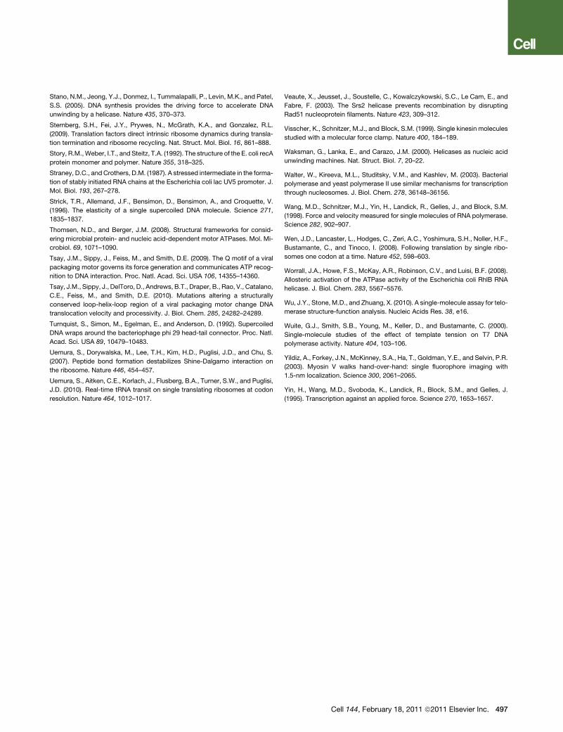
Stano, N.M., Jeong, Y.J., Donmez, I., Tummalapalli, P., Levin, M.K., and Patel,
S.S. (2005). DNA synthesis provides the driving force to accelerate DNA
unwinding by a helicase. Nature 435, 370–373.
Sternberg, S.H., Fei, J.Y., Prywes, N., McGrath, K.A., and Gonzalez, R.L.
(2009). Translation factors direct intrinsic ribosome dynamics during transla-
tion termination and ribosome recycling. Nat. Struct. Mol. Biol. 16, 861–888.
Story, R.M.,Weber, I.T., and Steitz, T.A. (1992). The structure of the E. coli recA
protein monomer and polymer. Nature 355, 318–325.
Straney, D.C., and Crothers, D.M. (1987). A stressed intermediate in the forma-
tion of stably initiated RNA chains at the Escherichia coli lac UV5 promoter. J.
Mol. Biol. 193, 267–278.
Strick, T.R., Allemand, J.F., Bensimon, D., Bensimon, A., and Croquette, V.
(1996). The elasticity of a single supercoiled DNA molecule. Science 271,
1835–1837.
Thomsen, N.D., and Berger, J.M. (2008). Structural frameworks for consid-
ering microbial protein- and nucleic acid-dependent motor ATPases. Mol. Mi-
crobiol. 69, 1071–1090.
Tsay, J.M., Sippy, J., Feiss, M., and Smith, D.E. (2009). The Q motif of a viral
packaging motor governs its force generation and communicates ATP recog-
nition to DNA interaction. Proc. Natl. Acad. Sci. USA 106, 14355–14360.
Tsay, J.M., Sippy, J., DelToro, D., Andrews, B.T., Draper, B., Rao, V., Catalano,
C.E., Feiss, M., and Smith, D.E. (2010). Mutations altering a structurally
conserved loop-helix-loop region of a viral packaging motor change DNA
translocation velocity and processivity. J. Biol. Chem. 285, 24282–24289.
Turnquist, S., Simon, M., Egelman, E., and Anderson, D. (1992). Supercoiled
DNA wraps around the bacteriophage phi 29 head-tail connector. Proc. Natl.
Acad. Sci. USA 89, 10479–10483.
Uemura, S., Dorywalska, M., Lee, T.H., Kim, H.D., Puglisi, J.D., and Chu, S.
(2007). Peptide bond formation destabilizes Shine-Dalgarno interaction on
the ribosome. Nature 446, 454–457.
Uemura, S., Aitken, C.E., Korlach, J., Flusberg, B.A., Turner, S.W., and Puglisi,
J.D. (2010). Real-time tRNA transit on single translating ribosomes at codon
resolution. Nature 464, 1012–1017.
Veaute, X., Jeusset, J., Soustelle, C., Kowalczykowski, S.C., Le Cam, E., and
Fabre, F. (2003). The Srs2 helicase prevents recombination by disrupting
Rad51 nucleoprotein filaments. Nature 423, 309–312.
Visscher, K., Schnitzer, M.J., and Block, S.M. (1999). Single kinesin molecules
studied with a molecular force clamp. Nature 400, 184–189.
Waksman, G., Lanka, E., and Carazo, J.M. (2000). Helicases as nucleic acid
unwinding machines. Nat. Struct. Biol. 7, 20–22.
Walter, W., Kireeva, M.L., Studitsky, V.M., and Kashlev, M. (2003). Bacterial
polymerase and yeast polymerase II use similar mechanisms for transcription
through nucleosomes. J. Biol. Chem. 278, 36148–36156.
Wang, M.D., Schnitzer, M.J., Yin, H., Landick, R., Gelles, J., and Block, S.M.
(1998). Force and velocity measured for single molecules of RNA polymerase.
Science 282, 902–907.
Wen, J.D., Lancaster, L., Hodges, C., Zeri, A.C., Yoshimura, S.H., Noller, H.F.,
Bustamante, C., and Tinoco, I. (2008). Following translation by single ribo-
somes one codon at a time. Nature 452, 598–603.
Worrall, J.A., Howe, F.S., McKay, A.R., Robinson, C.V., and Luisi, B.F. (2008).
Allosteric activation of the ATPase activity of the Escherichia coli RhlB RNA
helicase. J. Biol. Chem. 283, 5567–5576.
Wu, J.Y., Stone, M.D., and Zhuang, X. (2010). A single-molecule assay for telo-
merase structure-function analysis. Nucleic Acids Res. 38, e16.
Wuite, G.J., Smith, S.B., Young, M., Keller, D., and Bustamante, C. (2000).
Single-molecule studies of the effect of template tension on T7 DNA
polymerase activity. Nature 404, 103–106.
Yildiz, A., Forkey, J.N., McKinney, S.A., Ha, T., Goldman, Y.E., and Selvin, P.R.
(2003). Myosin V walks hand-over-hand: single fluorophore imaging with
1.5-nm localization. Science 300, 2061–2065.
Yin, H., Wang, M.D., Svoboda, K., Landick, R., Block, S.M., and Gelles, J.
(1995). Transcription against an applied force. Science 270, 1653–1657.
Cell 144, February 18, 2011 ª2011 Elsevier Inc. 497