Embed Size (px)
Citation preview

www.sciencedirect.com
c o r t e x 7 5 ( 2 0 1 6 ) 6 8e8 1
Available online at
ScienceDirect
Journal homepage: www.elsevier.com/locate/cortex
Research Report
Rhythm makes the world go round: An MEG-TMSstudy on the role of right TPJ theta oscillations inembodied perspective taking
Hongfang Wang a, Eleanor Callaghan a, Gerard Gooding-Williams a,Craig McAllister b and Klaus Kessler a,*
a Aston Brain Centre, Aston University, Aston Triangle, Birmingham, UKb School of Sport and Exercise Sciences, University of Birmingham, Edgbaston, Birmingham, UK
a r t i c l e i n f o
Article history:
Received 28 May 2015
Reviewed 14 August 2015
Revised 5 October 2015
Accepted 6 November 2015
Action editor Sven Bestmann
Published online 25 November 2015
Keywords:
Perspective taking
Embodiment
Magnetoencephalography (MEG)
Transcranial magnetic stimulation
(TMS)
Temporo-parietal junction (TPJ)
Abbreviations: MEG, Magnetoencephalogrparietal junction.* Corresponding author. Aston Brain Centre,E-mail address: [email protected] (K.
http://dx.doi.org/10.1016/j.cortex.2015.11.0110010-9452/© 2015 Elsevier Ltd. All rights rese
a b s t r a c t
While some aspects of social processing are shared between humans and other species,
some aspects are not. The former seems to apply to merely tracking another's visual
perspective in the world (i.e., what a conspecific can or cannot perceive), while the latter
applies to perspective taking in form of mentally “embodying” another's viewpoint. Our
previous behavioural research had indicated that only perspective taking, but not tracking,
relies on simulating a body schema rotation into another's viewpoint. In the current study
we employed Magnetoencephalography (MEG) and revealed that this mechanism of mental
body schema rotation is primarily linked to theta oscillations in a wider brain network of
body-schema, somatosensory and motor-related areas, with the right posterior temporo-
parietal junction (pTPJ) at its core. The latter was reflected by a convergence of theta
oscillatory power in right pTPJ obtained by overlapping the separately localised effects of
rotation demands (angular disparity effect), cognitive embodiment (posture congruence
effect), and basic body schema involvement (posture relevance effect) during perspective
taking in contrast to perspective tracking. In a subsequent experiment we interfered with
right pTPJ processing using dual pulse Transcranial Magnetic Stimulation (dpTMS) and
observed a significant reduction of embodied processing. We conclude that right TPJ is the
crucial network hub for transforming the embodied self into another's viewpoint, body
and/or mind, thus, substantiating how conflicting representations between self and other
may be resolved and potentially highlighting the embodied origins of high-level social
cognition in general.
© 2015 Elsevier Ltd. All rights reserved.
aphy; dpTMS, dual pulse Transcranial Magnetic Stimulation; pTPJ, posterior temporo-
Aston University, Aston Triangle, Birmingham, UK.Kessler).
rved.

c o r t e x 7 5 ( 2 0 1 6 ) 6 8e8 1 69
1. Introduction
Humans and other species are social animals and therefore
require specific information processing capacities that ensure
social functioning in cooperative and competitive situations.
While some aspects of social processing are shared with other
species, other aspects have only been observed in humans
(Frith & Frith, 2007; Tomasello, Carpenter, Call, Behne, &Moll,
2005). The latter typically involves representing what others
might be thinking or experiencing (Call & Tomasello, 1999),
while the former relies on simpler and more automatic pro-
cessing of others in relation to the environment (Kessler &
Rutherford, 2010; Michelon & Zacks, 2006). In both cases,
however, processing seems to ensure alignment of some sorts
between agents, enabling coordinated social behaviour (Frith
& Frith, 2007).
1.1. Perspective taking versus perspective tracking
Simple alignment may take on the form of tracking another'sperspective of the world, e.g., “Is the food visible or occluded
from the view of the alpha male?” (Brauer, Call, & Tomasello,
2005, 2007). In contrast to other species, however, humans
have the capacity to imagine another's perspective of the
world (Call & Tomasello, 1999; Frith & Frith, 2007; Tomasello
et al., 2005), e.g., when giving directions such as “turn left in
front of the building”. Such visuospatial perspective taking in
formof imagining theworld fromanother's viewpointmust be
distinguished from merely tracking what a conspecific can or
cannot see as observed in other species.
Nevertheless, apes and ravens have been reported to phys-
ically align themselves with humans, even moving around
obstacles in order to be able to see what a human can see
(Brauer et al., 2005; Bugnyar, St€owe, & Heinrich, 2004). Such
understanding of the required physical movement for aligning
viewpoints could reflect a proto-form of higher-level perspec-
tive taking. If this was the case, then perspective taking in
humansmayhave evolved fromphysical viewpoint alignment,
in other words, a mental simulation of adopting another'sviewpoint may have replaced actual movement execution.
1.2. The embodied nature of perspective taking
An increasing number of research findings indeed show that
perspective taking is linked to internal representations of the
body and its action and posture repertoire (van Elk & Blanke,
2014; Falconer & Mast, 2012; Surtees, Apperly, & Samson,
2013; Tcaci Popescu & Wexler, 2012; Tversky & Hard, 2009).
Kessler and Thomson (2010) directly manipulated partici-
pant's body posture during perspective taking (Fig. 1): When
the body was turned towards the target (posture “congruent”
with the direction ofmental self-rotation), response times and
error rates for directional judgements (“left/right”) from an-
other's perspective were significantly decreased compared to
when the body was turned away (“incongruent” posture). This
effect has been repeatedly replicated and extended (van Elk &
Blanke, 2014; Kessler & Rutherford, 2010; Kessler & Wang,
2012; Surtees et al., 2013; Tcaci Popescu & Wexler, 2012) and
suggests that high-level visuospatial perspective taking is
indeed based on a simulated rotation of the body (Kessler &
Wang, 2012). Importantly, Kessler and Rutherford (also
Kessler, Cao, O'Shea, & Wang, 2014; 2010) showed that during
simple perspective tracking (judging “visibility”) the posture
congruence effect was absent. This suggests that only the
more complex process of perspective taking is significantly
“embodied”, in the sense that humans mentally rotate their
own body representation into another's orientation in form of
a mental self-rotation.
1.3. The role of the temporo-parietal junction
Previous research in social cognitive neuroscience has impli-
cated the temporo-parietal junction (TPJ) as a crucial area
within a network generally engaged when inferring others'experiences and mental states (Arzy, Thut, Mohr, Michel, &
Blanke, 2006; Blanke et al., 2005; B€ogels, Barr, Garrod, &
Kessler, 2015; Van Overwalle & Baetens, 2009; Zacks &
Michelon, 2005) and particularly during high-level visuospa-
tial perspective taking (Arzy et al., 2006; Blanke et al., 2005;
B€ogels et al., 2015). Recent structural and functional in-
vestigations suggest subdivisions of TPJ along an anterior-
posterior and a ventral-dorsal dimension (Igelstr€om, Webb,
& Graziano, 2015; Mars et al., 2012). Converging results seem
to indicate that a posterior section of TPJ is particularly linked
to social processing (Carter & Huettel, 2013; Igelstr€om et al.,
2015; Mars et al., 2012).
A variety of notions have been proposed for the role of TPJ
involvement, e.g., suggesting a role in spatially transforming
frames of reference or in simultaneous co-representation of
several frames of reference (Schurz, Aichhorn, Martin, &
Perner, 2013). It has further been proposed that especially
the right TPJ controls conflicting representations of the self in
relation to others, such as suppressing the self when the
other's representation is task-relevant and vice versa
(Santiesteban, Banissy, Catmur, & Bird, 2012). However, work
by Blanke and colleagues (Arzy et al., 2006; Blanke et al., 2005)
using transcranial magnetic stimulation (TMS) and testing a
patient suffering from involuntary “out-of-body” experiences,
supports the notion that processing in TPJ could be related to
bodily representations and not merely to abstract spatial
processing. Indeed, based on lesion studies, areas in the pa-
rietal cortex including the TPJ (Berlucchi & Aglioti, 1997;
Berlucchi & Aglioti, 2010; Blanke et al., 2005; Buxbaum,
Giovannetti, & Libon, 2000; Tsakiris, Costantini, & Haggard,
2008; Wolpert, Goodbody, & Husain, 1998) have been associ-
ated with the so-called “body schema”, which has been
defined by Coslett and colleagues (e.g., Coslett, Buxbaum, &
Schwoebel, 2008; Medina, Jax, & Coslett, 2009) as a continu-
ously updated, dynamic representation of body part locations
based on proprioceptive and efference-copy information.
1.4. The current study
Here we employed the novel paradigm and posture manipula-
tion from Kessler and Rutherford (2010) and expected over-
lapping effects in the TPJ between visuospatial and body-
related transformations during a perspective taking task, in
contrast to a perspective tracking task. A confirmatory result
would highlight TPJ as the major network hub for embodied

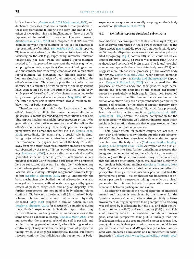
Fig. 1 e Stimuli and postures employed by Kessler and Rutherford (2010) and in the current study. Note that images were
presented in colour during the experiment and target objects were indicated in red colour (here in white). The top left image
shows an example for a “right” target from the avatar's perspective at 110� anticlockwise angular disparity, the top right
image shows an example for a “left” target from the avatar's perspective at 160� (clockwise), and the bottom left image
shows an example for a “visible” target from the avatar's perspective at 60� (anticlockwise). The bottom right images show
the two possible postures of the participant: body turned either clock- or anticlockwise, while gazing straight ahead. Note
that this induced either posture congruence or incongruence with the direction of mental self-rotation for any given
stimulus. Further explanations in the text.
c o r t e x 7 5 ( 2 0 1 6 ) 6 8e8 170
perspective transformations and would allow for unique con-
clusions about the type of processing carried outwithin TPJ and
its recently proposed subdivisions (Carter & Huettel, 2013;
Igelstr€om et al., 2015; Mars et al., 2012). Potentially, this could
substantiate a self-other controlmechanismproposed for right
TPJ (Santiesteban et al., 2012). Such a result would further
emphasise the embodied origins of social cognition, suggesting
that humans may have developed the capacity for mental
alignment by engaging the body representation system in
simulation mode (Gallese, 2013; Pezzulo, Iodice, Ferraina, &
Kessler, 2013; Wilson, 2002). This capacity may come with a
trade-off in the form of spontaneous, uncontrolled disem-
bodiment, that has also been linked to TPJ, hence, our findings
couldpotentially further elucidate the linkbetweenperspective
taking andspontaneousout-of-body-experiences (Blanke et al.,
2005; Blanke & Thut, 2007; Braithwaite et al., 2013).
2. Materials and methods
2.1. Participants
14 participants were tested in the Magnetoencephalography
(MEG) experiment at Glasgow University while a different
group of 15 participants were tested in the TMS experiment at
Aston University.
We obtained analysable MEG data from 12 participants (6
males, average age 23.3, all right-handed). Data from two
additional participants was excluded because of too noisy
data (dental implant), and for being on medication, respec-
tively. All participants had a maximum score of 5 on the “so-
cial skills” subscale of the Autism-Spectrum Quotient (Baron-
Cohen, Wheelwright, Skinner, Martin, & Clubley, 2001), based
on our previous research showing that low social skills (indi-
cated by larger values) may result in the engagement of
alternative processing strategies (Kessler & Wang, 2012).
In the TMS experiment 15 volunteers participated (6males,
average age 26.3, minimum 21 and maximum 37, 3 left-
handed). All participants were screened for contra-
indications (Keel, Smith, & Wassermann, 2001) and had a
maximum score of 5 on the “social skills” subscale of the
Autism-Spectrum Quotient (Baron-Cohen et al., 2001).
2.2. Experimental procedures
All experimental procedures complied with the Declaration of
Helsinki and were approved by the respective University
ethics committee.

c o r t e x 7 5 ( 2 0 1 6 ) 6 8e8 1 71
2.2.1. MEG Expt.The employed tasks and stimuli were adopted from Kessler
and Rutherford (2010, Expt. 1). In all stimuli an avatar was
presented seated at a round table shown from one of six
possible angular disparities (see Fig. 1: 60�, 110�, 160� clock-
wise and anticlockwise). The stimuli were coloured photo-
graphs (resolution of 1024� 768 pixels), taken from an angle of
65� above the plane of the avatar and table. The stimulus table
contained four grey spheres (placed around an occluder, cf.
Fig. 1). In each trial one of the spheres turned red indicating
this sphere as the target. From the avatar's viewpoint the
target could be visible/occluded (perspective tracking task) or
left/right (perspective taking task) and participants were asked
to make a judgement according to the avatar's perspective by
pressing the instructed key (Lumitouch® response pads): the
left key for “left” or “visible” targets and the right key for
“right” or “occluded” targets.1 For analysis we collapsed across
correct responses for left and right and across correct re-
sponses for visible and occluded, respectively. We also
collapsed across clockwise and anticlockwise orientations for
each angular disparity, after ensuring that the neural signa-
tures were comparable (no significant differences in source
space).
For each block of 120 trials (8 total per session) participants
were instructed to maintain one of two possible postures as
shown in Fig. 1, bottom right. The participant's posture in any
given block was always congruent with the mental rotation
direction required for half of the trials, while it was incon-
gruent with the other half. A blocked posture was essential for
avoiding movement artefacts in the MEG due to inter-trial
posture adjustments. The two tasks (perspective taking vs
tracking) were recorded in two separate sessions on different
days and the sequence was counterbalanced across
participants.
MEG data were acquired using a Magnes 3600, 248-channel
whole-head magnetometer (4D-Neuroimaging), sampled at
508.63 Hz and band-pass filtered between 0.1 and 200 Hz.
Stimulus resolution was 1024 � 768 pixels covering a visual
angle of 24� horizontal by 18� vertical. We employed an SR
Research remote Eyelink 1000 fort aborting trials (to be re-run
later) where participants blinked or moved their eyes away
from the screen centre (a box of dimensions 140 � 120 pixels,
covering the central target area, see Fig. 1).
1 Note that in Kessler and Rutherford (2010) we found the samebasic pattern of results with vocal responses (“left” or “right” forperspective taking and “in front” or “behind” for perspectivetracking) as with spatially mapped key presses. This is importantas vocal responses do not induce spatially incongruent stimulus-response mappings (see May & Wendt, 2013). Thus, since ourcurrent study replicated the pattern reported in Kessler andRutherford (2010) we are confident that our effects are not dueto spatial incompatibilities in stimulus-response mappings (seealso Kessler et al., 2014). Furthermore Surtees et al., (2013) re-ported a similar posture congruence effect in a task that did notrequire laterality judgements but judgements of visual appear-ance (e.g., does the other person perceive a digit as a “9” or a “6”?).This further rules out stimuluseresponse mappings as aconfound but also indicates that the posture effect is not only tiedto left/right or other directionality judgements but generalises tojudgements of visual experience.
Data were preprocessed & analysed using the Matlab®
toolbox Fieldtrip (Oostenveld, Fries, Maris,& Schoffelen, 2011).
Epochs were extracted from 600 msec before the visual stim-
ulus was shown until response. All epochs were detrended,
denoised and trials with large artefacts (e.g., strong muscle
artefacts) and continuously noisy channels were removed
(with max 6 out of 248 rejected channels and an average of
142.6 remaining trials per individual). ICA components were
then generated, visually inspected and removed if they re-
flected environmental noise and/or artefacts (such as heart
beats and muscle artefacts).
The power of frequencies between 2 and 32 Hz was
calculated using a Hanning taper (Grandke, 1983) with 3 cycles
per frequency. Planar gradient representations were calcu-
lated prior to sensor level analysis that used cluster-based
random permutation (Maris & Oostenveld, 2007). Conform-
ing to our previous research (e.g., B€ogels et al., 2015) we
employed a 2-step approach for emulating the interactions
between two factors in time and frequency analysis (e.g.,
task � posture; task � angle). We first calculated differences
between the two tasks, i.e., perspective tracking versus taking,
for each participant separately and then included the out-
comes of this 1st step difference into a group statistic that
compared a second factor, e.g., congruent versus incongruent
posture (or 60� vs 160�). The comparison at group level fol-
lowed the robust statistics approach described above. For
localising the power of theta-band oscillations, we used the
Dynamical Imaging of Coherent Sources (DICS, Gross et al.,
2001) approach for calculating spatial filters based on cross-
spectral densities for a timeefrequency tile centred on the
effects found at sensor level (3, 4, 5, 6 Hz; 0e660 msec).
2.2.2. TMS Expt.The stimuli were identical to the MEG experiment but we
simplified the paradigm by excluding trials with 110� angulardisparity and by excluding visibility judgements in order to
focus on the postulated posterior temporo-parietal junction
(pTPJ) involvement in perspective taking. In addition, we
randomly included trials with and without dual pulse TMS
stimulation, hence, a 2 � 2 � 2 repeated measures design was
employed with the factors “angular disparity” (160�/60�),“posture congruence” (congruent/incongruent), and “stimu-
lation” (dual pulse Transcranial Magnetic Stimulation e
dpTMSe vs control). The total number of 160 trials (20 trials in
each of the 8 design conditions) was delivered in 10 blocks of
16 trials each ¼ , where participants maintained the same
body posture (turned clockwise or anticlockwise, cf. Fig. 1
bottom right) throughout each block.
TMS was applied using a Magstim Super Rapid with a
70 mm diameter figure-of-eight stimulating coil, with
maximum magnetic field strength of 1.5 T. Prior to the
experiment three-dimensional brain models were created for
each participant in neuronavigation software (BrainSight® v2,
Rogue Research, Montreal, Canada), using each participants'structural MRI that was normalised into MNI space (Montreal
Neurological Institute template) with SPM8 software (Litvak
et al., 2011). The target sites for stimulation were defined in
normalised stereotactic space (MNI) and the coordinates were
based on the MEG group analysis (MNI coordinates: 50, �60,
32) reflecting a right pTPJ site. Brainsight® hard- and software

680
700
720
740
760
780
CongruentIncongruent
500
550
600
650
700
750
800
850
60 110 160
660
680
700
720
740
760
CongruentIncongruent
500
550
600
650
700
750
800
850
60 110 160
posture
posture angular disparity
angular disparity
Perspec ve Tracking (Visibility)
Perspec ve Taking (Le /Right)
Fig. 2 e Behavioural effects for response times (RT in msec on the y-axes). Significance is indicated as follows: * ¼ p < .05;
** ¼ p < .01; *** ¼ p < .001. “60”,”11000, “160” refer to the three angular disparities employed in the design (collapsed across
clockwise and anticlockwise orientations) and “congruent” and “incongruent” indicate the relationship between the
participant's posture and the target orientation (see also Fig. 1). Error bars denote standard error of mean. Further
explanations in the text.
c o r t e x 7 5 ( 2 0 1 6 ) 6 8e8 172
ensured continued accuracy of pulse application. dpTMS was
applied to right pTPJ in concordance with previous research
targeting the TPJ (e.g., Bosco, Carrozzo, & Lacquaniti, 2008).
Conforming to one of Bosco et al.’s (2008) conditions, the two
pulses were separated by 100 msec with the 1st pulse being
administered at 300 msec after stimulus onset (2nd pulse at
400 msec). Bosco et al. suggested that this would cause
interference lasting for approx. 300e500 msec after stimulus
onset. This period further overlapped with the time window
(350e550 msec) reported by Blanke et al. (2005), where single
pulse TMS affected perspective taking, and importantly, also
covered the peak of the theta (and alpha/beta) effects
observed in the currentMEG experiment (200e400msec, Fig. 3,
bottom). dpTMS was applied on 50% of the trials and pulses
were applied at 110% resting motor threshold as determined
in concordance with standard protocols (Rossini et al., 1994).
On all trials (also on those without dpTMS stimulation)
acoustic click sounds played binaurally via ear phones
ensured that participants could not distinguish between
dpTMS and control trials based on the sounds of the TMS coil
discharge alone.
3. Results
3.1. MEG experiment: behaviour
Response time data (RTs) shown in Fig. 2 were subjected to an
ANOVA that included angular disparity (60�, 110�, 160�)posture congruence (congruent vs incongruent), and task (left/
right vs visibility) as factors (see also Fig. 1). Based on previous
research (Kessler et al., 2014; Kessler & Rutherford, 2010;
Michelon & Zacks, 2006; Surtees et al., 2013), only for
perspective taking (left/right) but not for perspective tracking
(visibility) RTs were expected to slow down with increasing
angular disparity as a reflection of increased duration of
mental transformation. Only for perspective taking (left/right)
but not for perspective tracking (visibility) RTs were also ex-
pected to be faster for a congruent than for an incongruent
body posture as a reflection of body schema involvement in
the mental transformation (Kessler et al., 2014; Kessler &
Rutherford, 2010; Kessler & Thomson, 2010; Surtees et al.,
2013).

ecnereffiDthgiR/tfeL
TFR
160°:
Le /RightTask:
A
B
Hz:3025201510
50-.2 .2 .4 .6 sec
VisibilityHz:30
25201510
5
Hz:3025201510
5
160°vs. 60°
incongruent vs. congruent
Topography:alpha
Topography:theta
ggggggggggggggHz:30
25201510
5
0-.02 .02 .04 .06 sec
Hz:3025201510
5
0-.02 .02 .04 .06 sec
0-.2 .2 .4 .6 sec 0-.2 .2 .4 .6 sec
5 (x 10-23)
0
-5
5 (x 10-23)
0
-5
Visibility Task:160° vs. 60°:
NO significant clusters
incongruent vs. congruent:
NO significant clusters
Fig. 3 e General timeefrequency signatures. The top row of Panel A shows typical timeefrequency representations (TFRs)
for “left/right” and “visibility” judgements at 160� and their difference (left/right ¡ visibility) at the far right, with a theta
band increase and an alpha/beta band decrease in both tasks, yet, both frequency effects being more pronounced for “left/
right” than for “visibility” (y-axis: 2e30 Hz; x-axis: ¡200 msec pre-stimulus to þ700 msec post-stimulus time; colour-coded
scale shows power from ¡5 £ 10¡23 ¼ blue to þ5 £ 10¡23 ¼ red). Rows 2 and 3 depict the related topographies for the alpha
and theta band effects, respectively. Panel B shows two significant TFR cluster effects (for angle and posture, respectively)
for the “left/right” task in relation to the pre-trial baseline interval. The TFR graph and topographies on the left show the
effect of angular disparity (160� vs 60�), where 160� reveals a significantly stronger theta increase, while 60� shows a
significantly stronger alpha decrease. Note that effects involving 110� angular disparity are shown in Supplementary
Material, Fig. S2. The TFR graph and topography on the right shows the effect of posture congruence, where a congruent
posture reveals a significantly stronger theta increase and a numerically stronger, but non-significant alpha decrease.
Topography plots of significant clusters shown below each TFR depict significant channels (and related power topographies)
within a cluster (p < .05) as fully visible, while non-significant channels are reduced in visibility (70% opaque white). Note
that for the visibility task no significant clusters for angular disparity or posture congruence were observed, as indicated at
the bottom of Panel B (but see Fig. S1 for a pre- vs post-stimulus comparison for the visibility task, collapsed across all
conditions). Further explanations in the text.
c o r t e x 7 5 ( 2 0 1 6 ) 6 8e8 1 73

c o r t e x 7 5 ( 2 0 1 6 ) 6 8e8 174
Conforming to these expectations, the current results
replicated Kessler and Rutherford's findings (Kessler et al.,
2014; 2010), revealing a significant main effect of angular
disparity [F(2,10) ¼ 14.8, p < .001, h2p ¼ .747] for perspective
taking (left/right), where RTs increasedwith angle, and amain
effect of posture congruence [F(1,11) ¼ ¼ 10.1, p < .01,
h2p ¼ .478], with a congruent posture being significantly faster
than an incongruent posture. In contrast, perspective tracking
(visibility) only revealed a significant effect for angular
disparity [F(2,10) ¼ 12.2, p < .002, h2p ¼ .71], yet, where RTs
decreased with angle (Kessler et al., 2014). Significant in-
teractions for task � angle [F(2,10) ¼ ¼ 17.9, p < .001,
h2p ¼ ¼.782], for task � posture [F(1,11) ¼ ¼ 9.3, p < <.01,h2p ¼ .458] and for task � angle � posture [F(2,10) ¼ ¼ 25.9,
p < .001, h2p ¼ .839] confirmed the qualitative difference be-
tween the two tasks, as suggested by previous research
(Kessler et al., 2014; Kessler & Rutherford, 2010; Michelon &
Zacks, 2006).
3.2. MEG experiment: timeefrequency results for theta,alpha, beta
We replicated the pattern of behavioural results reported in
Kessler and Rutherford (Kessler & Rutherford, 2010) with
posture congruence and angular disparity effects for left/right,
but no such effects for visible/occluded judgements (as
confirmed by significant interactions with “task”). This indi-
cation of more intense embodied processing and higher
rotation demands during perspective taking compared to
tracking was also reflected in the MEG data where we did not
find any effect thatwas stronger for visibility compared to left/
right judgements, when we compared the two tasks directly
(see also Fig. 3, Panel A). Also note that when tested separately
for each task, posture congruence and angular disparity
revealed significant clusters for perspective taking but not for
tracking (Fig. 3 Panel B). Therefore, to complete the overall
picture we explored visibility judgements as a simple com-
parison between pre-stimulus baseline versus post-stimulus
task period (collapsed across all angular disparity and
posture congruence conditions). This analysis is reported in
the Supplementary Material (Fig. S1) and, in short, we
observed significant effects in alpha, beta, and theta fre-
quencies, indicating more intense processing during stimulus
presentation compared to pre-stimulus baseline. Importantly,
theta power differences localised in the frontal eye fields (FEF),
which has previously been related to visibility judgements
(Wallentin, Roepstorff, & Burgess, 2008) as well as to
perceiving another's gaze and line-of-sight (Grosbras, Laird, &
Paus, 2005).
Furthermore, we focussed our analysis of rotation de-
mands on the maximum angular disparity difference of 160�
versus 60�, since 110� revealed a pattern that was in-between
the two other angular disparities, thus, not adding funda-
mentally new insights. 110� did not differ significantly from
the other two angular disparities at theta but at alpha/beta
frequencies, which is reported in Supplementary Material
Fig. S2.
Conforming to the observed behavioural interaction ef-
fects of task � posture congruence and task � angular
disparity, the main timeefrequency (TFR) results were
revealed in 2-level analysis approaches (e.g., B€ogels et al.,
2015), comparing the two tasks at individual level and then
calculating an angle or posture effect at group level, thus,
approximating the interactions between task � angle and
task � posture congruence, respectively, while allowing for
robust random-permutation cluster statistics to control for
multiple comparison errors (see Section 2.2.1). A data-driven
analysis of frequencies between 2 and 32 Hz (see Section
2.2.1) was calculated conforming to this 2-level analysis
approach. Generally, all conditions followed a similar pattern
of post-stimulus theta-band (2e7 Hz) increase and an alpha/
beta-band decrease (8e25 Hz) compared to baseline (see
Fig. 3, Panel A). These are typical observations (Klimesch,
1999; Pfurtscheller & Lopes da Silva, 1999) associated with
processing of incoming stimuli (alpha/beta decrease) that
also require cognitive processing (theta increase). Although
the general pattern and topography was similar for both
tasks (see Fig. 3, Panel A), perspective taking (left/right)
revealed by far the stronger responses, i.e., theta increases as
well as alpha/beta decreases (see Differences in the far right
column of Fig. 3, Panel A). In fact, we did not find any effect
that was stronger for visibility compared to left/right judge-
ments (but see Fig. S1 for visibility judgements compared to
the baseline interval). Furthermore we found the most reli-
able effects across all contrasts in the theta band incl. higher
delta frequencies (2e7 Hz). We therefore focus our report on
these frequencies but report additional alpha/beta effects in
Supplementary Material (Fig. S3).
It is important to note that comparing the two tasks in the
MEG analysis provided us with a further contrast option that
could not be conducted based on behavioural responses alone,
or by analysing the tasks separately. Specifically, wewere able
to test if posture, disregarding congruency with the cognitive
target at hand, mattered more for perspective taking than for
perspective tracking. This directly relates to our general hy-
pothesis that the body schema would be engaged during
perspective taking but not during tracking: If that was the
case, then the neural representation of posture should be
more strongly engaged during left/right than during visibility
judgements. It is safe to assume that a body turned clockwise
versus anticlockwise results in different neural representa-
tions that code for the two different postures. If a particular
context is likely to use these posture representations on every
given trial of a block, e.g., a block of left/right judgements,
then the neural differences between the two postures should
be enhanced compared to a block where posture is irrelevant,
e.g., a block of visibility judgements. Hence, if posture was
more relevant during left/right compared to visibility judge-
ments, thenwe expected to find a stronger difference between
the two body postures (body turned clockwise vs anticlock-
wise, see Fig. 1) in the former compared to the latter, resulting
in what we termed a “posture relevance” effect. To clarify,
posture relevance is different from posture congruence in that
it is likely to reflect a tonic activity increase related to the body

vPMC
SM1SMA
pTPJ
SM1/SMA
OCC
latPFCinfSMG
Anti- vs. clockwisePosture:
Effect of posture relevance
Theta-power effects (in interaction x task)
160º vs. 60º:Effect of rotation demands
Incongruent vs. congruentPosture:
Embodied effect
pTPJpTPJ
-
pTPJ: BA 39 (40, 7)
-
SPL(BA40/7)
∑/3 ∑/3.5
.5
0
.25
.25
0
Fig. 4 e Theta interaction effects in sensor and source space. Three interactions of task (left/right vs visibility) with 1) angular
disparity (160� vs 60�), 2) posture congruence (incongruent vs congruent), and 3) posture relevance (anti- vs clockwise
posture). Bottom row: Topographies of interaction effects in the theta band (2e7 Hz, colour-coded scale shows power from
¡1 £ 10¡23 ¼ blue to þ1 £ 10¡23 ¼ red). Significant channels within a cluster (p < .05) are fully visible while non-significant
channels are reduced in visibility. (For effects at alpha and beta frequencies see Supplementary Material, Fig. S2.) Middle
row: Theta power source reconstructions for each of the three interaction effects. TPJ ¼ temporo-parietal junction;
OCC ¼ occipital cortex; SM1 ¼ primary sensorimotor cortex; SMA ¼ supplementary motor area; latPFC ¼ lateral prefrontal
cortex; SPL ¼ superior parietal lobule; vPMC ¼ ventrolateral premotor cortex. Colour scale shows normalised theta power
(red ¼ positive). Top image: The maximum overlap across the three interaction effects (average) is localised in the right
pTPJ, specifically Broadman area (BA) 39, extending into BA40 and BA7. Further explanations in the text.
c o r t e x 7 5 ( 2 0 1 6 ) 6 8e8 1 75
schema for the left/right compared to the visibility task (pre-
sented in separate blocks), disregarding specific trial param-
eters such as mental rotation direction, demands and
congruence.2
The interaction between task and angular disparity was
calculated for the maximum difference in angle, i.e., between
160� and 60� degrees, and revealed a significant cluster (p < .05;
Fig. 5, left column) in the theta band (2e7 Hz), lasting from 0 to
650 msec (Fig. 4, left). The 160� condition revealed a stronger
theta increase than 60� (for left/right but not for visibility). Theinteraction between task and posture congruence was re-
flected by a significant cluster (p < .05) in the theta band
(3e7 Hz) and lasted from 50 to 450 msec (Fig. 4, middle col-
umn). In reflection of the obtained behavioural effects (see
Fig. 2) posture congruence effects differed significantly be-
tween left/right and visibility judgements, with only the
former showing significantly stronger theta modulation in
2 Posture relevance was calculated as [L/R(anticlockwise) � visibility (anticlockwise)] � [L/R(clockwise) � visibility (clockwise)], while posture congruence as[L/R (incongruent) � visibility (incongruent)] � [L/R(congruent) � visibility (congruent)].
response to posture incongruence versus congruence.We also
observed the predicted “posture relevance” effect where the
two postures differedmore strongly for left/right compared to
visibility judgements, resulting in a significant cluster (p < .05)
in the theta band (2e7 Hz) that lasted from 0 to 650 msec
(Fig. 4, right column). This effect, reflecting higher relevance of
posture for left/right than for visibility judgements, further
supports stronger engagement of the body schema during
perspective taking (left/right) compared to mere perspective
tracking (visibility). Finally, the effects for all three in-
teractions seem to overlap over the right posterior hemi-
sphere (Fig. 4, bottom row), possibly indicating a source in the
right TPJ.
3.3. MEG experiment: source analysis for theta
Fig. 4 (middle row and top image) depicts the source re-
constructions for each of the three theta interaction effects
(with task) obtained with a similar 2-level approach as for
the sensor level analysis (see Section 2.2.1); the source co-
ordinates in MNI space are provided in Table 1. Firstly,
angular disparity localised in the posterior part of the right

550
600
650
700
750
800
850
60 160
RTs i
n m
sec
Angular Disparity
TMS congTMS incongCntrl congCntrl incong
dpTMS to right pTPJ (MNI: 50 -60 32)
*
Fig. 5 e TMS target site and response time results. Left: The same right pTPJ site (MNI coordinates taken from MEG group
analysis: 50, ¡60, 32) was targeted with dpTMS for each individual (MNI normalised) brain using Brainsight®. Right:
Response time results, shown as residuals after subtracting a congruent from an incongruent posture for each condition
separately (collapsed across clockwise and anticlockwise avatar locations and across left and right targets). The y-axis
denotes RT differences in msec and the x-axis contrasts control versus dpTMS trials for each of the two angular disparities.
Error bars denote standard error of mean. Further explanations in the text.
c o r t e x 7 5 ( 2 0 1 6 ) 6 8e8 176
TPJ (pTPJ), extending dorsally into dorsal TPJ and ventrally
into the lateral occipital complex, overlapping with the
extrastriate body area (OCC). More anterior sources include
sensorimotor (SM1) and frontal areas (SMA, latPFC), thus,
reflecting the topography of the widely distributed sensor
level cluster (Fig. 4, bottom left). Secondly, posture congru-
ence (Fig. 4, bottom middle; Table 1) also localised in the
right pTPJ extending into more superior areas of the poste-
rior parietal lobe (SPL) as well as to the right supplementary
Table 1 e Labels, Brodmann areas, and MNI coordinates for sou
Source label in Fig. 4 Brodmann areas
1) Contrast for angle (160� vs 60�)pTPJ 39/7 Right temporo-pa
OCC 18/19 Right occipital co
infSMG 2/40 Right supramarg
SM1/SMA 6/4/5 Right supplemen
latPFC 9/46 Right superior fro
e 8/6 Left superior fron
e 21/22 Left middle temp
e 44/45 Left inferior fron
2) Contrast for posture congruence
pTPJ 39 Right temporo-pa
SPL(BA40/7) 40/7 Right superior pa
SMA 6 Right supplemen
e 18 Left occipital cor
e 7 Left superior par
3) Contrast for posture relevance
pTPJ 39 Right temporo-pa
SM1 3/4 Right sensorimot
vPMC 6/44 Right ventral pre
e 18 Right occipital co
e 18/19 Left occipital cor
Average across the 3 contrasts (top, Fig. 4)
pTPJ 39 Right temporo-pa
e 17 Right occipital co
e 18/19 Left occipital cor
motor area (SMA). The posture relevance effect also local-
ised in the right pTPJ (Fig. 4, bottom right; Table 1) along
with right sensorimotor (SM1) and ventral premotor cortex
(vmPFC). Finally, Fig. 4 and Table 1 reveal that the maximum
overlap between the three effects is indeed located in the
right pTPJ, thus confirming our hypothesis that TPJ could be
the locus where the embodied self is transformed into an-
other's perspective and experience, possibly aligning bodies
as well as minds.
rces identified in Fig. 4.
Brain areas MNI coordinates
rietal junction: angular gyrus 34 �70 44
rtex/lateral occipital complex 26 �90 0
inal gyrus 64 �22 32
tary motor area, sensorimotor gyrus �2 �14 60
ntal gyrus/lateral prefrontal cortex 30 42 36
tal gyrus/SMA �22 14 52
oral gyrus �62 �30 4
tal gyrus/lateral PFC �57 18 12
rietal junction: angular gyrus 50 �60 24
rietal lobule 42 �58 60
tary motor area 26 �6 64
tex �38 �90 8
ietal lobule �30 �62 52
rietal junction: angular gyrus 54 �62 36
or gyrus 52 �18 60
motor cortex 54 6 16
rtex 6 �78 28
tex/lateral occipital complex �34 �94 �8
rietal junction: angular gyrus 50 �60 32
rtex 0 �96 �10
tex/lateral occipital complex �30 �96 �2

c o r t e x 7 5 ( 2 0 1 6 ) 6 8e8 1 77
3.4. TMS experiment: effects of dpTMS applied to rTPJ
We tested the proposed critical role of right pTPJ for embodied
processing during perspective taking (left/right). We targeted
the right pTPJ with a dual pulse TMS paradigm (dpTMS; e.g.,
Bosco et al., 2008) based on the coordinates obtained from the
MEG overlap analysis (Fig. 4, top; Fig. 5, left) and the time
window observed for the theta effects (Fig. 3) and in concor-
dance with previous research (see Methods). We applied the
1st pulse at 300 and the 2nd pulse at 400 msec after stimulus
onset. On all trials acoustic click sounds, mimicking TMS coil
discharges, were played via ear phones. The played sounds
were louder than the actual discharges; hence, participants
were unable to distinguish acoustically between dpTMS trials
and no-pulse control trials, which allowed us to mix TMS and
sham trials into a random trial-sequence. The binaurally
played sounds also masked the spatial asymmetry of the real
coil discharges over the right hemisphere, which otherwise
could have resulted in a spatial bias to the right.
The factor “stimulation” (dpTMS vs control) was included
as a within-subjects factor into a 2 � 2 � 2 repeated measures
ANOVA along with the factors “angular disparity” (60� vs 160�)and “posture congruence” (congruent vs incongruent). The
analysis revealed a significant main effect of “angular
disparity” [F(1, 14) ¼ 20.6, p < .0001, h2p ¼ .595], a significant
interaction between “angular disparity” and “posture
congruence” [F(1, 14) ¼ 7.8, p ¼ .014, h2p ¼ .359], and a signifi-
cant interaction between “stimulation” and “posture congru-
ence” [F(1, 14) ¼ 6.5, p ¼ .023, h2p ¼ .319]. All other effects did
not reach significance (all p > .1). The first two effects are in
line with our previous research showing faster RTs at low (60�)compared to high (160�) angular disparity and a significant
posture effect at high (160�) but not at low (60�) angular
disparity (Kessler & Thomson, 2010; Kessler & Wang, 2012).
The third effect is novel and can be interpreted, based on Fig. 5
(right graph), as a disruption of the posture congruence effect
by dpTMS to right pTPJ. Although the 3-way interaction be-
tween angle, posture, and stimulation was not significant
(p ¼ .381), Fig. 5 (right graph) reveals that dpTMS primarily
disrupted the posture effect where it existed in the first
instance, namely at 160�.
4. Discussion
Firstly, our current MEG Expt replicated previous behavioural
findings (Kessler et al., 2014; Kessler& Rutherford, 2010; Kessler
& Thomson, 2010; Kessler & Wang, 2012; Surtees et al., 2013)
showing a significant RT increase (Fig. 2) in relation to higher
angular disparity and posture incongruence for perspective
taking (left/right) in contrast to perspective tracking (visibility),
hence, further corroborating the notion of two distinct mech-
anisms (Michelon & Zacks, 2006). One mechanism seems to be
restricted to the simpler process of tracking another's line of
sight, while the other allows for imagining another's perspec-
tive by engaging an embodied process of mental self-rotation
into the other's orientation (Kessler et al., 2014; Kessler &
Rutherford, 2010; Michelon & Zacks, 2006). This clear behav-
ioural pattern (i.e., posture and disparity effects only in the left/
right task) allowed us to pursue the neural substrate of
perspective taking (left/right) in comparison to perspective
tracking (visibility). While all reported effects in the direct task
comparisonwere indeeddue tostrongeroscillatorymodulation
in the left/right task, we were nonetheless able to pinpoint FEF
as a major processing hub for the visibility task compared to a
pre-trial baseline period (2e7 Hz, see Fig. S1), replicating pre-
vious findings (Grosbras et al., 2005; Wallentin et al., 2008) and,
thus, confirming a potential role of FEF in inferring another'sline of sight.
Regarding perspective taking (in contrast to tracking) our
data-driven timeefrequency analysis revealed that modula-
tions of theta oscillations were a common theme (Fig. 4, bot-
tom row) amongst our three types of effects. “Rotation
demands” was reflected in higher theta power for 160� versus60� angular disparity, “cognitive embodiment” was reflected
by stronger theta for an incongruent versus a congruent
posture, and “posture relevance” was reflected by a stronger
theta contrast between anti- versus clockwise turned body
postures for perspective taking compared to tracking. Not only
was the frequency of interest (~2e7 Hz) in common across all
three effects, but also the primary cortical origin of these ef-
fects overlapped in the right pTPJ (Fig. 4 top image). This is in
agreement with previously reported involvement of right TPJ-
theta in high-level perspective taking and mentalizing (B€ogels
et al., 2015). In the subsequent TMS study we were able to
disrupt the posture congruence effect (“cognitive embodi-
ment”) by targeting right pTPJ with a dual pulse interference
paradigm (Fig. 5). However, we did not find a dpTMS effect on
angular disparity indicating that rotation demands were un-
affected by the stimulation. A more disruptive repetitive TMS
protocol might have affected both effects. However, our result
could also be related to the targeted site being drawn more
towards the body-related effects in the overlap (Fig. 4).
Potentially, it might be possible to selectively disrupt the ef-
fects of posture or angular disparity or both, by targeting
slightly different sites within right TPJ.
4.1. Implications for the role of TPJ
Our findings are in concordance with previous research that
has pinpointed TPJ, and pTPJ in particular, as a crucial area
within a network generally engaged when inferring others'experiences and mental states (Arzy et al., 2006; Blanke et al.,
2005; B€ogels et al., 2015; Van Overwalle & Baetens, 2009; Zacks
& Michelon, 2005). In addition, TPJ has also been related
directly to high-level visuospatial perspective taking and no-
tions of the role of TPJ either suggest an embodied contribu-
tion (Arzy et al., 2006; Blanke et al., 2005) or the deliberate
transformation of frames of reference and/or the co-
representation of egocentric and altercentric perspectives
(e.g., Santiesteban et al., 2012; Schurz et al., 2013). However,
TPJ does not seem to be confined to deliberate processing of
another's experience but has also been associated with
spontaneous forms of viewpoint changes, prominently sub-
sumed under the label of “out-of-body” experiences (Blanke
et al. 2005; Braithwaite & Dent, 2011; Braithwaite et al., 2013),
which is supportive of body-related processing in TPJ.
Our current empirical evidence allows reconciling
diverging views of the role of TPJ by suggesting it as the locus
of convergence between implicit body representation, i.e., the
c o r t e x 7 5 ( 2 0 1 6 ) 6 8e8 178
body schema (e.g., Coslett et al., 2008; Medina et al., 2009), and
deliberate processes that use simulated manipulations of
these representations to imagine the body (and mind) in an-
other(’s) viewpoint. This has implications on how the self is
represented in relation to another. Previous research
(Santiesteban et al., 2012) had proposed that TPJ controls
conflicts between representations of the self-in contrast to
representations of another. Santiesteban et al. (2012) reported
TPJ involvement when “the other” needed to be ignored while
focussing on the self (e.g., suppressing automatic imitation
tendencies), yet also when self-centred representations
needed to be suppressed to represent the other (e.g., when
adopting the other's perspective). Our current findings extend
and substantiate this rather vague notion of self- versus other
representations. As explained, our findings suggest that
humans simulate a rotation of their embodied self into the
other's orientation. Thus, we propose that a conflict arises
because of a simulated self where parts of the body schema
have been rotated outside the current location of the body,
while parts of the self and the body schema remain tied to the
body's current physical location (see also May, 2004). Without
the latter mental self-rotation would always result in full-
blown “out-of-body” experiences.
Therefore, our notion shifts the focus away from “the
other”, towards conflicts that arise between alternative
(physically vs mentally embodied) representations of the self.
This implies that humansmight represent others primarily by
generating an alternative representation of the self in the
other's circumstances (e.g., their body posture, viewpoint,
perspective, socio-emotional context, etc.; e.g., Pezzulo et al.,
2013). Accordingly, TPJ might play a crucial role in simu-
lating projected selves and controlling conflict with the self
that remains in the physical location of the body. This shift
away from “the other” towards alternative embodied selves is
corroborated by the role of TPJ in “out-of-body” experiences
(e.g., Blanke et al., 2005), where an alternative embodied self is
generated while no other is present. Furthermore, in our
previous research using the same basic paradigm as reported
here we substituted the avatar, i.e., “the other”, with an empty
chair, where participants had to imagine themselves being
located, while making left/right judgements towards target
objects (Kessler & Thomson, 2010, Expt. 2). Importantly, the
basic mechanism of embodied mental self-rotation was also
engaged in this versionwithout avatar, as suggested by typical
effects of posture congruence and angular disparity. This
further corroborates our notion of a body-schema-related
conflict in TPJ between a projected self (via simulated body-
schema rotation) and the self that remains physically
embodied (May, 2004 proposes a similar notion, but see
Kessler & Thomson, 2010; for discussion). Sometimes during
“out-of-body” experiences individuals report that they
perceive their self as being embodied in two locations at the
same time (so-called heautoscopy; Blanke & Mohr, 2005). This
indicates that the proposed split of the self is possible and
while it is being perceived as odd, when it is triggered un-
controllably, it may serve the crucial purpose of perspective
taking, when it is engaged deliberately. Indeed, our recent
research confirms that individuals who report “out-of-body”
experiences are quicker at mentally adopting another's body
orientation (Braithwaite et al., 2013).
4.2. TPJ linking separate functional subnetworks
In addition to the convergence of theta effects in right pTPJ, we
also observed differences in theta power localisation for the
three effects (Fig. 4, middle row). For rotation demands (160�
vs 60� angular disparity) we observed a more widely distrib-
uted topography (Fig. 4, bottom left), which could reflect ex-
ecutive function (latPFC) as well as visual processing (OCC) in
a theta-based network of brain areas. The lateral occipital
source overlaps with the extrastriate body area (EBA) and
suggests more intense visual processing of the avatar's body
(for review, Carter & Huettel, 2013), when rotation demands
are higher (160� vs 60�). In Kessler and Thomson (2010, Expt. 4;
also Kessler & Rutherford, 2010) we had argued that the
presence of another's body and their posture helps deter-
mining the accurate endpoint of the mental self-rotation
process e particularly at high angular disparities. Sustained
theta activation in the EBA observed here corroborates this
notion of another's body as an important visual parameter for
mental self-rotation. For the effect of angular disparity, right
TPJ activation extends into dorsal TPJ which has previously
been related to executive functions (Igelstr€om et al., 2015;
Mars et al., 2012). Overall the source configuration for the
angular disparity effect fits well with our interpretation that it
might reflect rotation demands that engage executive func-
tions and rely on rotation parameters.
Theta power effects for posture congruence localised in
right pTPJ and further areaswithin the superior parietal cortex
(BA 40/7) that have been associated with sensorimotor repre-
sentations and the body schema (Andersen, Snyder, Bradley,
& Xing, 1997; Wolpert et al., 1998). Activation of the pTPJ ex-
tends ventrally into EBA, further underlining processes that
integrate the perception of another's body (i.e., the avatar in
the scene) with the process of transforming the embodied self
into the other's orientation. Again, this dovetails nicely with
our previous behavioural findings (Kessler & Thomson, 2010;
Expt. 4), where we demonstrated an accelerating effect on
perspective taking if the avatar's body posture matched the
participants' posture. This emphasises the importance of an-
other's posture for perspective taking, not only as a visual
parameter for rotation, but also by generating embodied
resonance between participant and avatar.
The emerging picture of the neural signature of embodied
mental self-rotation is completed by the sources for the
“posture relevance“ effect, where stronger body schema
involvement during perspective taking compared to tracking
was reflected by localisations in right pTPJ and right ventro-
lateral premotor (vPMC) and sensorimotor (SM1) areas. This
could directly reflect the embodied simulation process
postulated for perspective taking. It is unlikely that this
localisation is due to the preparation of a motor response (key
press), since equivalent preparation processes could be ex-
pected for all conditions. vPMC specifically has been associ-
ated with embodied simulations and re-enactment in social
interaction (Gallese, 2013;Wheatley, Milleville,&Martin, 2007)

c o r t e x 7 5 ( 2 0 1 6 ) 6 8e8 1 79
and our results emphasise the integration with TPJ for simu-
lating a body rotation into another's viewpoint, possibly along
with sensorimotor feedback from the simulation (e.g., Tcaci
Popescu & Wexler, 2012) and/or sensorimotor conflict be-
tween physically versus mentally embodied self. Overall our
findings corroborate the notion of a simulated body rotation
that generates an updated efference copy within the body
schema, which in turn drives the actual visuospatial trans-
formation process (Kessler & Thomson, 2010; Tcaci Popescu &
Wexler, 2012; Zacks & Michelon, 2005).
4.3. Implications for the wider context of socialprocessing
Within a wider context our current findings and our previous
research suggest that high-level perspective taking is still
grounded in older action- and body-related brain systems, in
other words, that older systems have been re-purposed for
resolving new challenges (see also Gallese, 2013; Kessler &
Thomson, 2010; Parkinson & Wheatley, 2013; Pezzulo et al.,
2013; Wilson, 2002). This could explain the evolution of
perspective taking fromphysical alignment that is observed in
other species (Kessler & Thomson, 2010). More generally, the
embodied origin of mentalizing could be reflected by TPJ
activation in conjunction with other body-related brain areas.
This notion of visuospatial perspective taking as a develop-
mental and possibly evolutionary stepping stone for full-
blown theory of mind has recently found agreement
(Hamilton, Brindley, & Frith, 2009; Parkinson & Wheatley,
2013) as well as disagreement (Moll & Kadipasaoglu, 2013),
where the latter postulates that social empathy and perspec-
tive understanding precedes visuospatial perspective taking.
While we believe that our findings rather support the former,
we acknowledge that certain forms of joint attention may
predate even simple perspective tracking.
5. Conclusions
Significant aspects of information processing in humans are
not shared with other species. In the social domain such
processes have been typically related to explicitly represent-
ing the subjective experience and mental states of others.
However, some of these unique abilities still seem to depend
on “older” systems such as the body's movement repertoire.
The current research confirmed that the human capacity for
imagining another's perspective of the world is still signifi-
cantly “embodied”, in the sense that humans mentally rotate
their own body representation (body schema) into another'sorientation. Using MEG we found that brain oscillations at
theta frequency, originating from the right pTPJ reflected
cognitive as well as embodied processing elements. This was
subsequently confirmed using TMS, which disrupted
embodied processing effects, pinpointing right pTPJ as the
crucial network hub for transforming the embodied self into
another's viewpoint, body and/or mind. We propose that such
a “transformed embodied self”, projected into another's cir-
cumstances (e.g., their posture, orientation, perspective,
socio-emotional context, etc.), serves as the basis for repre-
senting and understanding others in various social scenarios.
Using state-of-the-art methodology our research elucidates
the embodied origins of high-level social processing in
humans, specifically highlighting the critical role of right pTPJ
and theta oscillations.
Acknowledgements
We would like to thank Frances Crabbe for help with MRI
scans and MEG data collection at Glasgow University.
Supplementary data
Supplementary data related to this article can be found at
http://dx.doi.org/10.1016/j.cortex.2015.11.011.
r e f e r e n c e s
Andersen, R. A., Snyder, L. H., Bradley, D. C., & Xing, J. (1997).Multimodal representation of space in the posterior parietalcortex and its use in planning movements. Annual Review ofNeuroscience, 20(1), 303e330.
Arzy, S., Thut, G., Mohr, C., Michel, C. M., & Blanke, O. (2006).Neural basis of embodiment: distinct contributions oftemporoparietal junction and extrastriate body area. TheJournal of Neuroscience, 26(31), 8074e8081.
Baron-Cohen, S., Wheelwright, S., Skinner, R., Martin, J., &Clubley, E. (2001). The Autism-Spectrum Quotient (AQ):evidence from Asperger syndrome/high-functioning autism,males and females, scientists and mathematicians. Journal ofAutism and Developmental Disorders, 31(1), 5e17.
Berlucchi, G., & Aglioti, S. (1997). The body in the brain: neuralbases of corporeal awareness. Trends in Neurosciences, 20(12),560e564.
Berlucchi, G., & Aglioti, S. M. (2010). The body in the brainrevisited. Experimental Brain Research, 200(1), 25e35.
Blanke, O., & Mohr, C. (2005). Out-of-body experience,heautoscopy, and autoscopic hallucination of neurologicalorigin: implications for neurocognitive mechanisms ofcorporeal awareness and self-consciousness. Brain ResearchReviews, 50(1), 184e199.
Blanke, O., Mohr, C., Michel, C. M., Pascual-Leone, A., Brugger, P.,Seeck, M., et al. (2005). Linking out-of-body experience and selfprocessing tomental own-body imagery at the temporoparietaljunction. The Journal of Neuroscience, 25(3), 550e557.
Blanke, O., & Thut, G. (2007). Inducing out-of-body experiences. InS. Della Salla (Ed.), Tall tales: popular myths about the mind andbrain (pp. 425e439). Oxford: Oxford University Press.
B€ogels, S., Barr, D. J., Garrod, S., & Kessler, K. (2015).Conversational interaction in the scanner: mentalizing duringlanguage processing as revealed by MEG. Cerebral Cortex, 25(9),3219e3234.
Bosco, G., Carrozzo, M., & Lacquaniti, F. (2008). Contributions ofthe human temporoparietal junction and MT/V5þ to thetiming of interception revealed by transcranial magneticstimulation. The Journal of Neuroscience, 28(46), 12071e12084.
Braithwaite, J. J., & Dent, K. (2011). New perspectives onperspective-taking mechanisms and the out-of-bodyexperience. Cortex, 47(5), 628e632.
Braithwaite, J. J., James, K., Dewe, H., Medford, N., Takahashi, C.,& Kessler, K. (2013). Fractionating the unitary notion ofdissociation: disembodied but not embodied dissociative

c o r t e x 7 5 ( 2 0 1 6 ) 6 8e8 180
experiences are associated with exocentric perspective-taking. Frontiers in Human Neuroscience, 7.
Brauer, J., Call, J., & Tomasello, M. (2005). All great ape speciesfollow gaze to distant locations and around barriers. Journal ofComparative Psychology, 119(2), 145e154.
Brauer, J., Call, J., & Tomasello, M. (2007). Chimpanzees reallyknow what others can see in a competitive situation. AnimalCognition, 10(4), 439e448.
Bugnyar, T., St€owe, M., & Heinrich, B. (2004). Ravens, Corvuscorax, follow gaze direction of humans around obstacles.Proceedings of the Royal Society of London. Series B: BiologicalSciences, 271(1546), 1331e1336.
Buxbaum, L. J., Giovannetti, T., & Libon, D. (2000). The role of thedynamic body schema in praxis: evidence from primaryprogressive apraxia. Brain and Cognition, 44(2), 166e191.
Call, J., & Tomasello, M. (1999). A nonverbal false belief task: theperformance of children and great apes. Child Development,70(2), 381e395.
Carter, R. M., & Huettel, S. A. (2013). A nexus model of thetemporaleparietal junction. Trends in Cognitive Sciences, 17(7),328e336.
Coslett, H. B., Buxbaum, L. J., & Schwoebel, J. (2008). Accuratereaching after active but not passive movements of the hand:evidence for forward modeling. Behavioural Neurology, 19(3),117e125.
van Elk, M., & Blanke, O. (2014). Imagined own-bodytransformations during passive self-motion. PsychologicalResearch, 78(1), 18e27.
Falconer, C. J., & Mast, F. W. (2012). Balancing the mind: vestibularinduced facilitation of egocentric mental transformations.Experimental Psychology, 59(6), 332.
Frith, C. D., & Frith, U. (2007). Social cognition in humans. CurrentBiology, 17(16), R724eR732.
Gallese, V. (2013). Mirror neurons, embodied simulation and asecond-person approach to mindreading. Cortex, 49(10),2954e2956.
Grandke, T. (1983). Interpolation algorithms for discrete Fouriertransforms of weighted signals. Instrumentation andMeasurement, IEEE Transactions on, 32(2), 350e355.
Grosbras, M. N., Laird, A. R., & Paus, T. (2005). Cortical regionsinvolved in eye movements, shifts of attention, and gazeperception. Human Brain Mapping, 25(1), 140e154.
Gross, J., Kujala, J., Hamalainen, M., Timmermann, L.,Schnitzler, A., & Salmelin, R. (2001). Dynamic imaging ofcoherent sources: studying neural interactions in the humanbrain. Proceedings of the National Academy of Sciences of the UnitedStates of America, 98(2), 694e699.
Hamilton, A. F. D., Brindley, R., & Frith, U. (2009). Visualperspective taking impairment in children with autisticspectrum disorder. Cognition, 113(1), 37e44.
Igelstr€om, K. M., Webb, T. W., & Graziano, M. S. (2015). Neuralprocesses in the human temporoparietal cortex separated bylocalized independent component analysis. The Journal ofNeuroscience, 35(25), 9432e9445.
Keel, J. C., Smith, M. J., & Wassermann, E. M. (2001). A safetyscreening questionnaire for transcranial magneticstimulation. Clinical Neurophysiology, 112(4), 720.
Kessler, K., Cao, L. Y., O'Shea, K. J., & Wang, H. F. (2014). A cross-culture, cross-gender comparison of perspective takingmechanisms. Proceedings of the Royal Society B-Biological Sciences,281(1785), 20140388. http://dx.doi.org/10.1098/Rspb.2014.0388.
Kessler, K., & Rutherford, H. (2010). The two forms of Visuo-SpatialPerspective Taking are differently embodied and subservedifferent spatial prepositions. [Original Research]. Frontiers inPsychology, 1. http://dx.doi.org/10.3389/fpsyg.2010.00213.
Kessler, K., & Thomson, L. A. (2010). The embodied nature ofspatial perspective taking: embodied transformation versussensorimotor interference. Cognition, 114(1), 72e88.
Kessler, K., & Wang, H. F. (2012). Spatial perspective taking is anembodied process, but not for Everyone in the same way:differences predicted by sex and social skills score. SpatialCognition and Computation, 12(2e3), 133e158.
Klimesch, W. (1999). EEG alpha and theta oscillations reflectcognitive and memory performance: a review and analysis.Brain Research Reviews, 29(2), 169e195.
Litvak, V., Mattout, J., Kiebel, S., Phillips, C., Henson, R., Kilner, J.,et al. (2011). EEG and MEG data analysis in SPM8. ComputationalIntelligence and Neuroscience, 2011.
Maris, E., & Oostenveld, R. (2007). Nonparametric statisticaltesting of EEG-and MEG-data. Journal of Neuroscience Methods,164(1), 177e190.
Mars, R. B., Sallet, J., Schuffelgen, U., Jbabdi, S., Toni, I., &Rushworth, M. F. (2012). Connectivity-based subdivisions ofthe human right “temporoparietal junction area”: evidence fordifferent areas participating in different cortical networks.Cerebral Cortex, 22(8), 1894e1903.
May, M. (2004). Imaginal perspective switches in rememberedenvironments: transformation versus interference accounts.Cognitive Psychology, 48(2), 163e206.
May, M., & Wendt, M. (2013). Visual perspective taking andlaterality decisions: problems and possible solutions. Frontiersin Human Neuroscience, 7.
Medina, J., Jax, S. A., & Coslett, H. B. (2009). Two-componentmodels of reaching: evidence from deafferentation in a Fitts'law task. Neuroscience Letters, 451(3), 222e226.
Michelon, P., & Zacks, J. M. (2006). Two kinds of visual perspectivetaking. Perception & Psychophysics, 68(2), 327e337.
Moll, H., & Kadipasaoglu, D. (2013). The primacy of social overvisual perspective-taking. Frontiers in Human Neuroscience, 7.
Oostenveld, R., Fries, P., Maris, E., & Schoffelen, J.-M. (2011).FieldTrip: open source software for advanced analysis of MEG,EEG, and invasive electrophysiological data. ComputationalIntelligence and Neuroscience, 2011, 1.
Parkinson, C., & Wheatley, T. (2013). Old cortex, new contexts: re-purposing spatial perception for social cognition. Frontiers inHuman Neuroscience, 7.
Pezzulo, G., Iodice, P., Ferraina, S., & Kessler, K. (2013). Sharedaction spaces: a basis function framework for social re-calibration of sensorimotor representations supporting jointaction. Frontiers in Human Neuroscience, 7.
Pfurtscheller, G., & Lopes da Silva, F. H. (1999). Event-related EEG/MEG synchronization and desynchronization: basic principles.Clinical Neurophysiology, 110(11), 1842e1857.
Rossini, P. M., Barker, A., Berardelli, A., Caramia, M., Caruso, G.,Cracco, R., et al. (1994). Non-invasive electrical and magneticstimulation of the brain, spinal cord and roots: basic principlesand procedures for routine clinical application. Report of anIFCN committee. Electroencephalography and ClinicalNeurophysiology, 91(2), 79e92.
Santiesteban, I., Banissy, M. J., Catmur, C., & Bird, G. (2012).Enhancing social ability by stimulating right temporoparietaljunction. Current Biology, 22(23), 2274e2277.
Schurz, M., Aichhorn, M., Martin, A., & Perner, J. (2013). Commonbrain areas engaged in false belief reasoning and visualperspective taking: a meta-analysis of functional brainimaging studies. Frontiers in Human Neuroscience, 7.
Surtees, A., Apperly, I., & Samson, D. (2013). The use of embodiedself-rotation for visual and spatial perspective-taking. Frontiersin Human Neuroscience, 7.
Tcaci Popescu, S., & Wexler, M. (2012). Spontaneous bodymovements in spatial cognition. [Original Research]. Frontiersin Psychology, 3. http://dx.doi.org/10.3389/fpsyg.2012.00136.
Tomasello, M., Carpenter, M., Call, J., Behne, T., & Moll, H. (2005).Understanding and sharing intentions: the origins of culturalcognition. Behavioral and Brain Sciences, 28(5), 675e691.discussion 691e735.

c o r t e x 7 5 ( 2 0 1 6 ) 6 8e8 1 81
Tsakiris, M., Costantini, M., & Haggard, P. (2008). The role of theright temporo-parietal junction in maintaining a coherentsense of one's body. Neuropsychologia, 46(12), 3014e3018.
Tversky, B., & Hard, B. M. (2009). Embodied and disembodiedcognition: spatial perspective-taking. Cognition, 110, 124e129.
Van Overwalle, F., & Baetens, K. (2009). Understanding others'actions and goals by mirror and mentalizing systems: a meta-analysis. Neuroimage, 48(3), 564e584.
Wallentin, M., Roepstorff, A., & Burgess, N. (2008). Frontal eyefields involved in shifting frame of reference within workingmemory for scenes. Neuropsychologia, 46(2), 399e408.
Wheatley, T., Milleville, S. C., & Martin, A. (2007). Understandinganimate agents: distinct roles for the social network andmirror system. Psychological Science, 18(6), 469e474.
Wilson, M. (2002). Six views of embodied cognition. PsychonomicBulletin & Reviews, 9(4), 625e636.
Wolpert, D. M., Goodbody, S. J., & Husain, M. (1998). Maintaininginternal representations: the role of the human superiorparietal lobe. Nature Neuroscience, 1(6), 529e533.
Zacks, J. M., & Michelon, P. (2005). Transformations ofvisuospatial images. Behavioral and Cognitive NeuroscienceReviews, 4(2), 96e118.