Embed Size (px)
Citation preview

RIK
EN
IMS A
nn
ual R
ep
ort 2
014
RIK
EN C
enter fo
r Integ
rative Med
ical Sciences
RIKEN IMSAnnual Report 2014
RIKEN Center for Integrative Medical Scienceshttp://www.ims.riken.jp/english/
RIKEN Yokohama Campus1-7-22 Suehiro-cho, Tsurumi-ku, Yokohama City, Kanagawa, 230-0045, Japan
Tel : 045-503-9111Fax : 045-503-9113Email : [email protected]
RIKEN Center for Integrative Medical Sciences
RIKEN IMSAnnual Report 2014
RIKEN 2015-046
Cover image: Preparation of glass-supportedlipid bilayers for analyzing molecular dynamicsof microclusters in Jurkat T cells.Image courtesy of Laboratory for MolecularLive-cell Quantification.

RIKEN Center for Integrative Medical SciencesOrganization Chart
Ryoji Noyori
RIKEN President
Core for Homeostatic Regulation
Core for Precise Measuring and Modeling
Lab. for Cell Signaling: Takashi Saito
Lab. for Lymphocyte Di�erentiation: Tomohiro Kurosaki
Lab. for Transcriptional Regulation: Ichiro Taniuchi
Lab. for Immune Cell Systems: Shigeo Koyasu
Lab. for Human Disease Models: Fumihiko Ishikawa
Lab. for Intestinal Ecosystem: Hiroshi Ohno
Lab. for Mucosal Immunity: Sidonia Fagarasan
Lab. for Gut Homeostasis: Kenya Honda
Lab. for Immune Homeostasis: Shohei Hori
Lab. for Skin Homeostasis: Masayuki Amagai
Lab. for Metabolic Homeostasis: Naoto Kubota
Lab. for Immune Crosstalk: Hilde Cheroutre
Lab. for In�ammatory Regulation: Takashi Tanaka
Lab. for Cytokine Regulation: Masato Kubo
Lab. for Homeostatic Network: Shigeo Koyasu
Lab. for Developmental Genetics: Haruhiko Koseki
Lab. for Integrative Genomics: Osamu Ohara
Lab. for Disease Systems Modeling: Hiroaki Kitano
Lab. for Cell Functional Imaging: Shigeo Koyasu
Lab. for Immunogenetics: Hisahiro Yoshida
Lab. for Integrated Bioinformatics: Todd Duane Taylor
Lab. for Tissue Dynamics: Takaharu Okada
Lab. for Integrated Cellular Systems: Mariko Okada
Lab. for Molecular Live-Cell Quanti�cation: Makio Tokunaga
Lab. for Metabolomics: Makoto Arita
Michiaki Kubo
Takashi Saito
Haruhiko Koseki
Toshihiro Tanaka
Deputy Director
Shigeo Koyasu
Director
Integrative Medical Sciences Planning O�ce
Access to RIKEN Yokohama Campus
Local Access
By BusTake the #08 bus from Platform 8 at the East Exit of Tsurumi Station (also accessible from the West Exit of Keikyu Tsurumi Station) and get off at the RIKEN Shidai Daigakuin Mae bus stop. The institute is across the street. All buses from this platform are bound for Fureyu.
Buses depart Tsurumi every 5–15 minutes. It takes about 15 minutes to arrive at RIKEN Yokohama. The fare is 220 yen in cash.
By TrainA 15-minute walk from JR Tsurumi-Ono Station (JR Tsurumi Line), which is directly accessible by transfer from JR Tsurumi Station.
Trains run about every 10 minutes during morning and evening rush hour, but less frequently at other times.
Searchable train timetables in English are available at http://www.hyperdia.com/en/
By TaxiUse the taxi stand at the East Exit of JR Tsurumi Station or the West Exit of Keikyu Tsurumi Station. The trip takes about 10 minutes and costs around 1,200 yen.
From the Airport
From Haneda Airport
Route 1Take the Keikyu Railways Airport Express* (blue kanji sign) for Yokohama and get off at Keikyu Tsurumi Station (27–29 minutes). Airport Express trains run every 10-15 minutes between 9:30 a.m. and 9:30 p.m. Next, follow the Local Access directions above to get to RIKEN Yokohama.
Route 2Take any train marked with a green (express), red or dark grey kanji sign to Keikyu Kamata Station. Transfer to the Keikyu Main Line and take a local train* toward Yokohama until Keikyu Tsurumi Station* (12 minutes).
*Only Airport Express (blue kanji sign) and local trains (dark grey kanji sign) stop at Keikyu Tsurumi Station. Note that Keikyu Tsurumi Station and JR Tsurumi Station are two different railway stations and are separated by a bus rotary (the stations are about 150 meters apart).
From Narita Airport
From Narita Airport Station take the JR Sobu Line (Rapid Express), Airport Limousine Bus or JR Narita Express* to JR Shinagawa Station. (JR Sobu Line is the most inexpensive option and takes about 1 hour and 15 minutes). From JR Shinagawa Station take the JR Keihin Tohoku Line (Yokohama direction) to JR Tsurumi Station (18 minutes). Next, follow the Local Access directions above to get to RIKEN Yokohama.
* A reserved seat express that requires payment of a surcharge in addition to train fare.
Searchable train timetables in English are available at http://www.hyperdia.com/en/
89

Core for Genomic Medicine Program for Medical Innovations
Lab. for Genotyping Development: Michiaki Kubo
Lab. for Genome Sequencing Analysis: Hidewaki Nakagawa
Lab. for Medical Science Mathematics: Tatsuhiko Tsunoda
Lab. for Statistical Analysis: Atsushi Takahashi
Lab. for Pharmacogenomics: Taisei Mushiroda
Lab. for International Alliance on Genomic Research:
Ming Ta Michael Lee
Lab. for Cardiovascular Diseases: Toshihiro Tanaka
Lab. for Autoimmune Diseases: Kazuhiko Yamamoto
Lab. for Digestive Diseases: Kazuaki Chayama
Lab. for Bone and Joint Diseases: Shiro Ikegawa
Lab. for Endocrinology, Metabolism and Kidney Diseases:
Shiro Maeda
Lab. for Respiratory and Allergic Diseases: Mayumi Tamari
Lab. for Immune Regulation: Masaru Taniguchi
Lab. for Immunotherapy: Shin-ichiro Fujii
Lab. for Vaccine Design: Yasuyuki Ishii
Lab. for Allergic Disease: Toshiaki Kawakami
RIKEN-TORII Joint Research Team: Masaru Taniguchi
Drug Discovery Antibody Platform Unit: Toshitada Takemori
Peter Sorger
Rudi Balling
Kiyoshi Takatsu
Hajime Karasuyama
Yutaka Kawakami
Michel Georges
Edison Tak-Bun Liu
Katsushi Tokunaga
Hiroyuki Aburatani
Max Cooper (chair)
Mark Lathrop (vice chair)
Ronald N. Germain
Paul W. Kincade
Bernard Malissen
William E. Paul
Dale Umetsu
Arthur Weiss
John O’Shea
Fiona Powrie
RIKEN Center for Integrative Medical Sciences Advisory Council
Masaru Taniguchi
Shizuo Akira
Senior Advisor
i

Organization ..........................................................................Contents .................................................................................
Director’s Report ...................................................................iii
iv
23456789
1011121314151617181920212223242526272829303132
333435
3637
383940414243444546
4747484849
495050515152
54
5454
54
Contents
Part 1 Lab Activities
Core for Homeostatic Regulation ...........................Laboratory for Cell Signaling ........................................Laboratory for Lymphocyte Di�erentiation ................Laboratory for Transcriptional Regulation ..................Laboratory for Immune Cell Systems ..........................Laboratory for Human Disease Models .......................Laboratory for Intestinal Ecosystem ............................Laboratory for Mucosal Immunity ...............................Laboratory for Gut Homeostasis ..................................Laboratory for Immune Homeostasis ..........................Laboratory for Skin Homeostasis .................................Laboratory for Metabolic Homeostasis ........................Laboratory for Immune Crosstalk ................................Laboratory for In�ammatory Regulation ....................Laboratory for Cytokine Regulation ............................
Core for Precise Measuring and Modeling .........Laboratory for Developmental Genetics ......................Laboratory for Integrative Genomics ...........................Laboratory for Disease Systems Modeling (LDSM) ...Laboratory for Immunogenetics ...................................Laboratory for Integrated Bioinformatics ....................Laboratory for Tissue Dynamics ..................................Laboratory for Integrated Cellular Systems .................Laboratory for Molecular Live-Cell Quanti�cation ...Laboratory for Metabolomics .......................................
Core for Genomic Medicine .......................................Laboratory for Genotyping Development ...................Laboratory for Genome Sequencing Analysis .............Laboratory for Medical Science Mathematics .............Laboratory for Statistical Analysis ................................Laboratory for Pharmacogenomics ..............................Laboratory for International Alliance on
Genomic Research ....................................................Laboratory for Cardiovascular Diseases ......................Laboratory for Autoimmune Diseases .........................
Laboratory for Digestive Diseases ................................Laboratory for Bone and Joint Diseases .......................Laboratory for Endocrinology, Metabolism and
Kidney Diseases ........................................................Laboratory for Respiratory and Allergic Diseases ......
Program for Medical Innovations ..........................Laboratory for Immune Regulation .............................Laboratory for Immunotherapy ....................................Laboratory for Vaccine Design .....................................Laboratory for Allergic Disease ....................................RIKEN-TORII Joint Research Team ............................Drug Discovery Antibody Platform Unit ....................
Central Facilities
FACS Laboratory ............................................................Confocal Laboratory ......................................................Genomics Laboratory .....................................................Animal Facility ................................................................
Young Chief Investigator Program .........................YCI Laboratory for Mathematical Modeling of
Immune System ........................................................YCI Laboratory for Stem Cell Competency ................YCI Laboratory for Immune Regeneration .................YCI Laboratory for Quantitative Omics ......................YCI Laboratory for Cellular Bioenergetic Network ...
Award Winners 2014 ....................................................Other Programs
RIKEN International Program Associate (IPA) ..........RIKEN Foreign Postdoctoral Researcher (FPR)
Program ......................................................................RIKEN Junior Research Associate (JRA) Program ....RIKEN Special Postdoctoral Researcher (SPDR)
Program .....................................................................
ii

Part 3 Events
56575859
60616263
646465
65
72
727373
7474
75757676777778
6666676768686969
70
70
Research highlights
Expanding the immune system’s memory ...................Finding the roots of a spinal condition ........................Gene regulation turned upside down ...........................A new target for allergy therapies .................................Twenty-three genetic variants identi�ed
that confer susceptibly to prostate cancer ..............An all or nothing proposition .......................................Dying cells trigger immunity ........................................A gut feeling on bacterial diversity ...............................
Projects
iPS Project .......................................................................Primary Immunode�ciency (PID) Project ..................Modeling Skin Diseases .................................................Impact of Host-gut Microbiota Interactions
on the Pathogenesis of Diabetes .............................
Part 2 Research Projects
Part 4 Data and Statistics
Linkage to RIKEN Program for Drug Discovery andMedical Technology Platforms (DMP) ...................
Humanized Mouse Research .........................................NKT Project ....................................................................Allergy Project ................................................................In�uenza Project .............................................................PGRN-CGM International Collaborative Studies ......Collaboration with Asian Institutes and SEAPharm ..International Cancer Genome Consortium (ICGC) ...
Commissioned Research
�e BioBank Japan Project ............................................Genome-guided drug �erapy Optimization Project
(G-TOP) ....................................................................
RIKEN IMS Summer Program (RISP) 2014 ......................�e IMS-JSI International Symposium on
Immunology 2014 ..........................................................�e 10th PGRN-RIKEN Strategic Alliance Meeting ........�e 1st IMS Symposiu ..........................................................RIKEN IMS-Monash University Collaborative
Agreement .......................................................................RIKEN IMS-LCSB Cooperative Agreement ......................
France-Japan Immunology Meeting ...................................RIKEN-McGill Mini-Workshop .........................................RIKEN IMS Advisory Council 2014 ..................................IMS Retreats ..........................................................................Harvard Summer School 2014 ............................................Adjunct Professorship Programs ........................................Guest Lectures 2014 .............................................................
8088
89Publlications 2014 .................................................................Budget, Personnel and Patent ..............................................
Access to RIKEN Yokohama Campus .....................
iii

synergistically to achieve their common objective - to understand the molecular and cellular networks that underlie homeostasis of each organ/tissue.
In the atopic dermatitis project, Drs. Yoshida and Masato Kubo are performing “wet lab” experimental studies, computational analyses and modeling are being done by Drs. Okada, Kitano and Ohara’s labs, and Drs. Amagai, Tamari and Tsunoda are working to connect the �ndings between mouse and human atopic dermatitis. Meanwhile, Dr. Taylor’s group has been developing an integrated database for the storage and distribution of the massive and various types of data generated by these di�erent approaches.
In the type 2 diabetes project, a comprehensive multi-omics strategy is being used to study the role of host-gut microbiota in-teractions in the pathogenesis of this disease. Human samples are collected in collaboration with Drs. Kadowaki and Yamazaki (�e Univ. of Tokyo), multi-omics analyses are done by Drs. Yamazaki, Hattori (�e Univ. of Tokyo) and IMS labs led by Drs. Ohara, Ohno, Arita, Kubota, Maeda and Taylor. �e goal is to identify disease risk factors by analyzing the meta data derived from this multi-omics approach.
In cancer genomics, IMS performed whole genome sequencing (WGS) and RNA-seq on 270 liver cancers and made comprehensive genomic pro�les of liver cancers. IMS group led by Dr. Nakagawa has deposited WGS data of liver cancer and released them as part of the Japanese International Cancer Genome Consortium (ICGC) project. IMS is also a member of the Pan-Cancer Analysis of Whole Genomes (PCAWG) project, where data from -3000 cancer WGS are analyzed in the same pipeline within the same computational environment by global e�orts within ICGC/TCGA. IMS is arrang-ing one of six academic “cloud” data centers worldwide and contrib-uting to data analysis for driver genes, mutational signatures, im-munogenomics and mitochondrial genomics of cancer in PCAWG, as well as providing data from 270 cancer WGS.
IMS continued to publish papers in signi�cant journals in 2014. Dr. Mariko Okada reported a positive feedback mechanism within a kinse signaling complex that functions as a switch for NF-кB ac-tivation (Science, 2014). Dr. Kondo in Dr. Koseki’s lab published a paper in Cell about polycomb regulatory mechanisms. Dr. Ikegawa discovered susceptibility genes for OPLL (ossi�cation of the pos-terior longitudinal ligament of the spine), a disease in which there is ectopic calci�cation, particularly in the cervical spine (Nature Genetics, 2014). �ere were 226 papers published by IMS investi-gators in 2014, and I hope that in the coming year our publications will continue to �ourish as pioneering works in integrative medical sciences.
Shigeo KoyasuDirector,RIKEN Center for Integrative Medical Sciences
RIKEN Center for Integrative Medical Sciences is a very new institute that began in 2013 as an ensemble of groups with di-
verse research backgrounds and interests. Because of this heteroge-neity, several e�orts have been undertaken to galvanize communi-cation and collaboration among researchers within the Center, with the hope of developing exciting new research directions and even new research �elds.
IMS held both its �rst and second Research Retreat in 2014. �e aim of these two retreats was to give IMS scientists a better un-derstanding of each other’s research focuses, approaches and tech-niques. Nearly 200 IMS researchers and students spent two days together at each of the retreats. In the �rst one, held in Shonan, Kan-agawa prefecture, each laboratory gave a general overview. For the second retreat, held in Narita, Chiba prefecture, the focus was on our many young researchers, who each had one minute to introduce their research focus. �ose retreats provided excellent opportuni-ties for PIs, young researchers and students to communicate with each other in a relaxed atmosphere.
�is past year, IMS also had a series of Research Seminars every month. Because IMS researchers have di�erent scienti�c back-grounds and cultures, speakers were requested to present their pro-jects so that they could be understandable to researchers in other �elds. It took more than a year for IMS researchers get to know each other and understand the focus and strategy of each labora-tory. Now the Center is trying to enhance communication between young researchers and postdocs.
In addition to those formal events, I started a monthly “Happy Hour” where PIs can talk freely. Researchers tend to use di�erent scienti�c languages and have di�erent way of thinking based on their scienti�c �elds, so I tried to motivate their free communi-cation on any topic of their choosing. �ere are always di�culties when people from di�erent cultures start something together, but I believe that when we �nally overcome them, our Center’s goal of “integrative medical sciences” will become a reality.
As evidence of progress toward this goal, several multidiscipli-nary center-wide research projects have already been launched to understand the pathogenesis of atopic dermatitis, type 2 diabetes, anaphylaxis, primary immunode�ciency and others, in which mul-tiple research groups from di�erent �elds work interactively and
Director’s Report
iv

Part 1
Lab Activities

The ultimate goal of the Core for Homeostatic Regulation is to elucidate the mechanisms of onset of human diseases and to create new scientific
paradigms. This Core clarifies the regulation of homeostasis in individuals, fo-cusing on their immune, metabolic and environmental response systems. In addition, the Core for Homeostatic Regulation will validate the disease mod-els established by the Core for Precise Measuring and Modeling in a multitier timeframe from before to after the onset of diseases.
The Core for Homeostatic Regulation is composed of 15 laboratories, which are divided into four areas;
[1] Immune homeostasisCell signaling (T. Saito), Lymphocyte di�erentiation (T. Kurosaki), Immune ho-meostasis (S. Hori), Metabolic homeostasis (N. Kubota)
[2] Lymphocyte developmentTranscriptional regulation (I. Taniuchi), Human disease models (F. Ishikawa)
[3] Mucosal immunityIntestinal ecosystem (H. Ohno), Mucosal immunity (S. Fagarasan), Immune cell systems (S. Koyasu), Gut homeostasis (K. Honda), Immune crosstalk (H. Cheroutre)
[4] Allergy and inflammationSkin homeostasis (M. Amagai), Inflammatory regulation (T. Tanaka), Cytokine regulation (M. Kubo)
All of these areas elucidate the basic mechanisms of immune regulation at cellular, tissue and systemic levels. We ultimately aim to analyze the onset of autoimmune diseases, metabolic disorders [1], primary immunodeficiency [2], inflammatory bowel disease and colitis [3], and atopic dermatitis and al-lergic diseases [4].
Figure: Lung inflammation causing asthma is induced by cross-talk between basophils and innate lymphoid cells ILC2 (natural helper cells): ILC2 produces IL-13 and IL-5 upon interaction with IL-4 derived from basophils.
Core forHomeostatic Regulation
2

Recent Major PublicationsRoncagalli R, Hauri S, Fiore F, Liang Y, Chen Z, Sansoni A, Kanduri K, Joly R, Malzac A, Lahdesmaki H, Lahes-maa R, Yamasaki S, Saito T, Malissen M, Aebersold R, Gstaiger M, Malissen B. Quantitative proteomic analy-sis of signalsome dynamics in primary T cells identifies the CD6 surface receptor as a LAT-independent TCR signaling hub. Nat Immunol 15, 384–92 (2014)
Kong KF, Fu G, Zhang Y, Yokosuka T, Casas J, Cano-nigo-Balancio AJ, Becart S, Kim G, Yates JR 3rd, Kro-nenberg M, Saito T, Gascoigne NR, Altman A. Protein kinase C-h controls CTLA-4-mediated regulatory T cell functions. Nat Immunol 15, 465–72 (2014)
Imanishi T, Ishihara C, Badr Mel S, Hashimoto-Tane A, Kimura Y, Kawai T, Takeuchi O, Ishii KJ, Taniguchi S, Noda T, Hirano H, Brombacher F, Barber GN, Akira S, Saito T. Nucleic acid sensing by T cells initiates Th2 cell differentiation. Nat Commun 5, 3566 (2014)
Invited presentationSaito T. Regulation of initial T cell activation and func-tional differentiation. Novo Nordisk Innovation Summit. Tokyo, Japan. October, 2014
Saito T. T cell activation and differentiation upon direct sensing of nucleic acids. Cold Spring Harbor Asia Con-ferences. Suzhou, China. September, 2014.
Saito T. Imaging of lymphocyte activation. The 37th Naito Conference, Niseko, Japan. July, 2014.
Saito T. Direct sensing of nucleic acids by T cells induces Th2 differentiation. FASEB Science Research Conference. Snowmass, USA. June, 2014
Saito T. Nucleic acids sensing by T cells induces co-stim-ulation to initiate Th2 cell differentiation. EMBO Confer-ence Lymphocyte signalling, Bertinoro, Italy. May, 2014
Figure: Sensing DNA/RNA from dying cells induc-es Th2 differentiation.Nucleic acids are recognized by naïve T cells and induce their co-stimulation. T cells readily incorporate DNA into endosomes as indicated by colocalization with dextran (left) and promote growth and IL-4 production. DNA released from dying cells upon infection/inflam-mation induces the initial IL-4 production by naive T cells, which subsequently induces Th2 differentiation.
We have investigated the mechanism and regulation of activation, di�eren-tiation and function of T cells, particularly from the signaling perspective:
spatiotemporal regulation of the assembly of signaling molecules, cytoskeletal reg-ulation of activation, and co-regulation by innate signaling in T cells.
Functional analysis of innate-related signals for T cell activation revealed that e�ector �1 but not �2 cells were directly activated through TLR2 for IFNγ pro-duction. Nucleic acids (NA) induce T cell co-stimulation for cytokine production and proliferation, a process that is independent of TLR/RLRs. Unlike innate cells, any form of DNA can induce T cell co-stimulation, particularly when complexed with LL37 or histones. NA-mediated co-stimulation is induced by an unknown unique sensor and induces �2 development. DNA released physiologically from dying cells upon infection or in�ammation induces initial IL-4 production by naïve T cells and then their �2 di�erentiation.
T cell activation is induced at the Immunological synapse (IS) formed between T cells and antigen-presenting cells. Analyzing the assembly of signaling mole-cules for T cell activation, we found that TCR microclusters as the initial site of T cell activation are surrounded by a ring of integrin/focal adhesion molecules that forms a structure termed a “microsynapse”. Microsynapses are formed transiently at the initial activation stage and are supported by the actin-cytoskeleton. �ey are essential for adhesion and activation of T cells, particularly upon weak TCR stimulation.
Laboratory for
Cell SignalingGroup Director: Takashi Saito
Lab activities
3

Recent Major PublicationsKurosaki T, Kometani K, Ise W. Memory B cells. Nat Rev Immunol (in press)
Ise W, Inoue T, McLachlan JB, Kometani K, Kubo M, Okada T, Kurosaki, T. Memory B cells contribute to rapid Bcl6 expression by memory TFH cells. Proc Natl Acad U S A 111, 11792–7 (2014)
Shinohara H, Behar M, Inoue K, Hiroshima M, Yasuda T, Nagashima T, Kimura S, Sanjo H, Maeda S, Yumoto N, Ki S, Akira S, Sako Y, *Hoffmann A, *Kurosaki T, *Oka-da-Hatakeyama M. Positive Feedback Within a Kinase Signaling Complex Functions as a Switch Mechanism for NF-κB Activation. (*co-corresponding authors) Sci-ence 344,760–4 (2014)
Invited presentationsKurosaki T. Involvement of transcription factors in gen-eration of memory B cells. The 43rd Annual Meeting of The Japanese Society for Immunology, Kyoto, Japan. December, 2014.
Kurosaki T. Mechanisms underlying rapid memory IgG responses. International Seminar Series: Institute for Basic Science (IBS), Gyeongbuk, Korea. December, 2014.
Kurosaki T. Regulatory functions of B lineage cells. France-Japan Immunology Meeting. Cassis, France. October, 2014.
Kurosaki T. Calcium signaling in B lymphocytes. 5th In-ternational Congress on Cell Membranes and Oxidative Stress: Focus on Calcium Signaling and TRP Channels, Isparta, Turkey. September, 2014.
Kurosaki T. Mechanisms underlying rapid memory IgG responses. The 2nd Symposium of International Immu-nological Memory and Vaccine Forum, La Jolla, USA. August, 2014.
Figure: Tools for influenza vaccine studyA. Single cell culture system for quantitative and quali-tative evaluationB. Fate mapping of specific repertoires upon vaccination
M emory humoral responses are typically more rapid, have a greater magnitude, and consist of antibodies of higher a�nity than in the primary response. �ere-
fore, development of long-term B cell memory is an important goal of vaccination. Our laboratory has now focused on clarifying the mechanisms of how memory B cells are generated and how their unique functions are performed.
Mechanisms of memory B cell generation In the case of CD8+ T cells, transcription factors that regulate the terminal-e�ector ver-sus memory T cell fates have been identi�ed. Several of them are known to function in pairs that form counter-regulatory axes. Based on this idea, we hypothesized that similar mechanisms are operating to generate memory B cells. In this regard, we have focused upon the transcription factor Bach2, because it is known to be an antagonistic factor for the terminal-di�erentiation factor Blimp-1. A�er antigen-stimulation, the expression of Bach2 decreased in the expansion phase, and then increased in the contraction phase. By using conditional knockout mice, we found that Bach2 facilitated expansion of an-tigen-speci�c B cells and subsequent generation of memory B cells. �us, restoration of Bach2 expression in the contraction phase is essential for memory B cell development.
Memory B cells against influenza virusTo develop better vaccines for various infectious diseases, we have developed a single B cell culture system to quantify the composition of the B cell repertoire and the speci�c activities against virus of individual components of the repertoire. For example, certain B cell repertoires may produce broadly cross-reactive antibodies that can protect against mutated in�uenza strains or di�erent subtypes of the virus. Further, to trace virus-specif-ic B cells, we have now generated B cell receptor knock-in mice to understand how these B cells acquire memory functions and survive longer. Overall, we have established several tools to understand memory B cell generation against in�uenza virus.
Laboratory for
Lymphocyte Di�erentiationGroup Director: Tomohiro Kurosaki
Lab activities
4

Lab activities
Recent Major PublicationsMajumder K, Koues OI, Chan EA, Kyle KE, Horowitz JE, Yang-Iott K, Bassing CH, Taniuchi I, Krangel MS, Oltz EM. Lineage-specific compaction of Tcrb requires a chromatin barrier to protect the function of a long-range tethering element. J Exp Med 212, 107–20 (2015)
Boucheron N, Tschismarov R, Göeschl L, Moser MA, Lagger S, Sakaguchi S, Winter M, Lenz F, Vitko D, Bre-itwieser FP, Müller L, Hassan H, Bennett KL, Colinge J, Schreiner W, Egawa T, Taniuchi I, Matthias P, Seiser C, Ellmeier W. CD4+ T cell lineage integrity is controlled by the histone deacetylases HDAC1 and HDAC2. Nat Immunol 15, 439–48 (2014)
Tanaka H, Naito T, Muroi S, Seo W, Chihara R, Miyamoto C, Kominami R, Taniuchi I. Epigenetic Thpok silencing limits the time window to choose CD4+ helper-lineage fate in the thymus. EMBO J 32, 1183–94 (2013)
Invited presentationsTaniuchi I. Transcriptional Regulation of T Cell Develop-ment in The Thymus. Functional Genomics and Experi-mental Medicine, Sendai, Japan. February, 2015.
Taniuchi I. Cell fate determination in the thymus. The 43rd Annual Meeting of The Japanese Society for Im-munology, Kyoto, Japan. December, 2014.
Taniuchi I. Transcriptional Regulation of CD4/CD8 Line-age Choice. France-Japan Immunology Meeting, Cassis, France. October, 2014.
Taniuchi I. Transcriptional Control of Helper/Cytotoxic Lineage Choice in The Thymus. RIKEN IMS-JSI Interna-tional Symposium on Immunology 2014, Yokohama, Japan. June, 2014.
Taniuchi I. Transcriptional Control of Helper/Cytotoxic Lineage Choice in The Thymus. Immunology 2014, AAI Annual Meeting, Pittsburgh, USA. May, 2014.
Figure: Derepression of CC chemokine expression by CD4+ T cells in Runx mutant mice.Time course measurement of CCL5 secretion by CD4+ and CD8+ T cells of wild type (Runx1/3+/+) and mutant mice lacking the VWRPY motif in both Runx1 and Runx3 proteins (Runx1/3∆V/∆V) following in vitro acti-vation (A) and lung histology of these mice (B). Scale bar: 20 µm.
One of the major questions in developmental biology is how extracellular in-formation is sensed by interface receptors and is integrated into a devel-
opmental program encoded by the genome. Research in my laboratory has been addressing this important issue by studying mechanisms that control helper ver-sus cytotoxic T cell di�erentiation in the thymus as a useful and unique model system. We have previously proposed that an antagonistic interplay between two transcription factors, �POK and Runx, serves as a central mechanism to separate these cell fates. �us, one of our research goals is to advance our understanding of how external information, recognition of peptide-MHC by the TCR in this case, is integrated into transcriptional and epigenetic control of the expression of these factors during the initial lineage selection process in the thymus. Currently we are focusing on functions of two nuclear factors, Bcl11b and SATB1, which both have turned out to be important for proper expression of �pok and Runx3 genes, in part by modifying local chromatin con�guration.
Our second objective is to unravel functions of Runx transcription factor com-plexes, which act as heterodimers of Runx and the non-DNA binding Cb� pro-tein, in the control of hematopoietic cell di�erentiation. Our goal is to understand regulatory mechanisms that modulate the function of Runx complexes as well as to identify novel Runx target genes involved in immune cell development. We are addressing these questions mainly by analyzing a series of mutant mouse strains harboring speci�c mutations in the Runx family genes and by identi�cation and characterization of Runx interacting molecules, including functional RNAs. Re-cently, we have found that the CC chemokine gene cluster is a novel Runx target, so that chemokine expression in CD4+ T cells should be repressed by Runx pro-teins to restrain the in�ammatory response (Figure).
Laboratory for
TranscriptionalRegulationGroup Director: Ichiro Taniuchi
5

Laboratory for
Immune Cell SystemsGroup Director: Shigeo Koyasu
Lab activities
Recent Major PublicationsVasanthakumar A, Moro K, Xin A, Liao Y, Gloury R, Kawamoto S, Fagarasan S, Mielke LA, Afshar-Sterle S, Masters SL, Nakae S, Saito H, Wentworth JM, Li P, Liao W, Leonard WJ, Smyth GK, Shi W, Nutt SL, Koyasu S, Kallies A. The transcriptional regulators IRF4, BATF and IL-33 orchestrate development and maintenance of ad-ipose tissue-resident regulatory T cells. Nat Immunol 16, 276–285 (2015)
Moro K, Koyasu S. Innate lymphoid cells, possible in-teraction with microbiota. Semin Immunopathol 37, 27–37 (2015)
Motomura Y, Morita H, Moro K, Nakae S, Artis D, Endo TA, Kuroki Y, Ohara O, Koyasu S, Kubo M. Basophil-de-rived interleukin-4 controls the function of natural helper cells, a member of ILC2s, in lung inflammation. Immunity 40, 758–71 (2014)
Invited PresentationsMoro K, Koyasu S. Innate Lymphoid cells and inflamma-tion. The 43rd Annual Meeting of The Japanese Society for Immunology, Kyoto, Japan. December, 2014.
Koyasu S. Role of natural helper cells, a member of group 2 innate lymphoid cells (ILC2s), in allergic inflammation. 2014 NHRI/IBMS Joint International Conference on Inflammation & Disease, Taipei, Taiwan. October, 2014.
Koyasu S, Motomura Y, Moro K. The role of basophils in the activation of lung ILC2 in allergic inflammation in the lung. EMBO Conference on Innate Lymphoid Cells 2014, Paris, France. October, 2014.
Koyasu S, Moro K. Role of Natural Helper Cells, a mem-ber of group 2 innate lymphoid cells (ILC2s), in health and diseases. Cold Spring Harbor Asia Conferences: Frontiers of Immunology in Health and Diseases, Su-zhou, China. September, 2014.
Figure: IL-33 regulates Tregs in visceral adipose tissue (VAT) to control adipose tissue homeo-stasis.Tregs in VAT, but not lymphoid tissues, express recep-tors for IL-33. IL-33 maintains VAT Tregs and induces their proliferation. On a high fat diet, Treg numbers in VAT decreased, which was associated with elevated blood glucose levels and impaired glucose tolerance. Administration of IL-33 restored the VAT Treg numbers and improved blood glucose levels/glucose tolerance.
We focus on the fat-associated lymphoid cluster (FALC), a new lymphoid tis-sue in mouse, rat and human mesentery, and on the Natural Helper (NH)
cell, an innate lymphocyte population, both of which we discovered. NH cells, now called group 2 innate lymphoid cells (ILC2), constitutively produce low levels of IL-5, IL-6 and IL-13 and support the proliferation of B1 cells in the peritoneal cavity and IgA production by B cells expressing surface IgA. NH cells respond to IL-33 and a combination of IL-2 and IL-25 during helminth infection and pro-duce large amounts of IL-5 and IL-13. IL-5 induces eosinophilia and IL-13 induces goblet cell hyperplasia during the innate phase of helminth infection (Moro et al., Nature 463, 540-4, 2010). NH cells are involved in allergic in�ammation in the lung and are involved in the steroid resistance of asthma. In collaboration with Dr. Masato Kubo’s laboratory, we showed in 2014 that basophil-derived IL-4 in combination with IL-33 plays a pivotal role in the activation of NH cells in the lung in�ammation triggered by a protease allergen, papain. We also examined the role of IL-33 in adipose tissue homeostasis in collaboration with Dr. Kallies’ lab-oratory at WEHI, Australia. We found that regulatory T cells (Tregs) in visceral adipose tissue (VAT) uniformly express the IL-33 receptor and proliferate in re-sponse to IL-33. Generation and/or maintenance of VAT Tregs require IL-33, as IL-33-de�cient mice lack VAT Treg, but they could be restored by administration of IL-33. Genetically obese NZO mice have reduced numbers of VAT Tregs and showed poor glucose tolerance along with high blood glucose. Administration of IL-33 ameliorated both Treg numbers and blood glucose levels/glucose tolerance. Our study demonstrated the importance of IL-33-mediated Treg induction/main-tenance in adipose tissue homeostasis. We are studying the interaction between ILC2s and Tregs in VAT and the role of ILC2 in adipose tissue homeostasis.
6

Laboratory for
Human Disease ModelsGroup Director: Fumihiko Ishikawa
Lab activities
Recent Major PublicationsAoki Y, Watanabe T, Saito Y, Kuroki Y, Hijikata A, Takagi M, Tomizawa D, Eguchi M, Eguchi-Ishimae M, Kaneko A, Ono R, Sato K, Suzuki N, Fujiki S, Koh K, Ishii E, Shultz LD, Ohara O, Mizutani S, Ishikawa F. Identification of CD34+ and CD34– leukemia-initiating cells in MLL-re-arranged human acute lymphoblastic leukemia. Blood 125, 967–80 (2015)
Saito Y, Yuki H, Kuratani M, Hashizume Y, Takagi S, Honma T, Tanaka A, Shirouzu M, Mikuni J, Handa N, Ogahara I, Sone A, Najima Y, Tomabechi Y, Wakiyama M, Uchida N, Tomizawa-Murasawa M, Kaneko A, Tanaka S, Suzuki N, Kajita H, Aoki Y, Ohara O, Shultz LD, Fukami T, Gogo T, Taniguchi S, Yokoyama S, Ishikawa F. A pyrrolo-pyrimidine derivative targets human primary AML stem cells in vivo. Sci Transl Med 5, 181ra52 (2013)
Invited PresentationsIshikawa F. Humanized mouse research. Human Immu-nology Forum, Kyoto, Japan. December, 2014.
Ishikawa F. Developing therapeutic strategies human AML stem cells. The 73rd Annual Meeting of the Japa-nese Cancer Association, Yokohama, Japan. September, 2014.
Ishikawa F. Humanized mouse for understanding nor-mal and diseased human immunity. The 42nd Annual Meeting of The Japan Society for Clinical Immunology, Tokyo, Japan. September, 2014.
Ishikawa F. Understanding human immune system and diseases using humanized mouse system. The 18th Annual Meeting of Japanese Association of Cancer Immunology, Matsuyama, Japan. July, 2014.
Ishikawa F. Developing Therapeutic Strategies Target-ing Human AML Stem Cells. The 5th JSH International Symposium 2014 in Hamamatsu, Hamamatsu, Japan. May, 2014.
Figure: CD34+CD38+ and CD34− cells, but not CD34+CD38− cells, initiate leukemia in vivo.CD34+CD38+, CD34−CD38+, and CD34+CD38−CD19−
CD33− cells were purified from human leukemic bone marrow samples and transplanted into newborn NOD/SCID/Il2rgKO mice. Recipients of CD34+CD38+ and CD34− cells developed leukemia. CD34− leukemic cells generated CD34+CD38+ cells in the recipient organs. CD34+CD38−CD19−CD33− cells were evaluated as normal HSC/HPC based on their capacity to give rise to normal B and T cell lineages.
T o directly analyze human hematopoiesis and immunity in vivo, we previously es-tablished a newborn NOD/SCID/Il2rgKO (NSG) xenotransplant model enabling
us to achieve high levels of human hematopoietic chimerism in the recipient mouse or-gans. Based on this model, we are creating mice with a humanized environment to better support human lympho/hematopoiesis, such as HLA-expressing humanized mice (Proc Natl Acad Sci USA, 2010) or human stem cell factor (SCF)-expressing humanized mice (Blood, 2012).
During FY2014 we have developed a humanized mouse model for a leukemia associ-ated with rearrangement of the mixed lineage leukemia (MLL) gene. In pediatric hema-tology/oncology, many children with acute leukemia can be cured with chemotherapy and/or stem cell transplantation. Nevertheless, a particular leukemia with the MLL-rear-rangement is still di�cult to treat, and more than half of the patients with MLL leukemia will succumb to the disease. �is rearrangement involves translocation of the MLL gene on chromosome 11q23 with other chromosomes, generating fusion genes with multiple partners such as AF4, AF9, and ENL and leading to the initiation leukemia.
Considering the poor prognosis of MLL leukemia, our laboratory started a collabo-ration with Prof. Mizutani and his colleagues at JPLSG (Japan Pediatric Leukemia Study Group) in 2010 to collect patient samples nationwide. A�er we obtained the samples, we sought to identify leukemia-initiating cells (LIC) by injecting di�erent cellular frac-tions, CD34+CD38−, CD34+CD38+, and CD34− cells into NOD/SCID/Il2rgKO newborns. We successfully recapitulated patient leukemia status in the recipient mice and identi�ed LICs in each of the di�erent translocations involving the MLL gene. Simultaneously, we found that CD34+CD38−CD19−CD33− cells are enriched for normal HSC/HPC activity. �rough a comparison of gene expression pro�les between normal HSCs and LICs we found that, among plasma membrane molecules, CD9, CD32, CD24, and CD180 were expressed by patient LICs but not by their HSCs. We will further validate whether these cell surface molecules are potential therapeutic targets for MLL-leukemia.
7

Laboratory for
Intestinal EcosystemGroup Director: Hiroshi Ohno
Lab activities
Recent Major PublicationsObata Y, Kimura, S, Nakato G, Iizuka K, Miyagawa Y, Nakamura Y, Furusawa Y, Sugiyama M, Suzuki K, Ebisa-wa M, Fujimura Y, Yoshida H, Iwanaga T, Hase K, Ohno H. Epithelial-stromal interaction via Notch signaling is essential for the full maturation of gut-associated lym-phoid tissues in mice. EMBO Rep 15, 1297–304 (2014)
Shima H, Watanabe T, Fukuda S, Fukuoka S, Ohara O, Ohno H. A novel mucosal vaccine targeting Peyer's patch M cells induces protective antigen-specific IgA responses. Int Imunol 26, 619–25 (2014)
Kato T, Fukuda S, Fujiwara A, Suda W, Hattori M, Kikuchi J, Ohno, H. Multiple omics uncovers host-gut microbial mutualism during prebiotic fructooligosac-charide supplementation. DNA Res 21, 469–80 (2014)
Invited PresentationsOhno H. Gut ecosystem, diseases and host defense. The 35th Annual Meeting of Japan Society for the Study of Obesity, Miyazaki Japan. October, 2014.
Ohno H. Gut microbe-derived butyrate can induce colonic Treg differentiation. 2014 Cold Spring Harbor Asia Conferences-Evolutionary Genetics and Genomics, Suzhou China. October, 2014.
Ohno H. Commensal microbe-derived butyrate epige-netically induces colonic regulatory T cell differentia-tion. International Conference of Beneficial Microbes 2014, Penang, Malaysia. May, 2014.
Ohno H. The Role of Short-Chain Fatty Acid Produced by Gut Microbiome on the Host Defense Mechanisms. Di-gestive Disease Week 2014, Chicago, USA. May, 2014.
Ohno H. Gut microbiota and metabolic regulation. The 87th Annual Meeting of the Japan Endocrine Society, Fukuoka, Japan. April, 2014.
Figure: Chimeric molecule composed of a GP2 monoclonal antibody with streptavidin can effi-ciently deliver biotinylated Salmonella antigens to the mucosal immune system for Salmonel-la-specific fecal IgA secretion and induction of protective immunity against Salmonella infec-tion.A, Schematic representation of the chimeric molecule composed of the Fab from an anti-GP2 monoclonal antibody with streptavidin (anti-GP2-SA). B, Mice were orally administered with PBS, biotinylated Salmonella Typhimurium lysate (bSL), bSL conjugated with strepta-vidin (SA), or bSL conjugated with anti-GP2-SA three times, and fecal S. Typhimurium-specific IgA was mea-sured. ***P < 0.001. C, Mice immunized in (B) were orally infected with S. Typhimurium and their survival was observed. (Refer to Shima et al., Int Immunol 26, 619–25, 2014 for details)
Gut microbiota play important roles in normal physiology as well as pathol-ogy of the host. However, the gut does not unconditionally accept com-
mensal microorganisms. Our intestinal immune system somehow senses the type and quantity of bacteria existing in the gut lumen and tries to contain the total number and composition of the gut microbiome. �e aim of this laboratory is to understand the mechanisms by which the host and its gut commensal microbiota interact, especially focusing on how gut microbes are delivered across the intesti-nal epithelial barrier to be recognized by the intestinal immune system, how gut microbiota shape host defense and immune systems, and how host-gut microbiota interactions a�ect host health and disease status.
�e delivery of particulate antigens such as bacteria is thought to be mainly achieved by a unique epithelial cell subset, M cells, residing in a limited region of the epithelial layer covering the lymphoid follicles of gut-associated lymphoid tissue such as Peyer’s patches. We are studying the function and di�erentiation of M cells at the molecular mechanistic level. We have identi�ed several bacterial uptake receptors on M cells, such as glycoprotein 2 (GP2) and cellular prion pro-tein. GP2 could be a good target for antigen delivery for e�cient vaccination. With this in mind, we screened GP2-speci�c aptamers. We also showed that a chimeric molecule consisting of a GP2 monoclonal antibody and streptavidin was able to e�ciently deliver biotinylated antigens to PPs for the induction of antigen-speci�c protective IgA.
For study of host-gut microbiota interactions, we employ a comprehensive multiple omics approach, combining exhaustive metagenomic, (meta)transcrip-tomic, and metabolomic analyses. By applying this approach, we have identi�ed gut microbes and their metabolites possibly involved in the rapid IgA secretion that occurs upon administration of the prebiotic �uctooligosaccharide.
8
Lab activities
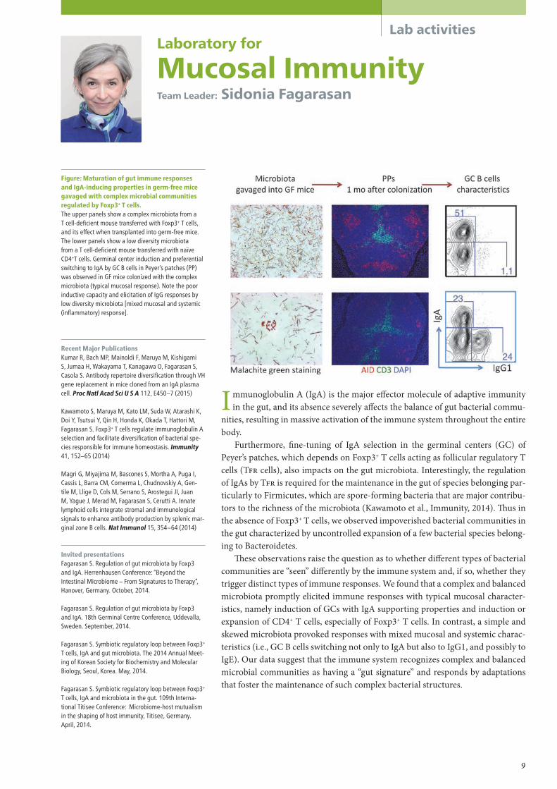
Figure: Maturation of gut immune responses and IgA-inducing properties in germ-free mice gavaged with complex microbial communities regulated by Foxp3+ T cells.The upper panels show a complex microbiota from a T cell-deficient mouse transferred with Foxp3+ T cells, and its effect when transplanted into germ-free mice. The lower panels show a low diversity microbiota from a T cell-deficient mouse transferred with naïve CD4+T cells. Germinal center induction and preferential switching to IgA by GC B cells in Peyer’s patches (PP) was observed in GF mice colonized with the complex microbiota (typical mucosal response). Note the poor inductive capacity and elicitation of IgG responses by low diversity microbiota [mixed mucosal and systemic (inflammatory) response].
Recent Major PublicationsKumar R, Bach MP, Mainoldi F, Maruya M, Kishigami S, Jumaa H, Wakayama T, Kanagawa O, Fagarasan S, Casola S. Antibody repertoire diversification through VH gene replacement in mice cloned from an IgA plasma cell. Proc Natl Acad Sci U S A 112, E450–7 (2015)
Kawamoto S, Maruya M, Kato LM, Suda W, Atarashi K, Doi Y, Tsutsui Y, Qin H, Honda K, Okada T, Hattori M, Fagarasan S. Foxp3+ T cells regulate immunoglobulin A selection and facilitate diversification of bacterial spe-cies responsible for immune homeostasis. Immunity 41, 152–65 (2014)
Magri G, Miyajima M, Bascones S, Mortha A, Puga I, Cassis L, Barra CM, Comerma L, Chudnovskiy A, Gen-tile M, Llige D, Cols M, Serrano S, Arostegui JI, Juan M, Yague J, Merad M, Fagarasan S, Cerutti A. Innate lymphoid cells integrate stromal and immunological signals to enhance antibody production by splenic mar-ginal zone B cells. Nat Immunol 15, 354–64 (2014)
Invited presentationsFagarasan S. Regulation of gut microbiota by Foxp3 and IgA. Herrenhausen Conference: “Beyond the Intestinal Microbiome – From Signatures to Therapy”, Hanover, Germany. October, 2014.
Fagarasan S. Regulation of gut microbiota by Foxp3 and IgA. 18th Germinal Centre Conference, Uddevalla, Sweden. September, 2014.
Fagarasan S. Symbiotic regulatory loop between Foxp3+ T cells, IgA and gut microbiota. The 2014 Annual Meet-ing of Korean Society for Biochemistry and Molecular Biology, Seoul, Korea. May, 2014.
Fagarasan S. Symbiotic regulatory loop between Foxp3+ T cells, IgA and microbiota in the gut. 109th Interna-tional Titisee Conference: Microbiome-host mutualism in the shaping of host immunity, Titisee, Germany. April, 2014.
Immunoglobulin A (IgA) is the major e�ector molecule of adaptive immunity in the gut, and its absence severely a�ects the balance of gut bacterial commu-
nities, resulting in massive activation of the immune system throughout the entire body.
Furthermore, �ne-tuning of IgA selection in the germinal centers (GC) of Peyer’s patches, which depends on Foxp3+ T cells acting as follicular regulatory T cells (TFR cells), also impacts on the gut microbiota. Interestingly, the regulation of IgAs by TFR is required for the maintenance in the gut of species belonging par-ticularly to Firmicutes, which are spore-forming bacteria that are major contribu-tors to the richness of the microbiota (Kawamoto et al., Immunity, 2014). �us in the absence of Foxp3+ T cells, we observed impoverished bacterial communities in the gut characterized by uncontrolled expansion of a few bacterial species belong-ing to Bacteroidetes.
�ese observations raise the question as to whether di�erent types of bacterial communities are “seen” di�erently by the immune system and, if so, whether they trigger distinct types of immune responses. We found that a complex and balanced microbiota promptly elicited immune responses with typical mucosal character-istics, namely induction of GCs with IgA supporting properties and induction or expansion of CD4+ T cells, especially of Foxp3+ T cells. In contrast, a simple and skewed microbiota provoked responses with mixed mucosal and systemic charac-teristics (i.e., GC B cells switching not only to IgA but also to IgG1, and possibly to IgE). Our data suggest that the immune system recognizes complex and balanced microbial communities as having a “gut signature” and responds by adaptations that foster the maintenance of such complex bacterial structures.
Laboratory for
Mucosal ImmunityTeam Leader: Sidonia Fagarasan
9

Lab activitiesLaboratory for
Gut HomeostasisTeam Leader: Kenya Honda
Recent Major PublicationsKawamoto S, Maruya M, Kato LM, Suda W, Atarashi K, Doi Y, Tsutsui Y, Qin H, Honda K, Okada T, Hattori M, Fagarasan S. Foxp3+ T cells regulate immunoglobulin a selection and facilitate diversification of bacterial spe-cies responsible for immune homeostasis. Immunity 41, 152–65 (2014)
Narushima S, Sugiura Y, Oshima K, Atarashi K, Hattori M, Suematsu M, Honda K*. Characterization of the 17 strains of regulatory T cell-inducing human-derived Clostridia. Gut Microbes 5, 333–9 (2014).
Obata Y, Furusawa Y, Endo TA, Sharif J, Takahashi D, At-arashi K, Nakayama M, Onawa S, Fujimura Y, Takahashi M, Ikawa T, Otsubo T, Kawamura YI, Dohi T, Tajima S, Masumoto H, Ohara O, Honda K, Hori S, Ohno H, Koseki H, Hase K. The epigenetic regulator Uhrf1 facilitates the proliferation and maturation of colonic regulatory T cells. Nat Immunol 15, 571–9 (2014)
Invited PresentationsHonda K. Th17 responses to epithelial adhesive intes-tinal microbes. Cold Spring Harbor Asia Conferences, Suzhou, China. September, 2014.
Honda K. Regulation of Th17 and Treg cells by the gut microbiota. 109th International Titisee Conference Microbiome-host mutualism in the shaping of host immunity, Titisee, Germany. April, 2014.
Figure: Schematic showing host-specific epitheli-al adhesion and Th17 cell induction by segment-ed filamentous bacteria (SFB).SFB induce Th17 cells in the small intestine of its nor-mal host, but not in a non-physiological host. Adhesion to epithelial cells is always associated with an increase of Th17 cells. Therefore, activation of epithelial cells by bacterial adhesion may be a prerequisite for SFB-medi-ated induction of Th17 cells.
The intestinal mucosa has a unique immune system composed of a variety of immune cell populations. �e development and function of these gut-unique
cells are known to be a�ected by the presence of the gut microbiota. Last year, we showed that a subset of gut commensal microbes belonging to the class Clostrid-ia was responsible for triggering production of colonic CD4+Foxp3+ regulatory T (Treg) cells, a key therapeutic target in a number of autoimmune and in�ammato-ry diseases. �is year, we have zeroed in on another subset of CD4+ T cells, �17 cells. Intestinal �17 cells are induced and accumulate in response to colonization with a subgroup of intestinal microbes, such as segmented �lamentous bacteria (SFB) and several extracellular pathogens. However, it was not clear what elements of these microbes speci�cally elicited �17 versus other immune cell responses in the intestine. We hypothesized that adhesion of microbes to intestinal epithelial cells (ECs) was one of the critical cues for �17 induction. SFB indigenous to mice (M-SFB) and rats (R-SFB) are genetically distinct host-speci�c members of the gut microbiota. Upon monocolonization in germ-free mice or rats, M-SFB and R-SFB showed host-speci�c adhesion to ECs of the small intestine (SI), accom-panied by host-speci�c induction of �17 cells. Adherent SFB elicited a unique gene-expression program in SI ECs and an SFB antigen-speci�c �17 response. Upon monocolonization in mice, intestinal pathogens including Citrobacter ro-dentium and Escherichia coli O157:H7 triggered similar �17 responses, whereas their adhesion-defective mutants failed to do so. Moreover, a mixture of 20 bac-terial strains, which were selected and isolated from fecal samples from a patient with ulcerative colitis (UC) on the basis of the ability to cause a robust induction of �17 cells in the mouse colon, also exhibited EC-adhesive characteristics. �ese �ndings de�ne �17 induction as a host response to EC-adhesive commensal and pathogenic microbes and provide clues into the better development of mucosal vaccines (manuscript submitted).
10

Lab activities
Recent Major PublicationsHori S. Lineage stability and phenotypic plasticity of Foxp3+ regulatory T cells. Immunol Rev 259, 159–72 (2014)
Obata Y, Furusawa Y, Endo TA, Sharif J, Takahashi D, At-arashi K, Nakayama M, Onawa S, Fujimura Y, Takahashi M, Ikawa T, Otsubo T, Kawamura YI, Dohi T, Tajima S, Masumoto H, Ohara O, Honda K, Hori S, Ohno H, Koseki H, Hase K. The epigenetic regulator Uhrf1 facilitates the proliferation and maturation of colonic regulatory T cells. Nat Immunol 15: 571–9 (2014)
Miyao T, Floess S, Setoguchi R, Luche H, Fehling HJ, Waldmann H, Huehn J, Hori S. Plasticity of Foxp3+ T cells reflects promiscuous Foxp3 expression in conven-tional T cells but not reprogramming of regulatory T cells. Immunity 36, 262–75 (2012)
Invited presentationsHori S. Foxp3-dependent control of regulatory T cell function and homeostasis. Academy of Immunology and Microbiology seminar series, Pohang, the Republic of Korea, March, 2015.
Hori S. Understanding genotype-phenotype relation-ships: lessons learned from disease-causing Foxp3 mutations. Monash Univ. & RIKEN IMS Workshop, Yoko-hama, Japan. August, 2014.
Figure: A model of regulatory T cell fate determi-nation and maintenanceDuring Treg cell differentiation, uncommitted precursor cells adopt either Treg or conventional T (Tconv) cell fates. The commitment to the Treg cell fate is made be-fore (and thus independently of) Foxp3 expression and is executed by a transcription factor network elicited by extrinsic signals from the extracellular environment. The same signals also induce epigenetic changes, including DNA demethylation of the Foxp3 locus. Foxp3 is incorporated into the pre-existing transcription factor network and the resulting “Foxp3 interactome” establishes the characteristic Treg cell phenotype and function in cooperation with the epigenetic modifica-tions. Although Treg cells may down-regulate Foxp3 expression under certain circumstances, these “latent” Treg cells retain the epigenetic memory of, and thus remain committed to, the Treg cell fate. On the other hand, when expressed in activated T cells without engagement of the epigenetic changes and the Foxp3 interactome, Foxp3 by itself cannot establish the char-acteristic Treg cell phenotype.
R egulatory T (Treg) cells expressing the transcription factor Foxp3 play an indispen-sable role in the establishment and maintenance of immunological self-tolerance
and tissue homeostasis. �is concept was �rmly established by the �nding that defective generation or function of Treg cells underlies a fatal autoimmune disease that develops in Foxp3-mutant mice and in humans su�ering from the IPEX syndrome. Recent �ndings that Foxp3+ Treg cells exert tissue-protective or immune-suppressive functions under di-verse circumstances have raised the question of what mechanisms ensure the robustness of Treg cell functions, and thus of immunological self-tolerance, in the face of various unpredictable perturbations in the extracellular environment. To answer this question, we have focused on the mechanisms that control lineage stability and adaptability of Treg cells in changing environments.
We have previously shown that Foxp3 expression per se does not specify the Treg cell lineage in that activated conventional T cells can promiscuously and transiently express Foxp3 while committed Treg cells can transiently and reversibly down-regulate Foxp3. Despite this phenotypic plasticity, Treg cells retain epigenetic memory of, and thus re-main committed to, Foxp3 expression and regulatory functions. We are now addressing the mechanisms underlying this epigenetic memory of Treg cell phenotype and function.
Another focus of our research is to understand how Foxp3 and Treg cells control immunological self-tolerance and tissue homeostasis. To address this question, we have addressed how Foxp3 gene mutations found in human IPEX impinge on Treg cells in vivo using knock-in mutagenesis in mice. Our analysis revealed that, while many mutations are amorphic or hypomorphic, one particular mutation acts as a gain-of-function mu-tation with respect to DNA binding and preferentially impairs Treg cell homeostasis in non-lymphoid tissues. By taking advantage of this unique animal model, we are currently investigating how Treg cells adapt to diverse and �uctuating tissue environments.
Laboratory for
Immune HomeostasisTeam Leader: Shohei Hori
11

Lab activitiesLaboratory for
Skin HomeostasisTeam Leader: Masayuki Amagai
Recent Major PublicationsYoshida K, Kubo A, Fujita H, Yokouchi M, Ishii K, Kawasaki H, Nomura T, Shimizu H, Kouyama K, Ebihara T, Nagao K, Amagai M. Distinct behavior of human Langerhans cells and inflammatory dendritic epidermal cells at tight junctions in patients with atopic dermati-tis. J Allergy Clin Immunol 134, 856–64 (2014)
Kubo A, Ishizaki I, Kubo A, Kawasaki H, Nagao K, Ohashi Y, Amagai M. The stratum corneum comprises three layers with distinct metal-ion barrier properties. Sci Rep 3, 1731 (2013)
Sasaki T, Shiohama A, Kubo A, Kawasaki H, IshidaYamamoto A, Yamada T, Hachiya T, Shimizu A, Okano H, Kudoh J, Amagai M. A homozygous nonsense mutation in the gene for Tmem79, a component for the lamellar granule secretory system, produces spontane-ous eczema in an experimental model of atopic derma-titis. J Allergy Clin Immunol 132, 1111–1120 (2013)
Invited PresentationsAmagai M. Towards antigen-specific immune sup-pression in pemphigus. Inflammatory Skin Disease Summit: The Translational Revolution, Vienna, Austria. November, 2014.
Amagai M. Clinical pictures as good teachers of basic science. 44th Annual Meeting of the European Society for Dermatological Research, Copenhagen, Denmark. September, 2014.
Matsui T. Functional Evolution of Mammalian Stratum Corneum by Retroposon-derived sequence. 16th Annu-al Meeting of Society for Evolutionary Studies, Japan, Osaka, Japan. August, 2014.
Amagai M. Skin as a site where immune system in-teracts with environment. RIKEN IMS-JSI International Symposium on Immunology 2014, Decoding Immune Complexity -Bench to Bedside-, Yokohama, Japan. June, 2014.
Amagai M. Epidermal barrier function and its dys-function in atopic diseases. Singapore International Conference on Skin Research, Biopolis, Singapore. March, 2014.
Figure: Barrier components of the skin, where immunity meets external antigens.Three major barrier components of the epidermis: stratum corneum, tight junctions, and the Langerhans cell network as an immunological barrier. The stratum corneum has at least thee layers with different func-tions, as visualized by TOF-SIMS (Time of flight second-ary ion mass spectrometry) (Kubo et al., Sci Rep, 2013).
When the immune system encounters external antigens in the skin, it tends to react to them. In contrast, when the immune system encounters anti-
gens in the gut, it tends to tolerate them. However, the exact mechanisms for these opposing immune reactions are still largely unknown. Our laboratory attempts to dissect and understand the skin as an immune organ. In particular, we are study-ing skin barrier formation, function, and its dysfunction in atopic diseases.
Skin is composed of three components: epidermis, dermis and subcutaneous fat tissue. Epidermis is a keratinized strati�ed squamous epithelium and forms an e�ective barrier, which is essential for the prevention of the invasion of microor-ganisms, chemical compounds and allergens into the body. �e epidermis is com-posed of four distinct layers; stratum basale (SB), stratum spinosum (SS), stratum granulosum (SG) and stratum corneum (SC), from bottom to top (Fig.).
Among many elements of the skin barrier, we are focusing on the SC as an air-liquid barrier, tight junctions as a liquid-liquid barrier, and the Langerhans cell network as an immunological barrier (Fig.). Tight junctions are formed in the second layer of the SG (SG2 cells) and Langerhans cells extend their dendrites above tight junctions to capture external antigens. Filaggrin de�ciency, which is a predisposing factor for atopic disease, enhances penetration of the SC by external antigens.
�e SC is 12 to 15 accumulated layers of corneocytes, which are terminally di�erentiated dead keratinocytes. �erefore, all the essential SC components are produced in SG1 cells. To understand the transcriptional activity of the SG layer, we have performed RNA-seq analysis of mouse SG layer cells and identi�ed vari-ous di�erentiation-speci�c genes. We also started to perform electron microscop-ic visualization of the SG and SC layers. Based on these analysis, we are planning to interpret cell biological changes in SG cells at the onset of dermatitis in several dermatitis model mice.
12

Lab activities
Recent Major PublicationsHashimoto S, Kubota N, Sato H, Sasaki M, Takamoto I, Kubota T, Nakaya K, Noda M, Ueki K, Kadowaki T. Insulin Receptor Substrate-2 (Irs2) in Endothelial Cells Plays a Crucial Role in Insulin Secretion. Diabetes 64, 876–86 (2015)
Takamoto I, Kubota N, Nakaya K, Kumagai K, Hashimo-to S, Kubota T, Inoue M, Kajiwara E, Katsuyama H, Oba-ta A, Sakurai Y, Iwamoto M, Kitamura T, Ueki K, Kad-owaki T. TCF7L2 in mouse pancreatic beta cells plays a crucial role in glucose homeostasis by regulating beta cell mass. Diabetologia 57, 542–53 (2014)
Kubota T, Kubota N, Kadowaki T. The role of endothelial insulin signaling in the regulation of glucose metabo-lism. Rev Endocr Metab Disord 14, 207–16 (2013)
Invited Presentations
Kubota N. Pathology and treatment strategy for type 2 diabetes with obesity. Diabetes symposium 2014 in TOHOKU, Sendai, Japan. December, 2014.
Kubota N. Zonation-dependent selective insulin resist-ance of the liver in obesity and type 2 diabetes. The 8th Diabetes Leading-edge Conference, Chiba, Japan. August, 2014.
Kubota N. Molecular mechanisms of glucose and lipid metabolism. Meet the Expert of Diabetes 2014, Tokyo, Japan. June 2014.
Kubota N. Therapeutic strategy for type2 diabetes. Shinshu Basal Insulin Seminar, Nagoya, Japan. March, 2014.
Figure: IRS-2 in the endothelial cells plays a cru-cial role in insulin secretion via islet blood flowWe have reported that IRS-2 expression levels in the endothelial cells reduced by hyperinsulinemia in obese and diabetic models. This reduction of IRS-2 expression levels also lead to obesity and type 2 diabetes. Endo-thelial-specific IRS-2 deficient (ETIrs2) mice is suitable as obese and diabetic models. Insulin secretion and islet blood flow decreased in ETIrs2 mice. ACE-inhibitor improved islet blood flow, resulting in restoration of insulin secretion. IRS-2 in the endothelial cells mediate insulin secretion through islet blood flow. Drugs for improvement of islet blood flow may be one of effective therapeutic strategy in obesity and type 2 diabetes subjects.
I n recent years there has been a rapid growth in the incidence of type 2 diabetes in both Western and Asian countries. �is high prevalence is most likely the result of
a complex interplay between genetic factors, such as reduced insulin secretion, and en-vironmental factors, such as high-fat diet and decreased physical activity. However, the precise molecular mechanisms underlying the development and progression of type 2 diabetes remain unclear. �e goal of our team is to identify molecular mechanisms of insulin secretion and insulin resistance.
Molecular mechanism of insulin secretionEndothelial cells mediate blood �ow, which is considered to be essential for insulin se-cretion. However, it is unclear whether endothelial cells are involved in the regulation of insulin secretion. We examined the relationship between insulin secretion and endothe-lial cells using the endothelial-speci�c Insulin Receptor Substrate-2 (IRS-2) knockout (ETIrs2KO) mice. Although insulin secretion from isolated islets was maintained, insulin secretion was signi�cantly impaired in the ETIrs2KO mice. �e islet blood �ow was also signi�cantly reduced these mice. Enalapril treatment, an ACE-inhibitor, improved the islet blood �ow, resulting in the restoration of insulin secretion in the ETIrs2KO mice. �ese data suggest that IRS-2 in the endothelial cells regulates islet blood �ow, mediating insulin secretion (Diabetes, in press).
Molecular mechanism of insulin resistance�e liver plays an important role in the control of glucose homeostasis. Interestingly, in patients with type 2 diabetes and obesity, hyperglycemia and hepatic steatosis o�en co-exist. �is would seem to indicate that the insulin signaling pathway is impaired in glu-coneogenesis but preserved in lipogenesis. �is phenomenon is referred to as “selective insulin resistance” and has recently received increasing attention. To address “selective insulin resistance” in the liver, we have been focusing our research on IRS-1 and IRS-2.
Laboratory for
Metabolic HomeostasisTeam Leader: Naoto Kubota
13

Lab activitiesLaboratory for
Immune CrosstalkTeam Leader: Hilde Cheroutre
Figure: Epithelial T cells (epiT cells) protect the mucosal barrier of the intestine from pathogen- and inflammation-induced pathology.T cells residing within the epithelium of the intestine are phenotypically heterogeneous but they are all specialized to protect the mucosal barrier against pathogen- and immune cell-induced pathology. In contrast to the T cells in the periphery, epiT cells are antigen-experienced T cells that encountered their antigen initially during selection in the thymus (agonist selected CD8aa TCRαβ and TCRγδ precursor cells) or as mature cells in the periphery (CD8αβ CTL and CD4 CTL). Although the various epiT cell subsets display different antigen specificity and MHC restriction and although they follow different paths of effector cell differentia-tion, they all acquire cytolytic and regulatory capacity. The functional specialization of epiT cell adapts them to provide optimal protection in the face of preserving the integrity of the delicate mucosal barrier.
Recent Major PublicationsVicente-Suarez I, Larange A, Reardon C, Matho M, Feau S, Chodaczek G, Park Y, Obata Y, Gold R, Wang-Zhu Y, Lena C, Zajonc DM, Schoenberger SP, Kronenberg M, Cheroutre H. Unique lamina propria stromal cells imprint the functional phenotype of mucosal dendritic cells. Mucosal Immunol 8, 141–51 (2015)
Mayans S, Stepniak D, Palida SF, Larange A, Dreux J, Arlian BM, Shinnakasu R, Kronenberg M, Cheroutre H, Lambolez F. αβT cell receptors expressed by CD4(-)CD8αβ(-) intraepithelial T cells drive their fate into a unique lineage with unusual MHC reactivities. Immu-nity 41, 207–18 (2014)
Fu G, Casas J, Rigaud S, Rybakin V, Lambolez F, Brzos-tek J, Hoerter JA, Paster W, Acuto O, Cheroutre H, Sauer K, Gascoigne NR. Themis sets the signal threshold for positive and negative selection in T-cell development. Nature 504, 441-5 (2013)
Invited PresentationsCheroutre H. Mucosal T cells: Same Players Different Strategies. UCLA 13T Immunology Forum Seminar Se-ries, Los Angeles, USA. December, 2014.
Cheroutre H. How the Gut Primes the Immune System. Annual Scientific Meeting of American College of Rheumatology (ACR), Boston, USA. November, 2014.
Cheroutre H. Mucosal Immunity: Taking Strategic Plan-ning a Step Further. The Dutch Society for Immunology Annual Symposium, "Mucosal Immunity", Lunteren, the Netherlands. April, 2014.
Cheroutre H. New Emerging Transcription Factor and Cytokine Networks at the Mucosal Interface of the In-testine. Keystone Symposium on Molecular and Cellular Biology: Emerging Cytokine Networks (J3). Session: Regulation of Tissue-Resident ILC and T Cells, Vancou-ver, Canada. January, 2014.
Our research continues to elucidate mechanisms of mucosal immune protec-tion and regulation. In a recent study, we uncovered an unexpected degree
of plasticity for CD4 T helper (�) cells which, upon antigenic stimulation, are able to terminate the expression of the � transcription factor, �POK, and dif-ferentiate into cytotoxic T lymphocytes (CTL). At steady state, CD4 CTLs remain quiescent and express a self-regulated phenotype. However under challenging conditions, these cells have the potential to transform into potent in�ammatory killer e�ector cells (Mucida et al., Nat Immunol, 2013).
Overall, based on the insights we are gaining from our research, a clear picture has begun to emerge showing that the immune defense of the intestine adapts to the local environment and specializes to provide the most e�cient and immediate protection in the face of preserving the integrity of the most critical mucosal bar-rier of the body.
In another study, we are aiming to understand the various mechanisms and processes that lead to Central Tolerance. Our previous research showed that in ad-dition to conventional selection, a process of so called “agonist” selection operates in the thymus and preserves self-speci�c thymocytes and functionally di�erenti-ates these precursor cells into bene�cial pre-programmed protective or regulatory T cells. In an e�ort to understand what factors control the decisive checkpoint during thymic selection, we identi�ed “�emis” as a critical switch. By making various kinds of mutant �emis knock-in mice, we found that localization of �emis protein in the nucleus is critical for its function. In addition to the mo-lecular mechanisms of �emis in the nucleus, we are now trying to elucidate the molecular and cellular factors and events that connect the Pre-TCR and TCR sig-nal strength with thymic selection and the fate decision of the developing T cells.
14

Lab activities
Recent Major PublicationsTanaka T, Shibazaki A, Ono R, Kaisho T. HSP70 medi-ates degradation of the p65 subunit of nuclear factor κB to inhibit inflammatory signaling. Sci Signal 7, ra119 (2014)
Yamazaki C, Sugiyama M, Ohta T, Hemmi H, Hamada E, Sasaki I, Fukuda Y, Yano T, Nobuoka M, Hirashima T, Iizuka A, Sato K, Tanaka T, Hoshino K, Kaisho T. Critical roles of a dendritic cell subset expressing a chemokine receptor, XCR1. J Immunol 190, 6071–82 (2013)
Invited PresentationsTanaka T. From Molecules to Diseases. Clarification of the molecular mechanisms that negatively regulate inflammatory responses and association analysis of autoimmune diseases by GWAS. The 3rd Akashi-cho rheumatic collaborative seminar in St Luke’s Interna-tional Hospital, Tokyo, Japan. October, 2014.
Tanaka T. Negative regulation for inflammatory re-sponses and its association with autoimmune diseases. Monash Univ. & RIKEN IMS Workshop, Yokohama, Japan. August, 2014
Figure: HSP70 is essential for PDLIM2-mediated degradation of NF-κB p65.PDLIM2 binds to the p65 subunit of NF-κB and pro-motes p65 polyubiquitination in the nucleus. PDLIM2 then targets p65 into discrete intranuclear compart-ments, called PML nuclear bodies. HSP70 binds to PDLIM2 and facilitates delivery of the NF-κB-PDLIM2 complex to the proteasome cooperatively with BAG1. Polyubiquitinated p65 is ultimately degraded by the proteasome in PML nuclear bodies.
The in�ammatory response is an important host defense mechanism to sense and eliminate invading microbial pathogens. Dendritic cells �rst detect
pathogens and activate the transcription factor NF-κB, which enters the nucleus and induces the expression of a series of in�ammation-related genes. �ese ini-tially helpful in�ammatory responses must be terminated at the appropriate time point, otherwise excessive responses can damage normal tissue and may cause au-toimmune diseases. Our research goal is to identify key regulators of in�amma-tion-related signal transduction pathways and to clarify the complete picture of the molecular mechanisms for regulating in�ammatory responses. We previously identi�ed PDLIM2 (PDZ and LIM domain protein-2), a nuclear protein that be-longs to a large family of LIM proteins, as one of the key factors negatively regu-lating in�ammatory responses. We demonstrated that PDLIM2 negatively regu-lates NF-κB activity and subsequent in�ammatory responses, acting as a nuclear ubiquitin E3 ligase targeting the p65 subunit of NF-κB. (Tanaka T, Nat Immunol, 2007). We have recently studied how PDLIM2-mediated p65 degradation is con-trolled and found that heat shock protein 70 (HSP70), a molecular chaperone, is required for PDLIM2 to degrade p65 and suppress NF-κB activation following in-�ammatory responses. In dendritic cells, HSP70 is detected only in the cytoplasm without stimulation, but it is translocated to the nucleus a�er TLR stimuli. HSP70 then associated with both PDLIM2 and BAG-1, a proteasome-associated protein, and promoted the transport of the NF-κB-PDLIM2 complex to the proteasome, thereby facilitating p65 degradation. Consistently, either HSP70 de�ciency or BAG-1 knockdown in dendritic cells leads to increased nuclear p65 protein levels and thus enhanced production of proin�ammatory cytokines in response to TLR stimuli (Tanaka T, Sci Signal, 2014). �ese studies should contribute to our under-standing of the pathogenesis of human autoimmune diseases and provide novel targets to develop new treatments for these diseases.
Laboratory for
Inflammatory RegulationTeam Leader: Takashi Tanaka
15

Lab activitiesLaboratory for
Cytokine RegulationTeam Leader: Masato Kubo
Recent Major PublicationsMotomura Y, Morita H, Moro K, Nakae S, Artis D, Koyasu S, Kubo M. Basophil-derived interleukin-4 controls the function of natural helper cells, a member of ILC2s, in lung inflammation. Immunity 40, 758–71 (2014)
Kurashima Y, Amiya T, Fujisawa K, Shibata N, Suzuki Y, Kogure Y, Hashimoto E, Otsuka A, Kabashima K, Sato S, Sato T, Kubo M, Akira S, Miyake K, Kunisawa J, Kiyono H. The enzyme Cyp26b1-mediates Inhibition of mast cell activation by fibroblasts to maintain skin-barrier homeostasis. Immunity 40, 530–41 (2014)
Noti M, Wojno ED, Kim BS, Siracusa MC, Giacomin PR, Nair MG, Benitez AJ, Ruymann KR, Muir AB, Hill DA, Chikwava KR, Moghaddam AE, Sattentau QJ, Alex A, Zhou C, Yearley JH, Menard-Katcher P, Kubo M, Obata-Ninomiya K, Karasuyama H, Comeau MR, Brown-Whitehorn T, de Waal Malefyt R, Sleiman PM, Hakonarson H, Cianferoni A, Falk GW, Wang ML, Spergel JM, Artis D. Thymic stromal lymphopoie-tin-elicited basophil responses promote eosinophilic esophagitis. Nat Med 19, 1005–13 (2013)
Invited PresentationsKubo M, Role of T follicular helper cells in influenza virus protection. France-Japan Immunology meeting, Cassis, France. October, 2014.
Kubo M, Notch regulates reciprocal expression of CCR7 versus CXCR5 to control central memory T cell generation. The 2nd Symposium of International Im-munological Memory and Vaccine Forum, La Jolla, USA. August, 2014.
Kubo M, Regulation of allergic airway inflammation by basophil and innate lymphoind cells. The 79th Annual Meeting of the Japanese Society of Interferon & Cy-tokine Research, Sapporo, Japan. June, 2014.
Kubo M, Understanding a role of cytokine signaling in homeostatic skin regulation. Shanghai Immunoderma-tology Forum 2014, Shanghai, China. May, 2014.
Kubo M, Regulation of allergic airway inflammation by basophil and innate lymphoind cells. Dry-eye allergy joint seminer, Tokyo, Japan. January, 2014.Conference on Skin Research, Biopolis, Singapore. March, 2014.
Figure: Cross-talk of basophils and group 2 in-nate lymphoid cells (ILC2s)/Natural helper (NH) cells in asthmatic responsesCysteine protease allergen induced IL-4 from basophils, and the basophil-derived IL-4 has a critical role in the secretion of IL-5, IL-13 and CCL11 from ILC2s/NH cells that lead to T-independent asthmatic airway inflam-mation.
Allergy is thought to be controlled by type 2 cytokines, including IL-4, IL-5, and IL-13. IL-4 promotes IgE production by B cells, IL-5 induces develop-
ment, recruitment, and activation of eosinophils, and IL-13 is tightly associated with multiple events in the e�ector phase of allergic responses, inducing airway hyper-responsiveness and hyperproduction of mucus. Recently, these type 2 cy-tokines were found to be secreted not only by TH2 cells but also by innate immune cells, including mast cells, basophils, eosinophils, and group 2 innate lymphoid cells (ILC2s), which have been discovered in the gut-associated mucosal tissues.
Allergic asthma is a chronic in�ammatory disease characterized by airway obstruction and wheezing. Conventional mouse models have illustrated the im-portance of TH2 cells and eosinophilic in�ammation and, more recently the im-portance of basophils and ILC2s. Asthmatic in�ammation is inducible by aller-gen proteases, including those from papain and house dust mites (HDM). �e HDM-derived protease Derp1 causes the airway in�ux of eosinophils and bron-choconstriction in asthma patients. Papain a plant-derived cysteine protease, causes occupational asthma. To examine the role of basophils and mast cells, we established a diphtheria toxin-based conditional deletion system using an Il4 en-hancer that we previously found was speci�c for IL-4 production by mast cells or basophils (Mas-TRECK and Bas-TRECK mice). Conditional deletion of baso-phils caused a resolution of papain-induced eosinophilia and mucus production. Resolution of eosinophilia was also observed in mice lacking IL-4 production speci�cally in basophils, indicating that basophil-derived IL-4 not only enhanced expression of CCL11, IL-5, IL-9 and IL-13 by natural helper (NH) cells, thus at-tracting eosinophils, but also promoted expansion of these ILC2s. �ese results demonstrate that IL-4 from basophils has an important role in the IL-33-NH cell-cytokine/chemokine axis, subsequently leading to protease allergen-induced airway in�ammation.
16

Toward the ultimate goal of obtaining a comprehensive understanding of the pathogenesis of human diseases, the functions of the Core for Precise
Measuring and Modeling are three pronged: production of mouse models, multiomics measurements and quantitative bioimaging, and bioinformatics/modeling of human disease processes. Through close interactions among these three branches of the core, we aim to collect a wide variety of quantita-tive data in order to build a computational and predictive network of the dis-ease process. As for the production of genetically engineered mice that will be used as models of human diseases, the laboratory for Developmental Ge-netics has begun to apply recent advances in genome engineering technol-ogy, e.g., CRISPR/Cas9-based genome editing, and thereby considerably en-hance the production capacity and power of the disease models. Regarding the precise quantitative measurements branch, one focus in 2014 has been to enhance the power of metabolite analysis by the Laboratory for Metabolom-ics (Photo). Together with mRNA/protein profiling by the Laboratory for Inte-grative Genomics, the enhanced multiomics measurements and bio imaging (Laboratory for Tissue Dynamics) will greatly contribute to exploration of the etiology of human diseases. After being processed by bioinformatics (Lab-oratory for Integrated Bioinformatics), the datasets are used for modeling (Laboratories for Disease Systems Modeling and Integrated Cellular Systems). As a leading IMS project, an atopic dermatitis model mouse, provided by the Laboratory for Immunogenetics, has been extensively analyzed from several di�erent angles, fully exploiting the analysis powers of this core. These e�orts should enable us to identify new biomarkers for early diagnosis and preven-tion of atopic dermatitis in the very near future.
Core for Precise Measuringand Modeling
Photo: The new mass spectrometry laboratory To enhance metabolomic and proteomic analyses in IMS, new laboratory space dedicated to mass spec-trometry was created.
17

Lab activitiesLaboratory for
Developmental GeneticsGroup Director: Haruhiko Koseki
Recent Major PublicationsObata Y, Furusawa Y, Endo TA, Sharif J, Takahashi D, Atarashi K, Nakayama M, Onawa S, Fujimura Y, Takahashi M, Ikawa T, Otsubo T, Kawamura YI, Dohi T, Tajima S, Masumoto H, Ohara O, Honda K, Hori S, Ohno H, Koseki H, Hase K. The epigenetic regulator Uhrf1 facilitates the proliferation and maturation of colonic regulatory T cells. Nat Immunol 15, 571–9 (2014)
Blackledge NP, Farcas AM, Kondo T, King HW, McGou-ran JF, Hanssen LL, Ito S, Cooper S, Kondo K, Koseki Y, Ishikura T, Long HK, Sheahan TW, Brockdorff N, Kessler BM, Koseki H, Klose RJ. Variant PRC1 complex-depend-ent H2A ubiquitylation drives PRC2 recruitment and polycomb domain formation. Cell 157, 1445–59 (2014)
Kondo T, Isono K, Kondo K, Endo TA, Itohara S, Vidal M, Koseki H. Polycomb potentiates meis2 activation in midbrain by mediating interaction of the promoter with a tissue-specific enhancer. Dev Cell 28, 94–101 (2014)
Invited PresentationsKoseki H. iPS-mediated induction of human NKT cells and their application for cancer therapy. The 35th An-nual Meeting of the Japanese Society of Inflammation and Regeneration, Okinawa, Japan. July, 2014.
Figure: Role of NP95 in embryonic and extra-embryonic tissuesWithout substrate (i.e. hemimethylated DNA), the NP95 SRA (SET and RING-finger associated) domain interacts with the neighboring TP (Tudor and PHD), assuming a mutually inhibitory conformation (top). Hemimethyl-ated DNA (5-cytosine methylation of only one strand of the CpG dyad) binding by NP95-SRA elicits two different downstream pathways. In embryonic cells, NP95 recruits DNMT1- the major DNA methyltrans-ferase, to maintain methylation upon DNA replication (left). Association of NP95 with naturally occurring hemimethylated DNA in extra-embryonic cells results in transcriptional activation through inhibition of the SETDB1 (H3K9 trimethyltransferase) and H3K9me3-me-diated repressive machinery (right).
T he Developmental Genetics Research Group focuses on the epigenetic regulation of organ development and stem cell functions by Polycomb group (PcG) proteins and
molecules that mediate DNA methylation.
Chromatin dynamics associated with activation of Polycomb-repressed genesHow PcG-repressed genes are activated upon receipt of developmental cues is poorly understood. We used the Meis2 locus as a model to identify the role of a tissue-speci�c enhancer in displacing the PcG complex from the promoter. Meis2 repression in early de-velopment depends on the binding of RING1B, an essential component of PcG, to the pro-moter. �is is coupled with the association of the promoter with another RING1B-bind-ing site (RBS) at the 3’ end of the Meis2 locus. During early midbrain development, a midbrain-speci�c enhancer (MBE) transiently associates with the promoter/RBS, form-ing a promoter/MBE/RBS tripartite interaction in a RING1-dependent manner. Subse-quently, the RING1B-bound RBS dissociates from the tripartite, leaving promoter/MBE engagement to activate Meis2 expression. �is study therefore demonstrates the role of PcG and/or related factors in Meis2 activation by regulating the topological transition of cis-regulatory elements.
Regulation of endogenous retroviral elements (ERVs) by the SRA protein NP95 ERVs are abundant in the mammalian genome and are transcriptionally silenced by re-pressive epigenetic marks such as DNA methylation and Histone H3 Lysine 9 (H3K9) methylation. However, during speci�c windows, such as early embryogenesis and pri-mordial germ cell (PGC) development, DNA methylation is globally erased, which poten-tially exposes the genome to aberrant activation of ERVs. We have found that functional inhibition of the SRA protein NP95, an essential component of the maintenance methyla-tion machinery, could mediate global demethylation and at the same time ensure silenc-ing of ERV sequences, particularly in the embryo proper. Absence of NP95 facilitates the transition from a DNA methylation-dependent state to an H3K9 methylation-dependent state contributing to ERV silencing. Paradoxically, NP95 also has a role in transcribing ERV sequences in the extra-embryonic tissues, such as the placenta, under normal phys-iological conditions, likely by destabilizing the H3K9 methylation pathway. Taken togeth-er, our observations reveal that the �delity of histone methylation-dependent proviral silencing is dependent upon timely maintenance DNA methylation.
18

Lab activitiesLaboratory for
Integrative GenomicsGroup Director: Osamu Ohara
Although “genomics” itself has always been an integrative science, our labora-tory name is “Integrative Genomics” to indicate and emphasize our intention
to gain new insights into regulation of complex biological systems by amalgamat-ing spatio-temporal multi-scale data. �us, besides our central support activities and involvement in collaborative/strategic programs at IMS, we have placed tech-nology development as a central mission of my group. In this regard, our current technology interest is in development of methods to compare and/or link macro-scopic genomic data with data at the single-cell resolution. Recent publications regarding measurements of protein secretion from single cells are examples of the outcomes of our e�orts in this direction. To further enhance such research activi-ties, we think that the technology of absolute quanti�cation of biomolecules would signi�cantly increase its importance and have begun to implement such technol-ogies at the protein level. SWATH (sequential window acquisition of all theoret-ical fragment ion spectra) is one such technology for high throughput, label-free protein quanti�cation based on mass spectrometry. However, to link SWATH data with the corresponding single-cell protein pro�les, we need high-quality anti-bodies for detection of speci�c proteins of interest as demonstrated in our recent publication (Shirasaki et al., Sci Rep, 2014). We are thus working on technolo-gy development of high-throughput production of recombinant antibodies with speci�ed a�nity/speci�city. �e combination of mass spectrometry-based and af-�nity-based proteomics approaches will enable us to open a new avenue leading to real spatio-temporal and multi-scale data at the protein level.
Recent Major PublicationsAoki Y, Watanabe T, Saito Y, Kuroki Y, Hijikata A, Takagi M, Tomizawa D, Eguchi M, Eguchi-Ishimae M, Kaneko A, Ono R, Sato K, Suzuki N, Fujiki S, Koh K, Ishii E, Shultz LD, Ohara O, Mizutani S, Ishikawa F.¿Identification of CD34+ and CD34- leukemia-initiating cells in MLL-re-arranged human acute lymphoblastic leukemia Blood 125, 967–80 (2015)
Liu T, Yamaguchi Y, Shirasaki Y, Shikada K, Yamagishi M, Hoshino K, Kaisho T, Takemoto K, Suzuki T, Kuranaga E, Ohara O, Miura M. Single-cell imaging of caspase-1 dynamics reveals an all-or-none inflammasome signal-ing response. Cell Rep 8, 974–82 (2014)
Shirasaki Y, Yamagishi M, Suzuki N, Izawa K, Nakahara A, Mizuno J, Shoji S, Heike T, Harada Y, Nishikomori R, Ohara O. Real-time single-cell imaging of protein secre-tion. Sci Rep 4, 4736 (2014)
Invited PresentationsOhara O. Construction of A Clinical Sequencing Pipeline for Genetic Diseases. The 59th Annual Meeting of the Japan Society of Human Genetics/The 21st Japanese Society for Gene Diagnosis and Therapy, Tokyo, Japan. November, 2014.
Hijikata A, Ohara O. Structural Bioinformatics of Human Genetic Diseases. The 86th Annual Meeting of The Ge-netics Society of Japan, Shiga, Japan. September, 2014.
Ohara O. Innovation and Invention in Genomics. 18th Meeting of Molecular Composite Medicine, Osaka, Japan. July, 2014.
Ohara O. What is “Next-Generation DNA Sequencing” for? Liaison Laboratory regular seminar FY2014, Kum-amoto, Japan. May, 2014.
Ohara O. Implementation of High-Volume Genomic Analyses by Microfluidics/microchip Technologies: To-wards Integrative Medical Sciences for Preventive Med-icine. International Conference on Electronics Packaging 2014, Toyama, Japan. April, 2014.
Figure: Principal Component Analysis (PCA) of protein profiles in four types of mouse tissues (N2, N3, N5, N16)A: PCA score plot shows separate clusters of four types of mouse tissues at different disease states. B: PCA loading plot shows the proteins responsible for separa-tion of the groups in the corresponding score plot.
19

Lab activities
Recent Major PublicationsWatanabe T, Kawakami E, Shoemaker JE, Lopes TJ, Matsuoka Y, Tomita Y, Kozuka-Hata H, Gorai T, Kuwaha-ra T, Takeda E, Nagata A, Takano R, Kiso M, Yamashita M, Sakai-Tagawa Y, Katsura H, Nonaka N, Fujii H, Fujii K, Sugita Y, Noda T, Goto H, Fukuyama S, Watanabe S, Neumann G, Oyama M, Kitano H, Kawaoka Y. Influenza virus-host interactome screen as a platform for antiviral drug development. Cell Host Microbe 16, 795–805 (2014)
Marti-Solano M, Birney E, Bril A, Della Pasqua O, Kitano H, Mons B, Xenarios I, Sanz F. Integrative knowledge management to enhance pharmaceutical R&D. Nat Rev Drug Discov 13, 239–40 (2014)
Hsin KY, Ghosh S, Kitano H. Combining machine learn-ing systems and multiple docking simulation packages to improve docking prediction reliability for network pharmacology. PLos One 8, e83922 (2013)
Invited PresentationsKitano H. Making comprehensible maps from compre-hensive knowledge - New approaches to information and knowledge management. Luxembourg, Life Sci-ence Hub in Europe, Tokyo, Japan. October, 2014.
Kitano H. Systems Biology and Applications. ICSB 2014, Melbourne, Australia. September, 2014.
Figure: Systems level approaches to investigate atopic dermatitis.The LDSM is investigating systems-level principles underlying diseases and developing multi-layer phys-iological models and detailed cellular and molecular interaction models by using gene expression data.
U nderstanding systems-level mechanisms of Atopic Dermatitis (AD) and support-ing computational systems biology needs of the center are missions of the LDSM.
During 2014, the LDSM has made signi�cant progress in developing basic analysis pipelines for RNA-seq analysis, network reconstruction, and dynamical modeling of AD, focusing on time series analysis involving pre- and post-onset of the disease.
On the technology development front, we have developed (1) a basic mRNA-seq anal-ysis pipeline tailored for IMS facilities, (2) a preliminary text-mining system for model reconstruction, (3) a genomics-systems biology integrated analysis pipeline using Gal-axy and Garuda platforms, etc. �ese have already proven to be useful for analyses and reproducibility. In the future, we will continue to improve and expand this technology infrastructure.
In terms of data analysis and modeling, we have completed an initial analysis of mRNA data provided by experimental IMS teams and identi�ed a set of critical genes that are implicated in a subset of AD outbreaks. We have also created a tissue speci�c gene analysis pipeline by using heterogeneous in-house experimental data that are im-portant in AD.
We have also conducted our own cell-based experiments to further validate our �nd-ings and to provide missing information. In parallel, we have developed a computational model of AD, initially focusing on keratinocytes. �is model will be integrated with other models developed by Dr. Mariko Okada (RIKEN) and Dr. Reiko Tanaka (ICL) to create a more comprehensive model of AD.
On the experimental side, we have established a mammalian cell-based experimental facility to verify a series of hypotheses derived from the model prediction, as well as a comprehensive siRNA screening system to identify possible roles of miRNA in AD. In addition, a yeast gTOW system is now up-and-running to generate robustness-pro�les to develop high precision yeast cell cycle models that should eventually contribute to un-derstanding the fragility of mammalian cellular homeostasis implicated in AD and other diseases.
Laboratory for
Disease Systems ModelingGroup Director: Hiroaki Kitano
20

Lab activities
Recent Major PublicationsObata Y, Kimura S, Nakato G, Iizuka K, Miyagawa Y, Nakamura Y, Furusawa Y, Sugiyama M, Suzuki K, Ebisa-wa M, Fujimura Y, Yoshida H, Iwanaga T, Hase K, Ohno H. Epithelial-stromal interaction via Notch signaling is essential for the full maturation of gut-associated lym-phoid tissues. EMBO Rep 15, 1297–304 (2014)
Miyai T, Hojyo S, Ikawa T, Kawamura M, Irie T, Ogura H, Hijikata A, Bin BH, Yasuda T, Kitamura H, Nakayama M, Ohara O, Yoshida H, Koseki H, Mishima K, Fukada T. Zinc transporter SLC39A10/ZIP10 facilitates antiapop-totic signaling during early B-cell development. Proc Natl Acad Sci U S A 111, 11780-–5 (2014)
Hojyo S, Miyai T, Fujishiro H, Kawamura M, Yasuda T, Hijikata A, Bin BH, Irie T, Tanaka J, Atsumi T, Murakami M, Nakayama M, Ohara O, Himeno S, Yoshida H, Koseki H, Ikawa T, Mishima K, Fukada T. Zinc transporter SL-C39A10/ZIP10 controls humoral immunity by modulat-ing B-cell receptor signal strength. Proc Natl Acad Sci U S A 111, 11786–91 (2014)
Invited PresentationsYoshida H. Genetic factors and atopic dermatitis dis-ease model. The 10th Tokyo Scientific Forum for Atopic Dermatitis and Psoriasis, Tokyo, Japan. December, 2014.
Yoshida H. Genetic factors and onset of atopic dermati-tis in murine disease model. The 113th Annual Meeting of the Japanese Dermatological Association, Kyoto, Japan. May, 2014.
Figure: Pre-symptomatic disease development in the Spade mutantPre-symptomatic events occurring in the Spade mu-tant were detected by histological, cytological and molecular biological analysis and are summarized in the figure as longitudinal steps. The pre-symptomatic events occurred first in epidermal tissues, and this was followed by local immune activation, local proteases expression, migration of innate immune cells to the lesion, microbial infection, and finally by a systematic immune reaction.
Before the onset of an in�ammatory disease, there must be an accumula-tion of many imperceptible pathogenic events in the human body as part of
pre-symptomatic disease development under the in�uence of genetic and envi-ronmental factors. If one could precisely monitor and understand these pre-symp-tomatic longitudinal multiple events occurring in a healthy individual, it should be possible to predict the timing of disease onset and to take measures to prevent it. �is strategy will be bene�cial for many of us in the coming era when everyone can know their own genome sequence information and genetic risk factors shortly a�er birth.
In our laboratory, we had been working to identify the genetic factors for al-lergic or immune disease development by phenotype screening of chemical mu-tagen, N-ethyl N-nitrosourea (ENU), -induced mutant mice on a C57BL/6J back-ground. Among the mutants identi�ed, we have focused on a dermatitis model in which the disease develops at approximately 8 weeks a�er birth, as detected by ear skin desquamation and scratching of the skin a few days a�er the initial symptoms. �is phase is followed by a �2 biased immune response detected by serum IgE, IgG1 and elevated histamine levels 3 weeks later and then, 8 weeks af-ter that, a �1 immune bias ensues, with elevated serum IgG2b and IgG2c. At this �nal stage, the pathological �ndings led to the diagnosis of a chronic in�amma-tory condition of the skin. �ese symptoms are in part compatible with those of human atopic dermatitis. �erefore we named this mutant Spade (Stepwise pro-gressive atopic dermatitis).
Using this disease model, we have precisely analyzed the pre-symptomatic pathological events as shown in the �gure. �e accumulation of all these events leads to the onset of dermatitis in the late stage. Now we are trying to identify the molecular networks responsible for each of these pre-symptomatic events.
Laboratory for
ImmunogeneticsTeam Leader: Hisahiro Yoshida
21

Lab activitiesLaboratory for
Integrated BioinformaticsTeam Leader: Todd D. Taylor
The main research focus of our laboratory is the development of computation-al tools and databases related to the analysis of metagenomic, and sometimes
bacterial genomic, sequence data. In addition, we are also developing some gener-al-purpose tools that are applicable to the wider research community.
Initially as part of a center project, we are developing an integrated database and sample-tracking system for the storage and distribution of massive and var-ious types of experimental data, including: �ow cytometry data, immunohisto-chemistry images, skin water loss measurements, blood pro�les, RNA-Seq data, and so on. For maximum �exibility, this database is being designed not to be data-, project-, species-, or center- speci�c. We will also integrate other useful outside data resources. We hope to develop a secure, �exible system that makes it easy to manage, access, analyze, integrate, and visualize various data types as per user requirements.
We are also developing a revolutionary new web-based tool, called iCLiKVAL (http://iclikval.riken.jp/), that uses the power of crowdsourcing to add valuable annotation information to the rapidly accumulating volume of scienti�c literature (e.g., millions of PubMed citations and other media types), resulting in a com-prehensive library of data that yields richer and more relevant literature search results. We will be actively promoting this resource in the coming year and we are strongly encouraging more and more scientists to get involved and contribute their valuable knowledge.
�e ultimate goal of our research is to develop tools capable of e�ciently processing and analyzing data from a variety of sources, with an emphasis on metagenomic data. With the advent of high-throughput next-generation sequenc-ers, and subsequent massive data output, we anticipate the need for better means of visualizing and understanding the results and we will continue to put more em-phasis in this critical area.
Recent Major PublicationsFoong C, Lau NS, Deguchi S, Toyofuku T, Taylor TD, Sudesh K, Matsui M. Whole genome amplification ap-proach reveals novel polyhydroxyalkanoate synthases (PhaCs) from Japan trench and nankai trough seawater. BMC Microbiol 14, 7. (2014) [Epub ahead of print]
International Glossina Genome Initiative. Genome se-quence of the tsetse fly (Glossina morsitans): vector of African trypanosomiasis. Science 344, 380–6 (2014)
Jinda W, Taylor TD, Suzuki Y, Thongnoppakhun W, Limwongse C, Lertrit P, Suriyaphol P, Trinavarat A, Atchaneeyasakul LO. Whole exome sequencing in Thai patients with retinitis pigmentosa reveals novel mutations in six genes. Invest Ophthalmol Vis Sci 55, 2259–68 (2014))
Invited PresentationsTaylor TD. Introduction to metagenomics; Metadata management, sampling considerations, experimental approaches, workflow summary. CCB-USM Workshop on Metagenomics, Penang, Malaysia. December, 2014.
Kim S.-W. Microbial community analysis using 16S rRNA; BioRuby for beginners. CCB-USM Workshop on Metagenomics, Penang, Malaysia. December, 2014.
Figure: Integrated database and sample-track-ing systemVarious types of wet-lab experimental results, such as flow cytometry information, immunohistochemistry im-ages, skin water loss measurements, and blood profiles (green arrows), will be input into the database directly from the respective labs. Other results, such as RNA-Seq data and analytical output (orange arrows), will be incorporated into the database using automated transfer protocols. All of the data will be available for viewing, tracking, downloading and analyzing by any authorized labs.
22

Lab activities
Recent Major PublicationsNatsuaki Y, Egawa G, Nakamizo S, Ono S, Hanakawa S, Okada T, Kusuba N, Otsuka A, Kitoh A, Honda T, Nakajima S, Tsuchiya S, Sugimoto Y, Ishii KJ, Tsutsui H, Yagita H, Iwakura Y, Kubo M, Ng Lg, Hashimoto T, Fuentes J, Guttman-Yassky E, Miyachi Y, Kabashima K. Perivascular leukocyte clusters are essential for efficient activation of effector T cells in the skin. Nat Immunol 15, 1064–9 (2014)
Kawamoto S, Maruya M, Kato LM, Suda W, Atarashi K, Doi Y, Tsutsui Y, Qin H, Honda K, Okada T, Hattori M, Fagarasan S. Foxp3+ T cells regulate immunoglobulin a selection and facilitate diversification of bacterial spe-cies responsible for immune homeostasis. Immunity 41, 152–65 (2014)
Moriyama S, Takahashi N, Green JA, Hori S, Kubo M, Cyster JG, Okada T. Sphingosine-1-phosphate receptor 2 is critical for follicular helper T cell retention in germi-nal centers. J Exp Med 211, 1297–305 (2014)
Invited PresentationsOkada T. Imaging of cellular dynamics during adaptive immune responses. International Symposium on Mul-ti-dimensional Fluorescence Live Imaging of Cellular Functions and Molecular Activities, Kyoto, Japan. January, 2015.
Okada T. Cellular dynamics of adaptive immune re-sponses in the lymph node. 44th Australasian Society for Immunology Annual Scientific Meeting, Wollon-gong, Australia. December, 2014.
Okada T. Imaging of anti-tumor immune response by cytotoxic T lymphocytes. The 73rd Annual Meeting of the Japanese Cancer Association, Yokohama, Japan. September, 2014.
Okada T. Imaging of adaptive immune responses in the lymph node. The 37th Naito Conference: Bioimaging-a paradigm shift for the life sciences, Hokkaido, Japan. July, 2014.
Okada T. Mechanism of follicular helper T cell localiza-tion in germinal centers. RIKEN IMS-JSI International Symposium on Immunology 2014, Yokohama, Japan. June, 2014.
Figure: S1PR2 is important for Tfh cell retention in the GC.(A) Two-photon imaging of S1PR2-sufficient (S1pr2+/+) or S1PR2-deficient (S1pr2V/V) OT-II Tfh cells (green) in the GC formed by Hy10 monoclonal B cells (blue) and the follicular mantle (FM), where polyclonal B cells (red) are accumulated. White are collagen fibers of lymph node capsules. (B) Cell tracking analysis of OT-II Tfh cells that access the interface zone from the GC. GC surfaces were reconstructed from the images in A. The tracks show migration paths of OT-II Tfh cells that en-tered the interface zone from the GC and then left the interface zone to the GC (yellow) or FM (pink).
The goal of the laboratory is to understand the mechanisms regulating cell mi-gration and interactions in the tissues that shape adaptive immune respons-
es. Currently, we have limited understanding of how generation of immunological memory and tolerance, two key features of adaptive immunity, are controlled by dynamic interactions among immune cells. For example, it is not understood how dynamics of B cells and helper T cells contribute to generation of humoral im-mune memory. As for cellular immunity mediated by cytotoxic T lymphocytes (CTLs), little is known about cell-cell interactions that regulate the CTL di�eren-tiation balance between e�ector and memory cells. Furthermore, cell-cell interac-tions that are required for peripheral tolerance of autoreactive B cells and CD8+ T cells are poorly understood.
�is year, we published our �nding about the role for S1PR2, the type-2 re-ceptor for sphingosine-1-phosphate, in follicular helper T (T�) cell localization in germinal centers. T� cells are essential for initiation and maintenance of T-de-pendent B cell responses, particularly germinal center (GC) reactions, which lead to high a�nity antibody production and humoral immune memory. A subpop-ulation of T� cells that has physical access to the GC is believed to be responsi-ble for controlling GC reactions. However, little was known about mechanisms of T� cell access to GCs except for a partial dependence on the chemokine receptor CXCR5. Using the S1pr2-reporter mice and real time two-photon microscopy, we demonstrated that S1pr2 was expressed at various levels by T� cells and that T� cells with high S1pr2 expression are retained in the GC in an S1PR2-dependent manner. Furthermore, we showed that double de�ciency of S1PR2 and CXCR5 in T cells severely impaired their localization to GCs and ability to support GC B cells. �us, our study suggests that S1PR2 plays a cooperative role with CXCR5 in T� cell biology.
Laboratory for
Tissue DynamicsTeam Leader: Takaharu Okada
23

Lab activitiesLaboratory for
Integrated CellularSystemsTeam Leader: Mariko Okada
The aims of the laboratory are to de�ne the general regulatory rules in sig-nal-transcription networks and to apply this knowledge of regulatory prin-
ciples to the understanding of development of human diseases. Mammalian sig-nal transduction pathways have developed to sense, sort and transfer a variety of extracellular information to transcription factors in the nucleus to regulate gene expression, thereby they link the genome and the environment. Interestingly, signaling pathways o�en control these processes in a nonlinear fashion and, in some cases, analogous graded doses of extracellular stimuli such as growth fac-tors and antigens promote digital activation of transcription factors. Time and space-resolved context-dependent network topology plays an essential function to realize these types of responses. We developed several mathematical models of signal-transcription networks in immune cell development and cancer. In B cell signaling, based on quantitative experiments and mathematical modeling, we identi�ed a positive feedback loop from IKK to TAK1 mediated by CARMA1 that induces switch activation of the NF-κB transcription factor. �e feedback loop contributes to determining the threshold for NF-κB-mediated B cell proliferation, thereby serving as a mechanism for digital activation of B cells. Our studies sug-gest that cellular complexity might arise from combinatorial regulation of bina-ry states of transcription factors. Moreover, to accomplish accurate prediction of combinatorial regulation of transcription factors in biological systems, we work together with our collaborators to develop computational algorithms that can be applied to the analysis of various Omics data.
Recent Major PublicationsNagashima T, Inoue N, Yumoto N, Saeki Y, Magi S, Vo-linsky N, Sorkin A, Kholodenko BN, Okada-Hatakeyama M. Feedforward regulation of mRNA stability by pro-longed extracellular signal-regulated kinase activity. FEBS J 282, 613–29 (2015)
Shinohara H, Behar M, Inoue K, Hiroshima M, Yasuda,T, Nagashima T, Kimura S, Sanjo H, Maeda S, Yumoto N, Ki S, Akira S, Sako Y, Hoffmann A, Kurosaki T, Oka-da-Hatakeyama M. Positive feedback within a kinase signaling complex functions as a switch mechanism for NF-κB activation. Science 344, 760–4 (2014)
Kajiyama K, Okada-Hatakeyama M, Hayashizaki Y, Ka-waji H, Suzuki H. Capturing drug responses by quantita-tive promoter activity profiling. CPT Pharmacometrics Syst Pharmacol 2, e77 (2013)
Invited PresentationsOkada-Hatakeyama M. Analog to Digital Conversion in Signal-Transcription Networks. The Third BMIRC Inter-national Symposium for Virtual Physiological Human, Izuka, Japan. March, 2015.
Okada-Hatakeyama M. Switch mechanism of biological network: implications for epigenetic regulation. RIKEN Epigenetics Project Meeting, Kobe, Japan. February, 2015.
Okada-Hatakeyama M. Omics cross-talk and cell regulation. The 87th Annual Meeting of the Japanese Biochemical Society, Kyoto, Japan. October, 2014.
Shinohara H. Swicth mechanism in B cell activation. IMSUT Seminar, Tokyo, Japan. June, 2014.
Figure: Modeling of Signal-transcription Network(A) The NF-κB activation pathway in BCR signaling. The letter P in a circle indicates the phosphorylated state of the amino acid residue. (B) Single cell imaging analysis of GFP-tagged RelA nuclear translocation. (top) A rep-resentative time-course response in WT and CARMA-1S578A cells after BCR stimulation with the M4 anti-IgM antibody. (bottom) M4 Dose response in WT (blue) and CARMA1S578A (green) cells. Nuclear-cytoplasmic (N/C) fluorescence ratios at 45 min after stimulation showed that the WT cells were digitally activated in response to increased concentrations of the M4 antibody, while CARMA1S578A cells instead showed a gradual increase.
24

Lab activities
Recent Major PublicationsAsakawa H, Yang HJ, Yamamoto TG, Ohtsuki C, Chikashige Y, Sakata-Sogawa K, Tokunaga M, Iwamoto M, Hiraoka Y, Haraguchi T. Characterization of nuclear pore complex components in fission yeast Schizosac-charomyces pombe. Nucleus 5, 149–62 (2014)
Stasevich TJ, Hayashi-Takanaka Y, Sato Y, Maehara K, Ohkawa Y, Sakata-Sogawa K, Tokunaga M, Nagase T, Nozaki N, McNally JG, Kimura H. Regulation of RNA polymerase II activation by histone acetylation in single living cells. Nature 516, 272–5 (2014)
Ito Y, Sakata-Sogawa K, Tokunaga M. A Facile Prepara-tion of Glass-supported Lipid Bilayers for Analyzing Mo-lecular Dynamics. Anal Sci 30, 1103–6 (2014), selected as Hot Article Award.
Invited PresentationsSakata-Sogawa K, Ito Y, Fukagawa A, Tokunaga M. Integrated imaging approach to the study of dynam-ics of chromatin. The 52th Annual Meeting of the Biophysical Society of Japan: Symposium Session on “Studies of dynamic chromatin structure and function to understand fundamentals of life”, Sapporo, Japan. September, 2014.
Figure: Facile Preparation of Glass-Supported Li-pid Bilayers for Analyzing Molecular DynamicsTime-lapse fluorescence images of Jurkat T cells ex-pressing CD3ζ-TagRFP-T after stimulation on a planar bilayer (A, B) and on an antibody-coated glass surface (C, D). The images in the rectangular white boxes of (A and C) were clipped and rearranged in chronological order (B and D, respectively). Representative micro-clusters are indicated by arrowheads. p: periphery, c: center. Bars, 3 µm.
Based on emerging techniques in molecular imaging, our long-term goal is the understanding of transcriptional regulation related to cell signaling. To
achieve this goal we quantify the interactions of signaling molecules and tran-scriptional regulators.
Development tools and methods for analyzing molecular dynamicsMany research programs focus on the molecular dynamics of living cells, an ap-proach that requires cells to be adhered to a substrate while retaining the innate motility of their surface molecules. Lipid bilayer-based systems ful�ll this require-ment, although current methods are complicated and their utility is limited. We developed a simple and rapid method for reproducible preparation of homogene-ous glass-supported lipid bilayers. Our method provides a facile means for bioim-aging and analysis of molecular dynamics both in vitro and in vivo, especially in living cells.
Integrated quantitative analysis of molecular dynamics and interactions�anks to the development of better techniques for �uorescent labeling of bio-molecules in living cells, the target of imaging analysis is being expanded in its spatio-temporal resolution. For example, photo-activatable GFP variants enable labeling of a limited number of the proteins, an approach that yields su-per-resolution images of two di�erent protein molecules. We are developing an integrated method for quantitative analysis of molecular dynamics and interac-tions, combining multiple methods of microscopy. Integrated use of multi-color single-molecule imaging, single molecule tracking, super-resolution micros-copy, �uorescence recovery a�er photobleaching (FRAP) and/or �uorescence cross-correlation spectroscopy (FCCS) provide a means to obtain new view of spatio-temporal and kinetic parameters of molecular dynamics and interactions.
Laboratory for
Molecular Live-Cell QuantificationTeam Leader: Makio Tokunaga
25

Lab activitiesLaboratory for
MetabolomicsTeam Leader: Makoto Arita
Liquid chromatography-tandem mass spectrometry (LC-MS/MS)-based lipi-domics involves the simultaneous and quantitative analysis of metabolites in
biological systems. When combined with proteomic, transcriptomic, and genom-ic pro�les (multi-omics pro�ling), it can greatly assist in understanding the role of lipids and their metabolites in normal physiological and/or pathological con-ditions. �is technology could potentially identify the metabolic �ngerprint of a disease for clinical diagnosis and treatment. Moreover, identi�cation of novel lipid metabolites with anti-in�ammatory and tissue-protective actions could lead to the development of novel therapeutics for human disease when sustained in�amma-tion is suspected as a key component of pathogenesis.
Dietary fatty acid balance is recognized as an important factor in immune reg-ulation and disease control. In 2014, using a combination of genetic and lipidomic approachs, we discovered the molecular mechanisms underlying the anti-in�am-matory and cardioprotective e�ects of omega-3 polyunsaturated fatty acids (PU-FAs). Omega-3 PUFAs such as EPA and DHA are widely thought to be cardiopro-tective, although the direct evidence and mechanisms underlying such e�ects had been unclear. We demonstrated that Fat-1 transgenic (Fat-1 Tg) mice, a genetic model with elevated omega-3 PUFA levels in cells and tissues, were resistant to pressure overload-induced cardiac remodeling and heart failure. Bone marrow transplantation experiments revealed that Fat-1 Tg bone marrow cells, but not Fat-1 Tg cardiac cells, contributed to the cardioprotective e�ect. LC-MS/MS-based li-pidomic analyses revealed signi�cant enrichment of EPA and the EPA-metabolite 18-HEPE in Fat-1 Tg macrophages, and that the 18-HEPE-rich milieu in Fat-1 Tg heart was formed by bone marrow-derived Fat-1 Tg macrophages. In vitro, 18-HEPE inhibited pro-in�ammatory activation of cardiac �broblasts in culture, and in vivo administration of 18-HEPE conferred resistance to maladaptive cardiac remodeling. �e use of 18-HEPE may become a novel therapeutic application for heart failure based on its anti-in�ammatory and anti-�brotic activities.
Recent Major PublicationsTani Y, Isobe Y, Imoto Y, Segi-Nishida E, Sugimoto Y, Arai H, Arita M. Eosinophils control the resolution of inflammation and draining lymph node hypertrophy through the proresolving mediators and CXCL13 path-way in mice. FASEB J 28, 4036–43 (2014)
Endo J, Sano M, Isobe Y, Fukuda K, Kang JX, Arai H, Arita M. 18-HEPE, an n-3 fatty acid metabolite released by macrophages, prevents pressure overload-induced maladaptive cardiac remodeling. J Exp Med 211, 1673–87 (2014)
Kubota T, Arita M, Isobe Y, Iwamoto R, Goto T, Yoshioka T, Urabe D, Inoue M, Arai H. Eicosapentaenoic acid is converted via ω-3 epoxygenation to the anti-inflamma-tory metabolite 12-hydroxy-17,18-epoxyeicosatetrae-noic acid. FASEB J 28, 586–93 (2014)
Invited PresentationsArita M. Metabolomics of n-3 PUFA and the regulation of inflammation. The 87th Annual Meeting of the Japa-nese Biochemical Society, Kyoto, Japan. October, 2014.
Arita M. Genetic and lipidomic approach for the an-ti-inflammatory properties of omega-3 fatty acid. 55th International Conference on the Bioscience of Lipids, Aberdeen, Scotland. June, 2014.
Arita M. Mediator lipidomics approach to understand the roles of fatty acid metabolism in controlling inflam-mation and tissue homeostasis, 18th Annual Meeting of Intestinal Microbiology, Tokyo, Japan. June, 2014.
Arita M. Lipid machinery involved in the resolution of inflammation. The 134th Annual Meeting of the Pharmaceutical Society of Japan, Kumamoto, Japan. March, 2014.
Arita M. Emerging roles of lipid mediators in controlling inflammation and resolution. Korea Society of Mo-lecular and Cellular Biology, Division of Lipid Biology Symposium, Busan, Korea. February, 2014.
Figure: Preserved cardiac function and less car-diac fibrosis in omega-3 PUFA-enriched Fat-1 Tg mice under pressure overloadMice were subjected to sustained pressure overload by transverse aortic constriction (TAC). Echocardiographic analysis revealed that the pressure overload-induced decline in percentage fractional shortening (%FS) observed in the WT mice was significantly alleviated in Fat-1 Tg mice (left). Pressure overload-induced perivas-cular and interstitial fibrosis and the accumulation of activated macrophages expressing Mac2 in the fibrotic tissue area were attenuated in Fat-1 Tg mice as com-pared to the WT mice (right). Azan staining (top) and immunohistochemistry for Mac2 (bottom) in ventricle sections from WT and Fat-1 Tg mice 4 weeks after TAC.
26

The Core for Genomic Medicine is performing genomic research on human diseases, especially the common diseases. The aims of Core for Genomic
Medicine are 1) to identify genetic variations related to disease susceptibility, disease outcome and drug responses (e�cacy/adverse reaction), 2) to provide useful information about possible molecular targets for drug discovery, 3) to examine the interactions between genetic and environmental factors to un-derstand the pathogenesis and the progression of diseases, and 4) finally to construct the evidence base for the implementation of personalized medicine.
To identify genetic variations related to disease susceptibility and drug re-sponses, the Core for Genomic Medicine first showed the proof of concept of the genome-wide association study (GWAS) in 2002. To advance this strat-egy, the Core for Genomic Medicine has organized laboratories to facilitate comprehensive genomic research on common diseases. To produce compre-hensive genomic information, the Laboratory for Genotyping Development is mainly working on large-scale SNP genotyping and genome sequencing for various diseases. The resulting huge amount of genomic variation data was mainly analyzed at the Laboratory for Statistical Analysis to extract signifi-cant genomic variations related to disease susceptibility and drug respons-es. These laboratories are in close communication with the research group of pharmacogenomics (Laboratory for Pharmacogenomics and Laboratory for International Alliance on Genomic Research), laboratories for disease-caus-ing mechanisms (Laboratory for Cardiovascular Diseases, Autoimmune Dis-eases, Digestive Diseases, Bone and Joint Diseases, Endocrinology, Metabo-lism and Kidney Diseases, and Respiratory and Allergic Diseases) and many other collaborators worldwide for further analyses. In addition to this strat-egy, the Laboratory for Genome Sequencing Analysis is mainly working on whole genome sequencing of cancer genomes to clarify the pathogenesis of carcinogenesis.
Core forGenomic Medicine
Photo: Twelve laboratories tackle the most ad-vanced research The Core for Genomic Medicine consists of four re-search groups and eight research teams. Each team is linked systematically with the others and works toward the implementation of personalized medicine.
27

Lab activitiesLaboratory for
Genotyping DevelopmentGroup Director: Michiaki Kubo
During the era of the SNP Research Center (FY2000-2007), our team estab-lished a high-throughput SNP genotyping system using a combined meth-
od of multiplex-PCR and the Invader assay. Using this system, our team contrib-uted to the establishment of the JSNP database (http://snp.ims.u-tokyo.ac.jp/index_ja.html) and to the success of the International HapMap Project Phase 1 (http://hapmap.ncbi.nlm.nih.gov/). From FY2003, our team has been working as the main genomic research facility for the BioBank Japan project and has gener-ated a large amount of SNP genotyping data for association studies of common diseases. In addition, since 2008 we have been performing genotyping of samples collected by NIH for a pharmacogenetic study, Pharmacogenomics Research Net-work (PGRN), under the PGRN-RIKEN CGM Global Alliance (http://bts.ucsf.edu/pgrn-cgm/). Moreover, we developed a new genotyping method to detect copy number variation (RETINA) that was published in Human Mutation (29, 182–9, 2008). Using RETINA, we developed a new genotyping method for the CYP2D6 gene, which has many functional variations combined with copy num-ber variations. From 2013, our team introduced next-generation sequencers to further analyze the association of genomic variations with various phenotypes, including disease susceptibility and drug responses. We also developed a rapid SNP genotyping system for clinical research and are performing several clinical intervention studies based on the genotype information in the Genome-guided drug Treatment Optimization Program (G-TOP) funded by MEXT. Our hope is to implement Genomic Medicine that will optimize medical care and health by use of genomic information.
Recent Major PublicationsCai Q, Zhang B, Sung H, Low SK, Kweon SS, Lu W, Shi J, Long J, Wen W, Choi JY, Noh DY, Shen CY, Matsuo K, Teo SH, Kim MK, Khoo US, Iwasaki M, Hartman M, Takahashi A, Ashikawa K, Matsuda K, Shin MH, Park MH, Zheng Y, Xiang YB, Ji BT, Park SK, Wu PE, Hsiung CN, Ito H et al. Genome-wide association analysis in East Asians identifies breast cancer susceptibility loci at 1q32.1, 5q14.3 and 15q26.1. Nat Genet 46, 886–90 (2014)
Zhang B, Jia WH, Matsuda K, Kweon SS, Matsuo K, Xiang YB, Shin A, Jee SH, Kim DH, Cai Q, Long J, Shi J, Wen W, Yang G, Zhang Y, Li C, Li B, Guo Y, Ren Z, Ji BT, Pan ZZ, Takahashi A, Shin MH, Matsuda F, Gao YT, Oh JH, Kim S, Ahn YO; Genetics and Epidemiology of Colorectal Cancer Consortium (GECCO), Chan AT, Chang-Claude J et al. Large-scale genetic study in East Asians identifies six new loci associated with colorectal cancer risk. Nat Genet 46, 533–42 (2014)
Okada Y, Wu D, Trynka G, Raj T, Terao C, Ikari K, Kochi Y, Ohmura K, Suzuki A, Yoshida S, Graham RR, Mano-haran A, Ortmann W, Bhangale T, Denny JC, Carroll RJ, Eyler AE, Greenberg JD, Kremer JM, Pappas DA, Jiang L, Yin J, Ye L, Su DF, Yang J, Xie G, Keystone E, Westra HJ, Esko T, Metspalu A et al. Genetics of rheumatoid arthri-tis contributes to biology and drug discovery. Nature 506, 376–81 (2014)
Invited PresentationsKubo, M. The impact of genome-wide association study on clinical medicine: future perspective. Mahidol Uni-versity Research Expo 2014, Nakhon Pathom, Thailand. December, 2014.
Kubo M. From genomic research to genomic medicine and prevention. The 59th Annual Meeting of the Japan Society of Human Genetics, Tokyo, Japan. November, 2014.
Kubo M. The BioBank Japan Project and the implemen-tation of Personalized Medicine. 41st IMSUT Founding Commemorative Symposium "New Frontiers in Human Genome Science," Tokyo, Japan. May, 2014.
Kubo M. Pharmacogenetics in the BioBank Japan project. The 3rd Meeting of South East Asian Pharma-cogenomics Research Network (SEAPharm), Jakarta, Indonesia. Apri, 2014.
Photo: Illumina Human Genotyping Array system (left) and HiSeq 2500 System (right)
28

Lab activities
Recent Major PublicationsFujimoto A, Furuta M, Shiraishi Y, Gotoh K, Kawakami Y, Arihiro K, Nakamura T, Ueno M, Ariizumi S, Hai Nguyen H, Shigemizu D, Abe T, Boroevich KA, Nakano K, Sasaki A, Kitada R, Maejima K, Yamamoto Y, Tanaka H, Shibuya T, Shibata T, Ojima H, Shimada K, Hayami S, Shigekawa Y, Aikata H, Ohdan H, Marubashi S, Yamada T, Kubo K, Hirano S, Ishikawa O, Yamamoto M, Yamaue H, Chayama K, Miyano S, Tsunoda T, Nakagawa H. Whole-genome mutational landscape of liver cancers displaying biliary phenotype reveals hepatitis impact and molecular diversity. Nat Commun 6, 6120 (2015)
Shiraishi Y, Fujimoto A, Furuta M, Tanaka H, Chiba K, Boroevich KA, Abe T, Kawakami Y, Ueno M, Gotoh K, Ariizumi S, Shibuya T, Nakano K, Sasaki A, Maejima K, Kitada R, Hayami S, Shigekawa Y, Marubashi S, Yamada T, Kubo M, Ishikawa O, Aikata H, Arihiro K, Ohdan H, Yamamoto M, Yamaue H, Chayama K, Tsunoda T, Miyano S, Nakagawa H. Integrated analysis of whole genome and transcriptome sequencing reveals diverse transcriptomic aberrations driven by somatic genomic changes in liver cancers. PLoS One 9, e114263 (2014)
Al Olama AA, Kote-Jarai Z, Berndt SI, Conti DV, Schu-macher F, Han Y, Benlloch S, …Breast and Prostate Cancer Cohort Consortium (BPC3), PRACTICAL (Prostate Cancer Association Group to Investigate Cancer-Asso-ciated Alterations in the Genome) Consortium, COGS (Collaborative Oncological Gene-environment Study) Consortium, GAME-ON/ELLIPSE Consortium, Cook MB, Nakagawa H, Wiklund F, Kraft P, Chanock SJ, Hender-son BE, Easton DF, Eeles RA, Haiman CA. A meta-analy-sis of 87,040 individuals identifies 23 new susceptibility loci for prostate cancer. Nat Genet 46, 1103–9 (2014)
Invited PresentationsNakagawa H. Decoding liver cancer genome by whole genome sequencing. 14th Japanese-German Cancer Workshop, Berlin, Germany. November, 2014.
Nakagawa H. SNP-based risk estimation for prostate cancer and personalized prostate cancer screening. The 52nd Annual Meeting of Japan Society of Clinical On-cology (JSCO2014), Yokohama, Japan. August, 2014.
Nakagawa H. Decoding liver cancer genome toward personalized medicine. 11th International Conference of the Asian Clinical Oncology Society in conjunction with the 19th Taiwan Joint Cancer Conference, Taipei, Taiwan. May, 2014.
Nakagawa H. Risk estimation of prostate cancer by genomic information and pseronalzied PSA screening. The 102nd Annual Meeting of the Japanese Urological Association, Kobe, Japan. April, 2014.
Nakagawa H. Whole genome pictures of live cancers and personalized medicine. Major Symposium of Amer-ican Association for Cancer Research (AACR) Annual Meeting 2014, San Diego, USA. April, 2014.
Figure: Genome-wide substitution patterns in hepatocellular (HCC) and biliary (LCB) phenotype liver cancersPrincipal component analysis (PCA) of the whole-genome substitution patterns of the 30 LCBs the 60 HCCs and other types of cancers. Hepa-titis-positive LCBs (black dots) overlap the HCC cluster (gray). LCBs developed in livers without hepatitis (blue dots) diverged from others. Hepati-tis-positive liver cancers (black and gray dots) are tightly clus-tered, as are melanomas and lung cancers, indicating that chronic hepatitis can strongly impact the somatic mutation signature.
We have been analyzing 300 whole genome sequencing (WGS) and RNA-seq datasets for liver cancer as a Japanese ICGC project, collaborating with the
National Cancer Research Center and the University of Tokyo. In this study, we have identi�ed several mutational clusters in non-coding regions and rearrange-ments, in addition to several coding driver mutations, in 300 liver cancer genomes and are now analyzing their biological and functional signi�cance in cancer de-velopment. �ese datasets were registered to ICGC and for the PanCancer WGS project (PCAWG) in ICGC/TCGA, which is contributing to 12.5% of the WGS dataset worldwide (300/2400). As one of PIs of PCAWG, we are involved with some collaborative projects, including driver gene analysis, mutational signatures, immune-genomics, and mitochondrial genomics, and have established several analytic pipelines for these missions in the cancer WGS project. Together with the University of Tokyo, we organized the Tokyo “cloud” data center for PCAWG, where six world-class genome centers play technically central roles in the analysis of ~5000 WGS datasets for the PCAWG vertical network. We systematically per-formed integrated analysis of WGS and RNA-seq of 22 HBV-related liver cancers, an e�ort that could improve the interpretation of the consequences of non-coding mutations and rearrangements (PLoS One, 2014). We also analyzed 30 WGS of biliary phenotype liver cancers and demonstrated the strong impact of chronic hepatitis on the mutational landscape in liver cancer (Nat Commun, 2015). As of 2014, we completed WGS/exome of 120 bile duct cancers, 23 IBD-related or mi-crosatellite instability (MSI) MSI colorectal cancers, and 20 chemo-radiation sen-sitive/resistant esophageal cancers, and WGS of 10 Vietnam families, who were a�ected with dioxin exposure and congenital diseases. We participated in global meta-analysis of prostate cancer GWAS and identi�ed 23 new loci for prostate cancer susceptibility, which have now reached 100 loci (Nat Genet, 2014), and were involved with meta-analysis of Asian prostate cancer GWAS.
Laboratory for
Genome SequencingAnalysisTeam Leader: Hidewaki Nakagawa
29

Lab activitiesLaboratory for
Medical ScienceMathematicsGroup Director: Tatsuhiko Tsunoda
O ur research centers around the development of mathematical methods to enhance understanding of genetic variation and its relationship to human disease. First,
to enhance ongoing GWAS analyses, we developed parallelized imputation pipelines to infer data for ungenotyped SNPs, developed methods to extract independent loci from the high-density data, and applied these methods to a number of GWAS. �is resulted in the identi�cation of several genes related to diseases/physiological traits, e.g. type 2 diabetes (Hum Mol Genet 23, 239–46, 2014), idiopathic scoliosis (Nat Genet 45, 676–9, 2013; J Med Genet 51, 401–6, 2014) and height (Hum Mol Genet 24, 1791–800, 2015). Also, we contributed to GWAS signal pathway analysis (Eur J Hum Genet, 23, 374–80, 2015), and conducted meta-analyses of atrial �brillation and BMI GWASes (Circulation 130, 1225–35, 2014; J Am Coll Cardiol 63, 1200–10, 2014; Hum Mol Genet 23, 5492–504, 2014). Using GWAS data, the Bayesian method, and a machine learning technique, we constructed an improved risk prediction system for type 2 diabetes (PLoS One 9, e92549, 2014). Recently, massively parallel sequencing technology has allowed the creation of comprehensive catalogs of genetic variation. However, to overcome relatively high se-quencing error rates, more sophisticated analysis methods are required. Expanding on our analysis of the �rst reported whole genome sequence of a Japanese individual (Nat Genet 42, 931–6, 2010), we developed methods for detecting SNVs and short indels in whole genome and exome sequencing data (Sci Rep 3, 2161, 2013). As an International Cancer Genome Consortium (ICGC) member (Nature 464, 993–8, 2010), we constructed an analytical pipeline based on this method to detect somatic alterations. Using this pipe-line, we reported our analyses of hepatocellular carcinoma (HCC) genomes (Nat Genet 44, 760–4, 2012), liver cancers with biliary phenotype (Nat Commun, 6, 6120, 2015), and transcriptomes associated with somatic mutations of HCC (PLoS One 9, e114263, 2014). Lastly, we applied our pipeline to various exome analyses, which resulted in the identi�-cation of several disease causing genes (J Hum Genet 59, 639–41, 2014; Clin Genet, doi: 10.1111/cge.12492; Circ Cardiovasc Genet 7, 466–74, 2014; Childs Nervous System, 31, 465–71, 2015; Human Genome Variation, 2, 15007, 2015).
Recent Major PublicationsFujimoto A, Furuta M, Shiraishi Y, Nguyen HH, Shigem-izu D, Gotoh K, Kawakami Y, Nakamura T, Ueno M, Ariizumi S, Shibata T, Abe T, Boroevich KA, Nakano K, Sasaki A, Kitada R, Maejima K, Yamamoto Y, Tanaka H, Shibuya T, Ojima H, Shimada K, Hayami S, Shigekawa Y, Aikata H, Arihiro K, Ohdan H, Marubashi S, Yamada T, Ishikawa O, Kubo M, Hirano S, Yamamoto M, Yamaue H, Chayama K, Miyano S, Tsunoda T*, Nakagawa H* (*: co-last). Whole-genome mutational landscape of liver cancers displaying biliary phenotype reveals hep-atitis impact and molecular diversity. Nat Commun 6, 6120 (2015)
He M+, Xu M+, Zhang B+, Liang J+, Chen P+, Lee JY+, Johnson TA+, Li H+, …, Kubo M, …, Okada Y, …, Takeuchi F, Tanaka T, …, Shen H*, Teo YY*, Mo Z*, Wong TY*, Lin X*, Mohlke KL*, Ning G*, Tsunoda T*, Han BG*, Shu XO*, Tai ES*, Wu T*, Qi L* (+: co-first, *: co-last). Meta-analysis of genome-wide association studies of adult height in East Asians identifies 17 nov-el loci. Hum Mol Genet 24, 1791–800 (2015)
Shigemizu D, Abe T, Morizono T, Johnson TA, Boroevich KA, Hirakawa Y, Ninomiya T, Kiyohara Y, Kubo M, Na-kamura Y, Maeda S, Tsunoda T. The construction of risk prediction models using GWAS data and its application to a type 2 diabetes prospective cohort. PLoS One 9, e92549 (2014)
Invited PresentationsTsunoda T. Exploring etiologies, sub-classification, and risk prediction of diseases based on big-data analysis of clinical and whole omics data in medicine. CREST Symposium on Big Data Application, Tokyo, Japan. March, 2015.
Tsunoda T. Medical science mathematics for cancer genome analysis. The 73rd Annual Meeting of the Japanese Cancer Association, Yokohama, Japan. Sep-tember, 2014.
Shigemizu D. Analysis for disease-causing mutations using whole-exome sequencing data. International Symposium on Bioinformatics and its Application, To-kyo, Japan. September, 2014.
Tsunoda T. Whole genome big data analysis for per-sonalized medicine. Fujitsu Knowledge, Inovation, and Drug Consoritum, Tokyo, Japan. April, 2014.
Tsunoda T. Whole genome big data analysis for com-plex diseases. 3rd Global COE Workshop between BGI and University of Tokyo - Advances in Medical Genom-ics, Tokyo, Japan. March, 2014.
Figure: Development and application of our new analysis methods
30

Lab activities
Recent Major PublicationsNakajima M, Takahashi A, Tsuji T, Karasugi T, Baba H, Uchida K, Kawabata S, Okawa A, Shindo S, Takeuchi K, Taniguchi Y, Maeda S, Kashii M, Seichi A, Nakajima H, Kawaguchi Y, Fujibayashi S, Takahata M, Tanaka T, Watanabe K, Kida K, Kanchiku T, Ito Z, Mori K, Kaito T, Kobayashi S, Yamada K, Takahashi M, Chiba K, Matsu-moto M, Furukawa K, Kubo M, Toyama Y, Ikegawa S. A genome-wide association study identifies susceptibility loci for ossification of the posterior longitudinal liga-ment of the spine. Nat Genet 46, 1012–6 (2014)
Low SK, Takahashi A, Mushiroda T, Kubo M. Ge-nome-wide association study: a useful tool to identify common genetic variants associated with drug toxicity and efficacy in cancer pharmacogenomics. Clin Cancer Res 20, 2541–52 (2014)
Low SK, Takahashi A, Ashikawa K, Inazawa J, Miki Y, Kubo M, Nakamura Y, Katagiri T. Genome-wide associa-tion study of breast cancer in the Japanese population. PLoS One 8, e76463 (2013)
Invited PresentationsTakahashi A. Japanese population structure evaluated by single nucleotide polymorphism. Research Meeting in National Institute of Genetics, Mishima, Japan. De-cember, 2014.
Takahashi A. Large scale genome analysis and clinical application. The 24 Annual Meeting of The Japanese Society of Clinical Neuropsychopharmacology / The 44 Annual Meeting of the Japanese Society of Neuropsy-chopharmacology, Nagoya, Japan. November, 2014.
Low SK. The role of common genetic variations in com-plex diseases and pharmacogenomics studies. Educa-tion Seminar in University of Sydney, Sydney, Australia. October, 2014.
Low SK. Genome-wide association studies identify genetic variants associated with drug efficacy and drug-induced toxicity. The 73rd Annual Meeting of Japanese Cancer Association, Yokohama, Japan. Sep-tember, 2014.
Low SK. 1. Path to personalized medicine: role of ge-netic contributions to pharmacogenomics studies, 2. Utilization of genome-wide association studies (GWAS) to decipher genetic architecture of complex diseases. Education Seminar in University of Malaya, Kuala Lum-pur, Malaysia. June, 2014.
Figure: Representative results of GWAS Analysis (a) relatedness beween subjects (b) population stratifi-cation (c) q-q plot (d) manhattan plot
The mission of our laboratory is to clarify the mechanisms of human diseases and traits from the viewpoint of statistics and informatics. Recent technolo-
gy developments have enabled us to investigate human variations over the entire genome. Massive amounts of genomic data are now available, and we try to iden-tify the genes associated with diseases/traits by performing analysis of these data.
Our center searches for genes associated with diseases and drug reactions. Our laboratory is in charge of performing GWAS and selecting candidate SNPs asso-ciated with diseases. We are performing many quality controls on the SNP data based on statistical genetics and statistics to obtain interpretable results. �en our laboratory is performing case-control association studies or quantitative analyses.
�e Biobank Japan project has collected samples from approximately 200,000 patients with 47 diseases, along with clinical and phenotypic information. IMS has genotyped an enormous number of the SNPs of individuals in the Biobank Japan. By using these data, we try to �nd new loci associated with diseases by GWAS. We also have constructed and developed GWAS systems to perform the GWAS in a shorter time. Individual genotype data have been accumulated and there are very large amounts of genomic data. �erefore, we have constructed a data system able to manage all these data. We are in charge of GWAS and statistical analysis at IMS and collaborate in many projects/consortiums to �nd novel loci associated with diseases, drug reactions and traits.
Our center has started whole genome and whole exome sequencing as part of the Tailor-made Medical Treatment Program. Our laboratory is attempting to establish a very accurate system to perform analysis of data produced by the next generation sequencer (NGS). Based on our system, we plan to �nd loci associated with diseases.
Laboratory for
Statistical AnalysisTeam Leader: Atsushi Takahashi
31
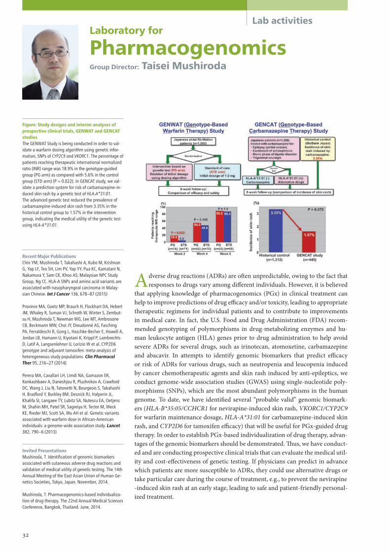
Lab activitiesLaboratory for
PharmacogenomicsGroup Director: Taisei Mushiroda
Adverse drug reactions (ADRs) are o�en unpredictable, owing to the fact that responses to drugs vary among di�erent individuals. However, it is believed
that applying knowledge of pharmacogenomics (PGx) in clinical treatment can help to improve predictions of drug e�cacy and/or toxicity, leading to appropriate therapeutic regimens for individual patients and to contribute to improvements in medical care. In fact, the U.S. Food and Drug Administration (FDA) recom-mended genotyping of polymorphisms in drug-metabolizing enzymes and hu-man leukocyte antigen (HLA) genes prior to drug administration to help avoid severe ADRs for several drugs, such as irinotecan, atomoxetine, carbamazepine and abacavir. In attempts to identify genomic biomarkers that predict e�cacy or risk of ADRs for various drugs, such as neutropenia and leucopenia induced by cancer chemotherapeutic agents and skin rash induced by anti-epileptics, we conduct genome-wide association studies (GWAS) using single-nucleotide poly-morphisms (SNPs), which are the most abundant polymorphisms in the human genome. To date, we have identi�ed several “probable valid” genomic biomark-ers (HLA-B*35:05/CCHCR1 for nevirapine-induced skin rash, VKORC1/CYP2C9 for warfarin maintenance dosage, HLA-A*31:01 for carbamazepine-induced skin rash, and CYP2D6 for tamoxifen e�cacy) that will be useful for PGx-guided drug therapy. In order to establish PGx-based individualization of drug therapy, advan-tages of the genomic biomarkers should be demonstrated. �us, we have conduct-ed and are conducting prospective clinical trials that can evaluate the medical util-ity and cost-e�ectiveness of genetic testing. If physicians can predict in advance which patients are more susceptible to ADRs, they could use alternative drugs or take particular care during the course of treatment, e.g., to prevent the nevirapine -induced skin rash at an early stage, leading to safe and patient-friendly personal-ized treatment.
Recent Major PublicationsChin YM, Mushiroda T, Takahashi A, Kubo M, Krishnan G, Yap LF, Teo SH, Lim PV, Yap YY, Pua KC, Kamatani N, Nakamura Y, Sam CK, Khoo AS; Malaysian NPC Study Group, Ng CC. HLA-A SNPs and amino acid variants are associated with nasopharyngeal carcinoma in Malay-sian Chinese. Int J Cancer 136, 678–87 (2015)
Province MA, Goetz MP, Brauch H, Flockhart DA, Hebert JM, Whaley R, Suman VJ, Schroth W, Winter S, Zembut-su H, Mushiroda T, Newman WG, Lee MT, Ambrosone CB, Beckmann MW, Choi JY, Dieudonné AS, Fasching PA, Ferraldeschi R, Gong L, Haschke-Becher E, Howell A, Jordan LB, Hamann U, Kiyotani K, Krippl P, Lambrechts D, Latif A, Langsenlehner U, Lorizio W et al. CYP2D6 genotype and adjuvant tamoxifen: meta-analysis of heterogeneous study populations. Clin Pharmacol Ther 95, 216–27 (2014)
Perera MA, Cavallari LH, Limdi NA, Gamazon ER, Konkashbaev A, Daneshjou R, Pluzhnikov A, Crawford DC, Wang J, Liu N, Tatonetti N, Bourgeois S, Takahashi H, Bradford Y, Burkley BM, Desnick RJ, Halperin JL, Khalifa SI, Langaee TY, Lubitz SA, Nutescu EA, Oetjens M, Shahin MH, Patel SR, Sagreiya H, Tector M, Weck KE, Rieder MJ, Scott SA, Wu AH et al. Genetic variants associated with warfarin dose in African-American individuals: a genome-wide association study. Lancet 382, 790–6 (2013)
Invited PresentationsMushiroda, T. Identification of genomic biomarkers associated with cutaneous adverse drug reactions and validation of medical utility of genetic testing. The 14th Annual Meeting of the East Asian Union of Human Ge-netics Societies, Tokyo, Japan. November, 2014.
Mushiroda, T. Pharmacogenomics-based individualiza-tion of drug therapy. The 22nd Annual Medical Sciences Conference, Bangkok, Thailand. June, 2014.
Figure: Study designs and interim analyses of prospective clinical trials, GENWAT and GENCAT studiesThe GENWAT Study is being conducted in order to val-idate a warfarin dosing algorithm using genetic infor-mation, SNPs of CYP2C9 and VKORC1. The percentage of patients reaching therapeutic international normalized ratio (INR) range was 18.9% in the genotype-guided group (PG arm) as compared with 5.6% in the control group (STD arm) (P = 0.022). In GENCAT study, we val-idate a prediction system for risk of carbamazepine-in-duced skin rash by a genetic test of HLA-A*31:01. The advanced genetic test reduced the prevalence of carbamazepine-induced skin rash from 3.35% in the historical control group to 1.57% in the intervention group, indicating the medical utility of the genetic test using HLA-A*31:01.
32

Lab activities
Recent Major PublicationsCaudle KE, Rettie AE, Whirl-Carrillo M, Smith LH, Mintzer S, Lee MT, Klein TE, Callaghan JT. Clinical pharmacogenetics implementation consortium guide-lines for CYP2C9 and HLA-B genotype and phenytoin dosing. Clin Pharmacol Ther 96, 542–8 (2014)
Song IW, Li WR, Chen LY, Shen LF, Liu KM, Yen JJ, Chen YJ, Chen YJ, Kraus VB, Wu JY, Lee MT*, Chen YT* (*: co-corresponding). Palmitoyl acyltransferase, Zdhhc13, facilitates bone mass acquisition by regulating postna-tal epiphyseal development and endochondral ossifica-tion: a mouse model. PLoS One 9, e92194 (2014)
Chen CH, Lee CS, Lee MT, Ouyang WC, Chen CC, Chong MY, Wu JY, Tan HK, Lee YC, Chuo LJ, Chiu NY, Tsang HY, Chang TJ, Lung FW, Chiu CH, Chang CH, Chen YS, Hou YM, Chen CC, Lai TJ, Tung CL, Chen CY, Lane HY, Su TP, Feng J, Lin JJ, Chang CJ, Teng PR, Liu CY, Chen CK, Liu IC, Chen JJ, Lu T, Fan CC, Wu CK, Li CF, Wang KH, Wu LS, Peng HL, Chang CP, Lu LS, Chen YT, Cheng AT. Variant GADL1 and Response to Lithium Therapy in Bipolar I Disorder. N Engl J Med 370, 119–28 (2014)
Invited PresentationsLee MT. International Clopidogrel Pharmacogenetics Consortium Genome Wide Association Study. 14th Joint Meeting of Coronary Revascularization, Busan, Korea. December, 2014.
Lee MT. Direct expression and methylation assessment of articular cartilage and subchondral bone on human osteoarthritic knee. The 59th Annual Meeting of the Japan Society of Human Genetics, Tokyo, Japan. No-vember, 2014.
Lee MT. Pharmacogenetic study of drug induced severe cutaneous disorders. Genomics of Rare Diseases Ser-bordisinn & 2014 Golden Helix Symposium, Belgrade, Serbia. October, 2014.
Lee MT. Potential application of pharmacogenomics in psychiatric diseases. 3rd Meeting of South East Asian Pharmacogenomics Research Network, Jakarta, Indone-sia. April, 2014.
Lee MT. Moving Towards Translation: Using Pharma-cogenetics as an example for personalized medicine. The Fourth Military Medical University, Shaanxi, China. February, 2014.
Figure: Using Immune Diversity to Elucidate the Underlying Mechanism of CBZ-induced SJS/TEN in the Japanese Population 1. HLA allele diversity in the population: Carriers with the HLA-A*3101 allele have a much higher risk to develop CBZ-
SJS/TEN. 2. Peptide diversity: Drug modification causes a shifted repertoire of new peptides that become “non-self ” antigens. 3. TCR diversity: Drug-specific TCRs may have a different TCR repertoire. This can explain why not all the HLA-A*3101
carriers develop CBZ-SJS/TEN.Granulysin is an effector molecule used by CD8 T cells to kill target cells.
The main aim of our laboratory is to identify genetic associations with diseas-es and with drug induced adverse events or drug e�cacy. It is hoped that the
discoveries from our research will identify useful biomarkers that could be used to predict drug-induced adverse events, guide drug use and could also be used in disease prediction/diagnosis. Another main focus of this laboratory is to promote collaborations both within Japan and internationally. We have set up collabora-tions/consortia in Asia (Southeast Asian Pharmacogenetics Consortium, SEA-Pharm) and Europe (Genomic Medicine Alliance) to foster collaboration with RIKEN and among the participating research groups. We also actively recruit young scientists from overseas to work at RIKEN and carry out research in SNP-based approaches, statistical analysis and biological analysis. Our goal is to estab-lish an international genetic research network that will make the Center for Inte-grative Medical Sciences a world leader in personalized medicine. Currently, our group’s main focus is on Pharmacogenetics (PGx) and the genetic study of com-plex diseases. For PGx studies, we aim to establish a functional analysis platform that not only allows us to study the interactions between HLA and drugs, but also can be used to con�rm the �ndings from genetic analysis for severe ADRs, many of which involve the HLA molecules. In addition, we will use genome-wide associ-ation study to identify genetic variants associated with 1. Anti-infectious drug-in-duced liver injuries. 2. Phenytoin and co-trimoxazole induced Stevens-Johnson Syndrome (SJS) and Toxic epidermal necrolysis. 3. Adverse reactions to non-ste-roidal anti-in�ammatory drugs. For complex diseases, we aim to identify genetic variants associated with Hippocampal Sclerosis in �ais and are performing an epigenetic and biomarker study of osteoarthritis (OA).
Laboratory for
International Alliance on Genomic ResearchTeam Leader: Ming Ta Michael Lee
33

Lab activitiesLaboratory for
Cardiovascular DiseasesGroup Director: Toshihiro Tanaka
Since cardiovascular diseases cause more than 15% of the deaths in the Japa-nese population and represent more than 20% of the total medical expenses
in Japan, it is socially important to discover the mechanisms of these disorders. We have been among the �rst to reveal genetic background e�ects in myocardi-al infarction (MI), atrial �brillation (AF), Kawasaki disease (KD), and peripheral artery disease (PAD) by comprehensive genetic analyses of the Japanese popula-tion followed by functional in vitro analyses. Our ultimate goal is to provide novel diagnostic/therapeutic approaches to such patients. To this end, we are extending our research area from genetics to molecular biology, including in vivo analyses of genetically engineered mice. Also, we are making e�orts to develop a new drug for MI that would elute from a stent, a powerful tool for coronary angioplasty.
Recent Major PublicationsSinner MF, Tucker NR, Lunetta KL, Ozaki K, Smith JG, Trompet S, Bis JC, Lin H, Chung MK, Nielsen JB, Lubitz SA, Krijthe BP, Magnani JW, Ye J, Gollob MH, Tsunoda T, Müller-Nurasyid M, Lichtner P, Peters A, Dolmatova E, Kubo M, Smith JD, Psaty BM, Smith NL, Jukema JW, Chasman DI, Albert CM, Ebana Y, Furukawa T, MacFarlane P, Harris TB, Darbar D, Dörr M, Holst AG, Svendsen JH, Hofman A, Uitterlinden A, Gudnason V, Isobe M, Malik R, Dichgans M, Rosand J, Van Wagoner DR; METASTROKE Consortium; AFGen Consortium, Ben-jamin EJ, Milan DJ, Melander O, Heckbert S, Ford I, Liu Y, Barnard J, Olesen MS, Stricker BH, Tanaka T, Kääb S, Ellinor PT. Integrating genetic, transcriptional, and func-tional analyses to identify five novel genes for atrial fibrillation. Circulation 130, 1225–35 (2014)
Lubitz SA, Lunetta KL, Lin H, Arking DE, Trompet S, Li G, Krijthe BP, Chasman DI, Barnard J, Kleber ME, Dörr M, Ozaki K, Smith AV, Müller M, Walter S, Agarwal SK, Bis JC, Brody JA, Chen LY, Everett BM, Ford I, Franco OH, Harris TB, Hofman A, Kääb S, Mahida S, Kathiresan S, Kubo M, Launer LJ, MacFarlane PW, Magnani JW, McKnight B, McManus DD, Peters A, Psaty BM, Rose LM, Rotter JI, Silbernagel G, Smith JD, Sotoodehnia N, Stott DJ, Taylor K, Tomaschitz A, Tsunoda T, Uitterlinden AG, Van Wagoner DR, Völker U, Völzke H, Murabito JM, Sinner MF, Gudnason V, Felix SB, März W, Chung M, Albert CM, Stricker BH, Tanaka T, Heckbert SR, Jukema JW, Alonso A, Benjamin EJ, Ellinor PT. Novel genetic markers associate with atrial fibrillation risk in Euro-peans and Japanese. J Am Coll Cardiol 63, 1200–10 (2014)
Invited PresentationsTanaka T. Genome and medicine. Forum on Cardiovas-cular Diseases, Osaka, Japan. June, 2014.
Onouchi Y. Genome wide association studies and Ka-wasaki disease. Pediatric Academic Societies and Asian Society for Pediatric Research Joint Meeting, Vancouver, Canada. May, 2014.
Tanaka T. Genetic epidemiology. 11th CEM Forum, Kyo-to, Japan. April, 2014.
Onouchi Y. Pathophysiological basis for genetic re-search in Kawasaki disease. American College of Cardi-ology 63rd Annual Scientific Session, Washington DC, USA. March, 2014.
Table1: Meta-analyses of SNP associations with AF by origin of the study population
34

Lab activities
M ost autoimmune diseases, such as rheumatoid arthritis (RA) and systemic lupus erythematosus, are multifactorial, involving both genetic and environmental fac-
tors. �e aim of our laboratory is to elucidate the etiology of these autoimmune diseases by identifying their genetic aspects. RA is one of the most common autoimmune diseases with an in�ammatory arthritis component. While the HLA-DRB1 gene polymorphism is the major determinant of RA susceptibility, several groups worldwide including our own have performed genome-wide association studies (GWAS) to �nd non-HLA risk loci. However, each individual GWAS lacked adequate statistical power and thus a sub-stantial proportion of risk loci remained undiscovered. In world-wide collaborations, we performed multi-ethnic meta-analyses of GWAS and have found more than 100 risk loci for RA so far.
As GWAS could only indicate the presence of disease-associated variants in the loci, we further investigated these candidate loci to seek disease-causal variants and elucidate their biological relevance to RA. Previous expression quantitative trait loci (eQTL) stud-ies that examined association between genetic variants and gene expression levels have suggested that the majority of autoimmune loci are eQTLs, where disease causing vari-ants a�ect expression of the responsible genes. �erefore, we believe it is essential to fo-cus on “gene expression” as an intermediate trait to dissect the etiology of “disease onset”. We are currently undertaking genome and transcriptome analysis of each immune cell type from Japanese individuals to establish eQTL catalogues of human immune cells. In addition, as the precise function of the responsible genes in vivo has been not yet clari�ed for most of the candidate loci, we are investigating the function of these genes includ-ing PADI4, CD244 FCRL3, and RTKN2 by using gene-targeting mice and disease-model mice. Furthermore, to facilitate the development of new therapies, we are performing a high-throughput screening of novel therapeutic compounds targeting RA risk genes in a collaboration with the RIKEN Program for Drug Discovery and Medical Technology Platforms.
Laboratory for
Autoimmune DiseasesTeam Leader: Kazuhiko Yamamoto
Figure: Understanding autoimmune diseases through eQTL studies We focus on the expression of genes as an intermedi-ate trait to understand the mechanisms of autoimmune diseases. We are performing eQTL studies for each immune cell type by using next generation sequencing technologies such as RNA-seq. By combining the data from GWAS and eQTL studies, we will unravel the mechanisms of disease.
Recent Major PublicationsYamamoto K, Okada Y, Suzuki A, Kochi Y. Genetics of rheumatoid arthritis in Asia—present and future. Nat Rev Rheumatol, in press
Kochi Y, Suzuki A, Yamamoto K. Genetic basis of rheu-matoid arthritis: a current review. Biochem Biophys Res Commun 452, 254–62 (2014)
Okada Y, Wu D, Trynka G, Raj T, Terao C, Ikari K, Kochi Y, Ohmura K, Suzuki A, Yoshida S, Graham RR, Mano-haran A, Ortmann W, Bhangale T, Denny JC, Carroll RJ, Eyler AE, Greenberg JD, Kremer JM, Pappas DA, Jiang L, Yin J, Ye L, Su DF, Yang J, Xie G, Keystone E, Westra HJ, Esko T, Metspalu A et al. Genetics of rheumatoid arthri-tis contributes to biology and drug discovery. Nature 506, 376–81 (2014)
Invited PresentationsYamamoto K. Current and future genetics of rheuma-toid arthritis in Asia. The 59th Annual Meeting of the Japan Society of Human Genetics, Tokyo, Japan. Tokyo, Japan. November, 2014.
Yamamoto K. A brief history of GWAS and next gen-eration genetics of RA. Rheumatoid arthritis genetics meeting, Boston, U.S.A. November, 2014.
Yamamoto K. Genetics and epigenetics in autoimmune diseases. 6th International Forum on Rheumatoid Ar-thritis, Beijing, China. September, 2014.
Yamamoto K. TGF-β3-producing CD4+CD25−LAG3+ regulatory T cells control B cell responses. Cold Spring Habor Asia Conferences “Frontiers of Immunology in Health and Diseases,” Suzhou, China. September, 2014.
Yamamoto K. T cell targeted therapies in autoimmune diseases. Advanced Target Therapies, Athens, Greece. March, 2014.
35

Lab activitiesLaboratory for
Digestive DiseasesTeam Leader: Kazuaki Chayama
Our laboratory has mainly used GWAS to investigate the host genetic factors under-lying various diseases and responsiveness to therapy, such as chronic HBV (Nat
Genet, 2009; Hum Mol Genet, 2011) and HCV infection (PLoS One, 2013), HCV-induced liver cirrhosis (J Hepatol, 2013) and cancer (Nat Genet, 2011), response to interferon therapy (J Gen Virol, 2011), and ribavirin-induced anemia (Gastroenterology, 2010). We have also participated in whole-genome sequencing analysis of liver cancers (Nat Genet, 2012).
We have now intensively investigated and veri�ed how to apply this genetic infor-mation to clinical practice (Hepatology, 2014; Antivir �er, 2014; Hepatol Res, 2014; J Gastroenterol, in press). We estimated haplotype structure of the IFNL4/IL-28B locus, consisting of 3 SNPs, and found that 2 of them may have better predictive impact on re-sponse to PEG-interferon/ribavirin therapy (J Gen Virol, 2014). We found ribavirin dose reduction during telaprevir/ribavirin/PEG-interferon triple therapy overcomes the e�ect of the ITPA gene polymorphism on ribavirin-induced anemia (J Viral Hepat, in press).
We have studied not only the human but also the hepatitis virus genome to predict and improve treatment response using in vitro and in vivo infection models (Gut, 2013; Antimicrob Agents Chemother, 2014). We studied drug resistance by using HCV-infect-ed chimeric mice treated with direct-acting antiviral agents (DAAs) and concluded that sequential use of DAAs should be avoided to prevent emergence of multidrug-resistant strains (Am J Gastroenterol, 2013).
In addition, we are currently investigating the host immune response to viral infec-tion to elucidate the mechanism of hepatitis (Hepatology, 2011&2012; Biochem Biophys Res Commun, 2013), since our GWAS suggested the importance of the immune system for the etiology of hepatitis B and C.
Our ultimate goals are (1) establishment of personalized medicine for various liver diseases and (2) contribution to development of new therapies, diagnostic methods, and prophylactic approaches to various liver diseases.
Recent Major PublicationsOchi H, Miki D, Hayes CN, Abe H, Hayashida Y, Kubo M, Chayama K. IFNL4/IL-28B haplotype structure and its impact on susceptibility to hepatitis C virus and treat-ment response in the Japanese population. J Gen Virol 95, 1297–1306 (2014)
Miki D, Ochi H, Takahashi A, Hayes CN, Urabe Y, Abe H, Kawaoka T, Tsuge M, Hiraga N, Imamura M, Kawakami Y, Aikata H, Takahashi S, Akuta N, Suzuki F, Ikeda K, Kumada H, Karino Y, Toyota J, Tsunoda T, Kubo M, Kamatani N, Nakamura Y, Chayama K. HLA-DQB1*03 confers susceptibility to chronic hepatitis C in Japanese: a genome-wide association study. PLoS One 8, e84226 (2013).
Abe Y, Aly HH, Hiraga N, Imamura M, Wakita T, Shi-motohno K, Chayama K, Hijikata M. Thromboxane A2 synthase inhibitors prevent production of infectious hepatitis C virus in mice with humanized livers. Gastro-enterology 145, 658–67.e11 (2013).
Figure: Haemoglobin levels by ITPA rs1127354 genotype during 24 weeks of telaprevir/riba-virin/peg-interferon therapy and 24 weeks of follow-upDecline of haemoglobin was significantly faster, and ribavirin was more extensively reduced in patients with ITPA SNP rs1127354 genotype CC than CA/AA. Post marketing-phase triple therapy resulted in a high sus-tained viral response rate in spite of extensive ribavirin dose reduction in a diverse patient population, indi-cating the importance of treatment continuation and appropriate management of adverse events.
36

Lab activities
Recent Major PublicationsNakajima M, Takahashi A, Tsuji T, Karasugi T, Baba H, Uchida K, Kawabata S, Okawa A, Shindo S, Takeuchi K, Taniguchi Y, Maeda S, Kashii M, Seichi A, Nakajima H, Kawaguchi Y, Fujibayashi S, Takahata M, Tanaka T, Watanabe K, Kida K, Kanchiku T, Ito Z, Mori K, Kaito T, Kobayashi S, Yamada K, Takahashi M, Chiba K, Mat-sumoto M, Furukawa KI, Kubo M, Toyama Y; Genetic Study Group of Investigation Committee on Ossification of the Spinal Ligaments, Ikegawa S. A genome-wide association study identifies susceptibility loci for ossi-fication of the posterior longitudinal ligament of the spine. Nat Genet 46, 1012–6 (2014)
Yamashita A, Morioka M, Kishi H, Kimura T, Yahara Y, Okada M, Fujita K, Sawai H, Ikegawa S, Tsumaki N. Statin treatment rescues FGFR3 skeletal dysplasia phe-notypes. Nature 513, 507–11 (2014)
Tsurusaki Y, Koshimizu E, Ohashi H, Phadke S, Kou I, Shiina M, Suzuki T, Okamoto N, Imamura S, Yamashita M, Watanabe S, Yoshiura K, Kodera H, Miyatake S, Nakashima M, Saitsu H, Ogata K, Ikegawa S, Miyake N, Matsumoto N. De novo SOX11 mutations cause Cof-fin-Siris syndrome. Nat Commun 5, 4011 (2014)
Invited PresentationsIkegawa S. Genomic study of common diseases. 1st Chinese Osteoarthritis Annual Congress, Nanjing, Chi-na. October, 2014.
Ikegawa S. Genomic study of skeletal diseases. 2nd Asia-Pacific Bone & Mineral Research Meeting, Seoul, Korea. May, 2014.
Ikegawa S. Genomic studies of bone and joint diseases: past, present and future. 1st Karolinska Workshop on Skeletal Dysplasia, Stockholm, Sweden. March, 2014.
Ikegawa S. Translational genomics in bone diseases. In-vited lecture, The Master Program for Clinical Pharma-cogenomics and Pharmacoproteomics, Taipei Medical University, Taipei, Taiwan. January, 2014.
Ikegawa S. Genomic study of common diseases: Road from genome to personalized medicine. Invited lecture, Taipei Medical University Hospital, Taipei, Taiwan. January, 2014.
Figure: Genome-wide association study (GWAS) identified susceptibility loci for ossification of the posterior longitudinal ligament of the spine Manhattan plot showing the –log10 P value from the GWAS. The values were plotted against their respective positions on the autosomal chromosomes. The red line represents the genome-wide significance threshold (P = 5 × 10⁻⁸). The blue line represents the threshold (P = 5 × 10⁻⁷) for selecting SNPs for the replication study.
Bone and joint diseases are serious problems throughout the world. We are working on identi�cation of new susceptibility genes for many common bone
and joint diseases, including osteoarthritis, osteoporosis, adolescent idiopathic scoliosis, lumbar disc herniation and ossi�cation of the posterior longitudinal lig-ament of the spine (OPLL). �is year, we succeeded in identi�cation of new sus-ceptibility genes for OPLL.
OPLL is one of the most common spinal disorders among the elderly and causes myelopathy and radiculopathy due to compression of the spinal cord and nerves by ectopic ossi�cation of the ligament. More than 1–2% of Japanese are su�ering from this intractable disease. To identify susceptibility genes for OPLL, we performed a genome-wide association study (GWAS), the �rst in the world for this disease. �rough the GWAS in ~8,000 Japanese individuals followed by a replication study using an additional ~7,000 Japanese individuals, we identi�ed six susceptibility loci for OPLL with genome-wide signi�cance of the associa-tion: 20p12.3 (rs2423294: P = 1.10 × 10⁻¹³), 8q23.1 (rs374810: P = 1.88 × 10⁻¹³), 12p11.22 (rs1979679: P = 4.34 × 10⁻¹²), 12p12.2 (rs11045000: P = 2.95 × 10⁻¹¹), 8q23.3 (rs13279799: P = 1.28 × 10⁻¹⁰) and 6p21.1 (rs927485: P = 9.40 × 10⁻⁹).
Analyses of gene expression in and around the loci using in silico and in vitro approaches suggested that several genes in the loci are involved in OPLL etiolo-gy through membranous and/or endochondral ossi�cation processes. Our results bring new insight into the etiology of OPLL as well as mechanisms of ectopic os-si�cation.
Laboratory for
Bone and Joint DiseasesTeam Leader: Shiro Ikegawa
37

Recent Major PublicationsKurashige M, Hanaoka K, Imamura M, Udagawa T, Kawaguchi Y, Hasegawa T, Hosoya T, Yokoo T, Maeda S. A comprehensive search for mutations in the PKD1 and PKD2 in Japanese subjects with autosomal dominant polycystic kidney disease. Clin Genet 87, 266–72 (2015)
DIAbetes Genetics Replication And Meta-analysis (DI-AGRAM) Consortium; Asian Genetic Epidemiology Net-work Type 2 Diabetes (AGEN-T2D) Consortium; South Asian Type 2 Diabetes (SAT2D) Consortium; Mexican American Type 2 Diabetes (MAT2D) Consortium; Type 2 Diabetes Genetic Exploration by Next-generation sequencing in multi-Ethnic Samples (T2D-GENES) Con-sortium. Genome-wide trans-ancestry meta-analysis provides insight into the genetic architecture of type 2 diabetes susceptibility. Nat Genet 46, 234–44 (2014)
Hara K, Fujita H, Johnson TA, Yamauchi T, Yasuda K, Horikoshi M, Peng C, Hu C, Ma RC, Imamura M, Iwata M, Tsunoda T, Morizono T, Shojima N, So WY, Leung TF, Kwan P, Zhang R, Wang J, Yu W, Maegawa H, Hirose H; DIAGRAM consortium, Kaku K, Ito C, Watada H, Tanaka Y, Tobe K, Kashiwagi A, Kawamori R, Jia W, Chan JC, Teo YY, Shyong TE, Kamatani N, Kubo M, Maeda S, Kadowaki T. Genome-Wide Association Study Identifies Three Novel Loci for Type 2 Diabetes. Hum Mol Genet 23, 239–46 (2014)
Figure: A meta-analysis of Japanese GWAS iden-tified 7 new loci for type 2 diabetes
Lab activitiesLaboratory for
Endocrinology, Metabolismand Kidney DiseasesTeam Leader: Shiro Maeda
Research activities for type 2 diabetes:We expanded our Japanese genome-wide association study (GWAS) for type 2 diabetes by increasing the number of examined SNPs (6,209,637 SNPs) and the number of par-ticipants (26,805 Japanese individuals, 5,976 patients with type 2 diabetes and 20,829 controls). A�er combining the results of candidate loci in the discovery stage and those in a follow-up case-control study (total number of participants, 30,392 type 2 diabetes cases and 34,814 controls) we identi�ed three new loci for type 2 diabetes in the Japanese, MIR129-LEP, GPSM1 and SLC16A13 (Hara K et al. Hum Mol Genet, 2014). �e associ-ations of these 3 loci with type 2 diabetes were not replicated in European populations, but the SLC16A13 locus was shown to be associated with type 2 diabetes in a Mexican population (Williams AL et al. Nature, 2014).
We further performed an independent Japanese GWAS for type 2 diabetes, in which ~7.5 million SNPs data obtained from genotype imputation performed by mini-MACH using directly genotyped data and reference data in the 1000 genomes (JPT+CHB+CHS, n = 275) were analyzed for 9,817 cases and 6,763 controls. �en these results were com-bined with GWAS data in the already published Japanese population described above. Ob-tained candidate SNP loci were further evaluated in an independent Japanese case-con-trol study and, a�er integration of all results, we have identi�ed 7 novel loci for type 2 diabetes in the Japanese (Fig.).
Simultaneously, in an international collaborative e�ort to identify common suscepti-bility loci for type 2 diabetes, we have been participating in a Trans-ethnic type 2 diabetes meta-analysis consortium; this analysis has identi�ed 7 additional susceptibility loci for type 2 diabetes.
Other research activities:We have been performing GWAS for diabetic nephropathy and diabetic retinopathy. We have also reported results of a comprehensive search for causal mutations in PKD1 and PKD2 in Japanese subjects with autosomal dominant polycystic kidney disease.
38

Recent Major PublicationsPark HW, Dahlin A, Tse S, Duan QL, Schuemann B, Martinez FD, Peters SP, Szefler SJ, Lima JJ, Kubo M, Tamari M, Tantisira KG. Genetic predictors associated with improvement of asthma symptoms in response to inhaled corticosteroids. J Allergy Clin Immunol 133, 664–9 (2014)
Ellinghaus D, Baurecht H, Esparza-Gordillo J, Rodríguez E, Matanovic A, Marenholz I, Hübner N, Schaarschmidt H, Novak N, Michel S, Maintz L, Werfel T, Meyer-Hof-fert U, Hotze M, Prokisch H, Heim K, Herder C, Hirota T, Tamari M, Kubo M, Takahashi A, Nakamura Y, Tsoi LC, Stuart P, Elder JT, Sun L, Zuo X, Yang S, Zhang X, Hoffmann P, Nöthen MM, Fölster-Holst R, Winkelmann J, Illig T, Boehm BO, Duerr RH, Büning C, Brand S, Glas J, McAleer MA, Fahy CM, Kabesch M, Brown S, McLean WH, Irvine AD, Schreiber S, Lee YA, Franke A, Weidinger S. High-density genotyping study identifies four new susceptibility loci for atopic dermatitis. Nat Genet 45, 808–12 (2013)
Tomita K, Sakashita M, Hirota T, Tanaka S, Masuyama K, Yamada T, Fujieda S, Miyatake A, Hizawa N, Kubo M, Nakamura Y, Tamari M. Variants in the 17q21 asthma susceptibility locus are associated with allergic rhinitis in the Japanese population. Allergy 68, 92–100 (2013)
Invited PresentationsTamari M. Genetic mechanisms of allergic diseases. The 3rd One Airway One disease in Osaka, Osaka, Japan. November, 2014.¿
Tamari M. Genome-wide association studies of allergic diseases. The 24th Congress of Interasma Japan/ North Asia, Nagoya. Japan. July, 2014.
Tamari M. Mechanisms of allergic diseases –cross talk between genes and environment-. The 31th Annual Meeting of the Japanese Society of Pediatric Intractable Asthma and Allergic Diseases, Nagoya, Japan. June, 2014.
Tamari M. Genome-wide association study of atopic dermatitis. The 79th Annual Meeting of the Japanese Society of Interferon and Cytokine Research, Sapporo, Japan. June, 2014.
Hirota T. Genetic studies for allergy susceptibility genes. The 26th Spring Meeting of Japanese Society of Aller-gology, Kyoto, Japan. May. 2014.
Figure: Manhattan plot of the Immunochip anal-ysis for atopic dermatitis We conducted an Immunochip analysis and validation studies. A total of four new atopic dermatitis suscep-tibility loci were identified with genome-wide signifi-cance: IL2-IL21, PRR5L, CLEC16A and ZNF652.
The aim of our project is to improve our understanding of the pathophysiolo-gy of human respiratory and allergic diseases, which are caused by a combi-
nation of genetic and environmental factors.We participate in the Global Alliance for Pharmacogenomics. We have con-
ducted genome-wide association studies (GWASs) and identi�ed a locus that is associated with improvement of asthma symptoms in response to inhaled corti-costeroids. �e related SNP lies in the intronic region of the FBXL7 gene and is associated with decreased expression of this gene in immortalized B cells derived from patients with asthma (J Allergy Clin Immunol, 2014).
A number of GWASs have been conducted for allergic diseases and several susceptibility loci have been identi�ed. We reported an immunochip analysis for atopic dermatitis, which revealed four new susceptibility loci, 4q27 (IL-2-IL21), 11p13 (PRR5L), 16p13.13 (CLEC16A-DEXI) and 17q21.32 (ZNF652) (Nat Gen-et, 2013). Candidate genes identi�ed by the previous GWASs and immunochip analysis suggest roles for barrier functions, innate-adaptive immunity, IL-1 family signaling, regulatory T cells and the vitamin D pathway in the pathogenesis of al-lergic diseases. To investigate whether polymorphisms identi�ed by these genetic studies could a�ect the susceptibility to and clinical phenotypes of diseases, we will conduct functional analyses and serological studies.
�e �rst GWAS of asthma in the European population identi�ed a locus on chromosome 17q21.1. Subsequent replication studies have identi�ed genetic var-iants on the 17q21.1 locus associated with asthma in di�erent ethnic populations. We reported that genetic variants in the 17q21.1 locus were associated with aller-gic rhinitis and were strongly correlated in cis with transcript levels of ORMDL3 in Epstein-Barr virus (EBV)-transformed lymphoblastoid cell lines in a Japanese population (Allergy, 2013).
We will conduct further cross-disciplinary studies combining genetics, im-munology and clinical epidemiology for translation of our research into clinical practice.
Laboratory for
Respiratory and Allergic DiseasesTeam Leader: Mayumi Tamari
Lab activities
39

Six original projects for clinical applications have been performed in 2014: 1) A biochemical drug using a PEGylated Cryj-1/2 fusion recombinant
protein for Cedar Pollinosis has been developed by the Torii pharmaceuti-cal company and IMS and introduced to the Japanese market. 2) A chemi-cal compound recently developed using the α-galactosylceramide (α-GalCer) analog, RCAI-X, selectively induces apoptosis of IgE but not IgG B cells and preferentially suppresses IgE production. It therefore potentially could be ap-plied to any type of allergic disorder, such as pollinosis, food allergy, as well as allergic asthma. 3) NKT cell-targeted therapy for head and neck tumors has been done in collaboration with Chiba University, and was authorized by the Japanese government as Advanced Medical Treatment B in 2013. 4) The arti-ficial adjuvant vector cell as an anti-tumor vaccine project has been accepted by the translational research network program and developed. This vaccine can be dosed with tumor antigen mRNA together with α-GalCer, so that it activates both innate and acquired protective immunity and also induces long-term memory. 5) The human iPS project for clinical use of in vitro gener-ated NKT cells has been accepted as a Center for Clinical Application Research (Type B) in the Research Center Network for Realization of Regenerative Med-icine, Japan in 2013. 6) A humanized mouse model for MLL gene-rearranged leukemia was established. Also, certain genes were identified that are di�er-entially expressed between normal stem cells and leukemic stem cells.
Program forMedical Innovations
Figure: Differences in gene expression between peripheral blood and iPSC-derived human NKT cells. Human NKT cells were generated from iPSC in OP9/dll-1 cultures. The iPS-derived and peripheral blood NKT cells were cultured for the indicated times with or with-out dendritic cells loaded with α-GalCer (DC/G). RNA sequence analysis was performed and the differences between expressed genes are depicted.
40

Recent Major PublicationsTarumoto N, Kinjo Y, Kitano N, Sasai D, Ueno K, Oka-wara A, Izawa Y, Shinozaki M, Watarai H, Taniguchi M, Takeyama H, Maesaki S, Shibuya K, Miyazaki Y. Exacer-bation of invasive Candida albicans infection by com-mensal bacteria or a glycolipid through IFN-γ produced in part by iNKT cells. J Infect Dis 209, 799–810 (2014)
Shimizu K, Sato Y, Shinga J, Watanabe T, Endo T, Asaku-ra M, Yamasaki S, Kawahara K, Kinjo Y, Kitamura H, Watarai H, Ishii Y, Tsuji M, Taniguchi M, Ohara O, Fujii S. KLRG+ invariant natural killer T cells are long-lived ef-fectors. Proc Natl Acad Sci U S A 111, 12474–9 (2014)
Ren Y, Dashtsoodol N, Watarai H, Koseki H, Quan C, Taniguchi M. Generation of induced pluripotent stem cell-derived mice by reprogramming of a mature NKT cell. Int Immunol 26, 551–61 (2014)
Invited PresentationsTaniguchi M. Development of a drug suppressing total IgE. The 51st Annual Meeting of Japanese Society of Pediatric Allergy and Clinical Immunology, Yokkaichi, Japan. November, 2014.
Taniguchi M. Discovery of NKT cells and their clinical application. 16th JSI Immunology Summer School , Sho-doshima Island, Japan. July, 2014.
Taniguchi M. Novel anti-cancer immunotherapy tar-geted on NKT cells. The 10th Hiroshima Liver Project Research Center Symposium, Hiroshima, Japan. July, 2014.
Taniguchi M. NKT cell-mediated adjuvant cell therapy on lung cancer and head and neck cancer. Federation of Clinical Immunology Societies 2014, Chicago, USA. June, 2014.
Taniguchi M. NKT cells as an ideal anti-tumor immuno-therapeutic. The 54th Annual Meeting of the Japanese Respiratory Society, Osaka, Japan. April, 2014.
Figure: Phase II Clinical Trial of NKT cell–targeted therapy for head and neck tumors The clinical study was carried out in collaboration with Prof. Okamoto, Chiba University Hospital. All 10 cases treated with the combination of α-GalCer/DCs and activated NKT cells showed dramatic clinical efficacy (Stable disease (SD) or partial remission (PR). Also note the significant correlation between clinical efficacy (PR in red, SD in black) and the number of NKT cells infil-trating the tumor in situ.
Laboratory for
Immune RegulationGroup Director: Masaru Taniguchi
Lab activities
Identification of novel NKT cell development pathway: We identi�ed an NKT cell subset developed at the thymic DN stage before selection by CD1d. �e DN NKT cells seem di�erent from NKT cells developing at the DP stage as judged by the following evidence: 1) RORγt, which is essential for generation and proliferation of NKT cells at the DP stage, is not necessary for DN NKT cell development, 2) both in-frame and out-of-frame Vα14Jα18 sequences are detected in DN fractions. Our �ndings reveal a novel NKT cell developmental pathway that is di�erent from the DP pathway.
Cell intrinsic requirement for the transcription factor Maf in the develop-ment of the IL-17 producing NKT cell subset: To clarify the role of Maf, a master regulator of IL-4 expression, in NKT cells, we used Maf-de�cient mice created by irradi-ation mixed fetal liver chimera. Maf was indispensable for the development of NKT17 cells in a cell intrinsic manner, but not for �1 and �2 type NKT cells. �erefore, our �ndings reveal a novel role for Maf in NKT17 cell di�erentiation.
Clinical trials of NKT-targeted therapy for lung cancer after surgery and for head and neck tumors: We are currently collaborating with the National Hospital Organization to carry out a randomized Phase II clinical trial on stage IIA-IIIA lung cancer a�er surgical tumor resection, which was approved as the advanced medical care assessment system B in 2014.
iPS-derived NKT cells: �e project on the development of human iPS from NKT cells and iPS-derived NKT cells has been accepted as the Center for Clinical Application Research (Type B) in the Research Center Network for Realization of Regenerative Medicine, Japan (headed by Dr. Koseki). We successfully developed human iPS-derived NKT cells with desired functions and established the method for functional NKT cells suitable for anti-cancer therapy.
41

Lab activitiesLaboratory for
ImmunotherapyTeam Leader: Shin-ichiro Fujii
The aims of the laboratory are to extend our basic studies for advancing immu-notherapy and translational research (TR), from basic studies back and forth
to the bedside in the �eld of cancer. To accomplish this, we have been conducting three NKT cell-related TR projects using the synthetic glycolipid, α-galactosyl-ceramide (α-GalCer). When NKT cells are activated in this manner, they acquire unique immunostimulatory features that include the rapid production of IFN-γ and NK cell activation, followed by innate immunity-mediated antitumor e�ects. Although NKT cells belong to the innate immune system, we recently discovered a new type of NKT cell, the memory like KLRG1+NKT cell, which can survive for a long period a�er activation (Fig.).
As a �rst TR project, we have attempted to establish a strategy linking innate and adaptive immunity. For this purpose, we have been studying the process of activation of adaptive immunity through full maturation of DCs that occurs soon a�er the activation of NKT cells in vivo. In addition, we are investigating novel delivery systems that have the potential to enhance antitumor immunity. We have developed arti�cial adjuvant vector cells as a new type of drug delivery system composed of NKT ligand and tumor associated antigen, linking innate and adap-tive immunity. We are also con�rming the induction of memory NKT cells us-ing this new type of vaccine. Under the RIKEN translational program and Tokyo University translational research program, we are making e�orts to work toward preclinical studies that will ultimately lead to clinical trials. Second, we have been working in a collaborative study with the RIKEN iPS-group to establish iPS-NKT cells. In this project, we focus on the preparation of primary NKT cells as the starting material for generating NKT cell-derived iPS cells and on the analysis of the function of iPS-derived NKT cells. �ird, we have been collaborating a joint clinical phase I / IIa study of NKT cell therapy with the National Hospital Organi-zation (NHO) for early stage of lung cancer patients a�er surgical tumor resection. In this study, we play an important role in the analyses of immune responses.
Recent Major PublicationsShimizu K, Sato Y, Shinga J, Watanabe T, Endo T, Asaku-ra M, Yamasaki S, Kawahara K, Kinjo Y, Kitamura H, Watarai H, Ishii Y, Tsuji M, Taniguchi M, Ohara O, Fujii S. KLRG+ invariant natural killer T cells are long-lived ef-fectors. Proc Natl Acad Sci U S A 111, 12474–9 (2014)
Shimizu K, Mizuno T, Shinga J, Asakura M, Kakimi K, Ishii Y, Masuda K, Maeda T, Sugahara H, Sato Y, Matsushita H, Nishida K, Hanada K, Dorrie J, Schaft N, Bickham K, Koike H, Ando T, Nagai R, Fujii S. Vaccina-tion with antigen-transfected, NKT cell ligand-loaded, human cells elicits robust in situ immune responses by dendritic cells. Cancer Res 73, 62–73 (2013)
Shimizu K, Asakura M, Shinga J, Sato Y, Kitahara S, Hoshino K, Kaisho T, Schoenberger SP, Ezaki T, Fujii S. Invariant NKT cells induce plasmacytoid DC cross-talk with conventional DCs for efficient memory CD8+ T cell induction. J Immunol 190, 5609–19 (2013)
Invited PresentationsFujii S. Development of a New Type of Cancer Vaccine “artificial Adjuvant Vector Cells (aAVC)”. Seminar in Dep. Cartilage & Bone Regeneration (Fuji-Soft), Gradu-ate School of Medicine, The University of Tokyo, Tokyo, Japan. December, 2014.
Fujii S. Immunological memory and antitumor immu-nity elicited by artificial adjuvant vector cells for cancer immunotherapy. The 43rd Annual Meeting of the Japanese Society for Immunology (JSI), Kyoto, Japan. December, 2014.
Fujii S. “Development of cancer vaccine triggered by in-nate immunity”. Seminar in Chiba University Graduate School of Medical and Pharmaceutical Sciences, Chiba, Japan. August 2014.
Fujii S. Development of a New Type of Cancer Vaccine Linking Innate and Adaptive immunity. Seminar in Jichi Medical University Graduate School of Medicine, Tochi-gi, Japan. July, 2014.
Fujii S. Development of a Novel Type of Cancer Vaccine Linking Innate and Adaptive immunity. The 22nd Inter-national Symposium on Molecular Cell Biology of Mac-rophages (MMCB2014), Kobe, Japan. June, 2014.
Figure: Generation of KLRG1+ memory-like NKT cellsWhen NKT cells are stimulated by NKT ligand-pulsed CD1d+ cells, the number of NKT cells transiently in-creases. After returning to the original steady state, some KLRG1+ memory-like NKT cells survived.
42

Recent Major PublicationsShimizu K, Sato Y, Shinga J, Watanabe T, Endo T, Asaku-ra M, Yamasaki S, Kawahara K, Kinjo Y, Kitamura H, Watarai H, Ishii Y, Tsuji M, Taniguchi M, Ohara O, Fujii S. KLRG+ invariant natural killer T cells are long-lived ef-fectors. Proc Natl Acad Sci U S A 111,12474–9 (2014)
Sakurai T, Inamine A, Iinuma T, Funakoshi U, Yonekura S, Sakurai D, Hanazawa T, Nakayama T, Ishii Y, Oka-moto Y. Activation of invariant natural killer T cells in regional lymph nodes as new antigen-specific immu-notherapy via induction of IL-21 and IFN-γ. Clin Exp Immunol 178, 65-74 (2014)
Hirai T, Ishii Y, Ikemiyagi M, Fukuda E, Omoto K, Namiki M, Taniguchi M, Tanabe K. A Novel approach inducing transplant tolerance by activated invariant natural killer T cells with costimulatory blockade. Am J Transplant 14, 554–67 (2014)
Figure: IL-21 expression by NKT cells treated with liposomal α-GalCer analogues C57BL/6 mice were injected intravenously with lipos-omal α-GalCer analogues, RCAI-X, Y or Z. Splenic NKT cells were sorted 2 or 20 h after the injection. IL-21 mRNA was assessed using a quantitative PCR method.
Laboratory for
Vaccine DesignTeam Leader: Yasuyuki Ishii
Lab activities
Natural killer T (NKT) cells perform immunoregulatory roles, such as the suppression of IgE responses, as well as having adjuvant roles in host de-
fense. Although the mechanism of immune activation by NKT cells is well under-stood, that of immune suppression remained unclear. In our previous studies, we showed that a liposome formulation of α-galactosylceramide (α-GalCer), a repre-sentative NKT cell ligand, could be preferentially delivered to a splenic B220-pos-itive cell subset. As this subset included IgE-expressing B cells, IL-21 derived from NKT cells played a role in the induction of B cell-speci�c apoptosis, and then IgE isotype-speci�c suppression. To develop this activity into an IgE-sup-pressive drug, we screened α-GalCer analogue compounds by assessing IL-21 ex-pression by NKT cells derived from mice injected intravenously with the liposo-mal compounds. �ree compounds, RCAI-X, Y and Z were selected. �e general toxicology and genetic toxicology were analyzed for each liposome formulation. All liposomal compounds were well tolerated in the mice when given up to three times in one week at intravenous injection doses up to 5 mg/kg over two weeks (6 total doses). In these pharmacological studies, liposomal RCAI-X enhanced IL-21 expression by NKT cells better than the other compounds and showed better suppression of in vivo secondary IgE antibody responses. Based on these results, we have decided to develop RCAI-X as the �rst candidate IgE-suppressive drug.
43

Lab activitiesLaboratory for
Allergic DiseaseTeam Leader: Toshiaki Kawakami
Role of histamine-releasing factor (HRF) in allergic diseasesHRF is a cytokine-like protein that can stimulate histamine release and cytokine (IL-4 and IL-13) production/secretion from IgE-sensitized basophils and mast cells. HRF ac-tivities are found in bodily �uids during the late phase of allergic reactions. We demon-strated that some, but not all, IgE and IgG molecules interact with HRF with low a�nity. By mapping the binding sites on both HRF and IgE/IgG molecules, we developed com-petitive inhibitors of HRF-IgE (or IgG) interactions. Using these inhibitors, we showed that HRF promotes allergic in�ammation in mouse models of anaphylaxis and asthma (Kashiwakura et al., 2012). For the past two years, we have been studying the role of HRF in food allergy. Using an ovalbumin-induced food allergy model, which is mast cell-, IgE-, and FcεRI-dependent, our numerous experiments have shown that HRF is involved in the promotion of allergic reactions including diarrhea (unpublished). Our more recent experiments treating diarrheic mice with several HRF inhibitors reduced both the oc-currence and clinical severity of diarrhea induced upon allergen challenge. �ese results strongly support the e�cacy of the HRF inhibitors as potential therapeutics for food al-lergy. We have also observed reduced levels of HRF-reactive IgGs in sera of food allergy patients a�er successful oral induction of tolerance. We also isolated four HRF-related human mRNA sequences, all of which could interfere with HRF-Ig interactions (unpub-lished).
Pathogenic mechanisms of atopic dermatitis (AD)AD is a chronic pruritic in�ammatory skin disease. In the AD project, we have been stud-ying its cellular and molecular mechanisms using our previously established in vivo AD induction model and the spontaneously occurring AD-like skin lesions in phospholipase C (PLC)-β3-de�cient mice. As we recently showed the importance of PLC-β3-mediated Stat5 regulation in mast cells in causing AD (Ando et al., 2014; Fig.), we are now studying whether Stat5 inhibitors can be utilized as new therapeutics for atopic dermatitis.
Figure: Hypothetical vicious cycle of allergic skin inflammation(A) Allergen-specific TH2 cells stimulate production/se-cretion of periostin by fibroblasts. Periostin in turn then stimulates keratinocytes to produce and secrete TSLP and other inflammatory cytokines, forming a feed-for-ward loop to exacerbate the skin allergic inflammation. Once sustained overexpression of TSLP is established, mast cells may play a more important role in persistent dermatitis than TH2 cells. PLC-β3 can regulate activities of the cellular elements of this network, such as prolif-eration of mast cells, periostin production/secretion by fibroblasts and TSLP production/secretion by keratino-cytes. (B) An inverse correlation between phosphorylat-ed STAT5 and PLC-β3 expression levels was observed in mast cells of a human atopic dermatitis patient. DC, dendritic cell; TH0, naïve CD4+ T cells.
Recent Major PublicationsKawakami T, Kashiwakura J, Kawakami Y. Histamine-re-leasing factor and immunoglobulins in asthma and allergy. Allergy Asthma Immunol Res 6, 6–12 (2014)
Ando T, Xiao W, Gao P, Namiranian S, Matsumoto K, To-mimori Y, Hong H, Yamashita H, Kimura M, Kashiwaku-ra J, Hata TR, Izuhara K, Gurish MF, Roers A, Rafaels NM, Barnes KC, Jamora C, Kawakami Y, Kawakami T. Critical role for mast-cell Stat5 activity in skin inflamma-tion. Cell Rep 6, 366–76 (2014)
Ando T, Matsumoto K, Namiranian S, Yamashita H, Glatthorn H, Kimura M, Dolan BR, Lee JJ, Galli SJ, Kawakami Y, Jamora C, Kawakami T. Mast cells are required for full expression of allergen/SEB-induced skin inflammation. J Invest Dermatol 133, 2695–705 (2013)
Invited PresentationsKawakami T. Critical role of mast-cell Stat5 in atopic dermatitis, Lessons from studies on phospholipase C-beta3. Academy of Immunology and Microbiology (AIM) Workshop on Immunology 2014, Pohang, Repub-lic of Korea. November, 2014.
Kawakami T. Roles of Stat5 in atopic dermatitis and hematopoietic malignancy. The 14th Annual Meeting of The Korean Atopic Dermatitis Association, Seoul, Republic of Korea. November, 2014.
Kawakami T. Introduction to mast cell biology. IMMU-NOLOGY 2014, Block Symposium: Mast cell biology, Pittsburgh, USA. May, 2014.
Kawakami T. Two tales of allergic inflammation. Micro-biology/Immunology Seminar Series, Montana State University, Bozeman, USA. April, 2014.
Kawakami T. the Breakfast Seminar entitled “New insights into IgE biology”. The Annual Meeting of The American Academy of Allergy, Asthma and Immunolo-gy, San Diego, USA. March, 2014.
44

Lab activities
Invited PresentationsFujimura T. Development of immunotherapeutic vaccine for Japanese cedar pollinosis and findings of therapeu-tic biomarkers for allergnen-specific immunotherapy. The 3rd Workshop of Hiroshima Research Center for Healthy Ageing (HiHA), Hiroshima, Japan. December, 2014.
Fujimura T. Allergen-specific immunotherapy for pol-linosis, up-to-date. The 7th Basic Seminar of Society of Atopy, Allergy, and Immunology in Veterinary Medicine, Tokyo, Japan. August, 2014.
Figure: Structure of the recombinant fusion vac-cine for Japanese cedar pollinosisRecombinant technology is used to conjugate two major allergens from Japanese cedar pollen, Cry j 1 and Cry j 2, and it is further chemically modified with poly-ethylene glycol to improve solubility.
Antigen-speci�c immunotherapy is considered to be the only curative treat-ment for allergy. In 2014, a standardized Japanese cedar pollen extract for
sublingual immunotherapy was commercially provided by TORII Pharmaceuti-cal Co., Ltd. as a curative vaccine for Japanese cedar pollinosis. However, only a crude extract from Japanese cedar pollen has been approved for clinical use by the Ministry of Health, Labour and Welfare in Japan. Vaccines using allergoids and modi�ed Cry j 1, a major allergen of Japanese cedar pollen, have been developed and used for pre-clinical trials. However, none of them has been commercially available for medical use due to poor clinical outcomes at later stage clinical trials or to the failure to �nd a cooperative pharmaceutical company to introduce them into the market. To �ll the critical gap between basic research and later stage drug development, RIKEN and TORII Pharmaceutical Co., Ltd. set up a joint research laboratory in IMS-RCAI in May 2010 and started to develop a vaccine.
For this vaccine, recombinant technology is used to conjugate two major aller-gens from Japanese cedar pollen, namely Cry j 1 and Cry j 2, and then it is further modi�ed with polyethylene glycol to improve solubility. Systemic injections of the vaccine into Cry j 1 or Cry j 2-sensitized mice prevented the increase of serum antigen-speci�c IgE normally observed following systematic or local sensitization with native Cry j 1 or Cry j 2 in dose-dependent manner. Furthermore, the vac-cine prevented the increase of Cry j 1 and Cry j 2 speci�c IgE a�er local sensiti-zation with Japanese cedar pollen in monkeys. We thus con�rmed animal POC (proof of concept) for this vaccine.
RIKEN-TORII Joint Research TeamTeam Leader: Masaru Taniguchi
45

Lab activities
Drug Discovery Antibody Platform UnitUnit Leader: Toshitada Takemori
Our laboratory is linked to the RIKEN Program for Drug Discovery and Med-ical Technology Platforms (DMP). �e aim of DMP is to contribute to the
identi�cation of new treatments for cancer and other diseases by promoting col-laboration within RIKEN for the development of innovative new pharmaceuticals and medical technologies.
�e use of monoclonal antibodies (mAbs) for cancer therapy has achieved signi�cant success in recent years. Tumor cell killing is mediated through direct action of the antibody (through receptor blockade or agonist activity), comple-ment-dependent cytotoxicity (CDC) and/or antibody-dependent cellular cytotox-icity (ADCC). �e antibody Fc is essential for mediating tumor cell killing through CDC and ADCC, but the antibody V-region is also important for the killing activ-ity with respect to the recognition sites and a�nity for the target molecules.
In this context, we have establish a system that enables us to easily and sim-ply evaluate the ADCC and CDC activities of mAbs in a large number of culture supernatants at any time point during hybridoma selection in HAT medium. Ac-cordingly we are able to preferentially select the best functional clones during the initial hybridoma culture, prior to mAb puri�cation. Furthermore, we established a mouse model to evaluate the anti-cancer activity of mAbs selected by this in vitro system.
Our laboratory is now developing mAb drugs for glioma and acute myeloid leukemia, AML. We are using several model systems to clarify the activity of mAbs obtained in our laboratory in terms of whether they could be applicable in medical therapies, prior to further analysis on the clinical side. In addition, we are ana-lyzing the mechanism of lymphoma-speci�c killing by a particular mAb without ADCC and CDC activities, which is independent of apoptosis and necrosis path-ways. �is task, if successful, may provide a new route for cancer cell eradication.
Recent Major PublicationsTakemori T. Generation of memory B cells. In Molecu-lar Biology of B Cells, 2nd Ed. Alt FW, Honjo T, Rad-bruch A and Reth M (eds.), Academic Press, London, UK, 227–32 (2014)
Takemori T, Kaji T, Takahashi Y, Shimoda M, Rajewsky K. Generation of memoryB cells inside and outside germinal centers. Eur J Im-munol 44, 1258–64 (2014)
Kaji T, Furukawa K, Ishige A, Toyokura I, Nomura M, Okada M, Takahashi Y, Shimoda M, Takemori T. Both mutated and unmutated memory B cells accumulate mutations in the course of the secondary response and develop a new antibody repertoire optimally adapted to the secondary stimulus. Int Immunol 25, 683–95 (2013)
Invited PresentationsTakemori, T. CD4 memory T cells develop and acquire functional competence by sequential cognate inter-actions and stepwise gene regulations. Symposium on the International Leibniz Research Cluster Immu-noMemory: Organization of Immunological Memory, Berlin, Germany. November, 2014.
Figure: A novel screening system for monoclonal antibodies (mAbs)mAb-X has the activity to kill the target cell indepen-dent of ADCC and CDC, as defined by FACS analysis after 4 h incubation in vitro. mAb-Y does not have such activity.
46

Central Facilities
Central Facilities in IMS provide all researchers in the Center with access to the most advanced equipment and technolo-
gies. Central Facilities consist of four sections; the FACS Laboratory managed by Dr. Takashi Saito, the Confocal Laboratory managed
�e Confocal Lab. provides equipment for cell and tissue imaging, and coordinates technical support. �ere are seven laser-scanning �uorescence microscopes and a super-resolution microscope avail-able to researchers at IMS.
1. Inverted Leica SP2 system with visible lasers for single-photon excitation and a femtosecond Ti:Sa laser for two-photon excita-tion.
2. Inverted Leica SP2 system with visible lasers for single-pho-ton excitation including a 405 violet laser. �is microscope is equipped with a chamber system that controls CO2 concentra-tion, temperature and humidity for live cell imaging.
3. Inverted Leica SP5 system with visible lasers for single-photon excitation including a 405 violet laser.
4. Upright Leica SP2 system with visible and UV lasers for sin-gle-photon excitation.
5. Inverted Leica SP8 system with visible lasers for single-photon excitation. SP8 is Leica’s newest system with improved optics.
6. Upright Leica SP5 system with two femtosecond Ti:Sa lasers for two-photon excitation. �is system utilizes resonant scanners
Photo: Upright SP5 two-photon microscope (#6)
Confocal Laboratory
that enable high-speed acquisition of large z-stacks for live tissue imaging.
7. Inverted Leica SP8 system with two femtosecond Ti:Sa lasers for two-photon excitation. �is system is equipped with two types of scanners (resonant and galvano) and hybrid detectors with high sensitivity and low background noise. One of the two Ti:-Sa lasers is connected to an optical parametric oscillator (OPO) that enables two-photon imaging by long wavelength excitation.
8. Inverted Nikon N-SIM/N-STORM super-resolution microscope for dual color imaging.
by Dr. Takaharu Okada, the Genomics Laboratory managed by Dr. Osamu Ohara, and the Animal Facility managed by Dr. Haruhiko Koseki.
Table: Instruments in the FACS Lab.
Machine types
FACS cell analyzer
FACS cell sorter
Mass-cytometer
Imaging flow cytometer
Machines
CaliburCanto II
Aria IIAria III
CyTOF2
ImageStreamX
42
32
1
1
# of machines
T he FACS Lab provides a range of support for �ow cytometry and cell sorting, techniques that are essential for nearly all im-
munological experiments. �e FACS Lab has added a new FACS Aria and is no longer using the older FACS Vantages. In addition to FACS, the lab installed ImageStreamX, a device that combines �ow cytometry with the visual detail of microscopy in a single platform, and upgraded CyTOF2, a mass-spectrometry-based cytometer that has the potential for analyzing more than 30 markers simultaneous-ly with metal-labeled antibodies.
For the users of the FACS machines (cell analyzers and cell sort-ers), Tomomi Aoyama and Noriko Yoza o�er various services: (1) Technical support and training: In 2014, the facility o�ered 16 tech-nical courses (6 for cell sorting and 10 for cell analysis). Cours-es were held at 3 di�erent levels, Calibur basic, Canto II and Aria basic. A total of 58 researchers took the courses in 2014. (2) Cell sorting operation service: �e FACS Lab provides a cell sorting operation service, in which researchers can ask an experienced op-erator to conduct the sorting experiment. In 2014, the lab provid-ed 266 such operation services. (3) Management/ maintenance of
FACS Laboratory
FACS machines: FACS machines are available for registered users 24 hours a day and reservations are accepted up to one month in advance through an internal website. In addition to the in-house FACS Lab sta�, engineers from Becton Dickinson visit once a week to provide maintenance and technical support.
Central Facilities
47

Central Facilities
The Laboratory for Integrative Genomics also serves as a tech-nical support service lab that provides genome- and pro-
teome-wide analysis for scienti�c research groups in the Center for Integrative Medical Sciences (IMS). �ey o�er a variety of services to suit the needs of di�erent labs. �ese include DNA sequencing, proteomics analysis, multiplex suspension array, DNA microarray (A�ymetrtix), cDNA/Genomic clone distribution, and Primer/labeled probe distribution for qRT-PCR analysis of immune cells (Table). Supplying advanced technologies on demand, they provide comprehensive interrogation of the nucleic-acid based information in a cell at single-base resolution with the Illumina HiSeq1500 and as well as proteomic approaches using the AB SCIEX TripleTOF 5600. Using the unbiased sequencer approach, the Genomics Lab-oratory has interrogated for: transcription units, mapping/genome annotation, alternative splice sites, and transcription factor binding sites. Our mass spectrometry system will make it possible to use quantitative proteomic approaches in various immunological stud-ies. �ese technologies will help to reveal additional hidden features of the dynamic genomic and proteomic landscape that are regulated by both genetic and epigenetic pathways in all organisms.
The animal Facility maintains over 50,000 mice in the SPF area, which also contains 550 germ-free or gnotobiotic mice in the
Vinyl Isolator rooms and Vinyl Isolator bio-bubble rooms for “hu-manized mice”, and 1,500 mice in an isolated area. Mouse lines are introduced into the SPF area by a combination of in vitro fertiliza-tion (IVF) and embryo transfer and 704 lines of cryostocks of genet-ic resources have been generated. �e animal facility also maintains relatively large colonies of several commonly used strains such as NOD/SCID/Il2rγKO mice, Rag1KO and cre deleters, and provided them to users on demand. �e facility has also provided technical assistance to generate knockouts (87 lines) and transgenic mice (10 lines). �e animal facility has attempted sanitization of knockout mice and built a system that creates germ-free mice (12 lines).
In addition, the facility has launched a new activity to improve the e�cacy of transplantation of human hematopoietic stem cells into NOD/SCID/Il2rγKO mice by “humanizing” the host strain. For this purpose, they have introduced large genomic fragments containing human genes encoding MHC, cytokines, adhesion mol-ecules, virus receptors and others into NOD/SCID/Il2rγKO mice. Up to now, they have established 15 BAC transgenic mice and 4 knock-in mice with con�rmed expression of human genes on a C57BL/6 background and begun back-crossing these mice onto the NOD/SCID/Il2rγKO mice using the speed-congenic method.
Beginning in April of this year, the animal facility has begun to generate KO and KI mice using the CRISPR/Cas system (68 con-structs prepared). In addition, they have been breeding germ-free C57BL/6N mice and started providing those to users beginning in June (20 - 40mice/month). Lastly, they have also generated an inter-nally available database for genetic resources.
Table: Services provided by the Genomics Lab in 2014
Next-generation DNA sequencing
RNA-sequencingChip-sequencingOthers (Exome etc)Total
Proteimics
Mass Spectrometry AnalysisMultiplex suspension array
Affymetrix Genechip(Exon array, Gene array, miRNA array)
HumanMouseTotal
Sanger DNA sequencing
36cm capillary50cm capillaryTotal
cDNA clone delivery
Primer/labeled probe delivery
# of samples
# of samples
# of samples
# of samples
# of clones
# of sets
# of teams
# of teams
# of teams
# of teams
# of teams
# of teams
739158
20917
11,968
473885
6,6405,536
12,176
23
105
962
12
113
134
171721
3
1
Genomics Laboratory
Animal Facility
Photo: Creation of germ-free mouse
48

YCI program
The Young Chief Investigator Program aims to provide a career path for young investigators who conduct multidisciplinary
research that will bridge immunology with other research �elds. In this program, the selected Young Chief Investigator (age below 40) will head an independent research laboratory but will have an ac-cess to mentoring by multiple senior specialists in related research �elds. Mentors provide guidance for experimental design, prepara-tion of papers and presentations, promotion of international visi-bility, and obtaining research funding. �e YCI laboratory will also share space, equipment and facilities with a host laboratory in IMS (Fig.) �e YCI Program Committee considers necessary changes in the Center’s support for each YCI and discusses the relevance and value of each YCI research project as part of the core research pro-jects at IMS.
�ere will be an initial 5-year appointment with the possibility of extending for an additional 2 years a�er evaluation by the Director and an internal committee. At that point, a Young Chief Investi-gator can leave IMS to take a position at another institution or be promoted to another type of position within IMS.
We are undertaking several mathematical works that are in-dispensable for construction of multi-scale mathematical
models. In this year, we especially focused on HIV infection in a lymph node (LN) network. We proposed a mathematical model which describes infection of CD4 positive T cells within a lymph node. Migration of T cells via blood and the lymphatic system is incorporated to simulate the spread of infection in vivo. Upon the in silico construction of a lymph node network, a volume proportion-ality assumption is employed to distinguish kinetic heterogeneity of the infectious process. �e basic reproduction number de�ned for the next generation matrix associated with our main model is calcu-lated to quantitatively characterize the spread of HIV infection. We then simulate combinational multi-drug therapy to investigate the e�ects of reverse-transcriptase and protease inhibitors on HIV in-fection. One important and interesting �nding from our theoretical study is the existence of the limit of drug treatment that was discov-ered by our incorporation of the network structure of lymph nodes. �is limit might partly explain why HIV can persist despite the suc-cessive administration of combinational multi-drug treatment.
YCI Laboratory for
Mathematical Modeling of Immune SystemYoung Chief Investigator: Shinji Nakaoka
Figure: HIV infection in a lymph node networkA schematic representation of HIV dynamics in a LN network (left) and the expected distribution of infected cells (right)
Young Chief Investigator Program
49

Adult tissue stem cells (TSCs) become impaired in their func-tions with age. TSC dysfunction and decreased regenerative
capacity are involved, at least in part, in disturbances of tissue ho-meostasis, e.g., ine�cient muscle repair, reduced bone mass, neuro-degenerative diseases, and dysregulation of hematopoiesis. �ere-fore, the restoration of TSC functions is expected to contribute to recovery of tissue homeostasis and improvements in our health.
Previously, we identi�ed the “competence change”, which is re-sponsible for the responsiveness of neural stem/progenitor cells (NSPCs) to extrinsic signals (Naka et al., Nat Neurosci, 2008). Moreover, further investigation revealed that competence regula-tion enabled us to control the neurogenic-to-gliogenic transition and restore neurogenic potential in developmentally-progressed gliogenic NSPCs (Naka-Kaneda et al., Proc Natl Acad Sci USA, 2014). Based on these �ndings, we have been investigating stem cell aging. Competence regulation is also involved in the aging of other TSCs, such as the decline in lymphopoiesis by hematopoietic stem cells (Fig.) and in the osteogenesis ability of mesenchymal stem/stromal cells. We aim to elucidate the central molecular machinery of stem cell aging and its in�uence on tissue homeostasis and, in turn, to develop a method for functional recovery of aged stem cells and restoration/maintenance of tissue homeostasis.
T , B and NK lymphocytes are generated from pluripotent hematopoi-etic stem cells (HSCs) through a successive series of lineage restric-
tion processes. Transcription factors (TFs) play a key role in regulating lineage-associated gene programs. Although many essential TFs, such as PU.1, Ikaros, GATA3, TCF-1, Bcl11b, E2A, EBF1 and Pax5 have been im-plicated in regulating the cell fate choice of lymphoid lineages, molecular mechanisms underlying the generation of these patterns during cell fate determination remain unexplored because of the lack of suitable experi-mental systems.
We have recently established stable multipotent progenitor cells, termed induced Leukocyte Stem (iLS) cells, which can be used to exam-ine gene regulatory networks during lymphoid lineage speci�cation from HSCs (Fig.). We have examined the transcriptional network during B cell commitment. B lineage commitment was completed within 7 days using the iLS system. Time-course gene expression analysis revealed the key TFs, EBF1, Pax5 and FoxO1 were upregulated 48 hrs a�er the induction. Rapid upregulation/downregulation of TFs was found before the induc-tion of the key factors. �e early responding TFs exhibited wave-like expression patterns, indicating that a successive transcriptional cascade is essential for B cell fate speci�cation. �is inducible culture system is useful for elucidating the mechanisms that orchestrate cell fate speci�ca-tion, commitment and di�erentiation during lymphocyte development. It can also be applied for ex vivo expansion of human hematopoietic stem/progenitors, which will be required for immune cell therapy or transplan-tation of HSCs.
YCI Laboratory for
Immune RegenerationYoung Chief Investigator: Tomokatsu Ikawa
YCI Laboratory for
Stem Cell CompetencyYoung Chief Investigator: Hayato Kaneda
Figure: Correction of the reduced B cell ratio in the peripheral blood of old mice by lentiviral transduction of a young MSC-secreting factorMesenchymal stem/stromal cells (MSCs) are well known to secrete a variety of homeo-static factors. However, no critical factors had been identified yet. We identified one of the young MSC-secreting factors and investigated its effects on age-related dysfunc-tions by lentiviral transduction into old mice. We found that the factor restored the B cell ratio to the young blood level (Factor) while the control vector (EGFP) had no effect.
Figure: Analytical cycle to identify the core transcriptional network con-trolling B cell fate specificationWe have established multipotent progenitors that have self-renewal activity. This was done by overexpressing Id3 in hematopoietic stem/progenitor cells and culturing the cells under B cell differentiating conditions. Id3 suppresses E2A activity and the multipotent progenitors acquire self-renewal capacity. The cells extensively proliferate for at least several months, still maintaining their multipotency. We named the cells, induced Leukocyte Stem (iLS) cells. Using iLS cells we are examining the transcriptional network during B lineage commitment.
YCI program
50

Quantitative Omics at the Single Cell & Single Molecule Level for Biological and Medical Sciences
A single cell may a�ect the behavior of a cell population, and the state of tissues and individuals. For example, at the onset of a disease, homeostasis is not destroyed simultaneously in an entire population of cells, but instead begins at the single cell level. In or-der to understand such phenomena at the molecular level, we wish to visualize the distribution of cell states by accurate system-wide measurements with single molecule and/or single cell resolution. To visualize the distribution and its shi� depending on the cell states, we are working on the development of highly accurate quanti�ca-tion methods.
We have been developing a molecular barcoding method in or-der to perform digital counting of RNA molecules genome-wide using a next generation sequencer. We are also developing a cell barcoding method to measure many cells at a time with single cell resolution. �ese techniques allow us to perform high throughput and high resolution analyses, which enable us to identify the state of biological systems based on the cell states and the cell number distribution of those cells. We are also working on applications to contribute to medical sciences.
T he overarching goal of our laboratory is to understand the role of cellular metabolism in the pathogenesis of complex diseases. Re-
search over the past decades has shown that monogenic mutations in metabolic pathways cause a wide variety of human diseases. However, more recent studies have highlighted the role of cellular metabolism in the development of a wide variety of complex human diseases. Our lab-oratory in particular has been studying the function of mitochondrial energy metabolism, which is associated with neurodegeneration, cardio-vascular disease, type 2 diabetes, and aging. We hope to identify novel pathways that restore or improve mitochondrial function through genetic and chemical screens and to examine their potential therapeutic value using genetically engineered mouse models and unique chemical probes.
Towards our goal, we have begun to explore the role of mitochondria in the innate immune pathway called the NLRP3 in�ammasome, which is involved in a variety of complex diseases. �e NLRP3 in�ammasome is activated by changes in cell physiology, including mitochondrial damage, although the molecular players involved have not been fully elucidated. We hope to leverage our expertise in high-throughput screens to system-atically identify genes and pathways involved in NLRP3 in�ammasome activation and to accurately place mitochondria in the context of this im-portant disease pathway.
YCI Laboratory for
Cellular Bioenergetic NetworkYoung Chief Investigator: Toshimori Kitami
YCI Laboratory for
Quantitative OmicsYoung Chief Investigator: Katsuyuki Shiroguchi
Figure: Identification of the overall state of an individual by system-wide measurements with single cell resolution
Figure: Chemical and genetic dissection of the NLRP3 inflammasome pathwayWe hope to identify genes and chemical compounds involved in NLRP3 inflammasome activation as a research tool for dissecting the role of mitochondria in this complex disease pathway.
YCI program
51

Photo 1
Photo 4
Photo 7
Photo 10
Photo 2
Photo 5
Photo 8
Photo 11 Photo 12
Photo 3
Photo 6
Photo 9
On April 15th, 2014, the Japanese Ministry of Education, Culture, Sports, Science and Technology (MEXT) awarded a Commendation for Science and Technology to Shigeo Koyasu (Photo 1), Director of IMS and Group Director of the Laboratory for Cell Systems. He won the Research Cat-egory Prize for his studies on innate immunity against par-asite infection. �is prize is awarded to investigators whose research is highly original and contributes to the science and technology of Japan.
�e Japan Society of Human Genetics (JSHG) awarded the Japan Society of Human Genetics (JSHG) Award to Michi-aki Kubo (Photo 2), Deputy Director of IMS and Group Di-rector of the Laboratory for Genotyping Development. He received the award for developing the foundation of genom-ic medicine using Genome-wide association study (GWAS) and its application to practice.
Kazuhiko Yamamoto (Photo 3), Team Leader of the Labora-tory for Autoimmune Diseases, received the 12th Takamine Memorial Daiichi Sankyo Prize on June 24th, 2014. He re-ceived the prize for his studies of antigen-speci�c immune responses in human autoimmune diseases. �is prize is given to an active researcher who is achieving remarkable feats in the progress and development of the life sciences, particularly in basic research �elds relating to the prevention and cure of disease as well as research with clear clinical ap-plications.
Yamamoto received the Medical Award of �e Japan Medical Association on November 1st, 2014, for his research of the molecular analyses of autoimmune diseases. He also received the 1st Human Immunology Research Award from the Japanese Society for Immunology for his study of human autoimmune diseases. �is award is given to an outstanding researcher who has contributed to the human immunology �eld.
Yuta Kochi (Photo 4), Senior Research Scientist in the Lab-oratory for Autoimmune Diseases, received the MEXT Prize for Young Investigators. �is prize is awarded to young sci-entists (under 40 years of age) in recognition of their crea-tive and original research and outstanding ability to develop scienti�c research projects. He received the award for his research on genetic factors among Japanese that a�ect their susceptibility to rheumatoid arthritis.
Award winners 2014
52

Award winners
Hiroaki Kitano (Photo 5), Group Director of the Laboratory for Disease Systems Modeling was selected as an ISSB (In-ternational Society for Systems Biology) Fellow for his out-standing and pioneering contributions to establish systems biology
Tomomitsu Hirota (Photo 6), Research Scientist in the Lab-oratory for Respiratory and Allergic Disease received �e Award for Young Scientist from �e Japan Society of Human Genetics for his study, “Identi�cation of susceptibility loci for allergic diseases by GWAS”.
Kenya Honda (Team Leader, Photo 7), Koji Atarashi (Visit-ing Researcher, Photo 8), and Takeshi Tanoue (Special Post-doctoral Researcher, Photo 9) of the Laboratory for Gut Ho-meostasis, received the second prize in the Gottfried Wage-ner Prize 2014 competition for their isolation of intestinal bacterial strains that strongly a�ect the host immune system.
Honda also received the Tatsuji Nomura Prize 2014 from �e Keio Medical Society on November 7th, 2014, for the identi�cation and isolation of potent immune modulatory bacteria from the gut microbiota.
Masayuki Amagai (Photo 10), Team Leader of the Labora-tory for Skin Homeostasis, received the Ogawa-Seiji Memo-rial Award from the Lydia O᾿Leary Memorial Pias Dermato-logical Foundation on May 31, 2014.
Shiro Ikegawa (Photo 11), Team Leader of the Laboratory for Bone and Joint Diseases, received the Russell A. Hibbs Best Basic Research Award from the Scoliosis Research Soci-ety for his presentation “A new genetic locus increases risk of idiopathic scoliosis in females.”
Hidewaki Nakagawa (Photo 12), Team Leader of the Labo-ratory for Genome Sequencing Analysis, received the Take-da Science Foundation, Visionary Research Fund Award for the study of plasma cell-free DNA sequencing of cancer pa-tients.
Zijin Guo, Research Scientist in the Laboratory for Intestinal Ecosystem, received the Outstanding Poster Presentation Award for his presentation, “Establishment of a new allergy mouse model without using adjuvant” at the 12th European Academy of Allergy and Clinical Immunology (EAACI) Im-munology Winter School held in Poiana Brasov, Romania.
Yoji Ogura, Visiting Scientist in the Laboratory for Bone and Joint Diseases received the Best Presentation Award at the 29th Annual Research Meeting of the Japanese Ortho-paedic Association. His presentation “GWAS Analyses of Adolescent Idiopathic Scoliosis: Identi�cation of a new ge-netic locus” was selected out of 852 presentations.
Rei Uematsu, Graduate Student in the Laboratory for Me-tabolomics, received the Young Scientist Award at the 13th Pharma-Bioforum of the Pharmaceutical Society of Japan for his oral presentation, “Eosinophils control the resolution of in�ammation trough the 12/15-lipoxygenase pathway.”
Kentaro Inoue, Postdoctoral Researcher in the Laboratory for Integrated Cellular Systems, received the Excellent Post-er Award for his poster presentation entitled “Mathematical modeling to reveal switch-like and oscillatory activation of NF-κB transcription factor” at IIBMP (Informatics in Biolo-gy, Medicine and Pharmacology) 2014.
Shinnosuke Matsueda, Graduate Student in the Laborato-ry for Metabolomics, received the Young Scientist Award at the 87th Annual Meeting of the Japanese Biochemical So-ciety. He was selected for his presentation “Analysis of the mechanism that regulates dynamics of eosinophil involved in the resolution of acute in�ammation”.
Takaharu Sasaki, Junior Research Associate in the Labo-ratory for Immune Cell Systems, received the Noyori Prize for poster at Noyori School 2014 for his presentation “In-volvement of secondary lymphoid tissues in the induction of obesity”.
53

Other programs
The Junior Research Associate Program was launched in 1996 to encourage young scientists with fresh ideas and youthful
enthusiasm to collaborate with, and learn from, senior scientists with years of experience. �is program provides part-time positions at RIKEN for young researchers enrolled in university Ph.D. pro-grams. �e JRA program serves the dual purpose of fostering the development of these young scientists while also energizing RIKEN with their innovative thinking.
�is year, 16 JRA students studied in IMS.
Hisashi Wada (Laboratory for Transcriptional Regulation)Ryuichi Murakami (Laboratory for Immune Homeostasis)Yujiro Yamamoto (Laboratory for Genome Sequencing Analysis)
RIKEN Junior Research Associate (JRA) Program
Akemi Fujiwara (Laboratory for Intestinal Ecosystem)Yusuke Sato (Laboratory for Immunotherapy)Tomohiro Miyai (Laboratory for Immune Cell Systems)Rintaro Ono (Laboratory for Human Disease Models)Hirotsugu Oda (Laboratory for Integrative Genomics)Yuki Furuichi (Laboratory for Skin Homeostasis)Tadashi Takeuchi (Laboratory for Intestinal Ecosystem)Rumiko Ono (Laboratory for In�ammatory Regulation)Takaharu Sasaki (Laboratory for Immune Cell Systems)Ryohei Aoyagi (Laboratory for Metabolomics)Kensuke Yamaguchi (Laboratory for Autoimmune Diseases)Yuma Sakamoto (Laboratory for Bone and Joint Diseases)Atsushi Ono (Laboratory for Digestive Diseases)
University) from Egypt studied in the Laboratory for Cell Signaling.Mei Suen Kong (Universiti Sains Malaysia, Malaysia) studied in the Laboratory for Cell Signaling.Kan Kaneko (University of Otago, New Zealand) studied in the Laboratory for Vaccine Design.Evangelia Eirini Tsermpini (University of Patras, Greece) studied in the Laboratory for International Alliance on Genomic Research.Chanyoung Shin (Tokyo Institute of Technology) from Korea studied in the Laboratory for In�ammatory Regulation.
IMS accepted four international students as RIKEN Interna-tional Program Associates (IPA). Under this IPA program, IMS
lab heads host international students from collaborating graduate schools and supervise their Ph.D. program as Joint Supervisors. �e students receive a daily living allowance and housing costs for up to a maximum of three years.
�e IPA students who studied at IMS in 2014 were
Mohamed El Sherif Gadelhaq Badr (Tokyo Medical and Dental
RIKEN International Program Associate (IPA)
The RIKEN Foreign Postdoctoral Researcher (FPR) program of-fers aspiring young foreign researchers with creative ideas and
who show promise of becoming internationally active in the future the opportunity to pursue innovative research at RIKEN under the direction of a RIKEN laboratory head. �e FPR Program is one of RIKEN’s initiatives to open up its facilities and resources to the fore-front of global science and technology.
RIKEN Foreign Postdoctoral Researcher (FPR) Program
In 2014, two young researchers studied at IMS as RIKEN FPRs.
Wooseok Seo studied in the Laboratory for Transcriptional Reg-ulation.Michelle Kendle Maslowski studied in the Laboratory for Intesti-nal Ecosystem.
RIKEN Special Postdoctoral Researcher (SPDR) Program
RIKEN’s Special Postdoctoral Researcher Program was institut-ed to provide young and creative scientists the opportunity to
be involved in autonomous and independent research in line with RIKEN objectives and research �elds. �e positions are competi-tive, but if selected, researchers receive salaries and research budg-ets (1million yen) from RIKEN and they are able to conduct their research at one of its laboratories.
�is year, 4 postdocs conducted their research at IMS through the SPDR program.Guillermo Juan Betancur Medina (Laboratory for Developmental Genetics)Saya Moriyama (Laboratory for Tissue Dynamics)Takeshi Tanoue (Laboratory for Gut Homeostasis)Jun Miyata (Laboratory for Metabolomics)
54

Part 2
Research Projects

The adaptive immune system has the ability to ‘re-member’ a given pathogen or cancer cell by produc-
ing memory T cells that can mount a rapid counterattack against future threats. �e innate immune system, on the other hand, is seen as more of a blunt instrument, only ca-pable of launching a broad defensive response against poten-tial invaders. A research team led by Shin-ichiro Fujii and colleagues from the RIKEN Center for Integrative Medical Sciences has now identi�ed a population of innate immune cells that display the attributes of memory cells and which may help keep tumor growth at bay.
Natural killer T (NKT) cells are recognized components of the innate immune system that are involved in the rapid response to virally infected cells and tumor formation. Re-cent studies, however, have hinted that a�er infection, some of these cells have many of the characteristics of memory cells. Spurred by clinical observations during NKT-cell ther-apy, Fujii’s team began investigating a subtype called invari-ant natural killer T (iNKT) cells.
“We have found that lung cancer patients treated with dendritic cells and a lipid called α-galactosylceramide, which together modulate iNKT-cell activity, showed a longer me-dian survival time, but we did not know the mechanism,” says Fujii. �eir research revealed that this activation specif-ically promotes the proliferation of iNKT cells expressing a
particular surface protein known as killer cell lectin-like re-ceptor subfamily G, member 1 (KLRG1), which is generally found on the surface of NK cells, adaptive immune cells, and memory T cells (Figure).
Examining the memory properties of these iNKT cells, Fujii’s group learned that mice treated with dendritic cells and α-galactosylceramide maintain reservoirs of KLRG1-ex-pressing iNKT cells in their lungs for up to nine months a�er treatment. Like memory T cells, these innate cells re-main poised for a quick response and generated a vigorous immune reaction against a second dose of α-galactosylcer-amide injected several weeks or even months a�er the in-itial treatment. Additional experiments showed that the KLRG1-expressing iNKT cells produced by mice inoculated with dendritic cells and α-galactosylceramide could sharp-ly reduce metastatic growth a�er injection with melanoma cells.
�ese �ndings reveal an additional layer of complex-ity for the innate immune system. “We have determined that the innate immune system can undergo a memory re-sponse,” says Fujii. His group is now exploring whether the same memory-cell population can be identi�ed and selec-tively stimulated in humans, o�ering a potential means for bolstering the protective response against cancer and other diseases.
Expanding the immune system’s memoryA newly identified subpopulation of innate immune cells can be ‘primed’ to provide a rapid response against the emergence of future threats, including tumor growth
Figure: Fluorescence microscopy image showing KLRG1 (green) on the surface of an NKT cell (nu-cleus stained blue). © 2015 Shin-ichiro Fujii, RIKEN Center for Integrative Medical Sciences
This article was reproduced from RIKEN Research with permissionhttp://www.riken.jp/en/research/rikenresearch/high-lights/7880/\
Original paperShimizu, K., Sato, Y., Shinga, J., Watanabe, T., Endo, T., Asakura, M., Yamasaki, S., Kawahara, K., Kinjo, Y., Kitamura, H., Watarai H., Ishii Y., Tsuji M., Taniguchi M., Ohara O., Fujii S. KLRG+ invariant natural killer T cells are long-lived effectors. Proc Natl Acad Sci U S A 111, 12474–12479 (2014).
Research highlights
56

Research highlights
Figure: The spinal cord runs through the spinal canal, which is protected by soft tissue.© Alexandr Mitiuc/iStock/Thinkstock
This article was reproduced from RIKEN Research with permissionhttp://www.riken.jp/en/research/rikenresearch/high-lights/7944/
Original paperNakajima, M., Takahashi, A., Tsuji, T., Karasugi, T., Baba, H., Uchida, K., Kawabata, S., Okawa, A., Shindo, S., Takeuchi, K., Taniguchi Y, Maeda S, Kashii M, Seichi A, Nakajima H, Kawaguchi Y, Fujibayashi S, Takahata M, Tanaka T, Watanabe K, Kida K, Kanchiku T, Ito Z, Mori K, Kaito T, Kobayashi S, Yamada K, Takahashi M, Chiba K, Matsumoto M, Furukawa K, Kubo M, Toyama Y. Genetic Study Group of Investigation Committee
Finding the roots of a spinal conditionA comprehensive genomic survey reveals six chromosomal regions containing risk factors for a spinal disorder particularly common in Japan
on Ossification of the Spinal Ligaments, Ikegawa S. A genome-wide association study identifies susceptibility loci for ossification of the posterior longitudinal liga-ment of the spine. Nat Genet 46, 1012–1016 (2014).
The spinal cord runs through a canal in the vertebrae that is lined with so� protective tissues. In patients with
a condition known as ossi�cation of the posterior longitudi-nal ligament of the spine (OPLL), bone begins to form with-in one of these tissues. �e resulting narrowing of the spinal canal compresses the spinal cord, in�icting motor weakness, numbness and pain.
A large consortium of researchers headed by Shiro Ike-gawa from the RIKEN Center for Integrative Medical Sci-ences has now uncovered a set of genetic factors that may contribute to the onset of this condition. Although the core causes of OPLL remain poorly understood, several indica-tors point to a strong hereditary component—including an unusually high prevalence among East Asians. “OPLL is one of the most common musculoskeletal diseases among mid-dle-aged and elderly people in Asia, and a�ects more than two percent of the Japanese population,” says Ikegawa.
As previous genetic studies on OPLL yielded ambiguous and contradictory data, Ikegawa teamed up with dozens of colleagues to search for additional risk factors across an un-precedented number of Japanese patients. �e team combed through genomic data from more than 1,600 individuals su�ering from OPLL and more than 13,000 healthy control subjects looking for DNA sequence variations with a statis-tically signi�cant association with susceptibility to this dis-
ease. �e results revealed six sites in the genome that appear to contain genetic risk factors, one of which had also been �agged in a previous genetic linkage analysis of OPLL.
Drilling deeper into these chromosomal segments, Ike-gawa and his colleagues identi�ed nearly a dozen candidate disease-associated genes, several of which exhibit suggestive functional links to OPLL. For example, the gene RSPO2 en-codes the R-spondin 2 protein, which helps to modulate the development and activity of bone-forming osteoblast cells. Another candidate gene, RSPH9, produces a protein that forms hair-like cellular structures known as cilia that play a prominent role in proper skeletal formation. Both of these genes �t into pathways that have been previously connect-ed, either directly or indirectly, to other skeletal disorders. Other candidate genes serve more general cellular roles or perform unknown functions.
�ese results were obtained through independent anal-yses of two separate cohorts of subjects, and the resulting statistical signi�cance gives Ikegawa great con�dence in the importance of the �ndings. “We have found the �rst set of OPLL genetic loci,” he says. “We now plan to identify the susceptibility genes within these loci and clarify the patho-logical mechanism of OPLL through functional studies.”
57

Molecular modi�cation of DNA and the histone pro-teins it is bound to is one of the key mechanisms re-
sponsible for regulating gene expression. Polycomb-group proteins play an important role in some of these modi�ca-tion processes, but the molecular mechanisms by which they bind to DNA and exert their e�ects have been poorly under-stood.
Takashi Kondo and colleagues from the Laboratory of Developmental Genetics at the RIKEN Center for Inte-grative Medical Sciences, in collaboration with researchers from the University of Oxford in the United Kingdom, have now shown that modi�cations involving Polycomb proteins occur in a manner that contradicts existing models.
Polycomb-group proteins are found in two complex-es, known as PRC1 and PRC2, and there are two forms of PRC1—the canonical and variant forms. Molecular modi�-cations involving these complexes play crucial roles in gene regulation, cellular di�erentiation and development. “Poly-comb-group proteins repress gene transcription by binding to DNA,” says Kondo. “�is is the main system that regulates transcription of developmental genes, and Polycomb sys-tems are also related to some cancers. Understanding their mechanism of action is therefore biologically very signi�-cant.”
PRC1 and PRC2 complexes always work together, but ex-
actly how has remained unclear. For some time, it has been thought that PRC2 must �rst bind to the DNA, which then allows binding or ‘recruitment’ of PRC1. However, as recent evidence suggests that this might not be the case, Kondo and his colleagues developed a new approach to investigate.
�e researchers inserted human DNA into mouse em-bryonic stem cells to cause the PRC1 complex to bind at spe-ci�c sites. �ey found that the variant form of PRC1, but not the canonical form, was able to recruit PRC2 to the DNA—the reverse of the long-accepted mechanism of PRC2 bind-ing prior to recruiting PRC1.
�is process is dependent on a component of the PRC1 complex called KDM2B, and deleting part of this protein prevented PRC1 from binding to DNA in cells. In live mice, genetically disrupting the function of KDM2B, thereby pre-venting PRC1 binding and PRC2 recruitment, had serious consequences: complete loss of KDM2B function was lethal before birth, while partial loss caused abnormalities in the skeleton due to incorrect development.
“Our �ndings indicate that the Polycomb regulatory mechanisms possibly rely on the activity of variant PRC1 rather than PRC2 or canonical PRC1,” explains Kondo. “�is means that studies on variant PRC1 may be more fruitful for investigating the mechanisms of developmental regulation and cancer development.”
Gene regulation turned upside downDNA binding by complexes that regulate developmental gene expression occurs in the reverse order to that expected
Figure: Polycomb-group proteins bind to DNA and repress expression of developmental genes. © koto_feja/iStock/Thinkstock
This article was reproduced from RIKEN Research with permissionhttp://www.riken.jp/en/research/rikenresearch/high-lights/7821/
Original paperBlackledge, NP., Farcas, AM., Kondo, T., King, HW., McGouran, JF., Hanssen, LLP., Ito, S., Cooper, S., Kondo, K., Koseki, Y., Ishikura, T., Long, HK., Sheahan TW., Brockdorff N., Kessler BM., Koseki H., Klose RJ. Variant PRC1 complex-dependent H2A ubiquitylation drives PRC2 recruitment and polycomb domain formation. Cell 157, 1445–1459 (2014)
Research highlights
58

Research highlights
Figure: Lung inflammation that causes asthma is linked to the secretion of interleukin-4 (IL-4) from immune cells called basophils.
This article was reproduced from RIKEN Research with permissionhttp://www.riken.jp/en/research/rikenresearch/high-lights/7815/
Original paperMotomura, Y., Morita, H., Moro, K., Nakae, S., Artis, D., Endo, TA., Kuroki, Y., Ohara, O., Koyasu, S., Kubo, M. Basophil-derived interleukin-4 controls the function of natural helper cells, a member of ILC2s, in lung inflam-mation. Immunity 40, 758–771 (2014)
A new target for allergy therapiesImmune cells called basophils are found to be key drivers of allergy-induced lung inflammation
Many particles and molecules in the environment can trigger allergic asthma in susceptible individuals. �e
allergic response to some of these allergens results in lung in�ammation that can lead to a narrowing of the airways and even severe respiratory di�culty. A research team led by Masato Kubo from the RIKEN Center for Integrative Medical Sciences has now identi�ed that a type of immune cell called a basophil is responsible for initiating a cascade of events that leads to in�ammation of the lung in mice a�er exposure to plant- and dust-mite-derived allergens.
Protein-chewing enzymes known as proteases that are derived from dust mites and plants can begin to break down cells in the outer layer of the lung, causing damage that can initiate a local in�ammatory reaction. While investigating the triggers of this in�ammation, the researchers found that these protease allergens induced lung in�ammation in nor-mal mice with a complete immune system, but not in mice genetically engineered to lack basophils.
Immune cells, including basophils, secrete the cytokine interleukin-4 (IL-4), which is known to play a role in the induction of asthma. �e extent to which basophil-derived IL-4 is involved in the induction of lung in�ammation, how-ever, was unknown. Kubo and his colleagues showed that proteases could increase the expression of IL-4 in basophils but not in other types of immune cells. In mice whose baso-
phils were unable to produce IL-4, protease treatment did not cause lung in�ammation, suggesting that basophil-de-rived IL-4 could be the main driver of protease-allergen-in-duced asthma in mice—a �nding that could also extend to humans.
Natural helper (NH) cells are another type of immune cell that is known to play a role in the body’s response to al-lergens. �e researchers showed that NH cells express the re-ceptor for IL-4, and that NH cells treated with IL-4 increase their expression of cytokines such as IL-5 and IL-13, which are also known to be involved in asthma induction. IL-4-treated NH cells also demonstrate a rise in expression of var-ious chemokine proteins known to attract large numbers of eosinophil immune cells to the lung (Figure). �is in�ux of eosinophils triggers in�ammation and narrowing of the air-way, which leads to asthmatic symptoms such as wheezing.
�e �ndings suggest that treatments that reduce the numbers of basophils, or that prevent the production of IL-4 by basophils, could be promising for the management of lung in�ammation and asthma caused by protease allergens.
59

A study conducted by Hidewaki Nakagawa of RIKEN Center for Integrative Medicine (IMS), in a very large
international collaboration e�ort including with the Univer-sity of Southern California, �e Institute of Cancer Research and the University of Cambridge, has revealed 23 new hu-man genetic susceptibility loci indicating risk for prostate cancer.
Prostate cancer is one of the most common malignan-cies in males throughout the world. Asian populations have the lowest incidence and mortality rate of prostate cancer in the world, but its incidence is rapidly increasing in Japan and other Asian countries. According to National Cancer Center of Japan, the age-adjusted incidence of prostate can-cer has increased 8 fold, from 7.1 out of 100 thousand people in 1975 to 56.0 in 2010. Although the mechanisms by which prostate cancer develops and progresses are not clear, there is considerable evidence that genetic factors are important in its etiology.
In 2010 and 2012, Nakagawa’s team reported 9 genetic variants associated with prostate cancer susceptibility in a Japanese population using a genome-wide association study (GWAS).
To identify additional prostate cancer susceptibility loci, the international research group analyzed genetic data of 87,040 men from European, African, Japanese and Latino ancestry. �is is the largest study of its kind and is the �rst that combines multiple studies across di�erent ethnic pop-ulations.
Past GWAS have identi�ed 77 variants associated with prostate cancer risk. �e 23 new variants identi�ed in the current study bring the total number of common genet-ic variants linked to prostate cancer to 100. �e new study shows that for European men assessed for the 100 common variants, the 10% at highest risk are 2.9 times more likely than the average person to develop prostate cancer, while the top 1% are 5.7 times more likely to develop the disease. �ese �ndings demonstrate the importance of conducting large-scale genetic studies in diverse populations for the dis-covery of new risk loci that continue to provide new insights into disease mechanisms for complex traits.
Twenty-three genetic variants identified that confer susceptibly to prostate cancer
Figure: Manhattan plot of the results from the multi-ancestry meta-analysis of overall prostate cancer risk. The 23 peaks above the green line (P=5×10⁻⁸) indicate an association with prostate cancer. The 77 already reported GWAS SNPs as-sociated with prostate cancer were omitted.
Original paperAl Olama AA, Kote-Jarai Z, Berndt SI, Conti DV, Schu-macher F, Han Y, Benlloch S, Hazelett DJ, Wang Z, Saun-ders E, Leongamornlert D, Lindstrom S, Jugurnauth-Lit-tle S, Dadaev T, Tymrakiewicz M, Stram DO, Rand K, Wan P, Stram A, Sheng X, Pooler LC, Park K, Xia L, Tyrer J, Kolonel LN, Le Marchand L, Hoover RN, Machiela MJ, Yeager M, Burdette L, Chung CC, Hutchinson A, Yu K, Goh C, Ahmed M, Govindasami K, Guy M, Tammela TL, Auvinen A, Wahlfors T, Schleutker J, Visakorpi T, Leinonen KA, Xu J, Aly M, Donovan J, Travis RC, Key TJ, Siddiq A, Canzian F, Khaw KT, Takahashi A, Kubo M, Pharoah P, Pashayan N, Weischer M, Nordestgaard BG, Nielsen SF, Klarskov P, Røder MA, Iversen P, Thibodeau SN, McDonnell SK, Schaid DJ, Stanford JL, Kolb S, Holt S,
Research highlights
Knudsen B, Coll AH, Gapstur SM, Diver WR, Stevens VL, Maier C, Luedeke M, Herkommer K, Rinckleb AE, Strom SS, Pettaway C, Yeboah ED, Tettey Y, Biritwum RB, Adjei AA, Tay E, Truelove A, Niwa S, Chokkalingam AP, Can-non-Albright L, Cybulski C, Wokołorczyk D, Kluźniak W, Park J, Sellers T, Lin HY, Isaacs WB, Partin AW, Brenner H, Dieffenbach AK, Stegmaier C, Chen C, Giovannucci EL, Ma J, Stampfer M, Penney KL, Mucci L, John EM, Ingles SA, Kittles RA, Murphy AB, Pandha H, Michael A, Kierzek AM, Blot W, Signorello LB, Zheng W, Albanes D, Virtamo J, Weinstein S, Nemesure B, Carpten J, Leske C, Wu SY, Hennis A, Kibel AS, Rybicki BA, Neslund-Dudas C, Hsing AW, Chu L, Goodman PJ, Klein EA, Zheng SL, Batra J, Clements J, Spurdle A, Teixeira MR, Paulo P, Maia S, Slavov C, Kaneva R, Mitev V, Witte JS, Casey G,
Gillanders EM, Seminara D, Riboli E, Hamdy FC, Coetzee GA, Li Q, Freedman ML, Hunter DJ, Muir K, Gronberg H, Neal DE, Southey M, Giles GG, Severi G; Breast and Prostate Cancer Cohort Consortium (BPC3); PRACTICAL (Prostate Cancer Association Group to Investigate Can-cer-Associated Alterations in the Genome) Consortium; COGS (Collaborative Oncological Gene-environment Study) Consortium; GAME-ON/ELLIPSE Consortium, Cook MB, Nakagawa H, Wiklund F, Kraft P, Chanock SJ, Henderson BE, Easton DF, Eeles RA, Haiman CA. A meta-analysis of 87,040 individuals identifies 23 new susceptibility loci for prostate cancer. Nat Genet 46, 1103–1109 (2014).
60

Research highlights
Figure: B cells (shown) are a type of lymphocyte that play a crucial role in the immune response. Licensed under CC BY 3.0 via Wikimedia Commons © BruceBlaus
This article was reproduced from RIKEN Research with permissionhttp://www.riken.jp/en/research/rikenresearch/high-lights/7820/
Original paperShinohara, H., Behar, M., Inoue, K., Hiroshima, M., Yasuda, T., Nagashima, T., Kimura, S., Sanjo, H., Maeda, S., Yumoto, N., Ki, S., Akira, S., Sako, Y., Hoffmann, A., Kurosaki, T., Okada-Hatakeyama, M. Positive feedback within a kinase signaling complex functions as a switch mechanism for NF-κB activation. Science 344, 760–764 (2014)
An all or nothing propositionA feedback signaling system forms the foundation for a cellular on–o� switch that regulates immune responses
The immune system contains a vast array of cell types and signaling pathways that help to regulate immune
response. Among these cell types are B cells, which have the unique ability to bind to speci�c antigens due to a transmem-brane receptor protein known as the B cell receptor (BCR) on the cell surface. �rough a combination of biochemical and mathematical modeling experiments, a research team led by Mariko Okada-Hatakeyama from the RIKEN Center for Integrative Medical Sciences has now gained a deeper understanding of the interplay between BCRs and another key component of the immune response, the NF-κB protein complex.
When a BCR recognizes a potential threat, it sets into motion a series of cellular events that promote a broader im-mune response. A key step in this process occurs when BCR signaling causes NF-κB to migrate from the cytoplasm to the nucleus, where it directly binds and activates numerous target genes that stimulate B cell maturation and antibody production.
Okada-Hatakeyama and her colleagues uncovered evi-dence that BCR-stimulated NF-κB activation operates via a binary switch mechanism in which any signal that crosses a set threshold triggers strong, long-lasting activation. Initial cell culture experiments showed that the core machinery of this switch resides in the interaction between two proteins: TAK1 and IKKβ. BCR signaling is initially transmitted from
TAK1 to IKKβ, which in turn activates NF-κB. However, the researchers also discovered a ‘positive feedback’ loop me-diated by phosphorylation of CARMA1 at Ser578, wherein activated IKKβ gives a further boost to TAK1 activation and thereby increases NF-κB activity. Computational simula-tions supported this model and con�rmed that interference with this feedback loop e�ectively kills the switch-like re-sponse.
�is on–o� signaling mechanism has important func-tional consequences. “All-or-none responses are very im-portant because a�er an ‘on’ response it is very di�cult to return to the basal state,” says Okada-Hatakeyama. “�is means that if a disease state develops, total recovery is very di�cult.” �ese �ndings could therefore prove helpful in understanding the role of NF-κB in certain cancers or in-�ammatory disorders. On the other hand, such signaling switches can also confer considerable stability to systems, eliminating the signaling ‘noise’ that can occur in more dy-namic signaling networks.
�e researchers now hope to explore the �nal impact of this switch mechanism at the level of NF-κB target gene activity. “We are also planning to rewire and modify this signaling network and see how NF-κB activation and B cell di�erentiation processes are changed in living systems,” says Okada-Hatakeyama.
61

The immune system produces various types of immune cells—some are pre-programmed to target pathogens
that the immune system has previously encountered, while others are ‘naive’ and retain the ability to mature or di�eren-tiate into speci�c cell types to target new invaders. Some of the triggers of this di�erentiation, however, remain poorly understood.
Takashi Saito, Takayuki Imanishi and colleagues from the Laboratory for Cell Signaling at the RIKEN Center for Integrative Medical Sciences have now led an international team of researchers that has found that nucleic acids, such as DNA and RNA, released from dying cells can trigger naive immune T cells to di�erentiate into T helper type 2 (�2) cells.
�e researchers performed a series of experiments where naive T cells were cultured with di�erent kinds of nucleic acid species to induce T-cell activation. �ey found that cer-tain classes of nucleic acids that tended to interact with other nucleic acids were more e�ective at activating T cells (Fig-ure), suggesting that these structural interactions enhance nucleic acid stability and uptake. Nucleic acids bound to var-ious antimicrobial peptides and proteins—typical of the nu-cleic acids released by dying cells—also tended to promote T-cell activation, indicating that activation occurs at sites of in�ammation or infection.
“Nucleic acids have previously been shown to be recog-nized by innate immune cells that present antigens to stim-ulate T cells,” explains Saito, “but this study clari�es that T cells are directly activated by the nucleic acids themselves.”
In many types of immune cells, nucleic acids bind to a class of proteins called Toll-like receptors (TLRs), which sense pathogen-associated molecular patterns to initiate in-nate responses and help regulate T cell-mediated adaptive immune responses. �e researchers were surprised to �nd that TLRs did not seem to play a role in nucleic-acid-driven T-cell di�erentiation.
Instead, the researchers found that exposure to nucleic acids induced in the naive T cells the expression of a tran-scription factor that is known to speci�cally drive �2 matu-ration. �is was evidenced by the secretion of proteins char-acteristic of �2 cells when naive T cells were cultured with nucleic acids.
�2 cells drive the immune response against parasitic worms and serve a key role in triggering allergic reactions. �e �ndings therefore suggest that blocking the ability of nucleic acids to induce �2 maturation could be a promising possible therapeutic approach to reducing the severity of al-lergies in humans.
Dying cells trigger immunityThe release of nucleic acids from dying cells induces the maturation of a type of immune cell that fights parasites and drives allergic reactions
Figure: DNA (red) is taken up inside naive T cells (green), leading to their activation and differen-tiation into T helper type 2 (Th2) cells. © 2014 T. Imanishi et al.
This article was reproduced from RIKEN Research with permissionhttp://www.riken.jp/en/research/rikenresearch/high-lights/7800/
Original paperImanishi, T., Ishihara, C., Badr, MESG., Hashimoto-Tane, A., Kimura, Y., Kawai, T., Takeuchi, O., Ishii, KJ., Tani-guchi, S., Noda, T., Hirano, H., Brombacher, F., Barber, GN., Akira, S., Saito, T. Nucleic acid sensing by T cells initiates Th2 cell differentiation. Nat Commun 5, 3566 (2014)
Research highlights
62

Research highlights
Figure: Gut bacteria from mice with rich (left) or poor (right) microbial communities. © 2014 Mikako Maruya, RIKEN Center for Integrative Medical Sciences
This article was reproduced from RIKEN Research with permissionhttp://www.riken.jp/en/research/rikenresearch/high-lights/7841/
Original paperKawamoto, S., Maruya, M., Kato, L. M., Suda, W., Ata-rashi, K., Doi, Y., Tsutsui, Y., Qin, H., Honda, K., Okada, T., Hattori M., Fagarasan S. Foxp3+ T cells regulate im-munoglobulin A selection and facilitate diversification of bacterial species responsible for immune homeosta-sis. Immunity 41, 152–165 (2014).
A gut feeling on bacterial diversityThe immune system interacts with microbes in the intestines to promote rich and balanced bacterial communities
The primary function of the human immune system is to �ght o� infections. Yet in the gut, the immune system
can also nourish communities of symbiotic microbes that help maintain proper health. Just how the body does this is not fully understood. Researchers led by Sidonia Fagarasan from the Laboratory for Mucosal Immunity at the RIKEN Center for Integrative Medical Sciences have now demon-strated that regulatory T cells play a role in shaping the com-munity of trillions of bacteria that inhabit the digestive tract.
“We found that the immune system is actively involved in the maintenance of rich and balanced bacterial communi-ties in the gut,” says Fagarasan. “Our study reveals a di�erent picture of the immune system, one involving sophisticated, two-way interactions between bacteria and immune cells.”
Fagarasan and her colleagues started by studying mice that were de�cient in various cellular and structural com-ponents of the gut immune system. �ey showed that these mice had reduced bacterial diversity. When the missing components were added back incrementally, they discov-ered that regulatory T cells—a type of immune cell that helps keep immune responses in check—were needed to control levels of an antibody called immunoglobulin A, which is produced in mucosal linings where it helps select for ‘good’ bacteria.
�e crosstalk between the immune system and bacteria
seemed to go both ways; the presence of more diverse gut �ora seemed to spur a positive feedback loop within the im-mune system. Fagarasan’s team tested this by introducing gut microbes taken from di�erent types of bacterial com-munities into mice born and bred in sterile incubators. �ey discovered that the immune system naturally responded to rich and balanced bacterial populations by inducing the proliferation of regulatory T cells and immunoglobulin A, which further supported a diversi�ed microbiome. In con-trast, less varied bacterial communities induced the prolif-eration of in�ammatory T cells and the antibodies immuno-globulin G and E.
“�is means that immune de�ciencies, regardless of whether inherited, acquired by external interventions or simply brought on by aging, will have an impact on the bac-terial communities, and likely reduce their diversity and ecologic stability,” notes Fagarasan.
When it comes to therapeutics for gut-associated diseas-es, many drug developers are now focused on developing probiotics or fecal transplants to introduce desired bacteria. According to Fagarasan, however, therapies that modify the immune system might also be necessary. “We need to think about correcting not only the bacterial imbalance but also re-establishing immune homeostasis,” she says.
63

Project
Figure: Strategy for the PID project at IMSThis figure shows a flowchart of how IMS will identify CVID causal gene(s) via a whole-exome sequencing (WES) approach.
Figure: iPS based approach for regeneration of functional NKT (iPS-NKT) cells.NKT derived iPSCs (NKT-iPSC) were separated into small clumps and plated onto OP9 feeder cells. On day 13, cells were transferred to co-culture with OP9-DLL1 feeder cells. On day 33, expression of the NKT cell specific TCR was confirmed by flow cytometry. These cells produced a substantial amount of INFγ¿upon DC/G stimulation.
As an important activity in former RCAI, we have constructed a network with pediatricians in Japan and are functioning as a
hub of the network in the PID project. With the help of the Kazusa DNA research Institute and the Clinical PID Study Group, RCAI had established a clinical archive for PID patients in Japan, termed PIDJ. Based on the success of PIDJ, RCAI extended the PID net-work to include Asia and created a new database, Resource of Asian Primary Immunode�ciency Diseases (RAPID). Given the presence of an existing active network in Japan and taking the opportuni-ty to merge with the strong human genetics teams coming from the former CGM, IMS has decided to continue focusing on PID. We continue to provide a genetic testing service for PID candidate genes in response to requests from pediatricians. �is activity fur-ther strengthens the PID network and contributes a great deal to bridging gaps between clinical and basic immunologists.
In addition to this screen for causative genetic variation(s), IMS has started to apply the whole-exome sequencing (WES) approach with the goal of identifying responsible gene mutations in CVID (common variable immunode�ciency) patients. In the past year, we performed WES on samples of �ve CVID patients. A�er validating these results, we are now focusing on mutations in two genes in two
Induced pluripotent stem (iPS) cells possess tremendous thera-peutic potential in the many areas, including regenerative med-
icine and immune therapy. We have begun an activity to apply iPS technology to both mouse and human immunology research and to development of therapeutics. On a collaborative basis with indi-vidual IMS research laboratories, the core facility for iPS research is engaged in developing e�cient protocols to reprogram various types of lymphocytes into iPS cells as well as to induce di�erentia-tion of iPS cells into a variety of lymphoid lineage cells. �is activity is partly supported by the Japan Science and Technology Agency (JST).
�is year, the facility established iPS cells from human natural killer T (NKT) cells. Because of the potent anti-tumor activity of NKT cells, tumor immunotherapy using activated NKT cells hasalready been approved in Japan as Advanced Medicine, and clinical trials have started. NKT-cell-targeted therapy, however, is restricted to a minority of patients, because o�en cancer patients su�er from a decrease in both the number and functional potency of NKT cells. NKT cells were transduced with Yamanaka factors, a�er which
di�erent patients, and have moved forward to perform function-al assays to verify the potential role of these genes in CVID. �is approach includes analyses of gene functions in cell lines and in mouse models, and reconstitution of hematopoiesis in the human-ized mouse system. We believe that the WES approach will identify novel CVID causal genes.
they established clones that formed colonies with human embry-onic stem cell-like morphology. When co-cultured with OP9/DLL1 cells, the NKT-iPS cells e�ciently di�erentiated into NKT cells (iPS-NKT). Stimulation of these iPS-NKT cells with dendritic cells that had been pulsed with the NKT cell ligand α-galactosylceramide (DC/G) resulted in the production of IFNγ. �e present study thus provides a novel method for cloning and expanding functional NKT cells, which can potentially be infused into autologous pa-tients and applied for NKT cell-mediated tumor immunotherapy.
iPS Project
Primary Immunodeficiency (PID) Project
64

Project
Schematic view of symbiosis and dysbiosis in disease pathogenesisIn healthy individuals, the gut microbiota is robust and resistant to perturbations, for example caused by antibiotics or bacterial/viral infections, and its compo-sition is maintained within a normal range to sustain homeostasis, a condition of symbiosis. However, in susceptible individuals, genetic predispositions may make it difficult to maintain homeostasis once the microbial composition or its diversity is perturbed and then the change becomes irreversible. The resultant abnormal microbiota, a condition of dysbiosis, can have a causative role in diseases by evoking a chronic inflammatory state.
Figure: Molecular and cellular events preceding atopic dermatitis onset in mouse skin tissueA genetic factor, in this case a point mutation in the Jak1 signal transduction molecule, induces protease overexpression in the epidermis which induces sequential events in various other cells in the skin tissue under the influence of environmental factors, ultimately resulting in atopic dermatitis.
Accumulating evidence from recent studies indicates that changes in gut microbiota composition and the loss of its di-
versity, a condition called dysbiosis, are not the consequence but rather the cause of various diseases including diabetes (Figure). As a Center project, we are using a comprehensive multiple omics ap-proach to study the role of host-gut microbiota interactions in the pathogenesis of type 2 diabetes. �e project is being done in collab-oration with Professors Takashi Kadowaki and Tsutomu Yamazaki from the University of Tokyo Hospital and Professor Masahira Hat-tori from the Center for Omics and Bioinformatics, the University of Tokyo. �e core participating Laboratories from RIKEN IMS are
T he pathogenesis of human diseases is a highly complex process because of the complexity of not only the pathogenetic insults and
homeostatic responses to them, but also of the structures of organs and tissues that include many di�erent types of cells. To tackle such complex-ity, IMS has initiated center-wide projects to understand the pathogenesis of atopic dermatitis (AD), autoimmune diseases, primary immunode�-ciency and others, in which multiple research groups work interactively and synergistically to achieve their common objectives. In each project, the �rst aim is to understand molecular and cellular networks underlying homeostasis of each organ/tissue.
In the atopic dermatitis project, Dr. Taylor’s group has been develop-ing an integrated database and sample-tracking system for the storage and distribution of massive and various types of experimental data in-cluding: �ow cytometry data, immunohistochemistry images, skin water loss measurements, blood pro�les, RNA-Seq data, etc. �ey have been working with colleagues in the experimental labs, Drs. Hisahiro Yoshida, Masato Kubo, Takaharu Okada and Masayuki Amagai, to better under-stand their data, and in the computational analysis and modeling labs led by Drs. Mariko Okada, Hiroaki Kitano, Osamu Ohara and Tatsushiko
Metabolic Homeostasis, Intestinal Ecosystem, Metabolomics, and Integrated Bioinformatics. Fecal metagenomic, metatranscriptomic and metabolomic data will be obtained from diabetic patients and healthy counterparts. Metabolomic data will also be obtained from blood and urine samples. In addition, exome sequencing and SNP analysis of disease susceptibility genes will be performed. �e goal is to identify disease risk factors, such as the presence or absence of certain bacteria and/or their metabolites, by analyzing the meta data derived from the comprehensive multiple omics approach combined with conventional clinical and genetic datasets.
Modeling Skin Diseases
Impact of Host-gut Microbiota Interactions on the Pathogenesis of Diabetes
Tsunoda, for their analysis requirements and preferences for capturing the data in the database.
Drs. Yoshida and Kubo generated AD mouse models and, using their samples, Dr. Ohara’s group has conducted transcriptome analysis. Drs. M. Okada and Kitano’s groups perform data-driven prediction of the order of events occurring in AD progression. Dr. T. Okada investigates nerve �ber structure in the dermis and epidermis by whole-mount staining of mouse ear skin and confocal �uorescence microscopy. Drs. Amagai and Tsuno-da have been working to connect the �ndings between mouse and human AD and discover the common mechanism of the disease onset.
65

Project
Figure: Differential expression of CD32 by nor-mal HSCs and leukemia-initiating-cells with the MLL rearrangementIn a case with the MLL-AF4 translocation, we found heterogeneous cell fractions, CD34+CD38+ and CD34– LICs as well as CD34+CD38– HSCs. CD34+CD38+ and CD34– LICs were further divided into CD33+ and CD33– fractions. All four fractions CD34+
CD38+CD19+CD33– cells, CD34+CD38+CD19+CD33+ cells, CD34-CD19+C33– cells, and CD34–CD19+CD33+ cells ex-press CD32. In contrast, CD34+CD38–CD19–CD33– HSCs are negative for the expression of CD32. Therefore, CD32 could be a therapeutic target for MLL-rearranged leukemia.
Through creation of humanized mouse models, we have been investigating normal and diseased human hematopoiesis. In
2014, we established an NSG xenotransplant model for mixed lin-eage leukaemia (MLL)-rearranged acute lymphoblastic leukemia in collaboration with the Japan Pediatric Leukemia Study Group (Aoki et al., Blood, 2015). Considering the fact that MLL leukemia is one of the most intractable of pediatric hematologic malignan-cies, we �rst aimed to identify leukemia-initiating-cells (LICs) by transplanting patient samples into NSG mice. MLL rearrangement occurs through translocations involving the MLL gene on chromo-some 11 (11q23) and other chromosomes, generating MLL fusion proteins with multiple partners such as AF4 and AF9. We found that CD34+CD38+ and CD34– cells initiated leukemia in cases with the MLL-AF4 translocation and that CD34– cells initiated leukemia
IMS collaborates with DMP to develop innovative new pharma-ceuticals and medical technologies by facilitating the transfer of
basic research within the institute. �e DMP was founded in RIKEN in 2010 in order to support all
phases of development of new therapeutics, from the discovery of promising targets to the identi�cation of potential lead compounds, such as small molecules and antibodies, and the acquisition of in-tellectual property rights to drugs and technologies that can then be brought to the development phase. �e implementation of drug discovery requires a di�erent technology, thus DMP established several medical technology platforms that promote research and
in cases with the MLL-AF9 translocation. In both translocations, CD34+CD38–CD19–CD33– cells appeared to be normal HSC/HPCs, as evidenced by their ability to di�erentiate into normal mature CD10–CD20+ B cells in the spleen, CD15+ neutrophils in the bone marrow, and CD4+CD8+ T cells in the thymus of the NSG recipi-ents. Transcriptome analyses identi�ed the gene signatures of nor-mal HSCs and LICs in MLL-rearranged leukemia. Genes that are di�erentially expressed between normal and malignant stem cells could be valuable therapeutic targets. Among plasma membrane molecules, we found that CD9, CD32, CD24, and CD180 are ex-pressed by LICs, but not by normal HSC/HPCs from MLL-rear-ranged leukemia patients.
development. IMS contributes to this e�ort in several ways, includ-ing by setting up a facility for development of antibody drugs.
IMS now has �ve programs in association with DMP, including Arti�cial adjuvant vector cells (Shin-ichiro Fujii), Leukemia treat-ment drugs targeting leukemic stem cells (Fumihiko Ishikawa), Cancer treatment with NKT cells (Haruhiko Koseki), Drugs for al-lergic diseases (Masaru Taniguchi), and a Mucosal vaccine delivery system (Hiroshi Ohno). �e Arti�cial adjuvant vector cells project for cancer therapy is now at the preclinical phase of drug develop-ment.
Linkage to RIKEN Program for Drug Discovery and Medical Technology Platforms(DMP)
Humanized Mouse Research
Figure: IMS links to the DMP project
66

Project
Figure: Comparison of the incorporation efficacy of α-GalCer and RCAI-X liposomes into splenic B220-positive cells.
We showed that delivery of α-galactosylceramide (α-GalCer), a representative ligand for invariant natural killer T (NKT)
cells, to subsets of splenic B220-positive cells, including IgE-ex-pressing B cells, led to IL-21 expression by iNKT cells and resulted in isotype-speci�c suppression of IgE responses. To develop this ac-tivity into an IgE-suppressive drug, we screened α-GalCer analogue compounds by assessing IL-21 expression in NKT cells derived from mice injected the liposomal compounds intravenously. �ree compounds, RCAI-X, Y and Z were selected. General toxicology and genetic toxicology testing were carried out in mice with each liposome formulation. All liposomal compounds were well toler-ated when given up to three times/week at intravenous injection doses up to 5 mg/kg over two weeks (6 total doses). During these pharmacological studies, we found that liposomal RCAI-X, but not the other compounds, enhanced IL-21 expression by NKT cells and suppressed the in vivo secondary IgE antibody response. Based on these results, we decided to develop the RCAI-X compound as the �rst candidate IgE-suppressive drug. In 2014, we were scheduled to complete the development of the RCAI-X manufacturing process with a collaborative company, Kaken Pharmaceutical Co. Ltd., but failed to do so due because too many steps were required for the synthesis of the RCAI-X compound. To recover from this develop-mental delay, we started to re-screen RCAI compounds, with the
NKT cells are known to enhance immune responses. �e med-ical innovation groups in IMS have launched projects aimed
at application of NKT cell therapy to cancer and allergic disease as follows.
First, based on our previous success using dendritic cells (DCs) loaded with the NKT ligand α-galactosylceramide (α-GalCer) for treatment of advanced lung cancer, we have begun a collaboration with the National Hospital Organization of a randomized phase IIa trial in early stage lung cancer and also a collaboration with Chiba University for a phase IIa trial for head and neck cancer. Second, based on our previous approaches in which we generated human iPS-derived T cells as well as mouse iPS-NKT cells, we successfully established and characterized human iPS-NKT cells during this �s-cal year. �is IMS iPS project on NKT cell-targeted therapy has been accepted as a clinical application research project, and is supported by the research center network for realization of regenerative med-icine. �ird, we established arti�cial adjuvant vector cells against cancer that are composed of tumor mRNA and α-GalCer, leading to activation of both innate and adaptive immunity. We have been working on preclinical studies through discussions with the Phar-maceuticals and Medical Devices Agency (PMDA). �is project has now been supported by the translational research network pro-
hope of �nding one whose synthetic process would be much sim-pler than that of RCAI-X. In parallel, we are attempting to establish a new RCAI-X synthesis process. �is project is supported by the RIKEN Drug Discovery and Medical Technology Platforms and the Scienti�c Research Fund from Health and Welfare.
gram. Fourth, we have developed a new chemical compound that can selectively be delivered to B cells, resulting in the preferential suppression of IgE production. �is drug could be applicable for asthma, pollinosis or food allergy. �e project is supported by the RIKEN Drug Discovery and Medical Technology Platforms and the Scienti�c Research Fund from Health and Welfare.
Figure: NKT projects in IMS as translational researchFour NKT projects have been launched in IMS as translational research projects. Three projects are for cancer treatment and one is for treatment of allergic diseases.
NKT Project
Allergy Project
67

Project
The U.S. NIH Pharmacogenomics Research Network (PGRN) is a consortium of research groups funded as individual cooper-
ative agreements by the NIH. PGRN investigators are top research-ers from U.S. academic institutions and conduct studies of variation in human genes relevant to drug metabolism, pharmacokinetics and pharmacodynamics, and the relationship of these genetic var-iations to drug responses. Principal investigators of the PGRN and RIKEN Center for Genomic Medicine (now RIKEN IMS Core for Genomic Medicine: CGM) held a series of discussions on the need to accelerate discoveries in pharmacogenomics (PGx) and launched the Global Alliance of Pharmacogenetics (GAP) in 2008.
In this international collaboration, the PGRN has been suc-cessfully assembling very large collection of DNA samples from well-phenotyped patients receiving speci�c drugs and drug com-binations in clinical trials conducted in the U.S. �e CGM focuses on high-throughput genome-wide SNP scans with technological and methodological expertise to identify genetic factors associat-ed with drug responses, risk of severe adverse drug reactions and non-response to medications. Moreover, in 2014, the CGM began
In�uenza infection is an annual health as well as socio-economic problem in our society. Although the current vaccination meth-
od e�ectively reduces the risk of death, the following issues still re-main.
(1) What are the human host factors that contribute to pathogene-sis during the acute phase of infection?It is widely suspected that the avian H5N1 In�uenza virus will become a future pandemic virus strain. �e lack of an animal model hampers better understanding of how this deadly virus causes severe pathogenic infectious disease in human, espe-cially in terms of the immune system. Using humanized mice, we investigated what factors impact the severe pathogenesis caused by avian H5N1 viral infection. Our gene expression pro-�ling studies demonstrated that type I interferon-related genes are upregulated in human-derived cells in humanized mice by H5N1 viral infection. We are now investigating what human host factors impact on the severe pathogenesis unique to the H5N1 virus.
(2) Generating a next-generation vaccine against a broad range of in�uenza viruses
a program to discover rare variants by targeted sequencing of se-lected genes or regions using next generation sequencing (NGS), an approach considered to be complementary to genome-wide asso-ciation studies (GWAS). Together, the PGRN-CGM capitalizes on these strengths to advance discoveries in PGx research. More than 30 collaborative studies for various drug responses are ongoing to identify genomic biomarkers, which will develop better and safer medications and realize the dream of global personalized medicine.
Several groups have succeeded in isolating human and rodent antibodies that are broadly reactive with various in�uenza virus strains including H1N1 and H5N1, �ndings that point to the feasibility of developing a universal vaccine. �ese antibodies mostly recognize the stem region of the Hemagglutinin (HA) protein, a region that is well-conserved at the amino acid se-quence level. We are designing synthetic peptides to induce B cell antibody responses that can broadly neutralize in�uenza virus infections. For the �rst step, using crystal structural data from the complex of HA antigen and broadly-reactive antibody, we performed in silico screening of HA peptides able to bind the broadly-reactive antibody. Among them, we identi�ed sev-eral peptides that can bind to the broadly-reactive antibodies in biochemical assays. For the next step, we will further modify and test their neutralization activity on various in�uenza virus strains.
Figure: Pharmacogenomics Research Network (PGRN) - RIKEN IMS Core for Genomic Medicine (CGM) strategic alliance.Please visit http://bts.ucsf.edu/pgrn-cgm/
Figure: Crafting universal vaccine for pandemic InfluenzaA) Cross-reactivity of a “Universal” antibody. B) Numerous influenza peptides are designed and their affinities for the “universal” antibody are predicted in silico. C) Selected peptides are subjected to biochemical binding analyses using the universal antibody. D) Potential peptides for the vaccine will be evaluated for their ability to generate “universal” antibody in vivo and to protect animals from infection with vari-ous influenza virus strains.
Influenza Project
PGRN-CGM International Collaborative Studies
68

Project
T he ICGC has been organized to launch and coordinate a large num-ber of research projects that have the common aim of comprehen-
sively elucidating the genomic changes present in many types of cancers. Its primary goals are to generate comprehensive catalogues of genomic abnormalities in di�erent cancer types and to make the data available to the entire research community with minimal restrictions. At the end of 2014, 74 cancer genome projects across 16 countries and the EU were on-going, and the ICGC released the genomic data from 12,232 cancer sam-ples as Release 17 (October, 2014). �e RIKEN group has been involved with liver cancer, which is one of the most common and deadly cancers worldwide, especially in Asia. We performed whole genome sequencing (WGS) and RNA-Seq for 270 liver cancers and called their somatic mu-tations by using our in-house pipeline. We deposited WGS data of all 270 liver cancers and released them as a Japanese ICGC project in Release 18. Combined with data from the National Cancer Research Center (234 samples), the Japanese ICCG project successfully completed the obligat-ed number (500) of cancer genomes. As an internal working group, we
O ne of the aims of the Laboratory of International Alliance on Genomic Research is to promote research collaborations around
the globe. We have established connections with the Institute of Biomed-ical Sciences at Academia Sinica in Taiwan, where we will explore several diseases and pharmacogenetic studies (PGx) together. In addition, we will also work closely with the Taiwan Biobank to study complex diseases and PGx across the two populations. It has been noticed that rare skin adverse reactions called SJS/TENS occur at a much higher frequency in East Asian and Southeast Asian populations and that these drug-induced adverse reactions have strong genetic associations. To tackle this prob-lem, we established the South East Asian Pharmacogenomics Research Network (SEAPharm) together with six Asian countries (Japan, Korea, Indonesia, Malaysia, Singapore, Taiwan, and �ailand). �e aim of the collaborative e�ort is to identify signi�cant PGx events important to the region so that we can identify genetic markers associated with these ad-verse drug reactions, which could lead to a reduction in these events. We are now focusing on the identi�cation of genetic associations in phenyto-in induced SJS/TEN and anti-TB induced liver injury. In addition, we also
are involved with benchmark comparison studies, where eleven genome centers analyze the same raw sequence data or the same DNA by each of their pipelines or platforms to compare their results in somatic mutation identi�cation (Manuscripts submitted). ICGC has launched a “pan-can-cer” whole genome project (PCAWG), where ~3000 cancer WGS data will be analyzed in the same pipeline within the same computational environ-ment; 600 researchers and technical sta� are involved world-wide in 16 theme working groups (Fig). We are contributing to this ambitious pro-ject as a member of a technical working group arranging six “cloud” data centers worldwide, PI and researchers for driver gene analysis, mutation-al signature, immunogenomics, and mitochondrial genomics, as well as by providing 300 WGS data to the PCAWG (10% contribution 300/3000).
aim to understand how the identi�ed genetic markers lead to the adverse events. It is hoped that the discoveries from our collaborative e�orts will identify useful biomarkers that can be used to predict drug-induced ad-verse events, guide drug use and aid in disease prediction/diagnosis.
Figure: Overview of the PCAWG (“Pan-CAncer” Whole Genome project) in ICGC/The Cancer Genome Atlas (TCGA).The ICGC is organizing six synchronized cloud data centers worldwide, each with 1 petabyte (PB) storage capacity, where 2000-3000 WGS pair datasets will be analyzed in the same computational environment. The centers are located in Chicago, USA; The European Bioinformatics Institute (EBI) located on the Wellcome Trust Genome Campus in London, UK; The German Cancer Research Center (Deutsches Krebsfor-schungszentrum, DKFZ; Barcelona Super Computer Center, Spain; Seoul Electronics and Telecommunications Research Institute, Korea; and, in Japan, The Institute of Medical Science, The University of Tokyo (IMSUT) and RIKEN, IMS. There are 16 working groups (WG) including those analyzing genomic structural variation (SV), cis-regulatory ele-ments, non-coding RNA, driver gene mutations, and immunogenomics.
Figure: South East Asian Pharmacogenomics Research Network (SEAPharm)
Laboratory for Genome Sequencing Analysis Laboratory for Digestive Diseases Laboratory for Medical Science Mathematics
International Cancer Genome Consortium (ICGC)
Collaboration with Asian Institutes and SEAPharm
69

Commissioned research
The Genome-guided drug �erapy Optimization Project was started in December, 2011 as a top-down project of MEXT to
validate the clinical utility, clinical e�cacy and cost-e�ectiveness of pharmacogenomic research �ndings by a clinical intervention study and to implement pharmacogenomic testing into clinical prac-tice [Project leader: Michiaki Kubo (FY2011–2014)]. �is project conducted three clinical interventional studies for the prevention of Carbamazepine-induced skin rush (GENCAT), genome-guided dose adjustment of Warfarin for safer anticoagulation (GENCAT), and genome-guided dose adjustment of tamoxifen for breast cancer therapy (TARGET-1). �e Core for Genomic Medicine and the Bi-oBank Japan organize all three projects in collaboration with many universities and hospitals in Japan. �e Research Group for Phar-macogenomics (Group Director: Taisei Mushiroda) also manages this project. GENCAT and GENWAT study completed the enroll-ment of study participants in 2014. Final analysis to validate the clinical e�cacy of pharmacogenomics testing is ongoing.
T he BioBank Japan project was started as a leading project of the Min-istry of Education, Culture, Sports, Science and Technology (MEXT)
in 2003 [Project leader: Yusuke Nakamura (FY2003–2011), Michiaki Kubo (FY2011-present)]. In the �rst 5-year period (FY2003–2007), the project constructed the BioBank Japan, which collected DNA, serum and clinical information from 200,000 patients who su�ered from at least one of the 47 target diseases. In the second 5-year period (FY2008–2012), the project performed GWAS for various diseases using the samples stored in the BioBank Japan and identi�ed many susceptibility genes for vari-ous diseases and drug responses. In the third 5-year period beginning in 2013, the project is expanding the BioBank infrastructure to collect DNA and clinical information from an independent cohort of 100,000 patients who su�er from at least one of 38 target diseases, including kidney can-cer, dementia and depression as new disease targets. In addition, collabo-ration with 3 National Centers (National Cancer Center, National Center of Neurology and Psychiatry and National Center for Child Health and Development) and 3 clinical research group (National Hospital Organiza-tion, Japan Clinical Oncology Group and Japan Children’s Cancer Group) was started in 2014. �e project is working on the biobanking of samples (DNA, plasma and tissue) collected by clinical research group and the genomic research using the samples stored in National Centers and clin-ical research groups. To standardize the method of tissue sampling, the project is collaborating with the Japan Society of Pathology to establish the standardized method of tissue sampling for genomic research. In con-junction with this collaboration, the Biobank Japan is expanding its ca-pacity of DNA bank and serum/plasma bank and establishing automated tissue bank. �e project completed GWAS of almost all samples collected at the �rst 5-year period and started whole genome sequencing of 1,000 patients as a pilot project.
Figure: Website of the Genome-guided drug Therapy Optimization Project (G-TOP)
Photo: DNA Storage (top) and blood serum storage (bottom)
Website: http://www.biobankjp.org/english/index.html
Website: http://www.biobankjp.org/pgx/index.html
The BioBank Japan Project
Genome-guided drug Therapy Optimization Project (G-TOP)
70

Part 3
Events

IMS was delighted to successfully organize the 9th RISP (RIK-EN IMS Summer Program). RISP began as the RCAI Interna-
tional Summer Program (RISP) in 2006 and has now been con-tinued by IMS, beginning last year. �e aim of this activity is to provide networking opportunities on a broad international scale for young immunologists as well as to encourage future collab-oration and postdoctoral training experiences in Japan. Due to the broadened research activities of the new IMS, the RISP 2014 program was modi�ed somewhat to include topics in genomic study to understand human diseases. �e internship program in which the participants perform research in the laboratories of IMS has been maintained. RISP 2014 was co-organized by the Chiba University Leading Graduate School Program.
In RISP 2014, forty-three graduate students and postdoctoral fellows from twelve countries, together with ten students from Chiba University, gathered at Yokohama from June 20th to 27th. �e program started with a tour of IMS research facilities, which include advanced two-photon microscopes, a HILO micro-scope for single molecule imaging and conventional cell sorters as well as a new mass spectrometry-based CyTOF cell sorting instrument. �e scienti�c sessions included 13 lectures by dis-tinguished senior scientists. RISP students also presented their research in both oral and poster sessions. �e RISP program ended with participation in a two day International Symposium on Immunology, co-organized by IMS and the Japanese Society for Immunology.
RISP2014 was a success; we received feedback comments on an evaluation survey and all students indicated that they would recommend this program to colleagues. From the other perspec-tive, many lecturers commented about how impressed they were with the quality of RISP students. IMS will again organize RISP in 2015, updating some of the lecture topics as necessary to keep pace with recent developments in this rapidly moving life science �eld.
The IMS-JSI International Symposium on Immunology, hosted by the RIKEN Center for Integrative Medical Sci-
ences (IMS), in conjunction with the Japanese Society for Immu-nology (JSI), was held on June 26-27 at the Paci�co Yokohama conference center. �e symposium entitled “Decoding Immune Complexity-Bench to Bedside-” included 18 internationally-rec-ognized speakers presenting their cutting-edged research and attracted close to 400 participants. �ere were four sessions, the �rst was “Human genetics and diseases”. To date, accumulating datasets from genomic research, such as genome-wide associa-tion studies (GWAS), have helped us to some extent in under-standing mechanisms of various diseases. However, it is still a big challenge to put all datasets into a comprehensive description of how human diseases arise. �is session gave us successful ex-amples of GWAS but also proposed a valuable complementary approach focusing on epigenetic modi�cation. In the “Environ-ment and immune responses” session, unique subsets of, for example, T cells were shown to play distinct roles in immune responses and to a�ect human diseases. One of the highlights of the “Roadmap to immune cells” session was the high-resolution analysis of hematopoietic cells using single-cell transplantation. Such an approach absolutely brings this research �eld up to the next level and provided exciting new insights. �e “Linking im-mune and nervous systems” session highlighted a research area studying interactions between nervous and immune systems, a new frontier that will become an even more important �eld in the coming years. Of note, the symposium this year attracted more individuals from the private sector, such as pharmaceutical companies, and this added value to the event.
RIKEN IMSSummer Program (RISP) 2014
The IMS-JSI International Symposium on Immunology 2014
Events
72

The Global Alliance for Pharmacogenomics (GAP), a col-laborative program between the former RIKEN Center
for Genomic Medicine (now �e Core for Genomic Medicine (CGM), RIKEN IMS) and the US National Institute of Health (NIH) Pharmacogenomics Research Network (PGRN), was started in 2008 with the objective of identifying the relationship between genetic variants and individual responses to drugs, in-cluding e�cacy and side e�ects. GAP strategic alliance meetings, held alternately in Japan and United States, allow for face-to-face discussions about the progress of ongoing projects and future di-rections for the PGRN-RIKEN CGM collaboration.
On June 25–26, 2014, CGM hosted the 10th PGRN-RIK-EN Strategic Alliance Meeting at the Business Support Floor of the Landmark Tower Yokohama, in Yokohama, Japan. Some 40 participants from PGRN and RIKEN attended this meeting, and it provided a valuable forum for exchanging information on ongoing collaborative activities: “Addition of bevacizumab to standard carboplatin-paclitaxel chemotherapy in patients with advanced ovarian cancer”, “Systems Pharmacology: genomic dis-coveries in a minority population”, “Updates on CALGB 80303”, “Patients Experiencing Breast Events While Receiving Aro-matase Inhibitors for Early Breast Cancer on NCIC CTG Trial MA.27”, and “Pathway GWAS: Expanding the universe of asth-ma pharmacogenetic studies”. Participants also explored �ve new collaborative research proposals from PGRN and �nally decid-ed to adopt three as new collaboration projects. In addition, Dr. Yukihide Momozawa from CGM presented a newly developed targeted resequencing method.
PGRN and RIKEN members had in-depth discussions and shared ideas to further enhance collaborations and another suc-cessful meeting concluded with plans for the next one.
O n February 28, 2014, the RIKEN Center for Integrative Medical Sciences (IMS) held its �rst public symposium on
new medical sciences for the advancement of personalized and preventive medicine at Station Conference Tokyo.
�is symposium was attended by 200 participants, including researchers from universities, various research institutes, and pharmaceutical and medical companies.
A�er the opening greetings by Mr. Yutaka Hishiyama, Dep-uty Director-General of the O�ce of Healthcare Policy in the Cabinet Secretariat and Mr. Yoshinori Horiuchi, Director of the Life Sciences Division in the Ministry of Education, Culture, Sports, Science and Technology (MEXT), Dr. Shigeo Koyasu, IMS Director (Acting Director at the time) introduced the IMS targets and strategies. Seven principal investigators from the center’s di�erent research sections spoke about their frontline research activities to advance personalized and preventive med-icine in terms of predicting disease in the individual and the de-velopment of preventive methods and treatments tailored to the individual.
�e keynote lectures were delivered by Dr. Masato Kasuga, President of the National Center for Global Health and Medicine and Dr. Kazuhiro Sakurada, Senior Researcher at Sony Comput-er Science Laboratories. Dr. Kasuga spoke about pathological diabetes from a homeostatic perspective, while Dr. Sakurada de-scribed a new systems medicine approach for proactive medical treatment.
The 10th PGRN-RIKEN Strategic Alliance Meeting
The 1st IMS Symposium
Events
73

International collaboration plays a key role in the �eld of medical sciences. IMS has already concluded many collabo-
ration agreements with research institutions worldwide but, so far, these have mostly been in Europe and the US. IMS had an opportunity to interact with Monash University in Melbourne, Australia in 2013. Monash University is very active in regener-ative medicine, immunology and systems biology and is well-known as the host of EMBL Australia at the Australian Regener-ative Medicine Institute. �eir research direction nicely �ts with IMS and thus collaboration with them can complement research activities on both sides and eventually improve the international visibility of IMS. More importantly, Monash University is also keen to connect Japanese and Australian research and industry partners through various research collaborations. In this con-text, IMS held a one-day symposium at the RIKEN Yokohama campus on August 5th, 2014, with delegates from Monash Uni-versity to exchange ideas for future collaborations. Taking this opportunity, IMS concluded a Memorandum of Understanding with Monash University through its Faculty of Biomedical and Psychological Sciences for enhancement of research interactions. Bottom-up collaborations have already started between some in-dividual laboratories. IMS and Monash University will continue to pursue more opportunities to enhance each others research activities through this collaboration.
O n October 10th, 2014, IMS and the University of Luxembourg acting for its Luxembourg Centre for Systems Biomedicine
(LCSB) agreed to a cooperative research program to understand the role of the immune system in health and diseases. �e agreement was signed during the visit of Luxembourg’s economic delegation to Japan, led by Luxembourg’s Crown Prince Guillaume.
Prior to the signing ceremony, a life science workshop was held at the Luxembourg Embassy in Japan. Mr. Mario Grotz, Director Gen-eral for Research, Luxembourg’s Ministry of Economy, Dr. Haruhiko Koseki, Deputy Director of IMS, Dr. Hiroaki Kitano, President of the Systems Biology Institute (SBI) and Group Director of IMS, and Pro-fessor Rudi Balling, Director of LCSB discussed their interdisciplinary research approaches and Drs. Koseki and Kitano introduced two on-going collaborative research projects between LCB and RIKEN or SBI.
A�er the seminars, Dr. Shigeo Koyasu, Director of IMS, and Profes-sor Balling signed the partnership agreement at Academy Hills Club in central Tokyo. �e agreement includes to 1) combine the Centers’ strengths in systems and experimental biology and clinical expertise to improve the understanding of molecular and cellular mechanisms underlying disease pathogenesis and eventually develop personalized medicine approaches and 2) seek funding for their activities through respective institutional funds or funding agencies for research pro-grams of Luxembourg and Japan.
�e day concluded with an o�cial reception in the presence of the Luxembourg’s royal couple Prince Guillaume and his wife Princess Stéphanie, Mr. Étienne Schneider, Deputy Prime Minister and Minis-ter of the Economy and Mr. François Bausch, Minister for Sustainable Development and Infrastructure.
RIKEN IMS-Monash University Collaborative Agreement
RIKEN IMS-LCSB Cooperative Agreement
Events
Photo: (From right to left) Mr. Étienne Schneider (Luxembourg’s Deputy Prime Minister and Minister of Economy), Professor Rudi Balling (Director, LCSB), Dr. Shigeo Koyasu (Director, RIKEN IMS) and Mr. François Bausch (Luxembourg’s Minister for Sustainable Development and Infrastructure)
Photo: Conclusion of a Memorandum of Understanding with Monash University.Dr. Shigeo Koyasu, IMS Director (left) and Professor John Carroll, Dean and Head of Biomedical and Sciences, Monash University (right) at the signing ceremony.
74

Joint workshops on Immunology between Pasture Institute/INSERM and the former RIKEN RCAI have been organized
several times in the past. �e most recent workshop was held on October 22–23 in Cassis, a beautiful city in Provence, France. �is time, there was a broader range of participating institutions and research on both sides of the Immunology workshop; France included the Pasture Institute, CIML Marseille and INSERM and Japan had RIKEN-IMS and IFReC Osaka University.
�e meeting started with a few words of welcome by the city mayor. �e workshop highlighted a wide range of immunology research, from basic to clinical applications. �ere were four ses-sions on the �rst day: Host-pathogen Interactions, Regulation of Mucosal Immunity, Cancer, and Autoimmunity and Tolerance, and three sessions on the second day: Lymphocyte Function and Dynamics, In�ammation and Immune System, and Im-mune Regulation by DC and Myeloid Cells. �irteen French and twelve Japanese speakers (6 from RIKEN-IMS, 6 from IFReC) presented cutting edge research in these sessions and discussed/exchanged critical information. At the close of the meeting, both sides agreed on the importance of having an exchange of young researchers between the participating laboratories, as well as continuing the joint workshop.
The RIKEN-McGill Mini-workshop was held on May 12-13, 2014 at IMS. RIKEN and McGill University, one of Cana-
da’s best-known and leading universities, signed an agreement in 2010 to cooperate for sta� exchange, collaborative research activities and joint academic meetings. Under this framework, a series of joint workshops have been held focusing on green chemistry and nanosciences in 2010 and 2012, and biomedical �elds in 2013. At the biomedical joint workshop in 2013, Drs. Piccirillo, Fritz and King from the Department of Microbiology and Immunology in McGill University and Drs. Koyasu, Koseki and Hori from RIKEN IMS agreed to initiate further discussion of collaborative research toward a better understanding of ho-meostatic mechanisms underlying host/pathogen interactions.
In May 2014, with the support by RIKEN-McGill collabora-tion funding, Drs. Piccirillo and Fritz visited IMS to attend the RIKEN-McGill joint workshop. On the �rst day, there were pres-entations by IMS and McGill researchers. Dr. Fritz introduced his research on the regulation of mucosal in�ammation, and Drs. Fagarasan, Ohno and Honda talked about bacteria-immune symbiosis in the gut and induction of colonic Treg or �17 cells. �en, Dr. Piccirillo discussed the functional dynamics of Treg in human. On the second day, Drs. Piccirillo and Fritz had one-on-one discussions with IMS researchers on potential topics for collaboration. A�er the meeting, researchers could �nd many common interests and agreed to facilitate the synergy between IMS and McGill University to solidify these prospective research themes.
France-Japan Immunology Meeting
RIKEN-McGill Mini-Workshop
Events
75

The inaugural IMS Advisory Council was held on May 28th and 29th, 2014 at RIKEN IMS in Yokohama. �e Advisory
Council members were Drs. Max Cooper (Chair), Mark Lath-rop (Vice Chair), Hiroyuki Aburatani, Rudi Balling, Michael Georges, Ronald Germain, Hajime Karasuyama, Yutaka Ka-wakami, Paul Kincade, Edison Tak-Bun Liu, Bernard Malissen, John O’Shea, William Paul, Fiona Powrie, Peter Sorger, Kiyoshi Takatsu, Katsushi Tokunaga, Dale Umetsu and Arthur Weiss. On the �rst day there were block reviews, where Advisory Council members reviewed individual laboratories in their �eld of ex-pertise. �e second day was the Assembly Meeting, where there were presentations outlining the Center’s Future Plans.
At the end of the two-day meeting, the Advisory Council summarized their �ndings and reported to Dr. Shigeo Koyasu (IMS Director, Acting) and Dr. Maki Kawai (RIKEN Executive Director). �ey commented that IMS was a logical joining of two great institutes and that the merger created the capacity to perform functional genomics, which could set IMS apart from other genomics institutes in the world. Dr. Cooper, the chair of the Advisory Council, commented that there were outstanding investigators present in the new institute and there had already been some integrated planning, thus the potential was there that this new institute would succeed. In terms of moving the IMS center forward during this transition period, the Advisory Council pointed out an urgent need of a permanent Director and, at least, a stable level of funding to help foster the success of the new center.
IMS held both its �rst and second Retreat in 2014. �e aim of these retreats was to give IMS researchers a better under-
standing of each other᾽s research focuses and techniques, be-cause IMS only started in 2013 as an ensemble of groups with heterogeneous research backgrounds.
�e �rst retreat was held at Shonan Village Center on Febru-ary 3rd and 4th, 2014. �e 183 researchers and students listened to talks from Dr. Koyasu and Dr. Kubo about the new Center’s strategies, and then each lab presented at least one poster to in-troduce the laboratory’s research focus and strategies. On the second day, nine selected young researchers gave oral presenta-tions.
For the second IMS Retreat, 191 IMS members gathered at Narita View Hotel on Oct. 9th and 10th, 2014. Dr. Rudi Balling, a member of the IMS Advisory Council, joined as a guest speak-er. On the �rst day, following Drs. Koyasu, Kubo, Balling and Ohara’s talks, young researchers or technical sta� each had one minute to introduce their research focus. During dinner, there was a surprise announcement from RIKEN that Dr. Koyasu had been appointed as the Director of IMS.
�anks to the organizers and the working group members of the 1st retreat (Takaharu Okada, Akihiro Fujimoto, Hisahi-ro Yoshida, Rieko Okoshi, Kozue Fujisawa, Mayumi Mirokuji, Akiko Imaizumi, Yosuke Nakayama, Hiroko Yamaguchi, Haru-ka Iwano) and the 2nd retreat (Tatsuhiko Tsunoda, Shin-ichiro Fujii, Takaharu Okada, Kozue Fujisawa, Mayumi Mirokuji, Aiko Iyama, Akiko Imaizumi, Yosuke Nakayama, Kie Sunatori), the two retreats provided excellent opportunities for researchers to communicate with each other, especially for the young scientists and students.
RIKEN IMS Advisory Council 2014
IMS Retreats
Events
76

IMS o�ers a summer internship program for undergraduate students from Harvard University. In this program students
do a research internship in the IMS laboratories, have basic bio-medical sciences lectures by IMS PIs and attend a Japanese lan-guage course. �ey also participate in the RIKEN IMS Summer Program (RISP) and the RIKEN IMS-JSI International Symposi-um on Immunology. �e participants receive a grade from IMS and course credit from Harvard. In 2014 from June 2 to August 11, there were three students from Harvard University (Elaine Dong, Karen L. Kennedy, and L. Mica Yoder), and one from Yale University (George Mo).
Ms. Dong conducted her research project in the Laboratory for In�ammatory Regulation (Dr. Tanaka), Ms. Kennedy in the Laboratory for Disease Systems Modeling (Dr. Kitano), Ms. Yo-der in the Laboratory for Human Disease Models (Dr. Ishikawa), and Mr. Mo in the Laboratory for Vaccine Design (Dr. Ishii). During their internships, the students had numerous discussions with IMS researchers and, at the end of the program, they gave oral presentations describing their research results.
In addition, they visited the Science Frontier High School and met with students who want to be scientists. On this occasion, they experienced Japanese culture and learned Sado (tea cere-mony) from the local high school students.
I MS collaborates with and accepts graduate students from 10 domestic university graduate schools. �ere are now a total
of 35 adjunct professors/associate professors in IMS (Table), and 119 students studied at IMS in 2014. On Apr 26, IMS held a brie�ng session on adjunct graduate school programs. Sixteen students participated from Hokkaido, Miyagi, Tokyo, Saitama, and Kanagawa prefectures. �e session provided an opportunity for students to visit and talk directly with lab leaders and to con-sider their future directions.
Harvard Summer School 2014
Adjunct Professorship Programs
Events
Graduate Program
Graduate School of Frontier Biosciences,Osaka University
Graduate School of Medicine, Osaka University
Graduate School of Medicine,Chiba University
Graduate School of Pharmaceutical Sciences, Chiba University
Graduate School of Biomedical Science,Tokyo Medical and Dental University
Graduate School of Medicine, Yokohama City University
Graduate School of Medical Life Science,Yokohama City University
Research Institute of Biological Sciences, Tokyo University of Science
Graduate School of Medicine, Kyoto University
Graduate School of Medicine, Keio University
Affiliated IMS Investigator
Tomohiro Kurosaki (Professor), Ichiro Tani-uchi (Visiting Professor)
Takashi Saito (Visiting Professor), Takashi Tanaka (Visiting Professor)
Takashi Saito (Visiting Professor), Haruhiko Koseki (Visiting Professor), Hiroshi Ohno (Visiting Professor), Shin-ichiro Fujii (Visiting Associate Professor), Yasuyuki Ishii (Visiting Associate Professor), Fumihiko Ishikawa (Visiting Associate Professor)
Osamu Ohara (Visiting Professor)
Takashi Saito (Visiting Professor)
Michiaki Kubo (Visiting Professor), Shiro Ikegawa (Visiting Professor), Mayumi Tamari (Visiting Professor), Tatsuhiko Tsunoda (Visit-ing Professor), Hidewaki Nakagawa (Visiting Professor), Taisei Mushiroda (Visiting Profes-sor), Atsushi Takahashi (Visiting Professor)
Hiroshi Ohno (Visiting Professor), Makoto Arita (Visiting Professor), Mariko Okada (Vis-iting Professor), Takaharu Okada (Visiting Associate Professor), Kazuyo Moro (Visiting Associate Professor)
Masato Kubo (Professor), Osamu Ohara(Visiting Professor), Shohei Hori(Visiting Associate Professor), Tadashi Yoko-suka (Visiting Associate Professor)
Fumihiko Ishikawa (Visiting Associate Professor)
Masayuki Amagai (Professor), Kenya Honda (Professor), Shigeo Koyasu (Visiting Profes-sor), Haruhiko Koseki (Visiting Professor)
Table: Joint graduate school programs
77

Guest Lectures 2014
Table: Guest Lectures Jan–Dec, 2014
Affiliation
Singapore Immunology Network-BMSI-A*STAR
Cancer and Inflammation Program, National Cancer Institute and Tel Aviv University
Singapore Immunology Network-BMSI-A*STAR
National Hospital Organization, Tokyo Medical Center
Department of Cell and Developmental Biology, University of Michigan Medical School
Laboratory of Genetics, Intramural Research Program, National Institute on Aging, National Institutes of Health
StaGen Co., Ltd.
Department of Medical and Molecular Genetics, King’s College London School of Medicine
Division of Cancer Biology, The Cancer Institute, Japanese Foundation for Cancer Research
Division of Biological Sciences, University of California San Diego
Department of Molecular and Medical Pharmacology, University of California Los Angeles
Department of Dermatology, Kyorin University School of Medicine
Epigenetics and Cell Fate, Le Centre National de la Recherche Scientifique (CNRS)
Department of Immunobiology, Yale University School of Medicine
Department of Internal Medicine, University of Texas Southwestern Medical Center
Department of Biology, Kyushu University
Department of Genetics, Osaka University Graduate School of Medicine
Department of Biochemistry and Molecular Biology, Mayo Clinic
Friedrich Miescher Institute for Biomedical Research (FMI)
Division of Biomedical Information Analysis, Iwate Tohoku Medical Megabank Organization
Cardiovascular Disease, Gladstone Institute
University of Cincinnati/ Cincinnati Children’s Hospital Medical Center
Department of Pathology and Immunology, Washington University School of Medicine
Speaker
Dr. Laurent Renia
Dr. Ruth Nussinov
Dr. Maria Curotto de Lafaille
Dr. Junzo Takeda
Dr. James Douglas Engel
Dr. David Schlessinger
Dr. Naoyuki Kamatani
Dr. Cameron Osborne
Dr. Eiji Hara
Dr. Masaki Miyazaki
Dr. Huiying Li
Dr. Tetsuo Shiohara
Dr. Pierre-Antoine Defossez
Dr. Yasutaka Okabe
Dr. Aktar Ali
Dr. Hiroshi Haeno
Dr. Tamotsu Yoshimori
Dr. James J. Lee
Prof. Antoine H.F.M. Peters
Prof. Atsushi Shimizu &Associate Prof. Tsuyoshi Hachiya
Dr. Yohei Hayashi
Dr. Satoshi Namekawa
Dr. Takeshi Egawa
Country
Singapore
Israel
Singapore
Japan
USA
USA
Japan
UK
Japan
USA
USA
Japan
France
USA
USA
Japan
Japan
USA
Switzerland
Japan
USA
USA
USA
Date
16-Jan-14
30-Jan-14
27-Jan-14
26-Feb-14
12-Mar-14
20-Mar-14
24-Apr-14
24-Apr-14
28-Apr-14
3-Jun-14
19-Jun-14
15-Aug-14
25-Sep-14
20-Oct-14
23-Oct-14
29-Oct-14
17-Nov-14
25-Nov-14
25-Nov-14
26-Nov-14
2-Dec-14
4-Dec-14
16-Dec-14
Title
Cerebral malaria: immunological mysteries at the blood brain barrier
How can computational structural biology help cancer research?
Regulation of IgE cell differentiation and memory
Anaphilaxis induced by muscle relaxant and cyclodextrin (in Japanese)
GATA transcription factor regulation of HSC homeostasis and T cell development
Genetics and aging
Can Drug Discovery in Japan Escape the Valley of Death?—Successful Examples of Genome-Based Drug Discovery (in Japanese)
High-resolution capture Hi-C to map long-range promoter contacts in human cells
Cellular senescence and cancer
The balance between the transcription factors E-protein and its agonist Id-protein that orchestrates naive T cell fate and maintains regulatory T cell function
The human skin microbiome in acne
Drug eruption: current status and the future challenges (in Japanese)
Biological functions of mammalian methyl-binding proteins
Functional specialization of tissue-resident macrophages
SNS-032: Targeting tumor angiogenesis, invasion and metabolism
Mathematical modeling of cancer progression
Autophagy: molecular mechanisms and pathophysiological basis (in Japanese)
Pulmonary responses following allergen provocation in mice result from the differential activation of airway eosinophils promoting the expression of IL-13
Epigenetic control of mammalian germ line and early embryonic development
Technology development for genome-based disease prediction (in Japanese)
Studies of patient-specific induced pluripotent stem cells: examples from ring-chromosomal patients
Polycomb functions in germline epigenome and sex chromosome inactivation
Transcriptional regulation of durable CD8+ T cell responses against microbial infection
78

Part 4
Data and Statistics

1 Abe, H., Hayes, C. N. & Chayama, K. New insight into the enhanced effect of pegylated interferon-alpha. Hepatology 60, 1435–1437 (2014)
2 Abe, J., Nakamura, K., Nishikomori, R., Kato, M., Mitsuiki, N., Izawa, K., Awaya, T., Kawai,
T., Yasumi, T., Toyoshima, I., Hasegawa, K., Ohshima, Y., Hiragi, T., Sasahara, Y., Suzuki, Y., Kikuchi, M., Osaka, H., Ohya, T., Ninomiya, S., Fujikawa, S., Akasaka, M., Iwata, N., Kawak-ita, A., Funatsuka, M., Shintaku, H., Ohara, O., Ichinose, H. & Heike, T. A nationwide survey of Aicardi-Goutieres syndrome patients identifies a strong association between dominant TREX1 mutations and chilblain lesions: Japanese cohort study. Rheumatology (Oxford) 53, 448–458 (2014)
3 Aguilar, H., Urruticoechea, A., Halonen, P., Kiyotani, K., Mushiroda, T., Barril, X., Ser-
ra-Musach, J., Islam, A., Caizzi, L., Di Croce, L., Nevedomskaya, E., Zwart, W., Bostner, J., Karlsson, E., Perez Tenorio, G., Fornander, T., Sgroi, D. C., Garcia-Mata, R., Jansen, M. P., Garcia, N., Bonifaci, N., Climent, F., Soler, M. T., Rodriguez-Vida, A., Gil, M., Brunet, J., Mar-trat, G., Gomez-Baldo, L., Extremera, A. I., Figueras, A., Balart, J., Clarke, R., Burnstein, K. L., Carlson, K. E., Katzenellenbogen, J. A., Vizoso, M., Esteller, M., Villanueva, A., Rodriguez-Pe-na, A. B., Bustelo, X. R., Nakamura, Y., Zembutsu, H., Stal, O., Beijersbergen, R. L. & Pujana, M. A. VAV3 mediates resistance to breast cancer endocrine therapy. Breast Cancer Res 16, R53 (2014)
4 Akamatsu, S., Hayes, C. N., Tsuge, M., Miki, D., Akiyama, R., Abe, H., Ochi, H., Hiraga, N.,
Imamura, M., Takahashi, S., Aikata, H., Kawaoka, T., Kawakami, Y., Ohishi, W. & Chayama, K. Differences in serum microRNA profiles in hepatitis B and C virus infection. J Infect DOI: 10.1016/j.jinf.2014.1010.1017 (2014) [Epub ahead of print]
5 Akamatsu, S., Hayes, C. N., Tsuge, M., Murakami, E., Hiraga, N., Abe, H., Miki, D., Imamura,
M., Ochi, H. & Chayama, K. Ribavirin dose reduction during telaprevir/ribavirin/peg-in-terferon therapy overcomes the effect of the ITPA gene polymorphism. J Viral Hepat DOI: 10.1111/jvh.12275 (2014) [Epub ahead of print]
6 Al Olama, A. A., Kote-Jarai, Z., Berndt, S. I., Conti, D. V., Schumacher, F., Han, Y., Benlloch, S.,
Hazelett, D. J., Wang, Z., Saunders, E., Leongamornlert, D., Lindstrom, S., Jugurnauth-Little, S., Dadaev, T., Tymrakiewicz, M., Stram, D. O., Rand, K., Wan, P., Stram, A., Sheng, X., Pool-er, L. C., Park, K., Xia, L., Tyrer, J., Kolonel, L. N., Le Marchand, L., Hoover, R. N., Machiela, M. J., Yeager, M., Burdette, L., Chung, C. C., Hutchinson, A., Yu, K., Goh, C., Ahmed, M., Govindasami, K., Guy, M., Tammela, T. L., Auvinen, A., Wahlfors, T., Schleutker, J., Visakorpi, T., Leinonen, K. A., Xu, J., Aly, M., Donovan, J., Travis, R. C., Key, T. J., Siddiq, A., Canzian, F., Khaw, K. T., Takahashi, A., Kubo, M., Pharoah, P., Pashayan, N., Weischer, M., Nordestgaard, B. G., Nielsen, S. F., Klarskov, P., Roder, M. A., Iversen, P., Thibodeau, S. N., McDonnell, S. K., Schaid, D. J., Stanford, J. L., Kolb, S., Holt, S., Knudsen, B., Coll, A. H., Gapstur, S. M., Diver, W. R., Stevens, V. L., Maier, C., Luedeke, M., Herkommer, K., Rinckleb, A. E., Strom, S. S., Pettaway, C., Yeboah, E. D., Tettey, Y., Biritwum, R. B., Adjei, A. A., Tay, E., Truelove, A., Niwa, S., Chokkalingam, A. P., Cannon-Albright, L., Cybulski, C., Wokolorczyk, D., Kluzniak, W., Park, J., Sellers, T., Lin, H. Y., Isaacs, W. B., Partin, A. W., Brenner, H., Dieffenbach, A. K., Stegmaier, C., Chen, C., Giovannucci, E. L., Ma, J., Stampfer, M., Penney, K. L., Mucci, L., John, E. M., Ingles, S. A., Kittles, R. A., Murphy, A. B., Pandha, H., Michael, A., Kierzek, A. M., Blot, W., Signorello, L. B., Zheng, W., Albanes, D., Virtamo, J., Weinstein, S., Nemesure, B., Carpten, J., Leske, C., Wu, S. Y., Hennis, A., Kibel, A. S., Rybicki, B. A., Neslund-Dudas, C., Hsing, A. W., Chu, L., Goodman, P. J., Klein, E. A., Zheng, S. L., Batra, J., Clements, J., Spur-dle, A., Teixeira, M. R., Paulo, P., Maia, S., Slavov, C., Kaneva, R., Mitev, V., Witte, J. S., Casey, G., Gillanders, E. M., Seminara, D., Riboli, E., Hamdy, F. C., Coetzee, G. A., Li, Q., Freedman, M. L., Hunter, D. J., Muir, K., Gronberg, H., Neal, D. E., Southey, M., Giles, G. G., Severi, G., Cook, M. B., Nakagawa, H., Wiklund, F., Kraft, P., Chanock, S. J., Henderson, B. E., Easton, D. F., Eeles, R. A. & Haiman, C. A. A meta-analysis of 87,040 individuals identifies 23 new susceptibility loci for prostate cancer. Nat Genet 46, 1103–1109 (2014)
7 Andersson, R., Gebhard, C., Miguel-Escalada, I., Hoof, I., Bornholdt, J., Boyd, M., Chen, Y.,
Zhao, X., Schmidl, C., Suzuki, T., Ntini, E., Arner, E., Valen, E., Li, K., Schwarzfischer, L., Glatz, D., Raithel, J., Lilje, B., Rapin, N., Bagger, F. O., Jorgensen, M., Andersen, P. R., Bertin, N., Rackham, O., Burroughs, A. M., Baillie, J. K., Ishizu, Y., Shimizu, Y., Furuhata, E., Maeda, S., Negishi, Y., Mungall, C. J., Meehan, T. F., Lassmann, T., Itoh, M., Kawaji, H., Kondo, N., Kawai, J., Lennartsson, A., Daub, C. O., Heutink, P., Hume, D. A., Jensen, T. H., Suzuki, H., Hayashizaki, Y., Muller, F., Forrest, A. R., Carninci, P., Rehli, M. & Sandelin, A. An atlas of active enhancers across human cell types and tissues. Nature 507, 455–461 (2014)
8 Ando, T., Xiao, W., Gao, P., Namiranian, S., Matsumoto, K., Tomimori, Y., Hong, H., Yamash-
ita, H., Kimura, M., Kashiwakura, J., Hata, T. R., Izuhara, K., Gurish, M. F., Roers, A., Rafaels, N. M., Barnes, K. C., Jamora, C., Kawakami, Y. & Kawakami, T. Critical role for mast cell Stat5 activity in skin inflammation. Cell Rep 6, 366–376 (2014)
9 Aoki, Y., Watanabe, T., Saito, Y., Kuroki, Y., Hijikata, A., Takagi, M., Tomizawa, D., Eguchi,
M., Eguchi-Ishimae, M., Kaneko, A., Ono, R., Sato, K., Suzuki, N., Fujiki, S., Koh, K., Ishii, E., Shultz, L. D., Ohara, O., Mizutani, S. & Ishikawa, F. Identification of CD34+ and CD34- leu-kemia-initiating cells in MLL-rearranged human acute lymphoblastic leukemia. Blood DOI: 10.1182/blood-2014-1103-563304 (2014) [Epub ahead of print]
10 Araki, Y., Ishii, Y., Abe, Y., Yoshizawa, J., Okamoto, F., Hozumi, Y. & Suzuki, T. Herman-sky-Pudlak syndrome type 4 with a novel mutation. J Dermatol 41, 186–187 (2014)
11 Arimura, Y., Isshiki, H., Onodera, K., Nagaishi, K., Yamashita, K., Sonoda, T., Matsumoto, T., Takahashi, A., Takazoe, M., Yamazaki, K., Kubo, M., Fujimiya, M., Imai, K. & Shinomura, Y. Characteristics of Japanese inflammatory bowel disease susceptibility loci. J Gastroenterol 49, 1217–1230 (2014)
12 Arner, E., Forrest, A. R., Ehrlund, A., Mejhert, N., Itoh, M., Kawaji, H., Lassmann, T., Lau-rencikiene, J., Ryden, M. & Arner, P. Ceruloplasmin is a novel adipokine which is overex-
Publlications 2014
N Engl J Med
Nature
Nat Rev Drug Discov
Cell
Science
Nat Genet Nat Immunol
Cell Stem Cell
Immunity
Neuron
J Am Coll Cardiol
Circulation
J Exp Med
Gut
Immunol Rev
Cell Host Microbe
J Allergy Clin Immunol
Hepatology
Am J Hum Genet
Nat Commun
Dev Cell
Proc Natl Acad Sci U S A
Blood
Leukemia
Cancer Res
Nucleic Acids Res
Diabetes
EMBO Mol Med
Clin Cancer Res
EMBO Rep
Clin Pharmacol Ther Cell Rep
Diabetologia
Hum Mol Genet J Bone Miner Res
Semin Immunopathol
Sci Signal Am J Transplant
Allergy
Others
Total
Journal Number ofPapers
54.4
42.4
37.2
33.1
31.5
29.6
25.0
22.2
19.7
16.0
15.3
14.9
13.9
13.3
12.9
12.2
11.2
11.2
11.0
10.7
10.4
9.8
9.8
9.4
9.3
8.8
8.5
8.2
8.2
7.9
7.4
7.2
6.9
6.7
6.6
6.5
6.3
6.2
6.0
1
5
1
1
2
6
7
1
6
1
1
1
2
1
1
1
5
1
1
6
1
10
2
1
1
1
3
1
1
1
3
3
1
6
1
1
1
1
1
135
226
Table: IMS Publlications Jan-Dec, 2014
Impact Factor(2013)
80

pressed in adipose tissue of obese subjects and in obesity-associated cancer cells. PLoS One 9, e80274 (2014)
13 Asakawa, H., Yang, H. J., Yamamoto, T. G., Ohtsuki, C., Chikashige, Y., Sakata-Sogawa, K., Tokunaga, M., Iwamoto, M., Hiraoka, Y. & Haraguchi, T. Characterization of nuclear pore complex components in fission yeast Schizosaccharomyces pombe. Nucleus 5, 149–162 (2014)
14 Ben-Omran, T., Lakhani, S., Almureikhi, M., Ali, R., Takahashi, A., Miyake, N., Matsumoto, N., Ikegawa, S., Superti-Furga, A. & Unger, S. Cono-spondylar dysplasia: clinical, radiograph-ic, and molecular findings of a previously unreported disorder. Am J Med Genet A 164A, 2147–2152 (2014)
15 Benitah, S. A., Bracken, A., Dou, Y., Huangfu, D., Ivanova, N., Koseki, H., Laurent, L., Lim, D. A., Meshorer, E., Pombo, A., Sander, M. & Xu, G. L. Stem cell epigenetics: looking forward. Cell Stem Cell 14, 706–709 (2014)
16 Bin, B. H., Hojyo, S., Hosaka, T., Bhin, J., Kano, H., Miyai, T., Ikeda, M., Kimura-Someya, T., Shirouzu, M., Cho, E. G., Fukue, K., Kambe, T., Ohashi, W., Kim, K. H., Seo, J., Choi, D. H., Nam, Y. J., Hwang, D., Fukunaka, A., Fujitani, Y., Yokoyama, S., Superti-Furga, A., Ikegawa, S., Lee, T. R. & Fukada, T. Molecular pathogenesis of spondylocheirodysplastic Ehlers-Danlos syndrome caused by mutant ZIP13 proteins. EMBO Mol Med 6, 1028–1042 (2014)
17 Blackledge, N. P., Farcas, A. M., Kondo, T., King, H. W., McGouran, J. F., Hanssen, L. L., Ito, S., Cooper, S., Kondo, K., Koseki, Y., Ishikura, T., Long, H. K., Sheahan, T. W., Brockdorff, N., Kessler, B. M., Koseki, H. & Klose, R. J. Variant PRC1 complex-dependent H2A ubiquitylation drives PRC2 recruitment and polycomb domain formation. Cell 157, 1445–1459 (2014)
18 Boonyarit, H., Mahasirimongkol, S., Chavalvechakul, N., Aoki, M., Amitani, H., Hosono, N., Kamatani, N., Kubo, M. & Lertrit, P. Development of a SNP set for human identification: A set with high powers of discrimination which yields high genetic information from naturally degraded DNA samples in the Thai population. Forensic Sci Int Genet 11, 166–173 (2014)
19 Boucheron, N., Tschismarov, R., Goschl, L., Moser, M. A., Lagger, S., Sakaguchi, S., Winter, M., Lenz, F., Vitko, D., Breitwieser, F. P., Muller, L., Hassan, H., Bennett, K. L., Colinge, J., Schreiner, W., Egawa, T., Taniuchi, I., Matthias, P., Seiser, C. & Ellmeier, W. CD4(+) T cell lin-eage integrity is controlled by the histone deacetylases HDAC1 and HDAC2. Nat Immunol 15, 439–448 (2014)
20 Bubier, J. A., Jay, J. J., Baker, C. L., Bergeson, S. E., Ohno, H., Metten, P., Crabbe, J. C. & Ches-ler, E. J. Identification of a QTL in Mus musculus for alcohol preference, withdrawal, and Ap3m2 expression using integrative functional genomics and precision genetics. Genetics 197, 1377–1393 (2014)
21 Cai, Q., Zhang, B., Sung, H., Low, S. K., Kweon, S. S., Lu, W., Shi, J., Long, J., Wen, W., Choi, J. Y., Noh, D. Y., Shen, C. Y., Matsuo, K., Teo, S. H., Kim, M. K., Khoo, U. S., Iwasaki, M., Hartman, M., Takahashi, A., Ashikawa, K., Matsuda, K., Shin, M. H., Park, M. H., Zheng, Y., Xiang, Y. B., Ji, B. T., Park, S. K., Wu, P. E., Hsiung, C. N., Ito, H., Kasuga, Y., Kang, P., Mariapun, S., Ahn, S. H., Kang, H. S., Chan, K. Y., Man, E. P., Iwata, H., Tsugane, S., Miao, H., Liao, J., Nakamura, Y., Kubo, M., Delahanty, R. J., Zhang, Y., Li, B., Li, C., Gao, Y. T., Shu, X. O., Kang, D. & Zheng, W. Genome-wide association analysis in East Asians identifies breast cancer susceptibility loci at 1q32.1, 5q14.3 and 15q26.1. Nat Genet 46, 886–890 (2014)
22 Caudle, K. E., Klein, T. E., Hoffman, J. M., Muller, D. J., Whirl-Carrillo, M., Gong, L., Mc-Donagh, E. M., Sangkuhl, K., Thorn, C. F., Schwab, M., Agundez, J. A., Freimuth, R. R., Huser, V., Lee, M. T., Iwuchukwu, O. F., Crews, K. R., Scott, S. A., Wadelius, M., Swen, J. J., Tyndale, R. F., Stein, C. M., Roden, D., Relling, M. V., Williams, M. S. & Johnson, S. G. Incorporation of pharmacogenomics into routine clinical practice: the Clinical Pharmacogenetics Implemen-tation Consortium (CPIC) guideline development process. Curr Drug Metab 15, 209–217 (2014)
23 Caudle, K. E., Rettie, A. E., Whirl-Carrillo, M., Smith, L. H., Mintzer, S., Lee, M. T., Klein, T. E. & Callaghan, J. T. Clinical pharmacogenetics implementation consortium guidelines for CYP2C9 and HLA-B genotypes and phenytoin dosing. Clin Pharmacol Ther 96, 542–548 (2014)
24 Chen, C. H., Lee, C. S., Lee, M. T., Ouyang, W. C., Chen, C. C., Chong, M. Y., Wu, J. Y., Tan, H. K., Lee, Y. C., Chuo, L. J., Chiu, N. Y., Tsang, H. Y., Chang, T. J., Lung, F. W., Chiu, C. H., Chang, C. H., Chen, Y. S., Hou, Y. M., Lai, T. J., Tung, C. L., Chen, C. Y., Lane, H. Y., Su, T. P., Feng, J., Lin, J. J., Chang, C. J., Teng, P. R., Liu, C. Y., Chen, C. K., Liu, I. C., Chen, J. J., Lu, T., Fan, C. C., Wu, C. K., Li, C. F., Wang, K. H., Wu, L. S., Peng, H. L., Chang, C. P., Lu, L. S., Chen, Y. T. & Cheng, A. T. Variant GADL1 and response to lithium therapy in bipolar I disorder. N Engl J Med 370, 119–128 (2014)
25 Chen, P., Takeuchi, F., Lee, J. Y., Li, H., Wu, J. Y., Liang, J., Long, J., Tabara, Y., Goodarzi, M. O., Pereira, M. A., Kim, Y. J., Go, M. J., Stram, D. O., Vithana, E., Khor, C. C., Liu, J., Liao, J., Ye, X., Wang, Y., Lu, L., Young, T. L., Lee, J., Thai, A. C., Cheng, C. Y., van Dam, R. M., Friedlander, Y., Heng, C. K., Koh, W. P., Chen, C. H., Chang, L. C., Pan, W. H., Qi, Q., Isono, M., Zheng, W., Cai, Q., Gao, Y., Yamamoto, K., Ohnaka, K., Takayanagi, R., Kita, Y., Ueshi-ma, H., Hsiung, C. A., Cui, J., Sheu, W. H., Rotter, J. I., Chen, Y. D., Hsu, C., Okada, Y., Kubo, M., Takahashi, A., Tanaka, T., van Rooij, F. J., Ganesh, S. K., Huang, J., Huang, T., Yuan, J., Hwang, J. Y., Gross, M. D., Assimes, T. L., Miki, T., Shu, X. O., Qi, L., Chen, Y. T., Lin, X., Aung, T., Wong, T. Y., Teo, Y. Y., Kim, B. J., Kato, N. & Tai, E. S. Multiple nonglycemic genomic loci are newly associated with blood level of glycated hemoglobin in East Asians. Diabetes 63, 2551–2562 (2014)
26 Chhibber, A., Mefford, J., Stahl, E. A., Pendergrass, S. A., Baldwin, R. M., Owzar, K., Li, M.,
Winer, E. P., Hudis, C. A., Zembutsu, H., Kubo, M., Nakamura, Y., McLeod, H. L., Ratain, M. J., Shulman, L. N., Ritchie, M. D., Plenge, R. M., Witte, J. S. & Kroetz, D. L. Polygenic inheritance of paclitaxel-induced sensory peripheral neuropathy driven by axon outgrowth gene sets in CALGB 40101 (Alliance). Pharmacogenomics J 14, 336–342 (2014)
27 Chin, Y. M., Mushiroda, T., Takahashi, A., Kubo, M., Krishnan, G., Yap, L. F., Teo, S. H., Lim, P. V., Yap, Y. Y., Pua, K. C., Kamatani, N., Nakamura, Y., Sam, C. K., Khoo, A. S. & Ng, C. C. HLA-A SNPs and amino acid variants are associated with nasopharyngeal carcinoma in Malaysian Chinese. Int J Cancer DOI: 10.1002/ijc.29035 (2014) [Epub ahead of print]
28 Cho, S. Y., Asharani, P. V., Kim, O. H., Iida, A., Miyake, N., Matsumoto, N., Nishimura, G., Ki, C. S., Hong, G., Kim, S. J., Sohn, Y. B., Park, S. W., Lee, J., Kwun, Y., Carney, T. J., Huh, R., Ikegawa, S. & Jin, D. K. Identification and in vivo functional characterization of novel com-pound heterozygous BMP1 variants in osteogenesis imperfecta. Hum Mutat DOI: 10.1002/humu.22731 (2014) [Epub ahead of print]
29 Chou, C. H., Lee, M. T., Song, I. W., Lu, L. S., Shen, H. C., Lee, C. H., Wu, J. Y., Chen, Y. T., Kraus, V. B. & Wu, C. C. Insights into osteoarthritis progression revealed by analyses of both knee tibiofemoral compartments. Osteoarthritis Cartilage DOI: 10.1016/j.joca.2014.1012.1020 (2014) [Epub ahead of print]
30 Cooper, D. N., Brand, A., Dolzan, V., Fortina, P., Innocenti, F., Lee, M. T. M., Macek, M. J., Al-Mulla, F., Prainsack, B., Squassina, A., Vayena, E., Vozikis, A., Williams, M. & Patrinos, G. P. Bridging genomics research between developed and developing countries: The Genomic Medicine Alliance. Personalized Medicine 7, 615–623 (2014)
31 Cooper, S., Dienstbier, M., Hassan, R., Schermelleh, L., Sharif, J., Blackledge, N. P., De Mar-co, V., Elderkin, S., Koseki, H., Klose, R., Heger, A. & Brockdorff, N. Targeting polycomb to pericentric heterochromatin in embryonic stem cells reveals a role for H2AK119u1 in PRC2 recruitment. Cell Rep 7, 1456–1470 (2014)
32 Date, Y., Nakanishi, Y., Fukuda, S., Nuijima, Y., Kato, T., Umehara, M., Ohno, H. & Kikuchi, J. In vitro evaluation method for screening of candidate prebiotic foods. Food Chem 152, 251–260 (2014)
33 Debette, S., Kamatani, Y., Metso, T. M., Kloss, M., Chauhan, G., Engelter, S. T., Pezzini, A., Thijs, V., Markus, H. S., Dichgans, M., Wolf, C., Dittrich, R., Touze, E., Southerland, A. M., Samson, Y., Abboud, S., Bejot, Y., Caso, V., Bersano, A., Gschwendtner, A., Sessa, M., Cole, J., Lamy, C., Medeiros, E., Beretta, S., Bonati, L. H., Grau, A. J., Michel, P., Majersik, J. J., Sharma, P., Kalashnikova, L., Nazarova, M., Dobrynina, L., Bartels, E., Guillon, B., van den Herik, E. G., Fernandez-Cadenas, I., Jood, K., Nalls, M. A., De Leeuw, F. E., Jern, C., Cheng, Y. C., Werner, I., Metso, A. J., Lichy, C., Lyrer, P. A., Brandt, T., Boncoraglio, G. B., Wichmann, H. E., Gieger, C., Johnson, A. D., Bottcher, T., Castellano, M., Arveiler, D., Ikram, M. A., Breteler, M. M., Padovani, A., Meschia, J. F., Kuhlenbaumer, G., Rolfs, A., Worrall, B. B., Ringelstein, E. B., Zelenika, D., Tatlisumak, T., Lathrop, M., Leys, D., Amouyel, P. & Dallongeville, J. Com-mon variation in PHACTR1 is associated with susceptibility to cervical artery dissection. Nat Genet DOI: 10.1038/ng.3154 (2014) [Epub ahead of print]
34 Deplus, R., Denis, H., Putmans, P., Calonne, E., Fourrez, M., Yamamoto, K., Suzuki, A. & Fuks, F. Citrullination of DNMT3A by PADI4 regulates its stability and controls DNA methylation. Nucleic Acids Res 42, 8285–8296 (2014)
35 Diefenbach, A., Colonna, M. & Koyasu, S. Development, differentiation, and diversity of innate lymphoid cells. Immunity 41, 354–365 (2014)
36 Ellmeier, W. & Taniuchi, I. The role of BTB-zinc finger transcription factors during T cell development and in the regulation of T cell-mediated immunity. Curr Top Microbiol Im-munol 381, 21–49 (2014)
37 Endo, J., Sano, M., Isobe, Y., Fukuda, K., Kang, J. X., Arai, H. & Arita, M. 18-HEPE, an n-3 fatty acid metabolite released by macrophages, prevents pressure overload-induced mala-daptive cardiac remodeling. J Exp Med 211, 1673–1687 (2014)
38 Endo, T. A. Quality control method for RNA-seq using single nucleotide polymorphism allele frequency. Genes Cells 19, 821–829 (2014)
39 Fang, H., Liu, X., Ramirez, J., Choudhury, N., Kubo, M., Im, H. K., Konkashbaev, A., Cox, N. J., Ratain, M. J., Nakamura, Y. & O'Donnell, P. H. Establishment of CYP2D6 reference samples by multiple validated genotyping platforms. Pharmacogenomics J 14, 564–572 (2014)
40 Forrest, A. R., Kawaji, H., Rehli, M., Baillie, J. K., de Hoon, M. J., Haberle, V., Lassmann, T., Kulakovskiy, I. V., Lizio, M., Itoh, M., Andersson, R., Mungall, C. J., Meehan, T. F., Schmeier, S., Bertin, N., Jorgensen, M., Dimont, E., Arner, E., Schmidl, C., Schaefer, U., Medvedeva, Y. A., Plessy, C., Vitezic, M., Severin, J., Semple, C., Ishizu, Y., Young, R. S., Francescatto, M., Alam, I., Albanese, D., Altschuler, G. M., Arakawa, T., Archer, J. A., Arner, P., Babina, M., Rennie, S., Balwierz, P. J., Beckhouse, A. G., Pradhan-Bhatt, S., Blake, J. A., Blumenthal, A., Bodega, B., Bonetti, A., Briggs, J., Brombacher, F., Burroughs, A. M., Califano, A., Cannis-traci, C. V., Carbajo, D., Chen, Y., Chierici, M., Ciani, Y., Clevers, H. C., Dalla, E., Davis, C. A., Detmar, M., Diehl, A. D., Dohi, T., Drablos, F., Edge, A. S., Edinger, M., Ekwall, K., Endoh, M., Enomoto, H., Fagiolini, M., Fairbairn, L., Fang, H., Farach-Carson, M. C., Faulkner, G. J., Favorov, A. V., Fisher, M. E., Frith, M. C., Fujita, R., Fukuda, S., Furlanello, C., Furino, M., Furusawa, J., Geijtenbeek, T. B., Gibson, A. P., Gingeras, T., Goldowitz, D., Gough, J., Guhl, S., Guler, R., Gustincich, S., Ha, T. J., Hamaguchi, M., Hara, M., Harbers, M., Harshbarger, J., Hasegawa, A., Hasegawa, Y., Hashimoto, T., Herlyn, M., Hitchens, K. J., Ho Sui, S. J., Hof-mann, O. M., Hoof, I., Hori, F., Huminiecki, L., Iida, K., Ikawa, T., Jankovic, B. R., Jia, H., Joshi, A., Jurman, G., Kaczkowski, B., Kai, C., Kaida, K., Kaiho, A., Kajiyama, K., Kanamori-Katay-ama, M., Kasianov, A. S., Kasukawa, T., Katayama, S., Kato, S., Kawaguchi, S., Kawamoto,
81

H., Kawamura, Y. I., Kawashima, T., Kempfle, J. S., Kenna, T. J., Kere, J., Khachigian, L. M., Kitamura, T., Klinken, S. P., Knox, A. J., Kojima, M., Kojima, S., Kondo, N., Koseki, H., Koyasu, S., Krampitz, S., Kubosaki, A., Kwon, A. T., Laros, J. F., Lee, W., Lennartsson, A., Li, K., Lilje, B., Lipovich, L., Mackay-Sim, A., Manabe, R., Mar, J. C., Marchand, B., Mathelier, A., Me-jhert, N., Meynert, A., Mizuno, Y., de Lima Morais, D. A., Morikawa, H., Morimoto, M., Moro, K., Motakis, E., Motohashi, H., Mummery, C. L., Murata, M., Nagao-Sato, S., Nakachi, Y., Nakahara, F., Nakamura, T., Nakamura, Y., Nakazato, K., van Nimwegen, E., Ninomiya, N., Nishiyori, H., Noma, S., Noazaki, T., Ogishima, S., Ohkura, N., Ohimiya, H., Ohno, H., Ohshi-ma, M., Okada-Hatakeyama, M., Okazaki, Y., Orlando, V., Ovchinnikov, D. A., Pain, A., Passi-er, R., Patrikakis, M., Persson, H., Piazza, S., Prendergast, J. G., Rackham, O. J., Ramilowski, J. A., Rashid, M., Ravasi, T., Rizzu, P., Roncador, M., Roy, S., Rye, M. B., Saijyo, E., Sajantila, A., Saka, A., Sakaguchi, S., Sakai, M., Sato, H., Savvi, S., Saxena, A., Schneider, C., Schultes, E. A., Schulze-Tanzil, G. G., Schwegmann, A., Sengstag, T., Sheng, G., Shimoji, H., Shimoni, Y., Shin, J. W., Simon, C., Sugiyama, D., Sugiyama, T., Suzuki, M., Suzuki, N., Swoboda, R. K., t Hoen, P. A., Tagami, M., Takahashi, N., Takai, J., Tanaka, H., Tatsukawa, H., Tatum, Z., Thompson, M., Toyodo, H., Toyoda, T., Valen, E., van de Wetering, M., van den Berg, L. M., Verado, R., Vijayan, D., Vorontsov, I. E., Wasserman, W. W., Watanabe, S., Wells, C. A., Winteringham, L. N., Wolvetang, E., Wood, E. J., Yamaguchi, Y., Yamamoto, M., Yoneda, M., Yonekura, Y., Yoshida, S., Zabierowski, S. E., Zhang, P. G., Zhao, X., Zucchelli, S., Summers, K. M., Suzuki, H., Daub, C. O., Kawai, J., Heutink, P., Hide, W., Freeman, T. C., Lenhard, B., Bajic, V. B., Taylor, M. S., Makeev, V. J., Sandelin, A., Hume, D. A., Carninci, P. & Hayashizaki, Y. A promoter-level mammalian expression atlas. Nature 507, 462–470 (2014)
41 Fu, C. H., Chen, W. T., Chang, P. Y., Lee, M. T. & Wen, M. S. Revalidation of CoaguChek XS plus system for INR monitoring in taiwanese patients: effects of clinical and genetic factors. Biomed J 37, 380–385 (2014)
42 Fujino, H., Imamura, M., Nagaoki, Y., Kawakami, Y., Abe, H., Hayes, C. N., Kan, H., Fukuhara, T., Kobayashi, T., Masaki, K., Ono, A., Nakahara, T., Honda, Y., Naeshiro, N., Urabe, A., Yokoyama, S., Miyaki, D., Murakami, E., Kawaoka, T., Hiraga, N., Tsuge, M., Hiramatsu, A., Hyogo, H., Aikata, H., Takahashi, S., Miki, D., Ochi, H., Ohishi, W. & Chayama, K. Predictive value of the IFNL4 polymorphism on outcome of telaprevir, peginterferon, and ribavirin therapy for older patients with genotype 1b chronic hepatitis C. J Gastroenterol 49, 1548–1556 (2014)
43 Fujisawa, D., Kashiwakura, J., Kita, H., Kikukawa, Y., Fujitani, Y., Sasaki-Sakamoto, T., Kuro-da, K., Nunomura, S., Hayama, K., Terui, T., Ra, C. & Okayama, Y. Expression of Mas-related gene X2 on mast cells is upregulated in the skin of patients with severe chronic urticaria. J Allergy Clin Immunol 134, 622–633 e629 (2014)
44 Funato, M., Uemura, O., Ushijima, K., Ohnishi, H., Orii, K., Kato, Z., Yamakawa, S., Nagai, T., Ohara, O., Kaneko, H. & Kondo, N. A complement factor B mutation in a large kindred with atypical hemolytic uremic syndrome. J Clin Immunol 34, 691–695 (2014)
45 Furukawa, Y., Teraguchi, S., Ikegami, T., Dagliyan, O., Jin, L., Hall, D., Dokholyan, N. V., Namba, K., Akira, S., Kurosaki, T., Baba, Y. & Standley, D. M. Intrinsic disorder mediates cooperative signal transduction in STIM1. J Mol Biol 426, 2082–2097 (2014)
46 Goswami, S., Yee, S. W., Stocker, S., Mosley, J. D., Kubo, M., Castro, R., Mefford, J. A., Wen, C., Liang, X., Witte, J., Brett, C., Maeda, S., Simpson, M. D., Hedderson, M. M., Davis, R. L., Roden, D. M., Giacomini, K. M. & Savic, R. M. Genetic variants in transcription factors are associated with the pharmacokinetics and pharmacodynamics of metformin. Clin Pharma-col Ther 96, 370–379 (2014)
47 Hanazato, M., Nakato, G., Nishikawa, F., Hase, K., Nishikawa, S. & Ohno, H. Selection of an aptamer against mouse GP2 by SELEX. Cell Struct Funct 39, 23–29 (2014)
48 Hara, K., Fujita, H., Johnson, T. A., Yamauchi, T., Yasuda, K., Horikoshi, M., Peng, C., Hu, C., Ma, R. C., Imamura, M., Iwata, M., Tsunoda, T., Morizono, T., Shojima, N., So, W. Y., Leung, T. F., Kwan, P., Zhang, R., Wang, J., Yu, W., Maegawa, H., Hirose, H., Kaku, K., Ito, C., Watada, H., Tanaka, Y., Tobe, K., Kashiwagi, A., Kawamori, R., Jia, W., Chan, J. C., Teo, Y. Y., Shyong, T. E., Kamatani, N., Kubo, M., Maeda, S. & Kadowaki, T. Genome-wide association study identifies three novel loci for type 2 diabetes. Hum Mol Genet 23, 239–246 (2014)
49 Hara, M., Nakamura, K., Nanri, H., Nishida, Y., Hishida, A., Kawai, S., Hamajima, N., Kita, Y., Suzuki, S., Mantjoro, E. M., Ohnaka, K., Uemura, H., Matsui, D., Oze, I., Mikami, H., Kubo, M. & Tanaka, H. Associations between hOGG1 Ser326Cys polymorphism and increased body mass index and fasting glucose level in the Japanese general population. J Epidemiol 24, 379–384 (2014)
50 Hara, M., Sakata, Y., Nakatani, D., Suna, S., Usami, M., Matsumoto, S., Ozaki, K., Nishino, M., Sato, H., Kitamura, T., Nanto, S., Hamasaki, T., Tanaka, T., Hori, M. & Komuro, I. Reduced risk of recurrent myocardial infarction in homozygous carriers of the chromosome 9p21 rs1333049 C risk allele in the contemporary percutaneous coronary intervention era: a pro-spective observational study. BMJ Open 4, e005438 (2014)
51 Hara, M., Sakata, Y., Nakatani, D., Suna, S., Usami, M., Matsumoto, S., Sugitani, T., Ozaki, K., Nishino, M., Sato, H., Kitamura, T., Nanto, S., Hamasaki, T., Tanaka, T., Hori, M. & Ko-muro, I. Renin-angiotensin-aldosterone system polymorphisms and 5-year mortality in sur-vivors of acute myocardial infarction: a report from the Osaka Acute Coronary Insufficiency Study. Int Heart J 55, 190–196 (2014)
52 Harada, A., Miya, F., Utsunomiya, H., Kato, M., Yamanaka, T., Tsunoda, T., Kosaki, K., Kane-mura, Y. & Yamasaki, M. Sudden death in a case of megalencephaly capillary malformation associated with a de novo mutation in AKT3. Childs Nerv Syst DOI: 10.1007/s00381-00014-02589-y (2014) [Epub ahead of print]
53 Hartmann, J., Karl, R. M., Alexander, R. P., Adelsberger, H., Brill, M. S., Ruhlmann, C., Ansel, A., Sakimura, K., Baba, Y., Kurosaki, T., Misgeld, T. & Konnerth, A. STIM1 controls neuronal Ca(2)(+) signaling, mGluR1-dependent synaptic transmission, and cerebellar motor behav-ior. Neuron 82, 635–644 (2014)
54 Hasegawa, Y., Ishikura, T., Hasegawa, T., Watanabe, T., Suzuki, J., Nakayama, M., Okamura, Y., Okazaki, T., Koseki, H., Ohara, O., Ikeno, M. & Masumoto, H. Generating a transgenic mouse line stably expressing human MHC surface antigen from a HAC carrying multiple genomic BACs. Chromosoma DOI: 10.1007/s00412-00014-00488-00413 (2014) [Epub ahead of print]
55 Hasegawa, Y., Tang, D., Takahashi, N., Hayashizaki, Y., Forrest, A. R. & Suzuki, H. CCL2 en-hances pluripotency of human induced pluripotent stem cells by activating hypoxia related genes. Sci Rep 4, 5228 (2014)
56 Hashimoto, S., Kubota, N., Sato, H., Sasaki, M., Takamoto, I., Kubota, T., Nakaya, K., Noda, M., Ueki, K. & Kadowaki, T. Insulin receptor substrate-2 (Irs2) in endothelial cells plays a crucial role in insulin secretion. Diabetes DOI: 10.2337/db2314-0432 (2014) [Epub ahead of print]
57 Hayashi, M., Hirota, T., Saeki, H., Nakagawa, H., Ishiuji, Y., Matsuzaki, H., Tsunemi, Y., Kato, T., Shibata, S., Sugaya, M., Sato, S., Tada, Y., Doi, S., Miyatake, A., Ebe, K., Noguchi, E., Ebi-hara, T., Amagai, M., Esaki, H., Takeuchi, S., Furue, M. & Tamari, M. Genetic polymorphism in the TRAF3IP2 gene is associated with psoriasis vulgaris in a Japanese population. J Dermatol Sci 73, 264–265 (2014)
58 Hayashi, M., Nakayama, T., Hirota, T., Saeki, H., Nobeyama, Y., Ito, T., Umezawa, Y., Fukuchi, O., Yanaba, K., Kikuchi, S., Nakagawa, H., Tsunemi, Y., Shibata, S., Sato, S., Tada, Y., Miya-take, A., Fujieda, S. & Tamari, M. Novel IL36RN gene mutation revealed by analysis of 8 Japanese patients with generalized pustular psoriasis. J Dermatol Sci 76, 267–269 (2014)
59 Hayashi, Y., Hirata, H., Watanabe, M., Yoshida, N., Yokoyama, T., Murayama, Y., Sugiyama, K., Arima, M., Fukushima, Y., Fukuda, T. & Ishii, Y. Epidemiologic investigation of hornet and paper wasp stings in forest workers and electrical facility field workers in Japan. Allergol Int 63, 21–26 (2014)
60 Hayes, C. N. & Chayama, K. Emerging treatments for chronic hepatitis C. J Formos Med Assoc DOI: 10.1016/j.jfma.2014.1009.1001 (2014) [Epub ahead of print]
61 He, M., Xu, M., Zhang, B., Liang, J., Chen, P., Lee, J. Y., Johnson, T. A., Li, H., Yang, X., Dai, J., Liang, L., Gui, L., Qi, Q., Huang, J., Li, Y., Adair, L. S., Aung, T., Cai, Q., Cheng, C. Y., Cho, M. C., Cho, Y. S., Chu, M., Cui, B., Gao, Y. T., Go, M. J., Gu, D., Gu, W., Guo, H., Hao, Y., Hong, J., Hu, Z., Hu, Y., Hwang, J. Y., Ikram, M. K., Jin, G., Kang, D. H., Khor, C. C., Kim, B. J., Kim, H. T., Kubo, M., Lee, J., Lee, N. R., Li, R., Li, J., Liu, J., Longe, J., Lu, W., Lu, X., Miao, X., Okada, Y., Ong, R. T., Qiu, G., Seielstad, M., Sim, X., Song, H., Takeuchi, F., Tanaka, T., Taylor, P. R., Wang, L., Wang, W., Wang, Y., Wu, C., Wu, Y., Xiang, Y. B., Yamamoto, K., Yang, H., Liao, M., Yokota, M., Young, T., Zhang, X., Kato, N., Wang, Q. K., Zheng, W., Hu, F. B., Lin, D., Shen, H., Teo, Y. Y., Mo, Z., Wong, T. Y., Lin, X., Mohlke, K. L., Ning, G., Tsunoda, T., Han, B. G., Shu, X. O., Tai, E. S., Wu, T. & Qi, L. Meta-analysis of genome-wide association studies of adult height in East Asians identifies 17 novel loci. Hum Mol Genet DOI: 10.1093/hmg/ddu1583 (2014) [Epub ahead of print]
62 Hendron, E., Wang, X., Zhou, Y., Cai, X., Goto, J., Mikoshiba, K., Baba, Y., Kurosaki, T., Wang, Y. & Gill, D. L. Potent functional uncoupling between STIM1 and Orai1 by dimeric 2-ami-nodiphenyl borinate analogs. Cell Calcium 56, 482–492 (2014)
63 Higashimoto, K., Jozaki, K., Kosho, T., Matsubara, K., Fuke, T., Yamada, D., Yatsuki, H., Maeda, T., Ohtsuka, Y., Nishioka, K., Joh, K., Koseki, H., Ogata, T. & Soejima, H. A novel de novo point mutation of the OCT-binding site in the IGF2/H19-imprinting control region in a Beckwith-Wiedemann syndrome patient. Clin Genet 86, 539–544 (2014)
64 Hirai, T., Ishii, Y., Ikemiyagi, M., Fukuda, E., Omoto, K., Namiki, M., Taniguchi, M. & Tanabe, K. A novel approach inducing transplant tolerance by activated invariant natural killer T cells with costimulatory blockade. Am J Transplant 14, 554–567 (2014)
65 Hirokawa, M., Morita, H., Tajima, T., Takahashi, A., Ashikawa, K., Miya, F., Shigemizu, D., Ozaki, K., Sakata, Y., Nakatani, D., Suna, S., Imai, Y., Tanaka, T., Tsunoda, T., Matsuda, K., Kadowaki, T., Nakamura, Y., Nagai, R., Komuro, I. & Kubo, M. A genome-wide association study identifies PLCL2 and AP3D1-DOT1L-SF3A2 as new susceptibility loci for myocardial in-farction in Japanese. Eur J Hum Genet DOI: 10.1038/ejhg.2014.1110 (2014) [Epub ahead of print]
66 Hishida, A., Takashima, N., Turin, T. C., Kawai, S., Wakai, K., Hamajima, N., Hosono, S., Nishida, Y., Suzuki, S., Nakahata, N., Mikami, H., Ohnaka, K., Matsui, D., Katsuura-Kamano, S., Kubo, M., Tanaka, H. & Kita, Y. GCK, GCKR polymorphisms and risk of chronic kidney dis-ease in Japanese individuals: data from the J-MICC Study. J Nephrol 27, 143–149 (2014)
67 Hishida, A., Wakai, K., Naito, M., Suma, S., Sasakabe, T., Hamajima, N., Hosono, S., Horita, M., Turin, T. C., Suzuki, S., Kairupan, T. S., Mikami, H., Ohnaka, K., Watanabe, I., Uemura, H., Kubo, M. & Tanaka, H. Polymorphisms of genes involved in lipid metabolism and risk of chronic kidney disease in Japanese - cross-sectional data from the J-MICC study. Lipids Health Dis 13, 162 (2014)
68 Hojyo, S., Miyai, T., Fujishiro, H., Kawamura, M., Yasuda, T., Hijikata, A., Bin, B. H., Irie, T., Tanaka, J., Atsumi, T., Murakami, M., Nakayama, M., Ohara, O., Himeno, S., Yoshida, H., Koseki, H., Ikawa, T., Mishima, K. & Fukada, T. Zinc transporter SLC39A10/ZIP10 controls humoral immunity by modulating B-cell receptor signal strength. Proc Natl Acad Sci U S A
82

111, 11786–11791 (2014)
69 Honjo, K., Kubagawa, Y., Suzuki, Y., Takagi, M., Ohno, H., Bucy, R. P., Izui, S. & Kubagawa, H. Enhanced auto-antibody production and Mott cell formation in FcmuR-deficient autoim-mune mice. Int Immunol 26, 659–672 (2014)
70 Hori, S. Lineage stability and phenotypic plasticity of Foxp3(+) regulatory T cells. Immunol Rev 259, 159–172 (2014)
71 Hoshii, T., Kasada, A., Hatakeyama, T., Ohtani, M., Tadokoro, Y., Naka, K., Ikenoue, T., Ika-wa, T., Kawamoto, H., Fehling, H. J., Araki, K., Yamamura, K., Matsuda, S. & Hirao, A. Loss of mTOR complex 1 induces developmental blockage in early T-lymphopoiesis and eradicates T-cell acute lymphoblastic leukemia cells. Proc Natl Acad Sci U S A 111, 3805–3810 (2014)
72 Hosoi, A., Matsushita, H., Shimizu, K., Fujii, S., Ueha, S., Abe, J., Kurachi, M., Maekawa, R., Matsushima, K. & Kakimi, K. Adoptive cytotoxic T lymphocyte therapy triggers a coun-ter-regulatory immunosuppressive mechanism via recruitment of myeloid-derived suppres-sor cells. Int J Cancer 134, 1810–1822 (2014)
73 Hwang, J. Y., Sim, X., Wu, Y., Liang, J., Tabara, Y., Hu, C., Hara, K., Tam, C. H., Cai, Q., Zhao, Q., Jee, S., Takeuchi, F., Go, M. J., Ong, R. T., Ohkubo, T., Kim, Y. J., Zhang, R., Yamauchi, T., So, W. Y., Long, J., Gu, D., Lee, N. R., Kim, S., Katsuya, T., Oh, J. H., Liu, J., Umemura, S., Jiang, F., Maeda, S., Chan, J. C., Lu, W., Hixson, J. E., Adair, L. S., Jung, K. J., Nabika, T., Bae, J. B., Lee, M. H., Seielstad, M., Young, T. L., Teo, Y. Y., Kita, Y., Takashima, N., Osawa, H., Lee, S. H., Shin, M. H., Shin, D. H., Choi, B. Y., Shi, J., Gao, Y. T., Xiang, Y. B., Zheng, W., Kato, N., Yoon, M., He, J., Shu, X. O., Ma, R. C., Kadowaki, T., Jia, W., Miki, T., Qi, L., Tai, E. S., Mohlke, K. L., Han, B. G., Cho, Y. S. & Kim, B. J. Genome-wide association meta-analysis identifies novel variants associated with fasting plasma glucose in East Asians. Diabetes DOI: 10.2337/db2314-0563 (2014) [Epub ahead of print]
74 Ikawa, T. Genetic and epigenetic control of early lymphocyte development. Curr Top Mi-crobiol Immunol 381, 1–20 (2014)
75 Ikegawa, S. Genetics of ossification of the posterior longitudinal ligament of the spine: a mini review. J Bone Metab 21, 127–132 (2014)
76 Imagawa, E., Kayserili, H., Nishimura, G., Nakashima, M., Tsurusaki, Y., Saitsu, H., Ikegawa, S., Matsumoto, N. & Miyake, N. Severe manifestations of hand-foot-genital syndrome asso-ciated with a novel HOXA13 mutation. Am J Med Genet A 164A, 2398–2402 (2014)
77 Imanishi, T., Ishihara, C., Badr Mel, S., Hashimoto-Tane, A., Kimura, Y., Kawai, T., Takeuchi, O., Ishii, K. J., Taniguchi, S., Noda, T., Hirano, H., Brombacher, F., Barber, G. N., Akira, S. & Saito, T. Nucleic acid sensing by T cells initiates Th2 cell differentiation. Nat Commun 5, 3566 (2014)
78 Initiative, I. G. G. Genome sequence of the tsetse fly (Glossina morsitans): vector of African trypanosomiasis. Science 344, 380–386 (2014)
79 Ise, W., Inoue, T., McLachlan, J. B., Kometani, K., Kubo, M., Okada, T. & Kurosaki, T. Memory B cells contribute to rapid Bcl6 expression by memory follicular helper T cells. Proc Natl Acad Sci U S A 111, 11792–11797 (2014)
80 Ishigaki, T., Sasaki, T., Serikawa, M., Minami, T., Okazaki, A., Yukutake, M., Ishii, Y., Kosaka, K., Mouri, T., Yoshimi, S. & Chayama, K. Comparative study of 4 Fr versus 6 Fr nasobiliary drainage catheters: a randomized, controlled trial. J Gastroenterol Hepatol 29, 653–659 (2014)
81 Ishii, Y., Koba, H., Kinoshita, K., Oizaki, T., Iwamoto, Y., Takeda, S., Miyauchi, Y., Nishimura, Y., Egoshi, N., Taura, F., Morimoto, S., Ikushiro, S., Nagata, K., Yamazoe, Y., Mackenzie, P. I. & Yamada, H. Alteration of the function of the UDP-glucuronosyltransferase 1A subfamily by cytochrome P450 3A4: different susceptibility for UGT isoforms and UGT1A1/7 variants. Drug Metab Dispos 42, 229–238 (2014)
82 Ishikawa, F. [Development of therapeutic strategies targeting AML stem cells]. Nihon Rin-sho 72, 992–996 (2014)
83 Islam, M. N., Itoh, S., Yanagita, T., Sumiyoshi, K., Hayano, S., Kuremoto, K., Kurosaka, H., Honjo, T., Kawanabe, N., Kamioka, H., Sakai, T., Ishimaru, N., Taniuchi, I. & Yamashiro, T. Runx/Cbfb signaling regulates postnatal development of granular convoluted tubule in the mouse submandibular gland. Dev Dyn DOI: 10.1002/dvdy.24231 (2014) [Epub ahead of print]
84 Isobe, Y. & Arita, M. Identification of novel omega-3 fatty acid-derived bioactive metabo-lites based on a targeted lipidomics approach. J Clin Biochem Nutr 55, 79–84 (2014)
85 Ito, Y., Sakata-Sogawa, K. & Tokunaga, M. A facile preparation of glass-supported lipid bilayers for analyzing molecular dynamics. Anal Sci 30, 1103–1106 (2014)
86 Itoh-Nakadai, A., Hikota, R., Muto, A., Kometani, K., Watanabe-Matsui, M., Sato, Y., Kobayashi, M., Nakamura, A., Miura, Y., Yano, Y., Tashiro, S., Sun, J., Ikawa, T., Ochiai, K., Kurosaki, T. & Igarashi, K. The transcription repressors Bach2 and Bach1 promote B cell development by repressing the myeloid program. Nat Immunol 15, 1171–1180 (2014)
87 Iwanaga, N., Ide, K., Nagashima, T., Tomita, T., Agari, Y., Shinkai, A., Kuramitsu, S., Oka-da-Hatakeyema, M., Kuzuyama, T. & Nishiyama, M. Genome-wide comprehensive analysis of transcriptional regulation by ArgR in Thermus thermophilus. Extremophiles 18, 995–1008 (2014)
88 Izuhara, Y., Matsumoto, H., Kanemitsu, Y., Izuhara, K., Tohda, Y., Horiguchi, T., Kita, H., Kuwabara, K., Tomii, K., Otsuka, K., Fujimura, M., Ohkura, N., Tomita, K., Yokoyama, A., Ohnishi, H., Nakano, Y., Oguma, T., Hozawa, S., Nagasaki, T., Ito, I., Inoue, H., Tajiri, T., Iwa-ta, T., Ono, J., Ohta, S., Tamari, M., Hirota, T., Yokoyama, T., Niimi, A. & Mishima, M. GLCCI1 variant accelerates pulmonary function decline in patients with asthma receiving inhaled corticosteroids. Allergy 69, 668–673 (2014)
89 Ji, Y., Schaid, D. J., Desta, Z., Kubo, M., Batzler, A. J., Snyder, K., Mushiroda, T., Kamatani, N., Ogburn, E., Hall-Flavin, D., Flockhart, D., Nakamura, Y., Mrazek, D. A. & Weinshilboum, R. M. Citalopram and escitalopram plasma drug and metabolite concentrations: genome-wide associations. Br J Clin Pharmacol 78, 373–383 (2014)
90 Jin, H., Arase, N., Hirayasu, K., Kohyama, M., Suenaga, T., Saito, F., Tanimura, K., Matsuoka, S., Ebina, K., Shi, K., Toyama-Sorimachi, N., Yasuda, S., Horita, T., Hiwa, R., Takasugi, K., Ohmura, K., Yoshikawa, H., Saito, T., Atsumi, T., Sasazuki, T., Katayama, I., Lanier, L. L. & Arase, H. Autoantibodies to IgG/HLA class II complexes are associated with rheumatoid arthritis susceptibility. Proc Natl Acad Sci U S A 111, 3787–3792 (2014)
91 Jinda, W., Taylor, T. D., Suzuki, Y., Thongnoppakhun, W., Limwongse, C., Lertrit, P., Suriyaph-ol, P., Trinavarat, A. & Atchaneeyasakul, L. O. Whole exome sequencing in Thai patients with retinitis pigmentosa reveals novel mutations in six genes. Invest Ophthalmol Vis Sci 55, 2259–2268 (2014)
92 Kato, L. M., Kawamoto, S., Maruya, M. & Fagarasan, S. Gut TFH and IgA: key players for reg-ulation of bacterial communities and immune homeostasis. Immunol Cell Biol 92, 49–56 (2014)
93 Kato, T., Fukuda, S., Fujiwara, A., Suda, W., Hattori, M., Kikuchi, J. & Ohno, H. Multiple omics uncovers host-gut microbial mutualism during prebiotic fructooligosaccharide supplemen-tation. DNA Res 21, 469–480 (2014)
94 Katsuura-Kamano, S., Uemura, H., Arisawa, K., Yamaguchi, M., Hamajima, N., Wakai, K., Okada, R., Suzuki, S., Taguchi, N., Kita, Y., Ohnaka, K., Kairupan, T. S., Matsui, D., Oze, I., Mikami, H., Kubo, M. & Tanaka, H. A polymorphism near MC4R gene (rs17782313) is asso-ciated with serum triglyceride levels in the general Japanese population: the J-MICC Study. Endocrine 47, 81–89 (2014)
95 Kawakami, T., Kashiwakura, J. & Kawakami, Y. Histamine-releasing factor and immunoglob-ulins in asthma and allergy. Allergy Asthma Immunol Res 6, 6–12 (2014)
96 Kawamoto, S., Maruya, M., Kato, L. M., Suda, W., Atarashi, K., Doi, Y., Tsutsui, Y., Qin, H., Honda, K., Okada, T., Hattori, M. & Fagarasan, S. Foxp3(+) T cells regulate immunoglobulin a selection and facilitate diversification of bacterial species responsible for immune homeo-stasis. Immunity 41, 152–165 (2014)
97 Keller, M. F., Reiner, A. P., Okada, Y., van Rooij, F. J., Johnson, A. D., Chen, M. H., Smith, A. V., Morris, A. P., Tanaka, T., Ferrucci, L., Zonderman, A. B., Lettre, G., Harris, T., Garcia, M., Bandinelli, S., Qayyum, R., Yanek, L. R., Becker, D. M., Becker, L. C., Kooperberg, C., Keating, B., Reis, J., Tang, H., Boerwinkle, E., Kamatani, Y., Matsuda, K., Kamatani, N., Nakamura, Y., Kubo, M., Liu, S., Dehghan, A., Felix, J. F., Hofman, A., Uitterlinden, A. G., van Duijn, C. M., Franco, O. H., Longo, D. L., Singleton, A. B., Psaty, B. M., Evans, M. K., Cupples, L. A., Rotter, J. I., O'Donnell, C. J., Takahashi, A., Wilson, J. G., Ganesh, S. K. & Nalls, M. A. Trans-ethnic meta-analysis of white blood cell phenotypes. Hum Mol Genet 23, 6944–6960 (2014)
98 Kim, B. S., Wang, K., Siracusa, M. C., Saenz, S. A., Brestoff, J. R., Monticelli, L. A., Noti, M., Tait Wojno, E. D., Fung, T. C., Kubo, M. & Artis, D. Basophils promote innate lymphoid cell responses in inflamed skin. J Immunol 193, 3717–3725 (2014)
99 Kimura, E., Kanzaki, T., Tahara, K., Hayashi, H., Hashimoto, S., Suzuki, A., Yamada, R., Yamamoto, K. & Sawada, T. Identification of citrullinated cellular fibronectin in synovial fluid from patients with rheumatoid arthritis. Mod Rheumatol 24, 766–769 (2014)
100 Kimura, S., Furuta, M., Sato, M. & Okada-Hatakeyama, M. A fast parameter estimation method for inferring reduced s-system models of genetic networks. In Proc. of 2014 In-ternational Conference on Information Systems and Computing Technology, 45–50 (2014).
101 Kobayashi, M., Shelley, W. C., Seo, W., Vemula, S., Lin, Y., Liu, Y., Kapur, R., Taniuchi, I. & Yoshimoto, M. Functional B-1 progenitor cells are present in the hematopoietic stem cell-deficient embryo and depend on Cbfbeta for their development. Proc Natl Acad Sci U S A 111, 12151–12156 (2014)
102 Kochi, Y., Suzuki, A. & Yamamoto, K. Genetic basis of rheumatoid arthritis: a current review. Biochem Biophys Res Commun 452, 254–262 (2014)
103 Kohno, T., Tsuge, M., Murakami, E., Hiraga, N., Abe, H., Miki, D., Imamura, M., Ochi, H., Hayes, C. N. & Chayama, K. Human microRNA hsa-miR-1231 suppresses hepatitis B virus replication by targeting core mRNA. J Viral Hepat 21, e89–97 (2014)
104 Kolek, M. J., Edwards, T. L., Muhammad, R., Balouch, A., Shoemaker, M. B., Blair, M. A., Kor, K. C., Takahashi, A., Kubo, M., Roden, D. M., Tanaka, T. & Darbar, D. A genome-wide asso-ciation study to identify genomic modulators of rate control therapy in patients with atrial fibrillation. Am J Cardiol 114, 593–600 (2014)
105 Kondo, T., Isono, K., Kondo, K., Endo, T. A., Itohara, S., Vidal, M. & Koseki, H. Polycomb potentiates meis2 activation in midbrain by mediating interaction of the promoter with a tissue-specific enhancer. Dev Cell 28, 94–101 (2014)
83

106 Kong, K. F., Fu, G., Zhang, Y., Yokosuka, T., Casas, J., Canonigo-Balancio, A. J., Becart, S., Kim, G., Yates, J. R., 3rd, Kronenberg, M., Saito, T., Gascoigne, N. R. & Altman, A. Protein ki-nase C-eta controls CTLA-4-mediated regulatory T cell function. Nat Immunol 15, 465–472 (2014)
107 Kosaka, K., Imamura, M., Hayes, C. N., Abe, H., Hiraga, N., Yoshimi, S., Murakami, E., Kawa-oka, T., Tsuge, M., Aikata, H., Miki, D., Ochi, H., Matsui, H., Kanai, A., Inaba, T. & Chayama, K. Emergence of resistant variants detected by ultra-deep sequencing after asunaprevir and daclatasvir combination therapy in patients infected with hepatitis C virus genotype 1. J Viral Hepat DOI:10.1111/jvh.12271 (2014) [Epub ahead of print]
108 Koseki, H., Imai, K., Nakayama, F., Sado, T., Moriwaki, K. & Taniguchi, M. Pillars article: homogenous junctional sequence of the V14+ T-cell antigen receptor alpha chain expanded in unprimed mice. Proc. Natl. Acad. Sci. U.S.A. 1990. 87: 5248–5252. J Immunol 193, 993–997 (2014)
109 Kubota, K., Ohnishi, H., Teramoto, T., Kawamoto, N., Kasahara, K., Ohara, O. & Kondo, N. Clinical and genetic characterization of Japanese sporadic cases of periodic Fever, aphthous stomatitis, pharyngitis and adenitis syndrome from a single medical center in Japan. J Clin Immunol 34, 584–593 (2014)
110 Kubota, T., Shimojo, N., Nonaka, K., Yamashita, M., Ohara, O., Igoshi, Y., Ozawa, N., Na-kano, T., Morita, Y., Inoue, Y., Arima, T., Chiba, K., Nakamura, Y., Ikegami, S., Masuda, K., Suzuki, S. & Kohno, Y. Prebiotic consumption in pregnant and lactating women increases IL-27 expression in human milk. Br J Nutr 111, 625–632 (2014)
111 Kurashige, M., Hanaoka, K., Imamura, M., Udagawa, T., Kawaguchi, Y., Hasegawa, T., Ho-soya, T., Yokoo, T. & Maeda, S. A comprehensive search for mutations in the PKD1 and PKD2 in Japanese subjects with autosomal dominant polycystic kidney disease. Clin Genet DOI: 10.1111/cge.12372 (2014) [Epub ahead of print]
112 Kurashima, Y., Amiya, T., Fujisawa, K., Shibata, N., Suzuki, Y., Kogure, Y., Hashimoto, E., Otsuka, A., Kabashima, K., Sato, S., Sato, T., Kubo, M., Akira, S., Miyake, K., Kunisawa, J. & Kiyono, H. The enzyme Cyp26b1 mediates inhibition of mast cell activation by fibroblasts to maintain skin-barrier homeostasis. Immunity 40, 530–541 (2014)
113 Kuroda, K., Hibi, D., Ishii, Y., Takasu, S., Kijima, A., Matsushita, K., Masumura, K., Watanabe, M., Sugita-Konishi, Y., Sakai, H., Yanai, T., Nohmi, T., Ogawa, K. & Umemura, T. Ochratoxin A induces DNA double-strand breaks and large deletion mutations in the carcinogenic target site of gpt delta rats. Mutagenesis 29, 27–36 (2014)
114 Kuwahara, M., Suzuki, J., Tofukuji, S., Yamada, T., Kanoh, M., Matsumoto, A., Maruyama, S., Kometani, K., Kurosaki, T., Ohara, O., Nakayama, T. & Yamashita, M. The Menin-Bach2 axis is critical for regulating CD4 T-cell senescence and cytokine homeostasis. Nat Commun 5, 3555 (2014)
115 Lachman, R. S., Burton, B. K., Clarke, L. A., Hoffinger, S., Ikegawa, S., Jin, D. K., Kano, H., Kim, O. H., Lampe, C., Mendelsohn, N. J., Shediac, R., Tanpaiboon, P. & White, K. K. Mucopolysaccharidosis IVA (Morquio A syndrome) and VI (Maroteaux-Lamy syndrome): under-recognized and challenging to diagnose. Skeletal Radiol 43, 359–369 (2014)
116 Lee, M. T., Mahasirimongkol, S., Zhang, Y., Suwankesawong, W., Chaikledkaew, U., Pavlidis, C., Patrinos, G. P. & Chantratita, W. Clinical application of pharmacogenomics: the example of HLA-based drug-induced toxicity. Public Health Genomics 17, 248–255 (2014)
117 Liu, M., Goss, P. E., Ingle, J. N., Kubo, M., Furukawa, Y., Batzler, A., Jenkins, G. D., Carlson, E. E., Nakamura, Y., Schaid, D. J., Chapman, J. A., Shepherd, L. E., Ellis, M. J., Khosla, S., Wang, L. & Weinshilboum, R. M. Aromatase inhibitor-associated bone fractures: a case-cohort GWAS and functional genomics. Mol Endocrinol 28, 1740–1751 (2014)
118 Liu, T., Yamaguchi, Y., Shirasaki, Y., Shikada, K., Yamagishi, M., Hoshino, K., Kaisho, T., Takemoto, K., Suzuki, T., Kuranaga, E., Ohara, O. & Miura, M. Single-cell imaging of caspase-1 dynamics reveals an all-or-none inflammasome signaling response. Cell Rep 8, 974–982 (2014)
119 Londono, D., Kou, I., Johnson, T. A., Sharma, S., Ogura, Y., Tsunoda, T., Takahashi, A., Mat-sumoto, M., Herring, J. A., Lam, T. P., Wang, X., Tam, E. M., Song, Y. Q., Fan, Y. H., Chan, D., Cheah, K. S., Qiu, X., Jiang, H., Huang, D., Group, T. I. C., International Consortium for Scoliosis, G., Su, P., Sham, P., Cheung, K. M., Luk, K. D., Gordon, D., Qiu, Y., Cheng, J., Tang, N., Ikegawa, S. & Wise, C. A. A meta-analysis identifies adolescent idiopathic scoliosis asso-ciation with LBX1 locus in multiple ethnic groups. J Med Genet 51, 401–406 (2014)
120 Low, S. K., Takahashi, A., Mushiroda, T. & Kubo, M. Genome-wide association study: a useful tool to identify common genetic variants associated with drug toxicity and efficacy in cancer pharmacogenomics. Clin Cancer Res 20, 2541–2552 (2014)
121 Lubitz, S. A., Lunetta, K. L., Lin, H., Arking, D. E., Trompet, S., Li, G., Krijthe, B. P., Chasman, D. I., Barnard, J., Kleber, M. E., Dorr, M., Ozaki, K., Smith, A. V., Muller-Nurasyid, M., Walter, S., Agarwal, S. K., Bis, J. C., Brody, J. A., Chen, L. Y., Everett, B. M., Ford, I., Franco, O. H., Harris, T. B., Hofman, A., Kaab, S., Mahida, S., Kathiresan, S., Kubo, M., Launer, L. J., Mac-farlane, P. W., Magnani, J. W., McKnight, B., McManus, D. D., Peters, A., Psaty, B. M., Rose, L. M., Rotter, J. I., Silbernagel, G., Smith, J. D., Sotoodehnia, N., Stott, D. J., Taylor, K. D., Tomaschitz, A., Tsunoda, T., Uitterlinden, A. G., Van Wagoner, D. R., Volker, U., Volzke, H., Murabito, J. M., Sinner, M. F., Gudnason, V., Felix, S. B., Marz, W., Chung, M., Albert, C. M., Stricker, B. H., Tanaka, T., Heckbert, S. R., Jukema, J. W., Alonso, A., Benjamin, E. J. & Ellinor, P. T. Novel genetic markers associate with atrial fibrillation risk in Europeans and Japanese. J Am Coll Cardiol 63, 1200–1210 (2014)
122 Magri, G., Miyajima, M., Bascones, S., Mortha, A., Puga, I., Cassis, L., Barra, C. M., Comer-ma, L., Chudnovskiy, A., Gentile, M., Llige, D., Cols, M., Serrano, S., Arostegui, J. I., Juan, M., Yague, J., Merad, M., Fagarasan, S. & Cerutti, A. Innate lymphoid cells integrate stromal and immunological signals to enhance antibody production by splenic marginal zone B cells. Nat Immunol 15, 354–364 (2014)
123 Mahajan, A., Go, M. J., Zhang, W., Below, J. E., Gaulton, K. J., Ferreira, T., Horikoshi, M., Johnson, A. D., Ng, M. C., Prokopenko, I., Saleheen, D., Wang, X., Zeggini, E., Abecasis, G. R., Adair, L. S., Almgren, P., Atalay, M., Aung, T., Baldassarre, D., Balkau, B., Bao, Y., Barnett, A. H., Barroso, I., Basit, A., Been, L. F., Beilby, J., Bell, G. I., Benediktsson, R., Bergman, R. N., Boehm, B. O., Boerwinkle, E., Bonnycastle, L. L., Burtt, N., Cai, Q., Campbell, H., Carey, J., Cauchi, S., Caulfield, M., Chan, J. C., Chang, L. C., Chang, T. J., Chang, Y. C., Charpentier, G., Chen, C. H., Chen, H., Chen, Y. T., Chia, K. S., Chidambaram, M., Chines, P. S., Cho, N. H., Cho, Y. M., Chuang, L. M., Collins, F. S., Cornelis, M. C., Couper, D. J., Crenshaw, A. T., van Dam, R. M., Danesh, J., Das, D., de Faire, U., Dedoussis, G., Deloukas, P., Dimas, A. S., Dina, C., Doney, A. S., Donnelly, P. J., Dorkhan, M., van Duijn, C., Dupuis, J., Edkins, S., Elliott, P., Emilsson, V., Erbel, R., Eriksson, J. G., Escobedo, J., Esko, T., Eury, E., Florez, J. C., Fontanillas, P., Forouhi, N. G., Forsen, T., Fox, C., Fraser, R. M., Frayling, T. M., Froguel, P., Frossard, P., Gao, Y., Gertow, K., Gieger, C., Gigante, B., Grallert, H., Grant, G. B., Grrop, L. C., Groves, C. J., Grundberg, E., Guiducci, C., Hamsten, A., Han, B. G., Hara, K., Hassanali, N., Hattersley, A. T., Hayward, C., Hedman, A. K., Herder, C., Hofman, A., Holmen, O. L., Hovingh, K., Hrei-darsson, A. B., Hu, C., Hu, F. B., Hui, J., Humphries, S. E., Hunt, S. E., Hunter, D. J., Hveem, K., Hydrie, Z. I., Ikegami, H., Illig, T., Ingelsson, E., Islam, M., Isomaa, B., Jackson, A. U., Jafar, T., James, A., Jia, W., Jockel, K. H., Jonsson, A., Jowett, J. B., Kadowaki, T., Kang, H. M., Kanoni, S., Kao, W. H., Kathiresan, S., Kato, N., Katulanda, P., Keinanen-Kiukaanniemi, K. M., Kelly, A. M., Khan, H., Khaw, K. T., Khor, C. C., Kim, H. L., Kim, S., Kim, Y. J., Kinnunen, L., Klopp, N., Kong, A., Korpi-Hyovalti, E., Kowlessur, S., Kraft, P., Kravic, J., Kristensen, M. M., Krithika, S., Kumar, A., Kumate, J., Kuusisto, J., Kwak, S. H., Laakso, M., Lagou, V., Lakka, T. A., Langenberg, C., Langford, C., Lawrence, R., Leander, K., Lee, J. M., Lee, N. R., Li, M., Li, X., Li, Y., Liang, J., Liju, S., Lim, W. Y., Lind, L., Lindgren, C. M., Lindholm, E., Liu, C. T., Liu, J. J., Lobbens, S., Long, J., Loos, R. J., Lu, W., Luan, J., Lyssenko, V., Ma, R. C., Maeda, S., Magi, R., Mannisto, S., Matthews, D. R., Meigs, J. B., Melander, O., Metspalu, A., Meyer, J., Mirza, G., Mihailov, E., Moebus, S., Mohan, V., Mohlke, K. L., Morris, A. D., Muhleisen, T. W., Muller-Nurasyid, M., Musk, B., Nakamura, J., Nakashima, E., Navarro, P., Ng, P. K., Nica, A. C., Nilsson, P. M., Njolstad, I., Nothen, M. M., Ohnaka, K., Ong, T. H., Owen, K. R., Palmer, C. N., Pankow, J. S., Park, K. S., Parkin, M., Pechlivanis, S., Pedersen, N. L., Peltonen, L., Perry, J. R., Peters, A., Pinidiyapathirage, J. M., Platou, C. G., Potter, S., Price, J. F., Qi, L., Radha, V., Rallidis, L., Rasheed, A., Rathman, W., Rauramaa, R., Raychaudhuri, S., Rayner, N. W., Rees, S. D., Rehnberg, E., Ripatti, S., Robertson, N., Roden, M., Rossin, E. J., Rudan, I., Rybin, D., Saaristo, T. E., Salomaa, V., Saltevo, J., Samuel, M., Sanghera, D. K., Saramies, J., Scott, J., Scott, L. J., Scott, R. A., Segre, A. V., Sehmi, J., Sennblad, B., Shah, N., Shah, S., Shera, A. S., Shu, X. O., Shuldiner, A. R., Sigurdsson, G., Sijbrands, E., Silveira, A., Sim, X., Sivapalaratnam, S., Small, K. S., So, W. Y., Stancakova, A., Stefansson, K., Steinbach, G., Steinthorsdottir, V., Stirrups, K., Strawbridge, R. J., Stringham, H. M., Sun, Q., Suo, C., Syva-nen, A. C., Takayanagi, R., Takeuchi, F., Tay, W. T., Teslovich, T. M., Thorand, B., Thorleifsson, G., Thorsteinsdottir, U., Tikkanen, E., Trakalo, J., Tremoli, E., Trip, M. D., Tsai, F. J., Tuomi, T., Tuomilehto, J., Uitterlinden, A. G., Valladares-Salgado, A., Vedantam, S., Veglia, F., Voight, B. F., Wang, C., Wareham, N. J., Wennauer, R., Wickremasinghe, A. R., Wilsgaard, T., Wilson, J. F., Wiltshire, S., Winckler, W., Wong, T. Y., Wood, A. R., Wu, J. Y., Wu, Y., Yamamoto, K., Yamauchi, T., Yang, M., Yengo, L., Yokota, M., Young, R., Zabaneh, D., Zhang, F., Zhang, R., Zheng, W., Zimmet, P. Z., Altshuler, D., Bowden, D. W., Cho, Y. S., Cox, N. J., Cruz, M., Hanis, C. L., Kooner, J., Lee, J. Y., Seielstad, M., Teo, Y. Y., Boehnke, M., Parra, E. J., Chambers, J. C., Tai, E. S., McCarthy, M. I. & Morris, A. P. Genome-wide trans-ancestry meta-analysis pro-vides insight into the genetic architecture of type 2 diabetes susceptibility. Nat Genet 46, 234–244 (2014)
124 Makita, N., Yagihara, N., Crotti, L., Johnson, C. N., Beckmann, B. M., Roh, M. S., Shigemizu, D., Lichtner, P., Ishikawa, T., Aiba, T., Homfray, T., Behr, E. R., Klug, D., Denjoy, I., Mastan-tuono, E., Theisen, D., Tsunoda, T., Satake, W., Toda, T., Nakagawa, H., Tsuji, Y., Tsuchiya, T., Yamamoto, H., Miyamoto, Y., Endo, N., Kimura, A., Ozaki, K., Motomura, H., Suda, K., Tanaka, T., Schwartz, P. J., Meitinger, T., Kaab, S., Guicheney, P., Shimizu, W., Bhuiyan, Z. A., Watanabe, H., Chazin, W. J. & George, A. L., Jr. Novel calmodulin mutations associated with congenital arrhythmia susceptibility. Circ Cardiovasc Genet 7, 466–474 (2014)
125 Marti-Solano, M., Birney, E., Bril, A., Della Pasqua, O., Kitano, H., Mons, B., Xenarios, I. & Sanz, F. Integrative knowledge management to enhance pharmaceutical R&D. Nat Rev Drug Discov 13, 239–240 (2014)
126 Masahata, K., Umemoto, E., Kayama, H., Kotani, M., Nakamura, S., Kurakawa, T., Kikuta, J., Gotoh, K., Motooka, D., Sato, S., Higuchi, T., Baba, Y., Kurosaki, T., Kinoshita, M., Shimada, Y., Kimura, T., Okumura, R., Takeda, A., Tajima, M., Yoshie, O., Fukuzawa, M., Kiyono, H., Fagarasan, S., Iida, T., Ishii, M. & Takeda, K. Generation of colonic IgA-secreting cells in the caecal patch. Nat Commun 5, 3704 (2014)
127 Masterson, J. C., McNamee, E. N., Fillon, S. A., Hosford, L., Harris, R., Fernando, S. D., Jedlic-ka, P., Iwamoto, R., Jacobsen, E., Protheroe, C., Eltzschig, H. K., Colgan, S. P., Arita, M., Lee, J. J. & Furuta, G. T. Eosinophil-mediated signalling attenuates inflammatory responses in experimental colitis. Gut DOI: 10.1136/gutjnl-2014-306998 (2014) [Epub ahead of print]
128 Matsuda, K., Takahashi, A., Middlebrooks, C. D., Obara, W., Nasu, Y., Inoue, K., Tamura, K., Yamasaki, I., Naya, Y., Tanikawa, C., Cui, R., Figueroa, J. D., Silverman, D. T., Rothman, N., Namiki, M., Tomita, Y., Nishiyama, H., Kohri, K., Deguchi, T., Nakagawa, M., Yokoyama, M., Miki, T., Kumon, H., Fujioka, T., Prokunina-Olsson, L., Kubo, M., Nakamura, Y. & Shuin, T. Genome-wide association study identified SNP on 15q24 associated with bladder cancer risk in Japanese population. Hum Mol Genet DOI: 10.1093/hmg/ddu1512 (2014)
129 Matsumoto, M., Baba, A., Yokota, T., Nishikawa, H., Ohkawa, Y., Kayama, H., Kallies, A.,
84

Nutt, S. L., Sakaguchi, S., Takeda, K., Kurosaki, T. & Baba, Y. Interleukin-10-producing plasmablasts exert regulatory function in autoimmune inflammation. Immunity 41, 1040–1051 (2014)
130 Matsuyama, M., Ishii, Y., Yageta, Y., Ohtsuka, S., Ano, S., Matsuno, Y., Morishima, Y., Yoh, K., Takahashi, S., Ogawa, K., Hogaboam, C. M. & Hizawa, N. Role of Th1/Th17 balance regulat-ed by T-bet in a mouse model of Mycobacterium avium complex disease. J Immunol 192, 1707–1717 (2014)
131 Mishima, Y., Wang, C., Miyagi, S., Saraya, A., Hosokawa, H., Mochizuki-Kashio, M., Nakaji-ma-Takagi, Y., Koide, S., Negishi, M., Sashida, G., Naito, T., Ishikura, T., Onodera, A., Nakay-ama, T., Tenen, D. G., Yamaguchi, N., Koseki, H., Taniuchi, I. & Iwama, A. Histone acetylation mediated by Brd1 is crucial for Cd8 gene activation during early thymocyte development. Nat Commun 5, 5872 (2014)
132 Miura, Y., Imamura, C. K., Fukunaga, K., Katsuyama, Y., Suyama, K., Okaneya, T., Mushiroda, T., Ando, Y., Takano, T. & Tanigawara, Y. Sunitinib-induced severe toxicities in a Japanese patient with the ABCG2 421 AA genotype. BMC Cancer 14, 964 (2014)
133 Miyai, T., Hojyo, S., Ikawa, T., Kawamura, M., Irie, T., Ogura, H., Hijikata, A., Bin, B. H., Yasuda, T., Kitamura, H., Nakayama, M., Ohara, O., Yoshida, H., Koseki, H., Mishima, K. & Fukada, T. Zinc transporter SLC39A10/ZIP10 facilitates antiapoptotic signaling during early B-cell development. Proc Natl Acad Sci U S A 111, 11780–11785 (2014)
134 Miyamoto, J., Mizukure, T., Park, S. B., Kishino, S., Kimura, I., Hirano, K., Bergamo, P., Rossi, M., Suzuki, T., Arita, M., Ogawa, J. & Tanabe, S. A gut microbial metabolite of linoleic acid, 10-hydroxy-cis-12-octadecenoic acid, ameliorates intestinal epithelial barrier impairment partially via GPR40-MEK-ERK pathway. J Biol Chem DOI: 10.1074/jbc.M1114.610733 (2014) [Epub ahead of print]
135 Mori, S., Kusumoto-Matsuo, R., Ishii, Y., Takeuchi, T. & Kukimoto, I. Replication interference between human papillomavirus types 16 and 18 mediated by heterologous E1 helicases. Virol J 11, 11 (2014)
136 Morihara, T., Hayashi, N., Yokokoji, M., Akatsu, H., Silverman, M. A., Kimura, N., Sato, M., Saito, Y., Suzuki, T., Yanagida, K., Kodama, T. S., Tanaka, T., Okochi, M., Tagami, S., Kazui, H., Kudo, T., Hashimoto, R., Itoh, N., Nishitomi, K., Yamaguchi-Kabata, Y., Tsunoda, T., Taka-mura, H., Katayama, T., Kimura, R., Kamino, K., Hashizume, Y. & Takeda, M. Transcriptome analysis of distinct mouse strains reveals kinesin light chain-1 splicing as an amyloid-beta accumulation modifier. Proc Natl Acad Sci U S A 111, 2638–2643 (2014)
137 Morikawa, H., Ohkura, N., Vandenbon, A., Itoh, M., Nagao-Sato, S., Kawaji, H., Lassmann, T., Carninci, P., Hayashizaki, Y., Forrest, A. R., Standley, D. M., Date, H. & Sakaguchi, S. Differential roles of epigenetic changes and Foxp3 expression in regulatory T cell-specific transcriptional regulation. Proc Natl Acad Sci U S A 111, 5289–5294 (2014)
138 Moriyama, S., Takahashi, N., Green, J. A., Hori, S., Kubo, M., Cyster, J. G. & Okada, T. Sphin-gosine-1-phosphate receptor 2 is critical for follicular helper T cell retention in germinal centers. J Exp Med 211, 1297–1305 (2014)
139 Moro, K. & Koyasu, S. Innate lymphoid cells, possible interaction with microbiota. Semin Immunopathol DOI: 10.1007/s00281-00014-00470-00284 (2014) [Epub ahead of print]
140 Motomura, Y., Morita, H., Moro, K., Nakae, S., Artis, D., Endo, T. A., Kuroki, Y., Ohara, O., Koyasu, S. & Kubo, M. Basophil-derived interleukin-4 controls the function of natural helper cells, a member of ILC2s, in lung inflammation. Immunity 40, 758–771 (2014)
141 Murakami, E., Imamura, M., Hayes, C. N., Abe, H., Hiraga, N., Honda, Y., Ono, A., Kosaka, K., Kawaoka, T., Tsuge, M., Aikata, H., Takahashi, S., Miki, D., Ochi, H., Matsui, H., Kanai, A., Inaba, T., McPhee, F. & Chayama, K. Ultradeep sequencing study of chronic hepatitis C virus genotype 1 infection in patients treated with daclatasvir, peginterferon, and ribavirin. Antimicrob Agents Chemother 58, 2105–2112 (2014)
142 Nagaoki, Y., Imamura, M., Kawakami, Y., Kan, H., Fujino, H., Fukuhara, T., Kobayashi, T., Ono, A., Nakahara, T., Naeshiro, N., Urabe, A., Yokoyama, S., Miyaki, D., Murakami, E., Kawaoka, T., Tsuge, M., Hiramatsu, A., Aikata, H., Takahashi, S., Hayes, C. N., Ochi, H. & Chayama, K. Interferon lambda 4 polymorphism affects on outcome of telaprevir, pegylated interferon and ribavirin combination therapy for chronic hepatitis C. Hepatol Res 44, E447–454 (2014)
143 Nagasawa, Y., Iio, K., Fukuda, S., Date, Y., Iwatani, H., Yamamoto, R., Horii, A., Inohara, H., Imai, E., Nakanishi, T., Ohno, H., Rakugi, H. & Isaka, Y. Periodontal disease bacteria specific to tonsil in IgA nephropathy patients predicts the remission by the treatment. PLoS One 9, e81636 (2014)
144 Nagashima, T., Inoue, N., Yumoto, N., Saeki, Y., Magi, S., Volinsky, N., Sorkin, A., Kholoden-ko, B. N. & Okada-Hatakeyama, M. Feedforward regulation of mRNA stability by prolonged extracellular signal-regulated kinase activity. FEBS J DOI: 10.1111/febs.13172 (2014) [Epub ahead of print]
145 Nagata, E., Kano, H., Kato, F., Yamaguchi, R., Nakashima, S., Takayama, S., Kosaki, R., Tonoki, H., Mizuno, S., Watanabe, S., Yoshiura, K., Kosho, T., Hasegawa, T., Kimizuka, M., Suzuki, A., Shimizu, K., Ohashi, H., Haga, N., Numabe, H., Horii, E., Nagai, T., Yoshihashi, H., Nishimura, G., Toda, T., Takada, S., Yokoyama, S., Asahara, H., Sano, S., Fukami, M., Ikegawa, S. & Ogata, T. Japanese founder duplications/triplications involving BHLHA9 are associated with split-hand/foot malformation with or without long bone deficiency and Gollop-Wolfgang complex. Orphanet J Rare Dis 9, 125 (2014)
146 Naka-Kaneda, H., Nakamura, S., Igarashi, M., Aoi, H., Kanki, H., Tsuyama, J., Tsutsumi, S., Aburatani, H., Shimazaki, T. & Okano, H. The miR-17/106-p38 axis is a key regulator of the neurogenic-to-gliogenic transition in developing neural stem/progenitor cells. Proc Natl Acad Sci U S A 111, 1604–1609 (2014)
147 Naka-Kaneda, H., Shimazaki, T. & Okano, H. MicroRNA-mediated regulation of the neuro-genic-to-gliogenic competence transition of neural stem/progenitor cells. Neurogenesis e29542–29541–29544 (2014)
148 Nakajima, M., Takahashi, A., Tsuji, T., Karasugi, T., Baba, H., Uchida, K., Kawabata, S., Oka-wa, A., Shindo, S., Takeuchi, K., Taniguchi, Y., Maeda, S., Kashii, M., Seichi, A., Nakajima, H., Kawaguchi, Y., Fujibayashi, S., Takahata, M., Tanaka, T., Watanabe, K., Kida, K., Kanchiku, T., Ito, Z., Mori, K., Kaito, T., Kobayashi, S., Yamada, K., Takahashi, M., Chiba, K., Matsumoto, M., Furukawa, K., Kubo, M., Toyama, Y. & Ikegawa, S. A genome-wide association study identifies susceptibility loci for ossification of the posterior longitudinal ligament of the spine. Nat Genet 46, 1012–1016 (2014)
149 Nakashima, C., Otsuka, A., Kitoh, A., Honda, T., Egawa, G., Nakajima, S., Nakamizo, S., Arita, M., Kubo, M., Miyachi, Y. & Kabashima, K. Basophils regulate the recruitment of eosinophils in a murine model of irritant contact dermatitis. J Allergy Clin Immunol 134, 100–107 (2014)
150 Narushima, S., Sugiura, Y., Oshima, K., Atarashi, K., Hattori, M., Suematsu, M. & Honda, K. Characterization of the 17 strains of regulatory T cell-inducing human-derived Clostridia. Gut Microbes 5, 333–339 (2014)
151 Natsuaki, Y., Egawa, G., Nakamizo, S., Ono, S., Hanakawa, S., Okada, T., Kusuba, N., Otsuka, A., Kitoh, A., Honda, T., Nakajima, S., Tsuchiya, S., Sugimoto, Y., Ishii, K. J., Tsutsui, H., Yagi-ta, H., Iwakura, Y., Kubo, M., Ng, L., Hashimoto, T., Fuentes, J., Guttman-Yassky, E., Miyachi, Y. & Kabashima, K. Perivascular leukocyte clusters are essential for efficient activation of effector T cells in the skin. Nat Immunol 15, 1064–1069 (2014)
152 Negishi, Y., Ishii, Y., Shiono, H., Akiyama, S., Sekine, S., Kojima, T., Mayama, S., Kikuchi, T., Hamano, N., Endo-Takahashi, Y., Suzuki, R., Maruyama, K. & Aramaki, Y. Bubble liposomes and ultrasound exposure improve localized morpholino oligomer delivery into the skeletal muscles of dystrophic mdx mice. Mol Pharm 11, 1053–1061 (2014)
153 Noti, M., Kim, B. S., Siracusa, M. C., Rak, G. D., Kubo, M., Moghaddam, A. E., Sattentau, Q. A., Comeau, M. R., Spergel, J. M. & Artis, D. Exposure to food allergens through inflamed skin promotes intestinal food allergy through the thymic stromal lymphopoietin-basophil axis. J Allergy Clin Immunol 133, 1390–1399, 1399 e1391–1396 (2014)
154 Obata, Y., Furusawa, Y., Endo, T. A., Sharif, J., Takahashi, D., Atarashi, K., Nakayama, M., Onawa, S., Fujimura, Y., Takahashi, M., Ikawa, T., Otsubo, T., Kawamura, Y. I., Dohi, T., Tajima, S., Masumoto, H., Ohara, O., Honda, K., Hori, S., Ohno, H., Koseki, H. & Hase, K. The epigenetic regulator Uhrf1 facilitates the proliferation and maturation of colonic regulatory T cells. Nat Immunol 15, 571–579 (2014)
155 Obata, Y., Kimura, S., Nakato, G., Iizuka, K., Miyagawa, Y., Nakamura, Y., Furusawa, Y., Sug-iyama, M., Suzuki, K., Ebisawa, M., Fujimura, Y., Yoshida, H., Iwanaga, T., Hase, K. & Ohno, H. Epithelial-stromal interaction via Notch signaling is essential for the full maturation of gut-associated lymphoid tissues. EMBO Rep 15, 1297–1304 (2014)
156 Ochi, H., Miki, D., Hayes, C. N., Abe, H., Hayashida, Y., Kubo, M. & Chayama, K. IFNL4/IL-28B haplotype structure and its impact on susceptibility to hepatitis C virus and treatment response in the Japanese population. J Gen Virol 95, 1297–1306 (2014)
157 Oda, H., Nakagawa, K., Abe, J., Awaya, T., Funabiki, M., Hijikata, A., Nishikomori, R., Funat-suka, M., Ohshima, Y., Sugawara, Y., Yasumi, T., Kato, H., Shirai, T., Ohara, O., Fujita, T. & Heike, T. Aicardi-Goutieres syndrome is caused by IFIH1 mutations. Am J Hum Genet 95, 121–125 (2014)
158 Ohara, M., Takahashi, H., Lee, M. T., Wen, M. S., Lee, T. H., Chuang, H. P., Luo, C. H., Arima, A., Onozuka, A., Nagai, R., Shiomi, M., Mihara, K., Morita, T. & Chen, Y. T. Determinants of the over-anticoagulation response during warfarin initiation therapy in Asian patients based on population pharmacokinetic-pharmacodynamic analyses. PLoS One 9, e105891 (2014)
159 Okada, M., Ikegawa, S., Morioka, M., Yamashita, A., Saito, A., Sawai, H., Murotsuki, J., Ohashi, H., Okamoto, T., Nishimura, G., Imaizumi, K. & Tsumaki, N. Modeling type II colla-genopathy skeletal dysplasia by directed conversion and induced pluripotent stem cells. Hum Mol Genet DOI: 10.1093/hmg/ddu1444 (2014) [Epub ahead of print]
160 Okada, Y., Wu, D., Trynka, G., Raj, T., Terao, C., Ikari, K., Kochi, Y., Ohmura, K., Suzuki, A., Yoshida, S., Graham, R. R., Manoharan, A., Ortmann, W., Bhangale, T., Denny, J. C., Carroll, R. J., Eyler, A. E., Greenberg, J. D., Kremer, J. M., Pappas, D. A., Jiang, L., Yin, J., Ye, L., Su, D. F., Yang, J., Xie, G., Keystone, E., Westra, H. J., Esko, T., Metspalu, A., Zhou, X., Gupta, N., Mirel, D., Stahl, E. A., Diogo, D., Cui, J., Liao, K., Guo, M. H., Myouzen, K., Kawaguchi, T., Coenen, M. J., van Riel, P. L., van de Laar, M. A., Guchelaar, H. J., Huizinga, T. W., Dieude, P., Mariette, X., Bridges, S. L., Jr., Zhernakova, A., Toes, R. E., Tak, P. P., Miceli-Richard, C., Bang, S. Y., Lee, H. S., Martin, J., Gonzalez-Gay, M. A., Rodriguez-Rodriguez, L., Rantapaa-Dahl-qvist, S., Arlestig, L., Choi, H. K., Kamatani, Y., Galan, P., Lathrop, M., Eyre, S., Bowes, J., Barton, A., de Vries, N., Moreland, L. W., Criswell, L. A., Karlson, E. W., Taniguchi, A., Yama-da, R., Kubo, M., Liu, J. S., Bae, S. C., Worthington, J., Padyukov, L., Klareskog, L., Gregersen, P. K., Raychaudhuri, S., Stranger, B. E., De Jager, P. L., Franke, L., Visscher, P. M., Brown, M. A., Yamanaka, H., Mimori, T., Takahashi, A., Xu, H., Behrens, T. W., Siminovitch, K. A., Momohara, S., Matsuda, F., Yamamoto, K. & Plenge, R. M. Genetics of rheumatoid arthritis
85

contributes to biology and drug discovery. Nature 506, 376–381 (2014)
161 Okamoto, N., Miya, F., Tsunoda, T., Kato, M., Saitoh, S., Yamasaki, M., Shimizu, A., Torii, C., Kanemura, Y. & Kosaki, K. Targeted next-generation sequencing in the diagnosis of neurodevelopmental disorders. Clin Genet DOI: 10.1111/cge.12492 (2014) [Epub ahead of print]
162 Okamoto, N., Miya, F., Tsunoda, T., Yanagihara, K., Kato, M., Saitoh, S., Yamasaki, M., Kane-mura, Y. & Kosaki, K. KIF1A mutation in a patient with progressive neurodegeneration. J Hum Genet 59, 639–641 (2014)
163 Park, H. W., Dahlin, A., Tse, S., Duan, Q. L., Schuemann, B., Martinez, F. D., Peters, S. P., Szefler, S. J., Lima, J. J., Kubo, M., Tamari, M. & Tantisira, K. G. Genetic predictors associated with improvement of asthma symptoms in response to inhaled corticosteroids. J Allergy Clin Immunol 133, 664–669 e665 (2014)
164 Potamias, G., Lakiotaki, K., Katsila, T., Lee, M. T., Topouzis, S., Cooper, D. N. & Patrinos, G. P. Deciphering next-generation pharmacogenomics: an information technology perspective. Open Biol 4, 140071 (2014)
165 Province, M. A., Goetz, M. P., Brauch, H., Flockhart, D. A., Hebert, J. M., Whaley, R., Suman, V. J., Schroth, W., Winter, S., Zembutsu, H., Mushiroda, T., Newman, W. G., Lee, M. T., Am-brosone, C. B., Beckmann, M. W., Choi, J. Y., Dieudonne, A. S., Fasching, P. A., Ferraldeschi, R., Gong, L., Haschke-Becher, E., Howell, A., Jordan, L. B., Hamann, U., Kiyotani, K., Krippl, P., Lambrechts, D., Latif, A., Langsenlehner, U., Lorizio, W., Neven, P., Nguyen, A. T., Park, B. W., Purdie, C. A., Quinlan, P., Renner, W., Schmidt, M., Schwab, M., Shin, J. G., Stingl, J. C., Wegman, P., Wingren, S., Wu, A. H., Ziv, E., Zirpoli, G., Thompson, A. M., Jordan, V. C., Nakamura, Y., Altman, R. B., Ames, M. M., Weinshilboum, R. M., Eichelbaum, M., Ingle, J. N. & Klein, T. E. CYP2D6 genotype and adjuvant tamoxifen: meta-analysis of heterogeneous study populations. Clin Pharmacol Ther 95, 216–227 (2014)
166 Qin, X., Jiang, Q., Matsuo, Y., Kawane, T., Komori, H., Moriishi, T., Taniuchi, I., Ito, K., Kawai, Y., Rokutanda, S., Izumi, S. & Komori, T. Cbfb regulates bone development by stabilizing runx family proteins. J Bone Miner Res DOI: 10.1002/jbmr.2379 (2014) [Epub ahead of print]
167 Ren, Y., Dashtsoodol, N., Watarai, H., Koseki, H., Quan, C. & Taniguchi, M. Generation of induced pluripotent stem cell-derived mice by reprogramming of a mature NKT cell. Int Immunol 26, 551–561 (2014)
168 Roncagalli, R., Hauri, S., Fiore, F., Liang, Y., Chen, Z., Sansoni, A., Kanduri, K., Joly, R., Malzac, A., Lahdesmaki, H., Lahesmaa, R., Yamasaki, S., Saito, T., Malissen, M., Aebersold, R., Gstaiger, M. & Malissen, B. Quantitative proteomics analysis of signalosome dynamics in primary T cells identifies the surface receptor CD6 as a Lat adaptor-independent TCR signaling hub. Nat Immunol 15, 384–392 (2014)
169 Sakaguchi, S., Hombauer, M., Hassan, H., Tanaka, H., Yasmin, N., Naoe, Y., Bilic, I., Moser, M. A., Hainberger, D., Mayer, H., Seiser, C., Bergthaler, A., Taniuchi, I. & Ellmeier, W. A novel Cd8-cis-regulatory element preferentially directs expression in CD44hiCD62L+ CD8+ T cells and in CD8alphaalpha+ dendritic cells. J Leukoc Biol DOI: 10.1189/jlb.1181HI1113-1597RR (2014) [Epub ahead of print]
170 Sakurai, T., Inamine, A., Iinuma, T., Funakoshi, U., Yonekura, S., Sakurai, D., Hanazawa, T., Nakayama, T., Ishii, Y. & Okamoto, Y. Activation of invariant natural killer T cells in regional lymph nodes as new antigen-specific immunotherapy via induction of interleukin-21 and interferon-gamma. Clin Exp Immunol 178, 65–74 (2014)
171 Saloura, V., Cho, H. S., Kiyotani, K., Alachkar, H., Zuo, Z., Nakakido, M., Tsunoda, T., Seiw-ert, T., Lingen, M., Licht, J., Nakamura, Y. & Hamamoto, R. WHSC1 promotes oncogenesis through regulation of NIMA-related kinase-7 in squamous cell carcinoma of the head and neck. Mol Cancer Res DOI: 10.1158/1541-7786.MCR-1114-0292-T (2014) [Epub ahead of print]
172 Sapkota, Y., Low, S. K., Attia, J., Gordon, S. D., Henders, A. K., Holliday, E. G., MacGregor, S., Martin, N. G., McEvoy, M., Morris, A. P., Takahashi, A., Scott, R. J., Kubo, M., Zondervan, K. T., Montgomery, G. W. & Nyholt, D. R. Association between endometriosis and the interleu-kin 1A (IL1A) locus. Hum Reprod DOI: 10.1093/humrep/deu1267 (2014) [Epub ahead of print]
173 Sasaki, T., Furusyo, N., Shiohama, A., Takeuchi, S., Nakahara, T., Uchi, H., Hirota, T., Tamari, M., Shimizu, N., Ebihara, T., Amagai, M., Furue, M., Hayashi, J. & Kudoh, J. Filaggrin loss-of-function mutations are not a predisposing factor for atopic dermatitis in an Ishigaki Island under subtropical climate. J Dermatol Sci 76, 10–15 (2014)
174 Schimmack, G., Eitelhuber, A. C., Vincendeau, M., Demski, K., Shinohara, H., Kurosaki, T. & Krappmann, D. AIP augments CARMA1-BCL10-MALT1 complex formation to facilitate NF-kappaB signaling upon T cell activation. Cell Commun Signal 12, 49 (2014)
175 Seki, S., Tsumaki, N., Motomura, H., Nogami, M., Kawaguchi, Y., Hori, T., Suzuki, K., Yahara, Y., Higashimoto, M., Oya, T., Ikegawa, S. & Kimura, T. Cartilage intermediate layer protein promotes lumbar disc degeneration. Biochem Biophys Res Commun 446, 876–881 (2014)
176 Shigemizu, D., Abe, T., Morizono, T., Johnson, T. A., Boroevich, K. A., Hirakawa, Y., Ninomiya, T., Kiyohara, Y., Kubo, M., Nakamura, Y., Maeda, S. & Tsunoda, T. The construction of risk prediction models using GWAS data and its application to a type 2 diabetes prospective cohort. PLoS One 9, e92549 (2014)
177 Shima, H., Watanabe, T., Fukuda, S., Fukuoka, S., Ohara, O. & Ohno, H. A novel mucosal vaccine targeting Peyer's patch M cells induces protective antigen-specific IgA responses. Int Immunol 26, 619–625 (2014)
178 Shimizu, K., Sato, Y., Shinga, J., Watanabe, T., Endo, T., Asakura, M., Yamasaki, S., Kawaha-ra, K., Kinjo, Y., Kitamura, H., Watarai, H., Ishii, Y., Tsuji, M., Taniguchi, M., Ohara, O. & Fujii, S. KLRG+ invariant natural killer T cells are long-lived effectors. Proc Natl Acad Sci U S A 111, 12474–12479 (2014)
179 Shinohara, H., Behar, M., Inoue, K., Hiroshima, M., Yasuda, T., Nagashima, T., Kimura, S., Sanjo, H., Maeda, S., Yumoto, N., Ki, S., Akira, S., Sako, Y., Hoffmann, A., Kurosaki, T. & Okada-Hatakeyama, M. Positive feedback within a kinase signaling complex functions as a switch mechanism for NF-kappaB activation. Science 344, 760–764 (2014)
180 Shiraishi, Y., Fujimoto, A., Furuta, M., Tanaka, H., Chiba, K., Boroevich, K. A., Abe, T., Ka-wakami, Y., Ueno, M., Gotoh, K., Ariizumi, S., Shibuya, T., Nakano, K., Sasaki, A., Maejima, K., Kitada, R., Hayami, S., Shigekawa, Y., Marubashi, S., Yamada, T., Kubo, M., Ishikawa, O., Aikata, H., Arihiro, K., Ohdan, H., Yamamoto, M., Yamaue, H., Chayama, K., Tsunoda, T., Miyano, S. & Nakagawa, H. Integrated analysis of whole genome and transcriptome sequencing reveals diverse transcriptomic aberrations driven by somatic genomic changes in liver cancers. PLoS One 9, e114263 (2014)
181 Shirakabe, K., Shibagaki, Y., Yoshimura, A., Koyasu, S. & Hattori, S. A proteomic approach for the elucidation of the specificity of ectodomain shedding. J Proteomics 98, 233–243 (2014)
182 Shirasaki, Y., Yamagishi, M., Suzuki, N., Izawa, K., Nakahara, A., Mizuno, J., Shoji, S., Heike, T., Harada, Y., Nishikomori, R. & Ohara, O. Real-time single-cell imaging of protein secre-tion. Sci Rep 4, 4736 (2014)
183 Shultz, L. D., Goodwin, N., Ishikawa, F., Hosur, V., Lyons, B. L. & Greiner, D. L. Human cancer growth and therapy in immunodeficient mouse models. Cold Spring Harb Protoc 2014, 694–708 (2014)
184 Shultz, L. D., Goodwin, N., Ishikawa, F., Hosur, V., Lyons, B. L. & Greiner, D. L. Subcapsular transplantation of tissue in the kidney. Cold Spring Harb Protoc 2014, 737–740 (2014)
185 Singh, A., Kim, O. H., Iida, A., Park, W. Y., Ikegawa, S. & Kapoor, S. A novel CANT1 muta-tion in three Indian patients with Desbuquois dysplasia Kim type. Eur J Med Genet DOI: 10.1016/j.ejmg.2014.1011.1006 (2014) [Epub ahead of print]
186 Sinner, M. F., Tucker, N. R., Lunetta, K. L., Ozaki, K., Smith, J. G., Trompet, S., Bis, J. C., Lin, H., Chung, M. K., Nielsen, J. B., Lubitz, S. A., Krijthe, B. P., Magnani, J. W., Ye, J., Gollob, M. H., Tsunoda, T., Muller-Nurasyid, M., Lichtner, P., Peters, A., Dolmatova, E., Kubo, M., Smith, J. D., Psaty, B. M., Smith, N. L., Jukema, J. W., Chasman, D. I., Albert, C. M., Ebana, Y., Fu-rukawa, T., Macfarlane, P. W., Harris, T. B., Darbar, D., Dorr, M., Holst, A. G., Svendsen, J. H., Hofman, A., Uitterlinden, A. G., Gudnason, V., Isobe, M., Malik, R., Dichgans, M., Rosand, J., Van Wagoner, D. R., Benjamin, E. J., Milan, D. J., Melander, O., Heckbert, S. R., Ford, I., Liu, Y., Barnard, J., Olesen, M. S., Stricker, B. H., Tanaka, T., Kaab, S. & Ellinor, P. T. Integrating genetic, transcriptional, and functional analyses to identify 5 novel genes for atrial fibrilla-tion. Circulation 130, 1225–1235 (2014)
187 Song, I. W., Li, W. R., Chen, L. Y., Shen, L. F., Liu, K. M., Yen, J. J., Chen, Y. J., Kraus, V. B., Wu, J. Y., Lee, M. T. & Chen, Y. T. Palmitoyl acyltransferase, Zdhhc13, facilitates bone mass acquisition by regulating postnatal epiphyseal development and endochondral ossification: a mouse model. PLoS One 9, e92194 (2014)
188 Stasevich, T. J., Hayashi-Takanaka, Y., Sato, Y., Maehara, K., Ohkawa, Y., Sakata-Sogawa, K., Tokunaga, M., Nagase, T., Nozaki, N., McNally, J. G. & Kimura, H. Regulation of RNA polymerase II activation by histone acetylation in single living cells. Nature 516, 272–275 (2014)
189 Sugimoto, Y., Wakai, K., Nakagawa, H., Suma, S., Sasakabe, T., Sakamoto, T., Takashima, N., Suzuki, S., Ogawa, S., Ohnaka, K., Kuriyama, N., Arisawa, K., Mikami, H., Kubo, M., Ho-sono, S., Hamajima, N. & Tanaka, H. Associations between polymorphisms of interleukin-6 and related cytokine genes and serum liver damage markers: a cross-sectional study in the Japan Multi-Institutional Collaborative Cohort (J-MICC) Study. Gene DOI: 10.1016/j.gene.2014.1012.1025 (2014) [Epub ahead of print]
190 Takaki, H., Honda, K., Atarashi, K., Kobayashi, F., Ebihara, T., Oshiumi, H., Matsumoto, M., Shingai, M. & Seya, T. MAVS-dependent IRF3/7 bypass of interferon beta-induction restricts the response to measles infection in CD150Tg mouse bone marrow-derived dendritic cells. Mol Immunol 57, 100–110 (2014)
191 Takamoto, I., Kubota, N., Nakaya, K., Kumagai, K., Hashimoto, S., Kubota, T., Inoue, M., Ka-jiwara, E., Katsuyama, H., Obata, A., Sakurai, Y., Iwamoto, M., Kitamura, T., Ueki, K. & Kad-owaki, T. TCF7L2 in mouse pancreatic beta cells plays a crucial role in glucose homeostasis by regulating beta cell mass. Diabetologia 57, 542–553 (2014)
192 Takasawa, K., Ono, M., Hijikata, A., Matsubara, Y., Katsumata, N., Takagi, M., Morio, T., Ohara, O., Kashimada, K. & Mizutani, S. Two novel HSD3B2 missense mutations with di-verse residual enzymatic activities for Delta5-steroids. Clin Endocrinol (Oxf) 80, 782–789 (2014)
193 Takemori, T., Kaji, T., Takahashi, Y., Shimoda, M. & Rajewsky, K. Generation of memory B cells inside and outside germinal centers. Eur J Immunol 44, 1258–1264 (2014)
86

194 Tamari, M. & Hirota, T. Genome-wide association studies of atopic dermatitis. J Dermatol 41, 213–220 (2014)
195 Tamari, M., Saeki, H., Hayashi, M., Umezawa, Y., Ito, T., Fukuchi, O., Nobeyama, Y., Yanaba, K., Nakagawa, H., Tsunemi, Y., Kato, T., Shibata, S., Sugaya, M., Sato, S., Tada, Y., Doi, S., Miyatake, A., Ebe, K., Noguchi, E., Fujieda, S., Ebihara, T., Amagai, M., Esaki, H., Takeuchi, S., Furue, M. & Hirota, T. An association study of 36 psoriasis susceptibility loci for psoriasis vulgaris and atopic dermatitis in a Japanese population. J Dermatol Sci 76, 156–157 (2014)
196 Tanaka, S., Hirota, T., Kamijo, A., Ishii, H., Hatsushika, K., Fujieda, S., Ishitoya, J., Masuyama, K. & Tamari, M. Lung functions of Japanese patients with chronic rhinosinusitis who under-went endoscopic sinus surgery. Allergol Int 63, 27–35 (2014)
197 Tanaka, S., Tanaka, K., Magnusson, F., Chung, Y., Martinez, G. J., Wang, Y. H., Nurieva, R. I., Kurosaki, T. & Dong, C. CCAAT/enhancer-binding protein alpha negatively regulates IFN-gamma expression in T cells. J Immunol 193, 6152–6160 (2014)
198 Tanaka, T., Shibazaki, A., Ono, R. & Kaisho, T. HSP70 mediates degradation of the p65 subu-nit of nuclear factor kappaB to inhibit inflammatory signaling. Sci Signal 7, ra119 (2014)
199 Tani, Y., Isobe, Y., Imoto, Y., Segi-Nishida, E., Sugimoto, Y., Arai, H. & Arita, M. Eosinophils control the resolution of inflammation and draining lymph node hypertrophy through the proresolving mediators and CXCL13 pathway in mice. FASEB J 10.1096/fj.1014-251132 (2014) [Epub ahead of print]
200 Tanimine, N., Tanaka, Y., Kobayashi, T., Tashiro, H., Miki, D., Imamura, M., Aikata, H., Tanaka, J., Chayama, K. & Ohdan, H. Quantitative effect of natural killer-cell licensing on hepatocellular carcinoma recurrence after curative hepatectomy. Cancer Immunol Res 2, 1142–1147 (2014)
201 Tarumoto, N., Kinjo, Y., Kitano, N., Sasai, D., Ueno, K., Okawara, A., Izawa, Y., Shinozaki, M., Watarai, H., Taniguchi, M., Takeyama, H., Maesaki, S., Shibuya, K. & Miyazaki, Y. Exacerba-tion of invasive Candida albicans infection by commensal bacteria or a glycolipid through IFN-gamma produced in part by iNKT cells. J Infect Dis 209, 799–810 (2014)
202 Tsurusaki, Y., Koshimizu, E., Ohashi, H., Phadke, S., Kou, I., Shiina, M., Suzuki, T., Okamoto, N., Imamura, S., Yamashita, M., Watanabe, S., Yoshiura, K., Kodera, H., Miyatake, S., Na-kashima, M., Saitsu, H., Ogata, K., Ikegawa, S., Miyake, N. & Matsumoto, N. De novo SOX11 mutations cause Coffin-Siris syndrome. Nat Commun 5, 4011 (2014)
203 Ueda, K., Ishikawa, N., Tatsuguchi, A., Saichi, N., Fujii, R. & Nakagawa, H. Antibody-coupled monolithic silica microtips for highthroughput molecular profiling of circulating exosomes. Sci Rep 4, 6232 (2014)
204 Wang, C. Q., Chin, D. W., Chooi, J. Y., Chng, W. J., Taniuchi, I., Tergaonkar, V. & Osato, M. Cbfb deficiency results in differentiation blocks and stem/progenitor cell expansion in he-matopoiesis. Leukemia DOI: 10.1038/leu.2014.1316 (2014) [Epub ahead of print]
205 Watanabe, T., Kawakami, E., Shoemaker, J. E., Lopes, T. J., Matsuoka, Y., Tomita, Y., Kozu-ka-Hata, H., Gorai, T., Kuwahara, T., Takeda, E., Nagata, A., Takano, R., Kiso, M., Yamashita, M., Sakai-Tagawa, Y., Katsura, H., Nonaka, N., Fujii, H., Fujii, K., Sugita, Y., Noda, T., Goto, H., Fukuyama, S., Watanabe, S., Neumann, G., Oyama, M., Kitano, H. & Kawaoka, Y. Influ-enza virus-host interactome screen as a platform for antiviral drug development. Cell Host Microbe 16, 795–805 (2014)
206 Wen, W., Zheng, W., Okada, Y., Takeuchi, F., Tabara, Y., Hwang, J. Y., Dorajoo, R., Li, H., Tsai, F. J., Yang, X., He, J., Wu, Y., He, M., Zhang, Y., Liang, J., Guo, X., Sheu, W. H., Delahanty, R., Kubo, M., Yamamoto, K., Ohkubo, T., Go, M. J., Liu, J. J., Gan, W., Chen, C. C., Gao, Y., Li, S., Lee, N. R., Wu, C., Zhou, X., Song, H., Yao, J., Lee, I. T., Long, J., Tsunoda, T., Akiyama, K., Takashima, N., Cho, Y. S., Ong, R. T., Lu, L., Chen, C. H., Tan, A., Rice, T. K., Adair, L. S., Gui, L., Allison, M., Lee, W. J., Cai, Q., Isomura, M., Umemura, S., Kim, Y. J., Seielstad, M., Hixson, J., Xiang, Y. B., Isono, M., Kim, B. J., Sim, X., Lu, W., Nabika, T., Lee, J., Lim, W. Y., Gao, Y. T., Takayanagi, R., Kang, D. H., Wong, T. Y., Hsiung, C. A., Wu, I. C., Juang, J. M., Shi, J., Choi, B. Y., Aung, T., Hu, F., Kim, M. K., Wang, T. D., Shin, M. H., Ji, B. T., Lee, Y. H., Young, T. L., Shin, D. H., Chun, B. Y., Cho, M. C., Han, B. G., Hwu, C. M., Assimes, T. L., Absher, D., Yan, X., Kim, E., Kuo, J. Z., Kwon, S., Taylor, K. D., Chen, Y. D., Rotter, J. I., Qi, L., Zhu, D., Wu, T., Mohlke, K. L., Gu, D., Mo, Z., Wu, J. Y., Lin, X., Miki, T., Tai, E. S., Lee, J. Y., Kato, N., Shu, X. O. & Tanaka, T. Meta-analysis of genome-wide association studies in East Asian-ancestry populations identifies four new loci for body mass index. Hum Mol Genet 23, 5492–5504 (2014)
207 Wen, Y. F., Wei, J. C., Hsu, Y. W., Chiou, H. Y., Wong, H. S., Wong, R. H., Ikegawa, S. & Chang, W. C. rs10865331 associated with susceptibility and disease severity of ankylosing spondy-litis in a Taiwanese population. PLoS One 9, e104525 (2014)
208 Whibley, N., Maccallum, D. M., Vickers, M. A., Zafreen, S., Waldmann, H., Hori, S., Gaffen, S. L., Gow, N. A., Barker, R. N. & Hall, A. M. Expansion of Foxp3(+) T-cell populations by Candida albicans enhances both Th17-cell responses and fungal dissemination after intra-venous challenge. Eur J Immunol 44, 1069–1083 (2014)
209 Wing, J. B., Ise, W., Kurosaki, T. & Sakaguchi, S. Regulatory T cells control antigen-specific expansion of Tfh cell number and humoral immune responses via the coreceptor CTLA-4. Immunity 41, 1013–1025 (2014)
210 Wu, A. C., Himes, B. E., Lasky-Su, J., Litonjua, A., Peters, S. P., Lima, J., Kubo, M., Tamari, M., Nakamura, Y., Qiu, W., Weiss, S. T. & Tantisira, K. Inhaled corticosteroid treatment modu-lates ZNF432 gene variant's effect on bronchodilator response in asthmatics. J Allergy Clin
Immunol 133, 723–728 e723 (2014)
211 Yamada, A., Maeda, K., Kiyotani, K., Mushiroda, T., Nakamura, Y. & Sugiyama, Y. Kinetic In-terpretation of the Importance of OATP1B3 and MRP2 in Docetaxel-Induced Hematopoietic Toxicity. CPT Pharmacometrics Syst Pharmacol 3, e126 (2014)
212 Yamamoto, Y., Miyamoto, M., Tatsuda, D., Kubo, M., Nakagama, H., Nakamura, Y., Satoh, H., Matsuda, K., Watanabe, T. & Ohta, T. A rare polymorphic variant of NBS1 reduces DNA repair activity and elevates chromosomal instability. Cancer Res 74, 3707–3715 (2014)
213 Yamashita, A., Morioka, M., Kishi, H., Kimura, T., Yahara, Y., Okada, M., Fujita, K., Sawai, H., Ikegawa, S. & Tsumaki, N. Statin treatment rescues FGFR3 skeletal dysplasia phenotypes. Nature 513, 507–511 (2014)
214 Yamazaki, S., Yamamoto, K., Tokunaga, M., Sakata-Sogawa, K. & Harata, M. Nuclear actin activates human transcription factor genes including the OCT4 gene. Biosci Biotechnol Biochem DOI: 10.1080/09168451.09162014.09972332 (2014) [Epub ahead of print]
215 Yanagihara, S., Goto, H., Hirota, T., Fukuda, S., Ohno, H. & Yamamoto, N. Lactobacillus aci-dophilus L-92 Cells Activate Expression of Immunomodulatory Genes in THP-1 Cells. Biosci Microbiota Food Health 33, 157–164 (2014)
216 Yasudo, H., Ando, T., Takeuchi, M., Nakano, H., Itonaga, T., Takehara, H., Isojima, T., Miura, K., Harita, Y., Takita, J. & Oka, A. Systemic lupus erythematosus complicated with liver cir-rhosis in a patient with Papillon-Lefevre syndrome. Lupus 23, 1523–1527 (2014)
217 Yasui, F., Kohara, M., Kitabatake, M., Nishiwaki, T., Fujii, H., Tateno, C., Yoneda, M., Morita, K., Matsushima, K., Koyasu, S. & Kai, C. Phagocytic cells contribute to the antibody-medi-ated elimination of pulmonary-infected SARS coronavirus. Virology 454–455, 157–168 (2014)
218 Yasukawa, S., Miyazaki, Y., Yoshii, C., Nakaya, M., Ozaki, N., Toda, S., Kuroda, E., Ishibashi, K., Yasuda, T., Natsuaki, Y., Mi-ichi, F., Iizasa, E., Nakahara, T., Yamazaki, M., Kabashima, K., Iwakura, Y., Takai, T., Saito, T., Kurosaki, T., Malissen, B., Ohno, N., Furue, M., Yoshida, H. & Hara, H. An ITAM-Syk-CARD9 signalling axis triggers contact hypersensitivity by stimulating IL-1 production in dendritic cells. Nat Commun 5, 3755 (2014)
219 Yatagai, Y., Sakamoto, T., Yamada, H., Masuko, H., Kaneko, Y., Iijima, H., Naito, T., Noguchi, E., Hirota, T., Tamari, M., Konno, S., Nishimura, M. & Hizawa, N. Genomewide association study identifies HAS2 as a novel susceptibility gene for adult asthma in a Japanese popula-tion. Clin Exp Allergy 44, 1327–1334 (2014)
220 Yokokura, Y., Isobe, Y., Matsueda, S., Iwamoto, R., Goto, T., Yoshioka, T., Urabe, D., Inoue, M., Arai, H. & Arita, M. Identification of 14,20-dihydroxy-docosahexaenoic acid as a novel anti-inflammatory metabolite. J Biochem 156, 315–321 (2014)
221 Yoshioka, K., Ishii, K., Kuramoto, T., Nagai, S., Funao, H., Ishihama, H., Shiono, Y., Sasaki, A., Aizawa, M., Okada, Y., Koyasu, S., Toyama, Y. & Matsumoto, M. A novel mouse model of soft-tissue infection using bioluminescence imaging allows noninvasive, real-time monitor-ing of bacterial growth. PLoS One 9, e106367 (2014)
222 Yukinaga, H., Shionyu, C., Hirata, E., Ui-Tei, K., Nagashima, T., Kondo, S., Okada-Ha-takeyama, M., Naoki, H. & Matsuda, M. Fluctuation of Rac1 activity is associated with the phenotypic and transcriptional heterogeneity of glioma cells. J Cell Sci 127, 1805–1815 (2014)
223 Zhang, B., Jia, W. H., Matsuda, K., Kweon, S. S., Matsuo, K., Xiang, Y. B., Shin, A., Jee, S. H., Kim, D. H., Cai, Q., Long, J., Shi, J., Wen, W., Yang, G., Zhang, Y., Li, C., Li, B., Guo, Y., Ren, Z., Ji, B. T., Pan, Z. Z., Takahashi, A., Shin, M. H., Matsuda, F., Gao, Y. T., Oh, J. H., Kim, S., Ahn, Y. O., Chan, A. T., Chang-Claude, J., Slattery, M. L., Gruber, S. B., Schumacher, F. R., Stenzel, S. L., Casey, G., Kim, H. R., Jeong, J. Y., Park, J. W., Li, H. L., Hosono, S., Cho, S. H., Kubo, M., Shu, X. O., Zeng, Y. X. & Zheng, W. Large-scale genetic study in East Asians identifies six new loci associated with colorectal cancer risk. Nat Genet 46, 533–542 (2014)
224 Zhang, B., Jia, W. H., Matsuo, K., Shin, A., Xiang, Y. B., Matsuda, K., Jee, S. H., Kim, D. H., Cheah, P. Y., Ren, Z., Cai, Q., Long, J., Shi, J., Wen, W., Yang, G., Ji, B. T., Pan, Z. Z., Matsuda, F., Gao, Y. T., Oh, J. H., Ahn, Y. O., Kubo, M., Thean, L. F., Park, E. J., Li, H. L., Park, J. W., Jo, J., Jeong, J. Y., Hosono, S., Nakamura, Y., Shu, X. O., Zeng, Y. X. & Zheng, W. Genome-wide association study identifies a new SMAD7 risk variant associated with colorectal cancer risk in East Asians. Int J Cancer 135, 948–955 (2014)
225 Zhang, H., Clemens, R. A., Liu, F., Hu, Y., Baba, Y., Theodore, P., Kurosaki, T. & Lowell, C. A. STIM1 calcium sensor is required for activation of the phagocyte oxidase during inflamma-tion and host defense. Blood 123, 2238–2249 (2014)
226 Zu, Y., Tan, M.-H., Chowbay, B., Lee, S. C., Yap, H., Lee, M. T. M., Lu, L.-S., Chang, C.-P. & Ying, J. Y. Nanoprobe-based Genetic Testing. Nano Today 2, 166–171 (2014)
87

Budget, Personnel and Patents
Number
Director
Senior Advisor
Deputy Director
Group Director
Team Leader
Deputy Team Leader
Senior Scientist
Postdoctoral Researcher
Special Postdoctoral Researcher
Foreign Postdoctoral Researcher
Research Scientist
Research Fellow
Student Trainee
International Program Associate
Research Associate
Junior Research Associate
Technical Staff
Coordinator
Assistant
Part-time Staff
Senior Technical Scientist
Technical Scientist
Research Consultant
Consultant
Senior Visiting Scientist
Visiting Scientist
Visiting Technician
Visiting Researcher
Temporary Staffing
Total
Category
1
2
4
8
25
6
22
17
4
2
42
9
98
5
8
16
126
2
32
19
2
7
2
13
8
142
32
6
11
671
JPY Million
Government funding for operations
Commissioned research
External competitive funding
Total
2013
2014
32
32
20
24
12
8
IMS Budget FY2014
Patents Total International patents (PCT)
Domestic patents (Japan)
3,421
1,586
1,032
6,040
Patents 2014There were 32 patents filed from January to December in 2014.
Personnel FY2014IMS Budget FY2014
88

RIKEN Center for Integrative Medical SciencesOrganization Chart
Ryoji Noyori
RIKEN President
Core for Homeostatic Regulation
Core for Precise Measuring and Modeling
Lab. for Cell Signaling: Takashi Saito
Lab. for Lymphocyte Di�erentiation: Tomohiro Kurosaki
Lab. for Transcriptional Regulation: Ichiro Taniuchi
Lab. for Immune Cell Systems: Shigeo Koyasu
Lab. for Human Disease Models: Fumihiko Ishikawa
Lab. for Intestinal Ecosystem: Hiroshi Ohno
Lab. for Mucosal Immunity: Sidonia Fagarasan
Lab. for Gut Homeostasis: Kenya Honda
Lab. for Immune Homeostasis: Shohei Hori
Lab. for Skin Homeostasis: Masayuki Amagai
Lab. for Metabolic Homeostasis: Naoto Kubota
Lab. for Immune Crosstalk: Hilde Cheroutre
Lab. for In�ammatory Regulation: Takashi Tanaka
Lab. for Cytokine Regulation: Masato Kubo
Lab. for Homeostatic Network: Shigeo Koyasu
Lab. for Developmental Genetics: Haruhiko Koseki
Lab. for Integrative Genomics: Osamu Ohara
Lab. for Disease Systems Modeling: Hiroaki Kitano
Lab. for Cell Functional Imaging: Shigeo Koyasu
Lab. for Immunogenetics: Hisahiro Yoshida
Lab. for Integrated Bioinformatics: Todd Duane Taylor
Lab. for Tissue Dynamics: Takaharu Okada
Lab. for Integrated Cellular Systems: Mariko Okada
Lab. for Molecular Live-Cell Quanti�cation: Makio Tokunaga
Lab. for Metabolomics: Makoto Arita
Michiaki Kubo
Takashi Saito
Haruhiko Koseki
Toshihiro Tanaka
Deputy Director
Shigeo Koyasu
Director
Integrative Medical Sciences Planning O�ce
Access to RIKEN Yokohama Campus
Local Access
By BusTake the #08 bus from Platform 8 at the East Exit of Tsurumi Station (also accessible from the West Exit of Keikyu Tsurumi Station) and get off at the RIKEN Shidai Daigakuin Mae bus stop. The institute is across the street. All buses from this platform are bound for Fureyu.
Buses depart Tsurumi every 5–15 minutes. It takes about 15 minutes to arrive at RIKEN Yokohama. The fare is 220 yen in cash.
By TrainA 15-minute walk from JR Tsurumi-Ono Station (JR Tsurumi Line), which is directly accessible by transfer from JR Tsurumi Station.
Trains run about every 10 minutes during morning and evening rush hour, but less frequently at other times.
Searchable train timetables in English are available at http://www.hyperdia.com/en/
By TaxiUse the taxi stand at the East Exit of JR Tsurumi Station or the West Exit of Keikyu Tsurumi Station. The trip takes about 10 minutes and costs around 1,200 yen.
From the Airport
From Haneda Airport
Route 1Take the Keikyu Railways Airport Express* (blue kanji sign) for Yokohama and get off at Keikyu Tsurumi Station (27–29 minutes). Airport Express trains run every 10-15 minutes between 9:30 a.m. and 9:30 p.m. Next, follow the Local Access directions above to get to RIKEN Yokohama.
Route 2Take any train marked with a green (express), red or dark grey kanji sign to Keikyu Kamata Station. Transfer to the Keikyu Main Line and take a local train* toward Yokohama until Keikyu Tsurumi Station* (12 minutes).
*Only Airport Express (blue kanji sign) and local trains (dark grey kanji sign) stop at Keikyu Tsurumi Station. Note that Keikyu Tsurumi Station and JR Tsurumi Station are two different railway stations and are separated by a bus rotary (the stations are about 150 meters apart).
From Narita Airport
From Narita Airport Station take the JR Sobu Line (Rapid Express), Airport Limousine Bus or JR Narita Express* to JR Shinagawa Station. (JR Sobu Line is the most inexpensive option and takes about 1 hour and 15 minutes). From JR Shinagawa Station take the JR Keihin Tohoku Line (Yokohama direction) to JR Tsurumi Station (18 minutes). Next, follow the Local Access directions above to get to RIKEN Yokohama.
* A reserved seat express that requires payment of a surcharge in addition to train fare.
Searchable train timetables in English are available at http://www.hyperdia.com/en/
89

RIK
EN
IMS A
nn
ual R
ep
ort 2
014
RIK
EN C
enter fo
r Integ
rative Med
ical Sciences
RIKEN IMSAnnual Report 2014
RIKEN Center for Integrative Medical Scienceshttp://www.ims.riken.jp/english/
RIKEN Yokohama Campus1-7-22 Suehiro-cho, Tsurumi-ku, Yokohama City, Kanagawa, 230-0045, Japan
Tel : 045-503-9111Fax : 045-503-9113Email : [email protected]
RIKEN Center for Integrative Medical Sciences
RIKEN IMSAnnual Report 2014
RIKEN 2015-046
Cover image: Preparation of glass-supportedlipid bilayers for analyzing molecular dynamicsof microclusters in Jurkat T cells.Image courtesy of Laboratory for MolecularLive-cell Quantification.