Embed Size (px)
Citation preview

Research ArticleRisk Factors and Medico-Economic Effect of Pancreatic Fistulaafter Pancreaticoduodenectomy
Renping Huang,1 Bing Liu,1 Hua Chen,2 Xuewei Bai,2 Rui Kong,2 Gang Wang,2
Yongwei Wang,2 Bei Sun,2 and Yinghui Guan1
1Vascular Surgery, Department of General Surgery, The First Affiliated Hospital of Harbin Medical University, Harbin 150001, China2Department of Pancreatic and Biliary Surgery, The First Affiliated Hospital of Harbin Medical University, Harbin 150001, China
Correspondence should be addressed to Bei Sun; [email protected]
Received 25 November 2014; Accepted 20 January 2015
Academic Editor: Gianfranco D. Alpini
Copyright © 2015 Renping Huang et al.This is an open access article distributed under the Creative CommonsAttribution License,which permits unrestricted use, distribution, and reproduction in any medium, provided the original work is properly cited.
The study aimed to uncover the risk factors for the new defined pancreatic fistula (PF) and clinical related PF (CR-PF) afterpancreaticoduodenectomy (PD) surgery and to evaluate the medico-economic effect of patients. A total of 412 patients wereclassified into two groups according to different criteria, PF and NOPF according to PF occurrence: CR-PF (grades B and C) andNOCR-PF (grade A) based on PF severity. A total of 28 factors were evaluated by univariate and multivariate logistic regressiontest. Hospital charges and stays of these patients were assessed.The results showed that more hospital stages and charges are neededfor patients in PF and CR-PF groups than in NOPF and NOCR-PF groups (𝑃 < 0.05). The excessive drinking, soft remnantpancreas, preoperative albumin, and intraoperative blood transfusion are risk factors affecting both PF and CR-PF incidence. Moreprofessional surgeons can effectively reduce the PF and CR-PF incidence. Patients with PF and CR-PF needmore hospital costs andstages than that in NOPF and NOCR-PF groups. It is critical that surgeons know the risk factors related to PF and CR-PF so as totake corresponding therapeutic regimens for each patient.
1. Introduction
Pancreaticoduodenectomy (PD) is performed for treatmentof patients with benign and malignant pancreatic and peri-ampullary diseases. Despite improved surgical technique andpostoperative care, the mortality of PD is high with mortalityrate up to 30%, due to its complex and challenging surgicalprocedure and high incidence of postoperative complications[1–4].
Pancreatic fistula (PF) is the one of the most frequentcomplications of PDandoccurswhenpancreatic anastomosisfails to heal during surgery [5, 6]. A definition and clinicalclassification of PF were proposed by the international studygroup of PF (ISGPF) in July, 2005 [1]. PF is defined aseither a measurable drainage from an operatively place ora continuous placed percutaneous drain with amylase atleast 3× normal serum activity 3 days postoperatively. TheISGPF classified the PF severity into grades A, B, and Cbased on the symptoms and treatment demand: grade A of
PF is transient, asymptomatic fistulas, just with elevateddrain amylase levels; grade B is clinical apparent fistulas thatneed diagnostic assessment and therapeutic management;grade C is severe and requires major deviations in clinicalmanagement. Patient, who is diagnosed as grades B and C,develops a clinical relevant PF (CR-PF).
The treatment of PF with an incidence ranging from 9.9%to 28.5% [7] will no doubt prolong the postoperative recoverytime and hospital stays and elevate the hospital cost andmortality of PD patients. Recent literatures have suggestedthat many factors could influence PF after PD, such as age,sex, operative time, anastomotic technique, intraoperativeblood loss, remnant pancreas texture, use of somatostatin,jaundice, and surgeons experience [3, 8–12]. However, theresearch about if these risk-related factors have impact onthe new defined and classified PF was relatively deficient.To evaluate the potential risk factors for PF and CR-PFpatients after PD and to further access the medico-economicconsequences of these patients, we collected data of 412
Hindawi Publishing CorporationGastroenterology Research and PracticeVolume 2015, Article ID 917689, 11 pageshttp://dx.doi.org/10.1155/2015/917689

2 Gastroenterology Research and Practice
patients who underwent PD during January 2007 and June2014 and analyzed by the univariate and multivariate tests inthe present study.
2. Materials and Methods
2.1. Patient Selection and Characteristics. Data of consecutivepatients who underwent PD surgery at our hospitalbetween January 2007 and June 2014 was collected in ourstudy. Patients were excluded if (a) they had incompleteinformation; (b) they performed entire pancreatectomy; (c)they died during the PD operation or after operation within3 days. According to these exclusion criteria, 34 patients wereexcluded.
Medical records of included patients were entered intoa database, including gender, ages, body mass index (BMI),smoking status, alcohol drinking status (excessive drinking ornot: excessive drinking is defined blood alcoholicity of morethan 0.08), preoperative complications (such as coronaryheart disease: this disease was determined by confirmedhistory of myocardial infarction, angina, or coronary revas-cularization), pathological diagnosis, diseased region, opera-tive duration, amount of intraoperative bleeding, amount ofintraoperative blood transfusion, residual pancreatic texture,pancreatic duct diameter, biochemical index in pre- and post-operation, volume of abdominal drainage, amylase contentin abdominal drainage, postoperative regimen, and hospitalstays and hospital charges.These patients with the occurrenceof PF were grouped into PF. The rest of PD patients withoutPF the occurrence of were defined as NOPF and groupedinto NOPF. CR-PF was defined as PF patients diagnosed asgrade B fistulas and grade C fistulas that needed clinical inter-vention, and NOCR-PF were defined as non-PF patients andgrade A PF patients that did not need clinical intervention.
2.2. Surgical Methods and Postoperative Care. PD was per-formed with or without pylorus-preservation (PP) by eitherlaparoscopic operation or laparotomy. The reconstructionof digestive tract was conducted by anastomosis includingbinding anastomosis to the jejunum, end-end invaginationanastomosis, end-side invagination anastomosis, and duct-mucosa anastomosis. Pancreatic duct stent was applied insome patients. One or two drainage tubes were placed at theanastomotic stoma of all surgeries.
Surgeons who performed PD operations ≥ 10 times peryear were considered as professional, the others were con-sidered as nonprofessional. Among all PD operations of ourpatients, 228 were performed by the professional surgeonsand 184 were performed by unprofessional surgeons. Patientswho hadmore than 300𝜇mol/L total bilirubin underwent bil-iary drainage during the operation.Those whose serum albu-min was less than 30 g/L in perioperative period were sup-plemented with albumin. Patients with hemoglobin less than70 g/L in perioperative period were treated with transfusion.Some patients were treated with somatostatin after surgery.
2.3. Statistical Analysis. Statistical analysis was performed bySPSS version 18.0 software. Data were expressed as 𝑥 ± 𝑠.
Categorical variables were analyzed by Fisher’s exact test andchi-square test, and comparison of quantitative variables wasanalyzed by independent sample 𝑡-test. Variables reachinga 𝑃 value of < 0.05 in a univariate analysis were includedin the multivariate analysis by using a logistic regressionmodel. The results of logistic regression model were assessedfor independence of risk factors. Statistical significance wasdefined at the 𝑃 < 0.05 level.
3. Results
3.1. Demographic Characteristics of PD Patients. A total of 412patients (260 men and 152 women) who underwent PD, witha mean age of 56 years (range from 22 to 79) undergoingPD between January 2007 and June 2014 were enrolled inour study. PF occurred in 126 (30.58%) of them, who weregrouped into PF.The other 286 (69.42%) patients without theoccurrence of PF were grouped into NOPF. Details regardingthe characteristics of these patients were listed in Table 1.Among PF patients, 52 were diagnosed as A grade, 58 werediagnosed as B grade, and 16 were diagnosed as C grade.There were no significant statistical differences in genders,age groups (<70 and ≥70), and BMI between PF and NOPFgroups, as well as CR-PF and NOCR-PF groups (Table 1).Unfortunately, 9 of them (2.18%) died after surgery.
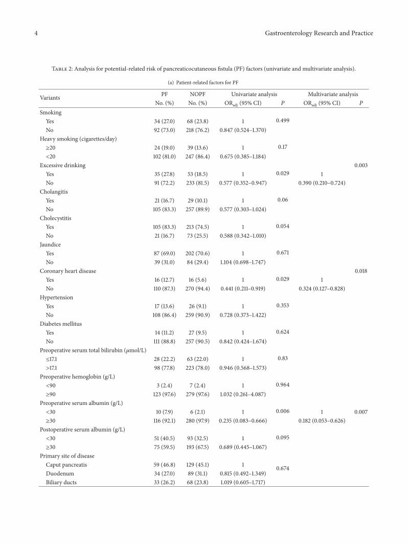
3.2. Potential-Related Factors for PF. Univariate logisticregression analysis showed that (Table 2(a)) therewere signif-icant associations between PF occurrence rates and patient-related factors of excessive drinking (𝑃 = 0.029), coronaryheart disease (𝑃 = 0.029), and preoperative albumin (𝑃 =0.006). Among comorbidities, a history of cholangitis, chole-cystitis, jaundice, hypertension, or diabetes mellitus wassimilar in PF and NOPF groups. Besides, the smoking habits,preoperative serum bilirubin, postoperative serum albumin,primary site of disease, pathologic diagnosis, and pancreaticduct diameter were also found to have no association with PFincidence.
Univariate logistic regression analysis of operative- andtherapeutic-related factors in PF and NOPF groups wasshowed in Table 2(b). There was no significant difference inPF rates between the preoperative biliary drainage treatmentor not. Early jejunal nutrition, operative time, and use ofsomatostatin after PD were also found to have no correlationwith PF rates. On the contrary, the pancreatic duct stentdrainage methods, excision methods, anastomosis methods,intraoperative blood loss (𝑃 = 0.003), intraoperative bloodtransfusion (𝑃 = 0.000), pancreatic duct stent drainage (𝑃 =0.000), excision methods (𝑃 = 0.016), methods of anastomo-sis (𝑃 = 0.005), intraoperative blood transfusion (𝑃 = 0.000),laparoscopic operation or not (𝑃 = 0.002), and professionalgroup or not (𝑃 = 0.000) markedly influenced the rate of PF.
3.3. Independence Risk Factors for PF. Based on the results ofthe above univariate analysis, additional multivariate analysiswas performed for evaluating the independence of risk fac-tors. As is showed in Table 3, both the excessive drinking andcoronary heart disease were proved to be the independent

Gastroenterology Research and Practice 3
Table 1: Demographic characteristics description.
Variants PF NOPF Total𝜒2 (𝑃 value) CR-PF NOCR-PF Total
𝜒2 (𝑃 value)
No. (%) No. (%) No. (%) No. (%) No. (%) No. (%)Gender
0.597 (0.440) 0.515 (0.473)Male 83 (65.9) 177 (61.9) 260 (63.1) 44 (59.5) 216 (63.9) 260 (63.1)Female 43 (34.1) 109 (38.1) 152 (36.9) 30 (40.5) 122 (36.1) 152 (36.9)
Age (year)0.077 (0.782) 0.619 (0.431)<70 113 (89.7) 259 (90.6) 372 (90.3) 65 (87.8) 307 (90.8) 372 (90.3)
≥70 13 (10.3) 27 (9.4) 40 (9.7) 9 (12.2) 31 (9.2) 40 (9.7)BMI
1.951 (0.377) 0.328 (0.849)<18.5 14 (11.1) 41 (14.3) 55 (13.3) 9 (12.2) 46 (13.6) 55 (13.3)18.5–25 90 (71.4) 208 (72.7) 298 (72.3) 53 (71.6) 245 (72.5) 298 (72.3)≥25 22 (17.5) 37 (12.9) 59 (14.3) 12 (16.2) 47 (13.9) 59 (14.3)
PF: patients undergoing pancreatic fistula after pancreaticoduodenectomy (PD); NOPF: PD patients without PF occur; CR-PF: PF patients diagnosed as gradeB fistulas and grade C fistulas; NOCR-PF: non-PF patients and grade A PF patients; BMI: body mass index.
risk factors for PF with odds ratio (ORs) of 0.390 (95% CI= (0.210–0.724), 𝑃 = 0.003) and 0.324 (95% CI = (0.127–0.828), 𝑃 = 0.018), respectively. The preoperative albumin(𝑃 = 0.007) was significantly higher in the PF group thanin the NOPF group (Table 3(a)). More intraoperative bloodtransfusion (𝑃 = 0.000) and harder remnant pancreas texture(𝑃 = 0.037) significantly reduced the PF risk. In addition,different methods of anastomosis, laparoscopic operation,and professional groupwere also included in the independentrisk factors affecting PF (𝑃 = 0.026). Though pancreaticduct stent drainage and excision method were proved to beassociated with PF in univariate analysis, the multivariateanalysis showed that they were not independent risk factors(𝑃 > 0.05) (Table 3(b)).
3.4. Potential-Related Factors for CR-PF. Univariate logisticregression analysis showed that (Table 3(a)) the patient char-acteristics such as cholangitis (𝑃 = 0.002), preoperativeserum albumin (𝑃 = 0.000), and texture of the remnantpancreas (𝑃 = 0.013) were significantly related to the CR-PF. The other patient characteristics, for example, smok-ing, excessive drinking, cholecystitis, jaundice, coronaryheart disease, hypertension, diabetes mellitus, preoperativeserum total bilirubin, preoperative hemoglobin, postoper-ative serum albumin, primary site of disease, pathologicdiagnosis, and diameter of pancreatic duct, had no influenceon CR-PF occurrence (all 𝑃 > 0.05).
Theoperative and therapeutic risk factors such as intraop-erative blood loss (𝑃 = 0.004), intraoperative blood transfu-sion (𝑃 = 0.002), pancreatic duct stent drainage (𝑃 = 0.007),and professional group were associated with an increasedincidence of CR-PF (𝑃 = 0.000), while the left factors werefound to have no significant association with the risk of CR-PF (Table 3(b)).
3.5. Independence Risk Factors for ORPF. When assessingthe independent effect of the potential risk factors on ORPFoccurrence in multivariate analysis, cholangitis, preopera-tive albumin, intraoperative blood transfusion, texture ofthe remnant pancreas, and professional group or not were
the significant associated factors (all 𝑃 < 0.05), whereasthe effect of pancreatic duct stent drainage methods had noindependent effect on ORPF.
3.6. Hospital Charges and Hospital Stays. Mean hospital stayswere shorter in the NOPF group andNOCR-PF patients thanin the PF andCR-PF patients, respectively (Table 4). By use ofnonparametric test analysis, there were significant differencesin hospital stays between PF and NOPF, as well as betweenCR-PF and NOCR-PF (both 𝑃 values were 0.000). The meancharges of the PF and NOPF patients were 56323.47 RMBand 83347.93 RMB, respectively, which exhibited significantdifference with each other by 𝑡 text (𝑃 = 0.000). Similarly,significant difference was found between CR-PF and NOCR-PF groups as well (𝑃 = 0.001), with mean hospital charges ofRMB 61339.84 and 81448.18, respectively.
4. Discussion
Effective management of PF has proven to be one of themost intractable challenges after PD surgery. Confront withthis adversity, there has been a shift therapeutic regimenfor management of PF, from a reactive “wait and see” to aproactive strategy that relies on early anticipation and timelyprevention [9, 13]. However, this approach depended onassumption and prediction of the risk for PF development.In the present study, we collected clinical data of 412 patientsin our hospital, analyzed the potential risk factors associatedwith PF and CR-PF, and evaluated the medico-economiceffect on these patients. Our results showed that the exces-sive drinking, coronary heart disease, preoperative albumin,intraoperative blood transfusion, texture of the remnant pan-creas, methods of anastomosis, laparoscopic operation, andprofessional group were independently associated with PFoccurrence. Among these risk factors of PF, the preoperativealbumin, intraoperative blood transfusion, texture of theremnant pancreas, and professional group were significantlyand independently associated with CR-PF. Though historyof cholangitis in patients was found insignificantly related
4 Gastroenterology Research and Practice
Table 2: Analysis for potential-related risk of pancreaticocutaneous fistula (PF) factors (univariate and multivariate analysis).
(a) Patient-related factors for PF
Variants PF NOPF Univariate analysis Multivariate analysisNo. (%) No. (%) ORadj (95% CI) 𝑃 ORadj (95% CI) 𝑃
Smoking0.499Yes 34 (27.0) 68 (23.8) 1
No 92 (73.0) 218 (76.2) 0.847 (0.524–1.370)Heavy smoking (cigarettes/day)
0.17≥20 24 (19.0) 39 (13.6) 1<20 102 (81.0) 247 (86.4) 0.675 (0.385–1.184)
Excessive drinking0.029
0.003Yes 35 (27.8) 53 (18.5) 1 1No 91 (72.2) 233 (81.5) 0.577 (0.352–0.947) 0.390 (0.210–0.724)
Cholangitis0.06Yes 21 (16.7) 29 (10.1) 1
No 105 (83.3) 257 (89.9) 0.577 (0.303–1.024)Cholecystitis
0.054Yes 105 (83.3) 213 (74.5) 1No 21 (16.7) 73 (25.5) 0.588 (0.342–1.010)
Jaundice0.671Yes 87 (69.0) 202 (70.6) 1
No 39 (31.0) 84 (29.4) 1.104 (0.698–1.747)Coronary heart disease
0.0290.018
Yes 16 (12.7) 16 (5.6) 1 1No 110 (87.3) 270 (94.4) 0.441 (0.211–0.919) 0.324 (0.127–0.828)
Hypertension0.353Yes 17 (13.6) 26 (9.1) 1
No 108 (86.4) 259 (90.9) 0.728 (0.373–1.422)Diabetes mellitus
0.624Yes 14 (11.2) 27 (9.5) 1No 111 (88.8) 257 (90.5) 0.842 (0.424–1.674)
Preoperative serum total bilirubin (𝜇mol/L)0.83≤17.1 28 (22.2) 63 (22.0) 1
>17.1 98 (77.8) 223 (78.0) 0.946 (0.568–1.573)Preoperative hemoglobin (g/L)
0.964<90 3 (2.4) 7 (2.4) 1≥90 123 (97.6) 279 (97.6) 1.032 (0.261–4.087)
Preoperative serum albumin (g/L)0.006<30 10 (7.9) 6 (2.1) 1 1 0.007
≥30 116 (92.1) 280 (97.9) 0.235 (0.083–0.666) 0.182 (0.053–0.626)Postoperative serum albumin (g/L)
0.095<30 51 (40.5) 93 (32.5) 1≥30 75 (59.5) 193 (67.5) 0.689 (0.445–1.067)
Primary site of disease
0.674Caput pancreatis 59 (46.8) 129 (45.1) 1Duodenum 34 (27.0) 89 (31.1) 0.815 (0.492–1.349)Biliary ducts 33 (26.2) 68 (23.8) 1.019 (0.605–1.717)

Gastroenterology Research and Practice 5
(a) Continued.
Variants PF NOPF Univariate analysis Multivariate analysisNo. (%) No. (%) ORadj (95% CI) 𝑃 ORadj (95% CI) 𝑃
Pathologic diagnosis
0.371
Caput pancreatis cancer 48 (38.1) 91 (31.8) 1Duodenal cancer 21 (16.7) 60 (21.0) 0.642 (0.348–1.184)Cholangiocarcinoma 30 (23.8) 62 (21.7) 0.906 (0.516–1.590)Pancreatitis 4 (3.2) 21 (7.3) 0.416 (0.134–1.294)Carcinoma of ampulla 6 (4.8) 20 (7.0) 0.588 (0.220–1.570)Others 17 (13.5) 32 (11.2) 1.141 (0.568–2.295)
Texture of the remnant pancreas 0.016 0.037Hard 20 (15.9) 77 (26.9) 1 1Soft 106 (84.1) 209 (73.1) 1.964 (1.136–3.394) 1.955 (1.042–3.669)
Diameter of pancreatic duct (mm) 0.496<3 68 (54.0) 142 (49.7) 1≥3 58 (46.0) 144 (50.3) 0.863 (0.566–1.318)
(b) Operative- and therapeutic-related factors for PF
Variants PF NOPF Univariate analysis Multivariate analysisNo. (%) No. (%) ORadj (95% CI) 𝑃 ORadj (95% CI) 𝑃
Preoperative biliary drainage treatmentYes 13 (10.3) 25 (8.7) 1 0.535No 113 (89.7) 261 (91.3) 0.798 (0.392–1.625)
Operative time (min) —<295 19 (15.1) 67 (22.9) 1 0.161 —≥295 107 (84.9) 226 (77.1) 0.669 (0.380–1.177) —
Intraoperative blood loss (mL) 0.003 — —<300 35 (27.8) 121 (42.3) 1 — —300–600 30 (23.8) 81 (28.3) 1.194 (0.675–2.113) — —600–900 28 (22.2) 45 (15.7) 2.089 (1.139–3.830) — —≥900 33 (26.2) 39 (13.6) 2.738 (1.498–5.005) — —
Intraoperative blood transfusion (mL) 0.000 0.000<300 41 (32.5) 136 (47.6) 1 1300–600 23 (18.3) 67 (23.4) 1.112 (0.616–2.008) 1.128 (0.556–2.290) 0.738600–900 31 (24.6) 57 (19.9) 1.754 (1.000–3.078) 2.574 (1.318–5.025) 0.006≥900 31 (24.6) 26 (9.1) 3.711 (1.969–6.995) 5.115 (2.364–11.069) 0.000
Pancreatic duct stent drainage 0.000 0.394No stent 40 (31.7) 49 (17.1) 1 1 0.570Internal drainage 77 (61.1) 174 (60.8) 0.518 (0.313–0.856) 0.819 (0.412–1.629) 0.178External drainage 9 (7.1) 63 (22.0) 0.162 (0.071–0.370) 0.476 (0.161–1.403) 0.624
Excision method 0.016 0.624Without PP 108 (85.7) 266 (93.0) 1 1With PP 18 (14.3) 20 (7.0) 2.300 (1.165–4.543) 1.240 (0.524–2.932)
Methods of anastomosis 0.005 0.026Binding anastomosis 3 (2.4) 22 (7.7) 1 1End-side invagination anastomosis 6 (4.8) 38 (13.3) 1.110 (0.251–4.911) 2.428 (0.439–13.432)End-end invagination anastomosis 106 (84.1) 194 (67.8) 3.922 (1.143–13.456) 5.510 (1.391–21.821)Duct-mucosa anastomosis 11 (8.7) 32 (11.2) 2.396 (0.595–9.642) 7.918 (1.619–38.722)
Laparoscopic operation 0.002 0.034Yes 10 (7.9) 3 (1.0) 1 1No 116 (92.1) 283 (99.0) 0.128 (0.034–0.477) 0.188 (0.040–0.883)

6 Gastroenterology Research and Practice
(b) Continued.
Variants PF NOPF Univariate analysis Multivariate analysisNo. (%) No. (%) ORadj (95% CI) 𝑃 ORadj (95% CI) 𝑃
Early jejunal nutrition 0.065Yes 25 (19.8) 81 (28.3) 1No 101 (80.2) 205 (71.7) 1.617 (0.970–2.694)
Use of somatostatin after PD 0.059Yes 84 (66.7) 161 (56.3) 1No 42 (33.3) 125 (43.7) 0.655 (0.422–1.017)
Professional group 0.000 0.000Yes 38 (30.2) 190 (66.4) 1 1No 88 (69.8) 96 (33.6) 4.718 (2.985–7.457) 3.925 (2.250–6.847)
—: themultivariate analysis of intraoperative blood loss was not performed in this study because of its corresponding relationship with intraoperative bloodtransfusion. Missing values: 3 diabetes mellitus patients and 2 hypertension patients were missing. OR: odds ratio; CI: confidence interval; ORadj: adjustedORs presented with 95% CI.
with PF, it was one of the risk factors that affected CR-PFsignificantly.
Although risk factors for PF and CR-PF have historicallybeen reported in the literature, their relevance in applicationhas been hampered by definitions of fistula [14]. Actually,several studies have tried to identify the risk factors associatedwith the PF development and many risk factors have beenproposed. However, only a few factors are independentfactors of PF and they vary among different studies. In thepresent study, total occurrence rate of PF after PD surgerywas 30.58%, which was slightly higher than previous results.The reasonmay be explained by the different definitions of PF.Though a normalized definition was proposed by ISGPF, thedefinition of PFmight in some case not be specific, because itincludes asymptomatic patients who are not clinically ill [6].Therefore, the study of risk factors affecting grades B and Cwill be more meaningful in clinical practice.
Generally, PF risk evaluation begins in the preoperativesetting, such as patient-related factors. In this study, weretrospectively analyzed the conditions of PF patients beforePD surgery and found that the preoperative serum albumin,history of coronary heart disease, and excessive drinkingwerethe independent risk factors associated with PF. With respectto CR-PF, the independent risk factors were preoperativeserum albumin and cholangitis.
Albumin in serum has properties of maintaining normalplasma osmotic pressure [15] and acid-base balance [16],antioxidant [17], scavenging free radical [18], and protectingmicrocirculation [19]. On the one hand, tissue edema causedby hypoproteinemia may lead to undesirable or anastomoticstoma healing and then increase the incidence of PF orCR-PF. On the other hand, the increased hypoproteinemiacomplications such as infection and diarrhea will influencethe PF and CR-PF more or less.
There is evidence from the current literatures that car-diovascular disease is a risk factor for PF [20], which wasconsistent with our findings, while the effect of coronaryheart disease on CR-PF was not significant. The reasonswhy coronary heart disease would be associated with PFare not well understood. Perhaps the cardiovascular andcerebrovascular diseases are surrogate for decreased visceral
perfusion result in anastomotic ischemia, or perhaps therelated medications to such patients compromise anasto-motic healing [21].Therefore, the association between cardio-vascular disease and PF should be well explained by reliableevidence from clinical outcomes.
Animal experiments and epidemiological studies havesuggested that alcohol had toxicity to pancreas [22, 23]. It wasproved to be an independent risk factor for PF occurrencein our patients undergoing PD. There are several hypotheseson the toxicity mechanisms of ethanol to pancreas. (a) Theethanol has toxic effect on pancreatic acinar cells and disturbsits metabolism [24]. (b) The accumulation of pancreaticstone protein induced by ethanol produces ulceration andinflammation of the ductules, and the ductule then leadsto atrophy, insufficiency, and fibrosis themselves [25]. (c)The disorders such as sphincter of Oddi dysfunction causedby ethanol have a connection with stenosis of ductule andregurgitation of duodenal juice [25]. (d) Excessive drinkingwill destroy the essential minerals and induce the oxygenradical in vivo in human, which are harmful to pancreas [24].We believe that the excessive drinking effect on PF will beclosely related to its effect on pancreas.
One of the interesting findings in our research is thatpatients with cholangitis will be more likely to suffer fromCR-PF than noncholangitis patients. Generally, mucosa inbiliary ducts is congestive in cholangitis patients, especiallyin patients with obstruction of biliary tract. Edema andinflammation usually happen in pancreatic tissues when thebile duct enlarged by obstruction. Therefore, it is harmfulto conduct the anastomosis and CR-PF is likely to occur. Inaddition, cholangitis is usually accompanied with increasedbacteria in the bile duct. The increased intraductal pressurecan lead to bacteria translocation or endotoxemia in thesepatients [26], which may be another indirect factors affectingCR-PF occurrence.
The factors of pathologic diagnosis, texture of the rem-nant pancreas and diameter of pancreatic duct have beenwidely accepted as the related risk factors of PF [12, 27–29]. Logistic analysis of regression showed that patients withsoft texture of the remnant pancreas had higher PF and CR-PF incidence than that in patients with hard texture. There

Gastroenterology Research and Practice 7
Table 3: Analysis for potential risk factors of pancreaticocutaneous fistula (CR-PF) factors (univariate and multivariate analysis).
(a) Patient-related factors for CR-PF
Variants CR-PF NOCR-PF Univariate analysis Multivariate analysisNo. (%) No. (%) ORadj (95% CI) 𝑃 ORadj (95% CI) 𝑃
Smoking 0.84Yes 19 (25.7) 83 (24.6) 1No 55 (74.3) 255 (75.4) 0.942 (0.529–1.678)
Heavy smoking (cigarettes/day) 0.191≥20 15 (20.3) 48 (14.2) 1<20 59 (79.3) 290 (85.8) 0.651 (0.342–1.239)
Excessive drinking 0.493Yes 18 (24.3) 70 (20.7) 1No 56 (75.7) 268 (79.3) 0.813 (0.449–1.470)
Cholangitis 0.002Yes 17 (23.0) 33 (9.8) 1No 57 (77.0) 305 (90.2) 0.363 (0.189–0.695)
Cholecystitis 0.075 0.004Yes 63 (85.1) 255 (75.4) 1 1No 11 (14.9) 83 (24.6) 0.536 (0.270–1.066) 0.321 (0.150–0.690)
Jaundice 0.054Yes 45 (60.8) 244 (72.2) 1No 29 (39.2) 94 (27.8) 1.673 (0.991–2.825)
Coronary heart disease 0.284Yes 8 (10.8) 24 (7.1) 1No 66 (89.2) 314 (92.9) 0.631 (0.271–1.465)
Hypertension 0.572Yes 9 (12.3) 34 (10.1) 1No 64 (87.7) 303 (89.9) 0.798 (0.365–1.745)
Diabetes mellitus 0.163Yes 4 (5.5) 37 (11.0) 1No 69 (94.5) 299 (89.0) 2.135 (0.736–6.188)
Preoperative serum total bilirubin (𝜇mol/L) 0.412≤17.1 19 (25.7) 72 (21.3) 1>17.1 55 (74.3) 266 (78.7) 0.784 (0.437–1.404)
Preoperative hemoglobin (g/L) 0.324<90 3 (4.1) 7 (2.1) 1≥90 71 (95.9) 331 (97.9) 0.501 (0.126–1.983)
Preoperative serum albumin (g/L) 0 0.000<30 9 (12.2) 7 (2.1) 1 1≥30 65 (87.8) 331 (97.9) 0.153 (0.055–0.425) 0.107 (0.031–0.363)
Postoperative serum albumin (g/L) 0.971<30 26 (35.1) 118 (34.9) 1≥30 48 (64.9) 220 (65.1) 0.990 (0.585–1.677)
Primary site of disease 0.935Caput pancreatis 34 (45.9) 154 (45.6) 1Duodenum 23 (31.1) 100 (29.6) 1.042 (0.580–1.872)Biliary ducts 17 (23.0) 84 (24.9) 0.917 (0.483–1.738)

8 Gastroenterology Research and Practice
(a) Continued.
Variants CR-PF NOCR-PF Univariate analysis Multivariate analysisNo. (%) No. (%) ORadj (95% CI) 𝑃 ORadj (95% CI) 𝑃
Pathologic diagnosis 0.322Caput pancreatis cancer 28 (37.8) 111 (32.8) 1Duodenal cancer 13 (17.6) 68 (20.1) 0.758 (0.368–1.563)Cholangiocarcinoma 15 (20.3) 77 (22.8) 0.772 (0.387–1.542)Pancreatitis 1 (1.4) 24 (7.1) 0.165 (0.021–1.274)Carcinoma of ampulla 4 (5.4) 22 (6.5) 0.721 (0.230–2.261)Others 13 (17.6) 36 (10.7) 1.432 (0.671–3.054)
Texture of the remnant pancreas 0.013 0.044Hard 9 (12.2) 88 (26.0) 1 1Soft 65 (87.8) 250 (74.0) 2.542 (1.215–5.319) 2.316 (1.205–5.234)
Diameter of pancreatic duct (mm) 0.400<3 41 (55.4) 169 (50.0) 1≥3 33 (44.6) 169 (50.0) 0.805 (0.485–1.334)
(b) Operative- and therapeutic-related factors for CR-PF
Variants CR-PF NOCR-PF Univariate analysis Multivariate analysisNo. (%) No. (%) ORadj (95% CI) 𝑃 ORadj (95% CI) 𝑃
Preoperative biliary drainage treatment 0.068Yes 11 (14.9) 27 (8.0) 1No 63 (85.1) 311 (92.0) 0.497 (0.235–1.054)
Operative time (min) 1.081<295 11 (14.9) 68 (20.1) 1≥295 63 (85.1) 270 (79.9) 0.693 (0.347–1.387)
Intraoperative blood loss (mL) 0.004 — —<300 20 (27.0) 136 (40.2) 1 — —300–600 14 (18.9) 97 (28.7) 0.981 (0.473–2.039) — —600–900 20 (27.0) 53 (15.7) 2.566 (1.279–5.148) — —≥900 20 (27.0) 52 (15.4) 2.615 (1.302–5.253) — —
Intraoperative blood transfusion (mL) 0.002 0.001<300 19 (25.7) 158 (46.7) 1 1300–600 18 (24.3) 72 (21.3) 2.079 (1.030–4.196) 2.311 (1.049–5.092) 0.038600–900 18 (24.3) 70 (20.7) 2.138 (1.058–4.321) 2.657 (1.207–5.851) 0.015≥900 19 (25.7) 38 (11.2) 4.158 (2.008–8.609) 5.337 (2.301–12.376) 0.000
Pancreatic duct stent drainage 0.007 0.542No stent 24 (32.4) 65 (19.2) 1 1Internal drainage 45 (60.8) 206 (60.9) 0.592 (0.335–1.045) 0.692 (0.359–1.334) 0.272External drainage 5 (6.8) 67 (19.8) 0.202 (0.073–0.562) 0.814 (0.248–2.671) 0.375
Excision method 0.068Without PP 63 (85.1) 311 (92.0) 1With PP 11 (14.9) 27 (8.0) 2.011 (0.949–4.264)
Methods of anastomosis 0.069Binding anastomosis 2 (2.7) 23 (6.8) 1End-side invagination anastomosis 3 (4.1) 41 (12.1) 0.841 (0.131–5.409)End-end invagination anastomosis 63 (85.1) 237 (70.1) 3.057 (0.702–13.314)Duct-mucosa anastomosis 6 (8.1) 37 (10.9) 1.865 (0.347–10.034)
Laparoscopic operation 0.231Yes 4 (5.4) 9 (2.7) 1No 70 (94.6) 329 (97.3) 0.479 (0.143–1.599)

Gastroenterology Research and Practice 9
(b) Continued.
Variants CR-PF NOCR-PF Univariate analysis Multivariate analysisNo. (%) No. (%) ORadj (95% CI) 𝑃 ORadj (95% CI) 𝑃
Early jejunal nutrition 0.238Yes 15 (20.3) 91 (26.9) 1No 59 (79.7) 247 (73.1) 1.449 (0.783–2.682)
Use of somatostatin after PD 0.069Yes 51 (68.9) 194 (57.4) 1No 23 (31.1) 144 (42.6) 0.608 (0.355–1.040)
Professional group 0.000 0.000Yes 16 (21.6) 212 (62.7) 1 1No 58 (78.4) 126 (37.3) 6.099 (3.362–11.066) 5.674 (2.867–11.230)
—: themultivariate analysis of intraoperative blood loss was not performed in this study because of its corresponding relationship with intraoperative bloodtransfusion. Missing values: 3 diabetes mellitus patients and 2 hypertension patients were missing. OR: odds ratio; CI: confidence interval; ORadj: adjustedORs presented with 95% CI.
Table 4: Hospital charges and hospital stays.
NOPF PF NOCR-PF CR-PFAverage hospital stays (d)
Normality test 𝑃 0.009 0.024 0.004 0.06𝑡-test/nonparametric test 𝑃 0.000a 0.000b
Mean (d) 22.25 39.08 23.33 46.42Standard deviation 15.80
Average hospital chargesNormality test 𝑃 0.223 0.617 0.279 0.915𝑡-test/nonparametric test 𝑃 0.000a 0.001b
Mean (yuan) 56323.47 83347.93 61339.84 81448.18Standard deviation 24360.81 32007.30 28166.70 31699.74Missing values: data missing for 8 patients in hospital charges. a𝑃 value of nonparametric test between PF and NOPF groups; b𝑃 value of 𝑡 test between NOCR-PF and CR-PF groups.
are several explanations for this association. Firstly, a softpancreas is more susceptible to injury and ischemia duringoperative dissection [29]. Meanwhile, exocrine function isusually preserved in the soft pancreas, leading to increasedsecretion of pancreatic juice and activation of proteolyticenzymes, which is more prone for PF development [30].However, our data did not provide evidence to supportpathologic diagnosis and diameter of pancreatic duct (≥3mmand <3mm) of influence factors for PF and CR-PF. Therelationship between them needs more studies in the future.
Blood transfusion in response to blood loss is con-sidered to be an immunosuppressive effect. In our study,the intraoperative blood transfusion was mainly caused byintraoperative blood loss, preoperative anemia, coagulationdisorders, and so forth. Though our results showed that theintraoperative blood transfusion was the risk factor influ-encing both PF and CR-PF, the full impact of intraoperativeblood transfusion is not well understood. Rapid blood loss, aswell as anemia and coagulation disorder, may cause ischemiaand poor healing of the pancreatic anastomosis, becauseof tissue edema from aggressive blood replacement in a“rebound” fashion [29]. In addition, other adverse effectssuch as complications of blood transfusion, disseminatedintravascular coagulation, and hemorrhagic tendency during
intraoperative blood transfusion may increase the risk of PFand CR-PF after PD surgery.
Hypertension has been noted as one of the risk factors inprevious studies [31, 32].They assumed that the pathophysio-logical effects of hypertension caused generalized atheroscle-rosis and therefore limited the microcirculation of the tissue.Thatwill negatively affect the healing process of PD.However,in our study, we found no significant association betweenhypertension and postoperative PD. Therefore, we stronglyrecommended more studies to resolve these controversialresults.
The PD surgery with laparoscope has been clinicallyapplied since its first description by Gagner and Pomp [33] in1994 [34, 35].Themultivariate analysis of 13 PD patients withlaparoscope (PF: 10, CR-PF: 4) and 309 patients with laparo-tomy showed that more patients treated with laparoscopedeveloped PF than that treated with laparotomy. Fortunately,the laparoscope treatment had no significant side effect onCR-PF. Limitations of this approach including inability topalpate the lesion, relatively narrow view, inaccurate location,and misestimates of tumor spread may be responsible forhigh PF occurrence. We believe that these limitations will beminimized as the experiential accumulation and technologi-cal improvement.

10 Gastroenterology Research and Practice
Other operative- and therapeutic-related factors such astreatment of residual pancreatic, application of pancreaticduct stent drainage, methods of anastomose, use of somato-statin after PD, and excision methods have suggested asso-ciating with the PF incidence [21, 32, 36]. Analysis of thesefactors in our study revealed that just anastomose methodswere associated with PF.The binding anastomosis was provedto be superior to end-side invagination anastomosis, end-endinvagination anastomosis, and duct-mucosa anastomosis,because of its less PF incidence. Instead of suture, bindinganastomosis can definitely minimize the leakage by avoidingany pinhole through the closure [37]. Furthermore, bindinganastomosis avoids the regurgitation of pancreatic juice bymaintaining higher blasting pressure in jejunum than othermethods [38]. However, this method was found to have norelationship with CR-PF.
Unquestionably, the complex anddifficult PDoperation isa challenge to surgeons.Therefore the skilled and experiencedsurgeons will be important factors related to PF incidence. Inour study, the PF and CR-PF incidence after PD surgery inprofessional group were 3 and 5.7 times more than nonpro-fessional group, respectively.The high-volume surgeons wereproved to have lower PF rate [39, 40], probably due to moreexperience for surgeons.Therefore, it is necessary for trainingPD surgeons and establishing professional group in future.
Patients with PF, especially with CR-PF after PD surgery,usually have to prolong the hospital stages and pay more forexternal surgery than patients in NOPF or NOCR-PF group.Our study also showed more hospital stages and charges inPF and CR-PF groups than in NOPF and NOCR-PF groups.Future studies that address the charge and hospital stages arerequired, in light of the rapid increase of technology.
5. Conclusion
In summary, the excessive drinking, coronary heart dis-ease, preoperative albumin, intraoperative blood transfu-sion (>600mL), soft remnant pancreas, and laparoscopicoperation were risk factors affecting PF incidence after PD.Binding anastomosis between remnant pancreas and jejunumcan effectively reduce the PF incidence compared with theother anastomosis methods, such as end-side invaginationanastomosis, end-end invagination anastomosis, and duct-mucosa anastomosis. The risk factors such as cholangitis,hypoproteinemia, intraoperative blood transfusion volume(>300mL), and soft remnant pancreas were significantlyassociated with CR-PF. Surgeons with more experience andprofession can significantly reduce the PF and CR-PF inci-dence when they perform PD surgery. More hospital stagesand charges in PF and CR-PF groups are needed than inNOPF and NOCR-PF groups. It is critical that surgeonsknow the risk factors related to PF and CR-PF so as to takecorresponding therapeutic regimens for each patient.
Conflict of Interests
The authors declare that there is no conflict of interestsregarding the publication of this paper.
Acknowledgments
This work was supported in part by grants from theResearch Special Fund For Public Welfare Industry of Health(201202007); the Research Fund for the Doctoral Programof Higher Education of China (20122307110012); and theNational Natural Science Foundation of China (81372613,81170431, 81302057, and 81470887).
References
[1] C. Bassi, C. Dervenis, G. Butturini et al., “Postoperative pan-creatic fistula: an international study group (ISGPF) definition,”Surgery, vol. 138, no. 1, pp. 8–13, 2005.
[2] J. L. Cameron, T. S. Riall, J. Coleman, and K. A. Belcher, “Onethousand consecutive pancreaticoduodenectomies,” Annals ofSurgery, vol. 244, no. 1, pp. 10–15, 2006.
[3] S. V. Shrikhande and M. A. D’Souza, “Pancreatic fistula afterpancreatectomy: evolving definitions, preventive strategies andmodern management,” World Journal of Gastroenterology, vol.14, no. 38, pp. 5789–5796, 2008.
[4] V. C. Njoku, T. J. Howard, C. Shen et al., “Pyogenic liver abscessfollowing pancreaticoduodenectomy: risk factors, treatment,and long-term outcome,” Journal of Gastrointestinal Surgery,vol. 18, no. 5, pp. 922–928, 2014.
[5] G. Balzano, A. Zerbi, G. Capretti, S. Rocchetti, V. Capitanio,and V. di Carlo, “Effect of hospital volume on outcome ofpancreaticoduodenectomy in Italy,” British Journal of Surgery,vol. 95, no. 3, pp. 357–362, 2008.
[6] G. Butturini, D. Daskalaki, E. Molinari, F. Scopelliti, A.Casarotto, and C. Bassi, “Pancreatic fistula: definition andcurrent problems,” Journal ofHepato-Biliary-Pancreatic Surgery,vol. 15, no. 3, pp. 247–251, 2008.
[7] N. Faccioli, G. Foti, E. Molinari et al., “Role of fistulography inevaluating pancreatic fistula after pancreaticoduodenectomy,”British Journal of Radiology, vol. 85, no. 1011, pp. 219–224, 2012.
[8] C. M. Schmidt, E. S. Powell, C. T. Yiannoutsos et al., “Pan-creaticoduodenectomy: a 20-year experience in 516 patients,”Archives of Surgery, vol. 139, no. 7, pp. 718–727, 2004.
[9] M. P. Callery,W. B. Pratt, andC.M. Vollmer Jr., “Prevention andmanagement of pancreatic fistula,” Journal of GastrointestinalSurgery, vol. 13, no. 1, pp. 163–173, 2009.
[10] E. C. H. Lai, S. H. Y. Lau, and W. Y. Lau, “Measures to preventpancreatic fistula after pancreatoduodenectomy: a comprehen-sive review,” Archives of Surgery, vol. 144, no. 11, pp. 1074–1080,2009.
[11] S. M. Strasberg, D. C. Linehan, P.-A. Clavien, and J. S. Barkun,“Proposal for definition and severity grading of pancreaticanastomosis failure and pancreatic occlusion failure,” Surgery,vol. 141, no. 4, pp. 420–426, 2007.
[12] N. O. MacHado, “Pancreatic fistula after pancreatectomy: defi-nitions, risk factors, preventive measures, and management—review,” International Journal of Surgical Oncology, vol. 2012,Article ID 602478, 10 pages, 2012.
[13] W. B. Pratt, S. K. Maithel, T. Vanounou, Z. S. Huang, M. P.Callery, and C. M. Vollmer Jr., “Clinical and economic vali-dation of the International Study Group of Pancreatic Fistula(ISGPF) classification scheme,” Annals of Surgery, vol. 245, no.3, pp. 443–451, 2007.
[14] C. Bassi, G. Butturini, E. Molinari et al., “Pancreatic fistularate after pancreatic resection: the importance of definitions,”Digestive Surgery, vol. 21, no. 1, pp. 54–59, 2004.

Gastroenterology Research and Practice 11
[15] P. D. Navar and L. G. Navar, “Relationship between colloidosmotic pressure and plasma protein concentration in the dog,”The American Journal of Physiology—Heart and CirculatoryPhysiology, vol. 233, no. 2, pp. H295–H298, 1977.
[16] J. Figge, T. H. Rossing, and V. Fencl, “The role of serum proteinsin acid-base equilibria,” Journal of Laboratory and ClinicalMedicine, vol. 117, no. 6, pp. 453–467, 1991.
[17] M. Roche, P. Rondeau, N. R. Singh, E. Tarnus, and E. Bourdon,“The antioxidant properties of serum albumin,” FEBS Letters,vol. 582, no. 13, pp. 1783–1787, 2008.
[18] M. Soriani, D. Pietraforte, and M. Minetti, “Antioxidant poten-tial of anaerobic human plasma: role of serum albumin andthiols as scavengers of carbon radicals,”Archives of Biochemistryand Biophysics, vol. 312, no. 1, pp. 180–188, 1994.
[19] M.W.Keller, S. S. Segal, S. Kaul, and B. Duling, “The behavior ofsonicated albuminmicrobubbles within the microcirculation: abasis for their use during myocardial contrast echocardiogra-phy,” Circulation Research, vol. 65, no. 2, pp. 458–467, 1989.
[20] M. L. DeOliveira, J. M.Winter, M. Schafer et al., “Assessment ofcomplications after pancreatic surgery: a novel grading systemapplied to 633 patients undergoing pancreaticoduodenectomy,”Annals of Surgery, vol. 244, no. 6, pp. 931–937, 2006.
[21] J. W. Lin, J. L. Cameron, C. J. Yeo, T. S. Riall, and K. D. Lille-moe, “Risk factors and outcomes in postpancreaticoduodenec-tomy pancreaticocutaneous fistula,” Journal of GastrointestinalSurgery, vol. 8, no. 8, pp. 951–959, 2004.
[22] G. A. Cote, D. Yadav, A. Slivka et al., “Alcohol and smoking asrisk factors in an epidemiology study of patients with chronicpancreatitis,” Clinical Gastroenterology and Hepatology, vol. 9,no. 3, pp. 266–273, 2011.
[23] D. Sophia, M. Gomathy, T. Shebin, P. Ragavendran, C. Arul-raj, and V. K. Gopalakrishnan, “Effect of Emilia sonchifolia(Linn.)DC on alcohol-induced oxidative stress in pancreas ofmale albino rats,”Asian Pacific Journal of Tropical Medicine, vol.4, no. 12, pp. 973–977, 2011.
[24] A. Vonlaufen, J. S. Wilson, R. C. Pirola, and M. V. Apte, “Roleof alcoholmetabolism in chronic pancreatitis,”Alcohol Researchand Health, vol. 30, no. 1, pp. 48–54, 2007.
[25] T. Stevens, D. L. Conwell, and G. Zuccaro, “Pathogenesis ofchronic pancreatitis: an evidence-based review of past theoriesand recent developments,” American Journal of Gastroenterol-ogy, vol. 99, no. 11, pp. 2256–2270, 2004.
[26] Y. Kimura, T. Takada, Y. Kawarada et al., “Definitions, patho-physiology, and epidemiology of acute cholangitis and chole-cystitis: tokyo guidelines,” Journal of Hepato-Biliary-PancreaticSurgery, vol. 14, no. 1, pp. 15–26, 2007.
[27] C. J. Yeo, J. L. Cameron, K. D. Lillemoe et al., “Does prophylacticoctreotide decrease the rates of pancreatic fistula and othercomplications after pancreaticoduodenectomy? Results of aprospective randomized placebo-controlled trial,” Annals ofSurgery, vol. 232, no. 3, pp. 419–429, 2000.
[28] J. W. Denbo, W. S. Orr, B. L. Zarzaur, and S. W. Behrman,“Toward defining grade C pancreatic fistula following pancre-aticoduodenectomy: Incidence, risk factors, management andoutcome,” HPB, vol. 14, no. 9, pp. 589–593, 2012.
[29] M. P. Callery, W. B. Pratt, T. S. Kent, E. L. Chaikof, and C. M.Vollmer Jr., “A prospectively validated clinical risk score accu-rately predicts pancreatic fistula after pancreatoduodenectomy,”Journal of the American College of Surgeons, vol. 216, no. 1, pp.1–14, 2013.
[30] S. E. Lee, J.-Y. Jang, C.-S. Lim et al., “Measurement of pancreaticfat by magnetic resonance imaging: predicting the occurrence
of pancreatic fistula after pancreatoduodenectomy,” Annals ofSurgery, vol. 251, no. 5, pp. 932–936, 2010.
[31] M. Distler, S. Kersting, F. Ruckert et al., “Chronic pancreatitisof the pancreatic remnant is an independent risk factor forpancreatic fistula after distal pancreatectomy,” BMC Surgery,vol. 14, no. 1, article 54, 2014.
[32] N. Akamatsu, Y. Sugawara, M. Komagome et al., “Risk factorsfor postoperative pancreatic fistula after pancreaticoduodenec-tomy: the significance of the ratio of themain pancreatic duct tothe pancreas body as a predictor of leakage,” Journal of Hepato-Biliary-Pancreatic Sciences, vol. 17, no. 3, pp. 322–328, 2010.
[33] M. Gagner and A. Pomp, “Laparoscopic pylorus-preservingpancreatoduodenectomy,” Surgical Endoscopy, vol. 8, no. 5, pp.408–410, 1994.
[34] L.W. Traverso and R. A. Kozarek, “Pancreatoduodenectomy forchronic pancreatitis: anatomic selection criteria and subsequentlong-term outcome analysis,” Annals of Surgery, vol. 226, no. 4,pp. 429–438, 1997.
[35] D. Kyuno, Y. Kimura, M. Imamura et al., “Pancreaticoduo-denectomy for biliary tract carcinomawith situs inversus totalis:difficulties and technical notes based on two cases,” WorldJournal of Surgical Oncology, vol. 11, no. 1, article 312, 2013.
[36] W.-H. Zhu, S. Li, D.-F. Zhang et al., “Risk factors and outcomeof pancreatic fistula after consecutive pancreaticoduodenec-tomy with pancreaticojejunostomy for patients with malignanttumor,” Chinese Journal of Cancer Research, vol. 22, no. 1, pp.32–41, 2010.
[37] S. Y. Peng, J. W. Wang, J. T. Li, Y. P. Mou, Y. B. Liu, and X.J. Cai, “Binding pancreaticojejunostomy—a safe and reliableanastomosis procedure,” HPB, vol. 6, no. 3, pp. 154–160, 2004.
[38] S. Y. Peng, J. W. Wang, W. Y. Lau et al., “Conventional versusbinding pancreaticojejunostomy after pancreaticoduodenec-tomy: a prospective randomized trial,” Annals of Surgery, vol.245, no. 5, pp. 692–698, 2007.
[39] S. Mukherjee, H. M. Kocher, R. R. Hutchins, S. Bhattacharya,and A. T. Abraham, “Impact of hospital volume on outcomesfor pancreaticoduodenectomy: a single UK HPB centre experi-ence,” European Journal of Surgical Oncology, vol. 35, no. 7, pp.734–738, 2009.
[40] N. Pecorelli, G. Balzano, G. Capretti, A. Zerbi, V. Di Carlo,and M. Braga, “Effect of surgeon volume on outcome followingpancreaticoduodenectomy in a high-volume hospital,” Journalof Gastrointestinal Surgery, vol. 16, no. 3, pp. 518–523, 2012.

Submit your manuscripts athttp://www.hindawi.com
Stem CellsInternational
Hindawi Publishing Corporationhttp://www.hindawi.com Volume 2014
Hindawi Publishing Corporationhttp://www.hindawi.com Volume 2014
MEDIATORSINFLAMMATION
of
Hindawi Publishing Corporationhttp://www.hindawi.com Volume 2014
Behavioural Neurology
EndocrinologyInternational Journal of
Hindawi Publishing Corporationhttp://www.hindawi.com Volume 2014
Hindawi Publishing Corporationhttp://www.hindawi.com Volume 2014
Disease Markers
Hindawi Publishing Corporationhttp://www.hindawi.com Volume 2014
BioMed Research International
OncologyJournal of
Hindawi Publishing Corporationhttp://www.hindawi.com Volume 2014
Hindawi Publishing Corporationhttp://www.hindawi.com Volume 2014
Oxidative Medicine and Cellular Longevity
Hindawi Publishing Corporationhttp://www.hindawi.com Volume 2014
PPAR Research
The Scientific World JournalHindawi Publishing Corporation http://www.hindawi.com Volume 2014
Immunology ResearchHindawi Publishing Corporationhttp://www.hindawi.com Volume 2014
Journal of
ObesityJournal of
Hindawi Publishing Corporationhttp://www.hindawi.com Volume 2014
Hindawi Publishing Corporationhttp://www.hindawi.com Volume 2014
Computational and Mathematical Methods in Medicine
OphthalmologyJournal of
Hindawi Publishing Corporationhttp://www.hindawi.com Volume 2014
Diabetes ResearchJournal of
Hindawi Publishing Corporationhttp://www.hindawi.com Volume 2014
Hindawi Publishing Corporationhttp://www.hindawi.com Volume 2014
Research and TreatmentAIDS
Hindawi Publishing Corporationhttp://www.hindawi.com Volume 2014
Gastroenterology Research and Practice
Hindawi Publishing Corporationhttp://www.hindawi.com Volume 2014
Parkinson’s Disease
Evidence-Based Complementary and Alternative Medicine
Volume 2014Hindawi Publishing Corporationhttp://www.hindawi.com