Embed Size (px)
Citation preview
i
RISK FACTORS AND TRENDS IN NEONATAL
MORTALITY IN A
SPECIAL CARE NEWBORN UNIT IN A
TERTIARY CARE HOSPITAL
IN BANGLADESH
By
Ananya Kumar
A thesis submitted to Johns Hopkins University in conformity with
the requirements for the degree of Master of Science.
Baltimore, Maryland
April 2019
© Ananya Kumar 2019
All rights reserved.
ii
Abstract
Background: Bangladesh’s neonatal mortality rate in 2017 was 18.4 deaths per 1000 livebirths.
Special care newborn units (SCANUs) have been established in hospitals country-wide to
improve neonatal survival.
Methods: We conducted a retrospective cohort study in the SCANU of Faridpur Medical
College Hospital, Bangladesh to describe the neonates admitted to the unit in 2018, describe
trends in admissions and deaths, and investigate risk factors associated with death within 48
hours of admission to the unit. Data were extracted from paper medical records at FMCH and
digitized. Neonates who were admitted alive to the unit between January and November 2018
with admission dates available in the medical records were described based on their demographic
and clinical characteristics. Logistic regression models were used to examine the relationship
between death within 48 hours of admission to the unit and potential demographic and clinical
risk factors.
Results: The study population consisted of 693 neonates; 38% (n=263) died in hospital, 24%
(166/693) within 48 hours of admission and 49% (n=340) left the unit against medical advice.
Sixty percent were admitted on the day of birth, 56% were male, and 82% were born prematurely
(before the completion of 37 weeks of pregnancy). There appeared to be an increase in
admissions in the latter half of the year. Each additional day of age was found to be associated
with a 5% reduction in the odds of death within 48 hours of admission to the unit (OR: 0.95,
95% CI: 0.9, 0.99); male sex (OR: 1.5, 95% CI: 1.04, 2.2), prematurity (OR: 2.5, 95% CI: 1.3,
4.7) and a perinatal asphyxia diagnosis (OR: 2.2, 95% CI: 1.4, 3.4) were all associated with an
increased odds.
iii
Conclusion: The high mortality among neonates in this unit, and the identification of risk factors
associated with death within 48 hours of admission, warrants further investigation to identify
strategies to improve neonatal outcomes as well as potential risk factors amongst maternal
characteristics. Other SCANUs in the country with similar treatment gaps may also require
attention and resources in order to improve child survival in these units.
Primary Reader: Emily S. Gurley
Secondary Reader: Melissa A. Marx

iv
Acknowledgements
I would like to thank Dr. Emily S. Gurley, Dr. Melissa A. Marx, and Dr. Kyu Han Lee for their
feedback in the preparation of this thesis. I would also like to thank Dr. Shams El Arifeen, Dr.
Farzana Islam and Dr. Sanwarul Bari from the International Center of Diarrhoeal Disease
Research, Bangladesh, and Dr. Abu Pervez and Dr. Chayon Deb from Faridpur Medical College
Hospital for their contributions to this project.
v
Table of Contents
Abstract ........................................................................................................................................... ii
Acknowledgements ........................................................................................................................ iv
List of Tables ................................................................................................................................. vi
List of Figures ............................................................................................................................... vii
Introduction ..................................................................................................................................... 1
Methods........................................................................................................................................... 6
Results ........................................................................................................................................... 11
Discussion ..................................................................................................................................... 15
Tables and Figures ........................................................................................................................ 23
Supplementary Appendices .......................................................................................................... 32
Bibliography ................................................................................................................................. 35
Biography ...................................................................................................................................... 41
vi
List of Tables
Table 1: Comparisons between Neonates based on Outcomes (Death, Discharged from Unit, Left
Against Medical Advice) and their Demographic, Clinical and Diagnostic Characteristics, Special Care
Newborn Unit, Faridpur Medical College Hospital, Bangladesh, 2018 ................................................. 23
Table 2: Demographic, Diagnostic and Clinical Characteristics of the Study Population for the
Regression Analysis, Special Care Newborn Unit, Faridpur Medical College Hospital, Bangladesh,
2018 ........................................................................................................................................................ 25
Table 3: Crude and Adjusted Risk Odds Ratio Estimates and 95% Confidence Intervals (CI) for
Neonatal Death within 48 hours of Admission to the SCANU, FMCH, Bangladesh, 2018 .................. 27
vii
List of Figures
Figure 1: Conceptual Framework Outlining Potential Risk Factors Associated with Neonatal
Death within the first 48 hours of Admission to the Special Care Newborn Unit (SCANU),
Faridpur Medical College Hospital, 2018 ................................................................................. 28
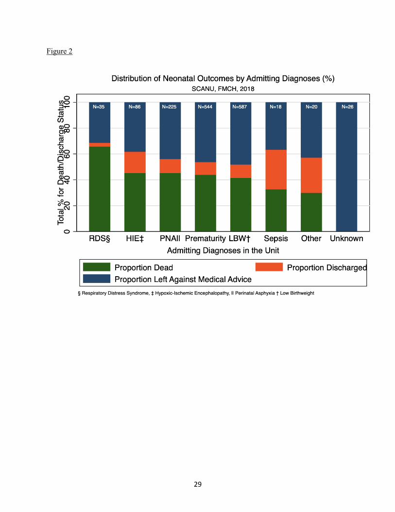
Figure 2: Distribution of Neonatal Outcomes by Admitting Diagnoses (%), SCANU, FMCH,
2018........................................................................................................................................... 29
Figure 3: Kaplan-Meier Curve of the Study Population (N = 693), SCANU, FMCH, 2018 ... 30
Figure 4: Trends in Neonatal Admissions and Deaths from January-November 2018, SCANU,
FMCH ....................................................................................................................................... 31
1
Introduction
The neonatal period is considered the most vulnerable period in a child’s life, with the
risk of dying being the highest in the first month of life (UN Inter-agency Group for Child
Mortality Estimation, 2018). Although a steady decline has been observed in neonatal mortality
globally, this decline has been much slower than the global decline observed in mortality
amongst older children from 1990 to 2017; the decline in neonatal mortality was approximately
51% in comparison to a 63% decline in mortality in children aged 1-4 years (UN Inter-agency
Group for Child Mortality Estimation, 2018). This trend has resulted in neonatal deaths
accounting for an increasingly larger proportion of global under-5 deaths in comparison to other
deaths classified as under-5 (United Nations, 2013), approximately 47% in 2017 (UNICEF Data,
2018).
Although neonatal mortality remains a global problem, the burden of neonatal deaths is
primarily focused in sub-Saharan Africa and South Asia (UNICEF Data, 2018). While neonatal
deaths accounted for a relatively lower proportion of deaths amongst children under-5 (37%) in
sub-Saharan Africa in 2017 in comparison to South Asia, neonatal deaths in South Asia
accounted for approximately 60% of under-5 deaths in the region in 2017 (UN Inter-agency
Group for Child Mortality Estimation, 2018 & UNICEF Data, 2018). South Asia’s neonatal
mortality rate in 2017 was 27 deaths per 1,000 live births – comparable with the neonatal
mortality rate in sub-Saharan Africa (27.2 deaths per 1,000 live births) and much higher than the
neonatal mortality rates in developed regions such as North America (4 deaths per 1,000 live
births) and Europe (3 deaths per 1,000 live births) (UN Inter-agency Group for Child Mortality
Estimation, 2018). Of the 2.5 million neonates who died in their first month of life in 2017
2
worldwide, South Asia accounted for 38% of the deaths, with 973,000 neonatal deaths in the
region that year (UN Inter-agency Group for Child Mortality Estimation, 2018).
Achieving global child mortality reduction goals has been difficult in this region.
Sustainable Development Goal #3 calls for all countries to reduce their neonatal mortality rates
to 12 deaths per 1,000 live births by 2030 (United Nations, 2018). Prior to the implementation of
the SDGs in 2015, Millennium Development Goal #4 called for a reduction of two-thirds in
mortality in children under the age of five from 1990 till 2015 (United Nations, 2015) – although
South Asia saw a substantial decline in neonatal mortality over the 25 years, (United Nations
Development Programme, 2015) most countries in the region did not achieve the global target
(Asian Development Bank, 2015).
Bangladesh was one of three South Asian countries to achieve MDG 4 (Asian
Development Bank, 2015). Bordered by India and Myanmar, Bangladesh’s population was
approximately 159 million people as of July 2018, making it the 8th most populous country in the
world at the time (Central Intelligence Agency, 2019). The GDP in 2017 was estimated at
approximately $261.5 billion. Twenty four percent of the population was considered to be below
the poverty line in 2016 (Central Intelligence Agency, 2019). Although factors such as
entrenched poverty and poor access to high-quality health services have hampered Bangladesh’s
progress towards achieving universal health care by 2032 (International Development Research
Centre, 2018), initiatives such as immunization campaigns, Vitamin A supplementation and
successful control of diarrheal diseases aided in Bangladesh meeting MDG 4’s requirements and
drastically improving the health of women and children in the country (International
Development Research Centre, 2018 & United Nations Development Programme, Bangladesh).
3
While improving neonatal survival was not an explicitly separate objective under the
MDGs like it is under the SDGs, Bangladesh’s commitment to improving newborn health in
particular is bolstered by the implementation of Special Care Newborn Units, commonly called
SCANUs. SCANUs were established to avert neonatal deaths due to preventable causes such as
infection and other birth complications, as part of a UNICEF newborn health initiative which
also included the provision of skilled healthcare workers and clean facilities (UNICEF
Bangladesh). The main services provided by SCANUs include care and management of sick
neonates, barring those who require surgical intervention and mechanical ventilation, as well as
follow-up for those deemed to be at high risk of adverse outcomes. The units also provide
referral services and employ medical professionals trained in newborn care (Directorate General
of Health Services, 2011). UNICEF set up SCANUs in four medical college hospitals (UNICEF
Bangladesh), while USAID and Save the Children have also installed SCANUs in the districts of
Jaintapur (U.S. Agency for International Development, 2019) and Lakshmipur (Save the
Children, Bangladesh, 2016) respectively. As of 2017, SCANUs have been established in 42
facilities in Bangladesh (Directorate General of Health Services, 2017). Despite the
implementation of the SCANUs, neonatal mortality is still a matter of concern in Bangladesh - as
of 2017, the neonatal mortality rate was 18.4 deaths per 1,000 live births (UNICEF Data, 2018),
considerably higher than the 2030 target.
Common risk factors for neonatal mortality worldwide include birth complications
associated with prematurity, as well as intrapartum-related complications, and diagnoses of
sepsis, meningitis, pneumonia and other congenital defects (Liu, L. et al, 2012). The state of
affairs is no different in South Asia – in 2017, prematurity and intrapartum complications were
determined to be the main causes of neonatal death in South Asia (UNICEF Data, 2018). Prior
4
studies have also established diagnoses of prematurity, birth asphyxia, neonatal sepsis, and
pneumonia as leading risk factors for neonatal mortality in Bangladesh (Khatun, F. et al, 2012 &
Chowdhury, H. R. et al, 2010). Maternal characteristics and socio-economic factors such as low
maternal age, low maternal education, lack of sanitation, birth rank, and lack of antenatal care-
seeking have also been indicated to be risk factors for neonatal mortality in the country (Kamal,
S. M. M., 2015 & Kamal, S. M. M. et al, 2012 & Roy, S. et al, 2018). While risk factors for
community-based neonatal cohorts have been extensively studied in Bangladesh with the help of
community health workers (Shah, R. et al, 2014 & Mitra, D. K., et al, 2018), risk factor profiles
in hospital settings such as the SCANUs are fewer. Previous research has shown that receiving
care at a district hospital or medical college in Bangladesh does not necessarily reduce the risk of
neonatal death (Halim A. et al, 2016), which indicates a need to understand what factors hamper
the effectiveness of hospital care in averting adverse outcomes in neonates. Evaluations of case
fatality rates associated with the services provided in SCANUs in India identified an association
between aseptic practices and positive neonatal outcomes in these units based on variation of the
case fatality rates associated with aseptic practice scores (Neogi S. B. et al, 2011); the same has
not been established for SCANUs in Bangladesh. Considering that SCANUs in Bangladesh have
been established in multiple districts across the country with the goal to improve neonatal
survival through the provision of life-saving treatments, characterizing their functioning can
allow for unique insight into the units studied and their impact on neonatal survival. Identifying
clinical risk factors for neonatal mortality in different facilities with SCANUs or similar units is
crucial if progress towards reducing neonatal deaths is to be made and has the potential for the
implementation of targeted interventions to address these factors. Seasonality in neonatal deaths
in Bangladesh has been previously established, with the occurrence of neonatal deaths increasing
5
in the winter (Becker, S., Weng, S., 1998); studying possible trends in seasonal clustering in the
admissions and deaths in the unit may highlight factors that affect neonatal survival and the
individual newborn periods throughout the course of the year.
This study aims to profile a SCANU in Faridpur Medical College Hospital in the district
of Faridpur, Dhaka, Bangladesh. We aim to describe the individuals admitted to the SCANU, as
well as any underlying seasonal patterns observed in the admissions and deaths in the unit in
2018. Additionally, we aim to preliminarily characterize potential risk factors associated with
early death post hospital admission i.e. death occurring within the first two days of admission to
the unit.
6
Methods
Study Design and Site
The study team conducted a retrospective longitudinal study of neonates who were
admitted to the Special Care Neonatal Unit (SCANU) at Faridpur Medical College Hospital
(FMCH) between 1st January 2018 and 30th November 2018. FMCH is a tertiary-level
healthcare facility in the district of Faridpur that serves as a referral hospital and has had an
active SCANU since 2009, which served as the primary study site for this study.
The SCANU primarily admits premature, low birthweight neonates considered at risk for
perinatal septicemia, asphyxia, neonatal jaundice, respiratory infections, and other birth
complications.
Data Extraction and Digitization
Hospital officials at FMCH extracted data from the paper medical records of all neonates
admitted to the unit in 2018. A data extraction instrument consisting of 120 variables was used to
extract and digitize the relevant data. A trained physician on the study team entered data from the
records including important dates for each neonate such as the date of admission to the ward, the
date of death or discharge, whichever came first, and the date of birth. Information on potential
risk factors including the age at admission in days, sex at birth, admitting and subsequent
diagnoses, location of the birth, delivery method, antibiotic regimen, treatments administered
such as intubation, oxygen masks, ventilation, and thermal regulation, pathogens detected,
sharing of an incubator, and geographic proximity to a neighboring incubator were also entered.
7
Analysis Methods
For the descriptive objectives of this study, we considered neonates eligible for the study
if they were between the ages of 0 and 28 days at admission to the unit, were admitted alive to
the SCANU between January and November 2018, and had available medical records detailing
the SCANU admission dates and outcomes. We considered the dates of admission to the
SCANU as the time origin in the study for each eligible neonate, and the date of hospital
discharge or death, whichever came first, as the end of observation time. Therefore, the duration
of time spent as a patient in the SCANU was defined using the date of admission and the date of
death or discharge, whichever of the latter came first. Individuals admitted between 1 to 23 hours
were considered to be admitted for one day. Neonates with missing discharge and death dates
were assumed to have absconded after being admitted to the SCANU for one day, since it was
definitively known that they were admitted to the unit for at least one day. No information on
antibiotics or treatments administered was recorded for these individuals, which indicates they
may not have been admitted to the unit for a substantial amount of time.
We calculated proportions for demographics and other measured characteristics to
compare the neonates that died while admitted to the unit to those that were discharged from the
unit and those that left the unit against medical advice. Differences in characteristics between the
groups were evaluated using 2 test statistics and the Kruskal-Wallis H test.
We calculated the proportion of neonates that died in the entire study population using
the total number of deaths in the study population as the numerator and the eligible study
population size as per the inclusion criteria, as the denominator.
We also calculated cause-specific case fatality proportions using the number of deaths in
those diagnosed with prematurity, low birthweight, sepsis, perinatal asphyxia, hypoxic-ischemic
8
encephalopathy and respiratory distress throughout the duration of the study. The denominator
was considered to be the number of neonates eligible for this analysis as per the inclusion criteria
stated. Individuals identified as premature or having low birthweight were assumed to have been
categorized as per the World Health Organization’s definitions: birth before the completion of 37
weeks of pregnancy was considered premature, and a birthweight below 2500 grams was
considered to be low (Zupan, J., & Åhman, E.,2006).
To analyze the occurrence of the deaths with respect to time spent admitted to the ward,
we set up a survival dataset and plotted a Kaplan-Meier curve to look at survival of the neonates
over time, in the eligible study population. The date of admission to the unit was designated as
the time of study entry, and the dates of death or discharge, depending on whichever date came
first, were designated as the time of study exit. The number of days spent as a patient in the unit
was considered as the time axis. In-ward death was specified as the event of interest, and
individuals who were discharged were considered censored. Neonates with no exit dates were
considered censored after one day of admission to the unit.
Using the dates of admission and the outcomes determined for each individual neonate,
we displayed potential seasonal trends in SCANU admissions and deaths from January till the
end of November in 2018.
For the second objective, we defined the outcome of the analysis as in-ward neonatal
death within the first 48 hours of admission to the SCANU between January and November in
2018, modeled as a binary variable. Preliminary data exploration indicated a large proportion of
deaths occurring within the first two days of admission - in order to characterize risk factors
associated with death soon after admission to the unit, we chose a cut-point of 48 hours. The
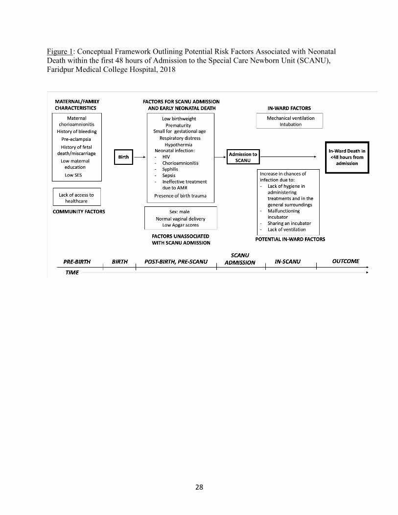
conceptual framework behind the analysis is described (Figure 1). For the purpose of this
9
analysis, individuals who left against medical advice within 48 hours of admission were
excluded in addition to the individuals already excluded for the descriptive objectives. We
calculated proportions for demographics and other measured characteristics to compare the
neonates that died in the first 48 hours of admission to those that survived beyond 48 hours from
admission. Differences in characteristics between the groups were evaluated using 2 test
statistics and the Wilcoxon-Mann-Whitney test.
To assess for potential risk factors associated with death within 48 hours of admission to
the unit, we used univariable and multivariable logistic regression models. The univariable
models were used to characterize possible associations between individual covariates such as age
at admission, sex, prematurity (defined as birth before the completion of 37 weeks of
pregnancy), low birthweight, diagnoses of sepsis, perinatal asphyxia, hypoxic-ischemic
encephalopathy or respiratory distress, home as a birth location, delivery methods, and incubator
sharing logistics and the outcome. We included covariates in the multivariable model based on
the biological rationale associated with risk factors previously established for neonatal mortality
as well as a p<0.1 specified a priori, for flexibility in the inclusion criterion for the adjusted
model. Mickey and Greenland have previously found that p<0.05 failed to identify important
covariates when screening them for inclusion in a multivariable model and concluded that
significance levels for p-values can be increased in order to decrease type II error when selecting
covariates (Mickey R. M., Greenland S., 1989). We subsequently ran the multivariable logistic
regression model to assess the effects of the chosen covariates such as age at admission, sex, and
diagnoses for prematurity, perinatal asphyxia and hypoxic-ischemic encephalopathy, to analyze
their potential associations with the risk for the outcome. Age was modeled as a continuous
variable, while sex and the diagnoses were modeled as binary variables. We reported the crude
10
and adjusted odds ratios with their 95% CIs as a measure of the strength of the associations, to
determine if the covariates functioned as risk factors for early death after admission to the ward.
Ethics Statement
Prior to analysis, data were de-identified via the removal of personally identifiable
information such as names and addresses. The study protocol was reviewed and approved by the
Research and Ethical Review Committees of the International Centre for Diarrheal Disease
Research, Bangladesh.
11
Results
Description of the Study Population
For the descriptive objectives of this study, of the 702 neonates admitted to the unit in
2018, 693 neonates were considered eligible for inclusion in the study population. We excluded
9 individuals due to their admission to the unit after 30th November 2018 (1.3%). The median
duration of time spent in the unit for the 693 neonates was 3 days, with an interquartile range of
3 days. The mean duration of time spent in the unit was 4 days, with 1 day being the minimum
amount of time spent in the unit and 22 days being the maximum.
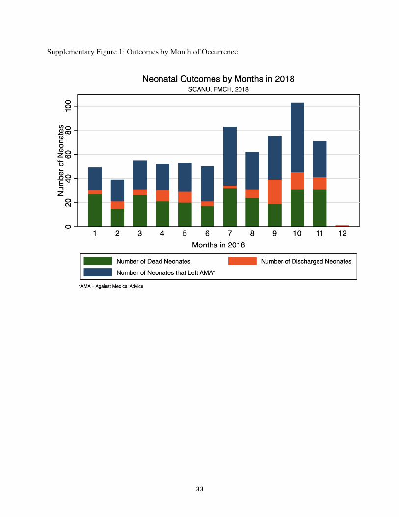
Over the 11-month period, in the population of 693 neonates, the proportion of neonates
that died in the unit was 38% (263 neonates). In the same time period, 49% (340 neonates)
absconded or were discharged against medical advice and 13% (90 neonates) were discharged. A
similar trend was observed in each month of admission, with discharge from the unit being the
least likely outcome across the 11-month period. (see Supplementary Figure 1).
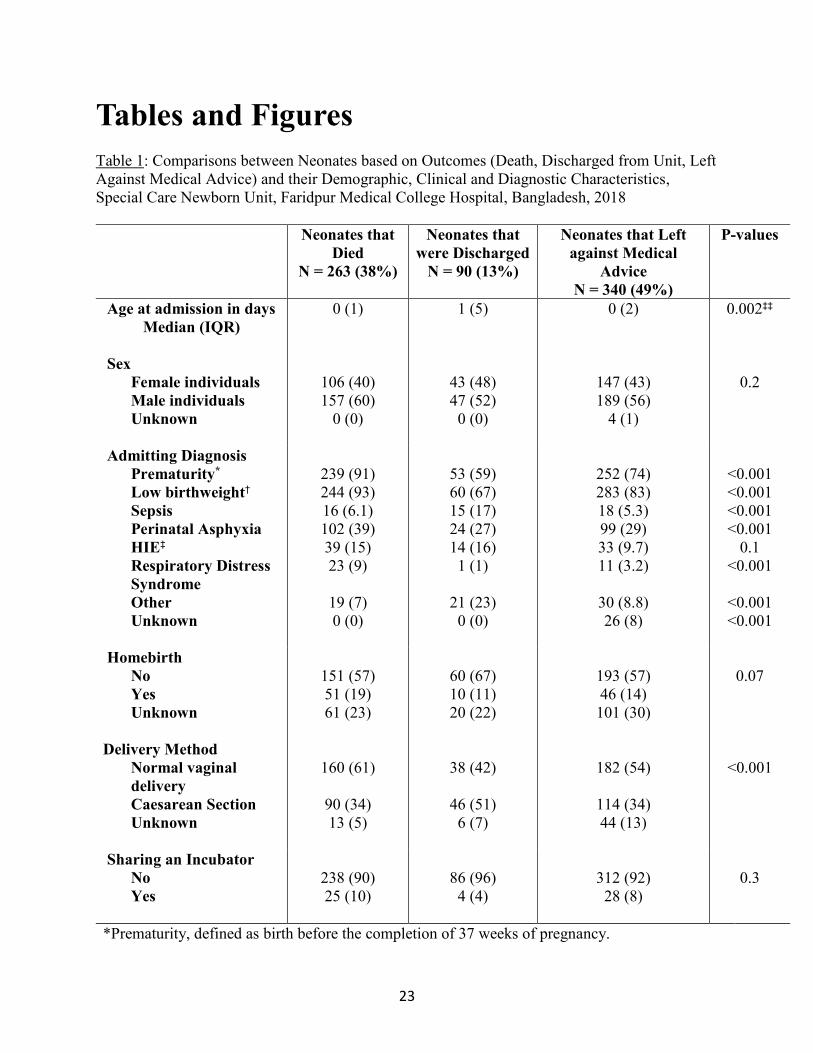
Comparisons between the neonates on the basis of their outcomes of death, discharge and
leaving against medical advice indicated that the median age of admission was less than 1 day
for those that died and those that left against medical advice. However, for those that were
discharged, the median age of admission was 1 day (p<0.001). Male neonates outnumbered
female neonates in those that died (60% male) and those that left against medical advice (56%
male). Individuals who died were more likely to receive diagnoses of prematurity (91%, 239
neonates), low birthweight (93%, 244 neonates), perinatal asphyxia (39%, 102 neonates) and
respiratory distress syndrome (9%, 23 neonates), in comparison to the other two outcome groups
(p<0.001), indicating a statistically significant difference in survival between the outcome
groups. However, individuals who were discharged had the largest proportion of those diagnosed
12
with sepsis (n=15, 17%) (p<0.001). Although the diagnosis of prematurity was based on the
occurrence of the birth before 37 weeks of pregnancy were completed, gestational age was not
routinely recorded in the medical files, leading to a lack of specificity within the diagnoses of
prematurity due to the unavailability of data. All three outcome groups indicated that more than
half of the births took place at locations that were not the home of the family. Of those who died,
61% (160 neonates) had been born through a normal vaginal delivery, in comparison to the 42%
(38 neonates) of those who were discharged and the 54% (182 neonates) of those who left
against medical advice who were born through the same method (p<0.001). Incubator-sharing
was not prevalent in the three groups, with 90% or more of the neonates in each group being
assigned their own individual incubator (Table 1).
The distribution of diagnoses across those that died, those who were discharged and those
who left against medical advice was also described (Figure 2). Those diagnosed with respiratory
distress had the largest proportion of deaths (66%, 23/35 neonates) of all the diagnoses, while the
collective proportion of those diagnosed with conditions other than prematurity, low birthweight,
sepsis, perinatal asphyxia, hypoxic-ischemic encephalopathy, and respiratory distress was 30%
(21/70 neonates). Individuals diagnosed with sepsis had the largest proportion of individuals who
recovered and were discharged (31%, 15/49 neonates), while individuals diagnosed with
respiratory distress had the smallest proportion of individuals discharged (3%, 1/35 neonates).
Proportions of those who left the unit against medical advice ranged from 31% to 48% across all
the diagnoses, barring those whose diagnoses were unknown. The outcomes of these individuals
remain uncertain, due to a lack of data regarding their decision to leave the unit as well and their
outcomes after exiting the unit.
13
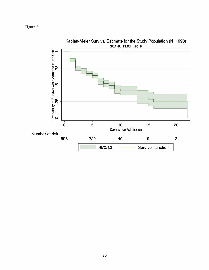
The Kaplan-Meier curve indicated a consistent decline in the probability of survival in the
first week of admission to the unit, with a drop in the probability of survival in the first two days
followed by a more staggered decline. The median survival time is approximately 8 days from
admission to the unit (Figure 3).
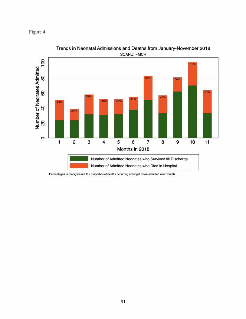
Trends in Admissions and Death in the Unit in 2018
Based on the trend analyses conducted, January had the highest proportion of deaths (53%,
27/51 neonates admitted that month), while September had the lowest proportion of deaths (24%,
19/81 neonates admitted that month). The proportion of those dead varies from 31% to 53% of
those admitted in the respective months, over the 11-month period. Barring January, the
proportion of those dead is less than 50% of the admitted in the remaining months. The number
of admissions ranged from 39 (February) to 58 (March) neonates in the first half of the year (up
till June). However, from July onwards, admissions appear to increase in comparison to the first
half of the year, with the number of admissions ranging from 55 (June) to 101(October) neonates
till November (Figure 4).
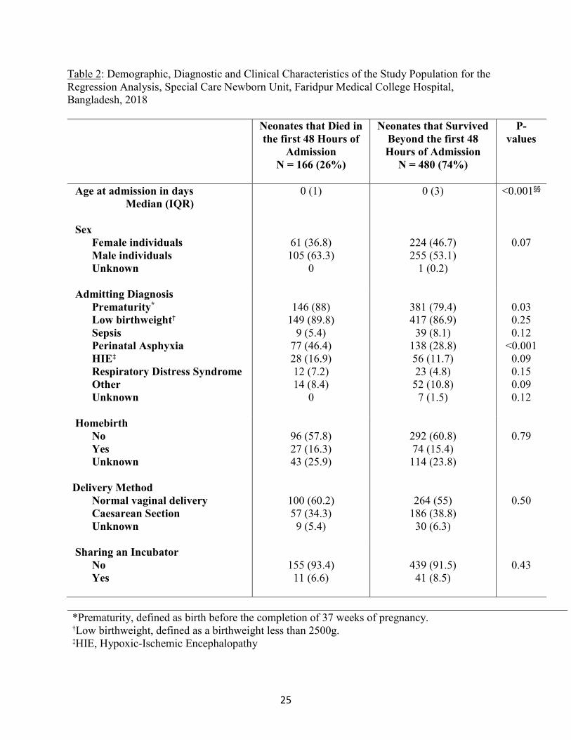
Risk Factor Identification for Death within 48 Hours of Admission
For the second objective, where we aimed to identify risk factors for death within 48 hours of
admission to the unit, we determined 646 neonates to be eligible for inclusion in the study
population for this analysis, after excluding 47 individuals who absconded within 48 hours of
admission to the unit. A quarter of the neonates (26%, 166 neonates) considered in this study
population died within 48 hours of admission to the unit. Of those that died within the first 48
hours, 88% were considered premature, compared to the 79% premature amongst those who
14
survived beyond the first 48 hours; the difference in survival between these two groups was
statistically significant. (p=0.03) The difference in survival beyond the first 48 hours was also
statistically significant amongst those with a diagnosis of perinatal asphyxia and those without -
nearly half of those who died within the first 48 hours were diagnosed with perinatal asphyxia
(46%), while only 29% of those that survived beyond the first 48 hours had the same diagnosis.
(p<0.001) (Table 2).
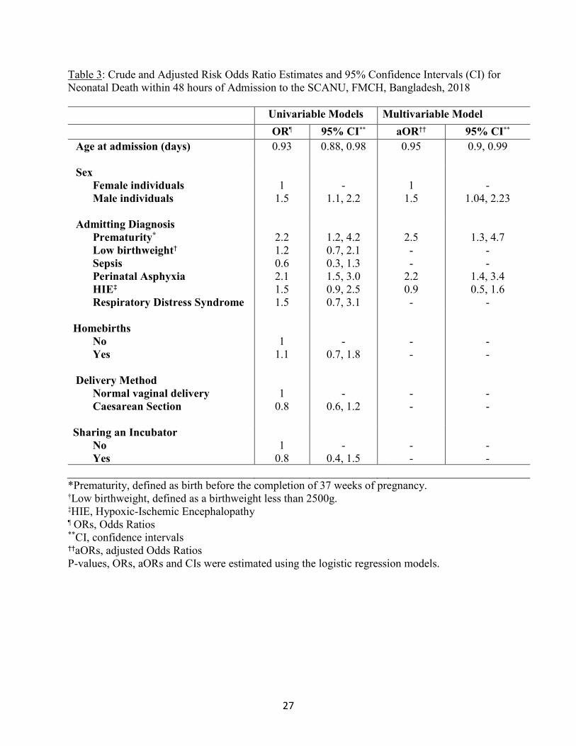
Finally, we presented crude and adjusted odds ratios for death within 48 hours of admission
to the unit to identify potential risk factors amongst demographic and clinical characteristics of
the patients (Table 3). The crude results from the univariable models indicated that each
additional day of age at admission (OR: 0.93, 95% CI: 0.88, 0.98) was associated with a
reduction in the odds of death within 48 hours of admission to the unit, while male sex (OR: 1.5,
95% CI: 1.1, 2.2), prematurity defined as birth before the completion of 37 weeks of pregnancy
(OR: 2.2, 95% CI: 1.2, 4.2), and a diagnosis of perinatal asphyxia (OR: 2.1, 95% CI: 1.5, 3.0)
indicated a higher odds of dying in the first 48 hours of admission to the unit. Although a
diagnosis of hypoxic-ischemic encephalopathy also indicated higher odds for the outcome, the
odds ratio estimate was only marginally significant (OR: 1.5, 95% CI: 0.9, 2.5). Adjusting for
these variables using a multivariable model indicated that an increase of one day in the age at
admission was associated with a 5% decrease in the odds of dying within 48 hours of admission.
(OR: 0.95, 95% CI: 0.9, 0.99). Male neonates had 1.5 times the odds of dying within 48 hours, in
comparison to female neonates (OR: 1.5, 95% CI: 1.04, 2.23). A diagnosis of prematurity
increased the odds of dying in the first 48 hours by 150% (OR: 2.5, 95% CI: 1.3, 4.7), while a
diagnosis of perinatal asphyxia increased the odds by 120% (OR: 2.2, 95% CI: 1.4, 3.4).
15
Discussion
Our analysis of the data revealed a large proportion of neonatal deaths in the unit (38%)
through 11 months in 2018. In comparison, the proportion of deaths amongst neonates attended
by hospital physicians in the United States is approximately 0.06% (Grünebaum, A. et al, 2017).
A study similar to ours was conducted in a neonatal intensive care unit in a tertiary care hospital
in India and reported a proportion of neonatal deaths of 46% (Tagare, A. et al, 2013) which
indicates that a proportion of deaths this high may not be uncommon in certain geographic
regions. Regardless, the high proportion of deaths in the SCANU is alarming, despite the fact
that neonates admitted to the unit are already sick and therefore may be less likely to survive.
Comparing the neonates based on their outcomes – whether they died, were discharged,
or left the unit against medical advice – indicated that the neonates who died were more similar
to those who left against medical advice than to those that were discharged. This is evidenced by
the differences in the median age of admission, proportions of those with specific diagnoses as
well as the method of birth delivery, where individuals who were discharged were older at
admission, healthier and born through caesarean sections in comparison to those in the other two
outcome groups. Caesarean sections have been previously shown to be associated with lower
odds of neonatal death in Bangladesh (Owais A. et al, 2013). Although we possess no specific
information as to the true outcomes of those individuals that left against medical advice, the
similarities between this group of neonates and those that died reveals that those who left may be
comparable to those who died. Possible reasons for these neonates being discharged against
advice may be due to the parents of these neonates choosing to have their children discharged
from the unit – either due to an inability to sustain payment of the hospital bills, or a desire to
16
have their child die at home instead of the hospital. The latter reason would assume that the
parents were aware of the full extent of their child’s illness and were making a conscious
decision while accounting for it.
Individuals who were discharged also presented with the largest proportion of individuals
diagnosed with sepsis. Sepsis has been established globally as a risk factor for neonatal mortality
(Lawn, J. E. et al, 2005) and has been identified previously as a cause of death amongst neonates
in Bangladesh (Khatun, F. et al, 2012). It is possible that we observe better survival outcomes
associated with sepsis diagnoses in our study population due to neonates with severe cases of
sepsis dying before they can be admitted to the SCANU, thus resulting in them being excluded
from the study population for this analysis. This would also explain the lower number of
neonates diagnosed with sepsis (n = 48), in comparison to other outcomes known to be risk
factors for neonatal death such as prematurity and perinatal asphyxia.
Considering the other diagnoses, respiratory distress syndrome, which has the highest
proportion of deaths attributed out of all the diagnoses in this study population, commonly
occurs in premature neonates due to a lack of anatomical development in the lungs (Hermansen,
C. L., & Lorah, K. N., 2007) and has an established association with neonatal mortality in South
Asian countries such as Pakistan (Bhutta Z, 1997).
The descriptive survival curve helped determine that deaths in this unit occurred closer to
the date of admission to the unit. This indicates that the neonates may have been severely ill on
admission, thereby skewing their chances of survival regardless of the setting they are in. Since
information on gestational age was not available to us, it is unclear if the neonates being
admitted were simply born too early to have a reasonable chance of surviving their hospital stay
17
and being discharged as healthy neonates. Information on low birthweight was missing as well –
although it is known to be only indirectly associated with neonatal death, 60-80% of the neonates
that die globally are classified as having low birthweights. (Lawn, J. E. et al, 2005).
The trend analyses indicated differences in the number of new admissions each month, as
well as the number that died each month. The increase in the number of admissions in the latter
half of the year indicates there may be a seasonality in the number of admissions – it is possible
that external drivers influence the occurrence of adverse neonatal outcomes or encourage
healthcare-seeking amongst parents in the later months of the year. Additionally, considering that
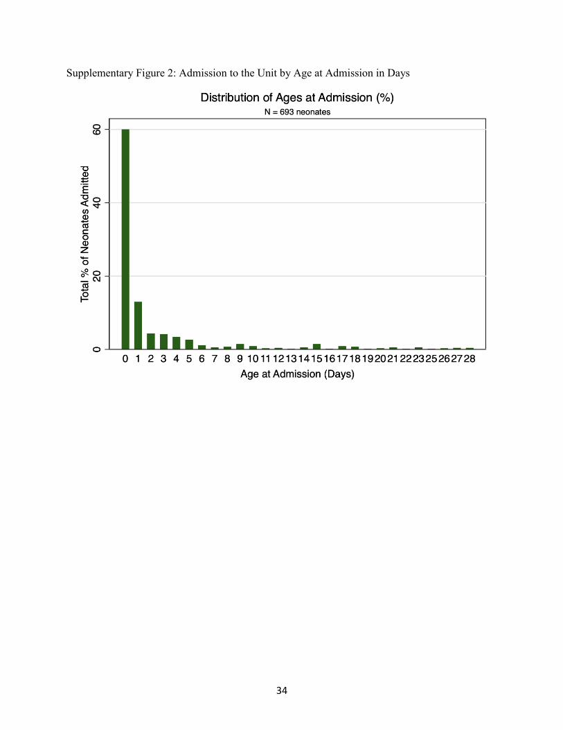
60% of the neonates (n = 416) overall were admitted to the unit on the day they were born (see
Supplementary Table 2), a seasonality in births may also be indicated. More data across multiple
years would be required to adequately characterize these hypothesized trends.
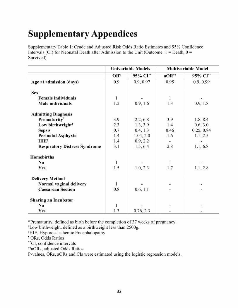
Neonates who died within 48 hours of admission to the unit were more likely to be male,
premature, and have diagnoses of perinatal asphyxia compared to those neonates that survived
beyond the first 48 hours from unit admission. These results were also corroborated by the
regression analysis, where lower age at admission, male sex, and diagnoses of prematurity and
perinatal asphyxia were identified as potential risk factors for death within the first 48 hours of
admission. Similar results are also observed when considering death in the unit as the primary
outcome using logistic regression models (see Supplementary Table 1). This aligns with the
literature on neonatal mortality in Bangladesh: prematurity is a well-established cause of death in
the neonatal period, as is asphyxia and the complications associated with it (Lawn, J. E. et al,
2005). A previous study identified prematurity as one of the main causes of neonatal death in
rural Bangladesh, and males accounted for 60% of the neonatal deaths (Owais A. et al, 2013).
18
Globally, the disparity in survival across gender categories in premature neonates is particularly
stark – males have worse survival than females when considering adverse neonatal outcomes
such as hemorrhages, and congenital malformations (Naeye R.L. et al, 1971). In addition to male
sex being a risk factor for mortality on the basis of perinatal characteristics such as low Apgar
scores, male neonates have also been found to be at higher risk for adverse neurological
outcomes and disability in comparison to female neonates, should they survive into infanthood
(Kent, A. L. et al, 2012). This ‘male disadvantage’ indicates that the effect of sex on neonatal
outcomes is independent of the biological disease processes involved, and that male sex is an
important risk factor for neonatal mortality (Naeye R.L. et al, 1971). Younger age also has an
established association with neonatal death - studies from Southeast Asian and sub-Saharan
African countries report a high burden of neonatal deaths in the first 3 days after birth (Sankar,
M. J. et al, 2016), indicating that younger neonates in these settings are more likely to die than
older neonates.
Limitations
One of the main limitations of the study is the lack of specificity in the data available
from the medical records. Due to a dearth of information regarding gestational age at birth of the
individuals included in the study population, the status of prematurity could not be stratified into
the sub-categories of extremely preterm (<28 weeks), moderately preterm (28-32 weeks) and late
preterm (32-37 weeks). The sub-categories of prematurity have been previously shown to have
different levels of survival at each stage, with the highest population attributable fraction
observed in extremely preterm individuals (Baqui, A. H. et al, 2013). Being unable to stratify
preterm individuals in our study population into these sub-categories may have obscured certain
nuanced effects that would have been reflected otherwise in the stratified estimates. Additionally,
19
neonatal birthweights were not reported – due to the lack of definition regarding how neonates
were classified as having a low birthweight, we were unable to assess its role in this analysis.
Since the definitions of prematurity and low birthweight were unclear, the default cutoffs of 37
weeks and 2500g respectively were assumed for the purpose of this study, however this
information was not specified in the medical records. There was also missing data on maternal
characteristics, such as maternal age, and therefore we could not assess for risk factors amongst
these characteristics, although such characteristics have established associations with neonatal
mortality (Kamal, S. M. M., 2015).
Another limitation lies in the fact that nearly half of the study population for the
descriptive objectives either absconded or was discharged against medical advice (49%).
Assumptions of survival for these individuals were not made due to the lack of data available on
what may have occurred after these neonates left the unit and the status of their true survival
outcomes. Assumptions of death could not be made since the individuals were alive when they
left the hospital. This group also includes neonates with missing discharge dates, whom we
assumed absconded after one day of admission – since this includes only 19 neonates (2.7%), it
is likely that this assumption will not bias estimates based on the outcomes. The descriptive
analyses indicated that these individuals were comparable demographically, clinically and
diagnostically to those who died, therefore the lack of certainty regarding the true outcomes of
those who absconded or left against medical advice possibly indicates that our reported
proportion of those who died is an underestimate. The lack of data on their survival outcomes
also affects our interpretation of the cause-specific case fatality proportions – it is possible that
more deaths may be attributed to specific diagnoses than reported in the descriptive analyses.
20
A limitation of the regression analysis may be that the comparison groups considered are
similar, due to the nature and structure of the outcome. In order to assess for risk factors
associated with early death on being admitted to the unit, a 48-hour cut-point was chosen to
separate the two groups. Although the group of neonates that died within the first 48 hours of
admission to the unit comprised 63% of the deaths that occurred in the study population overall,
36% of those that died by the end of the study did survive the first 48 hours and died in the
subsequent days. This indicates that the two groups considered in this case were inherently
structured to be similar in one aspect, since both groups contain individuals that died during their
time in the unit. This may possibly bias estimates and decrease the strength of the associations
characterized for the potential risk factors and the outcome chosen.
Strengths
The strengths lie in the longitudinal nature of the study; we were able to gain an idea of
the risk factors associated with death within 48 hours of admission and were able to characterize
how long neonates were admitted to the study and whether these durations differed across those
with different outcomes. This data may also allow us to look at risk factors associated with early
neonatal death (<7 days of age) in the future. Other studies conducted in SCANUs in Bangladesh
have mostly used cross-sectional data, or have data collected for less than half a year. This study
also succeeds in comprehensively describing the population of neonates admitted to the SCANU
at Faridpur Medical College Hospital based on their demographic and clinical characteristics and
helps preliminarily understand the unique population that is admitted to this ward.
Conclusions
21
Of all the neonates admitted to the unit from January through November 2018, 38% (n =
263) died during their time in the unit and 49% (n = 340) left against medical advice, while only
13% (n = 90) were discharged following recovery. Those who left against medical advice appear
demographically and clinically more similar to those that died while admitted to the ward than
those that were discharged from the unit. Approximately a quarter of the study population died
within 48 hours of admission to the unit. Factors found to be associated with death within 48
hours of admission to the unit included younger age at admission to the unit, prematurity, and
perinatal asphyxia. These findings emphasize the need for higher treatment capacity in order to
address gaps in treating younger, premature neonates diagnosed with perinatal asphyxia, as well
as assessing maternal health and identifying potential risk factors associated with neonatal death
in the unit.
Recommendations
Based on the high incidence of mortality in the unit, further investigation into the factors
associated with it is necessary if conditions in the unit are to be improved in order to support
neonatal survival and ensure better health outcomes. Medical records ought to include
information regarding the gestational age, as well as any other maternal information available
such as maternal infection and age, in order to fully clarify a neonate’s risk profile for mortality.
Evaluations of the services provided in the SCANU could be conducted, to assess their impact on
neonatal survival separately from the clinical characteristics outlined in this study; these could
include assessments of the functioning of the incubators, maintenance of aseptic practices in the
unit and the effects of incubator sharing on neonatal outcomes. Additionally, it is possible that
other SCANUs in Bangladesh are facing the same issues regarding the incidence of mortality – it

22
is important that these units also get the requisite resources to help them address the specific gaps
in treatment that are affecting unit-specific neonatal survival.
23
Tables and Figures
Table 1: Comparisons between Neonates based on Outcomes (Death, Discharged from Unit, Left
Against Medical Advice) and their Demographic, Clinical and Diagnostic Characteristics,
Special Care Newborn Unit, Faridpur Medical College Hospital, Bangladesh, 2018
Neonates that
Died
N = 263 (38%)
Neonates that
were Discharged
N = 90 (13%)
Neonates that Left
against Medical
Advice
N = 340 (49%)
P-values
Age at admission in days
Median (IQR)
0 (1) 1 (5) 0 (2) 0.002‡‡
Sex
Female individuals
Male individuals
Unknown
106 (40)
157 (60)
0 (0)
43 (48)
47 (52)
0 (0)
147 (43)
189 (56)
4 (1)
0.2
Admitting Diagnosis
Prematurity*
Low birthweight†
Sepsis
Perinatal Asphyxia
HIE‡
Respiratory Distress
Syndrome
Other
Unknown
239 (91)
244 (93)
16 (6.1)
102 (39)
39 (15)
23 (9)
19 (7)
0 (0)
53 (59)
60 (67)
15 (17)
24 (27)
14 (16)
1 (1)
21 (23)
0 (0)
252 (74)
283 (83)
18 (5.3)
99 (29)
33 (9.7)
11 (3.2)
30 (8.8)
26 (8)
<0.001
<0.001
<0.001
<0.001
0.1
<0.001
<0.001
<0.001
Homebirth
No
Yes
Unknown
Delivery Method
Normal vaginal
delivery
Caesarean Section
Unknown
151 (57)
51 (19)
61 (23)
160 (61)
90 (34)
13 (5)
60 (67)
10 (11)
20 (22)
38 (42)
46 (51)
6 (7)
193 (57)
46 (14)
101 (30)
182 (54)
114 (34)
44 (13)
0.07
<0.001
Sharing an Incubator
No
Yes
238 (90)
25 (10)
86 (96)
4 (4)
312 (92)
28 (8)
0.3
*Prematurity, defined as birth before the completion of 37 weeks of pregnancy.

24
†Low birthweight, defined as a birthweight less than 2500g. ‡HIE, Hypoxic-Ischemic Encephalopathy ‡‡P-value calculated using the Kruskal-Wallis H test.
P-values were estimated using Kruskal-Wallis H tests for medians and 2 test statistics for
differences in proportions. Pearson’s chi square test was the default test conducted.
25
Table 2: Demographic, Diagnostic and Clinical Characteristics of the Study Population for the
Regression Analysis, Special Care Newborn Unit, Faridpur Medical College Hospital,
Bangladesh, 2018
Neonates that Died in
the first 48 Hours of
Admission
N = 166 (26%)
Neonates that Survived
Beyond the first 48
Hours of Admission
N = 480 (74%)
P-
values
Age at admission in days
Median (IQR)
0 (1) 0 (3) <0.001§§
Sex
Female individuals
Male individuals
Unknown
61 (36.8)
105 (63.3)
0
224 (46.7)
255 (53.1)
1 (0.2)
0.07
Admitting Diagnosis
Prematurity*
Low birthweight†
Sepsis
Perinatal Asphyxia
HIE‡
Respiratory Distress Syndrome
Other
Unknown
146 (88)
149 (89.8)
9 (5.4)
77 (46.4)
28 (16.9)
12 (7.2)
14 (8.4)
0
381 (79.4)
417 (86.9)
39 (8.1)
138 (28.8)
56 (11.7)
23 (4.8)
52 (10.8)
7 (1.5)
0.03
0.25
0.12
<0.001
0.09
0.15
0.09
0.12
Homebirth
No
Yes
Unknown
Delivery Method
Normal vaginal delivery
Caesarean Section
Unknown
96 (57.8)
27 (16.3)
43 (25.9)
100 (60.2)
57 (34.3)
9 (5.4)
292 (60.8)
74 (15.4)
114 (23.8)
264 (55)
186 (38.8)
30 (6.3)
0.79
0.50
Sharing an Incubator
No
Yes
155 (93.4)
11 (6.6)
439 (91.5)
41 (8.5)
0.43
*Prematurity, defined as birth before the completion of 37 weeks of pregnancy. †Low birthweight, defined as a birthweight less than 2500g. ‡HIE, Hypoxic-Ischemic Encephalopathy

26
§§P-value calculated using the Wilcoxon-Mann-Whitney test.
P-values were estimated using Wilcoxon-Mann-Whitney tests for medians and 2 test statistics for
differences in proportions. Pearson’s chi square test was the default test conducted.
27
Table 3: Crude and Adjusted Risk Odds Ratio Estimates and 95% Confidence Intervals (CI) for
Neonatal Death within 48 hours of Admission to the SCANU, FMCH, Bangladesh, 2018
Univariable Models Multivariable Model
OR¶ 95% CI** aOR†† 95% CI**
Age at admission (days)
0.93 0.88, 0.98 0.95 0.9, 0.99
Sex
Female individuals
Male individuals
1
1.5
-
1.1, 2.2
1
1.5
-
1.04, 2.23
Admitting Diagnosis
Prematurity*
Low birthweight†
Sepsis
Perinatal Asphyxia
HIE‡
Respiratory Distress Syndrome
2.2
1.2
0.6
2.1
1.5
1.5
1.2, 4.2
0.7, 2.1
0.3, 1.3
1.5, 3.0
0.9, 2.5
0.7, 3.1
2.5
-
-
2.2
0.9
-
1.3, 4.7
-
-
1.4, 3.4
0.5, 1.6
-
Homebirths
No
Yes
1
1.1
-
0.7, 1.8
-
-
-
-
Delivery Method
Normal vaginal delivery
Caesarean Section
1
0.8
-
0.6, 1.2
-
-
-
-
Sharing an Incubator
No
Yes
1
0.8
-
0.4, 1.5
-
-
-
-
*Prematurity, defined as birth before the completion of 37 weeks of pregnancy. †Low birthweight, defined as a birthweight less than 2500g. ‡HIE, Hypoxic-Ischemic Encephalopathy ¶ ORs, Odds Ratios **CI, confidence intervals ††aORs, adjusted Odds Ratios
P-values, ORs, aORs and CIs were estimated using the logistic regression models.
28
Figure 1: Conceptual Framework Outlining Potential Risk Factors Associated with Neonatal
Death within the first 48 hours of Admission to the Special Care Newborn Unit (SCANU),
Faridpur Medical College Hospital, 2018
29
Figure 2
30
Figure 3
31
Figure 4
32
Supplementary Appendices
Supplementary Table 1: Crude and Adjusted Risk Odds Ratio Estimates and 95% Confidence
Intervals (CI) for Neonatal Death after Admission to the Unit (Outcome: 1 = Death, 0 =
Survived)
Univariable Models Multivariable Model
OR¶ 95% CI** aOR†† 95% CI**
Age at admission (days)
0.9 0.9, 0.97 0.95 0.9, 0.99
Sex
Female individuals
Male individuals
1
1.2
-
0.9, 1.6
1
1.3
-
0.9, 1.8
Admitting Diagnosis
Prematurity*
Low birthweight†
Sepsis
Perinatal Asphyxia
HIE‡
Respiratory Distress Syndrome
3.9
2.3
0.7
1.4
1.4
3.1
2.2, 6.8
1.3, 3.9
0.4, 1.3
1.04, 2.0
0.9, 2.2
1.5, 6.4
3.9
1.4
0.46
1.6
-
2.8
1.8, 8.4
0.6, 3.0
0.25, 0.84
1.1, 2.5
-
1.1, 6.8
Homebirths
No
Yes
1
1.5
-
1.0, 2.3
1
1.7
-
1.1, 2.8
Delivery Method
Normal vaginal delivery
Caesarean Section
1
0.8
-
0.6, 1.1
-
-
-
-
Sharing an Incubator
No
Yes
1
1.3
-
0.76, 2.3
-
-
-
-
*Prematurity, defined as birth before the completion of 37 weeks of pregnancy. †Low birthweight, defined as a birthweight less than 2500g. ‡HIE, Hypoxic-Ischemic Encephalopathy ¶ ORs, Odds Ratios **CI, confidence intervals ††aORs, adjusted Odds Ratios
P-values, ORs, aORs and CIs were estimated using the logistic regression models.
33
Supplementary Figure 1: Outcomes by Month of Occurrence
34
Supplementary Figure 2: Admission to the Unit by Age at Admission in Days

35
Bibliography
1. Asian Development Bank. (2015). MDG 4: Reduce Child Mortality. (n.d.). New Zealand,
6.
2. Baqui, A. H., Rosen, H. E., Lee, A. C. C., Applegate, J. A., El Arifeen, S., Rahman, S.
M., … Black, R. E. (2013). Preterm birth and neonatal mortality in a rural Bangladeshi
cohort: implications for health programs. Journal of Perinatology, 33(12), 977–981.
https://doi.org/10.1038/jp.2013.91
3. Becker, S., Weng, S. (1998). Seasonal patterns of deaths in Matlab, Bangladesh.
International Journal of Epidemiology, 27, 814-823. https://doi-
org.proxy1.library.jhu.edu/10.1093/ije/27.5.814
4. Bhutta, Z. (1997). Profile and outcome of the respiratory distress syndrome among
newborns in Karachi: risk factors for mortality. Journal of Tropical Pediatrics, 43(3),
143–148. https://doi.org/10.1093/tropej/43.3.143
5. Central Intelligence Agency (2019, April). South Asia: Bangladesh. The World Factbook.
Retrieved from https://www.cia.gov/library/publications/the-world-
factbook/geos/bg.html
6. Chowdhury, H. R., Thompson, S., Ali, M., Alam, N., Yunus, M., & Streatfield, P. K.
(2010). Causes of Neonatal Deaths in a Rural Subdistrict of Bangladesh: Implications for
Intervention. Journal of Health, Population and Nutrition, 28(4), 375–382.
https://doi.org/10.3329/jhpn.v28i4.6044
7. Directorate General of Health Services. (2011, November). Standard Operating
Procedure (SOP) For Newborn Care Services at Primary and Secondary Level Hospitals.
36
Retrieved from
http://www.dghs.gov.bd/licts_file/images/SOP/2011_Newborn_Care_SOP_29-11-11.pdf
8. Directorate General of Health Services. (2017, April) National Newborn Health Bulletin
Issue 2. Retrieved from https://www.healthynewbornnetwork.org/resource/bangladesh-
newborn-bulletin/
9. Grünebaum, A., McCullough, L. B., Arabin, B., Dudenhausen, J., Orosz, B., &
Chervenak, F. A. (2017). Underlying causes of neonatal deaths in term singleton
pregnancies: home births versus hospital births in the United States. Journal of Perinatal
Medicine, 45(3). https://doi.org/10.1515/jpm-2016-0200
10. Halim A., Dewez JE, Biswas A, Rahman F, White S, et al. (2016) When, Where, and
Why Are Babies Dying? Neonatal Death Surveillance and Review in Bangladesh. PLOS
ONE, 11(8): e0159388. https://doi.org/10.1371/journal.pone.0159388
11. Hermansen, C. L., & Lorah, K. N. (2007). Respiratory Distress in the Newborn.
American Family Physician, 76(7), 987-994.
12. International Development Research Centre (2018, January). Making healthcare
accessible in Bangladesh. Retrieved from https://www.idrc.ca/en/research-in-
action/making-healthcare-accessible-bangladesh
13. Kamal, S. M. M. (2015). What Is the Association Between Maternal Age and Neonatal
Mortality? An Analysis of the 2007 Bangladesh Demographic and Health Survey. Asia
Pacific Journal of Public Health, 27(2), NP1106–NP1117.
https://doi.org/10.1177/1010539511428949
37
14. Kamal, S. M. M., Ashrafuzzaman, M., & Nasreen, S. A. (2012). Risk Factors of Neonatal
Mortality in Bangladesh. Journal of Nepal Paediatric Society, 32(1), 37–46.
https://doi.org/10.3126/jnps.v32i1.4845
15. Kent, A. L., Wright, I. M. R., Abdel-Latif, M. E., & the New South Wales and Australian
Capital Territory Neonatal Intensive Care Units Audit Group. (2012). Mortality and
Adverse Neurologic Outcomes Are Greater in Preterm Male Infants. PEDIATRICS,
129(1), 124–131. https://doi.org/10.1542/peds.2011-1578
16. Khatun, F., Rasheed, S., Moran, A. C., Alam, A. M., Shomik, M. S., Sultana, M., …
Bhuiya, A. (2012). Causes of neonatal and maternal deaths in Dhaka slums: Implications
for service delivery. BMC Public Health, 12(1). https://doi.org/10.1186/1471-2458-12-84
17. Lawn, J. E., Cousens, S., & Zupan, J. (2005). 4 million neonatal deaths: When? Where?
Why? The Lancet, 365(9462), 891–900. https://doi.org/10.1016/S0140-6736(05)71048-5
18. Liu, L., Johnson, H. L., Cousens, S., Perin, J., Scott, S., Lawn, J. E., … Black, R. E.
(2012). Global, regional, and national causes of child mortality: an updated systematic
analysis for 2010 with time trends since 2000. The Lancet, 379(9832), 2151–2161.
https://doi.org/10.1016/S0140-6736(12)60560-1
19. Mickey, R. M., & Greenland, S. (1989). THE IMPACT OF CONFOUNDER
SELECTION CRITERIA ON EFFECT ESTIMATION. American Journal of
Epidemiology, 129(1), 125–137. https://doi.org/10.1093/oxfordjournals.aje.a115101
20. Mitra, D. K., Mullany, L. C., Harrison, M., Mannan, I., Shah, R., … Baqui, A. H. (2018).
Incidence and risk factors of neonatal infections in a rural Bangladeshi population: a
community-based prospective study. Journal of Health, Population and Nutrition, 37(1).
https://doi.org/10.1186/s41043-018-0136-2
38
21. Naeye R. L., Burt L. S., Wright D. L., Blanc W. A., Tatter D. (1971) Neonatal mortality,
the male disadvantage. PEDIATRICS, 48: 902–6.
22. Neogi, S. B., Malhotra, S., Zodpey, S., & Mohan, P. (2011). Assessment of special care
newborn units in India. Journal of Health, Population, and Nutrition, 29(5), 500-9.
23. Owais A, Faruque ASG, Das SK, Ahmed S, Rahman S, et al. (2013). Maternal and
Antenatal Risk Factors for Stillbirths and Neonatal Mortality in Rural Bangladesh: A
Case-Control Study. PLoS ONE 8(11): e80164.
https://doi.org/10.1371/journal.pone.0080164
24. Roy, S., & Haque, M. A. (2018). Effect of antenatal care and social well-being on early
neonatal mortality in Bangladesh. BMC Pregnancy and Childbirth, 18(1).
https://doi.org/10.1186/s12884-018-2129-y
25. Sankar, M. J., Natarajan, C. K., Das, R. R., Agarwal, R., Chandrasekaran, A., & Paul, V.
K. (2016). When do newborns die? A systematic review of timing of overall and cause-
specific neonatal deaths in developing countries. Journal of Perinatology, 36(S1), S1–
S11. https://doi.org/10.1038/jsp.2016.27
26. Save the Children, Bangladesh. (2016, October). Lakshmipur Gets Its First Special Care
Unit for Newborns. Retrieved from
https://bangladesh.savethechildren.net/news/lakshmipur-gets-its-first-special-care-unit-
newborns
27. Shah, R., Mullany, L. C., Darmstadt, G. L., Talukder, R. R., Rahman, S. M., Mannan, I.,
Arifeen, S. E., Baqui, A. H. (2014). Neonatal mortality, preterm birth, mortality risk,
Bangladesh. Paediatr Perinat Epidemiol, 28: 510-520. https://doi.org/10.1111/ppe.12145
39
28. U.S. Agency for International Development. (2019, March). Saving Newborn Lives in
Bangladesh. Retrieved from www.usaid.gov/news-information/photo-gallery/saving-
newborn-lives-bangladesh.
29. UNICEF Bangladesh. Saving Newborn Lives. Retrieved from
www.unicef.org/bangladesh/en/saving-newborn-lives.
30. UNICEF Data. (2018, March). Neonatal mortality. Retrieved from
https://data.unicef.org/topic/child-survival/neonatal-mortality/
31. UN Inter-agency Group for Child Mortality Estimation. (2018). Levels and Trends in
Child Mortality. Retrieved from
https://childmortality.org/files_v22/download/UN%20IGME%20Child%20Mortality%20
Report%202018.pdf
32. United Nations. (2013). The Millennium Development Goals Report. Retrieved from
http://www.un.org/millenniumgoals/2015_MDG_Report/pdf/MDG%202015%20rev%20
(July%201).pdf
33. United Nations. (2015). Millennium Development Goals and Beyond. Retrieved from
http://www.un.org/millenniumgoals/childhealth.shtml
34. United Nations. (2018). Sustainable Development Goal 3. Retrieved from
https://sustainabledevelopment.un.org/sdg3
35. United Nations Development Programme. (2015). Millennium Development Goal 4.
Retrieved from
https://www.undp.org/content/undp/en/home/sdgoverview/mdg_goals/mdg4.html
40
36. United Nations Development Programme, Bangladesh. Millennium Development Goal 4.
Retrieved from http://www.bd.undp.org/content/bangladesh/en/home/post-
2015/millennium-development-goals/mdg4.html
37. Tagare, A., Chaudhari, S., Kadam, S., Vaidya, U., Pandit, A., & Sayyad, M. G. (2013).
Mortality and Morbidity in Extremely Low Birth Weight (ELBW) Infants in a Neonatal
Intensive Care Unit. The Indian Journal of Pediatrics, 80(1), 16–20.
https://doi.org/10.1007/s12098-012-0818-5
38. Zupan, J., & Åhman, E. (2006). Neonatal and perinatal mortality: country, regional and
global estimates. Geneva: World Health Organization.
41
Biography
Ananya Kumar was born in 1995, in Chennai, India. She graduated magna cum laude from St.
Xavier’s College, Mumbai with a Bachelor of Science in microbiology and biochemistry in 2016
and went on to work as a project research intern in a neurobiology laboratory at the Indian
Institute of Science Education and Research, Pune.
In 2017, Ananya began her graduate studies at the Johns Hopkins Bloomberg School of Public
Health and is currently pursuing a Master of Science degree in Epidemiology, with a focus on
infectious diseases. She plans to graduate in May 2019. While at Johns Hopkins, Ananya worked
with the cholera dynamics group as well as the International Vaccine Access Center and served
as a teaching assistant for introductory epidemiology and outbreak investigation courses.