Embed Size (px)
Citation preview
Risks of Zoonosesfrom Dogs
On Sporting fields
Michael Hayward BVScChair,
Animal Welfare Advisory CommitteeACT.
Convenor Elect,Urban Animal Management Advisory Group
Australian Veterinary Association.
May 23
document.doc Page 1 of 31 May 23Version: 1.1

Contents
Executive summary........................................................................................................3Introduction....................................................................................................................4Helminths.......................................................................................................................5
Toxocara canis............................................................................................................5Uncinaria stenocephala..............................................................................................9Echinococcus granulosus.........................................................................................10
Protozoa........................................................................................................................13Giardia......................................................................................................................13Cryptosporidium......................................................................................................14Other Protozoa.........................................................................................................14
Bacteria.........................................................................................................................15Campylobacter.........................................................................................................15Salmonella................................................................................................................15Escherichia coli........................................................................................................16Leptospira.................................................................................................................17Other Bacteria..........................................................................................................17
Viruses..........................................................................................................................18Foxes............................................................................................................................18Possible solutions.........................................................................................................18
Dung beetles.............................................................................................................18Faecal disposal.........................................................................................................19
Conclusion....................................................................................................................21References....................................................................................................................23
document.doc Page 2 of 31 May 23Version: 1.1
Executive summary
This paper examines the various helminth, protozoan, bacterial and viral organisms which may occur in dog faeces and which may constitute a risk to human health. It also considers the relative risk posed by dogs which are deliberately exercised by their owners on sporting fields as opposed to the risks from stray dogs and foxes defaecating in these areas.
Worms such as the Dog Roundworm Toxocara canis and Hydatid Tapeworm Echinococcus granulosus, and bacteria such as Salmonella, Campylobacter and Escherichia coli are potential pathogens for humans in this area, with potentially serious consequences. However, in almost all cases the risk to humans of these organisms is positively associated with proximity to dogs (i.e. owning a dog), poor dog health (especially diarrhoea), and poor human hygiene such as playing in contaminated soil, eating soil, failure to wash hands after dog or soil contact, kissing dogs or allowing licking on the face, and eating during the above activities. Sporting activities on fields co-occupied by dogs is not mentioned in the veterinary or medical literature as a risk factor.
The incidence of zoonotic organisms in faeces from Canberra dogs is not known, but may be inferred to be low for various reasons including climate and the socio-economic and educational status of dog owners. The risk to those whose immune systems are impaired is significantly higher than to healthy people, who are more likely to be using the sporting fields for activities where they will be in contact with faeces or contaminated soil. Children are probably most at risk at ages where they “play in the dirt” in sand pits and the like, rather than once they start to play sport, but clearly some children of susceptible age will use sporting fields and other public parks
In the author’s opinion, risks to human health can be more effectively and more practically reduced by educating the public about the need to maintain good dog health, including the need for regular effective worming of pets, and by encouraging compliance with existing laws requiring the removal of faeces from public places. This should include provision of bins for the disposal of dog faeces and design of public open space to encourage dog owners to exercise and toilet their pets away from sporting fields and other areas of high human use.
AcknowledgmentsThe author thanks Dr David Jenkins, Australian Hydatid Control and Epidemiology Program, for data and adviceThe author is a veterinarian in small animal practice since 1982, including 17 years in Canberra and 1 year in Gundaroo.
document.doc Page 3 of 31 May 23Version: 1.1
Introduction
This review of the health risks to humans of dogs on sporting fields has been prepared at the request of Mr Ian Baird, Program Manager, Policy and Planning, Canberra Urban Parks and Places, on behalf of the Australian Veterinary Association. It is provided to assist the ACT Government as it reviews the Companion Animals Act 2000 (CAA200), particularly with respect to dog access to public places.
It is based upon a review of the scientific literature, but does not formally assess risk, because it is not being conducted by a specialist epidemiologist or risk manager. Such a review would take many months and cost several thousands of dollars. The current document was prepared by a volunteer in about two weeks.
However, the author is a veterinarian with more than twenty years experience, and a particular interest in parasitology. Veterinarians are trained in, and accept responsibility to safeguard against, zoonoses – that is, diseases which can be spread between animals and man.
This review does not consider physical risks associated with the presence of dogs on sporting fields, such as dog harassment, attack or bite on humans, nor the risk of tripping or falling over a dog. These risks are already prevented in the sense that they are legislated against. CAA2000 forbids anyone to take a dog onto a sporting ground when sport or training is in progress, or to allow a dog to be in a public place unaccompanied and/or unrestrained:
42 Prohibited places(3) A person must not take a dog onto a field or playing area where sport is being played or training for sport is being conducted.
44 Dogs in public places to be restrained(1) A carer must not be in a public place with a dog that is not restrained by a leash, unless the person is in an area designated as an area where dogs are not required to be restrained by a leash.Maximum penalty: 5 penalty units.(2) A keeper must not be in a public place with a dog that is not restrained by a leash, unless the person is in an area designated as an area where dogs are not required to be restrained by a leash.Maximum penalty: 5 penalty units.(3) The keeper of a dog commits an offence if the dog—
(a) is in a public place; and(b) is not with a carer.
This review considers the risks to human health associated with dog faeces and urine being deposited on sporting fields. In passing it comments on risks associated with dog excreta in other public places, and considers the risk from other animals, particularly wild canids. Three groups of organisms are considered to pose a risk: Helminths (worms), Protozoa, and Bacteria.
document.doc Page 4 of 31 May 23Version: 1.1
Helminths
Several helminth parasites of dogs may be transmitted to humans, namely the dog roundworm Toxocara canis, hookworms (Ancylostoma caninum, Uncinaria stenocephala), and hydatid tapeworm Echinococcus granulosus. The following parasites are also considered to be zoonotic, but will not be further considered here:
dog whipworm Trichuris canis 123 (unknown in humans in Australia) heartworm Dirofilaria immitis (uncommon in dogs in ACT, spread
indirectly via mosquitoes rather than directly or via faeces) flea tapeworm Dipylidium caninum (infection of humans occurs only via
swallowing the intermediate host, a flea)
Dogs can also act as transport hosts for the human roundworm Ascaris lumbricoides, whipworm Trichuris trichiura, and Coccidia (a protozoa) Isospora belli but to do so must ingest infected human faeces.4
Toxocara canisThe dog roundworm Toxocara canis is a common parasite of dogs throughout the world, particularly in young dogs. Puppies acquire infection pre-natally through the placenta, post-natally via the mammary glands, and from the environment (embryonated eggs, and larvae in paratenic (transport) hosts (rodents and birds)). A large proportion of larvae acquired do not complete their development to adult worms in the intestine, but rather remain as larval (infective) stages in the dog’s tissues, awaiting their opportunity to transfer to puppies. The male dog is largely a “dead end” host, since it cannot transmit larvae to its offspring via the placenta or mammary glands. Perhaps for this reason, adult male dogs carry heavier burdens of worms in their intestine and are a more important source of eggs contaminating the environment than adult females5.
Adult T. canis have a life span of about 4 months and females can produce 200,000 eggs / day6. However, infection rates are much lower in adult dogs than in puppies, and hence faecal shedding of eggs is much greater in pups than in adults. Puppies can shed 15,000 eggs per gram of faeces per day7.
1 Jonas, D 1981 “Parasitological findings in dogs and their importance for the protection of human health.” Praktische Tierarzt.. 62 (12): 1045-1046, 1048, 1050, 10522 Kasieczka, J 1982 “Contamination of open greens and of playgrounds in Vienna with the resting stages of dog and cat endoparasites pathogenic to man” Veterinarmedizinische Universitat, Vienna, Austria:. 216pp3 Scaini, C. J. Toledo, R. N. de. Lovatel, R. Dionello, M. A. Gatti, F. dos A. Susin, L. Signorini, V. R. M 2003 “Environmental contamination by helminth eggs and larvae in dog feces from central area of Cassino Beach, Rio Grande do Sul” Revista Da Sociedade Brasileira de Medicina Tropical. 36: (5) 617-6194 Traub RJ. Robertson ID. Irwin P. Mencke N. Thompson RC 2002 “The role of dogs in transmission of gastrointestinal parasites in a remote tea-growing community in northeastern India.” American Journal of Tropical Medicine & Hygiene. 67(5): 539-455 Dunsmore JD and Shaw SE 2000 “Clinical Parasitology of Dogs” Review No 31 Post Graduate Foundation in veterinary Science, University of Sydney
document.doc Page 5 of 31 May 23Version: 1.1
Human infection causes a syndrome called larva migrans. Visceral larva migrans (VLM) refers to the migration and sequestration of larvae of T canis (and other worms including the cat roundworm T. cati) in the internal organs (viscera) of humans – the most common site is the liver. Migration may also occur (with potentially serous consequences) to the brain, and eye (ocular larvae migrans (OLM)) where it can cause blindness or cause tissue changes similar to a retinal tumour resulting in eye removal. VLM has a mean age at diagnosis of 1-4 years, while for OLM it is 7-8 years6.
Human infection occurs by ingestion of embyonated eggs, containing the second stage (L2) larva. Embryonation occurs outside the host, from 10-15 days8, two weeks6, or 28 days9 - unembryonated eggs (i.e. those in fresh faeces) are not infective, and will pass through a human (or any other animal) without causing infection. All surveys show that infection rates are much higher in children, and are associated with pica or geophagia (soil eating), failing to wash hands before eating, and dog ownership (probably especially puppies). A rural background may increase risk, and socio-economic conditions are also important.
Under optimal conditions of moisture, oxygenation and temperature, eggs survive for many months or years, providing a reservoir of infection. The eggs are sticky and adhere to, and may be acquired from, the hair, mouth and environment of dogs, especially puppies. Chemicals (including Calcium cyanamide10, Lysol, formalin, hydrochloric acid and sodium hydroxide Dunsmore and Shaw 2000) are ineffective at killing the eggs. They are susceptible to heat (40 oC) and dessication. Heat (flame throwers) and Sodium hypochlorite 1% (followed by vigorous cleaning) can be used in concrete dog runs etc, or topsoil can be turned over so that the eggs are buried6. Prevention is accomplished by
1) maintaining dogs free of worms by regular administration of anthelminthics2) removal of dog faeces from the environment before the eggs are able to
embryonate (i.e. within 2 weeks), including burying 11
3) good personal hygiene. It should be noted that worming with common human antheminthics (Pyrantel, Mebendazole) are not effective against the larval stages of T. canis, and will not prevent development of disease.
The eggs will not embryonate at < 12 oC, although they will survive at – 25 oC. They die at 37 oC before embryonating. Adequate oxygen, and a relative humidity of 58-95%, are required for development12. Since only embyonated eggs are infective, fresh faeces (less than two weeks old), ground temperatures less than 12 or more than 37, or low humidty in the eggs micro-environment, all render faeces risk free for T. canis.8 Soulsby EJL 1968 “Helminths, Arthropods and Protozoa of Domesticated Animals” 6ed, Bailiere Tindall and Cassell, London9 Dunn AM 1978 “Veterinary Helminthology” 2ed, William Heinemann Medical Books, London10 Genchi, C. Manfredi, M. T. Calegari, M. Re. Di Sacco, B 1989 “Contamination of the environment by Toxocara canis eggs: role of a dog show, and effect of calcium cyanamide treatment on the ground.” Archivio Veterinario Italiano. 40: (2) 112-11711 Small, K 1996 “Dog parasites that can attack humans.” Agnote (Darwin). Department of Industries and Fisheries, Northern Territory, Darwin, Australia: No. 376, 2 pp6 Schantz PM and Stehr-Green JK 1995 “Toxocaral Larva Migrans” in Zoonoses Update 2 ed, American Veterinary Medical Association7 Kelly JD 1977 “Canine “Parasitology” Review No 17, Post Graduate Foundation in veterinary Science, University of Sydney
document.doc Page 6 of 31 May 23Version: 1.1
Eggs may be infective, but do not constitute a risk, if they are washed into soil5. In soil, they will not be ingested by people unless the soil is deliberately (or inadvertently) eaten. For this reason, children’s playgrounds, sand pits etc constitute a much higher risk, and it is appropriate that dogs be excluded from these areas, preferably by fencing.
Prevalence
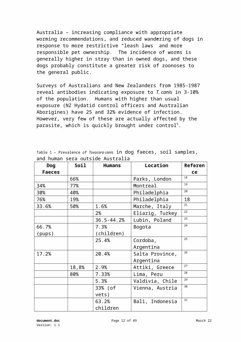
The prevalence of T. canis eggs in dog faeces and / or soil is commonly reported overseas (see table 1), and some data is available for Australia. Ng and Kelly found T. canis in 35.1% of stray dogs in Sydney in 1975; the prevalence was 63.8% were dogs less than 6 months of age13. A survey of 703 pound dogs in North-East Victoria revealed 37% infected with T. canis in 198214. In 1993, T. canis was found in 2.3-17.4% of faecal samples collected from parks, owned and stray dogs in Melbourne and Geelong 15. In Hobart, 10.9% of faecal specimens from urban dogs and 7.7% of park and beach specimens were positive for T. canis eggs, and 18% of samples from human blood donors were positive for Toxocara antibody (1995)16. More recently, only one sample of 180 soil samples from 9 locations in suburban Melbourne contained T. canis eggs in 2003, and the authors concluded that “the acquisition of the disease is unlikely to be from public parks”17. The presence of T. canis eggs in soil is significant, but the likelihood of these eggs becoming or remaining infectious due to the effects of heat, cold, low humidity or time needs to be considered.
Two factors are probably responsible for the apparent decline in worm eggs recovered from soil and faeces in Australia – increasing compliance with appropriate worming recommendations, and reduced wandering of dogs in response to more restrictive “leash laws” and more responsible pet ownership. The incidence of worms is generally higher in stray than in owned dogs, and these dogs probably constitute a greater risk of zoonoses to the general public.
Surveys of Australians and New Zealanders from 1985-1987 reveal antibodies indicating exposure to T. canis in 3-10% of the population. Humans with higher than usual exposure (NZ Hydatid control officers and Australian Aborigines) have 25 and 32% evidence of infection. However, very few of these are actually affected by the parasite, which is quickly brought under control5.
document.doc Page 7 of 31 May 23Version: 1.1
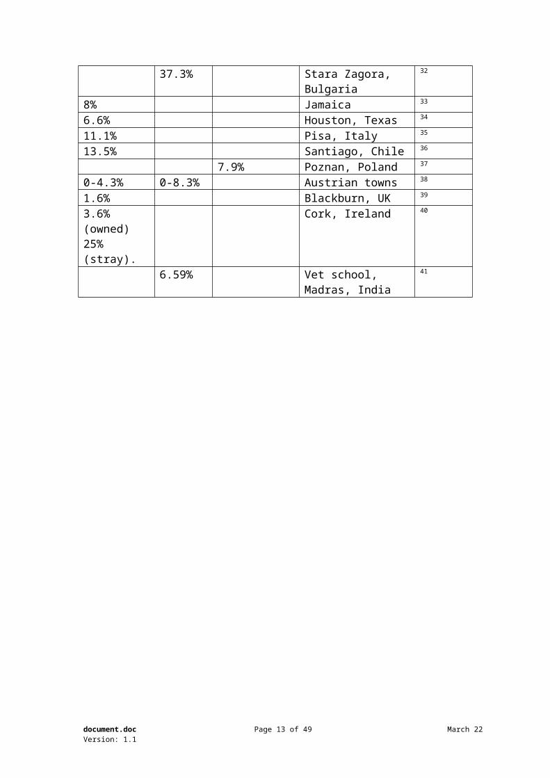
Table 1 – Prevalence of Toxocara canis in dog faeces, soil samples, and human sera outside Australia
Dog Faeces Soil Humans Location Reference66% Parks, London 18
34% 77% Montreal 19
30% 40% Philadelphia 20
76% 19% Philadelphia 1833.6% 50% 1.6% Marche, Italy 21
2% Eliazig, Turkey 22
36.5-44.2% Lubin, Poland 23
66.7% (pups) 7.3% (children) Bogota 24
25.4% Cordoba, Argentina 25
document.doc Page 8 of 31 May 23Version: 1.1
17.2% 20.4% Salta Province, Argentina
26
18,8% 2.9% Attiki, Greece 27
80% 7.33% Lima, Peru 28
5.3% Valdivia, Chile 29
33% (of vets) Vienna, Austria 30
63.2% children Bali, Indonesia 31
37.3% Stara Zagora, Bulgaria 32
8% Jamaica 33
6.6% Houston, Texas 34
11.1% Pisa, Italy 35
13.5% Santiago, Chile 36
7.9% Poznan, Poland 37
0-4.3% 0-8.3% Austrian towns 38
1.6% Blackburn, UK 39
3.6% (owned) 25%(stray).
Cork, Ireland 40
27 Vassalou, H. Kordatos, E. Platis, N. Vakalis, N 1998 “Seroprevalence study of toxocariasis in a district of West Attiki” Acta Microbiologica Hellenica. 43: (3) 258-26228 Lescano, S. A. Z. Chieffi, P. P. Peres, B. A. Mello, E. O. de. Velarde, C. N. Salinas, A. A. Rojas, C. E 1998 “Soil contamination and human infection by Toxocara sp. in the urban area of Lima, Peru” Memorias do Instituto Oswaldo Cruz. 93: (6) 733-73429 Navarrete, N. Rojas, E 1998 “Seroprevalence of toxocarosis in blood donors” Archivos de Medicina Veterinaria.. 30: (1) 153-15630 Deutz, A. Fuchs, K. Auer, H. Aspock, H 1996 “Serological examination of zoonoses in veterinarians. Part II: prevalence of antibodies against parasitological zoonoses” Wiener Tierarztliche Monatsschrift.. 83: (12) 353-35831 Chomel, B. B. Kasten, R. Adams, C. Lambillotte, D. Theis, J. Goldsmith, R. Koss, J. Chioino, C. Widjana, D. P. Sutisna, P “Serosurvey of some major zoonotic infections in children and teenagers in Bali, Indonesia” Southeast Asian Journal of Tropical Medicine & Public Health. 1993. 24: 2, 321-32632 Georgieva, D. A. Prelesov, P. N. Koynarski, V. T 1999 “Parasitological study of soil and sand samples from different regions of Stara Zagora” Bulgarian Journal of Veterinary Medicine. 2: (2/3) 125-12933 Robinson, R. D. Thompson, D. L. Lindo, J. F 1988 “The human public health significance of well-cared for dogs in Jamaica” West Indian Medical Journal. 37: Supplement, 2234 Arambulo, P. V., III. Steele, J. H 1976 “Urban dogs in Houston, Texas - parasitic infection and environmental health impact” International Journal of Zoonoses. 3: (2) 114-14435 Legrottaglie, R. Papini, R. Capasso, R. Cardini, G 2003 “Prevalence of Toxocara canis eggs in dog faecal deposits from urban areas of Pisa, Italy” Helminthologia. 40: (3) 173-17536 Castillo, D. Paredes, C. Zanartu, C. Castillo, G. Mercado, R. Munoz, V. Schenone, H 2000 “Environmental contamination of public squares and parks in Santiago, Chile, with Toxocara sp. eggs, 1999’ Boletin Chileno de Parasitologia. 55: (3/4) 86-9137 Luzna-Lyskov, A 2000 “Toxocarosis in children living in a highly contaminated area. An epidemiological and clinical study” Acta Parasitologica.. 45: (1) 40-4238 Kutzer, E. Golling, P. Wagneder, J 1997 “On the contamination of public green areas and children's playgrounds with eggs of Toxocara from carnivores in Austrian towns” Mitteilungen der Osterreichischen Gesellschaft fur Tropenmedizin und Parasitologie. 19: 71-7439 Al-Jabr, O. A. Storey, D. M. Akrigg, A. Bryden, A. S 1997 “Prevalence of Toxocara ova in dog faeces” Veterinary Record.. 140: (8) 211-212
document.doc Page 9 of 31 May 23Version: 1.1
6.59% Vet school, Madras, India
41
41 Gunaseelan, L. Ganesan, P. I. Ramadass, P. Basheer, M. A. Raghavan, N 1992 “Incidence of Toxocara ova in the environment” Indian Veterinary Journal. 69: (4) 308-30912 Levine ND 1968 “Nematode Parasites of Domestic Animals and Man” Burgess Publishing Co, Minnesota13 Ng, B. K. Y. Kelly, J. D 1975 “Anthropozoonotic helminthiases in Australasia: Part 3: Studies on the prevalence and public health implications of helminth parasites of dogs and cats in urban environments.” International Journal of Zoonoses. 2: (2) 76-9114 Blake RT and Overend DJ 1982 “The Prevalence of Dirofilaria immitis and other parasites in urban pound dogs in North-Eastren Victoria” Australian Veterinary Journal 58: (3) 111-11415 Johnston, J. Gasser, R. B 1993 “Copro-parasitological survey of dogs in southern Victoria.” Australian Veterinary Practitioner. 23: (3) 127-13116 Milstein, T. C. Goldsmid, J. M 1995 “The presence of Giardia and other zoonotic parasites of urban dogs in Hobart, Tasmania.” Australian Veterinary Journal. 72: (4) 154-15517 Carden, S. M. Meusemann, R. Walker, J. Stawell, R. J. MacKinnon, J. R. Smith, D. Stawell, A. M. Hall, A. J. H 2003 “Toxocara canis: egg presence in Melbourne parks and disease incidence in Victoria.” Clinical & Experimental Ophthalmology. 31: (2) 143-14618 Snow, K. R. Ball, S. J. Bewick, J. A 1987 “Prevalence of Toxocara species eggs in the soil of five east London parks.” Veterinary Record.. 120: (3) 66-6719 Ghadirian, E. Viens, P. Strykowski, H. Dubreuil, F 1976 “Epidemiology of toxocariasis in the Montreal area. Prevalence of Toxocara and other helminth ova in dogs and soil.” Canadian Journal of Public Health. 67: (6) 495-49820 Dubin, S. Segall, S. Martindale, J 1975 “Contamination of soil in two city parks with canine nematode ova including toxocara canis; a preliminary study.” American Journal of Public Health. 65: (11) 1242-124521 Habluetzel, A. Traldi, G. Ruggieri, S. Attili, A. R. Scuppa, P. Marchetti, R. Menghini, G. Esposito, F 2003 “An estimation of Toxocara canis prevalence in dogs, environmental egg contamination and risk of human infection in the Marche region of Italy.” Veterinary Parasitology. 113: (3/4) 243-25222 Kaplan, M. Godekmerdan, A. Kalkan, A. Erensoy, A. Ozden, M 1999 “Toxocara canis seroprevalence in Elazig region.” Saglik Bilimleri Dergisi, Firat Universitesi.. 13: (1) 51-5423 Zwolinski, J 2000 “The risk factors of Toxocara canis infestation in population of patients from the Lublin region.” Wiadomosci Parazytologiczne.. 46: (4) 463-47324 Acero, M. Mercedes Munoz, M. Florez, A. C. Santiago Nicholls, R 2001 “Toxocara canis: antibody seroprevalence and risk factors in children, Ciudad Bolivar, Bogota, 2000” Biomedica. 21: (3) 256-263. 2325 Gonzalez Peralta, J. Bernardes, G. Babini, S. Gonzalez, G. Anelo, A 2000 “First seroepidemiology study of toxocariasis in the city of Rio Cuarto, Cordoba, Argentina: preliminary results.” Revista de Medicina Veterinaria (Buenos Aires). 81: (4) 293-29626 Taranto, N. J. Passamonte, L. Marinconz, R. Marzi, M. C. de. Cajal, S. P. Malchiodi, E. L 2000 “Zoonotic parasitoses transmitted by dogs in the chaco of Salta Province” Medicina, Buenos Aires. 60: (2) 217-22040 O'Sullivan, E. N. 1995 “Epidemiological survey of canine toxocariasis in both the owned and stray dog populations of Cork county” Irish Veterinary Journal. 48: (7/8) 281-284
document.doc Page 10 of 31 May 23Version: 1.1
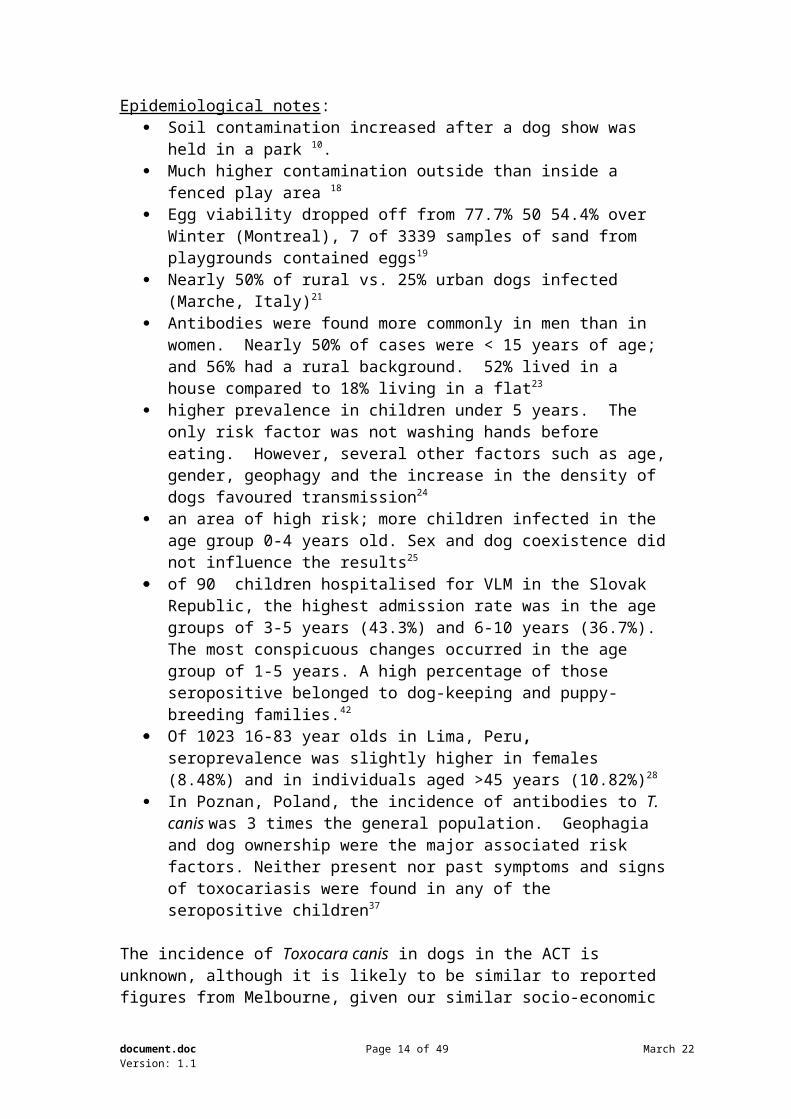
Epidemiological notes: Soil contamination increased after a dog show was held in a park 10. Much higher contamination outside than inside a fenced play area 18
Egg viability dropped off from 77.7% 50 54.4% over Winter (Montreal), 7 of 3339 samples of sand from playgrounds contained eggs19
Nearly 50% of rural vs. 25% urban dogs infected (Marche, Italy)21
Antibodies were found more commonly in men than in women. Nearly 50% of cases were < 15 years of age; and 56% had a rural background. 52% lived in a house compared to 18% living in a flat23
higher prevalence in children under 5 years. The only risk factor was not washing hands before eating. However, several other factors such as age, gender, geophagy and the increase in the density of dogs favoured transmission24
an area of high risk; more children infected in the age group 0-4 years old. Sex and dog coexistence did not influence the results25
of 90 children hospitalised for VLM in the Slovak Republic, the highest admission rate was in the age groups of 3-5 years (43.3%) and 6-10 years (36.7%). The most conspicuous changes occurred in the age group of 1-5 years. A high percentage of those seropositive belonged to dog-keeping and puppy-breeding families.42
Of 1023 16-83 year olds in Lima, Peru, seroprevalence was slightly higher in females (8.48%) and in individuals aged >45 years (10.82%)28
In Poznan, Poland, the incidence of antibodies to T. canis was 3 times the general population. Geophagia and dog ownership were the major associated risk factors. Neither present nor past symptoms and signs of toxocariasis were found in any of the seropositive children37
The incidence of Toxocara canis in dogs in the ACT is unknown, although it is likely to be similar to reported figures from Melbourne, given our similar socio-economic and educational status . Dr David Jenkins from the Australian Hydatid Control and Epidemiology Program, found about 10% of dogs producing any helminth eggs in their faeces in a survey at The ACT Dog Pound in the late 1990s. (pers. Comm).
ConclusionThe major risk factors for the development of Toxocara larva migrans in man are eating dirt, poor socio-economic status, ownership/prolonged contact with dogs, and perhaps rural lifestyle. Stray dogs are more likely to contaminate sporting fields than are owned dogs being deliberately exercised there, because they are likely to be carrying higher worm burdens due to infrequent antheminthic application. The brief contact with soil in sporting fields, or indeed with dog faeces, is unlikely to constitute a significant risk for Toxocara larva migrans. However, as for all the other pathogens discussed below, dog (and other animal) faeces should be excluded from children’s playgrounds and sandpits by appropriate fencing.
42 Kincekova, J. Reiterova, K. Dubinsky, P 1999 “Larval toxocariasis and its clinical manifestation in childhood in the Slovak Republic.” Journal of Helminthology 73: (4) 323-328
document.doc Page 11 of 31 May 23Version: 1.1
Uncinaria stenocephala
Hookworms are common parasites of dogs (and other species including man) throughout the world, but individual species tend to occur in specific geographical locations. The most common hookworm of dogs in Australia7 5 and also throughout the world43, Ancylostoma caninum, is a parasite of warm areas, being limited to north of 35 oS44. The canine hookworm most commonly implicated as the cause of the zoonotic disease cutaneous larva migrans (CLM) (creeping eruption) in Australia is A. braziliense 44, 5, which is limited to far northern Australia, but A. caninum and Uncinaria stenocephala can also cause this disease. U. stenocephala is a cool weather parasite, being limited to below 30 oS. Canberra lies just north of 35 oS, supposedly within the range for A. stenocephala, though this parasite has rarely if ever been recorded here. U. stenocephala is essentially the only hookworm recorded in dogs, cats, dingoes or foxes in the ACT.
Hookworms have a direct lifecycle, with eggs passed in faeces onto the ground, hatching into larvae which go through a succession of moults until they become infective (L3 stage). For U. stenocephala, the optimal temperature for development is 20 oC, with eggs hatching after 12 hours and L3 in 4 days. Development is delayed at lower temperatures (for example, L3 at 28 days at 7.5 oC), and no larvae become infective at 37 oC. 10% of eggs survive for only 2 days at –10 oC. Larvae do not develop in the centre of faeces, but eggs will hatch if the faeces are disturbed and break up into soil. Infective larvae crawl out of faecal material and onto vegetation. (Levine 1968). All, but especially early, larval stages are susceptible to desiccation and heat45.
Infection of dogs occurs via the mouth or by skin penetration, and the latter is the likely route in humans (except perhaps in small children). Humans with CLM typically are either:
Young children and adults who go barefoot in areas frequented by dogs, and children in contaminated playgrounds and sandpits
Travellers, particularly from tropical locations, especially those who have spent time on beaches where dogs defaecate
Tradesmen who work under houses, crawling over dirt contaminated with dog faeces, and nursery workers45
98 (0.7%) patients of 13,300 attending a travel related disease clinic of the University of Munich (Germany) had CLM. None of these had visited Australia, despite Australia being a common destination for Europeans, and despite records of CLM in Australia. Only visitors to tropical countries were affected, apparently with A. braziliense or, less commonly, with A. caninum46.43 Bowman DD 1992 “Hookworm Parasites of Dogs and Cats” Compendium of Continuing Education for the Practicing Veterinarian 14 (5) 585-59544 Stevenson WJ and Hughes KL 1980 “Synopsis of zoonoses in Australia” Commonwealth Department of Health, Canberra45 Hendrix CM, Bruce HS, Kellman NJ, Harrelson G and Bruhn BF 1996 “Cutaneous larva migrans and enteric hookworm infections” Journal of the American Veterinary Medical Association 209 (10) 1763-176746 Jelinek T. Maiwald H. Nothdurft HD. Loscher T 1994 “Cutaneous larva migrans in travelers: synopsis of histories, symptoms, and treatment of 98 patients” Clinical Infectious Diseases. 19
document.doc Page 12 of 31 May 23Version: 1.1
CLM is rare enough to be reported in medical journals when small numbers of cases are seen474849, the latter two cases specifying the involvement of Ancylostoma species.
(6):1062-6,
document.doc Page 13 of 31 May 23Version: 1.1
Prevalence
Uncinaria was found in 10% of Sydney dogs in 197513, 26.9% of pound dogs in a survey in North Eastern Victoria (1982) 14, hookworm eggs were recovered from 1.8% of dogs and 0.7% public places in Hobart in 199516, and it has been suggested that the incidence of hookworms declines with increasing latitude5. The incidence of CLM in Australia is not available from the medical research data base “Medline”.
Conclusion
Uncinaria is an unusual parasite of dogs in Canberra (personal experience), and is an uncommon cause of CLM worldwide. CLM occurs when there is relatively prolonged contact between bare skin and contaminated soil or vegetation. The risk of CLM from dog faeces on sporting fields is negligible to non-existent.
Echinococcus granulosus
Echinococcus granulosus, the Hydatid Tapeworm, is a tiny parasite of the small intestines of dogs, dingoes7 44, foxes 5051, and also wolves, jackals, coyotes, and African Lions52, causing no disease and no symptoms, even in massive numbers. Each worm produces about 1,000 eggs every two weeks5, and dogs can carry up to 300,000 worms, although domestic dogs do not usually carry such large numbers. Dingoes, on the other hand, and wild dogs infected with the “sylvatic strain” of Echinococcus, commonly carry heavy burdens53. Passage of large numbers of eggs in the faeces of dogs, especially mobile dogs, results in widespread contamination of pasture, bushland etc. Eggs are spread over wide areas by wind, insects, birds and the like, and a single dog could infect up to 30,000 hectares5. Eggs are susceptible to desiccation, but are very cold tolerant, and may survive in the field for at least a year. They are immediately infective.
Eggs are ingested by intermediate hosts, usually by grazing, but also accidentally by contact with dog faeces, soil, and eggs which have transferred onto the dog’s coat and mouth by rolling in or ingesting faeces, licking etc. Oncospheres (an intermediate stage) hatch from eggs in the intestine, penetrate the intestinal wall, and disperse throughout the body, but predominantly the liver and lungs. Here they form cysts which grow slowly and contain many thousands of protoscolices, which are infective for dogs if swallowed. It is these cysts which cause disease in humans, growing slowly over many years with few signs in some, causing pain or severe reactions in others, or causing serious disease if they form in the brain or other vital organ. Most humans require surgical treatment, although medication (albendazole) may also be used.
It has long been believed that there are two strains of E. granulosus in Australia which display morphological and antigenic differences. The “domestic strain” has a dog – sheep (or cattle) – dog cycle, while the “sylvatic (wildlife) strain” has a dingo – macropod – dingo cycle, although there is some overlap. The domestic strain has been recovered from dingoes, and is believed responsible for transmission to cattle in Queensland54, wild dogs53 55, and foxes51 56. Human hydatidosis in Australia is almost invariably caused by the domestic strain, although there has been one report of human
document.doc Page 14 of 31 May 23Version: 1.1
infection with the sylvatic strain. However, it is now considered that there is only one strain (the “sheep” strain) that is opportunistic, i.e. infecting whatever hosts are available - wildlife or domestic and occasionally human (D Jenkins, Australian Hydatid Control and Epidemiology Program, Canberra, pers. comm. April 2004).
Prevalence
The highest prevalence of infection in dogs in Australia occurs in south east NSW, with rural dogs recording 3.5-32.3% infection rates. Rates in Victoria (3%)[although none were found in pound dogs in north eastern Victoria in 1982 16], and Western Australia (0.7%), are lower, and the parasite has been eliminated in Tasmania and New Zealand. The parasite has also been recorded in dogs in Sydney5. 11.4% of farms in the ACT had dogs infected with E. granulosus in 197757.
Wild dogs and dingoes often have very high infection rates – 90% (SE Queensland54, 61% SE NSW58, 87%53, and although the sylvatic strain predominates, the domestic strain is also seen. Of significance is the 7% infection rate found in foxes killed on roads in Canberra56. Foxes can harbour the domestic strain of hydatids, probably in similar burdens to the sylvatic strain50, and a fox infected with the domestic strain was recovered near Gundaroo, just north of Canberra.
Infection rates in SE NSW have been reported at 5.4% in mature sheep, with some properties in the Cooma district having up to 42%59. The high prevalence in wild dogs around Canberra suggests the presence of hydatids in macropods in the area, but a survey of road kill in Canberra failed to recover any hydatid cysts56, implying that these urban foxes may have been infected from eating sheep and lambs.
Canberra lies in the area of greatest risk (on mainland Australia) for human hydatid disease, with a peak incidence of 26.2 cases / 100,000 in the period 1968-19735. Jenkins and Power report 195 new cases of hydatid disease in humans in NSW and the ACT between 1987-1992. Most of the patients lived in the eastern half of the State, including the ACT. It is of note that 60% of the patients living in major cities were born overseas60.
Most humans who contract Hydatid disease have a rural connection – commonly infection occurs when they are children but is not detected for years or decades. Many people in the ACT live on or visit farms nearby, where sheep are commonly raised. It is of concern that Canberran dogs also visit farms, or are exercised in the rural and bush-land in and around Canberra, often off lead and under-supervised. Such dogs who consume infected material from carcasses of sheep, pigs, kangaroos and wallabies which are less than three days dead (D Jenkins pers. comm.) may contract E. granulosus, and be excreting eggs in their faeces after about 6 weeks. These eggs constitute a risk to the humans, especially children, of the dog’s family, friends and neighbours, but also to strangers if the dog is exercised in public areas.
document.doc Page 15 of 31 May 23Version: 1.1
Fortunately, local veterinarians are very conscious of the risks, and are pro-active in educating their clients and ensuring that appropriate care is in place. Four rules for hydatid prevention are taught:
1. Don’t feed dogs raw meat or offal – use commercial foods2. Don’t allow dogs to wander or be unsupervised where they might eat from a
carcass3. Practice (and teach) good personal hygiene – don’t kiss dogs, don’t let them
lick you on the face, wash hands after touching dogs, don’t eat while playing with dogs
4. Worm at risk dogs with Praziquantel at least every 6 weeks.Most dog owners heed these warnings carefully, aware of the serious risk to their own and loved ones health.
There are others in the community who may be less exposed to this message and whose dogs may constitute a greater risk – those who use their dogs for hunting, or who bring the products of hunting back to feed dogs. This relatively small group of dogs is seen, in my experience, less frequently by veterinarians, and often has a lower standard of general health care. However, these dogs appear to be rarely if ever exercised outside their owner’s properties except when hunting, so the risk to the general community is probably low.
Hydatid disease is uncommon in humans who live in towns and cities, risk of infection increasing with duration of exposure to infected dogs and to contaminated country. The risk of human infection from sporting fields is negligible given:
Relatively brief contact with the sporting fieldRelatively low risk of dogs being exercised on sporting fields being exposed to
a source of infectionHigh compliance with worming recommendations in most Canberra dogs
(personal experience)
ConclusionCanberrans are at risk for Hydatid disease from:
Eggs from faeces dispersed into the environment through various natural processes not associated with direct presence of faeces,
Infected faeces from animals other than domestic dogs (foxes and perhaps wild dogs/dingoes) moving through suburban areas and over sporting fields.
Banning owned dogs from sporting fields will not reduce these risks.
document.doc Page 16 of 31 May 23Version: 1.1
Protozoa
GiardiaGiardia are protozoan parasites of the intestinal tract of many animal species, including man. Their presence is often sub-clinical, even in large numbers, although acute or chronic diarrhoea, sometimes with abdominal pain, may occur. In the past, Giardia from individual animal species were named for those species8, and it was thought that animal species of Giardia could not be spread to humans44. Partly because the morphology of Giardia from all host species is identical, their taxonomy has changed. There has also been much debate about their zoonotic potential.
The potential of Giardia as a zoonosis has been questioned since at least 197961, and was raised in Australia as early as 198662. Several Japanese studies have examined the likelihood of Giardia being zoonotic636465 - none of the owners of 307 infected dogs (out of 2652 tested – 11.5%) were infected, and the authors concluded that the risk for dog to human infection was very low. Gasser (1990) reported that techniques to determine whether Giardia could be spread from animals to man were available, and that direct transmission studies were necessary66. A review in 2000 commented that “the contribution of zoonotic transmission remains unclear”67. Human infection with Giardia mostly occurs via infected water or food contaminated with infected water; and presumably human sewage is the main source of such contamination. However, a recent survey of pristine water catchments (10 rivers in 7 National Parks) in eastern Australia found all to be infected with Giardia (and Cryptosporidium)68.
Prevalence
Giardia has been reported in 21% of 333 dogs in Perth62, and in 14.5% of dogs and 1.4% of park soil samples in Hobart16. Giardia also occur in the faeces of cats, and were reported in 14% of cats in the Perth study. A higher infection rate was recorded from refuge and breeding kennel dogs than from pets, due to higher living densities, environmental contamination rates, and perhaps lower general health and planes of nutrition.
Giardia infection in dogs appears to be common overseas, with prevalence reported at 11.5% in Japan (62-64), 14.5% in London (29.4% in dogs 6-12 months of age)69, 40% in Dublin, Ireland70, and 35.9% in puppies in the US71
ConclusionDespite the probable high incidence in dogs in the ACT, Giardia is a most unlikely risk for man from dog faeces. The parasite is mostly water born due to human faecal contamination, and even close proximity to dogs has not resulted in significant human infection.
document.doc Page 17 of 31 May 23Version: 1.1
CryptosporidiumCryptosporidium parvum is a tiny (4-5μm) coccidian, which is a primary pathogen causing diarrhoea in animals and man72. The organism survives for long periods in the environment, readily contaminates water from faeces, and is extremely difficult to kill with chemicals. Other species occur in animals and birds. C. parvum of bovine, but not human, origin can infect dogs73, a human and his dog were found to have different genotypes (cattle and dog, respectively) in Japan74, and in Osaka, all genetic isolates from dog faeces were of Cryptosporidium canis (previously known as the dog genotype), which is thought to be non-pathogenic in humans”75.
However, the dog strain has been recovered from 1 of 1680 patients with Cryptosporidiosis in England76, and from an HIV infected human in America77. Infection in immunocompetent people is regarded as moderate and self-limiting, which contrasts sharply with the prolonged severe diarrhoea in immunocompromised patients7879. Children, dairy farm workers, and travellers are also at risk, as are possibly the owners of infected dogs80. Transmission occurs by contaminated drinking and swimming water, food, and directly (faecal-oral), and livestock are commonly incriminated as the source of infection.
Cryptosporidium occurs in dogs worldwide(Edinburgh81, California 2%82, Ireland70, Spain 7.4%83, Colorado (3.8%)84), and in poor communities dogs are regarded as a possible source of infection (Guatemala85, Brazil86, Egypt87). However, Cryptosporidium had not been identified in dogs in Australia5, until 200088, and the prevalence in Australia is unknown, but likely to be very low.
ConclusionCryptosporidium from dogs is regarded as an extremely low risk zoonosis in Canberra.
Other ProtozoaOther potentially zoonotic protozoa recorded in Australian dogs include Entamoeba histolytica, but humans are regarded as the source of infection for dogs, rather then the reverse, and Toxoplasma gondii. Toxoplasma is an important, if clinically unusual, zoonotic parasite of cats. Humans may also be infected from raw or undercooked meat. Dogs have recently been identified as possible mechanical hosts of T. gondii if they ingest infected cat faeces – the sporulated oocysts may pass through into the dog’s faeces89. Unsporulated oocysts in cat faeces applied to dog fur, however, failed to sporulate. Given that cats only excrete Toxoplasma oocysts for about 2 weeks, the risk of infection from the faeces of dogs consuming cat faeces is negligible. Only naïve pregnant women (no previous immunity to Toxoplasma) and the immuno-incompetent are at risk for clinical disease90, and about half the Australian population have antibodies to this organism.
document.doc Page 18 of 31 May 23Version: 1.1
BacteriaBacteria are ubiquitous in the faeces of all animals, and contact between broken skin and faeces may lead to infection. Of course, soil borne organisms not associated with dog faeces pose exactly the same risk. Several bacteria found in the faeces of dogs are specifically regarded as zoonotic, including Campylobacter, Salmonella, Escherichia coli and other faecal coliforms.
CampylobacterCampylobacter are comma or gull wing shaped gram negative bacteria which may cause diarrhoea or abortion in man and animals. Human cholera is caused by a specific Campylobacter91. Different species occur in different hosts and cause different patterns of disease. Campylobacter is a worldwide zoonosis, and the leading cause of acute bacterial gastro-enteritis in England92. Campylobacter jejuni and C. upsaliensis may cause diarrhoea with blood and mucus in dogs, especially young dogs, and in people, especially children. Infection occurs by ingestion of contaminated food, water, raw milk, or the faeces of infected animals (especially young animals with diarrhoea).
Prevalence of Campylobacter varies from 0-49%; 13 of 30 normal and diarrhoeic dogs in Rome were shown to carry Campylobacter93, while 56% of healthy family dogs in Sweden carry the organism94. Clinically, it is a rare diagnosis in dogs in the ACT (personal experience).
Dog faeces in public areas do constitute a risk for Campylobacter gastro-enteritis, but the risk is higher in children exposed to faeces in play areas or by prolonged contact with dogs (e.g. ownership). The risk to human health of Campylobacter in dog faeces on sporting fields is considered to be low.
SalmonellaSalmonellae are gram-negative bacteria, commonly associated with food poisoning in people. The source of the organism is commonly human to human – there is a well understood carrier state which results in outbreaks when present in food handlers. Animals also carry (and may be affected by) all Salmonella strains except those causing Typhoid fever and related (paratyphoid) enteric fevers. Zoonotic infection is most commonly due to contamination of food, or the presence of the organism in food such as milk, chicken and eggs. Birds are commonly infected and shed the organism in faeces. The organism survives well in the environment and in water contaminated with organic material. It is unlikely, on the other hand, that it survives more than three weeks in water free of organic waste. Salmonellae are susceptible to heating and drying.
Pets, including dogs, cats and reptiles, are also a possible source of infection. Reptiles are a well recognised threat and the sale of some species are controlled in some countries because of Salmonella. In mammals, neonates, old animals, animals with concurrent infections, those undergoing the stress of transport, exercise95, malnutrition, feed changes, pregnancy or surgery are more susceptible to infection
document.doc Page 19 of 31 May 23Version: 1.1
and clinical disease. Human neonates and the elderly are most at risk, and risk is increased if taking antibiotics, suffering chronic or debilitating disease including neoplasia, or immunodeficient. Recovered animals may shed the organism (intermittently) for up to 3 months, but a carrier state is not reported in animals as it is for humans, except for highly host adapted strains such as S. dublin in cattle and S. pullorum in chickens.96
Dogs may acquire the organism from the environment, but raw or undercooked meat and eggs are a much more likely source, and all meat for pets should be cooked as if for human consumption. Milk consumed by pets should be pasteurised. Flies are an important source of contamination for Salmonellae as for other bacteria97
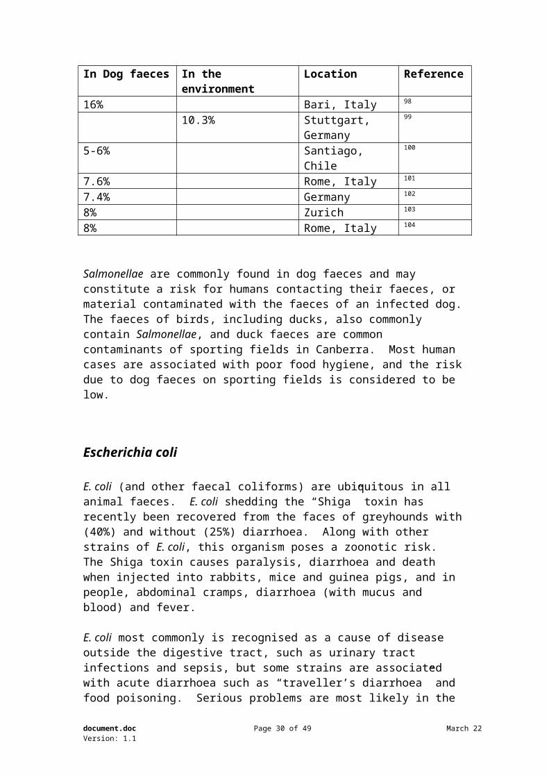
Prevalence
Table 2 – PrevalenceSalmonella recovered from dog faecesIn Dog faeces In the environment Location Reference16% Bari, Italy 98
10.3% Stuttgart, Germany 99
5-6% Santiago, Chile 100
7.6% Rome, Italy 101
7.4% Germany 102
8% Zurich 103
8% Rome, Italy 104
Salmonellae are commonly found in dog faeces and may constitute a risk for humans contacting their faeces, or material contaminated with the faeces of an infected dog. 47 Galanti B. Fusco FM. Nardiello S 2002 “Outbreak of cutaneous larva migrans in Naples, southern Italy” Transactions of the Royal Society of Tropical Medicine & Hygiene. 96 (5):491-2, 48 Malgor R. Oku Y. Gallardo R. Yarzabal I 1996 “High prevalence of Ancylostoma spp. infection in dogs, associated with endemic focus of human cutaneous larva migrans, in Tacuarembo, Uruguay.” Parasite. 3(2):131-4, 49 Richey TK. Gentry RH. Fitzpatrick JE. Morgan AM 1996 “Persistent cutaneous larva migrans due to Ancylostoma species” Southern Medical Journal. 89 (6): 609-11,50 Thompson RC 1982 “The susceptibility of the European red fox (Vulpes vulpes) to infection with Echinococcus granulosus of Australian sheep origin.” Annals of Tropical Medicine & Parasitology. 77(1):75-8251 Thompson RCA, Nicholas WL, Howell MJ and Kumaratilake LM 1985 “Echinococcus granulosus in a fox.” Australian Veterinary Journal. 62 (6):200-152 Bryan, R. T. Schantz, P. M 1989 “Echinococcosis (hydatid disease). Journal of the American Veterinary Medical Association. 195: (9) 1214-121753 Jenkins DJ. Morris B 1991 “Unusually heavy infections of Echinococcus granulosus in wild dogs in south-eastern Australia.” Australian Veterinary Journal. 68 (1): 36-754 Baldock FC. Thompson RC. Kumaratilake LM. Shield J 1985 “Echinococcus granulosus in farm dogs and dingoes in south eastern Queensland.” Australian Veterinary Journal. 62 (10):335-755 Grainger HJ. Jenkins DJ 1996 “Transmission of hydatid disease to sheep from wild dogs in Victoria, Australia.” International Journal for Parasitology. 26 (11):1263-7056 Jenkins DJ. Craig NA 1992 “The role of foxes Vulpes vulpes in the epidemiology of Echinococcus granulosus in urban environments.” Medical Journal of Australia. 157 (11-12):754-657 Christie M. Beard TC. Nicholas WL 1977 “The control of hydatid disease and ovine cysticercoses in the Australian Capital Territory and southern New South Wales.” Medical Journal of Australia. 165 (21):773-558 Morrison P, Stanton R and Pilatti E 1988 “Echinicoccus granulosus infection in wild dogs in south-eastern New South Wales” Australian Veterinary Journal 65 (3) 97-8
document.doc Page 20 of 31 May 23Version: 1.1
The faeces of birds, including ducks, also commonly contain Salmonellae, and duck faeces are common contaminants of sporting fields in Canberra. Most human cases are associated with poor food hygiene, and the risk due to dog faeces on sporting fields is considered to be low.
Escherichia coli
E. coli (and other faecal coliforms) are ubiquitous in all animal faeces. E. coli shedding the “Shiga” toxin has recently been recovered from the faces of greyhounds with (40%) and without (25%) diarrhoea. Along with other strains of E. coli, this organism poses a zoonotic risk. The Shiga toxin causes paralysis, diarrhoea and death when injected into rabbits, mice and guinea pigs, and in people, abdominal cramps, diarrhoea (with mucus and blood) and fever.
E. coli most commonly is recognised as a cause of disease outside the digestive tract, such as urinary tract infections and sepsis, but some strains are associated with acute diarrhoea such as “traveller’s diarrhoea” and food poisoning. Serious problems are most likely in the young or immuno-compromised91. Recent studies have established that dogs may act as reservoirs of pathogenic E. coli affecting the urinary tract and meninges105 106.
Environmental health authorities are concerned about bacterial contamination of waterways with animal faeces, including dogs, and large volumes of dog faeces are washed into storm water drains after heavy rain. The source of such bacterial contamination can be determined107 108 by genetic comparison.
E.coli from dog faeces is a possible risk for humans, although the organism is a common contaminant of water and food and these are more likely sources.
LeptospiraLeptospirosis is relatively common in livestock and feral pigs in Australia, and may occur in dogs and horses also. The organism attacks the kidneys and/or liver, and Leptospirosis in man is almost always a zoonosis contracted from animals, mainly through contact with infected urine. Humans have been infected from dogs in England109, Germany110, and dogs (along with other animals) have high levels of infection in areas with high human incidence such as Mexico111, Greece112, India 113, and Brazil114. Clinically normal dogs may shed the organism in their urine, constituting a threat to human health115.
However, the incidence of Leptospirosis in dogs in the NSW is low116, and in the ACT and region is very low (personal experience). Dickeson and Love concluded that dogs (and cats and horses) were unlikely to pose a risk to humans in south-eastern Australia. Most human infections are associated with rat urine, for example in cane fields in northern NSW and Queensland.
document.doc Page 21 of 31 May 23Version: 1.1
The risk of Leptospirosis from dog urine on sporting fields is considered to be negligible.
Other BacteriaAnaerobiospirillum spp. have been show to cause bacteraemia and diarrhoea in people. A study failed to grow the organism in human faeces but it was found in dog and cat faeces117, establishing pets as the likely source of human infection. No doubt many other bacteria from dog faeces are occasional sources of human infection, but like all diseases discussed, individual risk is highest in children and the immuno-compromised, especially where there is prolonged contact with dogs (i.e. ownership) and poor standards of hygiene (personal, and removal of dog excreta). Dog faeces on sporting fields are a low risk to humans.
document.doc Page 22 of 31 May 23Version: 1.1
VirusesRotavirus was found in 2 (3.5%), coronavirus in 7 (12.5%), parvovirus in 13 (23.2%) and both coronavirus and parvovirus in another 5 (8.9%) random dog faecal samples from Paris streets, but the infectivity of these dog strains for humans is not discussed118. In another study in Zurich, no enteric viruses were found103.
Viruses are an unlikely risk for humans from dog faeces.
FoxesFoxes are commonly seen in Canberra after dusk. Some clearly reside outside the city and visit to scavenge for food scraps, others (particularly those seen close to the city centre and in large institutions such as the Australian National University) almost certainly reside here.
Numerous studies both in Australia and overseas have demonstrated that foxes carry many of the same faecal organisms as dogs such as the helminths Toxocara canis, Uncinaria stenocephala, Trichuris vulpis, and antibodies to Toxoplasma gondii, amongst other organisms, in Yugoslavia119, Spain120, Ireland121, England122 and Australia56 51. Any consideration of risks of faeces on sporting fields must consider the possibility of contamination with fox faeces. Foxes, unlike dogs, never receive anthelminthic (worming) drugs and are not observed or treated for ill health such as bacterial diarrhoea.
Possible solutions
While this review was not intended to address solutions to the problem of dog faces on sporting fields and the attendant risks, two obvious solutions exist which will be
59 Howkins AB 1985 “A pilot survey into hydatid disease incidence levels in sheep and cattle of New South Wales.” Australian Veterinary Journal. 62(8):28860 Jenkins DJ. Power K 1996 “Human hydatidosis in New South Wales and the Australian Capital Territory, 1987-1992.” Medical Journal of Australia. 164 (1):18-2161 Eckert J. Wolff K 1979 “Giardiasis (lambliasis)--a zoonosis? (author's transl)” Schweizerische Rundschau fur Medizin Praxis. 68(45):1471-262 Swan JM. Thompson RC 1986 “The prevalence of Giardia in dogs and cats in Perth, Western Australia.” Australian Veterinary Journal. 63(4):110-263 Arashima Y. Iguchi K. Kubo N. Kumasaka K. Okuyama K. Kawano K. Harada M. Shimabukuro H. Saitoh T. Isa H. et al 1990 “Studies on the giardiasis as the zoonoses” Kansenshogaku Zasshi - Journal of the Japanese Association for Infectious Diseases. 64(3):295-864 Asano R. Hokari S. Murasugi E. Arashima Y. Kubo N. Kawano K 1991 “Studies on the giardiasis as the zoonosis. II. Giardiasis in dogs and cats” Kansenshogaku Zasshi - Journal of the Japanese Association for Infectious Diseases. 65(2):157-6165 Arashima Y. Kumasaka K. Kawano K. Asano R. Hokari S. Murasugi E. Iwashita E. Nishikawa S. Matsuo K. 1992 “Studies on the giardiasis as the zoonosis. III. Prevalence of Giardia among the dogs and the owners in Japan” Kansenshogaku Zasshi - Journal of the Japanese Association for Infectious Diseases. 66(8):1062-666 Gasser RB 1990 “Is giardiasis a zoonosis?” Australian Veterinary Journal. 67(12):456
document.doc Page 23 of 31 May 23Version: 1.1
briefly discussed.
Dung beetles
Dung beetles have been used for the disposal of dog faeces since at least 1995, when they were introduced in Warringah Shire in Sydney123. They have been introduced into the dog exercise courtyards of housing complexes in Melbourne to assist with faecal control. Dung beetles are attracted to faeces, and create a nest under the mound into which they transport the dung. The optimal species for dogs in this area, Ontophagus taurus, will remove 90% of the faecal material within 24 hours. A recent conference in America reported a 99.7% reduction in Cryptosporidium in the environment as a consequence of the actions of dung beetles on cattle faeces. Some helminth eggs and larvae, such as Toxocara, will be rendered unavailable by burying, while other more mobile species such as Uncinaria may still migrate on to vegetation. Clearly, dung beetles have great capacity to reduce the aesthetic and health problems of dog faeces.
However, there are limitations which need to be considered. The named specie is a summer active dung beetle, from November to April in this region. It hibernates over Winter. However, there are winter active species available, although they are larger and fly around at dusk. Dung beetles do not remove all of the faecal pat; they remove 90% of the interior but leave a shell which may look unchanged. Dung beetles are less active in sheltered areas such as under the canopy of trees, where dogs may go to defaecate, but would be active in the middle of sporting fields. Their health is affected by the protein content of the diet of the animal producing the dung, preferring higher protein diets. There has been concern about the effect of some anthelminthics passed in the faeces on dung, but most dogs are wormed intermittently (unlike livestock where prolonged activity formulations are often used), meaning that relatively few faecal pats would affect the beetles, if at all. The one long acting preparation used in dogs contains Moxidectin, which has been shown to be safe for dung beetles.
The above information was obtained from Dr John Sheehan1, a retired CSIRO scientists who has been researching and supplying dung beetles since 1965, and is happy to present his material.
Faecal disposal
The health problems associated with dog faeces, limited as they may be, can be largely eliminated by owners removing dog faeces from areas where people exercise or recreate. Various strategies are available to encourage this, including enforcing the carriage of faeces removal equipment, and fines for failure to remove faeces
46 Removal of faeces(1) The carer of a dog must hygienically dispose of any faeces dropped by the dog in a public place or in a stormwater drain or channel (whether on public or private land).Maximum penalty: 5 penalty units.
1 Dr John Sheehan 3 Prell Pl Hackett, 6248 0376, 9427 1140
document.doc Page 24 of 31 May 23Version: 1.1
(2) The carer of a dog must not take the dog into a public place or a stormwater drain or channel (whether on public or private land) unless the carer carries equipment suitable for the hygienic disposal of faeces dropped by the dog.Maximum penalty: 1 penalty unit.
Domestic Animals Act 2000
The clear limitation to this requirement is the lack of enforcement. However, most members of the public (personal observation) are willing to dispose of faeces as long as it is convenient. They dislike having to carry plastic bags containing faeces for long distances. The provision of more bins for faecal (and other waste) disposal in areas frequented by dogs and their owners has been shown to increase compliance rates.
Government can encourage compliance with current moves to ban plastic shopping bags by supplying biodegradable “poo bags”, available from a number of sources, together with biodegradable bin liners in special “poo bins”. These bins are distinctive, advertise the service provided by government, can be sponsored by local businesses, and tend to be ignored by vandals because of the association with dog faeces. Even normal litter bins will be used for faeces disposal if available. Special clip on bags for carriage of “poo bags” are also available and are commonly provided by councils with dog registration to encourage compliance with laws requiring removal of faeces. These are printed with messages and sponsors logos, and could be sponsored by local businesses and organisations.
Dogs can also be encouraged to eliminate in areas away from those used by the public. Many councils have installed “pooch patches” to attract dogs to toilet there – usually a patch of sandy soil (for drainage with posts to attract male dogs, and a litter / poo bin nearby(Pert 1996). Placing these near the entrances to areas frequented by dogs being exercised will encourage elimination there, rather than in areas shared with humans. Dogs are also more likely to defaecate in long grass than in short grass – maintaining grass short adjacent to walking paths but with long grass nearby will encourage dogs to defaecate there, rather than where people walk and play.
Finally, the provision of more, dedicated, dog exercise areas, especially safe off leash areas’ will attract dogs and their owners and reduce the use of sporting fields by dogs. If such areas are fenced, they will be much more attractive to dog owners (safety aspects) and contain dog faeces within the area. Clearly, owners should still be encouraged and required to clean up after their dog.
Further reading (especially *):Leather RL 1994 ”Legislation for urban animal management: experience with formulation and implementation of Scoop Law” National Urban Animal management Conference, Canberra, Australian Veterinary AssociationJackson, Virginia 1995 “Guidelines for designing and managing public open space” National Urban Animal management Conference, Melbourne, Australian Veterinary AssociationPert, Terry-Ann 1996 “Initiatives for the environment” National Urban Animal management Conference, Sydney, Australian Veterinary Association
document.doc Page 25 of 31 May 23Version: 1.1
Jackson, Virginia 1997 “Turning theory into practice Banyule City Council” National Urban Animal management Conference, Adelaide, Australian Veterinary Association*Jackson, Virginia 2000 “Faecal litter management - a local government priority for reasons of community health and environmental amenity” National Urban Animal management Conference, Hobart, Australian Veterinary Association
all available at http://www.ava.com.au/, then click “UAM” on the left, then “Conference Proceedings”
document.doc Page 26 of 31 May 23Version: 1.1
Conclusion
This review has shown that dog faeces may contain a variety of organisms which are pathogenic for man. It is clearly appropriate that the risk be reduced. However, the methods chosen must be due to a demonstrable need, and be cost effective and practical.
The incidence of parasites, bacteria and viruses is low in Canberra dogs due to a general high level of care by owners and our relatively high socio-economic status. 67 Slifko TR. Smith HV. Rose JB 2000 “Emerging parasite zoonoses associated with water and food.” International Journal for Parasitology. 30 (12-13):1379-9368 Buckley, R. Warnken, W 2003 “Giardia and Cryptosporidium in pristine protected catchments in Central Eastern Australia.” Ambio. 32: 2, 84-8669 Sykes, T. J. Fox, M. T 1989 “Patterns of infection with Giardia in dogs in London.” Transactions of the Royal Society of Tropical Medicine & Hygiene. 83: 2, 239-24070 Bauer D 1994 “The capacity of dogs to serve as reservoirs for gastrointestinal disease in children” Irish Medical Journal. 87 (6):184-571 Hahn, N. E. Glaser, C. A. Hird, D. W. Hirsh, D. C 1988 “Prevalence of Giardia in the feces of pups” Journal of the American Veterinary Medical Association. 192: 10, 1428-142972 Moon HW and Woodmansee DB 1995 “Cryptosporidiosis” in Zoonosis Updates 2 ed American Veterinary Medical Association.73 Darabus G. Olariu R 2003 “The homologous and interspecies transmission of Cryptosporidium parvum and Cryptosporidium meleagridis” Polish Journal of Veterinary Sciences. 6(3):225-874 Abe N. Kimata I. Iseki M 2002 “Identification of genotypes of Cryptosporidium parvum isolates from a patient and a dog in Japan” Journal of Veterinary Medical Science. 64(2):165-875 Abe N. Sawano Y. Yamada K. Kimata I. Iseki M 2002 “Cryptosporidium infection in dogs in Osaka, Japan” Veterinary Parasitology. 108(3):185-9376 Pedraza-Diaz S. Amar C. Iversen AM. Stanley PJ. McLauchlin J 2010 “Unusual cryptosporidium species recovered from human faeces: first description of Cryptosporidium felis and Cryptosporidium 'dog type' from patients in England.” Journal of Medical Microbiology. 50(3):293-677 Fayer R. Trout JM. Xiao L. Morgan UM. Lai AA. Dubey JP 2001 “Cryptosporidium canis n. sp. from domestic dogs” Journal of Parasitology. 87(6):1415-2278 Current WL. Reese NC. Ernst JV. Bailey WS. Heyman MB. Weinstein WM 1983 “Human cryptosporidiosis in immunocompetent and immunodeficient persons. Studies of an outbreak and experimental transmission.” New England Journal of Medicine. 308(21):1252-779 Robinson RA. Pugh RN 2002 “Dogs, zoonoses and immunosuppression” Journal of the Royal Society of Health. 122(2):95-880 Keusch GT. Hamer D. Joe A. Kelley M. Griffiths J. Ward H 1995 “Cryptosporidia--who is at risk?.” Schweizerische Medizinische Wochenschrift. Journal Suisse de Medecine. 125(18):899-90881 Simpson JW. Burnie AG. Miles RS. Scott JL. Lindsay DI 1988 “Prevalence of Giardia and Cryptosporidium infection in dogs in Edinburgh.” Veterinary Record. 123(17):44582 el-Ahraf A. Tacal JV Jr. Sobih M. Amin M. Lawrence W. Wilcke BW 1991 “Prevalence of cryptosporidiosis in dogs and human beings in San Bernardino County, California.” Journal of the American Veterinary Medical Association. 198(4):631-483 Causape AC. Quilez J. Sanchez-Acedo C. del Cacho E 1996 “Prevalence of intestinal parasites, including Cryptosporidium parvum, in dogs in Zaragoza city, Spain.” Veterinary Parasitology. 67(3-4):161-784 Hackett T. Lappin MR 2003 “Prevalence of enteric pathogens in dogs of north-central Colorado.” Journal of the American Animal Hospital Association. 39(1):52-685 Cruz JR. Cano F. Caceres P. Chew F. Pareja G 1985 “Infection and diarrhea caused by Cryptosporidium sp. among Guatemalan infants.” Journal of Clinical Microbiology. 26(1):88-9186 Newman RD. Wuhib T. Lima AA. Guerrant RL. Sears CL 1993 “Environmental sources of Cryptosporidium in an urban slum in northeastern Brazil.” American Journal of Tropical Medicine & Hygiene. 49(2):270-587 El-Hohary, A. H. Abdel-Latif, A. M 1998 “Zoonotic importance of cryptosporidiosis among some animals at Gharbia Province in Egypt.” Indian Journal of Animal Sciences. 68: 4, 305-307
document.doc Page 27 of 31 May 23Version: 1.1
Most dogs are fed commercial foods, reducing the risk of food borne pathogens. Most dogs are wormed regularly, minimising the risk they will be passing worm eggs in their faeces. Most dogs are not exercised when they are passing diarrhoeic faeces, reducing (but not eliminating) the passage of zoonotic bacteria. It is my experience that the dogs who are cared for by more responsible dog owners, who seek regular veterinary care and apply routine health management such as worming, are more likely to be exercised than are those dogs whose owners do not undertake such a responsible level of management. A higher risk for most of these organisms would be associated with faeces from stray dogs and foxes. One individual stray dog or fox
88 Morgan UM. Xiao L. Monis P. Fall A. Irwin PJ. Fayer R. Denholm KM. Limor J. Lal A. Thompson RC 2000 “Cryptosporidium spp. in domestic dogs: the "dog" genotype.” Applied & Environmental Microbiology. 66(5):2220-389 Lindsay, D. S. Dubey, J. P. Butler, J. M. Blagburn, B. L 1997 “Mechanical transmission of Toxoplasma gondii oocysts by dogs” Veterinary Parasitology. 1997. 73: 1/2, 27-3390 Dubey JP 1995) “Toxoplasmosis (revised 1995)” in Zoonosis Updates, American Veterinary Medical Association91 Davis BD, Dulbecca R, Eissen HN, Ginsberg HS and Wood WB 1973 Microbiology. Harper & Row International92 Williams LP 1995 “Campylobacteriosis” in Zoonosis Updates, American Veterinary Medical Association93 Figura, N 1991 “Campylobacter spp isolated from dog faeces.” Lancet (British edition) 338: 8779, 140394 Engvall EO. Brandstrom B. Andersson L. Baverud V. Trowald-Wigh G. Englund L 2003 “Isolation and identification of thermophilic Campylobacter species in faecal samples from Swedish dogs” Scandinavian Journal of Infectious Diseases. 35(10):713-895 Cantor, G. H. Nelson, S., Jr. Vanek, J. A. Everman, J. F. Eriks, I. S. Basaraba, R. J. Besser, T. E 1997 “Salmonella shedding in racing sled dogs” Journal of Veterinary Diagnostic Investigation. 9: 4, 447-44896 Pelzer KD 1995 “Salmonellosis” in Zoonosis Updates, American Veterinary Medical Association97 Urban, J. E. Broce, A 1998 “Flies and their bacterial loads in greyhound dog kennels in Kansas” Current Microbiology. 36: 3, 164-17098 Pire, E. Rizzo, G. Ricciardi, G 1978 “Dog faeces, a poorly known factor in environmental pollution. I. Healthy Salmonella carriers among household dogs” Igiene Moderna.. 71: 5, 711-72299 Schaeffert, R. M. Strauch, D 1979 “Naturally infected dog droppings from public parks and playgrounds as a possible source of infection with salmonellae and helminths” Annali Dell'Istituto Superiore di Sanita. 14: 2, 295-300100 Ledermann, C. W. Narvaez, F. Ledermann, F. Ledermann, P. Ledermann, M. A. 1980 “Salmonella in domestic animals. II. Dogs as carriers in two districts of Santiago.” Boletin del Instituto de Salud Publica de Chile. 21: 1/2, 10-12101 Fantasia, M. Filetici, E 1986 “Are dog stools a hazard of spreading Salmonella?” European Journal of Epidemiology. 2: 4, 318-319102 Niknafs, A 1992 “Salmonella in meat for dogs and dogs' faeces with a comparison of the conventional cultural detection and an enzyme-linked immunosorbent assay ("Equate"-ELISA-Kit).” Journal-Freie Universitat Berlin. No. 1646, 127 pp103 Hofstetter, H. Metzler, A. Palmer, D 1982 “Virological, bacteriological and parasitological investigations of canine faecal samples from public parks in Zurich” Schweizer Archiv fur Tierheilkunde. 124: 9, 457-459104 Filetici, E. Comi, R. Fantasia, M. Fantini, C 1986 “Salmonella serotypes in dogs at the Rome municipal kennels” La Clinica Veterinaria. 109: 2, 186-192107 Wallis, J. L. Taylor, H. D 2003 “Phenotypic population characteristics of the enterococci in wastewater and animal faeces: implications for the new European directive on the quality of bathing waters” Water Science & Technology. 47: 3, 27-32108 Harwood, V. J. Wiggins, B. Hagedorn, C. Ellender, R. D. Gooch, J. Kern, J. Samadpour, M. Chapman, A. C. H. Robinson, B. J. Thompson, B. C 2003 ‘Phenotypic library-based microbial source tracking methods: efficacy in the California collaborative study” Journal of Water & Health. 1: (4) 153-166
document.doc Page 28 of 31 May 23Version: 1.1
infected with Echinococcus granulosus could contaminate a large area or sporting and other public areas with dangerous eggs.
Infectivity of parasite eggs and other organisms is limited by their survival and ability to maturate. Most organisms are destroyed or unable to become infective if they are desiccated, as is likely in Canberra summers, particularly with reductions in water applied to sporting fields. Many organisms are temperature sensitive and will not survive or mature in winter temperatures, or when temperatures are too high. Some organisms become unavailable when faeces break down and are incorporated into soil.
109 Clegg, F. G. Heath, P. J 1975 “Subclinical L. icterohaemorrhagiae infection in dogs associated with a case of human leptospirosis” Veterinary Record. 96: (17), 385110 Weihe, W. H 1984 “Case of acute leptospirosis in a dog and a man in an experimental animal house” Deutsche Tierarztliche Wochenschrift. 91: (6) 227-230111 Vado-Solis, I. Cardenas-Marrufo, M. F. Jimenez-Delgadillo, B. Alzina-Lopez, A. Laviada-Molina, H. Suarez-Solis, V. Zavala-Velazquez, J. E 2002 “Clinical-epidemiological study of leptospirosis in humans and reservoirs in Yucatan, Mexico” Revista do Instituto de Medicina Tropical de Sao Paulo. 44: (6) 335-340112 Boutsini, S. Patakakis, M. Burriel, A. R. Kontos, B 2002 “Serologic evidence of mixed infection involving the zoonoses leishmaniasis and leptospirosis in Greek dogs” Microbiologica. 25: (4) 455-462105 Johnson JR. Stell AL. Delavari P 2001 “Canine feces as a reservoir of extraintestinal pathogenic Escherichia coli” Infection & Immunity. 69(3):1306-14106 Kurazono H. Nakano M. Yamamoto S. Ogawa O. Yuri K. Nakata K. Kimura M. Makino S. Nair GB 2003 “Distribution of the usp gene in uropathogenic Escherichia coli isolated from companion animals and correlation with serotypes and size-variations of the pathogenicity island.” Microbiology & Immunology. 47(10):797-802113 Kalimuthusamy Natarajaseenivasan. Marimuthu Boopalan. Krishnaswamy Selvanayaki. Suresh, S. R. Sivalingam Ratnam 2002 “Leptospirosis among rice mill workers of Salem, South India” Japanese Journal of Infectious Diseases. 55: (5) 170-173114 Lobo, E. A. Tautz, S. M. Lovatto, P. B 2003 “Serological pattern characterization of domestic animals potentially transmitters of leptospirosis in Santa Cruz do Sul, RS, Brasil” A Hora Veterinaria. 23: 134, 29-32115 Harkin, K. R. Roshto, Y. M. Sullivan, J. T. Purvis, T. J. Chengappa, M. M 2003 “Comparison of polymerase chain reaction assay, bacteriologic culture, and serologic testing in assessment of prevalence of urinary shedding of leptospires in dogs” Journal of the American Veterinary Medical Association. 222:(9) 1230-1233116 Dickeson, D. Love, D. N 1993 “A serological survey of dogs, cats and horses in south-eastern Australia for leptospiral antibodies” Australian Veterinary Journal. 70: (10) 389-390117 Malnick, H. Williams, K. Phil-Ebosie, J. Levy, A. S 1990 “Description of a medium for isolating Anaerobiospirillum spp., a possible cause of zoonotic disease, from diarrheal feces and blood of humans and use of the medium in a survey of human canine, and feline feces” Journal of Clinical Microbiology. 28: (6) 3380-3384118 Roseto, A. Lema, F. Cavalieri, F. Dianoux, L. Sitbon, M. Ferchal, F. Lasneret, J. Peries, J 1980 “Electron microscopy detection and characterization of viral particles in dog stools” Archives of Virology. 66: (2) 89-93119 Pavlovic, I. Kulisic, Z. Milutinovic, M1997 “The role of foxes (Vulpes vulpes L.) in the epizootiology and epidemiology of nematode parasitic zoonoses” Acta Veterinaria-Beograd. 47: (2/3) 177-182 120 Criado-Fornelio, A. Gutierrez-Garcia, L. Rodriguez-Caabeiro, F. Reus-Garcia, E. Roldan-Soriano, M. A. Diaz-Sanchez, M. A 2000 A parasitological survey of wild red foxes (Vulpes vulpes) from the province of Guadalajara, Spain” Veterinary Parasitology.. 92 (4) 245-251121 Wolfe, A. Hogan, S. Maguire, D. Fitzpatrick, C. Vaughan, L. Wall, D. Hayden, T. J. Mulcahy, G 2001 “Red foxes (Vulpes vulpes) in Ireland as hosts for parasites of potential zoonotic and veterinary significance” Veterinary Record. 149: (25) 759-763122 Smith, G. C. Gangadharan, B. Taylor, Z. Laurenson, M. K. Bradshaw, H. Hide, G. Hughes, J. M. Dinkel, A. Romig, T. Craig, P. S 2003 “Prevalence of zoonotic important parasites in the red fox (Vulpes vulpes) in Great Britain” Veterinary Parasitology. 118: (1/2) 133-142
document.doc Page 29 of 31 May 23Version: 1.1
Humans with normally functioning immune systems are at much less risk for most of the disease discussed than are young children, or those with incompetent immune systems. It is presumed that those with immunodeficiency diseases such as AIDS, or whose immune systems are affected by immuno-suppressive or anticancer drugs, are less likely to be active on sporting fields, and their greatest risk is in the home from their own pets. Children are probably most at risk at ages where they “play in the dirt” in sand pits and the like, rather than once they start to play sport, but clearly some children of susceptible age will use sporting fields and other public parks.
The risks to humans of dog faeces on sporting fields must also be balanced against the benefits to humans of exercising dogs. For many people, the time spent exercising their dog is the main exercise they get. Health benefits of pet ownership include reduction in stress, increased survival after cardio-vascular accidents and reduction in blood pressure. Pet owners make fewer visits to the doctor, amounting to an estimated saving of $3.86 billion to Australia’s annual health budget 124. This probably equates to a direct saving of $50 million to the ACT’s health budget. For many people, dogs are their main companions and friends, and one of their few links into society. A person walking a dog has been shown to be five times more likely to engage in conversation than someone walking alone125. Banning dogs from sporting fields will clearly not obviate these benefits, but may reduce them.
Unless a higher level of enforcement is available, one must also question the practicality of banning dogs from such areas. In many cases, sporting fields are the main open area available to dog owners, or are “on the way” to other more desirable locations. It will be very difficult for DAS or other CUPP staff to patrol these areas and enforce a ban.
A programme of education and encouraging compliance with existing laws seems much more appropriate. Most pet owners are willing to “do the right thing” if they understand why, and if it is made easy for them. The animal industry including veterinarians would support and participate in initiatives which encouraged a higher level of responsible pet ownership, including health care and faecal disposal. In addition, areas of higher risk such as children’s play areas should have more effective dog exclusion provisions including effective fencing.
In my opinion, the risks to humans of dog faeces on sporting fields do not justify the banning of dogs from these areas, and recommend that the government take other measures to minimise the small risks which do exist.
123 Pert TA 1996 “Initiatives for the Environment” National Urban Animal management Conference, Sydney. Australian Veterinary Association124 Contribution of the Pet care Industry to the Australian Economy 5 ed, BIS Schrapnel Pty Ltd – Australian Companion Animal Council Inc 2003125. Donelan Edward M 1995 “The benefits of living in the company of animals” National Urban Animal Management Conference, Melbourne; Australian Veterinary Association.
document.doc Page 30 of 31 May 23Version: 1.1
References
document.doc Page 31 of 31 May 23Version: 1.1