Embed Size (px)
Citation preview

Supramolecular Mimics of Heme-Protein Binding Sites
Roberto Fiammengo
2002
Ph.D. thesisUniversity of Twente
Also available in print:http://www.tup.utwente.nl/catalogue/book/index.jsp?isbn=9036518040
T w e n t e U n i v e r s i t y P r e s s

SUPRAMOLECULAR MIMICS OF HEME-PROTEIN
BINDING SITES

This work was supported by the Netherlands Research Council for Chemical Sciences
(CW) with financial aid from the Technology Foundation STW, project nr. 349-3985.
Publisher:
Twente University Press, P.O. Box 217, 7500AE Enschede, the Netherlands
www.tup.utwente.nl
Print: Ocè Facility Services, Enschede
© R. Fiammengo, Enschede, 2002
No part of this work may be reproduced by print, photocopy or any other means without
the permission in writing from the publisher.
ISBN 9036518040
SUPRAMOLECULAR MIMICS OF HEME-PROTEINBINDING SITES
PROEFSCHRIFT
ter verkrijging van
de graad van doctor aan de Universiteit Twente,
op gezag van de rector magnificus,
prof. dr. F. A. van Vught,
volgens besluit van het College voor Promoties
in het openbaar te verdedigen
door
Roberto Fiammengo
geboren op 19 september 1971
te Torino, Italië
op vrijdag 27 september 2002 te 13.15 uur.

Dit proefschrift is goedgekeurd door:
Promotor: Prof. dr. ir. D. N. Reinhoudt
assistent-promotor: Dr. M. Crego Calama

Ai miei genitori


Table of contents
CHAPTER 1GENERAL INTRODUCTION 1
CHAPTER 2SYNTHETIC SELF-ASSEMBLED MODELS WITH BIOMIMETIC FUNCTIONS 7
2.1 Introduction 7
2.2 Electron and energy transfer processes 8
2.2.1 Photosynthesis 9
2.2.2 Aspecific models for electron transfer processes 15
2.2.3 Cytochrome and redox cofactor activity mimics 17
2.3 Catalysis and enzyme mimics 18
2.4 Allosterism 21
2.5 Membrane Transport 22
2.6 Artificial Molecular Machines 24
2.7 Synthetic models for hemoglobin (Hb) and myoglobin (Mb) 26
2.7.1 Hemoglobin and myoglobin: cooperativity in O2-binding makes the difference 26
2.7.2 Synthetic models: stability and understanding of the O2 adduct 29
2.7.3 Synthetic systems having CoII in the active site: the stability issue 34
2.8 Concluding remarks 35
2.9 References and notes 36
CHAPTER 3NONCOVALENT SYNTHESIS OF CALIX[4]ARENE-CAPPED PORPHYRINS
IN POLAR SOLVENTS VIA IONIC INTERACTIONS 43
3.1 Introduction 43
3.2 Results and discussion 45
3.2.1 Synthesis 45
3.2.2 Self-assembly Studies 46
3.2.3 Ligand Binding Studies 56
3.3 Summary and Outlook 59
3.4 Experimental 59
3.4.1 General information and instrumentation 59
3.4.2 Binding studies 60

3.4.3 Synthesis 60
3.5 References 63
CHAPTER 4STRUCTURAL WATER SOLUBLE MODELS OF THE HEME-PROTEIN ACTIVE SITE
VIA SELF-ASSEMBLY 67
4.1 Introduction 67
4.2 Results and discussion 70
4.2.1 Synthesis 70
4.2.2 Assembly formation 70
4.2.3 Selective binding of nitrogenous ligands32 75
4.3 Conclusions 79
4.4 Experimental part 80
4.4.1 General information and instrumentation 80
4.4.2 Binding studies 80
4.5 References and notes 81
APPENDIX 88
CHAPTER 5TOWARDS WATER SOLUBLE FUNCTIONAL MODELS OF O2 BINDING HEME-PROTEINS
VIA SELF-ASSEMBLY 91
5.1 Introduction 91
5.2 Results and discussion 92
5.2.1 Synthesis 92
5.2.2 Assembly formation 92
5.2.3 O2 binding 93
5.2.4 Facilitated O2 transport across supported liquid membranes 100
5.3 Conclusions 101
5.4 Experimental part 102
5.5 References and notes 103
CHAPTER 6NONCOVALENT SECONDARY INTERACTIONS IN CO(II)SALEN COMPLEXES:
O2 BINDING AND CATALYTIC ACTIVITY IN CYCLOHEXENE OXYGENATION 107
6.1 Introduction 107
6.2 Results and discussion 109

6.3 Conclusions 116
6.4 Experimental part 116
6.5 References and notes 121
CHAPTER 7RECOGNITION OF CAFFEINE IN AQUEOUS SOLUTIONS 125
7.1 Introduction 125
7.2 Results and discussion 127
7.2.1 Synthesis. 127
7.2.2 Caffeine binding studies 127
7.2.3 Selectivity towards other ligands structurally related to caffeine 136
7.3 Conclusions and outlook 139
7.4 Experimental part 140
7.4.1 General information and instrumentation 140
7.4.2 Buffers 140
7.4.3 Binding studies 140
7.4.4 Synthesis 142
7.5 References and notes 145
SUMMARY 151SAMENVATTINGACKNOWLEDGEMENTSTHE AUTHOR
155159163

1
Chapter 1
GENERAL INTRODUCTION
What does “supramolecular chemistry” mean currently? And what is the impact of this
relatively recent field of science on the chemical community compared to more
traditional fields such as organic or inorganic chemistry? These and several other closely
related questions were addressed in March 2002 in a special issue of the renowned
international journal Science.∗
The term Supramolecular Chemistry was coined by Jean-Marie Lehn, who was awarded
the 1987 Nobel Prize in Chemistry together with Donald Cram and Charles Pederson. His
definition of “chemistry beyond the molecule” implied a direct interest towards
noncovalent interactions between molecules.1 It also introduced a new way of thinking
about interactions that had previously been studied in the fields of coordination chemistry
and biochemistry. As a matter of fact, chemists, possessing already more than 150 years
of experience with noncovalent interactions between molecules, have started to use these
interactions as tools for the construction of functional systems.
However, Reinhoudt and Crego-Calama have recently defined a supramolecular system
or a supramolecule as “a collection of atoms held together by covalent and noncovalent
bonds”2 stressing that covalent and noncovalent synthesis must be efficiently integrated
in order to obtain specific connectivity between atoms. This is an approach that Nature
has exploited for billions of years!
The first field of application for supramolecular chemistry was the creation of synthetic
receptors and molecular recognition systems.3,4 The high selectivity expressed by some
∗ “Supramolecular chemistry and self-assembly” in Science 2002, 295, 2395-2421.

Chapter 1
2
crown ether based receptors towards Na+ and K+ (rivaling the selectivity of the natural
antibiotic valinomycin) serves as an example of the level of control that has been
achieved.5
Soon after the introduction of supramolecular chemistry another term became frequently
used: self-assembly. According to Whitesides, self-assembly is not a formalized subject
and the term has been often overused.6 In principle, the definition of self-assembly refers
only to reversible processes that involve pre-existing components (separate or distinct
parts of a disordered structure), which can be controlled by proper design of the
components. The author believes that the attraction that humans have for “the appearance
of order from disorder” is one of the reasons for the attention that the self-assembly
concept generates. Despite the very philosophical nature of this statement, the true
scientific reasons remain also of very fundamental importance and concern, such as the
understanding of the emergence of life from a system of chemical reactions.
Furthermore, scientists are also very practical and have devoted their attention to the
possibility of building complex structures in a designed way in order to perform a
particular function.
In the context of supramolecular chemistry, functional devices are all those entities able
to perform actions such as energy, electron, and ion exchange (or transfer),7,8 and even
mechanical motion.9 Currently, the fabrication of molecular electronic systems is the
application field that perhaps attracts more consideration. In 2001, the first molecular-
scale logic and memory circuits were achieved by several research groups. This great
accomplishment has earned them Science’s Breakthrough of the Year.10
Besides the technological applications of self-assembled molecular devices there are
other motivations for the interest in this field. Self-assembly processes are omnipresent in
the biological world. One major advantage lent by self-assembly is the possibility of
having repair mechanisms that are normally impossible for systems created solely by
covalent synthesis. Furthermore, it is the most economical way to obtain highly
structured systems with specific functions. In fact, from quite a limited number of
building blocks (such as the four DNA nucleotides or the twenty natural amino acids),
Nature is able to assemble an almost infinite number of complex structures having the
desired biological roles. Some remarkable examples are the transfer and storage of

General introduction
3
genetic information in nucleic acids, the formation of cell membranes and the metabolic
activity of some enzymes.
However, considering the implementation of biomimetic functions into synthetic systems,
it should be emphasized that the most common approach to the problem has been via
covalent chemistry. The preparation of oxygenation catalysts (mimics of cytochrome
P450)11 and more generally of functional analogues of heme protein active sites,12
illustrate well the degree of sophistication reached using covalent models. Only recently
has the Nature-inspired self-assembly approach gained more credit.13 Nevertheless, many
biological functions have not yet been targeted in synthetic systems via self-assembly.
For instance, surprisingly there are no reports about synthetic model systems for O2
binding hemeproteins based on self-assembly.
The main focus of this thesis is aimed at the preparation of simple self-assembled mimics
of O2 binding hemeprotein active sites. Moreover, we have considered as a major target
the development of a model system that could be used in aqueous solution. The two main
reasons for this choice are that water is the most biologically relevant solvent, and the
possible use of these synthetic O2 carriers for biomedical applications (i.e. production of
O2-enriched air for patients that suffer from shortness of breath).
Chapter 2 reviewes the most recent efforts in the field of self-assembled systems with
biomimetic functions (sections 2.2 – 2.6). There are contributions spanning from the
mimicry of photosynthetic systems to the preparation of artificial molecular machines.
Section 2.7 summarizes the most important biological aspects connected to the field of O2
carriers and gives an overview of synthetic models obtained via classical covalent
synthesis.
Chapters 3 and 4 describe the results of designing and studying a self-assembly system
based on cationic porphyrins and anionic calix[4]arenes, both in polar organic solvents
and in aqueous solutions.
Chapter 5 deals with the O2 binding properties and the stability of these self-assembled
systems.
The work described in Chapters 6 and 7 is not strictly based on self-assembled systems
but shares with the preceding work the strategic use of noncovalent interactions which

Chapter 1
4
have been applied in the design of salen based O2 carriers or porphyrin based caffeine
receptors.
Chapter 6 describes the use of noncovalent interactions viz. hydrogen bonds, to induce
stabilization of the O2 adducts of CoII-salen complexes. Simple variations in the structure
of the complexes afford O2 adducts of different stability that have been used in the
development of a novel method (based on the observation of catalytic acivity in the
oxygenation reaction of cyclohexene) for testing their robustness.
Chapter 7 reports the combination of several noncovalent interactions, like hydrophobic
interactions, stacking, and metal coordination, for the design of the first water soluble
synthetic receptor for caffeine (a natural constituent of tea, coffee, guarana paste, cola
nuts, and cacao beans).
References
1. Lehn, J.-M. Angew. Chem. Int. Ed. Engl. 1988, 27, 89-112.
2. Reinhoudt, D. N.; Crego-Calama, M. Science 2002, 295, 2403-2407.
3. Lehn, J.-M. Supramolecular Chemistry: Concepts and Perspectives.; VCH:
Weinheim, Germany, 1995.
4. Comprehensive Supramolecular Chemistry; Atwood, J. L.; Davies, J. E. D.;
MacNicol, D. D.; Vögtle, F., Eds; Pergamon: Oxford, 1996.
5. Ungaro, R.; Arduini, A.; Casnati, A.; Pochini, A.; Ugozzoli, F. Pure Appl. Chem.
1996 , 68, 1213-1218.
6. Whitesides, G. M.; Grzybowski, B. Science 2002, 295, 2418-2421.
7. Balzani, V.; Scandola, F. Supramolecular Photochemistry; Ellis Horwood:
Chirchester, UK, 1991.
8. Gokel, G. W.; Mukhopadhyay, A. Chem. Soc. Rev. 2001, 30, 274-286.
9. Balzani, V.; Credi, A.; Raymo, F. M.; Stoddart, F. J. Angew. Chem. Int. Ed. 2000,
General introduction
5
39, 3348-3391.
10. Service, R. F. Science 2001, 294, 2442-2443.
11. Feiters, M. C.; Rowan, A. E.; Nolte, R. J. M. Chem. Soc. Rev. 2000, 29, 375-384.
12. Collman, J. P. Inorg. Chem. 1997, 36, 5145-5155.
13. Fiammengo, R.; Crego-Calama, M.; Reinhoudt, D. N. Curr. Opin. Chem. Biol.
2001, 5, 660-673.


7
Chapter 2
SYNTHETIC SELF-ASSEMBLED MODELS WITH BIOMIMETIC
FUNCTIONS∗∗∗∗
Synthetic systems prepared via self-assembly are becoming more frequently studied in the
field of biomimetic models. The implementation of biomimetic functions in these systems
may help to clarify natural processes and result in technological applications such as
artificial photosynthetic systems for solar energy collection. The first part of this chapter
is focused on self-assembled model systems (obtained from synthetic subunits) with
biomimetic function that have appeared in the literature since 1996. The second part of
the chapter focuses on O2 binding hemeproteins, which is the target function of the
biomimetic self-assembly strategy outlined in the following Chapters. Porphyrin based O2
carriers are mainly considered.
2.1 Introduction
Self-assembly processes are of paramount importance in biology. Nature uses self-
assembly to build up highly structured systems with specific functions. Astonishing
examples of this strategy are the transfer and storage of genetic information in nucleic
acids, the conversion of solar energy into biochemical energy by the photosynthetic
reaction center of plants and bacteria and the organization of proteins into efficient
molecular machines (DNA polymerase III holoenzyme).1 Complexity in biological
systems is required to promote energetically demanding processes under physiological
∗ Part of this chapter has been published as review: Fiammengo R., Crego-Calama M., Reinhoudt, D. N.
Curr. Opin. Chem. Biol. 2001, 5, 660-673.

Chapter 2
8
conditions. Self-assembly is the key to arrive at such systems in the most economic and
reliable way, minimizing the occurrence of errors and/or correcting them.
The use of self-assembly in synthetic model systems is thus a very powerful tool to
uncover some of the basic principles of the biological world. A better understanding of
the principles behind functioning natural systems provides also a strong motivation for
research towards possible technological applications in material science or in molecular
electronics. All of these applications that require rather large functional molecular
systems (nanodevices) are expected to benefit greatly from advances in the field of
nanotechnology.
Only examples in which the self-assembly process takes place in solution are relevant for
the experimental work described in the following chapters. Moreover, in agreement with
the aim of this thesis, systems that mimic only the structural features of natural systems
but not their activity (protein folding, membrane and bilayer formation, molecular
recognition) are not reviewed.
In sections 2.2-2.6 are described examples of electron and energy transfer processes,
enzyme mimics, allosteric systems, and artificial molecular machines implementing self-
assembly as the key feature. In section 2.7 the attention is focused on synthetic models
for hemoglobin and myoglobin, two O2 binding hemeproteins. Surprisingly there are no
examples reported so far of biomimetic O2 binders and transporters obtained via self-
assembly.
2.2 Electron and energy transfer processes
Much attention has been devoted to the mimicry and understanding of electron transfer
processes since they occur in a wide variety of biological systems. The efforts of the
scientific community have been mainly focused on photosynthetic systems and on
electron carriers such as cytochromes. The self-assembly approach has also been used to
study models for redox cofactor activity.

Synthetic self-assembled models with biomimetic functions
9
2.2.1 Photosynthesis
One of the most impressive series of chemical reactions that takes place in nature is the
conversion of water and carbon dioxide into carbohydrates using sunlight energy.
Biological light-harvesting systems such as photosystem I and II mediate this chain of
events: solar energy is absorbed by natural pigments (chromophoric units) and efficiently
transformed into chemical energy. Since the structural elucidation of Photosystem I and
II, the importance of the spatial arrangement of different chromophoric units in the so-
called light-harvesting system of the photosynthetic center has been recognized. For
instance, an intact Photosystem II complex in green plants contains up to 200 chlorophyll
molecules, absorbing and transferring the energy coming from sunlight. Green
photosynthetic bacteria also display large aggregates of chromophoric units
(bacteriochlorophylls-c, d, e) not supported by any protein. Therefore many model
systems consist of multiporphyrin systems (more general tetrapyrrole macrocycles) or
arrays in which the chromophoric units are placed in a predefined spatial arrangement
and are able to undergo photoinduced electron and energy transfer.
One of the most used approaches to the mimicry of the light-harvesting system of
bacteria is to synthesize porphyrin arrays by metal coordination chemistry. A very
detailed review on the construction of multiporphyrin systems using metal coordination
interactions covers most of the literature through 1999.2 The research in this field is still
very active and some interesting papers that appeared after this date are discussed. Hunter
and coworkers have used coordination chemistry to build relatively large light-harvesting
complexes making use of pyridine coordination to metal porphyrins. The formation of a
stable pentameric assembly constituted of one free-base porphyrin H2P (bearing four
pyridyl groups) with two molecules of a covalently linked Zn porphyrin dimer Zn2PD
was reported.3 The process is highly cooperative and gives rise to a well-defined three
dimensional structure. The two Zn2PD molecules act as antennas and efficiently transfer
energy to H2P. The same concept of cooperativity operates for the construction of an
unusually stable complex with approximately 12 CoII-porphyrin units.4 In a third example
a shape persistent cyclic array of six zinc porphyrins (cyclo-Zn6U) was used to study the
energy transfer process between porphyrins which differ only slightly in energy. In fact it
was possible to show quantitative energy transfer from uncoordinated zinc porphyrins to

Chapter 2
10
the pyridyl coordinated zinc porphyrins.5 Other groups have also used the interaction
between pyridyl moieties and zinc porphyrins for the preparation of photoactive
complexes. In particular, using picosecond time-resolved transient absorption
spectroscopy it was shown that intramolecular photoinduced charge separation and
recombination occur in a system constituted of two covalently linked zinc porphyrins and
a pyromellitimide acceptor.6
Very recently the first example of axial coordination of pyridyl porphyrins to zinc
phthalocyanines in edge-to-face arrays has been reported.7 The system has been
characterized using 1H NMR, UV and fluorescence spectroscopy. Photoinduced electron
transfer has been deduced from the fluorescence quenching of the tetrapyridyl porphyrin
as a function of the zinc phthalocyanine concentration. Moreover, it was possible to show
that an energy transfer pathway was not operative in the system since no phthalocyanine
emission was detected upon porphyrin excitation. The self-assembly strategy involving
metal coordination has been used to promote self-organization in a flexible photoactive
Zn-porphyrin/fullerene/Zn-porphyrin triad.8 Addition of DABCO to a solution of the triad
brings the two Zn porphyrins in a cofacial position and converts the system into a more
rigid tetrad that undergoes efficient energy and electron transfer. Considerably longer
charge-separated states were observed for the system upon photoexcitation.
Other interesting cyclic tetrapyrroles have been considered for the construction of self-
assembled systems via coordination chemistry. A closer mimic of natural light-harvesting
complexes involves the use of metallated chlorins as synthetic models for naturally
occurring bacteriochlorophylls;9 this will not be discussed here.10
Besides coordination chemistry to build up porphyrin arrays, other groups have used
hydrogen bonding interactions. Via a mix of coordination chemistry and hydrogen
bonding a nonameric porphyrin assembly was built. Energy transfer from eight zinc
porphyrins to a central free-base porphyrin was observed with 82% efficiency.11 In 1996
DeGrado and coworkers reported the preparation of a photosynthetic reaction center
maquette.12 Self-assembly of two homodi-α-helical peptides in a four helix bundle
structure brings two appended coproporphyrin I units together in a cofacial arrangement
(Figure 1a). The system is supposed to mimic the reaction center protein from the
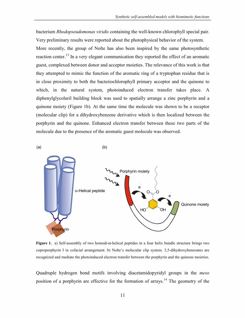
Synthetic self-assembled models with biomimetic functions
11
bacterium Rhodopseudomonas viridis containing the well-known chlorophyll special pair.
Very preliminary results were reported about the photophysical behavior of the system.
More recently, the group of Nolte has also been inspired by the same photosynthetic
reaction center.13 In a very elegant communication they reported the effect of an aromatic
guest, complexed between donor and acceptor moieties. The relevance of this work is that
they attempted to mimic the function of the aromatic ring of a tryptophan residue that is
in close proximity to both the bacteriochlorophyll primary acceptor and the quinone to
which, in the natural system, photoinduced electron transfer takes place. A
diphenylglycoluril building block was used to spatially arrange a zinc porphyrin and a
quinone moiety (Figure 1b). At the same time the molecule was shown to be a receptor
(molecular clip) for a dihydroxybenzene derivative which is then localized between the
porphyrin and the quinone. Enhanced electron transfer between these two parts of the
molecule due to the presence of the aromatic guest molecule was observed.
OH OH
O Oe
-
e-
Porphyrin moiety
Quinone moiety
Porphyrin
α-Helical peptide
(a) (b)
Figure 1. a) Self-assembly of two homodi-α-helical peptides in a four helix bundle structure brings two
coproporphyrin I in cofacial arrangement. b) Nolte’s molecular clip system. 3,5-dihydroxybenzoates are
recognized and mediate the photoinduced electron transfer between the porphyrin and the quinone moieties.
Quadruple hydrogen bond motifs involving diacetamidopyridyl groups in the meso
position of a porphyrin are effective for the formation of arrays.14 The geometry of the

Chapter 2
12
arrays is ultimately determined by the substitution pattern on the porphyrin, thus allowing
the formation of linear tapes as well as cyclic tetramers. Sessler and coworkers reported
the formation of a porphyrin-sapphyrin self-assembled complex.15 Sapphyrins,
pentapyrrolic “expanded porphyrin” macrocycles, are known to be excellent receptors for
a variety of anions. This property has been exploited for the formation of an assembly
with a porphyrin bearing a carboxylic acid group. The system was studied by 1H NMR in
order to prove the formation of the complex. UV and NMR expriments as well as X-ray
crystallographic evidence on model compounds showed that the two chromophores are
likely to be oriented perpendicular to each other. Nonetheless the system was shown to
undergo fast excitation transfer with high efficiency (the sapphyrin being the low lying
energy partner in the ensemble).
Another interesting aspect of photosynthetic systems that has attracted the attention of the
scientific community is the chain of events occurring after a photon has been absorbed by
the light-harvesting system, causing a photoinduced electron transfer. The charge
separation caused by the photon absorption needs to be stabilized efficiently and quickly
to allow conversion of the electromagnetic energy into chemical energy. In particular it is
interesting to focus attention on the so-called donor side in the chain of events happening
at Photosystem II. The photooxidized form of chlorophyll (formed upon electron transfer)
is regenerated with electrons coming from the catalyzed oxidation of water to oxygen
involving a redox active amino acid (named tyrosineZ) and a manganese cluster formed
by four high-valent Mn ions. A very recent excellent review16 summarizes the efforts of
Åkermark and Styring in this field which is also of paramount importance for practical
applications such as the conversion of solar energy into fuels (hydrogen). Their work
focuses on the use of ruthenium-manganese complexes. The basic idea was to use RuII
tris-bipyridine as a substitute for chlorophyll and to show that in dinuclear MnII – RuII
systems it is possible to have electron tranfer from the MnII moiety to the photogenerate
RuIII species.
A more sophisticated mimic17 involves the use of a phenolic moiety (covalently attached
to one of the RuII bipyridine ligands) that acts like the TyrZ residue (Figure 2a). In
particular, upon flash photolysis in acetonitrile or aqueous solution containing
methylviologen (MV2+) as an electron acceptor, electron transfer from the phenolic

Synthetic self-assembled models with biomimetic functions
substituent to the photogenerated RuIII occurs. The process is very fast (kET >107 s-1) in
the system containing two dipicolylamine arms that are hydrogen bonded to the phenolic
hydroxyl group. The importance of the hydrogen bonding network was clearly
demonstrated by comparison with systems that lack this structural element. It is known
that in the natural reaction center TyrZ is hydrogen bonded to His190. EPR experiments
performed in water during flash photolysis showed the formation of a neutral phenoxyl
radical in close resemblance to what is known to happen in nature to the TyrZ residue.
NN
R
NN
NNN N
OH NN
NH
COOEt
O
Figure 2. a) The Ru
introduce hydrogen bo
called donor side of P
reaction center.
In a related system
approach for the
semisynthetic appro
non-natural way to
myoglobin based t
located in the myo
mechanically linke
(BXV4+, as the acc
can be achieved b
involved in the reco
formation of the c
(a)
13
uN
N• 2 PF6
-
NN
Ru
NN
N
N
e- e-
Porphyrin
Polyether ch
Myoglobin
N+
+N+
II tris-bipyridine complex with an appended Tyr residue (chemically m
nding to the phenol group), mimicking the activity of the TyrZ residue
hotosystem II. b) The semisynthetic approach for the mimic of the pho
Shinkai and coworkers showed the potential of a semi
mimic of the photosynthetic reaction center (Figure
aches, natural building blocks are manipulated and/or conne
give an artificial system. This idea has been exploited to
riads containing a metal protoporphyrin (this time used a
globin pocket, a RuII tris(bipyridyl) moiety as the sensitiz
d (in the form of a catenane) cyclobis(paraquat-p-phenyl
eptor). The present case demonstrates the degree of comple
y the use of several different self-assembly processes.
nstitution of the apoprotein with the porphyrin appended tria
entral RuII complex starting from three different ligands an
(b)
ain
N+
N+
odified to
in the so-
tosynthetic
synthetic
2b).18 In
cted in a
prepare
s donor)
er, and a
ene) unit
xity that
They are
ds, in the
d in the

Chapter 2
14
synthesis of the catenane type ligand. Detailed investigations of the system showed the
expected vectorial, stepwise electron transfer giving rise to a long-lived charge separated
state, a chain of events that is known to operate in the natural photosynthetic reaction
center.
Another system based RuIItris(bpy)3 photosensitizer and BXV4+ acceptor should be
mentioned here.19 The well-known π-donor-acceptor complexes between
N,N’-dialkylbipyridinium salts and electron rich aromatic compounds such as
dialkoxybenzenes were used as the key feature for the noncovalent spatial organization of
the MXV4+ acceptor around the RuII center. Modified bpy ligands bearing one to two
dialkoxybenzene moieties were used to prepare dyads and polyads in a pseudorotaxane
fashion. Photophysical analysis of the system revealed that the electron transfer process
occurred in two distinct populations of the photosensitizer, viz. in the supramolecular
assembly and in the free photosensitizer.
One important consideration when attempting to gain valuable information about the
photosynthetic reaction center is that the size of a proposed model system should be
comparable to that of the natural system. For the investigation of large model systems,
dendrimers (hyperbranched, synthetic macromolecules with well-defined three-
dimensional structures) are ideal prototypes. Their inherent characteristics and structural
simplicity allow them to act as a bridge between the world of small molecular mimics and
of large and complex natural systems.20
The first of such systems is based on PAMAM (polyamidoamine) dendrimers with
20-residue α-helical peptides covalently linked to the peripheral amino groups.21
Different dendrimer generations (up to generation 4, G4) were synthesized and studied.
The molecular weight for the G4 dendrimer having 64 peptides linked to the PAMAM
scaffold is around 160 kD, remarkably high for a synthetic system. The peptides contain
one His residue in position 10 along the sequence. The authors showed that FeIII-or ZnII-
mesoporphyrin IX are complexed by the dendrimer, with each porphyrin coordinated
between two α-helical peptidic strands giving rise to a multiporphyrin array (the systems
are indicated as Fe-MP and Zn-MP respectively, Figure 3). Fluorescence studies in
solution showed photoinduced electron transfer from the porphyrin to methylviologen
(MV2+). More interestingly the system was shown to function effectively as an artificial

Synthetic self-assembled models with biomimetic functions
15
photosynthetic system. A solution containing triethanolamine as an electron donor, Zn-
MP as photosensitizer and MV2+ as electron acceptor was used to demonstrate the
production and accumulation of MV+ radicals upon photoirradiation. This result
confirmed that the three dimensional array of porphyrins, prepared by self-assembly with
the use of suitable peptide dendrimers, is an effective photosensitizer in the above
mentioned artificial photosynthetic system.
Zn- (Zn-MP)mesoporphyrin IX
α-Helical peptide
Figure 3. Light harvesting dendrimers.
2.2.2 Aspecific models for electron transfer processes
The focus of the following examples is not directly on the mimicry of the photosynthetic
reaction center as discussed previosly, but is somewhat more general in trying to
understand some of the basic features of the electron tranfer process itself.
Metallodendrimers have been reported as suitable candidates to mimic natural light-
harvesting systems and have been extensively studied and recently reviewed by Balzani
and coworkers.22 The strategy to build large polynuclear metal complexes is called by the
authors “complexes as metals/ complexes as ligands”. The function of the systems is due
to the intrinsic and specific properties of the building blocks (ability to undergo visible
light absorption, luminescence, and reversible multi-electron processes).
Fréchet and coworkers used a different type of chromophore as an antenna.23
Polybenzylether dendrons bearing a carboxylic acid functionality at the focal point (up to
generation 4) are light-harvesting units that can be self-assembled around lanthanide

Chapter 2
cations. The stoichiometry of the complexes is three dendritic wedges per cation. Studies
on the system showed both an antenna effect that allows the energy transfer from the
dendritic ligands to the central ion, and a shell effect leading to a decreased self-
quenching of lanthanide fluorescence. Despite the very interesting properties of dendritic
systems, their synthesis can be very demanding even if self-assembly is involved.
Interestingly, more easily accessible linear light-harvesting polymers can be prepared
following indications obtained from a dendritic model system.24 Copolymerization of a
RuII(bpy)3 functionalized monomer with a coumarin-2 functionalized monomer in a 1:3
ratio afforded a polymer that exhibited quantitative energy transfer from the coumarin-2
units to the RuII centers.
In naturally occurring electron transfer processes (both thermal and photoinduced),
hydrogen bonds and ionic interactions within the protein matrix play a very important
role. Some model systems have tried to quantify the importance of these interactions.
Sessler and coworkers used hydrogen bonds to mediate photoinduced electron transfer in
a dimethylaniline-anthracene ensemble.25 The ensemble was prepared via Watson-Crick
base pairing principles (Figure 4a). The donor and the acceptor moieties were attached
alternatively to one or the other base that participates in the recognition motif. This
approach provides two different diads that behave very differently upon photoexcitation.
Despite the fact that the reason for such different behavior was not understood, the
interesting conclusion is that not only is the nature of the interaction mediating the long
range electron transfer important (in this case hydrogen bonds), but also the
directionality.
e- e-
F
p
) )
(a16
NN
N N
O
HNH
H N
N
O
NHH
Donor
Acceptor
Rib
Rib
igure 4. Asymmetric self-assembled donor-acceptor in
rinciples, b) carboxylate-amidinium salt bridge.
(b
O
OH
N
NH
H
H
+
-
Donor
Acceptor
terfaces based on a) Watson-Crick base pairing

Synthetic self-assembled models with biomimetic functions
17
Similar conclusions were reached for electron transfer processes mediated by salt
bridges.26 Amidinium-carboxylate interactions were used as an asymmetric salt bridge
interface (Figure 4b) relevant for the understanding of naturally occurring proton-coupled
electron transfer processes. The rate of photoinduced electron transfer through the donor-
(amidinium-carboxylate)-acceptor salt bridge was 100 times slower than when the
interface was inverted to donor-(carboxylate-amidinium)-acceptor.
2.2.3 Cytochrome and redox cofactor activity mimics
The two most important energy-converting processes in biology are photosynthesis and
respiration. They both involve redox reactions and therefore enzymes and proteins with
redox active functionalities. Some model systems dealing with electron transfer processes
based on peptides have been reported and discussed in a recent review.27 Very promising
results have been obtained in model systems for cytochrome b, the membrane spanning
subunit of the mitochondrial cytochrome bc1, and of the photosynthetic cytochrome bf
complexes.
In this respect, some relevant features of systems reported recently by Willner and
Haehnel should be mentioned. They used the design principle of template-assembled
synthetic proteins in conjunction with an antiparallel four-helix bundle motif, to prepare a
cytochrome b model.28 First self-assembly was used to incorporate two FeIII-
protoporphyrin IX cofactors in the synthetic apoprotein. The electron transfer unit
obtained was then covalently attached to a self-assembled monolayer on a gold surface
bearing proper reactive groups. A third relevant self-assembly step involved the
formation of affinity complexes between a cytochrome-dependent native protein such as
nitrate reductase and the de novo synthesized electron-transporting protein attached to the
monolayer (Figure 5).29 With the aim of preparing a robust bioelectrocatalytic electrode,
the complexed nitrate reductase layer was further cross-linked. It should be mentioned
that other proteins could be fixed on the de novo protein modified monolayer, opening
the way to novel interesting technological applications in such fields as bioelectronic
devices.30
Redox processes are not limited to cytochromes and are also present in flavin-, quinone-
and pyrroloquinolinequinone-dependent enzymes. The use of model systems for the

Chapter 2
18
understanding of redox cofactor activity has been the subject of a recent review.31 Self-
assembly processes of interest were used in two systems based on flavin32 and 6-
azaflavin,33 respectively. These two publications focus on the role of noncovalent
interactions between the redox cofactor and the apoprotein (which is mimicked by a
much simpler molecular receptor) in modifying the redox properties of the system.
FeIII FeIII
Nitrate
Reductase
S
S
N N
O
O
O
H
S
S
N N
O
O
O
H
Au electrode
Figure 5. Molecular design of a bioelectrocatalytic electrode prepared via self-assembly followed by cross-
linking of the nitrate reductase layer.
2.3 Catalysis and enzyme mimics
In biotransformations, catalysis and therefore enzymes are of fundamental importance.
Model systems in this field are often called “artificial enzymes”. It is beyond the scope of
this chapter to give a detailed explanation of this term and of all the implications that
follow, but the interested reader is referred to an excellent review of Kirby that appeared
in 1996.34 The author provides a very critical analysis of enzyme model systems. One of
the basic definitions for enzyme mimics is that these systems must involve an initial
binding interaction between the substrate and the catalyst, thus giving rise to Michaelis-
Menten kinetics. Focusing on self-assembly processes in enzyme model systems, a very
broad classification based on the extent to which self-assembly is important could be
made viz. self-assembly for the recognition of the substrate only or self-assembly for both
the formation of the enzyme mimic and for the recognition of the substrate. The latter
case is described more in detail because it exploits the complete potential of the
noncovalent approach.

Synthetic self-assembled models with biomimetic functions
19
The group of Rebek reported the Diels-Alder reaction acceleration using self-assembled
molecular capsules (Figure 6).35 The system is formed upon dimerization of two
molecules having a concave surface and featuring complementary patterns of hydrogen
bonding sites. Complexation of the two reactants within the capsule and thus
enhancement of their concentration was regarded as the reason for the increased reaction
rate. One major problem common to many model systems involving reactions between
two different substrates34 is product inhibition and thus the lack of turnover.
NHN
N NHNN
NN
NH N
NNHRR RR
O
O
O
O
O
OO
OOH
OH
OH
OH
2
Figure 6. Self-assembled molecular capsules for the acceleration of Diels-Alder reactions.
A somewhat less characterized system has been reported involving the formation of
peptide self-aggregates in aqueous solutions.36 16-Mer peptides containing alanine and
lysine residues form very resistant macromolecular β-sheet structures involved in
multilayer sheets or micelles. These aggregates bind nucleotides and organic
phosphodiesters and are effective in the hydrolysis of bis(4-nitrophenyl)phosphate. A
103-104-fold increase in the reaction rate was observed in the presence of the above
mentioned peptide aggregates.
Hill and coworkers have addressed a very interesting aspect of natural systems viz. the
ability of repairing damage.37 A polyoxometalate catalyst self-assembles under the
reaction conditions and this self-assembly process has been related to the catalytic
Chapter 2
20
conversion of α-terpinene to p-cymene. As a consequence, any destructive event on the
catalyst can be repaired under the reaction conditions.
Mihara and coworkers designed α-helical peptides able to bind a Fe-mesoporphyrin.
With control of their three dimensional structure induced by the addition of
trifluoroethanol, these systems have N-demethylase activity.38 They further extended their
study showing the relationship between the haem-binding properties of the designed
peptides and the catalytic activity (peroxidase-like).39 In particular, they were able to
show that very tight haem-binding reduces the reactivity of the systems towards hydrogen
peroxide in a way that resembles the natural strategy to transform a haemprotein
(peroxidase-like) into an electron transfer protein (cytochrome-like) under physiological
conditions.
The group of Nolte has reported a very interesting self-assembled catalyst as an enzyme
mimic. This model features all the important components present in the natural
cytochrome P450 system, viz. molecular oxygen as the oxidizing agent, a
metalloporphyrin as the catalyst, an electron donor as the reducing agent, and a
membrane system that holds all the components together.40 Very detailed studies were
performed to understand the effect of the nature of the bilayer and vesicular aggregates
(and in particular of the charge carried by the different surfactants) on the system
reactivity. A number of different substrates could be epoxidized with turnover numbers
comparable to those observed for natural systems.
Finally, one catalytic system that involves self-assembly only in the recognition of the
substrate should be mentioned. Ghadiri and coworkers prepared a de novo designed
peptide ligase that catalyzes the condensation of short peptide fragments with rate
acceleration as high as 4100-fold.41 The catalytic activity was attributed to the efficient
binding in close proximity of the two reactive peptidic fragments. Also in this case
product inhibition was observed but the system represents the first example of a peptide
catalyst with designed substrate binding sites able to significantly accelerate a
bimolecular reaction.
Molecularly imprinted materials as enzyme mimics should also be mentioned. In these
systems self-assembly is often used as the first step to create a suitable binding site, using
a template molecule and functional monomers that are subsequently covalently fixed in a
Synthetic self-assembled models with biomimetic functions
21
second polymerization step. The template molecule is then washed away, leaving a
material that is able to recognize desired substrates and catalyze their reaction. However,
the field of molecularly imprinted materials is very broad and beyond the scope of this
overview. Furthermore, molecular imprinting has been recently reviewed with regard to
their applicability as enzyme models.42
2.4 Allosterism
Allosterism is a form of chemical feedback found in many biological processes, i.e.
cooperative dioxygen binding to hemoglobin and the hexamerization of arginine
repressor. A system presenting a number of recognition sites may show allosterism if
those binding sites are able to communicate upon substrate recognition. The biomimetic
design of allosteric systems has been focused on the initial binding of a substrate and
either the positive or negative effects (positive or negative allosterism, respectively) on
subsequent binding events. The allosterism can be heterotropic or homotropic, depending
on whether the second molecule is different or identical, respectively. The most
prominent synthetic allosteric systems deal with positive homotropic allosterism and are
well represented in the work of Shinkai and coworkers. They have chosen a tetrakis(4-
pyridyl)porphyrin to build up a cerium(IV) bis(porphyrinate) double decker system
(Figure 7).43 This complex shows slow rotation of the two porphyrin planes with respect
to one another at room temperature. The four pairs of 4-pyridyl groups are available as
hydrogen bond acceptor sites for diols, hydroxycarboxylic acid, and dicarboxylic acid.
The system displays a strong positive allosteric effect and is highly selective for BOC-
aspartic acid and 1,2-cyclohexanedicarboxylic acid. The observed allosterism can be
attributed to the successive suppression of the rotation of the porphyrin planes.
Modification of this system with two pairs of boronic acid groups allows the cooperative
recognition of two saccharide molecules in water.44 The same structure can also bind
oligosaccharides such as maltooligosaccharide and laminarioligosaccharide.

Chapter 2
22
N
N
N
NN
N N
N
NN
N N
N
N
N
N
CeIV
Dicarboxylic acid
Figure 7. Cerium(IV) bis(porphyrinate) double decker system showing positive allosterism upon
recognition of BOC-aspartic acid and 1,2-cyclohexanedicarboxylic acid.
2.5 Membrane Transport
Biological membranes are crucial for biochemical transformations, intercellular contacts,
organization, transport, energy transduction, and communication processes. Models of
functional micelles and vesicles45 and of molecular transport and organization in
supported lipid membranes46 have been recently reviewed. Hence, we focus on the
phenomenon of chemical transport through membranes by artificial self-assembled
channels. Very recently, Ghadiri and coworkers published an extensive review on
nanotubular structures of molecular dimensions that perform diverse biological functions,
with the emphasis on self-assembled systems.47 Nevertheless, three additional examples
not included in this review will be discussed.
A very interesting publication by Ghadiri and coworkers 48 describes a self-assembling
transmembrane peptide nanotube channel, which mediated the highly efficient transport
of glutamic acid. The nanotubes were formed by self-assembly via hydrogen bonds of
cyclic decapeptides (7 Å van der Waals internal diameter) containing an even number of
hydrophobic α-amino acids with alternating D and L configuration. Amino acid transport
was continuously monitored via an enzymatic assay that couples glutamine synthetase
activity with the reactions catalyzed by piruvate kinase and lactate dehydrogenase

Synthetic self-assembled models with biomimetic functions
23
(monitoring NADH oxidation at 340 nm). Conductance experiments were performed to
assess the behavior of a single channel.
R
R
R
R
R
R
R
R
R = O-Ac-Leu-N H2
R
HO
OMe
OMeNH
NH
O
OO
H
R OMe
MeO
O
O
N(CH3)3CH2
R = COOH+
(a)
(b)
Figure 8. Biomimetic models of ion channel proteins. a) Octa-leucine based dimers and b) amphiphilic
cholic acid derivatives with two different charged head groups (carboxylate or ammonium ions).
Dimeric rigid-rod β-barrels as biomimetic models of ion channel proteins were reported
by Matile and coworkers.49 The rigid rod octamers (octa-leucine) were prepared from
octa-anisol. NMR and CD experiments provided evidence of the self-assembly by
intermolecular β-sheet formation, and ESI-MS spectra strongly suggest the presence of
ionophoric dimers (Figure 8a) (internal pores diameters >5 Å). These structures are
capable of mediating efficient ion transport across lipid bilayers.
Kokube and coworkers50 prepared a new class of supramolecular transmembrane ion
channels. These systems were constructed by linking two units of amphiphilic cholic acid
methyl esters through biscarbamate bonds. This moiety was then modified to yield two
different structures, i.e. with a carboxylate group and an ammonium external head group,
respectively (Figure 8b). Both compounds gave rise to the formation of stable ion

Chapter 2
24
channels, characterized by relatively small conductances (5-20 pS) and long lasting (10
ms to 10 s) open states. The channels are able to discriminate K+ from Na+ and they
exhibit high cation/anion selectivity, which is modulated by the structure of the ionic
head group of the channels.
2.6 Artificial Molecular Machines
Natural molecular machines are extremely complicated systems rendering little chance of
creating artificial molecular machines in the near future that model the natural
counterparts. Nevertheless, in accordance with this far-reaching ambition, the number of
discerning publications dealing with artificial molecular machines has steadily increased
over the last few years. Recently, Stoddart, Balzani and coworkers have reviewed the
most significant developments in the field of artificial molecular machines. The review
focuses on supramolecular structures developed by noncovalent synthesis
(pseudorotaxanes) and by supramolecular assistance to covalent synthesis (rotaxanes51
and catenanes). Two natural molecular machines, i.e. F1-ATpase (a rotary motor) and
myosin (a linear motor) are also discussed. As the authors point out, the most interesting
systems from the point of view of molecular machines are the [2]-pseudorotaxanes in
which a guest molecule is threaded through the plane of a macrocycle. The reason for this
is that the threading and rethreading are reminiscent of the action of a linear motor.
Furthermore, the [2]-pseudorotaxanes are the only models that are self-assembled in the
strictest sense (thermodynamic control).
There are many examples of artificial molecular machines, based on pseudorotaxanes,
which undergo co-conformational change after chemical, electrochemical, and
photochemical stimuli. Photons or electrons are certainly the best energy source to power
molecular machines. It is well-established that N,N'-dialkylbipyridinium salts form π-
donor-acceptor complexes with different electron rich aromatic compounds. Stoddart,
Balzani and coworkers have extensively studied the supramolecular complexes
(pseudorotaxanes) between dialkoxybenzenes and bipyridinium cyclophanes. They have
shown that a Re-complex can be incorporated into the ring component of this type of
pseudorotaxane as a photosensitizer (Figure 9).52 This modification allows for the

Synthetic self-assembled models with biomimetic functions
25
fabrication of a sophisticated photochemically driven molecular machine of the
pseudorotaxane type. The dethreading and rethreading motions are triggered by
irradiation with visible light and oxygenation of the solution, respectively. The motions
can be easily monitored by UV/Vis absorption and luminescence spectroscopy. Many
dethreading/rethreading cycles can be performed with the same solution without any
appreciable loss of signal. The reductant scavenger is the limiting factor for the (in
principle) infinite cycle.
e-
OHO
O
OH O
O+
+
+
+
N
N
N
N
N
N
ReCO ClCO CO
OHO
O
OH O
O
+
+
+
+
N
N
N
N
N
N
ReCO ClCO CO
O2
Light
+
Reductant
Products
Figure 9. Mechanism of a linear molecular motor based on pseudorotaxane.
Harada and coworkers53 have prepared poly-pseudorotaxanes based on ionic polymers
and cyclodextrins. β-Cyclodextrins move along a polymer consisting of bipyridinium
(viologen) moieties bridged by polymethylene chains (molecular shuttles). To avoid the
escape of a cyclodextrin from the polymer chain, two molecular stoppers (rotaxane
formation) were attached to the end of the polymer chain. This molecular machine has a
shuttling behavior that is solvent and temperature sensitive and can be controlled by two
interactions viz. hydrophobic interaction between a cyclodextrin ring and a station
(polymethylene chain), and a repulsive interaction between the cyclodextrin ring and the
4,4'-bipyridinium linker.
In nature the chemical energy supplied by food is used to power the biological machines
that sustain life. Similarly, chemical energy input can be used to control
threading/dethreading processes in pseudorotaxanes. The system developed by Balzani

Chapter 2
26
and coworkers54 can be considered a molecular-level plug/socket device. The plug-in
function is based on the threading of a (±)binaphthocrown ether by a (9-
anthracenyl)benzylammonium ion. The association process can be reversed quantitatively
(plug out) by addition of a suitable base such a tributylamine. In the plug-in state
(pseudorotaxane), an energy-transfer process takes place. As a consequence of the
reversibility of the acid/base reactions, the energy transfer process can be switched on
and off at will.
2.7 Synthetic models for hemoglobin (Hb) and myoglobin (Mb)
Hemoglobin (Hb) and myoglobin (Mb) are the most known proteins involved in the
transport and storage of O2, fulfilling an essential role for the existence of a large variety
of living organisms on earth. The three dimensional structure of these compact globular
proteins, which are characterized by the presence of α-helical segments (labeled with
capital letters), is shown in Figure 10.
Synthetic models of these proteins have been invaluable tools in unraveling the subtle
complexities of reversible O2 binding and CO inhibition. However, no attempts have
been made so far to construct model systems in a noncovalent way. This is rather
surprising since self-assembly and noncovalent interactions are widely present in natural
systems.
2.7.1 Hemoglobin and myoglobin: cooperativity in O2-binding makes the difference
Evolution has led to the development of several proteins for O2 transport and storage. In
mammals, these functions are carried out by Hb (an O2 carrier) and Mb (for O2 storage in
the muscles). Recently a third type of globin, neuroglobin,55 has been discovered in
human and mouse brain tissues, where it apparently has a function comparable to that of
Mb in muscle cells. Other proteins able to bind dioxygen are hemerythrins56-58(storage)
and hemocyanins56 (transport) but their binding site is not a FeII porphyrin (heme) and
will not be considered since the research described in this thesis focuses on synthetic
models for heme containing O2 carriers.

Synthetic self-assembled models with biomimetic functions
27
A
β2 β1
α2 α1
A
C-terminus
HemoglobinMyoglobin
C-terminus
N-terminus
N-terminus
B
C
D
E
F
G
H
B E
HF
G
C
DF A
CD
BEG
H
Figure 10. Schematic three dimensional structures of myoglobin and hemoglobin.
The O2-binding site of Hb and Mb is a FeII protoporphyrin IX (the heme) located in a
hydrophobic cavity formed by the folding of the protein (Figure11).56,59
N
N
HO
OH
N
N
H
N
NN
N
Fe
O
OH
O O
Figure 11. Schematic representation of the heme i
protoporphyrin IX is coordinated axially to the proximal
E7) stabilizes the coordinated O2 via hydrogen bonding.
His E7
His F8
n O2 binding hemeprotein. The FeII atom of
histidine residue (His F8). The distal histidine (His

Chapter 2
28
The heme is held in its position by a multitude of noncovalent interactions, which include
hydrophobic interactions and the coordination of the FeII atom to the imidazole ring of
histidine F8 (proximal histidine).
Noncovalent interactions are also responsible for the differences between Mb and Hb. In
fact Mb is a monomer (one peptidic chain of 153 amino acids and one heme cofactor)
while Hb is a tetramer consisting of two α-globins (peptidic chains folded in globular
structures) and two β-globins, each very similar to Mb. The correct positioning of one
chain with respect to the others in the tetrameric structure is due to the existence of
noncovalent interactions such as salt-bridge formation and hydrophobic interactions. The
direct consequence of these different structural features between the two proteins is the
very well-known cooperativity in O2 binding displayed by Hb but not by Mb (see Figure
12).
Figure 12. Dioxygen saturation curve for Mb and Hb at pH 7.4 and 25°C
To describe the observed cooperativity based on a two-state allosteric model displayed by
Hb, Perutz introduced the symbols T and R.60,61 Two structures for each subunit (α or β)
are possible according to the model viz. a low O2 affinity structure (T or tense state) and a
high O2 affinity structure (R or relaxed state). These two states differ in the folding of the

Synthetic self-assembled models with biomimetic functions
29
peptidic chain (tertiary structure) of each subunit and also in the relative orientation of the
subunits in the tetramer (quaternary structure). The T and R states are present as an
equilibrium mixture, with preference for T at low O2 partial pressures. When the first O2
molecule binds to a T state subunit, a certain amount of strain is induced in the local
tertiary structure. This strain alters the interactions between the subunits, inducing a
change in their tertiary structure, which facilitates the binding of an additional O2
molecule (positive allosterism, see section 2.4).
2.7.2 Synthetic models: stability and understanding of the O2 adduct
Most work in this field has been concentrated on porphyrin systems for the preparation of
synthetic models for Hb and Mb active sites, because of the structural similarity between
these compounds and the heme cofactor. The porphyrinic scaffold offers a large number
of positions that can be chemically functionalized (Figure 13), and has led to the
preparation of very elaborate structures by covalent modification of the porphyrin
periphery. These modifications have allowed the protein environment in proximity to the
heme to be reproduced. Excellent reviews describing extensively the progress in this
eminent field of chemistry have been published.59,69-71 These efforts have resulted in a
large variety of porphyrins, which have been described as “capped”, “picket fence”,
“pocket”, “strapped”, “cofacial” and “picnic basket”. The leading idea behind many of
these synthetic systems is to understand the structure-activity relationship that could
explain efficient and stable O2 binding to natural heme proteins. Some examples are
shown in Figure 14 and their affinities for O2 are reported in Table 2.1. A more practical
reason for these studies is the achievement of an increased stability towards O2 (i.e.
systems in which undesired decomposition reactions triggered by O2 binding are
minimized) in view of technological applications such as O2 separation and storage.72

Chapter 2
30
Table 2.1. Dioxygen binding constants for some CoII and FeII porphyrins (see Figure 14) expressed as half
oxygenation pressure P1/2.a
Compound P1/2(O2)
(Torr)
Conditions
Fe-Cu-4 31 Torr 0.2M 1-MeIm, benzene, 20 °C62
FeSP-15 15 Torr 0.2M 1-MeIm, benzene, 20 °C62
Fe(Cap) 4.0 × 10³ Torr 1,2-diMeIm, toluene, 25 °C64
Co(Cap) 1.4 × 10⁵ Torr 1-MeIm, toluene, 15 °C64
C2-capped C5-strapped 110 Torr toluene, 0 °C68
Co TpivPP 140 Torr 1-MeIm, toluene, 25 °C67
Fe TpivPP 38 Torr 1,2-diMeIm, toluene, 25 °C66
a O2 partial pressure in the gas phase (in equilibrium with the porphyrin solution) under which 50% of the
porphyrin binds O2.
NH
N
N
NH
O OOHOH
OOH
OOH
NH
N
N
NH
OOH
OOH
NH
N
N
NH
OOH
OHO
NH
N
N
NH
O
OH
OH
O
OOH
NH
N
N
NH
OHO
NH
N
N
NH
(a) (b) (c)
(d) (e) (f)
Figure 13. Porphyrin structures commonly encountered in literature that are associated with natural and
synthetic heme protein systems. a) protoporphyrin IX; b) deuteroporphyrin IX; c) mesoporphyrin IX; d)
mesoporphyrin; e) coproporphyrin; f) meso-tetraphenylporphyrin.

Synthetic self-assembled models with biomimetic functions
31
NH
XO
NN N
NNH
X
NN N
N
OR
R
R
R
Fe
Cu
CO
N
N
NN N
N
R
R
O NH
ONH(CH2)
Fe
CO
n
N
N
(a) Fe-Cu-4 and Fe-Cu-5 (X=CH2 or CH2CH2)
(Chang 62)
(b) FeSP-13, FeSP-14 and FeSP-15 (n=5, 6, and 7)
NNHNH
NO
(CH2)O
(CH2)O
O
O
OO
(CH2)O
O
(CH2)
O
O
O
xx
xx
(c) CapH2 (x=2) and HmCapH2 (x=3) (Baldwin63 and Basolo64)
NNN
NM
NH
NH
O
O
NHO
NH
O
(d) TpivPP, M= FeII, CoII (Collman65-67) (e) C2-capped Cn-strapped n=4 or 5 (Baldwin68)
Figure 14. Superstructured porphyrin-based dioxygen carriers. a) ”cofacial”, b) “strapped”, c) “capped”, d)
“picket fence”, e) “capped-strapped”.
Proteins Hb and Mb as well as model systems (collectively designated with the term O2-
carriers) require that the iron atom be maintained in the ferrous (FeII) state in order to

Chapter 2
32
bind O2. This is not the thermodynamically stable state in the presence of O2 but the
proteins successful retard the oxidation to the inactive FeIII state. Even then, the met-Hb
(the oxidized FeIII form) content of our blood is usually around 3%.† Many factors
contribute to the overall stability of an O2-carrier such as the solvent, the electron density
on the FeII ion (partly determined by the axial ligand trans to the O2) and the steric
hindrance protecting the coordinated O2.59,70,73 However, one important conclusion can be
extracted from all the studies on O2-binding stability: the minimal requirement for a
model system to be stable towards oxidation is to have a superstructure that protects the
Fe-O2 moiety from reacting with a second Fe-porphyrin. The picket fence porphyrin
(FeTpivPP) reported by Collman represents one of the milestones in this field.65 The
compound has been reported to be remarkably stable due to the presence of the four
pivalamido moieties which indeed effectively protect the Fe-O2 moiety (see Figure 14).‡
Synthetic model systems should possess all (or at least most) of the important features of
natural systems viz. i) a hydrophobic environment surrounding the metal-O2 moiety, ii) a
proper axial ligand to enhance the binding of O2 to the metal, and iii) secondary
interactions between the metal-O2 moiety able to stabilize the complex.
The hydrophobic pocket, which in nature is achieved by burying the porphyrin within the
protein matrix, has been generally accomplished in synthetic systems by the use of
organic solvents and/or by the already mentioned superstructures around the porphyrin
core (see Figure 14). The axial ligand requirement has been met by adding nitrogenous
bases such as imidazole or pyridine derivatives or by covalent attachment of similar
nitrogenous bases to the porphyrin structure. The third prerequisite is probably the most
challenging to introduce into model systems because very subtle interactions such as
steric hindrance and hydrogen bonding are involved. In fact, the contribution of these
interactions in the natural systems is still a matter of debate among the scientific
† Therefore living organisms have repairing mechanisms: the enzyme NADH-cytochrome b5
oxidoreductase reduces met-Mb or met-Hb to the respective FeII deoxy species and thus prevents the
accumulation of the inactive forms.73
‡ When dissolved in an aprotic solvent such as toluene and in the presence of an axial base such as 1,2-
dimethylimidazole, this O2-carrier remains stable for periods as long as several months.

Synthetic self-assembled models with biomimetic functions
33
community.69,74 A reliable mimicry of these secondary interactions is especially
important to understand which factors induce destabilization of the competing complex
derived from binding of the endogenous toxic ligand carbon monoxide (CO) in natural
systems.
The hypothesis that polarity effects cause discrimination between O2 and CO and
favoritism for O2 can be traced back to the suggestion of Pauling in 1964.75 Since then,
many model systems have demonstrated the importance of these effects on O2 binding
(but not on CO) and are summarized in an excellent review that covers the literature until
1998.69,70 The most important contribution to selective stabilization of coordinated O2 has
generally been attributed to hydrogen bonding interactions. For instance, it has been
shown that a strong hydrogen bond between coordinated O2 and tyrosine B10 (with the
involvement of glutamine E7) in Ascaris Hb is responsible for its extraordinary O2
avidity, four orders of magnitude higher than that of human Hb.76 Recent contributions
from the group of Naruta have shown the existence of hydrogen bonds between the Fe-O2
moiety of a “single-coronet” porphyrin and hydroxyl groups attached to the binaphthyl
bridges of the porphyrin (Figure 15).77 Their work has also been extended to “twin-
coronet”78 porphyrins having thiolate axial ligands as models for cytochrome P450 (a
heme-protein which is responsible for O2 activation).79 80In all cases, Raman
spectroscopy was used to demonstrate that the coordinated O2 is hydrogen bonded to
inward pointing hydroxyl groups appended to the porphyrin superstructure.
OHHO
NNN
NFe
HN
NH
HN
O
O
HOPivOOPiv
Figure 15. “Single-coronet” porphyrin (derived from combination of porphyrin and binaphtyl scaffolds).
The inward pointing hydroxyl groups can hydrogen-bond to the O2 coordinated to the Fe atom.

Chapter 2
34
Very recently, progress in understanding the factors affecting Fe-O2 stabilization in
natural proteins has been made by using density functional theory (DFT) calculations in
combination with SAM1 semi-empirical electronic structure methods.81-83 These quantum
chemistry calculations were used to estimate the interaction energies associated with H-
bonds in the active site of Ascaris Hb, as well as in the α- and β-subunits of human Hb in
the R-state.81 The results confirmed the existence of strong hydrogen bonds for all three
Hbs and contradicted previous observations indicating the lack of such interaction for the
β-subunit of human Hb. The authors pointed out that the global equilibrium for binding
of a ligand to a heme-protein is a complex phenomena involving partition of the ligand
between the solvent and the protein matrix, transport across the protein matrix and finally
the binding process itself. Mutagenesis experiments on proteins may therefore provide
misleading results since the residues involved in H-bonding are also involved in the
kinetics of the process and a strong H-bond does not necessarily imply a high affinity
constant for such complex systems.
2.7.3 Synthetic systems having CoII in the active site: the stability issue
The stability of simple FeII porphyrins in oxygenated solutions is so low that for many
years attempts to mimic Hb and Mb were unsuccessful. It was then discovered that CoII
Schiff base complexes and CoII porphyrins can reversibly bind O2 in 1:1 ratio under
conditions that were found prohibitive for the stability of FeII pophyrins. These CoII
complexes-O2 adducts are further related to the natural systems by the necessity of an
axially coordinated donor ligand.59,84 In fact, simple CoII porphyrins offer easy
accessibility to pentacoordinated complexes upon addition of a nitrogenous base for axial
ligation. In contrast, this remains a serious challenge when FeII porphyrins are used, due
to their strong tendency to form hexacoordinate complexes. Moreover, both oxygenated
and deoxygenated CoII porphyrins are paramagnetic and can be studied by electron
paramagnetic resonance (EPR) and electron nuclear double resonance (ENDOR)
spectroscopies.84
The enhanced stability of CoII porphyrins allowed the preparation of coboglobins, the
cobalt analogues of Hb and Mb, by reconstitution of the proteins with CoII
protoporphyrin IX.85-89 The study of coboglobins permitted the direct observation of the

Synthetic self-assembled models with biomimetic functions
35
effect of the protein environment by comparison of the spectroscopic properties and the
O2 binding of the coboglobins with those of CoII protoporphyrin IX dimethylester in
solution.90
In addition to the use of simple porphyrins as synthetic models, the previously
synthesized superstructured porphyrins were also prepared with CoII (instead of FeII) as
the O2 binding unit. Three years after the first publication about FeTpivPP porphyrins,65
the group of Collman reported the preparation of the analogue CoTpivPP and studied its
O2 binding equilibrium.67,91 The group of Basolo then reported the preparation of CoII
“capped” porphyrins64 and found considerably smaller O2 affinities in comparison to
Collman’s TpivPP porphyrins (see Table 2.1). The reduced affinities were attributed to
greater conformational strain accompanying the formation of the six-coordinate O2
adducts. Interestingly, the so-called “homologous” capped porphyrins (HmCap, see
Figure 13) containing longer spacers unexpectedly gave even lower affinities.64
These examples clearly illustrate one of the serious limitations of using synthetic models
prepared by covalent modification of the porphyrin periphery. Such systems could in fact
have a much higher rigidity than the noncovalent counterparts constituting the natural
systems.
2.8 Concluding remarks
With increasing emphasis on function rather than on structure, the importance of covalent
synthesis is becoming surpassed by self-assembly processes. This development is also
clearly observed in the synthetic biomimetic model systems discussed in the first part of
this chapter (sections 2.2 – 2.6). A large variety of relatively simple building blocks,
which posses a specific chemical function, are connected in a rational way via
noncovalent interactions. The collective and cooperative action of such assemblies
compares favorably in many cases with the much more complex natural systems.
It is surprising that so far there are no examples reported in the literature concerning the
preparation and study of synthetic self-assembled model systems that mimic O2 binding
hemeproteins. Moreover, almost all the systems (based on porphyrins) studied so far are

Chapter 2
36
limited by insolubility in aqueous media,92 which would be the desirable solvent for a
closer mimicry of the natural systems.
Based on the many successful examples using self-assembly strategies and porphyrin
building blocks such as the reproduction of photosynthetic centers, cytochromes and
redox cofactor activities, as well as oxidation catalysts (sections 2.2 and 2.3), it is very
likely that a number of other biomimetic functions can be imitated by synthetic systems,
including O2 binding.
2.9 References and notes
1. Biochemistry, 4 ed.; Stryer, L.; W. H. Freeman and Company: New York, 1995.
2. Wojaczynski, J.; Latos-Grazynski, L. Coord. Chem. Rev. 2000, 204, 113-171.
3. Haycock, R. A.; Yartsev, A.; Michelsen, U.; Sundström, V.; Hunter, C. A. Angew.
Chem. Int. Ed. 2000, 39, 3616-3619.
4. Haycock, R. A.; Hunter, C. A.; James, D. A.; Michelsen, U.; Sutton, L. R. Org.
Lett. 2000, 2, 2435-2438.
5. Ambroise, A.; Li, J.; Yu, L.; Lindsey, J. S. Org. Lett. 2000, 2, 2563-2566.
6. Yamada, K.; Imahori, H.; Yoshizawa, E.; Gosztola, D.; Wasielewski, M. R.; Sakata,
Y. Chem. Lett. 1999, 235-236.
7. Li, X.; Ng, D. K. P. Eur. J. Inorg. Chem. 2000, 1845-1848.
8. Guldi. Dirk M.; Luo, C.; Swartz, A.; Scheloske, M.; Hirsch, A. Chem. Commun.
2001, 1066-1067.
9. Balaban, T. S.; Tamiaki, H.; Holzwarth, A. R.; Schaffner, K. J. Phys. Chem. B
1997, 101, 3424-3431.
10. Amakawa, M.; Tamiaki, H. Bioorg. Med. Chem. 1999, 7, 1141-1144. The article
reports the preparation of stable self-aggregates in a solid film state using design
Synthetic self-assembled models with biomimetic functions
37
principles very similar to those present in naturally occurring systems
(chlorosomes). However the work is more focused on structural aspects than on the
function of the system.
11. Kuroda, Y.; Sugou, K.; Sasaki, K. J. Am. Chem. Soc. 2000, 122, 7833-7834.
12. Rabanal, F.; DeGrado, W. F.; Dutton, P. L. J. Am. Chem. Soc. 1996, 118, 473-474.
13. Reek, J. N. H.; Rowan, A. E.; de Gelder, R.; Beurskens, P. T.; Crossley, M. J.; de
Feyter, S.; de Schryver, F.; Nolte, R. J. M. Angew. Chem. Int. Ed. 1997, 36, 361-
363.
14. Drain, C. M.; Shi, X.; Milic, T.; Nifiatis, F. Chem. Commun. 2001, 287-288.
15. Springs, S. L.; Gosztola, D.; Wasielewski, M. R.; Kral, V.; Andrievsky, A.; Sessler,
J. L. J. Am. Chem. Soc. 1999, 121, 2281-2289.
16. Sun, L.; Hammarström, L.; Åkermark, B.; Styring, S. Chem. Soc. Rev. 2001, 30, 36-
49. A detailed review with a large introductory section describing the most recent
progress towards the understanding of the Photosystem II reaction center.
17. Sun, L.; Burkitt, M.; Tamm, M.; Raymond, M. K.; Abrahamsson, M.;
LeGourriérec, D.; Frapart, Y.; Magnuson, A.; Kenéz, P. H.; Brandt, P.; Tran, A.;
Hammarström, L.; Styring, S.; Åkermark, B. J. Am. Chem. Soc. 1999, 121, 6834-
6842.
18. Hu, Y.-Z.; Tsukiji, S.; Shinkai, S.; Oishi, S.; Hamachi, I. J. Am. Chem. Soc. 2000,
122, 241-253.
19. David, E.; Born, R.; Kaganer, E.; Joselevich, E.; Durr, H.; Willner, I. J. Am. Chem.
Soc. 1997, 119, 7778-7790.
20. Hecht, S.; Fréchet, J. M. J. Angew. Chem. Int. Ed. 2001, 40, 74-91. This recent
review covers the application of dendrimers in biomimetic systems. Most of the
reported examples are not based on self-assembly.

Chapter 2
38
21. Sakamoto, M.; Ueno, A.; Mihara, H. Chem. Eur. J. 2001, 7, 2449-2458.
22. Balzani, V.; Campagna, S.; Denti, G.; Juris, A.; Serroni, S.; Venturi, M. Acc. Chem.
Res. 1998, 31, 26-34.
23. Kawa, M.; Fréchet, J. M. J. Chem. Mater. 1998, 10, 286-296.
24. Schultze, X.; Serin, J.; Adronov, A.; Frechet, J. M. J. Chem. Commun. 2001, 1160-
1161.
25. Sessler, J. L.; Sathiosatham, M.; Brown, C. T.; Rhodes, T. A.; Wiederrecht, G. J.
Am. Chem. Soc. 2001, 123, 3655-3660.
26. Kirby, J. P.; Roberts, J. A.; Nocera, D. G. J. Am. Chem. Soc. 1997, 119, 9230-9236.
27. Xing, G.; DeRose, V. J. Curr. Opin. Chem. Biol. 2001, 5, 196-200.
28. Rau, H. K.; Haehnel, W. J. Am. Chem. Soc. 1998, 120, 468-476.
29. Katz, E.; Heleg-Shabtai, V.; Willner, I.; Rau, H. K.; Haehnel, W. Angew. Chem. Int.
Ed. 1998, 37, 3253-3256. Even if self-assembled monolayers have not been
specifically considered in this review, it is worth mentioning this interesting
communication. Several distinct self-assembly steps are used and integrated with
covalent synthesis, resulting in novel electrodes for biosensor application.
30. Willner, I.; Hleg-Shabtai, V.; Katz, E.; Rau, H. K.; Haehnel, W. J. Am. Chem. Soc.
1999, 121, 6455-6468.
31. Rotello, V. M. Curr. Opin. Chem. Biol. 1999, 3, 747-751.
32. Breinlinger, E. C.; Keenan, C. J.; Rotello, V. M. J. Am. Chem. Soc. 1998, 120,
8606-8609.
33. Kajiki, T.; Moriya, H.; Hoshino, K.; Kondo, S.; Yano, Y. Chem. Lett. 1999, 397-
398.
34. Kirby, A. J. Angew. Chem. Int. Ed. 1996, 35, 707-724. This review provides very

Synthetic self-assembled models with biomimetic functions
39
clear definitions to introductory readers approaching the field of enzyme models
and mimics.
35. Kang, J.; Rebek J. Jr Nature 1997, 385, 50-52.
36. Pérez-Payá, E.; Forood, B.; Houghten, R. A.; Blondelle, S. E. J. Mol. Recognit.
1996, 9, 488-493.
37. Hill, C. L.; Zeng, H.; Zhang, X. J. Mol. Catal. A 1996, 113, 185-190.
38. Sakamoto, S.; Sakurai, S.; Ueno, A.; Mihara, H. Chem. Commun. 1997, 1221-1222.
39. Sakamoto, S.; Ueno, A.; Mihara, H. J. Chem. Soc., Perkin 2 1998, 2395-2404.
40. Schenning, A. P. H. J.; Spelberg, J. H. L.; Hubert, D. H. W.; Feiters, M. C.; Nolte,
R. J. M. Chem. Eur. J. 1998, 4, 871-880.
41. Kennan, A. J.; Haridas, V.; Severin, K.; Lee, D. H.; Ghadiri, M. R. J. Am. Chem.
Soc. 2001, 123, 1797-1803.
42. Ramström, O.; Mosbach, K. Curr. Opin. Chem. Biol. 1999, 3, 759-764.
43. Takeuchi, M.; Imada, T.; Shinkai, S. Angew. Chem. Int. Ed. 1998, 37, 2096-2099.
44. Sugasaki, A.; Ikeda, M.; Takeuchi, M.; Shinkai, S. Angew. Chem. Int. Ed. 2000, 39,
3839-3842.
45. Scrimin, P.; Tecilla, P. Curr. Opin. Chem. Biol. 1999, 3, 730-735.
46. Boxer, S. G. Curr. Opin. Chem. Biol. 2000, 4, 704-709.
47. Bong, D. T.; Clark, T. D.; Granja, J. R.; Ghadiri, M. R. Angew. Chem. Int. Ed.
2001, 40, 988-1011.
48. Sanchez-Quesada, J.; Kim, H. S.; Ghadiri, M. R. Angew. Chem. Int. Ed. 2001, 40,
2503-2506.
Chapter 2
40
49. Sakai, N.; Majumdar, N.; Matile, S. J. Am. Chem. Soc. 1999, 121, 4294-4295.
50. Kobuke, Y.; Nagatani, T. J. Org. Chem. 2001, 66, 5094-5101.
51. Collin, J.-P.; Dietrich-Buchecker, C.; Gavina, P.; Jimenez-Molero, M. C.; Sauvage,
J.-P. Acc. Chem. Res. 2001, 34, 447-487. The account is focused on linear machines
and motors based on rotaxanes. Especially interesting are Sauvage's examples of
molecular muscles. The authors have designed a multicomponent system able to
contract or stretch under direction of an external chemical signal.
52. Ashton, P. R.; Balzani, V.; Kocian, O.; Prodi, L.; Spencer, N.; Stoddart, J. F. J. Am.
Chem. Soc. 1998, 120, 11190-11191.
53. Harada, A. Acc. Chem. Res. 2001, 34, 456-464.
54. Ishow, E.; Credi, A.; Balzani, S. F.; Mandolini, L. Chem. Eur. J. 1999, 5, 984-989.
55. Burmester, T.; Weich, B.; Reinhardt, S.; Hankein, T. Nature 2000, 407, 520-523.
56. Niederhoffer, E. C.; Timmons, J. H.; Martell, A. E. Chem. Rev. 1984, 84, 137-203.
57. Brunold, T. C.; Solomon, E. I. J. Am. Chem. Soc. 1999, 121 , 8277-8287.
58. Brunold, T. C.; Solomon, E. I. J. Am. Chem. Soc. 1999, 121 , 8288-8295.
59. Jones, R. D.; Summerville, D. A.; Basolo, F. Chem. Rev. 1979, 79, 139-179.
60. Perutz, M. F. Nature 1970, 228, 726-734.
61. Perutz, M. F.; Fermi, G.; Luisi, B.; Shaanan, B.; Liddington, R. C. Acc. Chem. Res.
1987, 20, 309-321.
62. Ward, B.; Wang, C.; Chang, C. K. J. Am. Chem. Soc. 1981, 103, 5236-5238.
63. Almog, J.; Baldwin, J. E.; Dyer, R. L.; Peters, M. J. Am. Chem. Soc. 1975, 97, 226-
227.

Synthetic self-assembled models with biomimetic functions
41
64. Linard, J. E.; Ellis, P. E.; Budge, J. R.; Jones, R. D.; Basolo, F. J. Am. Chem. Soc.
1980, 102, 1896-1904.
65. Collman, J. P.; Gagne, R. R.; Reed, C. A.; Halbert, T. R.; Lang, G.; Robinson, W.
T. J. Am. Chem. Soc. 1975, 97, 1427-1439.
66. Collman, J. P.; Brauman, J. I.; Doxsee, K. M.; Halbert, T. R.; Suslick, K. S. Proc.
Natl. Acad. Sci. USA 1978, 75, 564-568.
67. Collman, J. P.; Brauman, J. I.; Doxsee, K. M.; Halbert, T. R.; Hayes, S. E.; Suslick,
K. S. J. Am. Chem. Soc. 1978, 100, 2761-2766.
68. Baldwin, J. E.; Cameron, J. H.; Crossley, M. J.; Dagley, I. J.; Hall, S. R.; Klose, T.
J. Chem. Soc., Dalton Trans. 1984, 1739-1746.
69. Collman, J. P.; Fu, L. Acc. Chem. Res. 1999, 32, 455-463.
70. Momentau, M.; Reed, C. A. Chem. Rev. 1994, 94, 659-698.
71. Traylor, T. G. Acc. Chem. Res. 1981, 14, 102-109.
72. Figoli, A.; Sager, W. F. C.; Mulder, M. H. V. J. Membrane Sci. 2001, 181, 97-110.
73. Shikama, K. Chem. Rev. 1998, 98, 1357-1373.
74. Spiro, T. G.; Kozlowski, P. M. Acc. Chem. Res. 2001, 34, 137-144.
75. Pauling, L. Nature 1964, 203, 182-183.
76. Goldberg, D. E. Chem. Rev. 1999, 99, 3371-3378.
77. Kossanyi, A.; Tani, F.; Nakamura, N.; Naruta, Y. Chem. Eur. J. 2001, 7, 2862-
2872.
78. Naruta, Y.; Tani, F.; Ishihara, N.; Maruyama, K. J. Am. Chem. Soc. 1991, 113,
6865-6872.
Chapter 2
42
79. Matsu-ura, M.; Tani, F.; Nakayama, S.; Nakamura, N.; Naruta, Y. Angew. Chem.
Int. Ed. 2000, 39, 1989-1991.
80. Tani, F.; Matsu-ura, M.; Nakayama, S.; Ichimura, M.; Nakamura, N.; Naruta, Y. J.
Am. Chem. Soc. 2001, 123, 1133-1142.
81. Scherlis, D. A.; Estrin, D. A. J. Am. Chem. Soc. 2001, 123 , 8436-8437.
82. Godbout, N.; Sanders, L. K.; Salzmann, R.; Havlin, R. H.; Wojdelski, M.; Oldfield,
E. J. Am. Chem. Soc. 1999, 121, 3829-3844.
83. Vogel, K. M.; Kozlowski, P. M.; Zgierski, M. Z.; Spiro, T. G. J. Am. Chem. Soc.
1999 , 121, 9915-9921.
84. Smith, T. D.; Pilbrow, J. R. Coord. Chem. Rev. 1981, 39, 295-283.
85. Ikeda-Saito, M.; Brunori, M.; Yonetani, T. Biochim. Biophys. Acta 1978, 533, 173-
180.
86. Dickinson, L. C.; Chien, J. C. W. Proc. Natl. Acad. Sci. USA 1980, 77, 1235-1239.
87. Chien, J. C. W.; Dickinson, L. C. Proc. Natl. Acad. Sci. USA 1972, 69, 2783-2787.
88. Hori, H.; Ikeda-Saito, M.; Yonetani, T. Nature 1980, 288, 501-502.
89. Hori, H.; Ikeda-Saito, M.; Yonetani, T. J. Biol. Chem. 1982, 257, 3636-3642.
90. Hoffman, B. M.; Petering, D. H. Proc. Natl. Acad. Sci. USA 1970, 67, 637.
91. Steiger, B.; Baskin, J. S.; Anson, F. C.; Zewail, A. H. Angew. Chem. Int. Ed. 2000,
39, 257-259.
92. Jimenez, H. R.; Momentau, M. New J. Chem. 1994, 18, 569-574.
.

43
Chapter 3
NONCOVALENT SYNTHESIS OF CALIX[4]ARENE-CAPPED
PORPHYRINS IN POLAR SOLVENTS VIA IONIC
INTERACTIONS∗∗∗∗
The first step towards a self-assembled system for the mimicry of O2 binding hemeprotein
is the preparation of a superstructured porphyrin architecture via noncovalent synthesis.
The work presented here focuses on the use of ionic interactions as the recognition motif
between building blocks (tetracationic porphyrins and tetraanionic calix[4]arenes). The
self-assembly process is studied in polar solvents and in water/organic solvent mixtures.
3.1 Introduction
There are few classes of chemical compounds that have attracted more attention from
chemists than metalloporphyrins. These compounds play an important role as prosthetic
groups in several proteins like hemoglobin,1 the cytochrome P-450 mono-oxygenase
family2 and peroxidases.3 Whereas the central metal ion (coordinated to the four pyrrolic
nitrogens) is the functional unit in molecular recognition and catalysis, the selectivity in
these processes as well as the stability of the systems are largely due to noncovalent
interactions with the surrounding protein structure. Such interactions are possible because
of the shielded position of the porphyrin within the pocket of the folded protein
backbone.
∗ Part of this chapter has been published: Fiammengo, R.; Timmerman, P.; de Jong, F.; Reinhoudt, D. N.
Chem. Commun. 2000, 2313-2314. Fiammengo, R.; Timmerman, P.; Huskens, J.; Versluis, K.; Heck, A. J.
R.; Reinhoudt, D. N Tetrahedron 2002, 58(4), 757-764.

Chapter 3
Synthetic chemists have extensively tried to mimic natural porphyrin systems via
covalent modification of the porphyrin periphery, resulting in a multitude of porphyrin
structures such as capped, strapped, picnic basket, twin coronet and others (see section
2.7.2).4-6 A noncovalent approach to the modification of the porphyrin periphery has been
attempted via coordination chemistry7 or via hydrogen-bond interactions,8 generally
resulting in multiporphyrin systems. However, little attention has been paid to the use of
ionic interactions.9-11 Among the systems consisting of a single porphyrin unit there are
examples of porphyrin-cyclodextrin complexes,12-14 porphyrins in phosholipid
vescicles,15 and porphyrins interacting with other chromophoric units16 or systems used
for sensing purposes.17
This chapter deals with the use of multiple ionic interactions for the self-assembly of
cationic zinc(II) meso-tetrakis(N-alkylpyridinium-3-yl) porphyrins 1a-d and anionic
25,26,27,28-tetrakis(2-ethoxyethoxy)-calix[4]arene tetrasulfonate 2 (Figure 1).
O OO O
-
O O OO
- - -SO3SO3 SO3 SO3
+R
NN N
R
NZn 2+
+
+
R
R
N
N
N
N
+
1a R=
1b R=
1c R=
1d R= NO
Figure 1. Molecular
counterions) and anion
1•2
44
O
structures of assemblies 1•2 obtained from cationic porphyrins 1a-d (PF6- as
ic calix[4]arene 2 (Na+ as counterions).
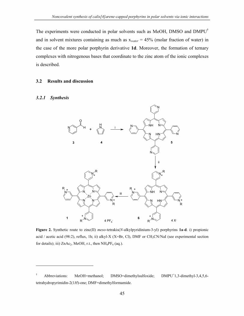
Noncovalent synthesis of calix[4]arene-capped porphyrins in polar solvents via ionic interactions
45
The experiments were conducted in polar solvents such as MeOH, DMSO and DMPU†
and in solvent mixtures containing as much as xwater = 45% (molar fraction of water) in
the case of the more polar porphyrin derivative 1d. Moreover, the formation of ternary
complexes with nitrogenous bases that coordinate to the zinc atom of the ionic complexes
is described.
3.2 Results and discussion
3.2.1 Synthesis
N
N
N
N
N
NH N
NH
N
O
H NH
+ i
4 PF6-
N
N
N
N
N
N N
N
R
R
R
R
Zn+
+
+
+
ii
iii N
N
N
N
N
NH N
NH
R
R
R
R
+
+
+
+4 X-
3 4 5
61
Figure 2. Synthetic route to zinc(II) meso-tetrakis(N-alkylpyridinium-3-yl) porphyrins 1a-d. i) propionic
acid / acetic acid (98:2), reflux, 1h; ii) alkyl-X (X=Br, Cl), DMF or CH3CN/NaI (see experimental section
for details); iii) ZnAc2, MeOH, r.t., then NH4PF6 (aq.).
† Abbreviations: MeOH=methanol; DMSO=dimethylsulfoxide; DMPU=1,3-dimethyl-3,4,5,6-
tetrahydropyrimidin-2(1H)-one; DMF=dimethylformamide.
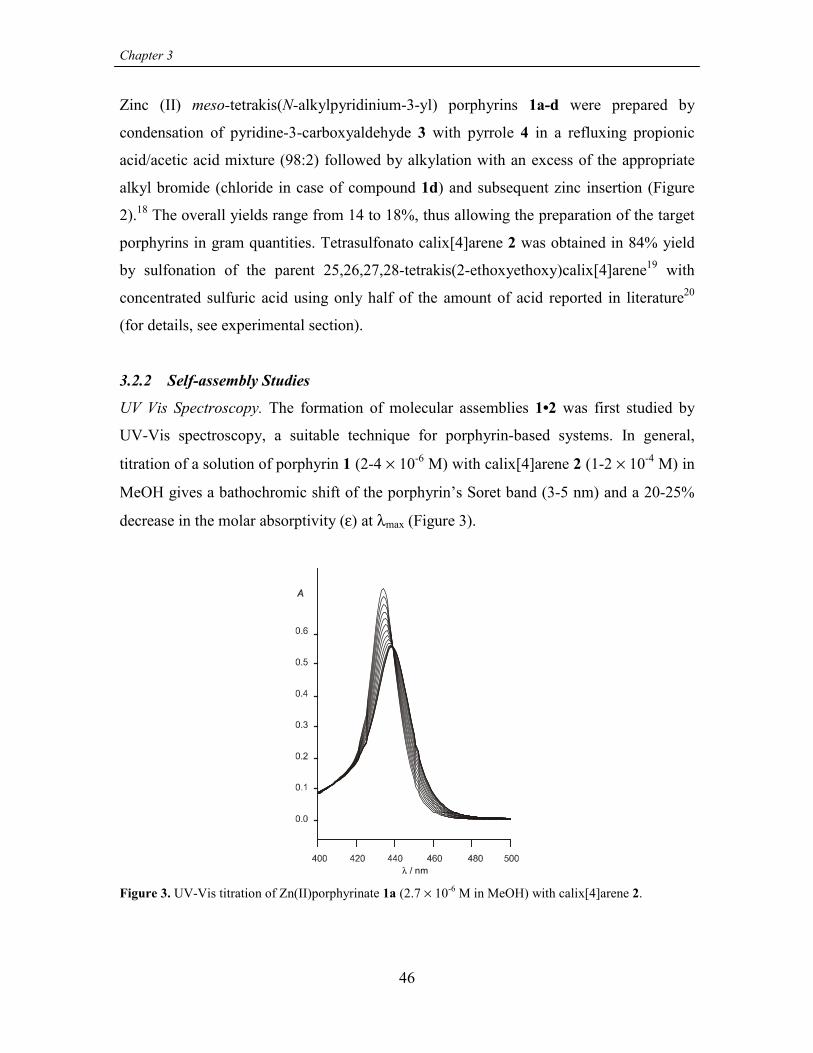
Chapter 3
46
Zinc (II) meso-tetrakis(N-alkylpyridinium-3-yl) porphyrins 1a-d were prepared by
condensation of pyridine-3-carboxyaldehyde 3 with pyrrole 4 in a refluxing propionic
acid/acetic acid mixture (98:2) followed by alkylation with an excess of the appropriate
alkyl bromide (chloride in case of compound 1d) and subsequent zinc insertion (Figure
2).18 The overall yields range from 14 to 18%, thus allowing the preparation of the target
porphyrins in gram quantities. Tetrasulfonato calix[4]arene 2 was obtained in 84% yield
by sulfonation of the parent 25,26,27,28-tetrakis(2-ethoxyethoxy)calix[4]arene19 with
concentrated sulfuric acid using only half of the amount of acid reported in literature20
(for details, see experimental section).
3.2.2 Self-assembly Studies
UV Vis Spectroscopy. The formation of molecular assemblies 1•2 was first studied by
UV-Vis spectroscopy, a suitable technique for porphyrin-based systems. In general,
titration of a solution of porphyrin 1 (2-4 × 10⁻⁶ M) with calix[4]arene 2 (1-2 × 10⁻⁴ M) in
MeOH gives a bathochromic shift of the porphyrin’s Soret band (3-5 nm) and a 20-25%
decrease in the molar absorptivity (ε) at λmax (Figure 3).
λ / nm
Figure 3. UV-Vis titration of Zn(II)porphyrinate 1a (2.7 × 10⁻⁶ M in MeOH) with calix[4]arene 2.

Noncovalent synthesis of calix[4]arene-capped porphyrins in polar solvents via ionic interactions
47
Fitting of the spectral data to a 1:1 binding model afforded association constants K1•2 in
the range of 107 M-1 (see Table 1). From these data it is possible to see that alteration of
the alkyl chains at the pyridyl nitrogens has almost no effect on the strength of the ion-
pair complex. This result confirms our hypothesis that assembly of the 1:1 complexes in
MeOH is primarily driven by the cooperative formation of four salt bridges between
molecular components 1 and 2. In order to exclude the possibility that the observed
spectral changes are the result of nonspecific aggregation of porphyrins 1, the UV
spectrum of porphyrin 1a was measured at various concentrations (between 3 × 10⁻⁷ and
4 × 10⁻⁵ M in MeOH). Aggregation of the porphyrin was not observed over this
concentration range.
Table 1. Association constants (K1•2) for the formation of assemblies 1•2
Assembly Solvent Log K1•2
1a•2 CH3OH 7.11 ± 0.04
1b•2 CH3OH 7.04 ± 0.20
1c•2 CH3OH 7.15 ± 0.09
1d•2 CH3OH 6.86 ± 0.06
1d•2 H2O/CH3OH (xwater=0.45) 5.43 ± 0.08a
1a•2 DMSO 6.21 ± 0.03
5.22 ± 0.06b
4.56 ± 0.03c
1a•2 DMPU 6.16 ± 0.01
1a•2 H2O/DMPU (xwater=0.26) 6.04 ± 0.02
T=25 ºC, [porphyrin] = 2–3 ×10⁻⁶ M (UV-vis), 1×10⁻⁴ M (ITC). Given errors are standard deviations of
three measurements. a ITC measurement, 1×10⁻² M Bu4NClO4; b 1 ×10⁻² M Bu4NClO4; c 1 ×10⁻² M NaClO4
Metal-free porphyrin 1a was used to study the effect of the metal on the assembly
formation. The log K1•2 value obtained in this case was 8.19 ± 0.19 M-1 and clearly shows
that the presence of the metal ion is not a prerequisite for complex formation. The even
higher K1•2 value is probably the consequence of the higher flexibility of the metal-free
porphyrin, allowing a better interaction with calix[4]arene 2. Therefore this result

Chapter 3
48
suggests that the metal center might still be available for ligand coordination and guest
recognition purposes (vide infra).
1H NMR Spectroscopy. 1H NMR spectroscopy measurements in CD3CN/D2O mixtures
(81:19) were performed to gain structural information about complexes 1•2. The spectra
of porphyrins 1 alone were studied first. It is well known that porphyrins like 1 can exist
in four different atropoisomeric forms (αααα, αααβ, αβαβ, and ααββ) due to the
non-symmetric substitution at the meso aromatic rings (Figure 4).21
αααα αβαβ
ααββαααβ
N+
NN
+
N+
NNN
NM
R
R
R
R
+
N+
N+
N
N+
NNN
NM
R
RR
R
+
N+
N+
N
N+
NNN
NM
R
R
RR
+
meso-pyridyl ringsβ-pyrrolic protons
N+
N+
N+
N+
NNN
NM
R
R
R
R
HH
Figure 4. Atropoisomeric forms for porphyrins 1.
When the N-alkyl moiety is in position 3 relative to the connection of the meso pyridyl
ring to the porphyrin macrocycle, atropoisomerization can occur on a time-scale
comparable to the NMR chemical shift time-scale. However, the coalescence temperature
can vary depending on the conditions under which the measurement is performed
(presence of salts, solvent, etc.)18 but the equilibrium remains still fast enough to prevent
the physical separation of the various isomers. For porphyrin 1a two signals were

Noncovalent synthesis of calix[4]arene-capped porphyrins in polar solvents via ionic interactions
49
observed for the 8 β-pyrrolic protons (COSY experiments show that the two signals
belong to adjacent protons) and one signal for three of the four different pyridyl protons
Hb-Hd (Figure 5) which would indicate the presence of only one isomer with fourfold
symmetry. However, proton Ha exhibits a pattern more complicated than expected for just
meta couplings with protons Hb and Hd. Moreover, VT NMR experiments showed
coalescence behavior for this signal, thus ruling out the possibility of only one isomer
being present.β− p r o t o n s
- 10° C
+ 20
9.010.0 (ppm)
+ 30
+ 40
+ 50
+ 60
+ 70
Ha Hb Hd Hc
Figure 5. VT 1H NMR experiments of porphyrin 1a (1.6 mM samples in CD3CN/D2O 81:19 mixtures).
Proton assignment as in Figure 6.
Based on this result we conclude that porphyrin 1a exists as a mixture of isomers
presenting accidental isochronicity for most of the NMR proton signals.21 The isomers
are slowly interconverting on the NMR time scale at 25 ºC in CD3CN/D2O (81:19) and
coalescence is observed around 60 ºC for the β-protons. Very similar 1H NMR spectra
were observed for porphyrins 1b-1d with the β-protons also appearing as singlets or as a
cluster of signals.

Chapter 3
50
a
b
c
HaHb Hd Hc
1a
calix 2N+
Ha
Hb
HcHd
10.0 9.0 8.0 7.0 (ppm)
Figure 6. 1H NMR of (a) porphyrin 1a (1.8 mM in CD3CN/D2O 81:19) at 298 K, (b) 1:1 mixture of 1a and
2 at 298 K, (c) 1:1 mixture of 1a and 2 at 343 K.
Upon addition of 1 equiv of calix[4]arene 2 at room temperature, the porphyrin proton
signals change in all cases to a complicate set of signals (see Figure 6 for 1a) indicating
the formation of assemblies 1•2. This result agrees well with the presence of several
isomers that are slowly interconverting on the NMR time scale (also observed for
porphyrins 1). The coalescence temperature measured for assembly 1a•2 is around 10 ºC
higher than for 1a alone.‡ In this assembly the porphyrin protons Hb, Hc, Hd present the
most upfield shifted signals (∆δ ~ 0.2–0.5 ppm), which suggests that they are in the
closest proximity to the calix[4]arene moiety.
Molecular simulation studies (CHARMm 24.0) support this interpretation.§ Figure 7
clearly shows that the distance between the oppositely charged groups is minimal when
calix[4]arene 2 approaches the porphyrin plane from the side opposite to where the alkyl
groups are situated. The estimated O-N distances (SO3 - N-alkylpyridine) are 2.74 Å and
‡ Ion-pair induced complexation has been reported to affect the kinetics of atropoisomerization.18
§ For simplicity only the αααα-porphyrin isomer is considered. In fact, porphyrin 1 is present as an
equilibrating mixture of atropoisomers αααα, αααβ, ααββ, and αβαβ in an expected statistical ratio of
1:4:2:1.21 Nevertheless, the same considerations mentioned for the αααα isomer would also be applicable
to the αααβ isomer (generally the most abundant), because both present two different porphyrin faces.

Noncovalent synthesis of calix[4]arene-capped porphyrins in polar solvents via ionic interactions
3.11 Å when the alkyl groups are pointing away or towards the calix[4]arene,
respectively.
Figure 7. Molecular s
(SO3 - N-alkylpyridine
For simplicity, the N-a
white) and compound 2
Small upfield shifts
aromatic and the
(according to the C
components within
temperature we co
CD3CN/D2O mixtu
¶ Below –10 ºC the D2O
Å
3.11Å
2.7451
imulation (CHARMm 24.0) of assembly 1•2 with indication of the O-N distances
). (A) N-alkyl groups pointing towards 2, (B) N-alkyl groups pointing away from 2.
lkyl groups shown are methyls. Compound 1 is in gray (the pyridyl N atoms are in
is in dark gray.
(0.03-0.08 ppm) were also observed upon assembly formation for the
methylene bridge protons of calix[4]arene 2, but their multiplicity
4v symmetry of 2) was unchanged. This indicates that exchange of the
the assembly is fast on the NMR chemical shift time scale (the lowest
uld access was –10 ºC).¶ Unfortunately, 2D ROESY experiments in
res failed to show any crosspeak between the two building blocks. The
/CD3CN solvent mixture separates into two different phases.

Chapter 3
reason for this is not entirely clear, but a similar observation was reported in the case of a
molecular capsule based on charged cyclodextrins in water.22
Figu
ESI
ESI
allo
of
con
(Fig
to
4PF
at m
pre
inta
also
[1a-R-3PF6]2+
re 8
-TO
-TO
ws
1a
cen
ure
porp
6+H
/z
sent
ct p
sho
[1a-4R-4PF6+H]+
. ESI-TOF mass s
F Mass Spectr
F mass spectr
the detection o
and 2 dissolve
tration showed
8). The spectr
hyrin 1a that
]+ and [1a-3R
1144 correspo
at m/z 634 an
orphyrin [1a-2
wed the same
[1a-2PF6]2+
pectra
omet
omet
f inta
d in
a sig
um a
has
-4PF
ndin
d 78
PF6]
deal
[1a-3R-4PF6]+
52
of (a) porphyrin 1a, (b) assembly 1a•2.
ry. Formation of assembly 1a•2 was a
ry.23 Using the appropriate experim
ct noncovalent complexes.24 Analysi
acetonitrile and further diluted w
nal at m/z 1173 for the doubly charg
lso shows two large peaks at m/z 68
lost four or three alkyl groups (R
6]+), under the mass spectrometric co
g to free calix[4]arene [2+Na]+. Ot
0 for the monodealkylated species [2+. ESI-TOF-MS analysis of a samp
kylation pattern.
[1a-2R-3PF6]+
l
en
s
it
e
1
n
he
1
le
[2+ Na]+
so invest
tal cond
of an equ
h MeOH
d species
and 828,
) respect
ditions a
r small
a-R-3PF6
of porp
[1a•2+2 Na]2+
b
a
igated by nano-
itions, ESI-MS
imolar mixture
to a 50 µM
[1a•2+2 Na]2+
corresponding
ively ([1a-4R-
nd a third peak
peaks are also
]2+ and for the
hyrin 1a alone

Noncovalent synthesis of calix[4]arene-capped porphyrins in polar solvents via ionic interactions
53
Enthalpic and Entropic Contribution to the Binding Process. For a better understanding
of the assembly process, the thermodynamic parameters associated with the formation of
1•2 were determined either using variable temperature UV/vis titration experiments or
isothermal titration microcalorimetry (ITC) measurements (Table 2).
Table 2. Thermodynamic parameters for the formation of 1•2
Assembly Solvent ∆H (kJ mol-1) ∆S (J mol-1K-1)
1a•2 MeOH -8.0 ± 6.7a +129 ± 24a
1d•2 MeOH/H2O
(xwater = 0.45)
+13.2 ± 0.4b +148 ± 3b
[porphyrin] = 2–3 × 10⁻⁶ M (UV-vis), 9.5 × 10⁻⁵ M (ITC). a VT UV, K1•2 measured at 7 different
temperatures (errors are standard deviations obtained from the linear regression); b ITC, 25 ºC, 1 × 10⁻² M
Bu4NClO4 (errors are standard deviations of three measurements).
In pure MeOH, the formation of assembly 1a•2 did not show any appreciable heat effect
in an attempted ITC measurement, thus preventing the use of this powerful technique in
this specific case. The thermodynamic parameters were thus indirectly determined by
van’t Hoff analysis of VT UV-studies (see Figure 9). The low enthalpy measured in this
way (especially if the error of the determination is considered) is in agreement with the
ITC observation. The process is therefore almost entirely entropy driven.
The formation of assembly 1d•2 could instead be conveniently studied in a MeOH/H2O
mixture (xwater = 45%) with Bu4NClO4 (1 × 10⁻² M) as the support electrolyte via ITC
measurements (see Figure 10) due to the more polar nature of porphyrin 1d. The
assembly formation is still strongly entropically driven (∆S = 148 ± 3 J K-1 mol-1). The
positive enthalpy change observed in this case (∆H = 13.2 ± 0.4 kJ mol-1) likely arises
from a positive enthalpy of desolvation, which overrides the expected negative enthalpy
for complex formation. K1•2 for the assembly 1d•2 in this solvent mixture is 1.43
logaritmic units (corresponding to 27 times) smaller than that measured in pure MeOH
(see Table 1), reflecting the weakening of electrostatic interactions in a solvent mixture
with a higher dielectric constant and containing 100 equivalents of Bu4NClO4.

Chapter 3
54
0
5
10
15
20
25
0.0032 0.0034 0.0036 0.0038 0.004 0.0042 0.0044
T -1 / K-1
ln K
12•
Figure 9. Van’t Hoff plot from VT UV-studies for the formation of assembly 1a•2 in MeOH (temperature
interval 243.1–298.2 K). Note that the concentration measure unit in these experiments is molality (m);
therefore K1•2 is expressed in m-1.
0 200.2
0.0
-0.2
0
0.0 0.5 1.0 2.0 2.5
Molar ratio
1.5
2
-0.4
40 60 80 100
Time (min)
µcal
/sec
Kcal
/mol
of i
njec
tant
Figure 10. ITC titration of porphyrin 1d (9.5 × 10⁻⁵ M in MeOH/H2O, xwater = 45%, [Bu4NClO4]=1 × 10⁻²
M) with calix[4]arene 2 at 25 °C. The plot shows the heat evolved per injection of titrant as a function of
[1d]/[2] ratio.

Noncovalent synthesis of calix[4]arene-capped porphyrins in polar solvents via ionic interactions
55
Solvent and Salt Effects. The stability of complexes 1•2 was investigated in different
polar solvents and in the presence of added salts. The complexes remain stable in a range
of polar solvents with a degree of complexation ≥ 95% at millimolar concentration (see
Table 1). For system 1a•2, the association constant in polar aprotic solvents, like DMSO
and DMPU (Log K1•2 ~ 6.2) is around one logaritmic unit (10 times) smaller than in
MeOH (Log K1•2 = 7.11). A similar value (Log K1•2 = 6.04) was also obtained when water
was added to DMPU (xwater = 0.26). These results indicate that MeOH is the superior
solvent for the assembly formation and suggest a stabilizing effect possibly due to the
inclusion of one (or two hydrogen-bonded25) solvent molecule(s) within the assembly
cavity. This idea is also supported by molecular simulation studies that confirm that one
MeOH molecule can be accommodated without any distortion of the cavity (Figure 11).#
Figure 11. Molecular simulation (CHARMm 24.0) of assembly 1•2: (A) empty cavity, (B) cavity filled
with MeOH. For simplicity, the N-alkyl groups shown are methyls.
When stability measurements were performed in DMSO in the presence of 10-2 M
Bu4NClO4 (4500 times the concentration of the building blocks), K1•2 was observed to
decrease only 10 times (Log K1•2 = 5.22) in comparison to pure DMSO (Log K1•2 = 6.21).
However, in the presence of 10-2 M NaClO4 an association constant 45 times lower was
obtained (Log K1•2 = 4.56). A more detailed study of K1•2 as a function of the NaClO4
concentration showed that the effect is probably due to a specific interaction of the Na+
cation with calix[4]arene 2. A logarithmic plot of K1•2 vs [NaClO4] shows a bimodal
# Also for these molecular simulation studies, only the αααα isomer has been considered. Under this
condition, the simulation shows that bulkier solvents like DMSO and DMPU might be less efficient in

Chapter 3
56
linear dependence of K1•2 with a slope of 1.91 ± 0.11 for [NaClO4]>2×10⁻³ M (Figure 12),
suggesting weak binding of two Na+ ions (β = 2.2 × 10⁻⁵ M-2) most likely to the lower rim
of the calix[4]arene moiety.
p[NaClO ]4
Log
K 12•
3.01.0 2.0 3.0 4.0 5.0
4.0
5.0
6.0
7.0
Figure 12. Effect of the presence of NaClO4 on the formation of assembly 1a•2.
The complexation appears to be cooperative as indicated by the sharp curvature at the
breaking point. In terms of molecular structure of calix[4]arene 2, the observed
cooperativity is most probably attributable to the preorganization of the ethoxy-ethoxy
chains after the first Na+ ion has been complexed to the lower rim oxygens.
3.2.3 Ligand Binding Studies
We have also studied the coordination of axial ligands L to the zinc center of assembly
1a•2 to give ternary complexes 1a•2•L. Zinc porphyrins are known to bind only one axial
ligand molecule and therefore represent the simplest model system to study axial
coordination to metalloporphyrins.
UV-vis titration experiments using 1-methylimidazole (1-MeIm) or pyridine as axial
ligands (L), clearly showed the formation of the expected ternary complexes 1a•2•L.
filling the cavity because they cause a distortion of the 1•2 complex.

Noncovalent synthesis of calix[4]arene-capped porphyrins in polar solvents via ionic interactions
57
0.8
Abs
0.4
0.6
0.2
0.0
500 550 600
λ (nm)
650
Abs
0.350 0.2
[1-MeIm] (M)0.4
0.45
0.55
0.65
Figure 13. Spectral evolution of a 1:1 mixture of porphyrin 1a and calix[4]arene 2 in MeOH (4×10⁻⁵ M,
assembly 1a•2 >95% in solution) titrated with 1-MeIm. The lower part shows the fitting of the titration
curve at 555 nm.
The observed spectral changes in the Q bands region (see Figure 13) were fitted to a 1:1:1
equilibrium model in order to obtain values for the equilibrium constant K1a•2•L (eqs. 1-3).
1a + 2 � 1a•2 K1•2 (1)
1a + L � 1a•L K1a•L (2)
1a + 2 + L � 1a•2•L K1a•2•L (3)
The binding constant for the axial ligand L to porphyrin 1a (K1a•L, eq. 2) was determined
in a separate experiment and K1•2 is the formation constant for assembly 1a•2 (see Table
1) under the same experimental conditions. The ratio K1a•2•L/K1•2 represents the ligand
affinity displayed by assembly 1a•2.
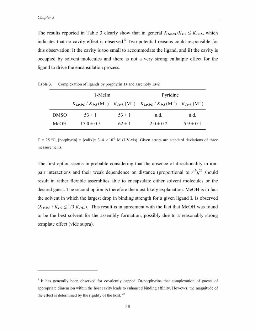
Chapter 3
58
The results reported in Table 3 clearly show that in general K1a•2•L/K1•2 ≤ K1a•L, which
indicates that no cavity effect is observed.$ Two potential reasons could responsible for
this observation: i) the cavity is too small to accommodate the ligand, and ii) the cavity is
occupied by solvent molecules and there is not a very strong enthalpic effect for the
ligand to drive the encapsulation process.
Table 3. Complexation of ligands by porphyrin 1a and assembly 1a•2
1-MeIm Pyridine
K1a•2•L / K1•2 (M-1) K1a•L (M-1) K1a•2•L / K1•2 (M-1) K1a•L (M-1)
DMSO 53 ± 1 53 ± 1 n.d. n.d.
MeOH 17.0 ± 0.5 62 ± 1 2.0 ± 0.2 5.9 ± 0.1
T = 25 ºC, [porphyrin] = [calix]= 3–4 × 10⁻⁵ M (UV-vis). Given errors are standard deviations of three
measurements.
The first option seems improbable considering that the absence of directionality in ion-
pair interactions and their weak dependence on distance (proportional to r-1),26 should
result in rather flexible assemblies able to encapsulate either solvent molecules or the
desired guest. The second option is therefore the most likely explanation: MeOH is in fact
the solvent in which the largest drop in binding strength for a given ligand L is observed
(K1•2•L / K1•2 ≤ 1/3 K1•L,). This result is in agreement with the fact that MeOH was found
to be the best solvent for the assembly formation, possibly due to a reasonably strong
template effect (vide supra).
$ It has generally been observed for covalently capped Zn-porphyrins that complexation of guests of
appropriate dimension within the host cavity leads to enhanced binding affinity. However, the magnitude of
the effect is determined by the rigidity of the host. 29

Noncovalent synthesis of calix[4]arene-capped porphyrins in polar solvents via ionic interactions
59
3.3 Summary and Outlook
This chapter reports the high thermodynamic stability of noncovalent calix[4]arene-
capped porphyrins in polar solvents and in organic solvent/water mixtures with as much
as xwater = 45%. These systems remain remarkably stable even upon addition of an excess
of electrolytes such as Bu4NClO4 or NaClO4.
The results described in this chapter constitute the basis for the development of a fully
water soluble system, i.e. a closer model for natural porphyrins under physiological
conditions (Chapters 4 and 5). Moreover, as described in Chapter 4, the recognition
properties of similar ion-pair driven assemblies in aqueous media are different from what
was observed here, favoring encapsulation (according to the larger influence of
hydrophobic interactions). The highly dynamic nature of these assemblies together with
their peculiar ligand recognition properties could be useful in catalytic applications.
3.4 Experimental
3.4.1 General information and instrumentation
All reagents used were purchased from Aldrich or Acros Organics and used without
further purification. All the reactions were performed under nitrogen atmosphere.
Compound 1b was prepared according to literature.18
Infrared spectra were recorded on a FT-IR Perkin Elmer Spectrum BX spectrometer and
only characteristic absorptions are reported. 1H NMR and 13C NMR spectra were
performed on a Varian Unity INOVA (300 MHz) or a Varian Unity 400 WB NMR
spectrometer. 1H NMR chemical shift values (300 MHz) are expressed in ppm relative to
residual CHD2OD (δ 3.30), CHD2CN (δ 1.93), or CHCl3 (δ 7.26). 13C NMR chemical
shift values (100 MHz) are expressed in ppm relative to residual CD3OD (δ 49.0) or
CD3CN (δ 1.3). UV-vis measurements were performed on a Varian Cary 3E UV-vis
spectrophotometer equipped with a Helma QX optical fiber probe (path length = 1.000
cm), using solvents of spectroscopic grade. Calorimetric measurements were carried out
using a Microcal VP-ITC microcalorimeter with a cell volume of 1.4115 ml. Elemental
analyses were carried out on a 1106 Carlo-Erba Strumentazione element analyzer.

Chapter 3
60
Electrospray ionization mass spectra were recorded on a Micromass LCT time-of-flight
mass spectrometer. Samples were introduced using a nanospray source. Molecular
simulation studies were carried out as described elsewhere.27
3.4.2 Binding studies
UV-Vis: Association constants for assemblies 1•2 were evaluated by measuring the
absorbance changes upon addition of increasing amount of a calix[4]arene 2 solution to a
porphyrin 1 solution (2 – 3×10⁻⁶ M). Each titration consisted of 15 to 20 additions. The
calix[4]arene solution was at a concentration high enough so that the degree of
complexation during the titration was between about 20 and 80%, and the overall dilution
was less than 3%. The experimental spectral changes for assembly formation were fitted
to a 1:1 binding model using a non-linear least squares fitting procedure28 that considered
the information coming from 6 different wavelenghts (model written with program
Scientist®, MicroMath®). The binding constants for axial ligands were determined
according to a 1:1:1 binding model using a non-linear least squares fitting procedure
considering the absorbance changes at one wavelength (the fitting procedure was
generally repeated for two different wavelengths and the results averaged, model written
with program Microsoft® Excel).
3.4.3 Synthesis
General procedure for the preparation of [5,10,15,20-tetrakis(N-alkylpyridinium-3-
yl)porphyrinato]zinc(II) (1a, 1c).
A solution of 5,10,15,20-tetra(3-pyridyl)porphyrin 5 (100 mg, 0.161 mmol) and the
appropriate alkylbromide (100-200 equivalents, p-t-butylbenzylbromide for 1a and 1-
bromo-3-methylbutane for 1c) in DMF (30-50 ml) was stirred for 8-17 h at 80 ºC. The
reaction was monitored by TLC (silica gel 60F254, CH3CN/(3 mg/ml NH4PF6 in H2O)
7:3). The DMF was evaporated under vacuum, the solid residue was triturated with
diethyl ether and subsequently dried under vacuum. The crude products were used
without further purification in the following metalation reaction.
Metallation with Zn was carried out by stirring the alkylated porphyrin in MeOH
saturated with ZnAc2 at room temperature for 1h, after which a solution of NH4PF6 in

Noncovalent synthesis of calix[4]arene-capped porphyrins in polar solvents via ionic interactions
61
water was added to precipitate the metallated porphyrin. The solid product (metallated
porphyrin 1 PF6 salt) was filtered off and washed extensively with water and ether.
5,10,15,20-tetrakis(N-p-t-butylbenzylpyridinium-3-yl)porphyrin (free base 1a)
Purification of an analytical sample of free-base 1a was performed by dissolving the
crude product in MeOH and precipitating by addition of a solution of NH4PF6 in water.
The precipitate was filtered off and washed extensively with water and ether. The
chloride salt of 1a was obtained by passing the PF6 salt through an ion-exchange column
(DOWEX 1-X8, 50-100 mesh, Cl-form) using CH3CN/H2O 1:1 v/v as the eluent. The
eluted solution was concentrated under vacuum and lyophilized to afford the product as a
brown solid. Yield: 85%.1H NMR (CD3OD) δ 10.10 (m, 4H), 9.58 (d, J=6.0 Hz, 4H), 9.44 (d, J=7.9 Hz, 4H), 9.07
(br s, 8H), 8.61 (m, 4H), 7.65 (m, 16H), 6.18 (s, 8H), 1.34 (s, 36H); 13C NMR δ 154.86,
150.40, 148.32, 145.79, 143.24, 131.71, 130.32, 128.47, 127.88, 114.41, 66.19, 35.73,
31.64; IR (KBr) 3414, 3068, 2962, 2868, 1628, 1499, 1465, 1196, 979, 794; UV-vis
(MeOH) 422, 512, 544, 586, 643; Anal. Calcd for C84H86N8Cl4×9 H2O: C 66.75, H 6.93
N 7.41, Found: C 66.85, H 6.57, N 7.30.
Hexafluorophosphate salt:1H NMR (CD3CN) δ 9.56 (m, 4H), 9.26 (m, 8H), 9.00 (s, 4H), 8.94(s, 4H), 8.52 (t, J=7.0
Hz, 4H), 7.58 (m, 16H), 6.02 (s, 8H), 1.30 (s, 36H), -3.15 (s, 2H).
[5,10,15,20-tetrakis(N-p-t-butylbenzylpyridinium-3-yl)porphyrinato]zinc(II) (1a)
Yield: 80% (alkylation + metallation)1H NMR (CD3OD) δ 9.93 (m, 4H), 9.48 (m, 4H), 9.35 (d, J=7.7 Hz, 4H), 8.99 (s, 4H),
8.93 (s, 4H), 8.54 (t, J=7.0 Hz, 4H), 7.64 (m, 16H), 6.13 (s, 8H), 1.33 (s, 36H); UV-vis
(MeOH) 433, 521, 560, 596; Anal. Calcd for C84H84N8P4F24Zn: C 54.51, H 4.57 N 6.05,
Found: C 54.15, H 4.49, N 5.91.
[5,10,15,20-tetrakis(N-(3-methylbutyl)pyridinium-3-yl)porphyrinato]zinc(II) (1c)
Yield: 91 % (alkylation + metallation)
Chapter 3
62
1H NMR (CD3CN) δ 9.53 (m, 4H), 9.20 (m, 8H), 9.00 (m, 8H), 8.47 (t, J=7.0 Hz, 4H),
4.85 (t, J=7.8 Hz, 8H), 2.15 (m, 8H), 1.87 (sept, J=6.5 Hz, 4H), 1.05 (d, J=6.5 Hz, 24H);13C NMR δ 151.36, 149.52, 147.09, 145.11, 143.96, 133.99, 128.02, 114.26, 62.02,
40.96, 26.80, 22.58; IR (KBr) 3107, 2960, 2875, 1629, 1498, 1200, 1159, 843, 558; UV-
vis (MeOH) 431, 520, 559, 597, 633; Anal. Calcd for Anal. Calcd for C60H68N8F24P4Zn:
C 46.60, H 4.43 N 7.25, Found: C 46.41, H 4.29, N 7.33.
[5,10,15,20-tetrakis(N-(4-acetylmorpholyl)pyridinium-3-yl)porphyrinato]zinc(II)
(1d)
5,10,15,20-tetra(3-pyridyl)porphyrin 5 (200 mg, 0.323 mmol) and chloroacetyl
morpholine (240 mg, 1.45 mmol) were dissolved in acetonitrile (30 ml). NaI (218 mg,
1.45 mmol) was added and the mixture was refluxed for 21 h. The solvent was then
removed under vacuum and the residue triturated with ether and redissolved in MeOH.
The product was precipitated by addition of NH4PF6 in water, filtered off and washed
with water and ether to afford crude alkylated porphyrin in 91% yield (0.28 mmol, 502
mg). Metallation with ZnAc2 as previously described, afforded 1d as a purple solid in
85% yield.1H NMR (CD3CN) δ 9.26 (m, 4H), 9.17 (d, J=8.0Hz, 4H), 8.91 (s, 12H), 8.36 (t, J=7.0
Hz, 4H), 5.63 (s, 8H), 3.63-3.36 (m, 32H); 13C NMR δ 164.02, 151.33, 150.43, 149.24,
146.21, 143.00, 133.97, 127.47, 114.17, 67.201, 66.96, 62.96, 46.17, 43.87; IR (KBr)
3101, 2932, 2866, 1661, 1450, 1246, 1112, 842, 558; UV-vis (MeOH) 432, 520, 559,
596, 636; Anal. Calcd for Anal. Calcd for C64H64N12O8F24P4Zn: C 43.32, H 3.64 N 9.47,
Found: C 43.31, H 3.42, N 9.42.
5,11,17,23-Tetrasulfonato-25,26,27,28-tetrakis(2-ethoxyethoxy)calix[4]arene,
tetrasodiumsalt (2). The compound was prepared by a slightly modified literature
procedure.20
25,26,27,28-tetrakis(2-ethoxyethoxy)calix[4]arene19 (2.00 g, 2.80 mmol) was dissolved in
H2SO4 (96%, 5 ml) and stirred at room temperature for 2 h. The reaction mixture was
then quenched by pouring it carefully into water and neutralized by addition of a NaOH
solution. The water was then removed under reduced pressure and the solid residue was

Noncovalent synthesis of calix[4]arene-capped porphyrins in polar solvents via ionic interactions
63
extracted overnight with MeOH using a Soxhlet extractor. The crude product was then
purified by reversed phase chromatography (Merck Lobar column, LiChroprep RP8, 40-
63 µm) eluting initially with pure water until no more inorganic sulfates were detected
(i.e. no precipitation observed upon addition of BaCl2 to the eluate). Pure 2 was obtained
by elution in a step-gradient to 70% EtOH/H2O (v/v). The eluted fractions were
concentrated under vacuum and lyophilized affording 2 as a white solid in 84% yield
(2.36 mmol, 2.65 g). The analytical data for the compound were identical to the reported
ones.20
3.5 References
1. Biochemistry, 4th ed.; Stryer, L.; W.H. Freeman & Co: New York, 1995.
2. Cytochrome P-450: Structure, Mechanism and Biochemistry, 2nd ed.; Ortiz de
Montellano, P. R., ed.; Plenum Press: New York, 1995.
3. Peroxidases in Chemistry and Biology; Everse, J.; Everse, K. E.; Grisham, M. P.,
ed.; CRC Press: Boca Raton, FL, 1991; Vol. I and II.
4. Feiters, M. C. Comprehensive Supramolecular Chemistry; Atwood, J. L.; Davies, J.
E. D.; MacNicol, D. D.; Vögtle, F.; Reinhoudt, D. N.; Lehn, J.-M., eds; Elsevier
Science Ltd. Pergamon: Elmsford, 1996; Vol. 10, pp 267-360.
5. Momenteau, M.; Reed, C. A. Chem. Rev. 1994, 94, 659-698.
6. Jones, R. D.; Summerville, D. A.; Basolo, F. Chem. Rev. 1979, 79, 139-179.
7. Wojaczynski, J.; Latos-Grazynski, L. Coord. Chem. Rev. 2000, 204, 113-171.
8. Drain, C. M.; Russel, K. C.; Lehn, J.-M. Chem. Commun. 1996, 337-338.
9. Ruhlmann, L.; Nakamura, A.; Vos, J. G.; Fuhrhop, J.-H. Inorg. Chem. 1998, 37,
6052.
10. Yamamoto, K.; Nakazawa.S.; Matsufuji, A.; Taguchi, T. J. Chem. Soc., Dalton

Chapter 3
64
Trans. 2001, 251-258.
11. Sirish, M.; Schneider, H.-J. Chem. Commun. 2000, 23-24.
12. Zhao, S.; Luong, J. H. T. J. Chem. Soc., Chem. Commun. 1995, 663-664.
13. Ribò, J. M.; Farrera, J.-A.; Valero, M. L.; Virgili, A. Tetrahedron 1995, 51, 3705-
3712.
14. Manka, J. S.; Lawrence, D. S. J. Am. Chem. Soc. 1990, 112, 2440-2442.
15. Lahiri, J.; Fate, G. D.; Ungashe, S. B.; Groves, J. T. J. Am. Chem. Soc. 1996, 118,
2347-2358.
16. Tecilla, P.; Dixon, R. P.; Slobodkin, G.; Alavi, D. S.; Waldeck, DH.; Hamilton, A.
D. J. Am. Chem. Soc. 1990, 112, 9408-9410.
17. Takeuchi, M.; Kijima, H.; Hamachi, I.; Shinkai, S. Bull. Chem. Soc. Jpn. 1997, 70,
699-705.
18. Mizutani, T.; Horiguchi, T.; Koyama, H.; Uratani, I.; Ogoshi, H. Bull. Chem. Soc.
Jpn. 1998, 71, 413-418.
19. Arduini, A.; Casnati, A.; Fabbi, M.; Minari, P.; Pochini, A.; Sicuri, A. R.; Ungaro,
R. Supramolecular Chemisrty 1993, 1, 235-246.
20. Casnati, A.; Ting, Y.; Berti, D.; Fabbi, M.; Pochini, A.; Ungaro, R.; Sciotto, D.;
Lombardo, G. G. Tetrahedron 1993, 49, 9815-9822.
21. Crossley, M. J.; Field, L. D.; Forster, A. J.; Harding, M. M.; Sternhell, S. J. Am.
Chem. Soc. 1987, 109, 341-348.
22. Hamelin, B.; Jullien, L.; Derouet, C.; Hervé du Penhoat, C.; Berthault, P. J. Am.
Chem. Soc. 1998, 120, 8438-8447.
23. Brody, M. S.; Schalley, C. A.; Rudkevich, D. M.; Rebek, Jr. J. Angew. Chem., Int.
Ed. 1999, 38, 1640-1644.
Noncovalent synthesis of calix[4]arene-capped porphyrins in polar solvents via ionic interactions
65
24. Staroske, T.; O'Brien, D. P.; Jørgensen, T. J. D.; Roepstorff, P.; William, D. H.;
Heck, A. J. R. Chem. Eur. J. 2000, 6, 504-509.
25. Bonar-Law, R. P.; Sanders, J. K. M. J. Am. Chem. Soc. 1995, 117, 259-271.
26. Hossain, M. A.; Schneider, H.-J. Chem. Eur. J. 1999, 5, 1284-1290.
27. Timmerman, P.; Weidmann, J.-L.; Jolliffe, K. A.; Prins, L. J.; Reinhoudt, D. N.;
Shinkai, S.; Frish, L.; Cohen, Y. J. Chem. Soc., Perkin Trans. 2 2000, 2077-2089.
28. Leggett, D. J.; Kelly, S. L.; Shiue, L. R.; Wu, Y. T.; Chang, D.; Kadish, K. M.
Talanta 1983, 30, 579-586.
29. Rudkevich, D. M.; Verboom, W.; Reinhoudt, D. N. J. Org. Chem. 1995, 60, 6585-
6587.

67
Chapter 4
STRUCTURAL WATER SOLUBLE MODELS OF THE HEME-
PROTEIN ACTIVE SITE VIA SELF-ASSEMBLY∗
The noncovalent strategy outlined in Chapter 3 for the preparation of cage-like
complexes with anionic calix[4]arenes and cationic porphyrins has been applied to the
preparation of water soluble structural mimics of heme-protein active sites. Novel
porphyrin-peptide conjugates have been used as building blocks. The complexes
encapsulate small nitrogenous bases such as 4-methylpyridine and 1-methylimidazole but
bind the bulkier caffeine on the solvent exposed porphyrin face.
4.1 Introduction
Synthetic models of heme-protein active sites have been investigated extensively to
understand the basic structural features associated with the functioning of different
biological systems.1-3 From the structural point of view, the active site of a heme-protein
is essentially a hydrophobic cavity of appropriate dimensions enclosed between the
protein matrix and the metal porphyrin. This cavity is accessible to exogenous ligands
like O2 and CO in the case of hemoglobin and myoglobin,4 or to a substrate molecule in
case of cytochrome P450 (see Figure 11 Chapter 2 for a schematic representation of the
active site in O2 binding heme-protein).5
Two different approaches to the mimicry of heme-protein active sites are described in the
literature i.e. covalent1,2 and noncovalent6,7 synthesis. Using the first approach, a great
∗ Part of this chapter has been submitted for publication: Fiammengo, R.; Wojciechowski, K.; Crego-
Calama, M.; Timmerman, P.; Reinhoudt, D. N.
Chapter 4
68
number of synthetic model systems have been prepared in which the hydrophobic cavity
(resembling the natural active site) is formed combining covalently porphyrins with
organic substituents. Remarkable examples of this strategy are the “picket fence”,8,9
“pocket”,10 and “capped”11,12 porphyrins. However, most of these systems suffer from the
limitation of being water not soluble,13,14 which is an important requisite when mimicry
of a natural system is attempted. Covalent porphyrin-peptide conjugates have also been
widely used to model the heme group and its closest proteic environment in water.3
In addition to the synthetic simplicity, the main advantage of the noncovalent approach is
that it allows more accurate reproduction of the interactions between the porphyrin core
and the proteic matrix in natural proteins.15 Water soluble peptide-porphyrin
combinations based on metal coordination have been obtained,3,16 but major challenges
such as control over the folding properties of the chosen peptides remain.
According to the major focus of this thesis viz. the development of self-assembly systems
that possess biomimetic functions,17 the work reported in this chapter concerns water
soluble self-assembled models of heme-protein active sites. These are the first examples
of simple structural analogues of the heme-proteins active site obtained via self-assembly
of cage-like complexes by ionic interaction.18-20
In Chapter 3 the formation of strong 1:1 complexes between tetracationic Zn-
porphyrinates and tetraanionic tetrasulfonato calix[4]arene 2 (see Chart 1) in polar
organic solvents such as methanol, DMSO and DMPU, or in organic solvent/water
mixtures has been described.21 Unfortunately, the solubility of the assemblies in pure
water was too low. Here, an improved design of the porphyrin building blocks, which
allows the self-assembly process to occur in pure water, is presented. The short peptides
attached to the pyridyl nitrogens of porphyrins 1 (M = Zn2+, Co2+ see Chart 1) are
responsible for the relatively large water solubility of assemblies 1•2 (concentrations up
to 2-5 mM).22 These ensambles exhibited association constants K1•2 in aqueous carbonate
buffer solution as high as 105 M-1 (for structure of the assembly see Figure 2).
Furthermore, in previous noncovalent synthetic models for heme-protein reported by
other groups,6,23 no attention was given to the binding properties of the metal porphyrin
moiety. In Chapter 3 it was shown that self-assembled cage-like porphyrin-calix[4]arene
complexes form ternary complexes upon addition of nitrogenous ligands.21 Nevertheless,
Structural water soluble models of heme-protein active site via self-assembly
69
binding of the added ligands to the Zn-porphyrin moiety in polar solvents consistently
occurred on the solvent exposed porphyrin face, probably due to unfavorable competition
with the solvent for the assembly cavity. In contrast, the data reported here show that,
using water as the solvent for assemblies 1•2, coordination of nitrogenous bases to the
Zn-center affords ternary complexes with diverse topology. The structure of the
complexes is directed by the sterical requirements of the base used. Small bases such as
4-methyl pyridine are complexed preferentially inside the hydrophobic cavity of
assemblies 1c•2 and 1e•2, while larger molecules such as caffeine are bound outside the
cavity (1c•2) on the solvent-exposed porphyrin face. Moreover, CoII porphyrins (Co-1b)
can also be self-assembled with calix[4]arene 2, thus opening the way towards functional
model of heme-protein active-sites (for instance O2 binding mimics).
Chart 1
1a R= NH
O
O
NH
1b R= N
O
O
NH
1c R= NH
O
O
NH
NH
O
1d R= N
O
O
NHO
NH
· 4 Cl-
MN
N
N
N
N
N N
N
R
R
R
R
+
+
+
+
1 a-e
O
O
4
SO3- Na+
21e R= N
O
O

Chapter 4
70
4.2 Results and discussion
4.2.1 Synthesis
Alkylation of the 5,10,15,20-tetra(3-pyridyl)porphyrin pyridyl nitrogens with polar side
chains derived from amino acid, dipeptide amides, or morpholine affords water soluble
porphyrins 1a-e.24 Calix[4]arene tetrasulfonate 2 was prepared according to the procedure
described in Chapter 3.
Assemblies 1•2 are easily prepared by mixing aqueous solutions of the molecular
building blocks 1a-e and 2. Remarkably, no precipitation is observed upon addition of up
to 3 equivalents of calix[4]arene 2 to porphyrin solutions of concentrations up to 2-5
mM,25 conditions ensuring >95% assembly formation (based on K1•2, vide infra).
4.2.2 Assembly formation
ITC studies. Self-assembly of Zn porphyrins 1a-e with calix[4]arene tetrasulfonate 2 was
studied in aqueous solutions using isothermal titration microcalorimetry (ITC). The
association constants (see Tables 1 and 2) are quite insensitive to the nature of the
N-substituents. Under similar conditions, the largest difference in binding strength is less
than a factor of two for the series of porphyrins 1a-e.
Table 1. Association constants (K1•2) for cationic porphyrins 1a-e (0.090-0.105 M) and anionic
calix[4]arene 2 (sodium carbonate buffer, pH = 9.6, I = 0.010 M).
Log K1•2 ∆H° (kJ mol-1) ∆S° (J mol-1 K-1)
1a•2 4.90 ± 0.02 -23.9 ± 0.3 -14 ± 1
1b•2 4.82 ± 0.02 -32.5 ± 0.3 -17 ± 1
1c•2 4.68 ± 0.02 -25.8 ± 0.4 -3 ± 2
1d•2 4.90 ± 0.02 -28.9 ± 0.3 +3 ± 1
1e•2 4.69 ± 0.01a
5.62 ± 0.05b
5.43 ± 0.08c
-25.5 ± 0.3
-20.8 ± 0.3
+13.2 ± 0.4
+5 ± 1
+38 ± 2
+148 ± 3a Carbonate buffer pH = 9.7, I = 0.020 M; b no added salts; c MeOH/water (xwater = 0.45), 1 × 10⁻² M
Bu4NClO4.21

Structural water soluble models of heme-protein active site via self-assembly
71
Self-assembly of complexes 1a-d•2 in carbonate buffered solutions (I = 0.010 M) is
characterized by negative enthalpy and small (close to zero) entropic contributions (Table
1). However, at a higher buffer concentration (I = 0.045 M, Table 2) the formation of 1a-
d•2 is accompanied by larger negative enthalpy and entropy changes. The changes tend to
compensate each other and the net result is a moderate decrease of the association
constants (2-5 times, ∆LogK1•2~0.4-0.7).
Table 2. Association constants (K1•2) for cationic porphyrins 1a-e (0.090 - 0.105 M) and anionic
calix[4]arene 2 (sodium carbonate buffer, pH = 10, I = 0.045 M).
Log K1•2 ∆H° (kJ mol-1) ∆S° (J mol-1 K-1)
1a•2 4.26 ± 0.04 -33 ± 3 -29 ± 12
1b•2 4.40 ± 0.02 -36 ± 1 -38 ± 5
1c•2 4.11 ± 0.03 -43 ± 2 -64 ± 8
1d•2 4.23 ± 0.02 -40 ± 1 -54 ± 5
Co-1b•2 3.80 ± 0.04 -42 ± 4 -71 ± 15
Measurements on the less soluble assembly 1e•2 also confirm the correlation between
changes in buffer concentration and changes in thermodynamic parameters. In the
absence of buffer, formation of 1e•2 is even less enthalpically favored (∆H° = -20.8 kJ
mol-1) but much more entropically favored (∆S° = +38 J mol-1 K-1). The overall effect is
an association strength approximately one order of magnitude higher (Log K1•2 = 5.62 vs
4.69 in the presence of carbonate buffer, I = 0.020 M). These observations suggest that
assembly formation in aqueous solution is primarily driven by multiple ionic interactions
between the two building blocks.
Qualitatively, complex formation is expected to present a negative enthalpy (coulombic
energy) and negative entropy (bimolecular reaction). However, desolvation of the ionic
groups upon assembly formation is accompanied by a positive entropic contribution
(release of ordered solvent molecule to the bulk) and a positive (endothermic) enthalpic
contribution. In polar solvents and aqueous solutions the contribution of desolvation is as
relevant (and sometimes even more important) as the contribution associated with

Chapter 4
72
complexation.26-28 Therefore, the balance between these two contributions is the reason
for the rather large differences observed in the measured thermodynamic parameters
when the same assembly process takes place in different media. Here, the data suggest
that at lower salt concentration (resulting in a more ordered solvent shell around the ionic
groups) the importance of desolvation in driving the assembly process becomes greater.
For instance, desolvation dominates the self-assembly of 1e•2 in MeOH/water (xwater =
0.45) with 0.01 M of Bu4NClO4, resulting in a positive ∆H° = +13.2 kJ mol-1 and positive
∆S° = +148 mol-1 K-1).21
UV-vis studies. Formation of assembly 1e•2 was confirmed by UV-vis
spectrophotometric titrations.29 Addition of a solution of calix[4]arene 2 to 1e in aqueous
carbonate buffer (I = 7.2 mM) produced spectral variations in the porphyrin Soret band
region (400-480 nm) similar to what was previously observed for the formation of
assembly 1e•2 in methanol.21 The spectral variations (bathochromic shift ∆λmax = 2-3 nm,
∆ε at λmax = 10-15%) could be fitted to a 1:1 model.
Table 3. Association constants (K1e•2 in M-1) for the formation of assembly1e•2 in water at 25 ºC, from UV-
vis titrations.
Medium Log K1•2
NaNO3, I~10 mM, pH = 8.0 5.28 ± 0.03
NaNO3, I~10 mM, pH = 4.2a 5.24 ± 0.02
NaNO3, I~10 mM, pH = 9.5b 5.24 ± 0.02
Phosphate buffer, I = 8.0 mM, pH = 7.0 5.23 ± 0.02
Carbonate buffer, I = 7.2 mM, pH = 9.4 5.38 ± 0.01
CH3OH21 6.86 ± 0.06a pH adjusted with 0.1 M HCl; b pH adjusted with 0.1 M NaOH.
Hydrophilic anions such as nitrates, phosphates or carbonates were chosen in these
studies because preliminary experiments indicated that anions such as borates and
perchlorates were causing aggregation. Formation of assembly 1e•2 was observed in
basic, neutral, and acidic solutions. The presence of various electrolytes had no influence

Structural water soluble models of heme-protein active site via self-assembly
73
on the association constant K1•2 (Table 3). These data compare well with those obtained
via ITC considering that the slightly different conditions (Log K1•2 = 5.62, in the absence
of added salts and 4.69 in carbonate buffer I = 0.020 M, see Table 2).
Conductometric studies. Additional proof for the 1:1 stoichiometry for assembly 1e•2
was obtained from a conductometric titration. Changes in conductivity of the solution
were observed during the titration of porphyrin 1e with calix[4]arene 2 due to the
formation of the ion-pair complex. Extrapolation of the linear sections of the plot
afforded the end-titration point (the point were the two solid lines intersect), which was
found to be 0.85 equivalents. This is in reasonable agreement with the proposed
stoichiometry (Figure 1).
15
17
19
21
23
25
27
0.0 0.2 0.4 0.6 0.8 1.0 1.2 1.4[calix]/[porph]
cond
[ µS/
cm]
Figure 1. Conductometric titration of porphyrin 1e (4.3 × 10⁻⁵ M) with calix[4]arene 2 (8.3 × 10⁻⁴ M),
H2O, T = 25 ºC.
1H NMR studies. Structural information about assembly 1c•2 was obtained from 1H-NMR
experiments. The addition of 1 equiv of calix[4]arene 2 to porphyrin 1c in D2O/CD3OD
(9:1) produced large upfield shifts of the calix[4]arene protons due to the anisotropic
shielding from the porphyrin ring. The methylene bridges (Hb, Figure 2) and the CH2
directly connected to the lower rim oxygens (Hc) are the most upfield shifted (∆δ = 0.7-
0.85 ppm), due to their closer position to the porphyrin center.30 Moreover, all the signals
are significantly broadened, indicating an exchange equilibria rate approaching the
chemical shift time scale.

Chapter 4
74
The porphyrin pyridyl ring signals (Hd-g) are also shifted upfield (∆δ 0.15-0.40 ppm) and
significantly changed in their appearance, indicating the existence of several slowly
interconverting isomers.21 However, the signals relative to the i-Pr groups (Mei-Pr and Hi-
Pr) move in the opposite direction (downfield). Studies on ligand binding to tetracationic
porphyrins 1c-d showed that in aqueous solution the rather hydrophobic dipeptide chains
are not freely exposed to the bulk solvent but organized to some extent in the proximity
of the porphyrin.24 Evidence for these interactions is the unusual high-field resonance of
the i-Pr groups terminating the peptidic chains (Figure 2).31 The observed downfield
shifts are therefore attributed to the displacement of the peptide chains from the porphyrin
plane caused by the assembly formation.
34 2 1 ppm10 89 7 6
(c)
(b)
(a)
Mei-Pr
Hi-Pr
N
NN
NNN
NZn
R
R+
+ +
O OO O
-
OO OO
- - -SO3 SO3SO3 SO3
Ha
Hf
He
HdHβ
Hβ
Hg
Hb
HbHcHc
Ha
O
ONH
NH
HN
O
Hi-Pr
Me
Me
Me
Mei-Pr
Mei-Pr
f
a cb b
d
β
e,g
Figure 2. Spectral changes upon assembly 1c•2 formation in D2O/CD3OD (9:1), (for simplicity only one
peptide arm and three of the four pyridyl rings are shown). (a) Calix[4]arene 2, 1 mM. (b) Assembly 1c•2, 1
mM. (c) Porphyrin 1c, 1 mM. * Residual CD2HOD signal.
Structural water soluble models of heme-protein active site via self-assembly
75
4.2.3 Selective binding of nitrogenous ligands32
In the naturally occurring heme-protein the position occupied by the metal porphyrin
within the peptidic matrix is such that the two different porphyrin faces can be
distinguished. At the vicinal site, coordination of an amino acid residue to the metal
center takes place, while the more open nature of the distal site allows binding of
exogenous ligands (see Chapter 2 Figure 11). These structural characteristics have been
reproduced in many covalent synthetic models.1 However, similar efforts using the
noncovalent approach have been less common.6,23 Our self-assembled heme-protein
mimics 1•2 present two differentiated porphyrin faces, one enclosing a small cavity in
conjuction with calix[4]arene 2 and the other open to the solvent (but susceptible to the
influence of the peptidic chains, vide infra).
UV-vis measurements were used to study the formation of ternary complexes between
porphyrins 1, calix[4]arene 2 and a ligand L. The experimental data were fitted to a 1:1:1
mathematical model encompassing the simultaneous occurrence of three equilibria in
solution (equations 1-3).33
1 + 2 � 1•2 K1•2 (1)
1 + L � 1•L K1•L (2)
1 + 2 + L � 1•2•L K1•2•L (3)
The coordination strength of the axial ligand L to porphyrin 1 (K1•L in eq. 2) was
determined in separate experiments. The affinity displayed by the assembly 1•2 for a
ligand L is given by the ratio K1•2•L/K1•2 = Krec. According to this model, Krec = 0.5 × K1•L
if calix[4]arene 2 is completely blocking one porphyrin side without any other effect on
the metal coordination ability (this conclusion should apply to large guests that cannot be
encapsulated). On the other hand, for covalently capped Zn-porphyrins34 it has been
observed that complexation of guests of appropriate dimension within the host cavity
leads to enhanced binding affinity and therefore Krec ≥ K1•L should be expected.35-38
However, the magnitude of this effect is determined by the rigidity of the host, the
possibility of additional specific interactions between the ligand and the porphyrin
superstructure (e.g. H-bonds),39,40 and by the solvent.41-44
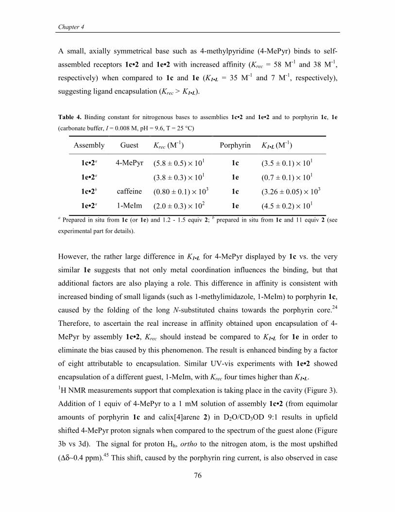
Chapter 4
76
A small, axially symmetrical base such as 4-methylpyridine (4-MePyr) binds to self-
assembled receptors 1c•2 and 1e•2 with increased affinity (Krec = 58 M-1 and 38 M-1,
respectively) when compared to 1c and 1e (K1•L = 35 M-1 and 7 M-1, respectively),
suggesting ligand encapsulation (Krec > K1•L).
Table 4. Binding constant for nitrogenous bases to assemblies 1c•2 and 1e•2 and to porphyrin 1c, 1e
(carbonate buffer, I = 0.008 M, pH = 9.6, T = 25 °C)
Assembly Guest Krec (M-1) Porphyrin K1•L (M-1)
1c•2a 4-MePyr (5.8 ± 0.5) × 101 1c (3.5 ± 0.1) × 101
1e•2a (3.8 ± 0.3) × 101 1e (0.7 ± 0.1) × 101
1c•2b caffeine (0.80 ± 0.1) × 103 1c (3.26 ± 0.05) × 103
1e•2a 1-MeIm (2.0 ± 0.3) × 102 1e (4.5 ± 0.2) × 101
a Prepared in situ from 1c (or 1e) and 1.2 - 1.5 equiv 2; b prepared in situ from 1c and 11 equiv 2 (see
experimental part for details).
However, the rather large difference in K1•L for 4-MePyr displayed by 1c vs. the very
similar 1e suggests that not only metal coordination influences the binding, but that
additional factors are also playing a role. This difference in affinity is consistent with
increased binding of small ligands (such as 1-methylimidazole, 1-MeIm) to porphyrin 1c,
caused by the folding of the long N-substituted chains towards the porphyrin core.24
Therefore, to ascertain the real increase in affinity obtained upon encapsulation of 4-
MePyr by assembly 1c•2, Krec should instead be compared to K1•L for 1e in order to
eliminate the bias caused by this phenomenon. The result is enhanced binding by a factor
of eight attributable to encapsulation. Similar UV-vis experiments with 1e•2 showed
encapsulation of a different guest, 1-MeIm, with Krec four times higher than K1•L.1H NMR measurements support that complexation is taking place in the cavity (Figure 3).
Addition of 1 equiv of 4-MePyr to a 1 mM solution of assembly 1c•2 (from equimolar
amounts of porphyrin 1c and calix[4]arene 2) in D2O/CD3OD 9:1 results in upfield
shifted 4-MePyr proton signals when compared to the spectrum of the guest alone (Figure
3b vs 3d). The signal for proton Hh, ortho to the nitrogen atom, is the most upshifted
(∆δ∼0.4 ppm).45 This shift, caused by the porphyrin ring current, is also observed in case

Structural water soluble models of heme-protein active site via self-assembly
77
of complexation of 4-MePyr to porphyrin 1c alone (Figure 3c). However, small but
reproducible upfield shifts (∆δ = 0.05-0.06 ppm) are observed for protons Hi and Hl when
binding of 4-MePyr to 1c•2 is compared to binding to 1c (Figure 3b vs Figure 3c). This
observation supports the formation of the inclusion complex, with the observed shifts
caused by the shielding from the calix[4]arene moiety. Additionally, proton Hh, (closer to
the porphyrin, but further away from the calix[4]arene) does not show any additional shift
when binding to 1c•2 is compared to 1c.
2 1 0 ppm
(c)
(b)
(a)
(d)
Mei-Pr
b
l
Hh
Hi
HlHl
Hl
N
10 89 7
i
Figure 3. 1H NMR spectra of (a) assembly 1c•2, (b) 1c•2 + 1 eq 4-MePyr, (c) porphyrin 1c + 1 eq 4-
MePyr, (d) 4-MePyr. 1 mM solutions in 9:1 D2O/CD3OD. Only the structure of the ligand is shown for
clarity.
Moreover, complexation experiments using the bulkier ligand caffeine show no
encapsulation. From UV-vis titrations a Krec = 800 M-1 for the self-assembled receptor
1c•2 was calculated. This value is one fourth of K1•L for 1c (3260 M-1). The decreased
affinity confirms that caffeine is too big to fit in the cavity and binding is taking place
only on the open face of the self-assembled receptor. This decrease also suggests larger

Chapter 4
78
unfavorable interactions with the long peptidic chains, which are probably arranged
predominantly on the open face of the porphyrin as a consequence of the ion-pair
complex 1c•2 formation (vide supra).
Structural information about the complex is gained by 1H NMR experiments performed
under the same conditions used for 4-MePyr (Figure 4). The binding mode of caffeine to
porphyrins 1a-e (and other similar ones) is substantially different from the binding mode
of ligands like imidazole or pyridine derivatives.24 The steric bulk of the ligand (due to
methyl group Meo) and the tendency towards stacking are responsible for a non-
perpendicular arrangement of caffeine with respect to the porphyrin plane. Nevertheless,
the data suggest direct interaction with the Zn atom via the nitrogen N-9.
34 2 1 0ppm
(c)
(b)
(a)
(d)
Hp
Meo
MenMem
N
N
N
N9O
O
Mei-Pr
Hi-Pr
10 89 7
Figure 4. 1H NMR spectra of (a) assembly 1c•2, (b) 1c•2+1 equiv caffeine, (c) porphyrin 1c+1 equiv
caffeine, (d) caffeine. (1 mM solutions in 9:1 D2O/CD3OD). For assignment protons Mei-Pr and Hi-Pr see
Figure 2. Only the structure of the ligand is shown for clarity.
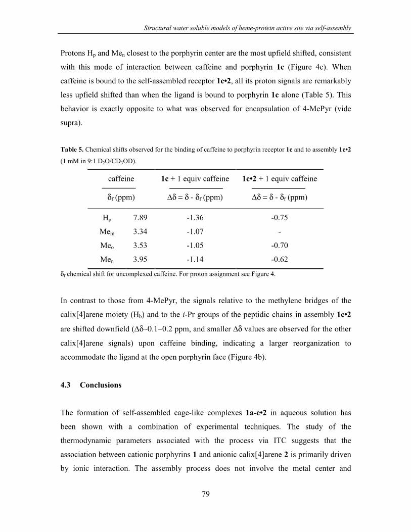
Structural water soluble models of heme-protein active site via self-assembly
79
Protons Hp and Men closest to the porphyrin center are the most upfield shifted, consistent
with this mode of interaction between caffeine and porphyrin 1c (Figure 4c). When
caffeine is bound to the self-assembled receptor 1c•2, all its proton signals are remarkably
less upfield shifted than when the ligand is bound to porphyrin 1c alone (Table 5). This
behavior is exactly opposite to what was observed for encapsulation of 4-MePyr (vide
supra).
Table 5. Chemical shifts observed for the binding of caffeine to porphyrin receptor 1c and to assembly 1c•2
(1 mM in 9:1 D2O/CD3OD).
caffeine
δf (ppm)
1c + 1 equiv caffeine
∆δ = δ - δf (ppm)
1c•2 + 1 equiv caffeine
∆δ = δ - δf (ppm)
Hp 7.89 -1.36 -0.75
Mem 3.34 -1.07 -
Meo 3.53 -1.05 -0.70
Men 3.95 -1.14 -0.62
δf chemical shift for uncomplexed caffeine. For proton assignment see Figure 4.
In contrast to those from 4-MePyr, the signals relative to the methylene bridges of the
calix[4]arene moiety (Hb) and to the i-Pr groups of the peptidic chains in assembly 1c•2
are shifted downfield (∆δ∼0.1−0.2 ppm, and smaller ∆δ values are observed for the other
calix[4]arene signals) upon caffeine binding, indicating a larger reorganization to
accommodate the ligand at the open porphyrin face (Figure 4b).
4.3 Conclusions
The formation of self-assembled cage-like complexes 1a-e•2 in aqueous solution has
been shown with a combination of experimental techniques. The study of the
thermodynamic parameters associated with the process via ITC suggests that the
association between cationic porphyrins 1 and anionic calix[4]arene 2 is primarily driven
by ionic interaction. The assembly process does not involve the metal center and

Chapter 4
80
formation of ternary complexes upon addition of suitable ligands has been achieved. The
topology of these complexes depends on the molecular dimensions of the guest. The
possibility of selectively addressing the desired porphyrin face by appropriate choice of
the ligand may be used to obtain more complex architectures such as quaternary
complexes with two different ligands. It is also shown that “inert” zinc porphyrins can be
replaced by cobalt porphyrin, allowing the evolution from structural to functional models
of heme-proteins (see Chapter 5).
4.4 Experimental part
4.4.1 General information and instrumentation1H NMR spectra were performed on a Varian Unity INOVA (300 MHz). Chemical shift
values are expressed in ppm (δ) relative to residual CHD2OD (δ 3.30). UV-vis
measurements were performed on a Varian Cary 3E UV-vis spectrophotometer equipped
with a Helma QX optical fiber probe (path length=1.000 cm), using solvents of
spectroscopic grade. Calorimetric measurements were carried out using a Microcal
VP-ITC microcalorimeter with a cell volume of 1.4115 mL.
Synthesis of porphyrins 124 and of calix[4]arene 2,21 buffer preparation, dilution
experiments to confirm the absence of porphyrin aggregation and UV-vis studies of the
assembly formation are described in Chapters 3 and 7. Cobalt porphyrin Co-1b has been
prepared using the same free base precursor24 used to prepare Zn-1b. Metallation with
CoAc2 was carried out under the same experimental conditions used for the insertion of
Zn.
4.4.2 Binding studies
Capsule formation and UV-vis studies of guest recognition: Assembly 1c•2 and 1e•2
were prepared in situ by addition of calix[4]arene 2 to the porphyrin in carbonate buffer
solution (I = 0.008 M, pH = 9.6) in order to reach > 80% assembly formation in the final
solution. The amount of calix[4]arene 2 needed was calculated from the assembly
formation constants K1•2 measured under the same conditions used for the guest
recognition experiments. Eleven equiv of 2 were used to prepare assembly 1c•2 for

Structural water soluble models of heme-protein active site via self-assembly
81
titration with caffeine and 1.2-1.5 equiv were instead used to prepare assemblies 1c•2 and
1e•2 when titrated with 4-MePyr or 1-MeIm. The binding of guests to porphyrins 1c, 1e
and to assemblies 1c•2 and 1e•2 was evaluated by UV-vis titration. Spectral variation as a
function of ligand concentration was monitored in the Soret band region (400-480 nm)
for caffeine or in the Q-band region (500-630 nm) for 4-MePyr and 1-MeIm. Porphyrin
concentrations were 2.6 × 10⁻⁶ M and × 10-5 M, respectively.
Each titration consisted of 13 to 20 data points. The experimental spectral changes were
fitted to a 1:1 or to a 1:1:1 binding model (see text). A nonlinear least-squares fitting
procedure considering simultaneously 6 different wavelenghts was used in the case of 1:1
complexation (model written with the program Scientist®, MicroMath®). A nonlinear
least-squares fitting procedure, considering separately 4-5 different wavelenghts followed
by an average of the results was used for 1:1:1 complexation (model written with the
program Microsoft® Excel).
Microcalorimetry (ITC): Aliquots of a calix[4]arene 2 solution (1.05 mM) in carbonate
buffer (I = 0.010 M, pH = 9.6 or I = 0.045 M, pH = 10.0) contained in a buret were added
to the porphyrin 1 solution (in the same buffer) contained in the calorimetric cell
(porphyrin concentration 0.090-0.105 mM). Data were fitted for metalated receptors and
to a 1:2 (receptor/guest) model in the case of free base receptor 1d. Fittings of the
experimental data to a 1:1 binding model were obtained using Origin® implemented with
the calorimetric set up provided by Microcal Inc.
4.5 References and notes
1. For detailed review articles about covalent superstrucutered porphyrins as model
systems for heme proteins see: Collman, J. P.; Fu, L. Acc. Chem. Res. 1999, 32,
455-463. Collman, J. P. Inorg. Chem. 1997, 36, 5145-5155. Collman, J. P.;
Eberspacher, T.; Fu, L.; Herrmann, P. C. J. Mol. Cat. A 1997, 117, 9-20.
Momentau, M.; Reed, C. A. Chem. Rev. 1994, 94, 659-698. Traylor, T. G. Acc.
Chem. Res. 1981, 14, 102-109. Jones, R. D.; Summerville, D. A.; Basolo F. Chem.
Rev. 1979, 79, 139-179. For other more recent examples of superstructured

Chapter 4
82
porphyrins: Tani, F.; Matsu-ura, M.; Nakayama, S.; Ichimura, M.; Nakamura, N.;
Naruta, Y. J. Am. Chem. Soc. 2001, 123, 1133-1142. Kossanyi, A.; Tani, F.;
Nakamura, N.; Naruta, Y. Chem. Eur. J. 2001, 7 , 2862-2872. Gazeau, S.; Pècaut,
J.; Marchon, J.-C. Chem. Commun. 2001, 1644-1645. Nakagawa, H.; Nagano, T.;
Higuchi, T. Org. Lett. 2001, 3, 1805-1807. Starnes, S. D.; Rudkevich, D. M.;
Rebek, J., Jr. J. Am. Chem. Soc. 2001, 123, 4659-4669. Smeets, S.; Asokan, F.;
Motmans, F.; Dehaen, W. J. Org. Chem. 2000, 65, 5882-5885. Schwenninger, R.;
Ramondenc, Y.; Wurst, K.; Schlögl, J.; Kräutler, B. Chem. Eur. J. 2000, 6, 1214-
1223.
2. Porphyrin-core dendrimers: Hecht, S.; vladimirov, N.; Fréchet, J. M. J. J. Am.
Chem. Soc. 2001 , 123, 18-25. Pollak, K. W.; Sandford, E. M.; Fréchet, J. M. J. J.
Mat. Chem. 1998, 8, 519-527. Jiang, D.-L.; Aida, T. J. Am. Chem. Soc. 1998, 120,
10895-10901. Jiang, D.-L.; Aida, T. J. Macromol. Sci., Pure Appl. Chem. 1997,
A34, 2047-2055. Jin, R.-H.; Aida, T.; Inoue, S. Chem. Commun. 1993, 1260-1262.
Weyermann, P.; Gisselbrecht, J.-P.; Boudon, C.; Diederich, F.; Gross, M. Angew.
Chem., Int. Ed. Engl. 1999, 38, 3215-3219. Collman, J. P.; Fu, L.; Zingg, A.;
Diederich, F. Chem. Commun. 1997, 193-194. Bhyrappa, P.; Vaijayanthimala, G.;
Suslick, K. S. J. Am. Chem. Soc. 1999, 121, 262-263. Bhyrappa, P.; Young, J. K.;
Moore, J. S.; Suslick, K. S. J. Am. Chem. Soc. 1996, 118, 5708-5711.
3. For peptide-based heme-protein models see Lombardi, A.; Nastri, F.; Pavone, V.
Chem. Rev. 2001, 101, 3165-3189 and references therein.
4. Spiro, T. G.; Kozlowski, P. M. Acc. Chem. Res. 2001, 34, 137-144.
5. For supramolecular cytochrome P450 mimics in which the reproduction of the
substrate binding site is taken into consideration see Feiters, M. C.; Rowan, A. E.;
Nolte, R. J. M. Chem. Soc. Rev. 2000, 29, 375-384 and references cited therein.
6. For noncovalent modification of porphyrin in aqueous solution using complexation
with cyclodextrins, see: Zhao, S.; Luong, J. H. T. Chem. Commun. 1995, 663-664.
Ribò, J. M.; Farrera, J.-A.; Valero, M. L.; Virgili, A. Tetrahedron 1995, 51, 3705-
Structural water soluble models of heme-protein active site via self-assembly
83
3712. Dick, D. L.; Rao, T. V. S.; Sukumaran, D.; Lawrence, D. S. J. Am. Chem.
Soc. 1992, 114, 2664-2669. Carofiglio, T.; Fornasier, R.; Lucchini, V.; Rosso, C.;
Tonellato, U. Tetrahedron Lett. 1996, 37, 8019-8022. Venema, F.; Nelissen, H. F.
M.; Berthault, P.; Birlirakis, N.; Rowan, A. E.; Feiters, M. C.; Nolte, R. J. M. Chem.
Eur. J. 1998, 4, 2237-2250. Michels, J. J.; Fiammengo, R.; Timmerman, P.;
Huskens, J.; Reinhoudt, D. N. J. Incl. Phenom. 2001, 41, 163-172.
7. Norsten, T. B.; Chichak, K.; Branda, N. R. Tetrahedron 2002, 58, 639-651.
8. Collman, J. P.; Gagne, R. R.; Reed, C. A.; Halbert, T. R.; Lang, G.; Robinson, W.
T. J. Am. Chem. Soc. 1975, 97, 1427-1439.
9. Collman, J. P.; Brauman, J. I.; Doxsee, K. M.; Halbert, T. R.; Suslick, K. S. Proc.
Natl. Acad. Sci. USA 1978, 75, 564-568.
10. Collman, J. P.; Brauman, J. I.; Collins, T. J.; Iverson, B. L.; Lang, G.; Pettman, R.
B.; Sessler, J. L.; Walters, M. A. J. Am. Chem. Soc. 1983, 105, 3038-3052.
11. Ellis, P. E. Jr.; Linard, J. E.; Szymanski, T.; Jones, R. D.; Budge, J. R.; Basolo, F. J.
Am. Chem. Soc. 1980, 102, 1889-1896.
12. Linard, J. E.; Ellis, P. E. Jr.; Budge, J. R.; Jones, R. D.; Basolo, F. J. Am. Chem.
Soc. 1980, 102, 1896-1904.
13. Jimenez, H. R.; Momentau, M. New J. Chem. 1994, 18, 569-574.
14. Vinogradov, S. A.; Lo, L.-W.; Wilson, D. F. Chem. Eur. J. 1999, 5, 1338-1347.
15. For examples of noncovalent porphyrin-peptide systems (with a single porphyrin
unit) considering explicitly the interactions between the porphyrin core and the
peptidic environment see: Huffman, D. L.; Suslick, K. S. Inorg. Chem. 2000, 39,
5418-5419. Liu, D.; Williamson, D. A.; Kennedy, M. L.; Williams, T. D.; Morton,
M. M.; Benson, D. R. Huffman, D. L.; Suslick, K. S. J. Am. Chem. Soc. 1999, 121,
11798-11812.
Chapter 4
84
16. For other examples of noncovalent porphyrin-peptide systems (with a single
porphyrin unit) see: Tomizaki, K.; Murata, T.; Kaneko, K.; Miike, A.; Nishino, N.
J. Chem. Soc., Perkin Trans. 2 2000, 1067-1074. Sakamoto, S.; Obataya, I.; Ueno,
A.; Mihara, H. Chem. Commun. 1999, 1111-1112. Sakamoto, S.; Ueno, A.; Mihara,
H. Chem. Commun. 1998, 1073-1074. Sakamoto, S.; Sakurai, S.; Ueno, A.; Mihara,
H. Chem. Commun. 1997, 1221-1222. Sharp, R. E.; Diers, J. R.; Bocian, D. F.;
Dutton, P. L. J. Am. Chem. Soc. 1998, 120, 7103-7104. Liu, D.; Lee, K.-H.; Benson,
D. R. Chem. Commun. 1999, 1205-1206. Arnold, P. A.; Shelton, W. R.; Benson, D.
R. J. Am. Chem. Soc. 1997, 119, 3181-3182. Karpishin, T. B.; Vannelli, T. A.;
Glover, K. J. J. Am. Chem. Soc. 1997, 119, 9063-9064. Choma, C. T.; Lear, J. D.;
Nelson, M. J.; Dutton, P. L.; Robertson, D. E.; DeGrado, W. F. J. Am. Chem. Soc.
1994, 116, 856-865.
17. Fiammengo, R.; Crego-Calama, M.; Reinhoudt, D. N. Curr. Opin. Chem. Biol.
2001, 5, 660-673.
18. Fiammengo, R.; Timmerman, P.; de Jong, F.; Reinhoudt, D. N. Chem. Commun.
2000, 2313-2314.
19. For 5,10,15,20-tetrakis(4-N-methylpyridyl)porphyrin (free-base) – tetrasulfonato
calix[4]arenes assemblies: Di Costanzo, L.; Geremia, S.; Randaccio, L.; Purrello,
R.; Lauceri, R.; Sciotto, D.; Gulino, F. G.; Pavone, V. Angew. Chem., Int. Ed. Engl.
2001, 40, 4245-4247. Lang, K.; Kubát, P.; Lhoták, P.; Mosinger, J.; Wagnerová, D.
M. Photochem. Photobiol. 2001, 74, 558-565.
20. For the first well defined cage-like complexes obtained via ionic interactions for
guest encapsulation see: Corbellini, F.; Fiammengo, R.; Timmerman, P.; Crego-
Calama, M.; Versluis, K.; Heck, A. J. R.; Luyten, I.; Reinhoudt, D. N. J. Am. Chem.
Soc. 2002, 124, 6569-6575. For other assemblies obtained via ionic interactions
with no inner space for guest encapsulation see: Grawe, T.; Schrader, T.; Zadmard,
R.; Kraft, A. J. Org. Chem. 2002, 67, 3755-3763. Grawe, T.; Schrader, T.; Gurrath,
M.; Kraft, A.; Osterod, F. Org. Lett. 2000, 2, 29-32. Grawe, T.; Schrader, T.;
Gurrath, M.; Kraft, A.; Osterod, F. J. Phys. Org. Chem. 2000, 13, 670-673.

Structural water soluble models of heme-protein active site via self-assembly
85
21. Fiammengo, R.; Timmerman, P.; Huskens, J.; Versluis, K.; Heck, A. J. R.;
Reinhoudt, D. N. Tetrahedron 2002, 58, 757-764. See also Chapter 3.
22. The range of concentrations for these water soluble systems is comparable to the
solubility of natural heme-proteins viz. 160 g/l for hemoglobin in human blood,
which corresponds to ~2.5 mM.
23. Karpishin, T. B.; Vannelli, T. A.; Glover, K. J. J. Am. Chem. Soc. 1997, 119, 9063-
9064.
24. Fiammengo, R.; Crego-Calama, M.; Timmerman, P.; Reinhoudt, D. N. J. Am.
Chem. Soc. 2002 submitted. See also Chapter 7.
25. The solubility of assemblies 1 (d-e)•2 are ≤1 mM due to the higher hydrophobicity
of the porphyrin moieties.
26. Schneider, H.-J.; Kramer, R.; Simova, S.; Schneider, U. J. Am. Chem. Soc. 1988,
110, 6442-6448.
27. Diederich F.; Dick, K.; Griebel, D. J. Am. Chem. Soc. 1986, 108, 2273-2286.
28. Gelb, R. I.; Lee, B. T.; Zompa, L. J. J. Am. Chem. Soc. 1985, 107, 909-916.
29. [porphyrin 1e] = 2-3 × 10⁻⁶ M. [Calix[4]arene 2] = 8.3 × 10⁻⁴ M. Very good
linearity between absorbance and concentration was observed in a dilution
experiments performed in the range 8 × 10⁻⁵ – 1 × 10⁻⁷ M, confirming non
aggregation of the porphyrin under these conditions.
30. The closer position of protons Hb and Hc to the porphyrin center can be better
visualized in the molecular simulation structure (CHARMm 24.0) of assembly 1•2
(see Figure 7, Chapter 3).
31. In contrast, 1a-b bearing single amino acid based (short) chains do not show these
interactions.
32. Basic pH was chosen for the assembly process because it is the same media where
Chapter 4
86
complexation of nitrogenous bases is also studied in order to neglect the protonation
equilibria of the bases. Ionic strength I ~ 0.008 M for the titrated porphyrin solution
was chosen to equalize the ionic strength of the calix[4]arene solution (8 × 10⁻⁴ M)
used as titrant. In this way constant ionic strength was ensured throughout the
titration while keeping at a minimum level the amount of added salts, which have
been shown to decrease the constant for assembly formation K1•2.
33. Binding of the neutral ligands to calix[4]arene 2 has been excluded according to
previous studies on this compound, see: Arena, G.; Contino, A.; Gulino, F. G.;
Magrì, A.; Sciotto, D.; Ungaro, R. Tetrahedron Lett. 2000, 41, 9327-9330. Arena,
G.; Contino, A.; Gulino, F. G.; Magrì, A.; Sansone, F.; Sciotto, D.; Ungaro, R.
Tetrahedron Lett. 1999, 40, 15967-1600.
34. For general reviews on supramolecular modified porphyrins used for ligand
recognition: Weiss, J. J. Incl. Phenom. 2001, 40, 1-22. Robertson, A.; Shinkai, S.
Coord. Chem. Rev. 2000, 205, 157-199.
35. Rudkevich, D. M.; Verboom, W.; Reinhoudt, D. N. J. Org. Chem. 1995, 60, 6585-
6587.
36. Middel, O.; Verboom, W.; Reinhoudt, D. N. J. Org. Chem. 2001, 66, 3998-4005.
37. Imai, H.; Uemori, Y. J. Chem. Soc., Perkin Trans. 2 1994, 1793-1797.
38. Elemans, J. A. A. W.; Claase, M. B.; Aarts, P. P. M.; Rowan A. E.; Schenning, A.
P. H. J.; Nolte, R. J. M. J. Org. Chem. 1999, 64, 7009-7016.
39. Bonar-Law, R. P.; Sanders, J. K. M. J. Am. Chem. Soc. 1995, 117, 259-271.
40. Bonar-Law, R. P.; Sanders, J. K. M. J. Chem. Soc., Chem. Commun 1991, 574-577.
41. Imai, H.; Munakata, H.; Takahashi, A.; Nakagawa, S.; Ihara, Y.; Uemori, Y. J.
Chem. Soc., Perkin Trans. 2 1999, 2565-2568.
42. Benson, D. R.; Valentekovich, R.; Knobler, C. B.; Diederich, F. Tetrahedron 1991,

Structural water soluble models of heme-protein active site via self-assembly
87
47, 2401-2422.
43. Imai, H.; Nakagawa, S.; Kyuno, E. J. Am. Chem. Soc. 1992, 114, 6719-6723.
44. Collman, J. P.; Brauman, J. I.; Fitzgerald, J. P.; Hampton, P. D.; Naruta, Y.;
Sparapany, J. W.; Ibers, J. A. J. Am. Chem. Soc. 1988, 110, 3477-3486.
45. The magnitude of the shift is consistent with that observed for the ortho proton of
pyridine complexed by a Zn-porphyrin-cyclophane receptor (∆δ~0.45 ppm for a 2.2
mM receptor solution in CD3OD, 1.3 equiv of pyridine), see ref. 42.
Chapter 4
88
Appendix
CRYSTALLIZATION ATTEMPTS OF WATER-SOLUBLE
ASSEMBLIES 1•2∗∗∗∗
Methodology1-3 and results
Three main crystallization techniques are based on vapor diffusion: hanging drop, sitting
drop, and sandwich drop. The hanging drop technique was used in the attempted
crystallization of the assemblies 1•2. In a typical experiment the hanging drop is prepared
from 4 µl of assembly 1•2 solution mixed with 2 µl of reservoir solution containing the
precipitating agent and occasionally other additives (see experimental section for further
details). The drop is then further equilibrated with the reservoir solution. During this time
diffusion of the volatiles (water and/or organic solvents) from the drop to the reservoir
takes place due to the different vapor pressure of the two solutions. The main parameters
influencing the evaporation kinetics and therefore the velocity of nucleation are:
temperature, initial drop volume and dilution with respect to the reservoir, the nature of
the additive and precipitating agents added and finally the concentration of the species to
be crystallized.
Unfortunately, the formation of crystals was not observed in any case, but rather the
assemblies tended to precipitate as amorphous solids (Figure 1) or to separate as viscous
oils.
∗ For this work Dr. Silvano Geremia (Centro di Eccellenza di Biocristallografia, Dipartimento di Scienze
Chimiche, University of Trieste, Italy) is gratefully acknowledged.
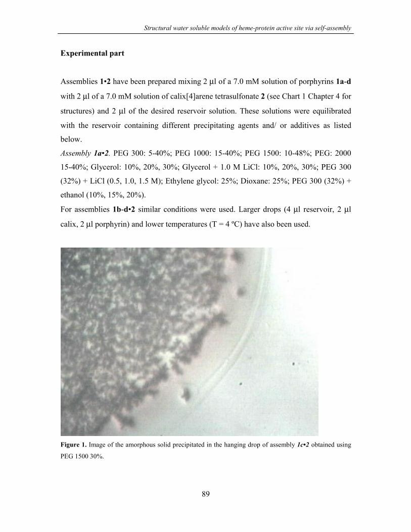
Structural water soluble models of heme-protein active site via self-assembly
89
Experimental part
Assemblies 1•2 have been prepared mixing 2 µl of a 7.0 mM solution of porphyrins 1a-d
with 2 µl of a 7.0 mM solution of calix[4]arene tetrasulfonate 2 (see Chart 1 Chapter 4 for
structures) and 2 µl of the desired reservoir solution. These solutions were equilibrated
with the reservoir containing different precipitating agents and/ or additives as listed
below.
Assembly 1a•2. PEG 300: 5-40%; PEG 1000: 15-40%; PEG 1500: 10-48%; PEG: 2000
15-40%; Glycerol: 10%, 20%, 30%; Glycerol + 1.0 M LiCl: 10%, 20%, 30%; PEG 300
(32%) + LiCl (0.5, 1.0, 1.5 M); Ethylene glycol: 25%; Dioxane: 25%; PEG 300 (32%) +
ethanol (10%, 15%, 20%).
For assemblies 1b-d•2 similar conditions were used. Larger drops (4 µl reservoir, 2 µl
calix, 2 µl porphyrin) and lower temperatures (T = 4 ºC) have also been used.
Figure 1. Image of the amorphous solid precipitated in the hanging drop of assembly 1c•2 obtained using
PEG 1500 30%.
Chapter 4
90
References
1. Randaccio, L.; Geremia, S.; Stener, M.; Toffoli, D.; Zangrando, E. Eur. J. Inorg.
Chem. 2002, 93-103.
2. Randaccio, L.; Furlan, M.; Geremia, S.; Slouf, M.; Srnova, I.; Toffoli, D. Inorg.
Chem. 2000, 39, 3403-3413.
3. Randaccio, L.; Geremia, S.; Nardin, G.; Slouf, M.; Srnova, I. Inorg. Chem. 1999, 38,
4087-4092.

91
Chapter 5
TOWARDS WATER SOLUBLE FUNCTIONAL MODELS OF O2
BINDING HEME-PROTEINS VIA SELF-ASSEMBLY∗
Water soluble noncovalent mimics for O2 binding heme-proteins have been prepared
using the ion-pair complexes of calix[4]arenes and porphyrins as described in Chapter 3
and 4. To obtain functional models, the inert Zn porphyrins have been replaced by
CoIIporphyrins, which are able to bind O2. These novel functional models have been
studied in relation to their O2 binding ability and their use in O2/N2 membrane
separation.
5.1 Introduction
Transport and storage of O2 is of fundamental importance to most living organisms. In
mammals, these two functions are carried out by heme-proteins such as hemoglobin and
myoglobin.1 Surprisingly, there are no examples reported so far of biomimetic O2 binders
and transporters obtained via noncovalent synthesis in water. This Chapter deals with
self-assembly of tetracationic CoII porphyrins and tetraanionic calix[4]arenes to form
water soluble O2 binding heme-protein models. The strategy followed for the preparation
of these systems has been described in detail in Chapters 3 and 4 in which structural
models of heme-protein based on Zn-porphyrins are described. Nevertheless, to obtain
functional models, the inert Zn2+ ion has to be replaced by Fe2+ or Co2+ since these Fe and
Co porphyrins are able to bind O2. Functional models are generally more attractive than

Chapter 5
92
structural models because of their potential applications, e.g. as synthetic O2 carriers.
Actually, the idea of using synthetic O2 carriers for the separation of O2 and N2 has long
been pursued. In particular, supported liquid membranes employing synthetic O2 carriers
have been recognized for their potential use in biomedical applications. They could be
used in small portable devices to produce O2 enriched air for patients suffering from
respiratory or lung diseases such as asthma and emphysema.2 The self-assembled O2
carriers presented in this Chapter might be especially interesting in this area because of
their water solubility3 and modest synthetic efforts. From the biomimetic point of view,
water solubility is very important because biological processes take place in aqueous
solutions. Only a few reports have dealt with binding of O2 to simple water soluble CoII
porphyrins,4,5 and the use of covalent superstructured porphyrins in water has only been
reported by Collman and coworkers.6-8 In general, CoII porphyrins are chosen because of
their higher stability towards autoxidation, which eventually lead to the inactivation of
the carrier, compared to FeII porphyrins.9
5.2 Results and discussion
5.2.1 Synthesis
Metallation of free base porphyrins 1 with cobalt II was performed at 40 ºC using 10-20
equiv of Co(Ac)2 in deoxygenated MeOH. Porphyrins 1a and 1b (Figure 1) were isolated
as hexafluorophosphate salts by precipitation upon addition of NH4PF6.10,11 The
preparation of tetrasulfonato calix[4]arene 2 has been reported elsewhere.10
5.2.2 Assembly formation
The assembly formation between CoII porphyrins 1b and calix[4]arene 2 has been studied
via ITC. Assembly 1b•2 is formed with LogK1•2 = 3.80 ± 0.04 in aqueous carbonate
buffered solution at pH 10 and I = 0.045 M (K1•2 in M-1). This result clearly confirms that,
∗ Prof. J. Reedijk and Mr. G. A. van Albada (University of Leiden, The Netherlands) are gratefully
acknowledged for the support offered with EPR measurements. We thank Prof. M. Wessling (University of
Twente, The Netherlands) for helpful discussions on membrane measurements.
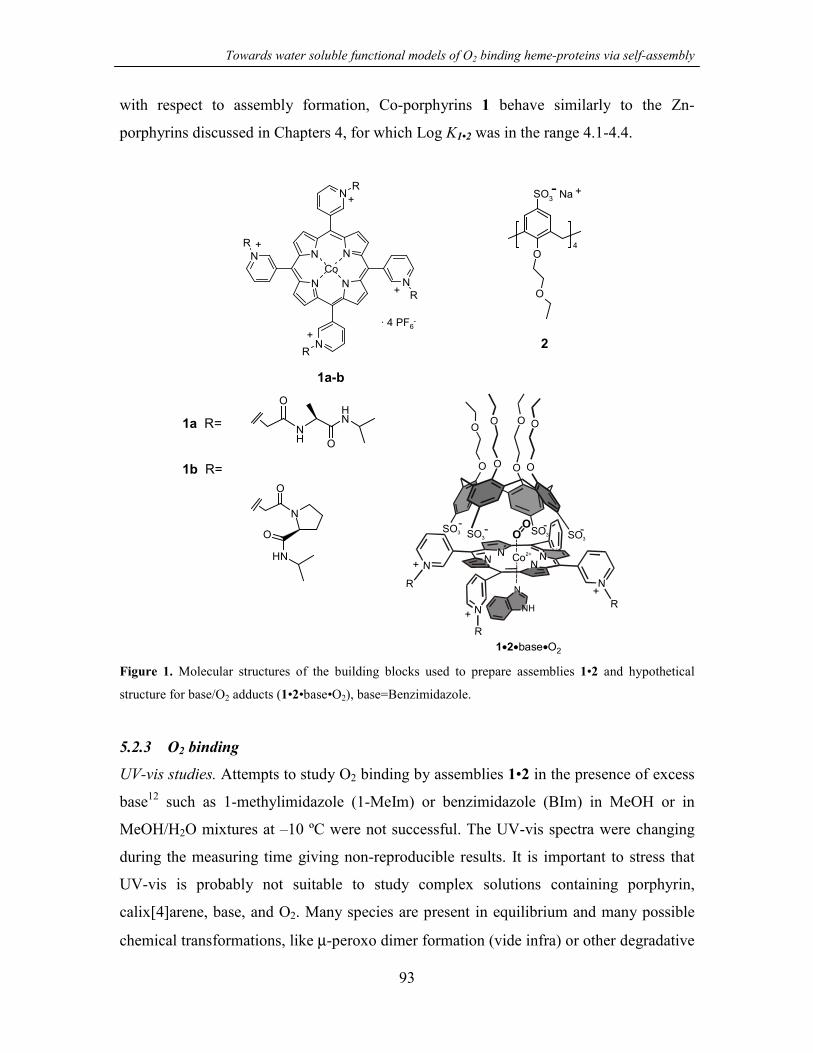
Towards water soluble functional models of O2 binding heme-proteins via self-assembly
93
with respect to assembly formation, Co-porphyrins 1 behave similarly to the Zn-
porphyrins discussed in Chapters 4, for which Log K1•2 was in the range 4.1-4.4.
· 4 PF6-
CoN
N
N
N
N
N N
N
R
R
R
R
+
+
+
+
1a-b
O
O
4
SO3- Na +
2
1a R=
1b R=
NH
O
O
NH
N
O
O
NH
OO
O OO O
-
O O OO
- - -SO3SO3 SO3 SO3
+ N
N N
R
NCo2+
+
+
R
R
N
N
N
N
+
N
NH
1 2• • •base O2
Figure 1. Molecular structures of the building blocks used to prepare assemblies 1•2 and hypothetical
structure for base/O2 adducts (1•2•base•O2), base=Benzimidazole.
5.2.3 O2 binding
UV-vis studies. Attempts to study O2 binding by assemblies 1•2 in the presence of excess
base12 such as 1-methylimidazole (1-MeIm) or benzimidazole (BIm) in MeOH or in
MeOH/H2O mixtures at –10 ºC were not successful. The UV-vis spectra were changing
during the measuring time giving non-reproducible results. It is important to stress that
UV-vis is probably not suitable to study complex solutions containing porphyrin,
calix[4]arene, base, and O2. Many species are present in equilibrium and many possible
chemical transformations, like µ-peroxo dimer formation (vide infra) or other degradative

Chapter 5
94
processes could take place.13 Attempts to measure O2 binding to porphyrin 1b alone in
the presence of BIm in MeOH (or in MeOH/H2O mixtures) were also not successful,
probably due to the use of polar protic solvents. To confirm this hypothesis, O2 binding
was studied in acetonitrile. The UV-vis spectral changes (Figures 2 and 3) occurring upon
equilibration of a solution containing 1b and 1-MeIm (0.15 M) with O2/N2 gas mixtures
of varying composition (expressed as O2 partial pressure pO2) were followed. The
measurements were performed at –30 ºC to promote binding and to reduce the possibility
of irreversible oxidation of the porphyrin.
0.0
0.1
0.2
0.3
0.4
Abs
500 520 540 580 600
λ (nm)
Figure 2. Spectral changes as a function of increasing pO2 in equilibrium with a solution of 1b at –30 ºC.
[1b] = 2.1 × 10⁻⁵ M, in CH3CN, [1-MeIm] = 0.15 M. Max. pO2 = 76 Torr.
The spectral variations at 553 (Figure 3) and 551 nm in response to the increase in pO2
were fitted to a 1:1 binding isotherm using a nonlinear least-squares fitting procedure
affording a binding constant of KO2 = 0.137 Torr-1 (half oxygenation pressure P1/2 = 1/KO2
= 7.3 Torr). Despite the fact that tetracationic alkylpyridinium porphyrins have been
known for more than 30 years,14 their O2 binding ability of Co derivatives has not been
reported in quantitative terms. However, the P1/2 value determined for 1b can be
compared to the values for the simple CoII-protoporphyrinIX-dimethylester, for which

Towards water soluble functional models of O2 binding heme-proteins via self-assembly
95
values of P1/2 in non polar and polar solvents are available (axial base 1-MeIm, –23 ºC,
P1/2 = 417 Torr in toluene and P1/2 = 12.6 Torr in DMF).15
Abs
pO2 (Torr)
Figure 3. Absorbance changes at 553 nm as a function of the pO2 in equilibrium with a 2.1 × 10⁻⁵ M
solution of 1b in CH3CN, [1-MeIm] = 0.15 M. Solid line is the fitted curve according to a 1:1 binding
isotherm.
EPR studies. Because of the complications encountered in the UV-vis measurements
(vide supra), EPR (electron paramagnetic resonance) studies were performed to directly
detect the formation of O2 complexes (Figure 4).16 CoII porphyrins and their 1:1 complex
with O2 are paramagnetic species and can be studied via EPR spectroscopy. The Co2+ ion
(3d7) is low spin in porphyrins with one unpaired electron residing in the 2zd -orbital. O2
binds to Co affording end-on complexes for which the electron density for the unpaired
electron is largely delocalized on the terminal oxygen atom. For this reason these
complexes have been described as superoxo-like complexes (CoIII-O2- formalism).
The EPR spectra for porphyrins 1a and 1b recorded in frozen 40:60 MeOH/H2O mixtures
at 77K (Figures 5a and 6a) are very similar and characteristic of CoII porphyrins (axially
symmetric molecules with S = 1/2, I = 7/2). These spectra were obtained in the presence
of O2 since the solutions were prepared in open air. However, there is no spectral
evidence for O2 binding under these conditions.17

Chapter 5
96
Co P baseII •Co P baseII •
Co P base + H OIII 2 2 2•
O2
η1-superoxo complex µ-peroxo complex
Co P baseIII •
Co P baseIII •
OO
Co P baseIII •
OO-
Figure 4. Equilibria and reactions for CoII porphyrin systems upon oxygenation. Base indicates a
coordinating molecule like a nitrogenous base or a solvent molecule (MeOH, H2O in this study).
200
B (mT)250 300
a
b
350 400
Figure 5. EPR spectra of a frozen solution of a) 1a (1.26 × 10⁻³ M) and b) 1a•2 (1a 1.26 × 10⁻³ M, 1.7
equiv 2) in 40:60 MeOH/H2O at 77 K.
The EPR spectra are mainly characterized by two components of the g tensor viz. g⊥ and
g// (Table 1). Subsequently, solutions of 1a and 1b in 40:60 MeOH/H2O were left in
contact with air for periods up to 20 h at room temperature. After this time no decrease in
signal intensity of the EPR spectra was observed, showing that no oxidation (formation of
CoIIIporphyrin species) or µ-peroxo dimer formation (2:1 CoIIporphyrin-O2 complexes,

Towards water soluble functional models of O2 binding heme-proteins via self-assembly
97
formally described as diamagnetic CoIII–O-O–CoIII species) takes place under these
conditions (see Figure 4).
200
B (mT)250 300 350 400
a
b
Figure 6. EPR spectra of a frozen solution of a) 1b (1.28×10⁻³ M) and b) 1b•2 (1b 1.28×10⁻³ M, 1.7 equiv
2) in 40:60 MeOH/H2O at 77K.
Table 1. EPR parameters for 1a and 1b in 40:60 MeOH/H2O frozen solutions at 77 K.
g⊥ g// A// (G)
1a 2.324 2.044 84
1b 2.324 2.044 86
3(DMSO)18 2.317 2.042 80
3(DMF)18 2.335 2.061 83
When imidazole (Im) or BzI was added to 1a (or 1b) in 40:60 MeOH/H2O in the
presence of air, the formation of O2 adducts CoIIP•base•O2 was indicated by the
appearance of a new signal at g~2.01 in the EPR spectra.19 The O2 adduct is the

Chapter 5
98
dominating species in frozen solutions of porphyrin 1a (or 1b) containing 3 equivalents
of imidazole. In contrast, a large fraction of CoIIP•base complex was still present even
when using 7-8 equivalents of the weaker base BzI (Figure 7).20 Moreover, samples
containing Im underwent much faster µ-peroxo dimer formation21 as shown by the loss of
the EPR signal in measurements performed after 30-50 min of standing at room
temperature. For samples containing BzI the signal intensity remained unchanged when
the spectra were recorded after 13 h at room temperature. This observation indicates that
O2 complexes are not present at room temperature (note that EPR spectra are recorded at
77K), which is due to the weaker basicity of BzI. Support for this conclusion is the large
reduction in signal intensity (~80%) showing µ-peroxo dimer formation observed when
1b solution containing BzI is kept at around –30 ºC for 3h (lower temperatures strongly
favor the formation of CoII porphyrin-O2 complexes). These results also indicate that for
solutions containing Im there must be an appreciable concentration of CoII porphyrin-O2
complex already at room temperature in the presence of air, which is quickly converted to
µ-peroxo dimers. Finally, it is worth noting that porphyrin 1a gave systematically higher
amounts of O2 complex than 1b under identical conditions for reasons not yet understood.
200 250 300 350 400-1500
-1000
-500
0
500
1000
B (mT)
Inte
nsity
B (mT)200 250 300 350 400
-1000
-500
0
500
1000
1500
Figure 7. EPR spectra of a frozen solution of (left) 1a (1.26 × 10⁻³ M) with 8.0 equiv of benzimidazole in
the presence of air; (right) 1b (1.28 × 10⁻³ M) with 7.9 equiv of benzimidazole in the presence of air. In
both spectra the signal on the right (*) at g ~ 2.32 is assigned to CoIIP•base while the other signal (•) at g ~
2.01 is assigned to CoIIP•base•O2 complex.
Assemblies 1a•2 and 1b•2, prepared from 1.26-1.28 × 10⁻³ M porphyrin solutions and 1.7
equiv of calix[4]arene 2, do not bind O2 in the absence of an axial bases (see Figures 5b

Towards water soluble functional models of O2 binding heme-proteins via self-assembly
99
and 6b).22 However, upon addition of Im or BzI, O2 complexes were obtained as observed
for 1a and 1b. These are possibly quaternary complexes formed by one porphyrin unit,
one calix[4]arene, one base and one O2 molecule. The presence of the signal at g ~2.32
indicates that conversion to the O2 complex was not quantitative (Figure 8). The choice
of imidazole and benzimidazole as axial bases was dictated by their different molecular
dimensions, which can affect their spatial location in complexes 1•2•base. It was
previously shown for analogous ion-pair complexes based on Zn-porphyrins (section
4.2.3) that small nitrogenous ligands such as 4-methylpyridine and 1-MeIm can be
encapsulated in the cavity formed between calix[4]arene 2 and porphyrins 1, while the
bulkier caffeine is bound outside on the solvent exposed porphyrin face. A similar
behavior is expected for imidazole (encapsulation) and benzimidazole (external
complexation). Unfortunately the EPR spectra did not provide any indications about the
location of the axial base in the assembly and consequently about the location of O2.
Furthermore, independently from the base used, no appreciable difference was noted for
1a•2 when compared with the measurements performed with 1a in the absence of 2.
Slightly larger amounts of O2 adduct were obtained for 1b•2 compared to 1b (larger
signal for CoIIP•base•O2 complex vs CoIIP•base complex in Figure 8 compared to Figure
7-right).
200 250 300 350 400
-1000
-500
0
500
1000
1500
Inte
nsity
B (mT)
Figure 8. EPR spectrum of a frozen solution of 1b•2 (1b 1.28 × 10⁻³ M, 1.7 equiv 2) with 8.0 equiv of
benzimidazole in the presence of air. The signal at g ~ 2.32 (*) is assigned to CoIIP•base, the signal at g ~
2.01 (•) is assigned to CoIIP•base•O2 complex.

Chapter 5
100
Surprisingly, when solutions of 1a•2 and 1b•2 containing benzimidazole were left at
room temperature for 1-2 h a clear decrease in signal intensity could be observed, thus
indicating formation of diamagnetic CoIII species. This transformation occurs faster than
for 1a and 1b under identical conditions. These results contradict the expectation that
binding of benzimidazole at the solvent exposed porphyrin face will induce binding of O2
within the cavity, preventing the formation of µ-peroxo dimers. However, it is important
to note that there are other possible oxidative mechanisms leading to EPR-inactive CoIII
species.13 One of these may be favored by the calix[4]arene moiety in close proximity to
the bound O2.
Thus, these preliminary studies show that binding of O2 to self-assembled complexes 1•2
in the presence of an axial base is taking place, but it is hard to describe how
calix[4]arene 2 is affecting O2 binding. Further structural studies will be necessary to
clarify the the complex structure.
5.2.4 Facilitated O2 transport across supported liquid membranes
Separation of O2 and N2 from air is an important process for the production of these two
industrially important gases. For large scale preparation of pure gases the method of
choice is distillation using cryogenic plants. However, for many industrial applications,
especially involving combustion processes, high purity O2 is not necessary and O2
enriched air can be a valid substitute. In this respect, membrane separation processes may
play a crucial role due to the lower costs compared to cryogenic distillation.2,23 The
separation of N2 and O2 have been achieved using polymeric membranes24 with selective
permeability for O2, or supported liquid membranes25,26 in which the liquid phase is a
solution containing a selective and reversible O2 carrier immobilized in the pores of a thin
porous polymer. In the latter case, transport of O2 involves chemical binding of O2 to the
carrier and diffusion limited transport along the concentration gradient created across the
membrane, between the feed and the permeate side.27 Therefore, such membranes are
often called facilitated transport membranes.28
Self-assembled O2 carrier solutions were prepared dissolving porphyrins 1, calix[4]arene
2 and 1-MeIm in water (2.0 × 10⁻³ M, 6.0 × 10⁻³ M, and 2.0 × 10⁻² M., respectively). The
hydrophilic porous membranes were loaded by immersion in the carrier solutions. The

Towards water soluble functional models of O2 binding heme-proteins via self-assembly
101
membranes were then placed in a gas permeation set-up and the feed and the permeate
gas streams were analyzed for their content in O2 and N2. Preliminary results of these
measurements have been reported elsewhere.29 Here only one example is reported in
which a commercially available membrane (Millipore VMWP) was loaded with a
solution containing 1b, 2, and 1-MeIm. The feed gas composition was 20% O2 and 80%
N2. Facilitated O2 transport was observed, even though the facilitation factor was only a
modest 1.15 (selectivity coefficient 2.3-2.4) with respect to the membrane loaded with
only H2O (facilitation factor = 1, selectivity = 2, see Figure 9). Decrease in membrane
selectivity was observed after 4-6 h indicating possible degradation of the carrier.
Enhanced O2 transport was observed only if all the components necessary for the self-
assembled carrier were present.
1.31.51.71.92.12.32.52.72.9
50 150 250 350Time (min)
Sele
ctiv
ity
Figure 9. Facilitated O2 transport (dots) observed for a Millipore VMWP membrane loaded with a solution
of assembly 1b•2 in water in the presence of 1-MeIm at 10 ºC (1b = 2.0 × 10⁻³ M, 3 equiv 2, 10 equiv of 1-
MeIm). The solid line shows the O2/N2 selectivity of the membrane loaded with only H2O. Feed gas
composition 20% O2 / 80% N2.
5.3 Conclusions
Even though the results reported in this Chapter are not optimal, for the application of
these carriers in membrane technology, self-assembled complexes 1•2 can be considered
a novel class of water-soluble functional models for O2 binding heme-proteins. A great
advantage of these systems is the availability due to the noncovalent synthetic assembly

Chapter 5
102
in their preparation. However, the main drawback is the complexity of the system in
which multiple equilibria and species may contribute to the overall response towards
changes from the outside (for instance in O2 concentration). More studies are needed to
fully understand the systems and possibly to design a second generation of more efficient
self-assembled O2 carriers.
5.4 Experimental part
Mass spectra were measured using a Perkin Elmer/PerSeptive Biosystem Voyager-DE-
RP MALDI-TOF mass apectrometer (PerSeptive Biosystem, Inc., Framingham, MA,
USA) equipped with delayed extraction. UV-vis measurements were performed on a
Varian Cary 3E UV-vis spectrophotometer equipped with a Helma QX optical fiber
probe (path length=1.000 cm), using solvents of spectroscopic grade. Calorimetric
measurements were carried out using a Microcal VP-ITC microcalorimeter with a cell
volume of 1.4115 mL. X-Band EPR spectra were recorded on a Jeol RE2x electron spin
resonance spectrometer using DPPH (diphenylpicrylhydrazyl, g=2.0036) as a standard.
All the EPR measurements were performed in frozen solutions (40:60 MeOH/H2O) at 77
K.
Membrane preparation and the sweep gas permeation set-up used to study the facilitated
O2 transport across supported liquid membranes,29 O2 binding experiments in solution,30
synthesis of free base porphyrins 111 and of calix[4]arene 210 have been described in
detail elsewhere.
Preparation of porphyrins 1a and 1b
Free base porphyrin 1 was dissolved in deoxygenated MeOH containing 10-20 equiv of
Co(Ac)2. The solution was stirred 1 h at 40 ºC under N2 (insertion of the metal was
followed by UV-vis spectroscopy). Porphyrins 1a and 1b (1b MALDI-TOF MS: m/z
1898.7 [M-PF6]+, calculated for [C80H92N16O8P3F18Co]+ 1898.6) were isolated as
hexafluorophosphate salts by precipitation upon addition of NH4PF6.

Towards water soluble functional models of O2 binding heme-proteins via self-assembly
103
5.5 References and notes
1. For an overview on the field of natural and biomimetic O2 carriers see Chapter 2.7
of this thesis.
2. Oxygen Complexes and Oxygen Activation by Transition Metals; Martell, A. E.;
Sawyer, D. T., Ed.; Plenum Press: New York, 1988.
3. Extensive work has been carried out using membranes containing aqueous solutions
of hemoglobin or of bis(histidinato)-cobalt(II) as carriers. See ref 2 and references
therein.
4. De Bolfo, J. A.; Smith, T. D.; Boas, J. F.; Pilbrow, J. R. J. Chem. Soc., Dalton
Trans. 1976, 1495-1500.
5. Evans, D. F.; Wood, D. J. Chem. Soc., Dalton Tras. 1987, 3099-3101.
6. Zhang, X.; Uffelman, E.; Collman, J. P. U. S. Patent 5384397 A, 1995.
7. Zhang, X.; Uffelman, E. S.; Collman, J. P. U. S. Patent 5274090 A, 1993.
8. For reversible O2 binding to FeII porphyrins in aqueous media see Komatsu, T.;
Hayakawa, S.; Yanagimoto, T.; Kobayakawa, M.; Nakagawa, A.; Tsuchida, E. Bull.
Chem. Soc. Jpn. 2001, 74, 1703-1707. Tsuchida, E.; Komatsu, T.; Arai, K.;
Yamada, K.; Nishide, H.; Fuhrhop, J. H. Langmuir 1995, 11, 1877-1884.
9. Jones, R. D.; Summerville, D. A.; Basolo, F. Chem. Rev. 1979, 79, 139-179.
10. Fiammengo, R.; Timmerman, P.; Huskens, J.; Versluis, K.; Heck, A. J. R.;
Reinhoudt, D. N. Tetrahedron 2002, 58, 757-764. See also Chapter 3.
11. Fiammengo, R.; Crego-Calama, M.; Timmerman, P.; Reinhoudt, D. N. 2002, J. Am.
Chem. Soc. submitted. See also Chapter 7.
12. The presence of an axial base is necessary to enhance the electron density on the Co
atom thus increasing the binding ability of CoIIporphyrins for O2.

Chapter 5
104
13. Busch, D. H.; Alcock, N. W. Chem. Rev. 1994, 94, 585-623.
14. Hambright, P.; Fleischer, E. B. Inorg. Chem. 1970, 9, 1757-1761.
15. Stynes, H. C.; Ibers, J. A. J. Am. Chem. Soc. 1972, 94, 5125-5127.
16. Walker, F. A. J. Am. Chem. Soc. 1970, 92, 4235-4244.
17. It has been observed that the similar 5,10,15,20-tetrakis(N-methylpyridinium-4-
yl)porphyrinatocobalt(II) can be converted almost quantitatively to the O2 complex
if a solution of the porphyrin in water is bubbled with O2 for three minutes at r.t.
before cooling. See ref 5.
18. Araullo-McAdams, C.; Kadish, K. M. Inorg. Chem. 1990, 29, 2749-2757.
19. Assignment of the g tensor principal axis and determination of the g and ACo
matrixes for CoIIporphyrin-O2 complex is generally difficult from simple X-band
CW EPR spectra and required more sophisticated spectroscopic techniques for
accurate determinations. See Van Doorslaer, S.; Schweiger, A. J. Phys. Chem. B
2000, 104, 2919-2927.
20. The amount of BzI was not further increased since a difference in the spectrum of
1a was only observed in going from 1 to 3 equiv and not for further additions.
21. Under the experimental conditions used in this Chapter for the EPR spectra, µ-
peroxo dimer formation is the main cause for the loss of the EPR signal. See ref 5.
22. The measured K1•2 allows for the prediction that 1.7 equiv of calix[4]arene 2 are
necessary to obtain higher than 85% of assembly formation at concentrations in the
millimolar range used for EPR experiments.
23. Figoli, A.; Sager, W. F. C.; Mulder, M. H. V. J. Membrane Sci. 2001, 181, 97-110.
24. Lonsdale, H. K. J. Membr. Sci. 1982, 10, 81-181.
25. Baker, R. W.; Roman, I. C.; Lonsdale, H. K. J. Membr. Sci. 1987, 31, 15-29.
Towards water soluble functional models of O2 binding heme-proteins via self-assembly
105
26. Johnson, B. M.; Baker, R. W.; Matson, S. L.; Smith, K. L.; Roman, I. C.; Tuttle, M.
E.; Lonsdale, H. K. J. Membr. Sci. 1987, 31, 31-67.
27. Mulder, M. H. V. Basic Principles of Membrane Technology; Kluwer Academic
Publisher: Dordrecht, 1996.
28. The mathematical model for the transport mechanism can be found in refs 2, 26-27.
29. For more details about the O2 transport results see: Figoli A. Synthesis of
nanostructured mixed matrix membranes for facilitated gas separation, PhD Thesis
(ISBN 90-365-1673-0), University of Twente, 2001, p 139-152.
30. See Experimental part Chapter 6.

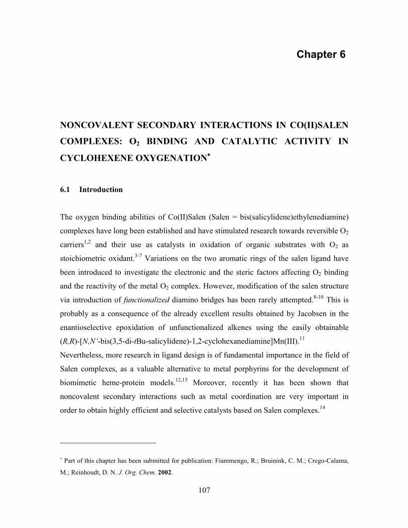
107
Chapter 6
NONCOVALENT SECONDARY INTERACTIONS IN CO(II)SALEN
COMPLEXES: O2 BINDING AND CATALYTIC ACTIVITY IN
CYCLOHEXENE OXYGENATION∗∗∗∗
6.1 Introduction
The oxygen binding abilities of Co(II)Salen (Salen = bis(salicylidene)ethylenediamine)
complexes have long been established and have stimulated research towards reversible O2
carriers1,2 and their use as catalysts in oxidation of organic substrates with O2 as
stoichiometric oxidant.3-7 Variations on the two aromatic rings of the salen ligand have
been introduced to investigate the electronic and the steric factors affecting O2 binding
and the reactivity of the metal O2 complex. However, modification of the salen structure
via introduction of functionalized diamino bridges has been rarely attempted.8-10 This is
probably as a consequence of the already excellent results obtained by Jacobsen in the
enantioselective epoxidation of unfunctionalized alkenes using the easily obtainable
(R,R)-[N,N’-bis(3,5-di-tBu-salicylidene)-1,2-cyclohexanediamine]Mn(III).11
Nevertheless, more research in ligand design is of fundamental importance in the field of
Salen complexes, as a valuable alternative to metal porphyrins for the development of
biomimetic heme-protein models.12,13 Moreover, recently it has been shown that
noncovalent secondary interactions such as metal coordination are very important in
order to obtain highly efficient and selective catalysts based on Salen complexes.14
∗ Part of this chapter has been submitted for publication: Fiammengo, R.; Bruinink, C. M.; Crego-Calama,
M.; Reinhoudt, D. N. J. Org. Chem. 2002.

Chapter 6
108
In this Chapter the preparation of three novel Co(II)Salen complexes 1-3 bearing (for 1
and 2) an OH functionalized diamino bridge is reported (Figure 1). The presence of the
hydroxyl groups in 1 increases the affinity for oxygen due to hydrogen bonding with the
Co-coordinated O2 (Figure 2).
N
O
N
O
R R
H H
Co
tBu tBu
tBu tBu
1: R=OH (cis-syn)2: R=OH (cis-anti)3: R=H
N
O
N
O
HH
Co
4tBu tBu
tBu tBu
Figure 1. Structures of Co(II)Salens 1-4
Co
ON N
O O
O
OHOH
tBu
tBu tButBu
Co
ON N
O O
OHOH
O
tBu
tBu tButBuB B
2•O2•B
Figure 2. Schematic representation of ternary complexes 1•O2•B and 2•O2•B showing hydrogen-bonding
stabilization of Co-coordinated O2 (left). B=base (1-MeIm or propionaldehyde in the text).
To the best of our knowledge, this is the first time that the role of hydrogen bonding as
noncovalent secondary interactions in Salen complexes has been considered.15 This
design mimics the hydrogen-bonding interaction operated by the distal His residue in
hemoglobin and myoglobin or by Tyr in O2 avid Ascaris hemoglobin.16,17 Moreover,
Co(II)Salen 1-3 and the commercially available 4 have been tested in the aerobic

Noncovalent secondary interactions in Co(II)Salen complexes
109
oxidation of cyclohexene (Scheme 3). Reduced catalytic activity is observed as a
consequence of the Co-O2 complex stabilization.
6.2 Results and discussion
Synthesis. Co(II)Salenes 1 and 2 were prepared starting from the two diastereomeric cis-
1,2-diamino-cis-4,5-dihydroxycyclohexanes (5 and 6 respectively). The corresponding
dihydrochloride salts (5-Cl and 6-Cl) were synthesized from cis-1,2,3,6-tetraphtalic
anhydride 12 following a slightly modified literature procedure (Scheme 1).18 Catalytic
osmylation of Cbz-protected 1,2-diaminocyclohex-4-ene 15 using NMO (4-
methylmorpholine-N-oxide) as the primary oxidant and DABCO as the ligand afforded a
mixture of diastereomeric cis diols 16 and 17 in 36:64 ratio (80% yield) at room
temperature.19 Deprotection via catalytic hydrogenation and precipitation with HCl in
EtOH afforded the desired salts.
O
O
ONCO
NCO NH3
NH3
NH
NH
Cbz
Cbz
NH
NHOH
OHCbz
Cbz
NH
NHOH
OH
Cbz
Cbz
+NH3Cl
NH3ClOH
OHNH3Cl
NH3ClOH
OH
6-Cl 5-Cl
12 13 14(Cl-salt) 15
1617(cis-anti-cis) (cis-syn-cis)
+
+
+
+
+
+
a, b, a c d
e
f, g
--
--
Scheme 1. a TMS-N3, THF, reflux, 3h. b SOCl2, CCl4, cat. DMF, 50°C, 2.5 h. c conc. HCl, THF/acetone, rt,
14 h. d Cbz-Cl, N,N-diisopropyl-N-ethylamine, THF/H2O, 0°C, 3 h. e NMO, OsO4 (0.02 equiv), DABCO
(0.2 equiv) acetone/H2O, rt, 72 h. 16 and 17 separation by column chromatography. f 10% Pd/C, H2, EtOH,
rt, 5 h. g HCl/EtOH, rt.

Chapter 6
110
Salen ligands 9-11 were prepared by condensation of 3,5-di-t-butyl-2-
hydroxybenzaldehyde 8 with diamine 5-6 or with cis-1,2-diaminocyclohexane 7,
respectively, in refluxing ethanol in moderate to good yields.20 Subsequent metalation
with CoAc2 afforded Co(II)Salen 1-3 in 93-95% yield (Scheme 2).
+
O
OH
8tBu
tBu
N
OH
N
OH
R R
H H
9-11
tBu
tBu tBu
tBu
1-3
EtOH, reflux
26-81%
Co(OAc)2,EtOH, reflux >93%
NH2
NH2
R
R
5: R=OH (cis-syn)6: R=OH (cis-anti)7: R=H
Scheme 2
Structural diversity is introduced in Co(II)Salen complexes 1-4 via the 1,2-
diaminocyclohexane bridge. The stereochemistry of the two amino groups is (meso)-cis
in 1-3 and (R,R)-trans in 4. As a result, 1-3 have non-planar structures, with the
cyclohexyl ring being at roughly 90º to the Co(II)-N2O2 plane.21 Therefore, this spatial
arrangement should shield the Co-coordinated O2. In contrast, as evident from an earlier
X-ray crystal structure,22 the commercially available Co(II)Salen 4 is roughly planar.
Additionally, two cis hydroxyl groups have been introduced on the cyclohexane ring of 1
and 2 in position 4 and 5. The orientation of these two OH groups is either syn or anti to
the two amino groups (see Scheme 1). A CPK model of Co(II)Salen 1 shows that the syn
arrangement of the cis-4,5-hydroxyl groups is necessary for one OH to point directly
towards the Co-O2 moiety, thus adopting the required orientation to form a hydrogen
bond with O2. No hydrogen bond is possible when the hydroxyl groups are arranged anti
as in Co(II)Salen 2 and 3 (Figure 2).
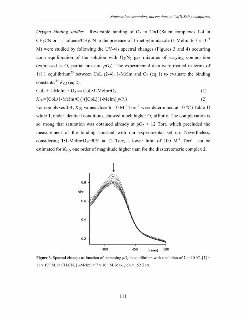
Noncovalent secondary interactions in Co(II)Salen complexes
111
Oxygen binding studies. Reversible binding of O2 to Co(II)Salen complexes 1-4 in
CH3CN or 1:1 toluene/CH3CN in the presence of 1-methylimidazole (1-MeIm, 6-7 × 10⁻³
M) were studied by following the UV-vis spectral changes (Figures 3 and 4) occurring
upon equilibration of the solution with O2/N2 gas mixtures of varying composition
(expressed as O2 partial pressure pO2). The experimental data were treated in terms of
1:1:1 equilibrium23 between CoL (2-4), 1-MeIm and O2 (eq 1) to evaluate the binding
constants,24 KO2 (eq 2).
CoL + 1-MeIm + O2 � CoL•1-MeIm•O2 (1)
KO2=[CoL•1-MeIm•O2]/([CoL][1-MeIm] pO2) (2)
For complexes 2-4, KO2 values close to 10 M-1 Torr-1 were determined at 10 ºC (Table 1)
while 1, under identical conditions, showed much higher O2 affinity. The complexation is
so strong that saturation was obtained already at pO2 < 12 Torr, which precluded the
measurement of the binding constant with our experimental set up. Nevertheless,
considering 1•1-MeIm•O2>90% at 12 Torr, a lower limit of 100 M-1 Torr-1 can be
estimated for KO2, one order of magnitude higher than for the diastereomeric complex 2.
400 450 λ (nm) 500
0.4
0.2
0.6
Abs
0.8
Figure 3. Spectral changes as function of increasing pO2 in equilibrium with a solution of 2 at 10 ºC. [2] =
11 × 10-5 M, in CH3CN, [1-MeIm] = 7 × 10⁻³ M. Max. pO2 = 152 Torr.

Chapter 6
112
This result agrees with the proposed hydrogen bonding stabilization of Co-coordinated O2
which, according to the three-dimensional arrangement of its hydroxyl groups, is specific
for 1. Moreover, the similar and lower KO2 determined for 2-4 show that the spatial
arrangement of the diamino bridges of these complexes has no appreciable influence on
O2 binding.
0.7
0.75
0.8
0.85
0.9
0 25 50 75 100 125 150
Abs
pO2 (Torr)
Figure 4. Absorbance changes at 416 nm (�) and at 424 nm (�) as function of the pO2 in equilibrium with
a 11 × 10-5 M solution of 2 in CH3CN, [1-MeIm] = 7 × 10⁻³ M. Solid lines are the best fitting according to a
1:1:1 model (see text).
Table 1. Formation constants of ternary complexes CoL•1-MeIm•O2 in CH3CN solutions at 10 ºC. [CoL] =
6-10 × 10-5 M, [1-MeIm] = 6-7 × 10⁻³ M.
KO2 (M-1Torr-1)
1 ≥100a
2 8.4
3 13.6
4 9.5b
a estimated value (see text). b 1:1 toluene/acetonitrile
In the absence of 1-methylimidazole, O2 binding to 1-4 was neither observed at 10 nor at
–10 ºC. This result confirms that strong O2 complexation to 1 is due to secondary
interactions (hydrogen bonding) stabilizing the ternary complex 1•1-MeIm•O2 and not to
alteration of the metal center electronic properties.

Noncovalent secondary interactions in Co(II)Salen complexes
113
Oxygenation of cyclohexene. Co(II) Schiff’s base complexes3 as well as
Co(II)porphyrins25 catalyze the oxidation of organic substrates (e.g. alkenes and alkanes)
with O2 as the bulk oxidant in the presence of reducing agents such as aldehydes.
Therefore, oxidation of cyclohexene with O2 in the presence of propanal (Scheme 3) was
chosen as a model reaction to study the relationship between noncovalent secondary
interactions stabilizing O2 binding and the catalytic activity of complexes 1-4. A
mechanistic study of the oxygenation of cyclohexene with Co(II)Salens 1-4 is beyond the
scope of this work. However, it has been reported for similar systems that the reaction
probably starts with the formation of a CoL•B•O2 complex (where B = aldehyde)26 in
close analogy with the formation of CoL•1-MeIm•O2 under the equilibrium conditions
reported here.
CH3CH2COOH
O
+ O+ O2 + +1-4
18 19
CH3CH2CHO
Scheme 3
Complexes 1-4 efficiently catalyze cyclohexene oxidation under mild conditions (20 °C,
1 atmosphere of O2, 0.5 mol% of catalyst, propionaldehyde as co-reductant) affording,
independently from the catalyst used, two oxygenation products27 viz. cyclohexene oxide
18 and 2-cyclohexene-1-one 19 in 4.7 ± 0.5 ratio (18/19) at 70 % conversion (Figure 5).28
The constant product selectivity observed for different metal catalyst shows a common
catalytic pathway for all the reactions, which probably involves radical chain reactivity, a
well-documented pathway for Co complexes catalyzed oxidations.5,7,29 Nevertheless,
different catalysts exhibit distinct reactivities, as evident from the measured induction
times (tind), initial reaction rate (ri), and half-life times for cyclohexene conversion (t1/2)
(Table 2). These differences can be related to the catalyst structure and in particular to the
hydrogen bonding ability of 1 vs 2-4 and possibly to the steric hindrance around the metal
center caused by the orientation of the cyclohexyl ring with respect to the Co(II)-N2O2
plane (coplanar as in 4 vs bent as in 1-3).
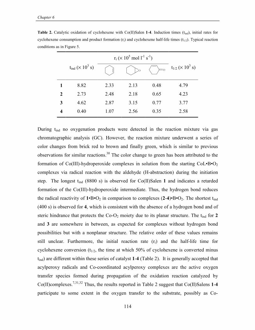
Chapter 6
114
Table 2. Catalytic oxidation of cyclohexene with Co(II)Salen 1-4. Induction times (tind), initial rates for
cyclohexene consumption and product formation (ri) and cyclohexene half-life times (t1/2). Typical reaction
conditions as in Figure 5.
ri (× 105 mol l-1 s-1)
tind (× 103 s)O O
t1/2 (× 103 s)
1 8.82 2.33 2.13 0.48 4.79
2 2.73 2.48 2.18 0.65 4.23
3 4.62 2.87 3.15 0.77 3.77
4 0.40 1.07 2.56 0.35 2.58
During tind no oxygenation products were detected in the reaction mixture via gas
chromatographic analysis (GC). However, the reaction mixture underwent a series of
color changes from brick red to brown and finally green, which is similar to previous
observations for similar reactions.30 The color change to green has been attributed to the
formation of Co(III)-hydroperoxide complexes in solution from the starting CoL•B•O2
complexes via radical reaction with the aldehyde (H-abstraction) during the initiation
step. The longest tind (8800 s) is observed for Co(II)Salen 1 and indicates a retarded
formation of the Co(III)-hydroperoxide intermediate. Thus, the hydrogen bond reduces
the radical reactivity of 1•B•O2 in comparison to complexes (2-4)•B•O2. The shortest tind
(400 s) is observed for 4, which is consistent with the absence of a hydrogen bond and of
steric hindrance that protects the Co-O2 moiety due to its planar structure. The tind for 2
and 3 are somewhere in between, as expected for complexes without hydrogen bond
possibilities but with a nonplanar structure. The relative order of these values remains
still unclear. Furthermore, the initial reaction rate (ri) and the half-life time for
cyclohexene conversion (t1/2, the time at which 50% of cyclohexene is converted minus
tind) are different within these series of catalyst 1-4 (Table 2). It is generally accepted that
acylperoxy radicals and Co-coordinated acylperoxy complexes are the active oxygen
transfer species formed during propagation of the oxidation reaction catalyzed by
Co(II)complexes.7,31,32 Thus, the results reported in Table 2 suggest that Co(II)Salens 1-4
participate to some extent in the oxygen transfer to the substrate, possibly as Co-

Noncovalent secondary interactions in Co(II)Salen complexes
115
coordinated acylperoxy species, and not only in the initiation step. Consequently,
hydrogen bonding and steric hindrance also play an important role in stabilizing these
intermediate species during the propagation step of the reaction. In fact, 1 is the least
reactive catalyst presenting the smallest ri and the longest t1/2, while 4 is the most reactive
one having the shortest t1/2. The ri values for 4 are somehow smaller than expected.
However, this catalyst also exhibits a slightly different concentration vs time profile when
compared to catalysts 1-3. Actually, the oxidation time course obtained for catalyst 4
resembles an autocatalytic reaction (Figure 6). Probably for this reason the initial rates (ri)
for 4 (Table 2) are the smallest in the series while t1/2 is the shortest, indicating higher
reactivity.
O
O
012 14 16108
20
80
100
40
60
Figure 5. Concentration vs time profile for cyclohexene (0.18 M in CH3CN) oxidation catalyzed by
Co(II)Salen 1 (0.005 equiv). Conditions: 20 °C, O2 1 atm, propanal (4 equiv).
Chapter 6
116
03 4 510
20
80
100
40
60
O
O
Figure 6. Concentration vs time profile for cyclohexene oxidation catalyzed by Co(II)Salen 4. Conditions
as in Figure 5. Note especially the slightly sigmoidal shape for the cyclohexene curve.
6.3 Conclusions
O2 affinity studies on novel Co(II)Salen complexes 1-3 (and on commercially available
4) have shown that noncovalent secondary interactions and especially hydrogen bonding
largely increase O2 binding (1 vs 2-4). The observed enhanced O2 binding for 1 is also
reflected in the lower catalytic reactivity in the cyclohexene oxidation. The simple
variations introduced on the salen ligand skeleton, and in particular on the diamino
bridge, show substantial control over the binding properties of Co(II)Salen complexes by
secondary interactions. These results might allow introduction of an alternative model to
the heavily exploited field of biomimetic superstructured porphyrins.
6.4 Experimental part
1H NMR and 13C NMR spectra were performed on a Varian Unity INOVA (300 MHz) or
a Varian Unity 400 WB NMR spectrometer. 1H NMR chemical shift values (300 MHz)
are expressed in ppm relative to residual CHD2OD (δ 3.30) or CHCl3 (δ 7.26). 13C NMR
chemical shift values (100 MHz) are expressed in ppm relative to residual CD3OD (δ

Noncovalent secondary interactions in Co(II)Salen complexes
117
49.0) or CDCl3 (δ 76.9). Infrared spectra were recorded on a FT-IR Perkin Elmer
Spectrum BX spectrometer using KBr pellets. Elemental analyses were carried out using
a 1106 Carlo-Erba Strumentazione element analyzer. MS FAB spectra were measured on
a Finnigan MAT 90 spectrometer with m-nitrobenzylalcohol (NBA) as the matrix. GC
spectra were recorded on a Varian 3400 gas chromatograph equipped with a flame
ionization detector (FID), using a capillary column (30 m, 0.25 µm film thickness) with
DB-5MS stationary phase (for temperature program, response factor determination see
Supplementary Informations).
General procedure for the synthesis of Salenes 9 and 10
3,5-di-t-butyl-2-hydroxybenzaldehyde 8 (280 mg, 1.2 mmol) was added to a solution of
cis-1,2-diamino-cis-4,5-dihydroxycyclohexane dihydrochloride salt 5-Cl18 (or 6-Cl)18
(120 mg, 0.55 mmol) and NaHCO3 (100 mg, 1.2 mmol) in 10 ml of EtOH at room
temperature. The reaction mixture was stirred for 1 hour at 75 ºC, and the solvent was
removed under vacuum. The product was extracted from the solid residue with CHCl3
and recrystallized from CH3OH.
(1R,2S,3R,4S)-[N,N’-bis(3,5-di-tBu-salicylidene)-4,5-dihydroxy-1,2-
cyclohexanediamine 10
Yield: 26%. 1H NMR (400 MHz, CDCl3): δ (ppm) 12.88 (bs, 2H), 8.26 (s, 2H), 7.27 (d,
2H, J=2.2 Hz), 6.96 (d, 2H, J=2.2 Hz), 3.95-3.86 (m, 2H), 3.59-3.52 (m, 2H), 2.56 (bs,
2H), 2.35-2.25 (m, 2H), 1.98-1.90 (m, 2H), 1.28 (s, 18H), 1.18 (s, 18H). 13C NMR (100
MHz, CDCl3): δ (ppm) 166.59, 158.08, 140.38, 137.01, 127.62, 126.40, 117.86, 69.96,
67.90, 35.23, 34.30, 31.64, 29.59. IR: (cm-1) 3369, 2958, 2910, 2870, 1629, 1467, 1439,
1391, 1362, 1274, 1251, 1203, 1173, 1100, 1039, 968, 773. FAB-MS: m/z 578.3 ([M]+,
calcd 578.4). Anal. calcd for C36H54N2O4: %C 74.70, %H 9.40, %N 4.84; found: %C
74.25, %H 9.50, %N 4.75.

Chapter 6
118
(1R,2S,3S,4R)-[N,N’-bis(3,5-di-tBu-salicylidene)-4,5-dihydroxy-1,2-
cyclohexanediamine 9
Yield: 65% yield. 1H NMR (400 MHz, CDCl3): δ (ppm) 13.46 (bs, 2H), 8.40 (s, 2H),
7.37 (d, 2H, J=2.3 Hz), 7.07 (d, 2H, J=2.3 Hz), 4.38-4.31 (m, 2H), 3.90-3.80 (m, 2H),
2.25 (bs, 2H), 2.21-2.10 (m, 4H), 1.40 (s, 18H), 1.29 (s, 18H). 13C NMR (100 MHz,
CDCl3): δ (ppm) 166.64, 158.22, 140.28, 136.82, 127.34, 126.31, 118.01, 68.71, 35.22,
34.31, 34.01, 31.67, 29.64. IR: (cm-1) 3422, 2957, 2909, 2871, 1628, 1459, 1439, 1390,
1362, 1274, 1250, 1202, 1174, 1088, 1027. FAB-MS: m/z 578.3 ([M]+, calcd 578.4).
Anal. calcd for C36H54N2O4: %C 74.70, %H 9.40, %N 4.84; found: %C 73.99, %H 9.52,
%N 4.76.
Preparation of (1R,2S)-[N,N’-bis(3,5-di-tBu-salicylidene)-1,2-cyclohexanediamine 11
3,5-di-t-butyl-2-hydroxybenzaldehyde 8 (490 mg, 2.1 mmol) was added to a solution of
cis-1,2-diaminocyclohexane 7 (120 µl, 1.0 mmol) in 20 ml of EtOH at room temperature.
The reaction mixture was stirred for 1 hour at 75 ºC, and gradually cooled down to room
temperature to allow precipitation of the product. The bright yellow solid was then
filtrated and washed several times with cold CH3OH. Yield: 81% (445 mg, 0.81 mmol).1H NMR (400 MHz, CDCl3): δ (ppm) 13.77 (bs, 2H), 8.37 (s, 2H), 7.36 (d, 2H, J=2.3
Hz), 7.07 (d, 2H, J=2.3 Hz), 3.63-3.57 (m, 2H), 2.09-1.92 (m, 2H), 1.82-1.73 (m, 2H),
1.67-1.55 (m, 2H), 1.42 (s, 18H), 1.29 (s, 18H). 13C NMR (100 MHz, CDCl3): δ (ppm)
165.41, 158.40, 140.00, 136.80, 127.03, 126.17, 118.15, 69.53, 35.24, 34.30, 31.70,
30.65, 29.67, 22.94. IR: (cm-1) 2997, 2955, 2864, 1630, 1598, 1459, 1441, 1390, 1362,
1273, 1251, 1204, 1174, 1134, 1083, 989, 879, 853, 827, 773. FAB-MS: m/z 546.3 ([M]+,
calcd 546.4), 547.3. Anal. calcd for C36H54N2O2: %C 79.07, %H 9.95, %N 5.12; found:
%C 78.99, %H 10.13, %N 5.25.
General procedure for the synthesis of Co(II)Salen 1-3
Salens 9-11 (1 equiv) were dissolved under nitrogen in degassed EtOH (typically 0.7
mmol in 10 ml). A Co(Ac)2·4H2O solution in degassed EtOH (1.5 equiv in 5 ml) was then
added causing an immediate color change from bright yellow to deep red. The reaction
mixture was heated to reflux for 1 hour under nitrogen and dried in vacuo. The product

Noncovalent secondary interactions in Co(II)Salen complexes
119
was extracted from the solid residue with CHCl3, dried under vacuum, and washed with
CH3OH affording 1-3 as brick red/dark red solids which were always stored under
nitrogen at –30 ºC.
Co(II)Salen 1 Yield: 97%. FAB-MS: m/z 635.3 ([M]+, calcd. 635.3). Anal. calcd for
C36H52N2O4Co: %C 68.01, %H 8.24, %N 4.41; found: %C 67.74, %H 8.36, %N 4.35.
Co(II)Salen 2 Yield: 100%. FAB-MS: m/z 635.3 ([M]+, calcd. 635.3). Anal. calcd for
C36H52N2O4Co: %C 68.01, %H 8.24, %N 4.41; found: %C 67.60, %H 8.51, %N 4.26.
Co(II)Salen 3 Yield: 93%. FAB-MS: m/z 603.3 ([M]+, calcd. 603.3). Anal. calcd for
C36H52N2O2Co: %C 71.62, %H 8.68, %N 4.64; found: %C 72.03, %H 8.45, %N 4.80.
Oxygen binding measurements
O2 affinities were measured spectrophotometrically under equilibrium conditions with a
Varian Cary 3E UV-vis spectrophotometer equipped with a Helma QX optical fiber
probe (path length = 1.000 cm), using solvents of spectroscopic grade. Solutions of
Co(II)Salenes 1-4 (6-10 × 10-5 M) in CH3CN containing 1-MeIm (6-7 × 10⁻³ M) were
prepared under nitrogen using degassed solvent. The solutions were transferred to a
measuring chamber thermostated at 10 °C and equipped with the UV-vis optical probe.
O2 content in the gas layer in equilibrium with the solution in the chamber was varied
using the flow method.33 pO2 was varied between 3.9 and 152 Torr.
General procedure for the catalytic oxidation of cyclohexene
In a an external-cooling jacketed reactor at 20 oC with vigorous stirring, a freshly
prepared solution of Co(II)Salen (1-4, 0.005 equiv respect to cyclohexene) and 1,2-
dichlorobenzene as the internal standard (200 µl) in CH3CN (25 ml) was equilibrated (2-5
min) with O2 (1 atm). Propanal (1.4 ml, 20 mmol) and cyclohexene (500 µl, 5.0 mmol)
were then added simultaneously to start the reaction. The reaction course was monitored
via GC analysis: 50 µl samples were withdrawn from the reaction mixture and diluted

Chapter 6
120
with 250 µl of CH2Cl2 before injection. The detected products were epoxide 18,
unsaturated ketone 19, propionic acid and traces of propionaldehyde.
GC analysis of the catalytic oxidation of cyclohexene
y = 0.7642x + 0.0039, R2 = 0.9996
y = 0.5534x - 0.0137, R2 = 0.9972
y = 1.0203x + 0.0006, R2 = 0.9999
0
0.5
1
1.5
2
2.5
3
0.0 0.5 1.0 1.5 2.0 2.
area ratio (Ax / A21)
[x] /
[21]
Figure 7. Response factors for 18-20
Table 3. Retention times (t) for analytical samples of compounds present in the cyclohe
reaction mixture
t (min)
20 1.8
O
OH
1.9-2.4
O
18 3.5
O
O
O 4.3
O 19 4.6
Cl Cl
21 6.0
0
219
18
5
xene oxidation
Noncovalent secondary interactions in Co(II)Salen complexes
121
GC method: injector 200 ºC; detector 280 ºC; column: 50 ºC (hold 0.5 min), ramp 7
ºC/min to 75 ºC (hold 0.1 min), ramp 25 ºC/min to 120 ºC.
The retention times observed for compounds possibly present in the reaction mixture are
summarized in Table 3 (determined using commercially available samples). Response
factors were determined for products 18 and 19 and for cyclohexene 20 using 1,2-
dichlorobenzene as internal standard (Figure 7).
6.5 References and notes
1. For an X-ray structure of a 1:1 O2-Co(II)Salen complex carrying 1-MeIm as an
axial base see: Avdeef, A.; Schaefer, W. P. J. Am. Chem. Soc. 1976, 98, 5153-5159.
2. For review articles see: Jones, R. D.; Summerville, D. A.; Basolo, F. Chem. Rev.
1979, 79, 139-179 and Niederhoffer, E. C.; Timmons, J. H.; Martell, A. E. Chem.
Rev. 1984, 84, 137-203. For more recent works see: Miyokawa, K.; Kinoshita, S.;
Etoh, T.; Wakita, H. Bull. Chem. Soc. Jpn. 1992, 65, 3374-3377. Bhattacharya, S.;
Mandal, S. S. J. Chem. Soc., Chem. Commun. 1995, 2489-2490. Krebs, J. F.;
Borovik, A. S. Chem. Commun. 1998, 553-554. Nishide, H.; Mizuma, H.; Tsuchida,
E.; McBreen, J. Bull. Chem. Soc. Jpn. 1999, 72, 1123-1127. Sharma, A. C.;
Borovik, A. S. J. Am. Chem. Soc. 2000, 122, 8946-8955.
3. For review articles see: Mukayama, T.; Yamada, T. Bull. Chem. Soc. Jpn. 1995, 68,
17-35. Bailey, C. L.; Drago, R. S. Coord. Chem. Rev. 1987, 79, 321-332.
4. Bernardo, K.; Leppard, S.; Robert, A.; Commenges, G.; Dahan, F.; Meunier, B.
Inorg. Chem. 1996, 35, 387-396.
5. Rhodes, B.; Rowling, S.; Tidswell, P.; Woodward, S.; Brown, S. M. J. Mol. Cat. A
1997 , 116, 375-384.
6. Kureshi, R. I.; Khan, N. H.; Abdi, S. H. R.; Bhatt, A. K.; Iyer, P. J. Mol. Catal.
1997, 121, 25-31.
7. Kholdeeva, O. A.; Vanina, M. P. React. Kinet. Catal. Lett. 2001, 73, 83-89.

Chapter 6
122
8. Zanello, P.; Cini, R.; Cinquantini, A.; Orioli, P. L. J. Chem. Soc., Dalton Trans.
1983, 2159-2166.
9. Corden, B. B.; Drago, R. S.; Perito, R. P. J. Am. Chem. Soc. 1985, 107, 2903-2907.
10. Blaauw, R.; Kingma, I. E.; Laan, J. H.; van der Baan, J. L.; Balt, S.; de Bolster, M.
W. G.; Klumpp, G. W.; Smeets, W. J. J.; Spek, A. L. J. Chem. Soc., Perkin 1 2000,
1199-1210.
11. Jacobsen, E. N. Catalytic Asymmetric Synthesis; Ojima, I., Ed.; VCH: New York,
1993; pp 159-199.
12. For covalent superstructered porphyrins as model systems for heme-proteins, see:
Collman, J. P.; Fu, L. Acc. Chem. Res. 1999, 32, 455-463. Collman, J. P. Inorg.
Chem. 1997, 36, 5145-5155. Momentau, M.; Reed, C. A. Chem. Rev. 1994, 94, 659-
698. For supramolecular modified porphyrins see: Weiss, J. J. Incl. Phenom. 2001,
40, 1-22. Robertson, A.; Shinkai, S. Coord. Chem. Rev. 2000, 205, 157-199. Tani,
F.; Matsu-ura, M.; Nakayama, S.; Ichimura, M.; Nakamura, N.; Naruta, Y. J. Am.
Chem. Soc. 2001, 123, 1133-1142. Gazeau, S.; Pècaut, J.; Marchon, J.-C. Chem.
Commun. 2001, 1644-1645. Starnes, S. D.; Rudkevich, D. M.; Rebek, J., Jr. J. Am.
Chem. Soc. 2001, 123, 4659-4669.
13. For Co(II) Schiff base "lacunar complexes" as biomimetic O2 carriers: Busch, D.
H.; Alcock, N. W. Chem. Rev. 1994, 94, 585-623. Rybak-Akimova, E. V.; Marek,
K.; Masarwa, M.; Busch, D. H. Inorg. Chim. Acta 1998, 270, 151-161. See also ref
2.
14. Cooperative bimetallic mechanism in epoxide ring-opening reaction catalyzed by
Co(III)Salen and Cr(III)Salen complexes: Jacobsen, E. N. Acc. Chem. Res. 2000,
33, 421-431. Ready, J. M.; Jacobsen, E. N. J. Am. Chem. Soc. 2001, 123, 2687-
2688. Ready, J. M.; Jacobsen, E. N. Angew. Chem. Int. Ed. 2002, 41, 1374-1377.
Supramolecular metallocleft catalysts (UO2Salophen): van Axel Castelli V.; Dalla
Cort, A.; Mandolini, L.; Reinhoudt, D. N.; Schiaffino, L. Chem. Eur. J. 2000, 6,
1193-1198. van Axel Castelli, V.; Dalla Cort, A.; Mandolini, L. J. Am. Chem. Soc.

Noncovalent secondary interactions in Co(II)Salen complexes
123
1998, 120, 12688-12689.
15. Hydrogen bond stabilization of the Co-coordinated O2 has been considered for a
Co(II) Schiff base complex obtained from salicyl aldehyde and L-Serine. However
no binding studies have been performed to quantify the interaction, see:
Punniyamurthy, T.; Bhatia, B.; Reddy, M. M.; Maikap, G. C.; Iqbal, J. Tetrahedron
1997, 53, 7649-7670.
16. Goldberg, D. E. Chem. Rev. 1999, 99, 3371-3378.
17. Hydrogen-bonding interactions and reactivity of coordinated O2 in natural and
biomimetic porphyrin systems: Harris, D. L.; Loew, G. H. J. Am. Chem. Soc. 1994,
116, 11671-11674. Chang, C. K.; Avilés, G.; Bag, N. J. Am. Chem. Soc. 1994, 116,
12127-12128.
18. Witiak, D. T.; Rotella, D. P.; Filippi, J. A.; Gallucci, J. J. Med. Chem. 1987, 30,
1327-1336.
19. No further attempt to increase the diastereoselectivity of the osmylation reaction
was made.
20. Judged from NMR spectra condensation of diamino diols 5-6 with aldehyde 8 was
quantitative. Poor crystallization from methanol lowered the yield in isolated
products 9-10 in comparison to 11.
21. van Doorn, A. R.; Schaafstra, R.; Bos, M.; Harkema, S.; van Eerden, J.; Verboom,
W.; Reinhoudt, D. N. J. Org. Chem. 1991, 56, 6083-6084.
22. Leung, W.-H.; Chan, E. Y. Y.; Chow, E. K. F.; Williams, I. D.; Peng, S.-M. J.
Chem. Soc., Dalton Trans. 1996, 1229-1236.
23. Binding of O2 has often been considered as a 1:1 equilibrium between O2 and
CoL•B. Experimentally, it is required that [B] be high enough to have CoL•B as the
predominant species in solution before O2 addition. This condition is difficult to
achieve with complexes 1-4 in CH3CN. For instance, binding constants of < 10 M-1
Chapter 6
124
for pyridine to CoL complexes similar to 3 and 4 in CHCl3 are reported, see ref 24.
24. O2 binding constants for Co(II)Salen complexes similar to 3 and 4 have been
reported for pyridine and DMF solutions at 20 °C. However, [O2] = 1 M was
assumed as standard state and therefore the values are not directly comparable with
our results. See: Cesarotti, E.; Gullotti, M.; Pasini, A.; Ugo, R. J. Chem. Soc.,
Dalton Trans. 1977, 757-763.
25. Mandal, A. K.; Iqbal, J. Tetrahedron 1997, 53, 7641-7648.
26. Kalra, S. J. S.; Punniyamurthy, T.; Iqbal, J. Tetrahedron Lett. 1994, 35, 4847-4850.
27. 2-Cyclohexen-1-ol (product of allylic oxidation) was not detected by GC in all the
cases reported here.
28. GC analysis showed that conversion of up to 98% could be reached by extension of
the reaction time (overnight). A control experiment was carried out using the same
reaction conditions in the absence of catalyst. No oxidation of the cyclohexene or
aldehyde was observed.
29. Sheldon, R. A.; Kochi, J. K. Metal-Catalyzed Oxydations of Organic Compounds;
Academic Press: New York, 1981.
30. Hamilton, D. E.; Drago, R. S.; Zombeck, A. J. Am. Chem. Soc. 1987, 109, 374-379.
31. Mastrorilli, P.; Nobile, C. F.; Suranna, G. P.; Lopez, L. Tetrahedron 1995, 51,
7943-7950.
32. Nam, W.; Kim, H. J.; Kim, S. H.; Ho, R. Y. N.; Valentine, J. S. Inorg. Chem. 1996,
35, 1045-1049.
33. Collman, J. P.; Brauman, J. I.; Doxsee, K. M.; Halbert, T. R.; Hayes, S. E.; Suslick,
K. S. J. Am. Chem. Soc. 1978, 100, 2761-2766.

125
Chapter 7
RECOGNITION OF CAFFEINE IN AQUEOUS SOLUTIONS*
7.1 Introduction
Caffeine is probably one of the most widely used drugs in the world. It is a natural
constituent of tea, coffee, guarana paste, cola nuts, and cacao beans. It is also added to
many popular soft drinks and is a component of pharmacological preparations and over-
the-counter medications including analgesics (where caffeine acts as an adjuvant), diet
aids, and cold/flu remedies. In all its consumable forms caffeine is present in aqueous
solutions or mixtures. Recently, three synthetic systems that recognize caffeine in non
polar solvents (CHCl3, CH2Cl2) have been reported.1-3 These receptors bind caffeine
mainly via hydrogen-bonding interactions. However, for applications in sensor and/or
separation technology, it is of major interest to control and direct molecular interactions
between caffeine and synthetic receptors in water.
In aqueous solution, the interaction of caffeine with polyphenols, which are present in
coffee and especially in tealeaves, has been studied.4-7 Additionally, flat aromatic
molecules such as ethidium bromide (DNA intercalator) form weak complexes (Ka ~ 10-
200 M-1) with caffeine predominantly via aspecific hydrophobic interactions.8 For
theophylline complexation (see Table 5) more sophisticated target-binding RNAs
(aptamers)9 and combinatorial techniques have been used. This system binds theophylline
in aqueous solution very tightly (Ka = 3.1×10⁶ M-1) and selectively in comparison to
caffeine (Ka = 286 M-1).10 Attempts to obtain caffeine selective systems have been
reported in the field of molecularly imprinted polymers.11-13 However, none of these has a
* This work has been submitted for publication: Fiammengo, R.; Crego-Calama, M.; Timmerman, P.;
Reinhoudt, D. N. J. Am. Chem. Soc. 2002.

Chapter 7
126
simple and easily accessible molecular structure that recognizes caffeine in aqueous
solutions.
In this chapter a series of novel receptors that bind caffeine in aqueous solutions, viz.
water-soluble peptide-porphyrin conjugates Zn-1 (Chart 1) are reported.14-16 Remarkably,
the association constants of complexes between Zn-1 and caffeine (Ka ~ 6000 M-1 for
Zn-1a-d and Zn-1f, Table 1) are up to 20 times higher than the RNA based system10 and
only 5-6 times lower than the highest binding constant measured with an artificial
receptor in non polar organic solvents.1
Chart 1
1a R=
1b R= NO
O
· 4 Cl-
ZnN
N
N
N
N
N N
N
R
R
R
R
+
+
+
+
Zn-1a-ea
1c R=
NH
OO
O
OH
1d R=NH
O
O
NH
1e R=NH
O
O
NH
NH
O
1f R= N
O
O
NH
1g R= N
O
O
NHO
NH
a Zn-1b has PF6- as counterion.
Recognition of caffeine in aqueous solutions
127
The design of these water-soluble receptors for caffeine combines the flat aromatic
surface of a porphyrin, to favor hydrophobic interactions, with a Lewis-acidic metal
center for ligand coordination. The four alkyl groups on the pyridine nitrogens bear polar
functionalities to increase the water solubility of the system. Moreover, they can also
influence the binding ability. In fact, due to the non-symmetrical substitution of the
pyridyl rings (nitrogen atom in position 3) the alkyl chains are oriented at an angle with
respect to the porphyrin plane, providing a somewhat shielded recognition site.17 The
alkyl groups used in this work are aminoacid or dipeptide moieties as well as simple
hydrocarbon chains or amides derived from morpholine. The sensing system possesses a
built-in transduction mechanism to monitor the complexation of caffeine, viz. the intrinsic
UV-vis activity of the porphyrin core.
Using this system, complexation of caffeine and a number of structurally related
molecules was studied in buffered aqueous solution (sodium carbonate buffer pH = 9.6 or
sodium oxalate buffer pH = 3.7).18 The binding mechanism was also investigated using
isothermal titration microcalorimetry (ITC) and NMR spectroscopy.
7.2 Results and discussion
7.2.1 Synthesis.
Porphyrin Zn-1 were prepared following known literature procedures.19,20 Alkylation of
commercially available 5,10,15,20-tetra(3-pyridyl)porphyrin with pentylbromide in DMF
(Zn-1a)19 or with the appropriate chloroacetyl amide derivative in CH3CN afforded
receptors Zn-1b-g.20 Chloroacetamido amino acid or dipeptide amides used in the
alkylation reactions were prepared by solution-phase peptide chemistry starting from
Boc-amino acids (EDC/HOBt coupling reactions).
7.2.2 Caffeine binding studies
Binding of caffeine to water-soluble receptors 1 can be expressed in terms of the
equilibria21,22 described by equations 1 or 2 and 3 (depending whether the porphyrin is
metalated (Zn-1) or free-base (H-1)) where Ki is the intrinsic binding constant accounting
for the presence of two degenerate binding sites. The replacement of a water molecule

Chapter 7
128
coordinated to the Zn by an added ligand (eq. 1) has been already reported for several
other water-soluble porphyrins.23-26
Zn-1•H2O + L � Zn-1•L + H2O Ka = [Zn-1•L]/([Zn-1•H2O][L]) (1)
H-1 + L � H-1•L
H-1•L + L � H-1•L2
K1 = [H-1•L]/([H-1][L]), Ki = K1/2
K2 = [H-1•L2]/([H-1•L][L])
(2)
(3)
For the free-base porphyrins reported here it is assumed that there are two available
binding sites corresponding to the two porphyrin faces (eq. 2-3).27 Investigation of the
binding phenomenon using different experimental techniques (UV spectroscopy and
microcalorimetry) in different concentration ranges (vide infra), support this model.
Unfortunately, comparison of the binding properties of receptors 1 with other previously
reported systems proved to be difficult. In fact, several reports have considered the
complexation of neutral guests with the planar, symmetrical 5,10,15,20-tetrakis(1-
methylpyridinium-4-yl)porphyrin. Generally a 1:1 stoichiometry has been assumed based
on the occurrence of isosbestic points during spectrophotometric titrations.16,28-30
However, a detailed analysis by several research groups has shown that neither their
presence nor their absence has any decisive value in diagnosing the number of species in
the mixture.31
Controversial opinions have been expressed about the coordination mode of nitrogenous
bases to 5,10,15,20-tetrakis-(N-alkylpyridinium-4-yl)Zn-porphyrinate in water. It has
been proposed that the interaction of ligand heteroatoms with the Zn atom is not strong
enough to lead to axial coordination and that face-to-face association dominates the
binding.30 Other groups have treated the binding of pyridine and 1-methylimidazole
according to a water-exchanging equilibrium (eq. 1).25,32,33 Association constants Ka =
3800 M-1 and 4900 M-1 were reported for the two ligands, respectively.32 However, the
magnitudes of these Kas were not consistent with the binding of nitrogenous bases to
water soluble Zn-porphyrins (14-29 M-1 for pyridine25,33 and 36-69 M-1 for imidazole25)
reported by other groups.
UV-vis spectroscopy studies. The binding of caffeine to receptors 1 was evaluated by
monitoring the UV-vis intensity variations occurring in the Soret band region (between
400 and 480 nm) as a function of increasing guest concentration (see Tables 1 and 3).
Bathochromic shifts between 4 and 5 nm upon complexation were observed for all

Recognition of caffeine in aqueous solutions
129
receptors, except Zn-1c (described later in more detail). The largest relative changes in
molar absorptivity were between 36 and 43% (∆ε < 0). The spectral changes, showing
well defined isosbestic points in all cases, were fitted according to eq. 1 for metalated
receptors Zn-1 or to eq. 2-3 for free-base porphyrins.
Table 1. Binding constants of caffeine to porphyrins Zn-1 in aqueous solutions (sodium carbonate buffer, I
= 0.008 M, pH = 9.6), obtained by UV-vis and ITC titrations.
Host Ka (× 103 M-1)
Zn-1a 6.2 ± 0.1 (5.15 ± 0.04)c
Zn-1ba 6.5 ± 0.1
Zn-1c 4.28 ± 0.08 (6.09 ± 0.07)b
Zn-1d 5.66 ± 0.05 (6.72 ± 0.08)c
Zn-1e 3.26 ± 0.05 (2.75 ± 0.04)c
Zn-1f 6.43 ± 0.08 (6.06 ± 0.06)b
Zn-1g 2.83 ± 0.02
a PF6- as counterion. b Sodium oxalate buffer, I = 0.008 M, pH = 3.7. c Data obtained via ITC, sodium
carbonate buffer I = 0.11 M, pH = 10.3.
From the Ka values (Table 1) it is clear that caffeine complexation is influenced by the
N-alkyl chain length. For receptors Zn-1a-d and Zn-1f bearing short alkyl chains (5-8
atoms) the Ka value is roughly two times higher than for receptors Zn-1e and Zn-1g
bearing longer chains (11 atoms). Therefore longer chains are hindering, to some extent,
the binding of caffeine. It should be noted that receptors 1, even though soluble in water,
bear chains that are to some extent hydrophobic to promote the formation of a
hydrophobic binding site. However, chain hydropobicity can also cause stronger
interaction with the porphyrin core, thus disfavoring the binding process.1H NMR spectrometry studies. Structural analysis of receptors Zn-1 by 1H NMR also
support the interaction of the longer peptidic chains with the porphyrin platform (Figures
1,2).34
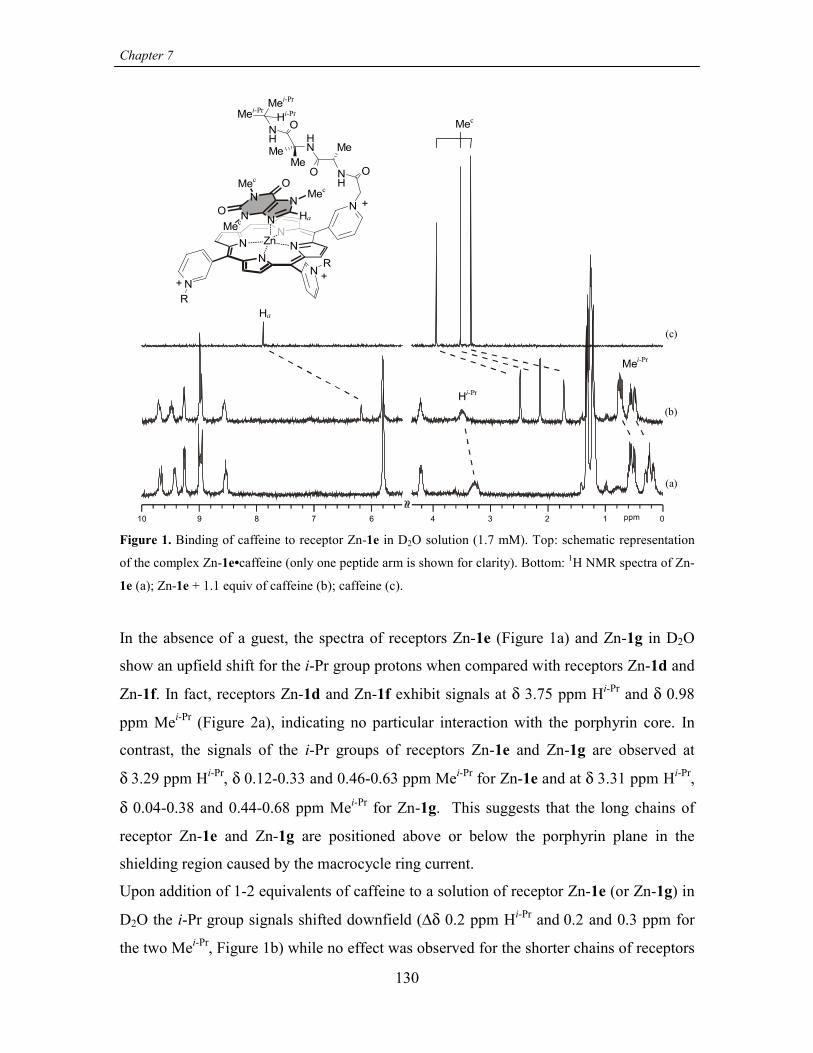
Chapter 7
130
10 89 7 6 34 2 1 0ppm
N
NN
NNN
NZn
NH
O
NH
O
OHi-Pr
Hi-Pr
NH
MeMeMe
Mei-PrMei-Pr
Mei-Pr
R
R
N
NN
N
O
O
Mec
Mec
Mec
Mec
Ha
Ha
+
+ +
(c)
(b)
(a)
Figure 1. Binding of caffeine to receptor Zn-1e in D2O solution (1.7 mM). Top: schematic representation
of the complex Zn-1e•caffeine (only one peptide arm is shown for clarity). Bottom: 1H NMR spectra of Zn-
1e (a); Zn-1e + 1.1 equiv of caffeine (b); caffeine (c).
In the absence of a guest, the spectra of receptors Zn-1e (Figure 1a) and Zn-1g in D2O
show an upfield shift for the i-Pr group protons when compared with receptors Zn-1d and
Zn-1f. In fact, receptors Zn-1d and Zn-1f exhibit signals at δ 3.75 ppm Hi-Pr and δ 0.98
ppm Mei-Pr (Figure 2a), indicating no particular interaction with the porphyrin core. In
contrast, the signals of the i-Pr groups of receptors Zn-1e and Zn-1g are observed at
δ 3.29 ppm Hi-Pr, δ 0.12-0.33 and 0.46-0.63 ppm Mei-Pr for Zn-1e and at δ 3.31 ppm Hi-Pr,
δ 0.04-0.38 and 0.44-0.68 ppm Mei-Pr for Zn-1g. This suggests that the long chains of
receptor Zn-1e and Zn-1g are positioned above or below the porphyrin plane in the
shielding region caused by the macrocycle ring current.
Upon addition of 1-2 equivalents of caffeine to a solution of receptor Zn-1e (or Zn-1g) in
D2O the i-Pr group signals shifted downfield (∆δ 0.2 ppm Hi-Pr and 0.2 and 0.3 ppm for
the two Mei-Pr, Figure 1b) while no effect was observed for the shorter chains of receptors

Recognition of caffeine in aqueous solutions
131
Zn-1d (Figure 2b) and Zn-1f. Therefore it is likely that the side chains in receptors Zn-1e
and Zn-1g are folded back over the porphyrin plane and are displaced upon caffeine
complexation. On the contrary, the shorter alkyl chains of receptors Zn-1d and Zn-1f do
not hinder the complexation of caffeine. Furthermore, caffeine signals also undergo large
upfield shifts between 1 and 2 ppm for proton Ha on C-8 (see Figures 1b, 2b) and
between 0.9 and 1.7 ppm for the three methyl groups (Mec) upon complexation with all
receptors 1 (metalated or not).
34 2 1 0
(b)
(a)
ppm
Mei-Pr
Hi-Pr
Mec
Figure 2. Binding of caffeine to receptor Zn-1d in D2O solution (1.7 mM). The signals for the i-Pr groups
of the receptor are not shifted (see text). Signals for caffeine Mec are upfield shifted when compared to their
position in the absence of receptor, confirming complexation. (a) Zn-1d; (b) Zn-1d + 2.4 equiv of caffeine.
ITC studies. ITC experiments also confirm the influence of the chain length on caffeine
complexation (Table 1). The enthalpograms, i.e. the evolved heat per added mole of
caffeine during the calorimetric titration, were fitted to 1:1 model and good agreement
with the binding constants obtained via UV titrations was found. The same trend is
observed with the Ka for Zn-1d approximately two times higher than the Ka for Zn-1e.
Thermodynamic parameters for caffeine binding to porphyrins 1 can be extracted from
the ITC experiments (Table 2). In general, large negative enthalpy and entropy
contributions are observed for the complexation of caffeine. The large negative enthalpy
change associated with the formation of the complex is consistent with a substantial
contribution of hydrophobic and stacking interactions to the binding process.

Chapter 7
132
Table 2. Thermodynamic parameters for caffeine binding to porphyrin Zn-1 in aqueous solutions
determined via ITC (sodium carbonate buffer, I = 0.11 M, pH = 10.3). For Ka values see Table 1.
Host ∆H° (kJ mol-1) ∆S° (J mol-1 K-1)
Zn-1a -36.7 ± 0.1 -52.2 ± 0.3
Zn-1d -34.5 ± 0.2 -42.5 ± 0.8
Zn-1e -39.3 ± 0.2 -65.9 ± 0.8
Negative entropic contributions have also been observed by Mizutani and coworkers for
binding of amine and amino esters to Zn-porphyrin receptors and have been analyzed in
terms of induced-fit type complexes.35 Conformational restrictions upon complex
formation and/or receptor reorganization (as observed from NMR measurements for
Zn-1e and Zn-1g) cause entropy losses (∆S° < 0) which are only partially compensated by
the entropic gain from desolvation. Accordingly, the long chain receptor Zn-1e has a
significantly more negative ∆S° value (-65.9 J mol-1 K-1) than Zn-1d (-42.5 J mol-1 K-1),
which agrees with the necessity of displacing the longer alkyl chains of Zn-1e from the
porphyrin faces. Even though the binding strength of Zn-1a is similar to that of the other
short chain receptors (Ka ∼ 6000 M-1), the thermodynamic parameters are not comparable
due to the quite different nature of the N-substituents and their influence on the solvation
of the receptor.
Free-base porphyrin receptors. To gain more information about the binding mode and to
assess the role of the Zn atom, UV and ITC studies on metal free receptors 1c and 1d
were performed (Figures 3 and 4). UV titration curves for the complexation of caffeine
were fitted to 1:1 and 2:1 caffeine to receptor stoichiometry. Despite the presence of
isosbestic points, better fits were obtained using the 2:1 binding model, although the
differences are too small to discard the possibility of 1:1 stoichiometry. On the other
hand, fitting to a 2:1 model gave better agreement between the UV and ITC
measurements.36

Recognition of caffeine in aqueous solutions
133
0.0
0.2
0.4
0.6
0.8
A
400 440420 460
λ (nm)
480440420 460
λ (nm)
Figure 3. Changes in the UV-vis absorption spectra of receptors free-base-1d (left) and Zn-1d (right) upon
addition of caffeine. [Porphyrin] ~ 2.5 µM in carbonate buffered solution (I = 0.008 M, pH = 9.6).
-15
-10
-5
0
0 50 100 150 200Time (min)
µcal
/sec
0.0 0.5 1.0 1.5 2.0 2.5 3.0 3.5-6
-4
-2
0
Molar Ratio
kcal
/mol
e of
inje
ctan
t
-20
-10
0
0 50 100 150 200Time (min)
µcal
/sec
0.0 1.0 2.0 3.0 4.0 5.0 6.0-8
-6
-4
-2
0
Molar Ratio
kcal
/mol
e of
inje
ctan
t
Figure 4. Microcalorimetric titrations of receptors free-base-1d (left) and Zn-1d (right) with caffeine.
[Porphyin] ~ 1 mM in carbonate buffered solution (I = 0.11 M, pH = 10.3).
Binding of caffeine Ki (Table 3) to the free base receptors (taking into account the
presence of two degenerate binding sites) is around four times weaker than for the
corresponding metalated hosts. For example, considering both the UV and the ITC data,

Chapter 7
134
Zn-1d and free-base 1d bind caffeine with Ka ~ 6100 M-1 and Ka ~ 1600 M-1,
respectively.
Table 3. Binding constants for caffeine to free-base porphyrins 1 in aqueous solutions
Host Ki (× 103 M-1)a K2 (× 103 M-1)
UVb 1c 1.4 0.41
UVb 1d 1.5 0.37
ITCc,d 1d 1.8 0.52
a Ki = K1/2. For definitions of K1 andK2 see eq. 2, 3. b Error margin ± 10%. Sodium carbonate buffer, I =
0.008 M, pH = 9.6. c Sodium carbonate buffer I = 0.11 M, pH = 10.3. d Thermodynamic parameters: ∆H°i =
-27.7 ± 0.1 kJ mol-1, ∆S°i = -30.8 ± 0.8 J mol-1 K-1
and ∆H°2 = -13.2 ± 0.2 kJ mol-1, ∆S°2 = 7.8 ± 0.8 J mol-1
K-1.
Comparison of the thermodynamic parameters for metal-free 1d (∆H°i = -27.7 kJ mol-1
and ∆S°i = -30.8 J mol-1 K-1) and Zn-1d (∆H° = -34.5 kJ mol-1 and ∆S° = -42.5 J mol-1 K-
1) shows that the difference between the two receptors in the binding process is mostly
due to enthalpy. Thus, the more negative enthalpic contribution (which overrules a more
negative entropic contribution) for Zn-1d suggests a direct interaction of the guest with
the Zn center. Caffeine can coordinate to the Zn center via the imidazole nitrogen N-9
(see structure Table 5).37 However, the methyl group at N-3 prevents a good
perpendicular arrangement with respect to the porphyrin plane (the effect is partially
compensated by a position of the Zn atom above the porphyrin plane, towards the axial
ligand24). Therefore, caffeine likely binds to receptor Zn-1 in a tilted way, with an angle
smaller than 90˚ between the molecular planes of caffeine and porphyrin. Support for this
proposed complex structure is corroborated by a report of complex
[Rh2(acetato)4(caffeine)2] in which binding of the two caffeine moieties at an angle
different from the optimal does not prevent metal coordination at nitrogen N-9.38
Additional evidence arises from various modes for the binding of bicyclic nucleobases
such as adenine and guanine (structurally similar to caffeine) to metalloporphyrins
reported in the literature. In some cases direct metal coordination39-41 was invoked,

Recognition of caffeine in aqueous solutions
135
whereas others used only hydrophobic interactions.42 The experiments reported here
show that the presence of Zn is an important factor for enhanced binding affinity of
caffeine.
Tyr-substituted porphyrin Zn-1c. Tyr moieties were introduced affording receptor Zn-1c
to possibly enhance the binding of caffeine through stacking with the phenol units.
However, the Tyr-based chains are long (10 atoms) and hydrophobic enough to be
positioned on top of the porphyrin plane,43 which may hinder caffeine binding like the
chains of Zn-1e and Zn-1g.1H NMR spectra of Zn-1c and free-base 1c in D2O confirm the folding of the peptide
chains towards the porphyrin core, as shown by the upfield shifted signals for the four
aromatic protons of the phenol rings (6.78 and 6.18 ppm compared to 7.07 and 6.66 ppm
for the spectrum recorded in CD3OD).44 Moreover, different from the non-ionizable side
chains of Zn-1e and Zn-1g, the phenol OH moiety can be deprotonated45 and a CPK
model suggests that the phenolate moiety could coordinate intramolecularly to the Zn.46
This hypothesis was verified by studying the UV-vis spectrum of the receptor under
different conditions. The UV-vis spectrum of Zn-1c in carbonate buffered solution (pH =
9.6) showed a 7 nm red shifted Soret (in comparison to Zn-1d and Zn-1f) and a quite
marked hypochromicity which was not caused by aggregation (Figure 5). When dissolved
in an oxalate buffer or in pure water (pH = 3.7 and ~7 respectively) these effects were not
observed and the spectrum closely resembles that of the other receptors. Metal-free 1c
had a much smaller response to the basic pH (∆λmax = 0 nm and only a modest
hypochromicity, Figure 5).
0
0.5
1
1.5
2
2.5
350 400 450 500 550 600 650
λ (nm)
A
0
0.5
1
1.5
2
2.5
350 400 450 500 550 600 650
λ (nm)
A
Figure 5. UV-vis spectra for receptors Zn-1c (left) and metal-free-1c (right) at pH 7 (black traces) and at
pH 9.6 (gray traces).

Chapter 7
136
The intramolecular coordination of the Tyr moiety to the Zn is also expected to lower the
caffeine binding because of competition for the metal center (although only at basic pH).
Binding of caffeine to receptor Zn-1c in basic solutions is observed with Ka = 4280 M-1.
The binding strength is indeed reduced by the competitive effect played by the phenolate
moieties, since at pH 3.7, where the amount of phenolate form is negligible, stronger
binding (Ka = 6090 M-1) is observed.
The data also show that receptor Zn-1c binds caffeine two times stronger then receptors
Zn-1e and Zn-1g, which have substituents with comparable chain length (see Table 1).
This result may indicate stabilizing interactions between the Tyr phenol rings and the
electron deficient caffeine π-system.
7.2.3 Selectivity towards other ligands structurally related to caffeine
1-Methylimidazole (1-MeIm). Imidazole-based ligands, resembling the 5-membered ring
of caffeine,47 are known to coordinate to metal porphyrins in aqueous solution.25,33,48
Binding of 1-methylimidazole to receptors 1 was studied by UV-vis spectroscopy through
the spectral changes in the Q-band region induced during the titrations (500-630 nm, see
Figure 6). The results (Table 4) show that the binding mechanism is substantially
different from that observed in the case of caffeine and predominantly involves metal
coordination. A division between receptors bearing short or long chains, similar to what
was observed for caffeine binding, can also be made in case of 1-MeIm binding.
However, the effect of the chain length on the binding strength is in opposite direction.
All short chain receptors (Zn-1a, Zn-1b, Zn-1d and Zn-1f) show similar binding
constants (Ka ~ 50 M-1) while Zn-1e and Zn-1g bind 1-MeIm three times stronger (Ka ~
150 M-1). Coordination of 1-methylimidazole to the Zn atom takes place perpendicular to
the porphyrin plane. This indicates that the ligand is small enough that no reorganization
of the alkyl chains of Zn-1e and Zn-1g is necessary to accommodate it. Moreover the
higher stability of the complexes obtained with Zn-1e and Zn-1g (with respect to the
short chain receptors) is probably due to the hydrophobic pocket created by the peptidic
chains on the porphyrin plane.
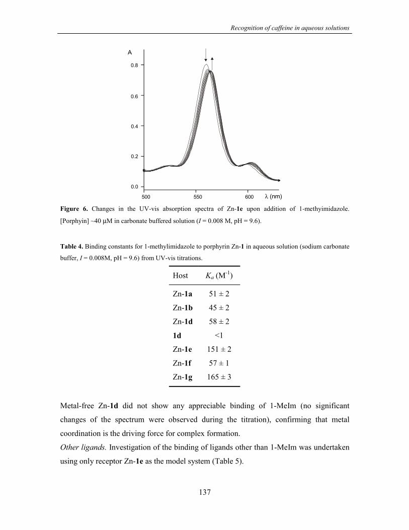
Recognition of caffeine in aqueous solutions
137
0.0
500 550 600
0.2
0.4
0.6
0.8
A
λ (nm)
Figure 6. Changes in the UV-vis absorption spectra of Zn-1e upon addition of 1-methyimidazole.
[Porphyin] ~40 µM in carbonate buffered solution (I = 0.008 M, pH = 9.6).
Table 4. Binding constants for 1-methylimidazole to porphyrin Zn-1 in aqueous solution (sodium carbonate
buffer, I = 0.008M, pH = 9.6) from UV-vis titrations.
Host Ka (M-1)
Zn-1a 51 ± 2
Zn-1b 45 ± 2
Zn-1d 58 ± 2
1d <1
Zn-1e 151 ± 2
Zn-1f 57 ± 1
Zn-1g 165 ± 3
Metal-free Zn-1d did not show any appreciable binding of 1-MeIm (no significant
changes of the spectrum were observed during the titration), confirming that metal
coordination is the driving force for complex formation.
Other ligands. Investigation of the binding of ligands other than 1-MeIm was undertaken
using only receptor Zn-1e as the model system (Table 5).

Chapter 7
138
Table 5. Binding constants for different ligands to porphyrin Zn-1e in aqueous solution (sodium carbonate
buffer, I = 8 mM, pH = 9.6)
Guest Ka (M-1)
caffeine N
NN
N
O
O
12
6 543
7 89 (3.26 ± 0.05) × 103
theophyllineN
NH
N
N
O
O
12
6 543
7 89 (2.52 ± 0.05) × 103
uracil NH
NH
O
O
< 5
thymine NH
NH
O
O
< 5
1-methylimidazoleN
N(1.51 ± 0.02) × 102
imidazole N
NH
(1.56 ± 0.02) × 102
4-methylpyridineN
(3.5 ± 0.1) × 101
Imidazole coordinates to Zn-1e with the same strength as 1-methylimidazole (Ka = 156
M-1, 20 times weaker than caffeine) while 4-methylpyridine is bound with a significantly
smaller affinity (Ka = 35 M-1). These observations clearly confirm that hydrophobic or
π-stacking interactions play an important role in stabilizing the caffeine complex with
Zn-1e in addition to metal coordination. The interactions of Zn-1e with guest molecules
that lack the nitrogen donor atom and resemble the six-member ring of caffeine (like
uracil or thymine) were also studied. These molecules might also be able to associate to

Recognition of caffeine in aqueous solutions
139
Zn-1e if the binding mode is predominantly π-stacking. Interestingly, UV-vis titrations
did not show any appreciable binding (< 5 M-1). Therefore, strong binding is favored by
the bicyclic structure of caffeine, supporting the involvement of hydrophobic interactions.
The structurally closely related theophylline (which lacks only the N-7 methyl group of
caffeine) also shows complexation by tetracationic porphyrin Zn-1e indicating a similar
binding mode. However, it is interesting to note that in view of the small structural
difference between the two molecules, the binding strength is distinctly lower.
7.3 Conclusions and outlook
Although receptors Zn-1, and especially Zn-1c, Zn-1e and Zn-1g, are not yet suitable for
practical use (sensors), the results reported in this chapter show for the first time
recognition of caffeine in aqueous solution and provide a detailed view of the binding
process from several experimental techniques. The data show that even porphyrin Zn-1a
bearing simple alkyl chains binds caffeine. However, the use of amino acid or peptide
chains allows the introduction of additional interactions. In particular, if the chains on the
pyridyl rings are longer than 10 atoms (Zn-1c, Zn-1e and Zn-1g) interaction with the
porphyrin surface and therefore influence on the recognition site become possible. The
balance of the various contributions to binding is very subtle and involves competition for
the hydrophobic surface (Zn-1e and Zn-1g) and additional stabilizing stacking
interactions (Zn-1c).
Influence of the alkyl chains on guest recognition has also been shown for a much smaller
guest such as 1-methylimidazole, which binds to porphyrins Zn-1 via a different
mechanism. In fact, the longer chain receptors showed enhanced binding for
1-methylimidazole.
The present work also shows that influence on the recognition abilities of our synthetic
receptors can be achieved even though only very simple porphyrin-peptide conjugates are
used (one or two aminoacids with thus far relatively small differences in the side chain
nature). Much more complex porphyrin-peptide conjugates bearing chains as long as 20
amino acids have been reported so far in the literature for the mimic of heme-protein.49
The present approach is expected to have great potential for the preparation of novel

Chapter 7
140
water-soluble receptors, the affinity of which may be tailored by the proper choice of
peptidic moieties.
7.4 Experimental part
7.4.1 General information and instrumentation
All reagents used were purchased from Aldrich or Acros Organics and used without
further purification. All the reactions were performed under nitrogen atmosphere. 1H
NMR and 13C NMR spectra were performed on a Varian Unity INOVA (300 MHz) or a
Varian Unity 400 WB NMR spectrometer. 1H NMR chemical shift values (300 MHz) are
expressed in ppm (δ) relative to residual CHD2OD (δ 3.30), CHD2CN (δ 1.93), or CHCl3
(δ 7.26). 13C NMR chemical shift values (100 MHz) are expressed in ppm (δ) relative to
residual CD3OD (δ 49.0) or CD3CN (δ 1.3). UV-vis measurements were performed on a
Varian Cary 3E UV-vis spectrophotometer equipped with a Helma QX optical fiber
probe (path length = 1.000 cm), using solvents of spectroscopic grade. Mass spectra were
measured using a Perkin Elmer/PerSeptive Biosystem Voyager-DE-RP MALDI-TOF
mass apectrometer (PerSeptive Biosystem, Inc., Framingham, MA, USA) equipped with
delayed extraction. A 337nm UV Nitrogen laser producing 2 ns pulses was used and the
mass spectra were obtained in the linear and reflectron mode.
7.4.2 Buffers
Carbonate buffer (I = 0.11 M, pH = 10.3) was prepared dissolving dry Na2CO3 (723 mg)
and dry NaHCO3 (573 mg) in distilled water (250 ml). The buffer was used as such or
eventually diluted to obtain the desired ionic strength.
Oxalate buffer (I = 0.008 M, pH = 3.7) was prepared adding oxalic acid (683 mg) to a
NaOH solution (8.2 ml, 1 M) and diluting to 100 ml.
7.4.3 Binding studies
Dilution experiments: Proof that the receptors used in this study were in their monomeric
form was obtained by recording the UV-vis spectra of the receptor over a range of
concentrations (1.0-9.0 × 10⁻⁶ M, in the presence of the buffer) for a total of 7-12
Recognition of caffeine in aqueous solutions
141
experimental observations. Plot of the absorbances vs concentrations were always linear
(correlation > 0.995) indicating no aggregation under the measuring conditions
([porphyrins] between 2.4 and 4.8 × 10⁻⁶ M).
Aggregation was also absent at higher porphyrin concentrations (2.8 × 10⁻³ M) as proved
for receptor Zn-1e by a microcalorimetric (ITC) experiment. A solution of Zn-1e in water
(in the microburette) was added in aliquots to the sample cell containing initially only
water. The resulting enthalpogram, i.e. the evolved heat per added mole of 1e is
featureless and only shows a small constant endothermic effect which is attributed to
dilution of the porphyrin solution.
UV-Vis: Binding of guests to receptor 1 was evaluated by monitoring the spectral
variation upon addition of guest solution either in the Soret band region (400-480 nm) or
in the Q-band region (500-630 nm) of the porphyrin spectra. Changes in the Q-band
region were monitored in cases where preliminary measurements indicated a smaller
binding constant. For these measurements the concentration of receptors 1 was 10-20
times higher then for the titrations monitored in the Soret band region (see text for typical
values). In all cases the titration covers the 20%-80% interval of complex formation.
Each titration consisted of 13 to 20 data points. The experimental spectral changes were
fitted to a 1:1 binding model using a nonlinear least-squares fitting procedure considering
simultaneously 6 different wavelenghts (model written with program Scientist®,
MicroMath®). In the case of 2:1 binding, the data were fitted also using a nonlinear least-
squares fitting procedure considering simultaneously 2 different wavelenghts (model
written with program Microsoft® Excel).
Microcalorimetry (ITC): Calorimetric measurements were carried out using a Microcal
VP-ITC microcalorimeter with a cell volume of 1.4115 mL. Aliquots of a 16.8 mM
caffeine solution in sodium carbonate buffer (I = 0.11 M, pH = 10.3) contained in the
buret were added to the receptor solution (in the same buffer) contained in the
calorimetric cell (receptor concentration 0.7-1.6 mM). Data were fitted to a 1:1 model for
metalated receptors and to a 1:2 (receptor/guest) model in the case of free base receptor
1d. Fittings of the experimental data to the appropriate complexation model were
obtained using Origin® implemented with the calorimetric set up provided by Microcal
Inc.

Chapter 7
142
7.4.4 Synthesis
Preparation of N-chloroacetyl derivatives of amino acid amides/esters and dipeptide
amides. Amino acid amides and dipeptide amides were prepared by standard solution-
phase peptide chemistry.
Couplings: A mixture of Boc amino acids (1.0 equiv), EDC (1.1 equiv), HOBt (1.0
equiv) and DIEA (1.0 equiv) in CH2Cl2 (or CH3CN) under N2 was stirred at 0 °C for 5-10
minutes. A solution of the desired amino compound (1.1 equiv) and DIEA (1.2 equiv) in
CH2Cl2 (or CH3CN) was added and the reaction mixture was allowed to warm up to r.t.
The final concentration of the Boc-amino acid in the mixture was between 0.1 and 0.3 M.
The reaction course was monitored by TLC (silica gel, CHCl3/CH3CH2OH 9:1). After
disappearance of the Boc-amino acid, the reaction mixture was diluted (~10 times) with
EtOAc and washed with citric acid (0.5 M), NaHCO3 (5%), and brine. The organic layer
was then dried over Na2SO4 and the solvent removed under vacuum. Yields were
between 75 and 90%.
Boc- deprotection: TFA/CH2Cl2 1:1, 30 min. to 1h under N2.
Chloroacetylation: Chloroacetyl chloride (1.0 equiv) was reacted with the desired amino
compound under biphasic conditions (CH2Cl2/saturated aqueous Na2CO3) at 0 °C for
45-60 minutes. The reaction mixture was then treated as the other coupling reaction
mixtures. Yields were between 65 and 90%.
Preparation of tetracationic porphyrin receptors (1). The synthesis of porphyrins Zn-1a
and Zn-1b has been published previously.19,20 Receptors Zn-1c-g have been prepared
under the same conditions reported for Zn-1b with a slightly modified workup procedure.
The alkylation reaction mixture was dried under vacuum and the solid residue was
triturated and washed extensively with ether and CH2Cl2. Then the product was extracted
by dissolution with the minimum amount of CH3CN. The solution was evaporated and
the residue redissolved in CH3OH. ZnAc2 (~10 equiv) was added and the reaction
mixture was stirred at 40 °C for 1h. The excess ZnAc2 was removed via reverse-phase
chromatography (stationary phase RP-8, eluent: from H2O to 1:1 H2O/CH3CN/0.1% TFA
mixture.) The eluted solution was first concentrated under vacuum and then lyophilized.
The solid residue was redissolved in H2O/CH3CN 1:1 and transformed in the

Recognition of caffeine in aqueous solutions
143
tetracholoride salt by ion-exchange column (DOWEX 1-X8, 50-100 mesh, Cl-form).
Receptors Zn-1 were obtained as solids after concentration and lyophilization. Combined
yields for alkylation and metallation ranged between 70 and 85%. (Free-base receptors 1c
and 1d were prepared in analogous manner, the reverse phase and ion exchange columns
following directly the alkylation step).
Due to the hygroscopic nature of the chloride salts, satisfactory elemental analyses were
not obtained. However, purity of the compounds was confirmed by analytical HPLC.
5,10,15,20-tetrakis(N-(AcetylTyr-OMe)pyridinium-3-yl) porphyrin (1c). Prepared
from 5,10,15,20-tetra(3-pyridyl)porphyrin and chloroacetyl-Tyr-OMe.
Metal Free-1c: 1H NMR (CD3CN) δ 9.46 (s, 4H), 9.33 (d, J= 7.9 Hz, 4H), 9.12 (d, J=6.1
Hz, 4H), 9.04 (s, 8H), 8.54 (m, 4H), 7.62 (d, J=7.4 Hz, 4H), 7.15 (bs, 4H) 7.05 (m, 8H),
6.65 (m, 8H), 5.60 (AB system, 8H), 4.76 (m, 4H), 3.66 (m, 12H), 3.09 (dd, J=14.0, 4.9
Hz, 4H), 2.96 (dd, J=14.0, 7.5 Hz, 4H), -3.07 (s, 2H).
UV-vis (H2O) 421, 516, 546, 583. MALDI-TOF MS: m/z 1667.5 [M-Cl]+ (calculated for
[C88H82N12O16Cl3]+ 1667.5).
Zn-1c: 1H NMR (CD3OD) δ 9.79 (bm, 4H), 9.40 (bm, 4H), 9.32 (d, J=6.3 Hz, 4H), 9.10
(m, 8H), 8.57 (t, J=7 Hz, 4H), 7.07 (m, 8H), 6.66 (m, 8H), 5.79 (AB system, 8H), 3.71
(m, 12H), 3.17 (dd, J=13.9, 5.5 Hz, 4H), 2.97 (dd, J=13.9, 8.8 Hz, 4H). 1H NMR (D2O) δ
9.33 (m, 8H), 9.08 (bm, 4H), 8.83 (m, 8H), 8.45 (bm, 4H), 6.78 (m, 8H), 6.18 (m, 8H),
5.58 (m, 8H), 3.59 (m, 12H), 2.91 (bm, 4H), 2.60 (bm, 4H). UV-vis (H2O) 433, 520,
560, 598; MALDI-TOF MS: m/z 1729.5 [M-Cl]+ (calculated for [C88H80N12O16Cl3Zn]+
1729.4).
5,10,15,20-tetrakis(N-(AcetylAla-NH-i-Pr)pyridinium-3-yl) porphyrin (1d). Prepared
from 5,10,15,20-tetra(3-pyridyl)porphyrin and chloroacetyl-Ala-NH-i-Pr.
Metal Free-1d: 1H NMR (CD3OD) δ 10.02 (m, 4H), 9.50 (m, 8H), 9.20 (bs, 8H), 8.65 (m,
4H), 5.88 (m, 8H), 4.41 (m, 4H), 3.93 (m, 4 H), 1.43 (m, 12H), 1.19-1.08 (m, 24H).
UV-vis (H2O) 420, 515, 544, 580. MALDI-TOF MS: m/z 1409.8 [M-Cl]+ (calculated for
[C72H86N16O8Cl3]+ 1407.6).

Chapter 7
144
Zn-1d: 1H NMR (CD3CN) δ 9.54 (m, 4H), 9.24 (m, 4H), 9.15 (m, 4H), 8.95 (s, 8H), 8.40
(m, 4H), 8.25 (m, 4H), 7.05 (m, 4H), 5.68 (AB system, 8H), 4.38 (m, 4H), 3.90 (m, 4H),
1.34 (d, J=7.1 Hz, 12H), 1.02 (m, 24H). 1H NMR (D2O) δ 9.68 (bs, 4H), 9.40 (bm, 4H),
9.23 (d, J=6.3 Hz, 4H), 8.97 (m, 8H), 8.52 (m, 4H), 5.77 (s, 8H), 4.25 (m, 4H), 3.75 (sept,
J=6.5 Hz, 4H), 1.33 (d, J=7.2 Hz, 12H), 0.95 (m, 24H). 13C NMR (CD3CN) δ 165.02,
151.40, 150.18, 149.01, 145.96, 143.51, 133.70, 127.21, 114.00, 63.29, 51.01, 42.65,
22.47, 18.84. IR (KBr): 3314, 3101, 2984, 1679, 1545, 1458, 1207, 1140, 843, 801, 724.
UV-vis (CH3OH) 433, 519, 559, 597, 638. MALDI-TOF MS: m/z 1470.8 [M-Cl]+
(calculated for [C72H84N16O8Cl3Zn]+ 1469.5).
[5,10,15,20-tetrakis(N-(AcetylAlaAib-NH-i-Pr)pyridinium-3-yl)porphyrinato]
zinc(II) (1e). Prepared from 5,10,15,20-tetra(3-pyridyl)porphyrin and chloroacetyl-Ala-
Aib-NH-i-Pr.1H NMR (D2O) δ 9.68 (m, 4H), 9.42 (m, 4H), 9.26 (d, J=6.2 Hz, 4H), 8.98 (m, 8H), 8.54
(m, 4H), 5.80 (s, 8H), 4.21 (m, 4H), 3.29 (bm, 4H), 1.33 (d, J=7.2 Hz, 12H), 1.22 (m,
24H), 0.63-0.46 (m, 12H), 0.33-0.12 (m, 12H). UV-vis (CH3OH) 432, 519, 560, 597,
640. MALDI-TOF MS: m/z 1809.4 [M-Cl]+ (calculated for [C88H112N20O12Cl3Zn]+
1809.7).
[5,10,15,20-tetrakis(N-(AcetylPro-NH-i-Pr)pyridinium-3-yl)porphyrinato]zinc(II)
(1f). Prepared from 5,10,15,20-tetra(3-pyridyl)porphyrin and chloroacetyl-Pro-NH-i-Pr.1H NMR (D2O) δ 9.66 (m, 4H), 9.40 (m, 4H), 9.20 (m, 4H), 9.06-8.75 (m, 8H), 8.52 (m,
4H), 5.94 (bs, 8H), 4.38 (m, 4H), 3.85-3.55 (m, 12 H), 2.24 (bm, 4H), 2.09-1.80 (bm,
12H), 0.98 (m, 24H). (CH3OH) 432, 519, 561, 597, 640. MALDI-TOF MS: m/z 1574.6
[M-Cl]+ (calculated for [C80H92N16O8Cl3Zn]+ 1573.6).
[5,10,15,20-tetrakis(N-(AcetylProAib-NH-i-Pr)pyridinium-3-yl)porphyrinato
zinc(II) (1g). Prepared from 5,10,15,20-tetra(3-pyridyl)porphyrin and chloroacetyl-Pro-
Aib-NH-i-Pr.
1H NMR (D2O) δ 9.68 (m, 4H), 9.44 (m, 4H), 9.23 (m, 4H), 8.99 (m, 8H), 8.55 (m, 4H),
5.97 (s, 8H), 4.37 (bm, 4H), 3.71 (bm, 8H), 3.31 (bm, 4H), 2.24 (m, 4H), 2.01 (m, 8H),

Recognition of caffeine in aqueous solutions
145
1.86 (m, 4H), 1.25 (m, 24H), 0.68-0.44 (m, 12H), 0.38-0.04 (m, 12H). (CH3OH) 433,
520, 558, 597, 638. MALDI-TOF MS: m/z 1913.9 [M-Cl]+ (calculated for
[C96H120N20O12Cl3Zn]+ 1913.8).
7.5 References and notes
1. Waldvogel, S. R.; Fröhlich, R.; Schalley, C. A. Angew. Chem., Int. Ed. 2000, 39,
2472-2475.
2. Goswami, S.; Mahapatra, A. K.; Mukherjee, R. J. Chem. Soc., Perkin Trans. 1
2001, 2717-2726.
3. Ballester, P.; Barceló, M. A.; Costa, A.; Deyà, P. M.; Morey, J.; Orell, M.; Hunter,
C. A. Tetrahedron Lett. 2000, 41, 3849-3853.
4. Charlton, A. J.; Davis, A. L.; Jones, D. P.; Lewis, J. R.; Davies, A. P.; Haslam, E.;
Williamson, M. P. J. Chem. Soc., Perkin Trans. 2 2000, 317-322.
5. Martin, R.; Lilley, T. H.; Falshaw, C. P.; Halsam, E.; Begley, M. J.; Magnolato, D.
Phytochemistry 1986, 26, 273-279.
6. Martin, R.; Lilley, T. H.; Bailey, N. A.; Falshaw, C. P.; Haslam, E.; Magnolato, D.;
Begley, M. J. J. Chem. Soc., Chem. Commun. 1986, 105-106.
7. Gaffney, S. H.; Martin, R.; Lilley, T. H.; Haslam, E.; Magnolato, D. J. Chem. Soc.,
Chem. Commun. 1986, 107-109.
8. Zdunek, M.; Piosik, J.; Kapuscinski, J. Biophys. Chem. 2000, 84, 77-85.
9. Frauendorf, C.; Jäschke, A. Bioorgan. Med. Chem. 2001, 9 , 2521-2524.
10. Jenison, R. D.; Gill, S. C.; Pardi, A.; Polisky, B. Science 1994, 263, 1425-1428. The
values are reported as dissociation constants Kd = 1/Ka .
11. Villamena, F. A.; De La Cruz, A. A. J. Appl. Polym. Sci. 2001, 82, 195-205.

Chapter 7
146
12. Kobayashi, T.; Murawaki, Y.; Reddy, P. S.; Abe, M.; Fujii, N. Anal. Chim. Acta
2001, 435, 141-149.
13. Carter, S. R.; Rimmer, S. Adv. Mater. 2002, 14, 667-670.
14. For recent reviews about porphyrin receptors see: (a) Weiss, J. J. Incl. Phenom.
2001, 40, 1-22. (b) Robertson, A.; Shinkai, S. Coord. Chem. Rev. 2000, 205, 157-
199. (c) Ogoshi, H.; Mizutani, T. Curr. Opin. Chem. Biol. 1999, 3, 736-739. (d)
Ogoshi, H.; Mizutani, T. Acc. Chem. Res. 1998, 31, 81-89.
15. For other water soluble receptors based on cationic (N-alkyl)pyridyl porphyrins see:
(a) Sirish, M.; Chertkov, V. A.; Schneider, H.-J. Chem. Eur. J. 2002, 5, 1181-1188.
(b) Schneider, H.-J.; Tianjun, L.; Sirish, M.; Malinovski, V. Tetrahedron 2002, 58,
779-786. (c) Sirish, M.; Schneider, H.-J. J. Am. Chem. Soc. 2000, 122, 5881-5882.
16. Sirish M.; Schneider, H.-J. Chem. Commun. 2000, 23-24.
17. A statistical mixture of atropoisomers is expected to be present in solution. In this
case the αααβ isomer, the most abundant one, together with the αααα isomer
should account for 62.5% of the mixture. NMR measurements did not provide any
indications about isomer distribution. Changes induced by complexation seem to
occur but are also difficult to be quantified. See Mizutani, T.; Horiguchi, T.;
Koyama, H.; Uratani, I.; Ogoshi, H. Bull. Chem. Soc. Jpn. 1998, 71, 413-418.
18. A basic buffer was chosen to allow comparison of the data obtained for caffeine
with the one obtained for more basic guests (Tables 4 and 5) without having to
consider protonation equilibria (see Rekharsky, M. V.; Nakamura, A.; Hembury, G.
A.; Inoue, Y. Bull. Chem. Soc. Jpn. 2001, 74, 449-457). Furthermore, as shown for
receptor Zn-1f in Table 1, there is not any appreciable effect on caffeine binding if
the acidic oxalate buffer is used (with the exception of Zn-1c due to the ionization
equilibrium involving the Tyr OH group).
19. Mizutani, T.; Horiguchi, T.; Koyama, H.; Uratani, I.; Ogoshi, H. Bull. Chem. Soc.
Jpn. 1998, 71, 413-418.
Recognition of caffeine in aqueous solutions
147
20. Fiammengo, R.; Timmerman, P.; Huskens, J.; Versluis, K.; Heck, A. J. R.;
Reinhoudt, D. N. Tetrahedron 2002, 58, 757-764.
21. Caffeine is known to dimerize or self-aggregate in aqueous solutions, but the
association constants are very small (5-8 M-1) and do not affect the binding process
in our experiments. See Horman, I.; Dreux, B. Helv. Chim. Acta 1984, 67, 754-764
and Charlton, A. J.; Davis, A. L.; Jones, D. P.; Lewis, J. R.; Davies, A. P.; Haslam,
E.; Williamson, M. P. J. Chem. Soc., Perkin Trans. 2 2000, 317-322)
22. Porphyrins concentration in the UV experiments is between 2.4 and 4.8 × 10⁻⁶ M
and preliminary dilution experiments showed no aggregation in the range 1.0-9.0
× 10-6 M. Study of the binding equilibria by ITC required much higher porphyrins
concentration (1.2-1.8 × 10⁻³ M). Aggregation was also not observed in these
conditions as proven directly by a microcalorimetric experiment on porphyrin Zn-1e
(see experimental part).
23. Imai, H.; Misawa, K.; Munakata, H.; Uemori, Y. Chem. Lett. 2001, 688-689.
24. Weiss, J. J. Incl. Phenom. 2001, 40, 1-22.
25. Imai, H.; Munakata, H.; Takahashi, A.; Nakagawa, S.; Ihara, Y.; Uemori, Y. J.
Chem. Soc., Perkin Trans. 2 1999, 2565-2568.
26. Stacking of a second caffeine molecule on the distal porphyrin face (the one not
occupied by coordination of the first molecule) has not been considered since the
Kas obtained from measurements in different concentration ranges (UV and ITC)
were in good agreement.
27. For an example in which the occurrence of 2:1 complexes is considered see ref. 36.
28. Liu, T.; Schneider, H.-J. Angew. Chem., Int. Ed. 2002, 41 , 1368-1370.
29. Jasuja, R.; Jameson, D. M.; Nishijo, C. K.; Larsen, R. W. J. Chem. Phys. B 1997,
101, 1444-1450.
Chapter 7
148
30. Schneider, H.-J.; Wang, M. J. Org. Chem. 1994, 59, 7464-7462.
31. Connors, K. A. Binding Constants; John Wiley & Sons., Inc.: Canada, 1987; pp
141-187.
32. D' Souza, F.; Deviprasad, G. R.; Zandler, M. E. J. Chem. Soc., Dalton Trans. 1997,
3699-3703.
33. Mizutani, T.; Wada, K.; Kitagawa, S. J. Am. Chem. Soc. 1999, 121, 11425-11431.
34. The signals have a complex multiplicity pattern, which agrees with the presence of
different conformational isomers interconverting slowly on the NMR chemical shift
time scale at room temperature.
35. Mizutani, T.; Wada, K.; Kitagawa, S. J. Org. Chem. 2000, 65, 6097-6106.
36. This is not surprising since ITC measurements performed at concentrations 500-
1000 times higher than UV measurements have a higher contribution coming from
the existence of 2:1 species.
37. Aoki, K.; Yamazaki, H. J. Chem. Soc., Chem. Commun. 1980, 186-188.
38. The two caffeine molecules are found asymmetrically reclined between the two
dirhodium-diacetate planes in order to accommodate base stacking. See Aoki, K.;
Yamazaki, H. J. Chem. Soc., Chem. Commun. 1980, 186-188.
39. Kuroda, Y.; Hatakeyama, H.; Inakoshi, N.; Ogoshi, H. Tetrahedron Lett. 1993, 34,
8285-8288.
40. Ogoshi, H.; Hatakeyama, H.; Kotani, J.; Kawashima, A.; Kuroda, Y. J. Am. Chem.
Soc. 1991, 113, 8181-8183.
41. Ogoshi, H.; Hatakeyama, H.; Yamamura, K.; Kuroda, Y. Chem. Lett. 1990, 51-54.
42. Pasternack, R. F.; Gibbs, E. J.; Gaudemer, A.; Antebi, A.; Bassner, S.; De Poy, L.;
Turner, D. H.; Williams, A.; Laplace, F.; Lansard, M. H.; Merienne, C.; Perrée-
Recognition of caffeine in aqueous solutions
149
Fauvet, M. J. Am. Chem. Soc. 1985, 107, 8179-8186.
43. Stacking interactions between the Tyr phenol moiety and tetracationic 5,10,15,20-
tetrakis-(N-methylpyridinium-4-yl)porphyrinato zinc(II) chloride salt give
stabilization to the porphyrin - Tyr complex in aqueous solution. See: (a) Mikros,
E.; Gaudamer, A.; Pasternack, R. Inorg. Chim. Acta 1988, 153, 199-200. (b)
Verchére-Bèaur, C.; Mikros, E.; Perrèe-Fauvet, M.; Gaudamer, A. J. Inorg.
Biochem. 1990, 40, 127-139.
44. The difference observed between Zn-1c and metal-free 1c is = 0.1 ppm.
45. The pKa value for the Tyr side chain is around 10.07 as reported in Handbook of
Chemistry and Physics, 58th Ed.; CRC Press Inc.: Cleveland, Ohio, 1977; p. C-767.
No mention is made about the ionic strength under which this value was measured.
46. Similar intramolecular coordination has been shown for pyridyl appended Zn-
tetraphenyl porphyrins in non-polar solvents, see: Walker, F. A.; Benson, M. J. Am.
Chem. Soc. 1980, 102, 5530-5538.
47. From electronic considerations imidazole and 1-methylimidazole should be better
ligands than caffeine due to increased basicity.
48. Cramer, R. E.; Ho, D. M.; van Doorne, W.; Ibers, J. A.; Norton, T.; Kashiwagi, M.
Inorg. Chem. 1981, 20, 2457-2461.
49. Lombardi, A.; Nastri, F.; Pavone, V. Chem. Rev. 2001, 101, 3165-3189.

151
Summary
This Ph.D. thesis describes the preparation, characterization, and study of supramolecular
systems for the mimicry of heme-protein binding sites.
A general introduction about the recent development of supramoleular chemistry is
outlined in Chapter 1. Special attention is given to the preparation of functional systems
and in particular to self-assembled structures with biomimetic functions due to their
relevance in the elucidation of the properties of natural biological systems.
In Chapter 2 the recent literature concerning the study of self-assembled model systems
(obtained from synthetic subunits) with biomimetic functions is reviewed. Only examples
in which the self-assembly process takes place in solution are considered and the
contributions are grouped by biomimetic function. In sections 2.2 - 2.6 examples of
electron and energy transfer processes, enzyme mimics, allosteric systems, and artificial
molecular machines are described. The second part of the chapter (section 2.7) is more
directly connected with the central part of this thesis viz. the preparation of self-
assembled synthetic analogues of O2 binding heme-proteins. This section focuses on the
most important biochemical aspects of O2 binding heme-proteins and gives an overview
of the synthetic models obtained via “classical” covalent synthesis.
In Chapter 3 the preparation of noncovalent calix[4]arene capped porphyrins in polar
solvents via ionic interactions is described. The preparation of a superstructured
porphyrin architecture via noncovalent synthesis is the first necessary step towards the
mimicry of O2 binding hemeproteins. Tetracationic zinc(II) meso-tetrakis(N-
alkylpyridinium-3-yl) porphyrins and anionic 25,26,27,28-tetrakis(2-ethoxyethoxy)-
calix[4]arene tetrasulfonate are the building blocks synthesized for this purpose. These
building blocks self-assemble due to ionic interactions in solvents such as methanol,
DMSO, 1,3-dimethyl-3,4,5,6-tetrahydropyrimidin-2(1H)-one, and mixtures containing as
much as xwater = 45% (molar fraction of water). The systems (structural mimics of heme-

152
proteins) have been studied with UV-vis, 1H NMR, and mass spectrometry (ESI-TOF).
The thermodynamic parameters associated with the assembly process have been
determined via variable temperature experiments and, when possible, via isothermal
titration microcalorimetry (ITC). The assemblies described in this chapter are remarkably
stable under a variety of conditions (formation constant ~ 106-107 M-1 in neat solvents, ~
105 M-1 in the presence of salts).
Self-assembly in polar solvents is just the first step towards the development of heme-
protein model systems via noncovalent synthesis. Therefore, the preparation of water-
soluble systems is definitely a major advance considering the biological relevance of
water. Its achievement requires not only water-soluble building blocks but also that the
assemblies remain water-soluble once they are formed. Thus Chapter 4 deals with water-
soluble structural mimics of heme-proteins. In the design of the porphyrin-based building
block the short peptides attached to the macrocycle are responsible for the relatively large
water solubility of the assemblies (concentrations up to 2-5 mM). Formation of ternary
complexes upon addition of nitrogenous base ligands has been observed. These ligands
coordinate to the metal center of the assembly, and the structure of the complexes is
directed by the sterical requirements of the base used. Small bases such as 4-methyl
pyridine are complexed preferentially inside the hydrophobic cavity of the assemblies
while larger molecules such as caffeine are bound outside the cavity at the solvent
exposed porphyrin face.
The “inert” zinc porphyrins have been replaced by cobalt porphyrins (able to bind O2) to
afford the functional models of heme-proteins described in Chapter 5. This chapter deals
with the self-assembly of tetracationic cobalt(II) porphyrins and tetraanionic
calix[4]arenes to prepare water soluble O2 binding heme-protein models. The O2 binding
ability has been tested using UV-vis and electron paramagnetic resonance (EPR)
spectroscopy. Even though the complex nature of these systems (due to the presence of
multiple equilibria) prevents a full description of the situation in solution, they have been
tested in membranes for O2/N2 separation. A modest but significant facilitated O2
transport (facilitation factor = 1.15, selectivity coefficient = 2.3-2.4) is observed for
membrane loaded with the self-assembled systems with respect to the membrane loaded
with only H2O (facilitation factor = 1.0, selectivity = 2.0).

153
Other organic ligands besides porphyrins can be used for the preparation of O2 binding
metal complexes, among them Salen (bis(salyylidene)ethylenediamine) ligands. The
supramolecular design of novel cobalt(II) Salen complexes able to stabilize the Co-
coordinated O2 via hydrogen-bonding is the topic of Chapter 6. This design mimics the
hydrogen-bonding interaction operated by the distal His residue in hemoglobin and
myoglobin or by Tyr in O2 avid Ascaris hemoglobin. The affinity for O2 is increased ten
times in comparison to systems lacking such interaction. These Co(II)Salen complexes
have also been tested in relation to their catalytic activity in the oxidation of cyclohexene
with the aim of studying the relation existing between stabilization of the Co-O2 moiety
and reactivity. The more thermodynamically stable Co-O2 complexes yield lower
reactivity.
During the studies on water-soluble porphyrin systems (Chapter 4), the much higher
coordination strength of caffeine compared to other nitrogenous bases was noticed and
led to the study of these cationic porphyrins (and some novel derivatives) as potential
sensors for caffeine, the results of which are reported in Chapter 7. The remarkable
affinity observed for caffeine in water (Ka~6000 M-1) is due to a combination of metal
coordination and stacking interactions. The results reported in this chapter show for the
first time recognition of caffeine in aqueous solution and provide a detailed view of the
binding process from several experimental techniques. The use of amino acid or peptide
chains (of appropriate length) allows the introduction of additional interactions, which
can be used to influence the recognition site. The intrinsic UV-vis activity of the
porphyrin core and the relevance of working directly in aqueous solution are interesting
features in view of sensing applications.


Samenvatting
Dit proefschrift beschrijft de synthese, karakterisatie en bestudering van supramoleculaire
systemen voor het nabootsen van de heem-proteinen bindingsplaatsen.
Een algemene inleiding over de recente ontwikkelingen in de supramoleculaire chemie is
uiteengezet in hoofdstuk 1. Speciale aandacht is besteed aan de synthese van functionele
systemen, met name aan zelf-assemblerende structuren met biomimetische
functionaliteiten, die relevant zijn in de opheldering van de eigenschappen van
natuurlijke biologische systemen.
In hoofdstuk 2 is een overzicht gegeven van de recente literatuur betreffende de
bestudering van zelf-assemblerende model systemen (verkregen uit synthetische sub-
eenheden) met biomimetische functionaliteiten. Alleen voorbeelden van systemen die
zelf-assembleren in oplossing zijn in beschouwing genomen en gerangschikt naar
biometische functie. In de paragrafen 2.2 – 2.6 zijn voorbeelden beschreven van elektron-
energie overdrachtsprocessen, enzym nabootsingen, allosterische systemen en
kunstmatige moleculaire machines. Het tweede gedeelte van dit hoofdstuk (paragraaf 2.7)
is meer direct verbonden met het centrale gedeelte van dit proefschrift, te weten de
synthese van zelf-assemblerende synthetische analogen van O2 bindende heem-proteinen.
Deze paragraaf richt zich op de meest belangrijke biochemische aspecten van O2
bindende heem-proteinen en geeft een overzicht van synthetische modellen verkregen via
“klassieke” covalente synthese.
In hoofdstuk 3 is de synthese beschreven van niet-covalente calix[4]areen-porfyrine
complexen via ionische interacties in polaire oplosmiddelen. De synthese van een
suprastructurele porfyrine architectuur via niet-covalente synthese is de eerste
noodzakelijke stap richting de nabootsing van O2 bindende heem-proteinen.
Tetracationische zink(II) meso-tertrakis(N-alkylpyridinium-3-yl) porfyrines en anionische
25,26,27,28-tetrakis(2-ethoxyethoxy)-calix[4]areen tetrasulfonaat zijn de
155

gesynthetiseerde bouwstenen voor dit doel. Deze bouwstenen zelf-assembleren als
gevolg van ionische interacties in oplosmiddelen zoals methanol, DMSO, 1,3-dimethyl-
3,4,5,6-tetrahydropyrimidin-2(1H)-on en mengsels die zoveel als xwater = 45% (molfractie
water) bevatten. De systemen (structurele nabootsingen van heem-proteinen) zijn
bestudeerd met UV-vis, 1H NMR en massa spectroscopie (ESI-TOF). De
thermodynamische parameters van het zelf-assemblerende proces zijn bepaald met
variabele temperatuur experimenten en waar mogelijk met behulp van isothermische
titratie microcalorimetrie (ITC). De assemblages beschreven in dit hoofdstuk zijn
opmerkelijk stabiel onder een verscheidenheid aan condities (vormingsconstante ~ 106-
107 M-1 in pure oplosmiddelen, ~ 105 M-1 in aanwezigheid van zouten).
Zelf-assemblage in polaire oplossingen is slechts de eerste stap naar de ontwikkeling van
heem-proteinen model systemen via niet-covalente synthese. De synthese van water-
oplosbare systemen is daarom absoluut de belangrijkste vooruitgang gezien de
biologische relevantie van water. Succes vereist niet alleen water-oplosbare bouwstenen,
maar ook dat de assemblages na vorming oplosbaar blijven in water. Hoofdstuk 4
behandelt de water-oplosbare structurele nabootsingen van heem-proteinen. In het
ontwerp van de porfyrine bouwstenen zijn de korte peptiden verantwoordelijk voor de
relatief grote oplosbaarheid van de assemblages (concentraties tot 2-5 mM). Vorming van
driedelige complexen is waargenomen bij toevoeging van stikstofhoudende basische
liganden. Deze liganden coördineren aan het metaal centrum van de assemblage en de
structuur van de complexen wordt bepaald door de sterische eigenschappen van de
gebruikte base. Kleine basen zoals 4-methyl pyridine worden bij voorkeur
gecomplexeerd binnenin de hydrofobe holte van de assemblages terwijl grotere basen
zoals cafeïne aan de buitenkant van de holte aan het oplosmiddel blootgestelde porfyrine
worden gebonden.
De “inerte” zink porfyrines zijn vervangen door kobalt porfyrines (in staat om O2 te
binden) om de functionele modellen van heem-proteinen te verschaffen is beschreven in
hoofdstuk 5. Dit hoofdstuk behandelt de zelf-assemblage van tetracationische kobalt(II)
porfyrines met tetraanionishe calix[4]arenen voor de synthese van water oplosbare O2
bindende heem-protein modellen. De O2 binding bekwaamheid is getest met
gebruikmaking van UV-vis en elektron paramagnetische resonantie spectroscopie (EPR).
156

Hoewel de complexe oorsprong van deze systemen in oplossing (als gevolg van meerdere
evenwichten) een volledige beschrijving van de situatie verhindert, zijn zij getest in
membranen voor O2/N2 scheiding. Een bescheiden maar significante gefaciliteerde O2
transport (faciliteitsfactor = 1.15, selectiviteitscoëfficiënt = 2.3-2.4) is waargenomen voor
een membraan “geladen” met de zelf-assemblerende systemen in vergelijking met de
membranen met enkel H2O (faciliteitsfactor = 1.0, selectiviteit = 2.0).
Andere organische liganden naast porfyrines kunnen gebruikt worden voor de synthese
van O2 bindende metaal complexen, waaronder Salen (bis(salyylidene)ethylenediamine)
liganden. Hoofdstuk 6 beschrijft het supramoleculaire ontwerp van nieuwe kobalt(II)
Salen complexen die in staat zijn de Co-O2 via waterstof-binding te stabiliseren. Dit
ontwerp bootst de waterstof-bindende interactie na van de distal His residu in
hemoglobine and myoglobine of van Tyr in O2 avid Ascaris hemoglobine. De affiniteit
voor O2 is 10 keer groter in vergelijking met systemen die deze interactie missen. Deze
Co(II) Salen complexen zijn ook getest met betrekking tot hun katalytische activiteit in
de oxidatie van cyclohexeen met als doel de relatie tussen stabilisatie van de Co-O2
eenheid en reactiviteit te bestuderen. De meer thermodynamisch stabiele Co-O2
complexen vertonen een lagere reactiviteit.
Tijdens de studies aan water-oplosbare porfyrine systemen (hoofdstuk 4) is een veel
hogere coördinatie sterkte van cafeïne in vergelijking met andere stikstofhoudende basen
waargenomen. Deze cationische porfyrines (en enkele nieuwe derivaten) zijn daarom
bestudeerd als mogelijke sensoren voor cafeïne en de resultaten zijn beschreven in
hoofdstuk 7. De waargenomen merkwaardige affiniteit voor cafeïne in water
(Ka~6000M-1) is te danken aan een combinatie van metaal coördinatie en stapelings
interacties. De resultaten beschreven in dit hoofdstuk laten voor de eerste keer de
herkenning van cafeïne in waterige oplossing zien en verschaffen een gedetailleerde kijk
van het bindingsproces met een aantal experimentele technieken. Het gebruik van amino
zuren of peptide ketens (van geschikte lengte) maakt de introductie van bijkomende
interacties mogelijk, die gebruikt kunnen worden om de herkenningsplaats te
beïnvloeden. De intrinsieke UV-vis activiteit van de porfyrine kern en de relevantie van
het direct werken in waterige oplossingen zijn interessante eigenschappen met het oog op
sensor toepassingen.
157


Acknowledgements
The work described in this thesis would not have been possible without the contributions
of many friends and colleagues from the University of Twente and from other research
groups in The Netherlands.
First of all I would like to thank my promotor David Reinhoudt for giving me the
possibility of working in a very stimulating research group with all the freedom I could
ever desire. David, you gave me the opportunity of presenting my work in conferences all
around the world and you always stimulate me in writing publications. From you I have
learned that being able to communicate the results is as important as carrying out a
careful scientific investigation. Thank you for all of this.
I would like to thank my daily supervisors Mercedes Crego-Calama and Peter
Timmerman.
Dear Merce you have started to supervise me after more than two years the project was
started. Nevertheless, you manage to catch up very soon and to give me very precious
suggestions during the long talks we had in the lab. You had also the “honor” (or the
pain) to go through the correction of the largest part of my writings, including this thesis.
I really appreciate the way you did it. You always stimulated me in being clear and
simple in my explanations and, as I told you already many times, I always liked the final
result. Muchisimas gracias also for your friendship.
Dear Peter I would like to thank you very much for your supervision during more than
half of my Ph.D. project. Thank you for being always available for work (and no work)
discussions. I appreciated your enthusiasm.
159

Special thanks to Jurriaan Huskens and Prof. Feike de Jong for the help in developing the
mathematical models that describe the chemical equilibria in the systems object of my
studies.
The work described in Chapter 5 has been performed partially at the University of Leiden
and I would like to thank Prof. Jan Reedijk and Mr. Ge van Albada for all what they have
taught me about EPR spectroscopy and for helpful discussions. The membrane-related
part of this work has been carried out in collaboration with the Membrane Technology
group (University of Twente) of Prof. Matthias Wessling. I would like to thank him for
the many clarifying discussions we had about the membrane permeation experiments and
I want to extend my acknowledgement to Alberto Figoli with who I shared the work in
the lab. Alberto grazie anche per la tua amicizia al di fuori delle lunghe ore di lavoro.
Prof. Albert Heck and Kees Versluis from the University of Utrecht are gratefully
acknowledged for the mass spectrometry measurements reported in Chapter 3.
Special thanks goes to the students that have worked on this project trying some of my
ideas but also putting their valuable original contribution: Christiaan Bruinink, Dennis
Rots, Kamil Wojciechowski, Davy Martin. You all did an excellent work and it was a
real pleasure to collaborate with you!
I would like to thank Richard Egberink for the work he has carried out as part of the
project and especially for always being present and helpful with computer-stuff, safety
issues, and synthetic discussions.
Joop Toevank, Irene Wolbers, Marc Brower, and Marcel de Bruine thank you for all your
help throughout the years. You really did (and Marcel still do) a great job to fulfill the
request of such a large group of chemists. Thanks also to Hanny Visser, Tieme Stevens,
Roel Fokkens and Annemarie Montanaro-Christenhusz for their indispensable analysis
work.
160

Carla Weber-van der Ploeg, I have to thank you so much for all the help you gave me
with the administrative duties, especially in this last period before the “Promotie”.
Becky, Mrinal being Americans you had to play a very important role: proofreaders of
my concept thesis! You did it in an excellent way and your suggestions helped me to
improve the overall quality of this thesis. Thank you.
One of the nicest things of this four years in Enschede has been the possibility of working
in an incredibly stimulating international atmosphere. With many of the people I also had
the occasion of enjoy the free time in Enschede. I would like to thank you all and I
already apologize if I forget somebody. Niels, Arianna, Kjeld, Jasper, Leonard, Maria,
Francesca Cardullo, Jessica, Xue-Mei, Kazu, Tsutomu, Piotr, Anja, Lourdes, Olga,
Miguel, Juanjo, Mattijs, Paolo, Monica, Emiel, Stefano, Petra, Barbara, Romina, Mario,
Susanna, Lorenza, Micol, and Luca, I consider myself lucky to have friends like you.
Francesca grazie per essermi stata amica in questi anni, per essere stata spesso a sentire le
mie elucubrazioni e le mie boiate, per quando ero nervoso con il mondo intero e per
quando le cose andavano bene.
Tommaso e Marta, thanks for being my paranimfen but especially for all the time we
spent together. Tommaso sei un vero amico, e da te ho cercato di imparare a essere
puntuale, disponibile e...sportivo.
Da ultimi vorrei ringraziare voi mamma, papà e Lucio per essermi sempre stati vicino
durante questi anni e avere sostenuto le mie scelte anche se mi hanno portato lontano da
voi.
Grazie di cuore a tutti,
Roberto
161


The author
Roberto Fiammengo was born in Torino (Italy) on September 19, 1971. In 1990 he
obtained his diploma with specialization in industrial chemistry at the Technical
Highschool “E. Fermi” in Mantova (Italy). In June 1997 he graduated full marks in
Chemistry at the University of Padova (Italy). During his undergraduate training period
at the Department of Organic Chemistry he worked on asymmetric oxidations mediated
by titanium complexes and on stereoselective Diels-Alder reactions under the supervision
of Prof. F. di Furia and Prof. G. Licini. His M.Sc. thesis is entitled "(-)-trans-benzo[e]-
1,4-dithiine-S,S’-dioxide: a new C2 simmetry dienophile". From July 1997 to October
1997 he continued the work on stereoselective oxidations and from November 1997 to
August 1998 he worked on the derivatization of tetrasubstituted α-amino acids, under the
supervision of Prof. G. Modena (Dep. of Organic Chemistry, University of Padova) in
collaboration with DSM (Geleen, The Netherlands). In September 1998 he joined the
group of Supramolecular Chemistry and Technology (SMCT) at the University of
Twente as a Ph.D. candidate under the supervision of Prof. dr. ir. D. N. Reinhoudt. The
research performed during the period 1998-2002 is described in this thesis. Currently he
is working as a post-doc in the group of Prof. P. Scrimin (Dep. of Organic Chemistry,
University of Padova) on synthetic models of metallonucleases.
163